
Phytochemical-Based Nano-Pharmacotherapeutics for Management of Burn Wound Healing
by
 , , ,
, , ,
Abdul Qadir
1 ,
,
Samreen Jahan
1,
Mohd Aqil
1,
Musarrat Husain Warsi
2,*,
Nabil A. Alhakamy
3,
Mohamed A. Alfaleh
3,4,
Nausheen Khan
5 and
Athar Ali
6 1
Department of Pharmaceutics, School of Pharmaceutical Education & Research, Jamia Hamdard, New Delhi 110062, India
2
Department of Pharmaceutics and Industrial Pharmacy, College of Pharmacy, Taif University, Al-Haweiah, Taif 21974, Saudi Arabia
3
Department of Pharmaceutics, Faculty of Pharmacy, King Abdulaziz University, Jeddah 21589, Saudi Arabia
4
Vaccines and Immunotherapy Unit, King Fahd Medical Research Center, King Abdulaziz University, Jeddah 21589, Saudi Arabia
5
Department of Pharmacognosy and Phytochemistry, School of Pharmaceutical Education & Research, Jamia Hamdard, New Delhi 110062, India
6
Centre for Transgenic Plant Development, Department of Biotechnology, Jamia Hamdard, New Delhi 110062, India
*
Author to whom correspondence should be addressed.
Gels 2021, 7(4), 209; https://doi.org/10.3390/gels7040209
Submission received: 28 September 2021
/
Revised: 31 October 2021
/
Accepted: 11 November 2021
/
Published: 13 November 2021
(This article belongs to the Special Issue Advances in Nanogels)
Abstract
:Medicinal plants have been used since ancient times for their various therapeutic activities and are safer compared to modern medicines, especially when properly identifying and preparing them and choosing an adequate dose administration. The phytochemical compounds present in plants are progressively yielding evidence in modern drug delivery systems by treating various diseases like cancers, coronary heart disease, diabetes, high blood pressure, inflammation, microbial, viral and parasitic infections, psychotic diseases, spasmodic conditions, ulcers, etc. The phytochemical requires a rational approach to deliver the compounds to enhance the efficacy and to improve patients’ compatibility. Nanotechnology is emerging as one of the most promising strategies in disease control. Nano-formulations could target certain parts of the body and control drug release. Different studies report that phytochemical-loaded nano-formulations have been tested successfully both in vitro and in vivo for healing of skin wounds. The use of nano systems as drug carriers may reduce the toxicity and enhance the bioavailability of the incorporated drug. In this review, we focus on various nano-phytomedicines that have been used in treating skin burn wounds, and how both nanotechnology and phytochemicals are effective for treating skin burns.
1. Introduction
Skin is the largest visible and vulnerable organ of the human body. It protects our body from environmental changes and dehydration [1,2]. There are certain skin conditions, such as burns and other substantial loss of the outer layer of the skin (epidermis), which acts as the barricade that prevents the skin from degeneration and microbial incursion and balances the fluid levels of the body. In such conditions, both nutritional and electrolytes constituents get demolished. Hence, skin wounds can drastically impact human health [3]. Various diseases, such as eczema, herpes zoster, rosacea, and psoriasis, can cause harm to skin; however, burns are the major cause of skin damage [4]. According to the World Health Organization (WHO), an estimated 180,000 deaths are caused by burns annually [5]. A burn injury may result from hot and cold materials and vulnerability to chemicals and radiations. Burn wounds are of three types and classified by the profundity: (1) superficial (first degree), (2) partial thickness (second degree), and (3) full thickness (third degree) [6,7]. Healing of burn wounds is a complicated process, and it proceeds through various phases, including inflammation, proliferation, and remodeling. These phases should occur in the proper order and time sequence for better would healing as changes in any of the phases may cause a delay in the healing process [8,9]. After epidermal injury, platelet activation leads to control of blood loss and results in clot formation, which is the first step in wound healing mechanism [10]. Nanotechnology offers an excellent outlook to fast-track persistent wound healing by altering the different phases of healing with high payloads of phytoconstituents [11]. Over the last two centuries, the use of plants extracts in wound healing has increased due to the presence of active compounds in these extracts [12]. Herbal medicines are widely accepted because of their efficacy and low level of adverse effects [13]. For skin-related diseases and other disorders, utilization of herbal constituents is accepted by 80% of population [14]. Using different plant extracts, various studies have been performed to observe the pharmacological action of constituents on various disease. From 2011 to present day, the use of herbal medicines has increased from $18 million to $26 billion; it is also estimated that 50% of approved herbal drugs are provided worldwide [15,16].
Nanocarriers with herbal drugs have gathered significant recognition for their potential and distinctive attributes in numerous domains of human activity [17].
The combination of nanotechnology with natural drugs would be a novel development for enhancing the medicinal effect of these natural drugs [18]. To increase the acceptability of these compounds by patients and to prevent the need for repeated administration, the phytochemical needs an approach that can encourage the delivery of active components in a sustained release format. Novel drug delivery systems help achieve the required therapeutic effects with reduced adverse events and enhance the bioavailability of herbal constituents [19,20].
Comprehensive searches were done on Google Scholar and PubMed databases pertaining to herbal-based pharmaceuticals for burn wound, nano-drug delivery applications in burn healings, and past to present evolution of nano-phytomedicines for the management of burn wound healing. We focused mainly on the last 15 years of works in this area, although some older references were also included to provide validity to the review.
2. Wound Healing Process
Would healing is a complicated process, and it progresses through various phases, including inflammation, proliferation, and remodeling. Involvement of fibroblasts, leukocytes, and monocytes in the healing process aid in reconstituting the destructed skin (Figure 1). Vitamins E and C play crucial roles in wound healing as these are key factors in this process. Vitamin K prevents severe bleeding, carotenoids restore the skin epithelial layer and tissues, and phytosterols have antimicrobial and anti-inflammatory effects [21]. Would healing also involves the use of biochemical genetic reprogramming to reinstate the skin health. Recent research has shown that the use of phytochemicals has active constituents that have the capability to induce wound healing with less side effects [22].
2.1. Inflammatory Phase
The inflammatory phase starts 4–5 days after the skin damage has already occurred. Once injury occurs, intravascular platelets mediate homeostasis, which is responsible for clot formation over the wound to stop further bleeding [23]. Post-homeostasis, thrombin activates platelets, which releases multiple growth factors, such as epidermal growth factor (EGF), insulin growth factor-1 (IGF-1), platelet derived growth factor (PDGF), fibroblast growth factor (FGF), and transforming growth factor (EGF, IGF-1, PDGF, FGF, and TGF, respectively) [24,25]. These growth factors activate neutrophils, monocytes, leukocytes, and macrophages, which act as a shield for skin to prevent further damage and initiate the wound healing process [26,27].
2.2. The Proliferative Phase
The proliferative phase involves cell proliferation and migration and takes 3–15 days to activate [28]. Once PDGF is released by platelets during the inflammatory phase, formation of new blood vessels and capillaries occurs [29]. Following this step, angiogenesis and migration of fibroblasts takes place to form granulation tissues [30]. Once the fibroblasts process is complete, a new extracellular matrix (ECM) consisting of collagen and proteoglycans is produced. Some fibroblasts undergo fission into myofibroblasts, which helps with the contraction of the wounded area [31]. Following this step, activation of keratinocytes, which migrate to the injured area and completes the last step, that of re-epithelialization, occurs [32].
2.3. Re-Modeling Stage
The re-modeling stage continues to change over the first several weeks to several years after the wound occurs. Collagen I replace collagen III, which consists of newly synthesized ECM, and the fresh collagen fibers develop into an assembled lattice composition that enhances the intensity of healed tissues [33,34].
3. The Impact of Antibiotics and Antioxidant Properties of Plants
Since ancient times, various plants have been used for treatment of different diseases and are currently in use worldwide. Due to their antimicrobial activities, natural constituents, such as aminoglycosides [35], beta-lactams [36], glycopeptides [37], quinolones [38], sulfonamides [39], and tetracyclines [40] have been utilized for wound healing treatment. For wound therapy, the benefits of plant extracts or phytochemical have been recognized as has the existence of antioxidants in numerous plants extracts. The presence of oxygen free radicals causes disruption in the wound healing process. Stress caused by oxidation slows down the healing process and causes additional damage to tissues. An antioxidant confers protection from oxygen free radicals by reducing their effects; this process assists in the wound healing process. Active compounds possessing antimicrobial effects play an important role as they neutralize free oxygen radicals and enhance the wound healing process [41,42]. Antioxidants have been used to hasten healing activity by extending antioxidant effects throughout the healing process. It seems the existence of antioxidants is essential to facilitate recuperation from persistent skin injury; Table 1 shows plants with both antimicrobial and antioxidant properties [43]. Natural metabolites influence the wound healing process by introducing various growth factors, such as EGF and FGF, which affect cellular movement [44]. Animal studies have indicated that herbal compounds encourage anti-inflammatory and antimicrobial activities for wound treatment by promoting regeneration of skin cells and displacing connective tissues [45]. Table 1 shows the names of natural compounds having antioxidant and antibiotic activities toward wound healing.
The efficacy of Centella asiatica has been studied broadly in animal models. This herb helps heal the incision-induced injuries. In one study, it was concluded that the level of antioxidants increases extensively in the presence of this herb, leading to improved healing activity [46]. Asiaticoside is extracted from Centella asiatica and produces a better capacity for injury healing process in both chronic and immediate healing as it has fibroblast proliferating activity [47].
Leaf extracts from Chromolaena odorata contain flavonoids and this herb has been used widely in the wound healing activity due to the free radical approach, which has shown a conclusive promotion in healing activity [48]. This extract was shown to cause improvement in the fibroblast proliferation and keratinocyte and endothelial cell activity, and to stimulate keratinocyte migration [48].
Quercus infectoria Olivier possesses anti-inflammatory, anti-bacterial, and antioxidant properties. The ethanolic extract of gallic acid, ellagic acid, and syringic acid form the active constituents of tannins, which might be the reason for antioxidant effects resulting in enhanced healing activity. In this study, the incision wound animal model showed better healing activity after stimulation with antioxidants, which caused enhancement of the superoxide dismutase and catalase levels, both of which are influential antioxidant enzymes [49].
Wounds, burns, and internal and external ulcers can be treated by Buddleja globasa (common name: orange bell Buddleja). This herb is also used traditionally in Chile for the treatment of ulcers and burns. This herb was tested for its capability to stimulate fibroblast growth and antioxidant activity in vitro. Testing was specific as the effect of the aqueous solution of B. globasa on these two processes is considered as the first stage in the tissue repair cycle [50]. It was proven that the damage caused by oxygen free radical causes a delay in the healing process; thus, an antioxidant was needed to reverse this delay [51]. This process was achieved because of the presence of flavonoids and caffeic acid in the extract. Buddleja leaves have other applications in the skin layer formation that is part of wound healing [52].
Curcumin is a derivative of Curucuma longa, which is also known as turmeric and Haldi. C. longa has numerous biological properties, which consist of antioxidant, initiation of enzyme detoxification, and prevention of degenerative diseases [53]. Dermal application of these substances to patients resulted in enhanced wound healing activity and provided tissue defense from oxidation-induced damage [54].

Table 1.
Phytochemicals having antibiotic, antimicrobial, and antioxidants activity on wound healing.
Table 1.
Phytochemicals having antibiotic, antimicrobial, and antioxidants activity on wound healing.
| Medicinal Plants | Family | Active Ingredients | Activities | Reference |
|---|---|---|---|---|
| Acacia Senegal L. | Leguminosae | Saponins, alkaloids, and malic acid | Antimicrobial, anti-inflammatory, and antioxidant activity | [55] |
| Acalypha indica L. | Euphorbiaceae | Flavanoids, alkaloids, saponins | Antioxidant and antimicrobial activity | [55] |
| Allophylus rubifolius L. | Sapindaceae | - | Antioxidant, antibacterial, and anti-inflammatory activity | [55] |
| Anagallis arvensis L. | Primulaceae | flavonoids, saponins, glycosides, alkaloids, and anthraquinones | Antioxidant, anti-inflammatory, and antimicrobial activity | [56] |
| Anogeissus dhofarica L. | Combretaceae | - | Antioxidant and antimicrobial activity | [41] |
| Aloevera L. | Liliacae | Saponins, acemannan, and anthraquinone | Antimicrobial activity | [57] |
| Anethum graveolens L. | Umbelliferae | - | Anti-inflammatory and antibacterial activity | [58] |
| Aristolochia bracteolate L. | Aristolochiaceae | - | Antimicrobial and antioxidant activity | [59] |
| Alternanthera brasiliana L. | Amaranthaceae | - | Antimicrobial activity | [60] |
| Achillea millefolium L. | Asteraceae | Isovaleric acid, salicylic acid, sterols, flavonoids, tannins, and coumarins | Antimicrobial activity | [15] |
| Acanthus polystachyus L. | Acanthaceae | Tannins, flavonoids, saponins, polyphenols, terpenoids, glycosides, and anthraquinones | Anti-inflammatory and antioxidant activity | [61] |
| Becium dhofarense L. | Lamiaceae | - | Antioxidant activity | [55] |
| Bridelia ferruginea L. | Phyllanthaceae | Flavonoids, tannins, saponins, and terpenoids | Antioxidant and antibacterial activity | [62] |
| Buddleja globosa L. | Scrophulariaceae | - | Antioxidant activity | [50] |
| Centella asiatica L. | Araliaceae | Flavonoids | Antioxidant activity | [46,63,64] |
| Chromolaena odorata L. | Asteraceae | Alkaloids, flavonoids, flavanone, essential oils, phenolics, saponins, tannins, and terpenoids | Antimicrobial, antioxidant, and anti-inflammatory activity | [48] |
| Clerodendrum infortunatum L. | Lamiaceae | Flavonoids | Antioxidant and antimicrobial activity | [65] |
| Combretum smeathmanii L. | Combretaceae | Alkaloids, coumarins, flavonoids, saponins, terpenes, and sterols | Antioxidant and antimicrobial activity | [66] |
| Cordia perrottettii L. | Boraginaceae | - | Antioxidant activity | [55] |
| Curcuma longa L. | Zingiberaceae | Glycosides, tannins, and flavonoids | Anti-inflammatory, antimicrobial, and antioxidant activity | [53,54,67] |
| Crassocephalum crepidioides L. | Asteraceae | Phenolic, flavonoid, and essential oil | Anti-inflammatory and antioxidant activity | [68] |
| Cinnamomum verum L. | Lauraceae | Tannins | Anti-inflammatory, antimicrobial, and antioxidant activity | [69] |
| Dendrophthoe falcata L. | Loranthaceae | - | Antioxidant and antimicrobial activity | [70] |
| Eucalyptus globulus L. | Myrtaceae | Alkaloids, flavonoids, saponin, tannin, carbohydrates, and glycosides etc. | Anti-inflammatory activity | [71] |
| Ficus asperifolia L. | Moraceae | Flavonoids, phenolics, alkaloids, and tannins | Antioxidant and antimicrobial activity | [72] |
| Gossypium arboreum L. | Malvaceae | Alkaloids, phenolic compounds, terpenoids, tannins, saponins flavonoids, cardiac glycosides, and protein | Antioxidant and antimicrobial activity | [72] |
| Gunnera perpensa L. | Gunneraceae | Alkaloids, ellagic acids, flavonoids, phenols, benzoquinones, proanthocyanidins, tannins, and minerals | Anti-inflammatory, antioxidant, and antibacterial activity | [73] |
| Hippophae rhamnoides L. | Elaeagnaceae | Flavonoids, tannins, triterpenes, glycerides of palmitic, stearic, oleic acids, vitamins (C, E, K), and amino acids | Anti-inflammatory, antimicrobial, and antioxidant activity | [74] |
| Holoptelea integrifolia L. | Ulmaceae | Terpenoids, saponins, tannins, phenols, alkaloids, flavonoids, glycosides, and quinines | Anti-inflammatory, antibacterial, and antioxidant activity | [75] |
| Memecylon edule L. | Melastomataceae | - | Anti-inflammatory activity | [76] |
| Moringa peregrina L. | Moringaceae | Proteins, vitamins, beta-carotene, amino acid, and phenolics | Anti-inflammatory, antimicrobial and antioxidant activity | [53,77] |
| Olea europaea L. | Oleaceae | Flavonoids, iridoids, secoiridoids, flavanones, biophenols, triterpenes, benzoic acid | Antioxidant activity | [53,78] |
| Phyllanthus muellerianus L. | Phyllanthaceae | Isoquercitrin, rutin, astragalin, phaselic acid, gallic acid, caffeic acid, methylgallate | Anti-inflammatory and antioxidant activity | [71] |
| Plagiochasma appendiculatum L. | Aytoniaceae | - | Antioxidant and antimicrobial activity | [79] |
| Pluchea Arabica L. | Asteraceae | Anthocyanins, phenolic acids, flavonoids, and carotenoids | Antioxidant activity | [53] |
| Quercus infectoria L. | Fagaceae | Tannin | Anti-inflammatory and antibacterial | [49] |
| Rhizophora mangle L. | Rhizophoraceae | Triterpenes, tannins and their glycosides | Antimicrobial and antioxidant activity | [80] |
| Secamone afzelii L. | Apocynaceae | alkaloids, tannins, cardiac glycosides and saponins | Antioxidant activity | [50] |
| Trigonella foenum L. | Papilionaceae | Carbohydrates, proteins, lipids, fibers, flavonoids, alkaloids, and saponins | Anti-inflammatory and antioxidant activity | [81] |
4. Nanotechnology Involvement in Wound Healing Enhancement
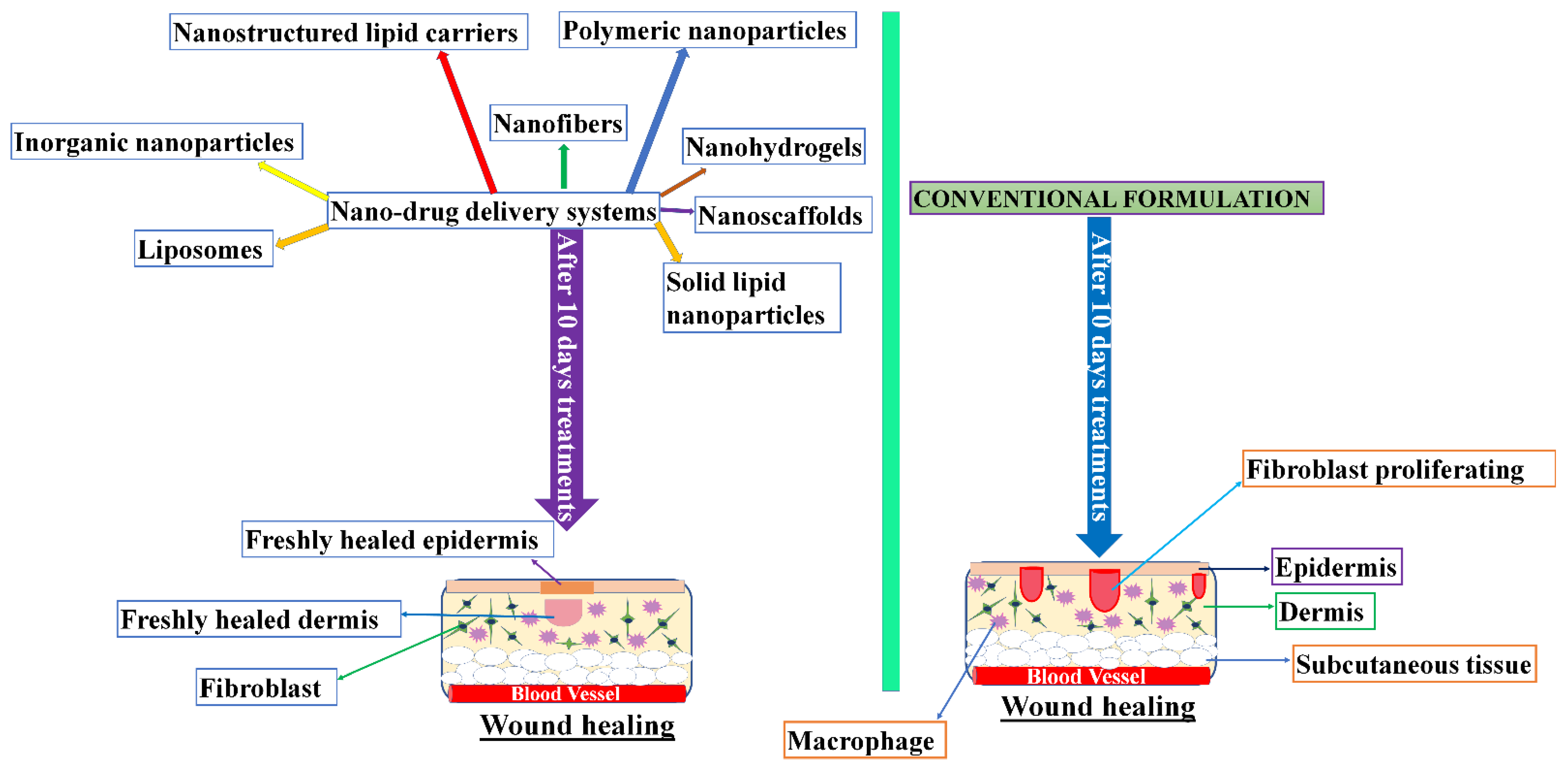
Nano-drug delivery systems enormously influence the potential of drugs’ medicinal effects and also protect the drugs from deterioration. Wound healing and skin re-formation involves various nano-delivery systems, such as those contained in organic nanoparticles, lipid nanoparticles, liposomes, polymeric nanoparticles, nanohydrogels, and nanofibers. These nano-systems show better efficacy compared to conventional systems (Figure 2).
In the past decades, a steady increase in the filing of patents based on herbal nano-formulations has been recorded. The key factor behind this increase is the capability of nano-formulations to overcome solubility drawbacks and bioavailability problems faced by conventional systems. One of most frequently filed patents is for curcumin, the multifunctional phytoceutical, extensively used in the treatment of tumors, cancers, and skin disorders. Other herbal based nano-formulations patents include carotenoids (nano-particles), silymarin (nano-particles), Panax ginseng (liquid mixture), Syzygium cumini, Tinospora cordifolia, Trigonella foenum-graecum, Withania somnifera (nano-emulsion, nanoencapsulation, nano-dispersion, or synergistic liquid mixture), and Arbutin (emulsified nanoparticles) [82].
4.1. Nanofibers
Nanofibers are formed by unbreakable polymer chains of natural and synthetic compounds, which act as a sheet of nanofibers when placed on the skin to improve the tissues [83]. Nanofibers imitate collagen fibrils in the ECM, which can be formed from the synthetic or natural compounds and have numerous qualities that provide benefits to the wound healing process [83]. Nanofibers are beneficial for wound healing because they have a permeable construction and great orifice connection. Nanofibers have the capability to keep moisture at a suitable level. The synthesis of nanofibers with phytochemicals in nanofibrous materials has yielded tremendous results in the area of wound healing as these fibers have the capability to reduce the incision mark because of their porosity, which allows movement of oxygen [84]. Emodin (1,3,8-trihydroxy-6-methyl-anthraquinone) is an anthraquinone derivative that is found in the roots of Rheum officinale L. and is used extensively for wound healing as it has antimicrobial and anti-inflammatory activities. It produced a positive result when used for acute skin injuries [85]. The nanofibers of emodin in polyvinylpyrrolidone were harmless, anti-allergenic, bioactive, and dissolved at a rapid rate when compared to the pure compound. Re-epitheliazation was shown to have occurred at the wounded area, which hastened the healing process [86]. To increase the composition of collagen in human cells to 100%, emodin was incorporated in cellulose acetate nanostructure fibers [87]. The development of herbal constituents in cellulose acetate nanofibers promotes wound healing by using biomaterials as an interactive dressing material. Asiaticoside is extracted from C. asiatica, and the incorporation of trisachharide triterpene into cellulose acetate nanofibers produces an antioxidative effect during the early stages of the injury healing [88]. Increases in types I and III pro-collagen mRNAs were shown to enhance skin fibroblasts by elevating the protein levels [89]. Curcumin incorporated into cellulose acetate caused an improvement in fibroblast proliferation, enhanced collagen synthesis, and protected the dermal fibroblast cells from oxidative stress caused by hydrogen peroxide (H2O2) [90].
The active constituent of turmeric curcumin (1,7-bis (4-hydroxy-3-methoxyphenyl)-1,6-heptadiene-3,5-dione) is a polyphenolic compound, which is obtained from C. longa L. Curcumin is an active ingredient and its use is widely accepted for wound healing because it possesses various properties, such as anti-inflammatory, antibacterial, and antioxidant ones [91]. For epidermal injury healing, curcumin has been used in various in-vivo animal models [92]. Re-epithelization occurs during the early stages and enhances coagulation synthesis because it releases TGFβ1, which leads to increases in the number of blood vessels and cell granulation [93]. Liakos et al. [94] suggested that essential oils, such as cinnamon, lemongrass, and peppermint, can be used as antimicrobial agents. These electro-spun cellulose-based nanofibrous dressings were shown to prevent the Escherichia coli growth and required lesser quantities of oils. These dressings did not show any kind of cytotoxic effects and appears to be safe to use.
It has been reported that the curcumin loaded poly(ε-caprolactone)/gum tragacanth (PCL/GT) led to an improvement in the mechanical properties and tensile strengths of nanofibers and had a positive impact on collagen content for the treatment of diabetic wounds. By 15 days after injury, this moiety led to rapid wound healing by causing regeneration of the epithelial layer [95]. Bromelain-loaded chitosan nanofibers produced favorable wound healing results. It was observed for the second degree burn and has positive impact. The chitosan 2% w/v bromelain showed better physiochemical results compared to chitosan 4% w/v bromelain and was effective in reducing burn-induced injuries [96]. Bixin-loaded polycaprolactone (PCL) nanofibers maintained and accelerated wound healing activity in excisional wounds and effectively reduced the scar tissue area on the diabetic mice [97]. Alfalfa nanofibers yielded better results with respect to skin regeneration as these nanofibers possess antibacterial activity and bioactive phytoestrogens that work as a building block for the dressings for regenerative wounds [98].
4.2. Polymeric Nanoparticles (PNPs)
Polymeric nanoparticles are biocompatible colloidal systems that have risen in importance for both biomedical and bioengineering applications [99]. They are generally integrated by charged polymers and connected by interactivity of cationic and anionic chains of groups [100]. When drugs are incorporated into polymeric systems, this process prevents the deterioration caused by proteases found in the injury and delivered in stages to lower the frequency of administration [101]. Polymeric nanomaterials are widely utilized because of their antibacterial and wound healing activities [102]. For re-generation of skin injury, keratinocyte growth factor (KGF) is an impressive and potent growth factor [103]. It was observed that the KGF consists of self-assembled nanovesicles that enhances healing of the injured tissue cells of the skin by enhancing epithelization and skin re-modeling [104,105]. Recently, PNPs have been formulated by poly-lactic-co-glycolic acid (PLGA) and some of the other combinations in polymeric systems, including alginate, gelatin, and chitosan [106]. PLGA is approved by the Food and Drug Administration (FDA) for use in PNPs. The size of the PGLA NPS is 1–200 nm, which provides the benefits of biodegradability, biocompatibility, and being innocuous [107]. PLGA particles are generally formulated by emulsification of lipophilic compounds utilizing numerous surfactants and organic solvents [108]. The development of EGF-loaded nanoparticles for injury healing using PGLA yielded a positive response with respect to fibroblast proliferation and enhancement of the healing activity in the full thickness wounded skin. EGF plays an importance role in mediating the de-differentiation of keratinocytes into an epithelial linage and to reestablishing the epithelial barrier [109]. One of the studies also suggested that PGLA might produce a biocompatible system for growth factor delivery. To reduce lactate levels and enhance wound healing activity, the peptide defense host, known as LL37, was incorporated into PGLA nanoparticles [110]. Natural polymers, such as chitosan, have been chiefly considered for wound healing activity because of antibacterial and biocompatibility activities [111]. Chitosan is cationic in nature and has been utilized for the inhibition of microbial-induced infections [112]. Chitosan nanovesicles (150–300 nm) are generally formulated utilizing the method of ionic gelation [113]. Nowadays, chitosan is widely accepted in wound treatment, and it can also be utilized as a prophylactic agent to inhibit the infection development and enhance healing activity [114,115]. Studies have shown that polylactic acid-loaded chitosan magnetic eugenol nanospheres had improved prevention and development of biofilm compared to pure chitosan, whilst performing endothelial proliferation [116]. Most of the studies concerning this topic have reported that nanovesicles containing chitosan and analogs might enhance healing activity by improving inflammatory cell function and restoring fibroblasts and osteoblast functions [117]. In two different studies, it was observed that chitosan-loaded nanovesicles improved the coagulation by binding to red blood cells (RBC) and ameliorating the function of inflammatory cells. In another study, the chitosan nanovesicles were used as the compounds in bandages outlined for the skin wound and, hence, enhanced healing activity in both humans and animals [115,118].
4.3. Dendrimers
Dendrimers are nanoscale (1–10 nm) systems with homogeneous structures that are monodispersed in polymer macromolecule that can be used for both therapeutic and diagnostic purposes. Subunits of phenyl acetylene were used to develop dendrimers [119,120]. In addition, functional groups present on the surface of dendrimers can operate as antibacterial agents. Dendrimers cause detachment of contaminated tissues and may extend the phase of inflammation and slow injury diminution in addition to promoting re-epithelization and better wound healing activity [121]. The interaction between positively and negatively charged groups present on dendrimers and on the bacterial cell wall would lead to the bacterial structure disturbance [121]. In another study, silver-loaded dendrimer NSs were observed to show anti-inflammatory and anti-microbial activities in a synergistic manner. These properties were also shown to prevent inflammation and enhance healing activity [122].
4.4. Metallic Nanoparticles
Metal-based nanoparticles are widely utilized as they produce antibacterial, antimicrobial, and anti-inflammatory effects. The chemical and physical structures of nanoparticles are important for determining the propensity of a nanoparticle to enter and/or bind to target cells with the capacity to interact with their biological machinery and elicit a response. The metal-based nanoparticles are widely accepted in medicine, and the most acceptable metallic nanoparticles are silver- and gold-based nanostructures. Herbal plants are widely accepted in the development of metallic nanoparticles because of their low levels of side effects and more therapeutic effects as compared to the conventional dosage form [123]. Most of the herbal extracts, such as Cladophora fascicularis [124], Aerva lanata [125], Hippophae rhamnoides [126], Eucommia ulmoides [127], Black tea leaf [128], Averrhoa bilimbi [129], Salicornia brachiate [130], Abelmoschus esculentus [131], olive leaf [132], Ipomoea carnea [133], geranium [134], and Cissus arnotiana [135] have been incorporated into metallic nanoparticles
Silver nanoparticles are widely used as they possess antimicrobial, antibacterial, and anti-inflammatory properties [136]. The solubility and bioactivity of the silver particles at the wounded area depend on the size of silver particles; the smaller the size is, the stronger the contact with the will skin be. Silver nanoparticle vesicle sizes range from 1 to 100 nm. In one study, the silver–silver chloride nanoparticles combined with lower grapheme oxide nanovesicles induced an escalation of the healing process because it generated a higher number of oxygen free radicals rather than free the silver ions. A positive impact on the antibacterial activity on both Gram-negative and -positive bacteria has been shown, and, hence, these particles can enhance wound healing activity as shown in in-vivo studies in mice [137]. ACTICOAT is an alternate form of silver antimicrobial barrier wound dressing, which prevents the complication of prior agents. It slows down the bacterial activity, which leads to a reduction in inflammation and causes an improvement in the healing process [138]. The plant-based bio-prepared nanoparticles reveal potential for wound remedy and bacterial infection prevention [139]. Different methods for the preparation of silver nanoparticles are used. Photochemical and chemical reduction are the two most widely used methods [140]. Different plant extracts have been incorporated into silver nanoparticles for wound healing containing alkaloids, glycoside, corticosteroids and essential oils [141]. Cassia roxburghii prepared silver nanoparticles show the potential for wound healing enhancement as these particles have significant antibacterial and antifungal activities [142].
The active constituent of Drosera binata is naphthoquinones, primarily plumbagin. D. binata silver nanoparticles show better antibacterial activity against Staphylococcus aureus without affecting human keratinocytes. It was also inconclusive as to whether it is D. binata extract or its pure form (3-chloroplumbagin) that would have effective results for antibiotics and, hence, enhance wound healing [143]. Extracts of grape pomace were also combined with silver nitrate, and grape-silver nanoparticle-stabilized liposomes were developed by Castangia et al. The resulting nano-formulation showed potential to offer a significant shield of keratinocytes and fibroblasts to combat oxidative stress, thus, avoiding cell damage and death [144]. The other highly acceptable nanoparticles in different applications, such as wound treatment, re-epithelization, and particularly drug delivery, include gold nanoparticles [145].
Their chemical stability and capability to absorb near-infrared (NIR) light combined with their positive impact and antibacterial activity will strengthen the wound healing process [146]. Gold nanoparticles have the potential to penetrate bacterial tissues and cause alterations in the cell membrane, which causes inhibition of bacterial activity [147], and also prevents bacteria from developing reactive oxygen species [148].
Gold nanoparticles are synthesized with collagen, gelatin, and chitosan to yield effective injury recovery activity and also helps to achieve the biocompatibility [149]. Chitosan-loaded gold nanoparticles showed enhanced results in the healing process as these particles increase free radical scavenging and improve biocompatibility; in the model, these particles enhance the formation of cells and lead to an improvement in hemostasis by increasing the healing activity in comparison to pure chitosan [150]. The resulting metabolites from Indigofera aspalathoides Vahl. (Papilionaceae), which is also known as Shivanarvembu, are extracted from plants and used for wound healing. The histopathology results demonstrate that the I. aspalathoides silver nanoparticles have a better effect on wound healing in mice. When treated with plant extract, the granulation tissue which possesses fibroblasts, collagen fibers, minimal edema, and newly developed blood vessels were noted [151]. The other forms of metallic nanoparticles are gold and copper oxide nanovesicles that improve wound healing, which leads to fast injury healing and slows down the infection development. Both silver and gold nanoparticles are formed by incorporating Coleous forskohlii root extracts. These particles exhibit antimicrobial activity and antioxidant activities and have a positive effect on re-epithelization at the site of wound, which enhances connective tissue formation and causes an increase in proliferation and remodeling rates of dermal cells [152]. The development of both titanium dioxide and copper oxide nanoparticles of Moringa oleifera and Ficus religiosa leaf extracts, respectively, were shown to enhance wound healing and decrease the removal wound site in rats [153].
4.5. Nanohydrogels
For wound treatment, nanohydrogels are considered to be effective carriers as they possess three-dimensional polymeric networks. Due to their permeable network, they have the capability to absorb the liquid, which helps the wound to keep hydrated and enhance the wound healing process by keeping the proper oxygen level. Due to their effectiveness, compatibility, and showing beneficial results on skin revitalization, nanohydrogels have become widely accepted [154].
To improve wound healing activity, the gellan cholesterol nanohydrogel is immersed in baicalin. The baicalin-loaded nanohydrogels manifest ideal efficacy for skin repair and also act as inflammation inhibitors when applied to an epidermal inflammation mice model in in-vivo studies [155]. The freshly developed nanocrystal bacterial cellulose hydrogels instantly stick to fibroblasts, support human dermal fibroblast morphology, restrict the relocation of cells, enhance the proliferation of cells, and influence the nine expressions of genes connected to healing of injury. These genes include interleukins 6 and 10, granulocyte-macrophage colony-stimulating factor, matrix metalloproteinase 2 (IL-6 and -10, GM-CSF, MMP-2, respectively), and TGF-β; hence, nanohydrogels play an important role in skin regeneration [156].
4.6. Liposomes
Liposomes appear to be an important vehicle for topical delivery; they are harmless and environmentally safe and possess high drug loading efficiency, long-term stability, biological acceptability with skin in addition to having the capability to incorporate both hydrophobic and hydrophilic drugs in water and bilayer cavities [157]. Liposomes successfully shield the injury site and build a humid habitat at the site of injury, which is beneficial for the healing of the wounded skin. Taking all these characteristics into consideration, liposomes have become widely accepted in skin regeneration and injury treatment [158]. A study on propylene glycol liposome nanocarriers demonstrated numerous merits in comparison to other nano-systems. This system showed the tendency to enhance the stability, retention, and permeation in the tissues of skin [159]. It surmised that propylene glycol ameliorate the elasticity of vesicle containing bilayer of phospholipids. Hence, it improved the permeation into the skin. Moreover, the particles size of liposomes should be 150 nm for better drug perforation into the skin layers [160]. Liposomes with silk fibroin hydrogels were prepared to stabilize the basic fibroblast growth factor (bFGF) that maintained the activity of proliferation of cells on wound fluids; it also enhances the healing process by inspiring angiogenesis [161]. Rabelo et al. assessed the gelatin-membrane consisting of usnic acid-loaded liposomes and obtained encouraging results for wound healing. These results showed that the membrane of liposomes prominently manages the second-grade infection on porcine model [162]. Furthermore, with improved collagen, accumulation on cellularized granulation tissue was discovered in the treated group of liposomal membrane, which when compared to one of the commercial products improved the granulation tissue maturation and repaired the scars [163]. Argan-liposomes and argan-hyalurosomes have been successfully developed by incorporating neem oil into them. These formulations were extremely biocompatible and could protect skin cells from oxidative stress effectively with improved efficacy of oil. Moreover, formulations stimulate wound closure substantially more effectively than oil dispersion [164]. The efficacy of mangiferin (employed in cure of skin lesions) was enhanced by modifying transferosomes with propylene glycol and glycerol. Improved deposition of mangiferin was observed in epidermal and dermal layer and fibroblasts were protected from oxidative stress and intensified their propagation [165].
4.7. Inorganic Nanoparticles
Inorganic nanoparticles are those derived from the inorganic materials and include carbon-, metal-, and ceramic-based nanovesicles that accelerate tissue repair and re-modeling. These particles deliver assistance in the region of medicines, counting cancer, imaging, and drug delivery; however, their utilization in tissue regulation and skin re-modeling is new, it also provides adhesion in tissue and enhanced antimicrobial activity in injury healing [166].
4.8. Lipid Nanoparticles
Lipid nanoparticles were designed to overcome the stability limitation of liposomes due to the lipid bilayer. Lipid nanovesicles consist of two types: (1) solid lipid nanoparticles (SLNs) and (2) nanostructured lipid carriers (NLCs). The preparation of lipid nanovesicles amid lipids molecules does not include the use of any potentially harmful biotic solvents [167]. In a study, both SLN- and NLC-loaded rh-EGF (epidermal growth factor) for chronic injury treatment were formulated by the emulsification followed by an ultrasonication method; however, the NLC process included no organic solvent and showed better entrapment efficiency. The results of both formulations show capabilities to enhance cell proliferation when compared with free rh-EGF and considerably enhance the healing activity for wound closure, re-establish the process of inflammation, and facilitate re-epithelization [168].
In another study, development of SLNs with the elastase inhibitor serpin A1 and antimicrobial peptide LL37 had a synergistic impact on injury healing. SLNs promoted the closure of injury in cells of fibroblasts and keratinocytes. Moreover, it also led to improvement in the activity of antibacterial against S. aureus and E. coli when compared with the LL37- and A1-treated groups [169].
5. Future Perspective and Conclusions
The main aim of this review article was to describe the advantages of using nano-systems for use in the wound healing process. The distinctive physiochemical properties of nano-systems make them a perfect candidate for the application of wound healing process. The wound therapy process by nanotechnological systems demonstrates better therapeutic effect compared to the conventional therapy for wound healing. Nanotechnological systems can change one or more than one phase of wound healing during the process, as it possesses antibacterial, anti-inflammatory, and anti-proliferation activities. Worldwide, the research has been conducted on natural and herbal compounds due to their more therapeutic effects and lesser side effects. There is a need for the development of improved systems for the delivery of drugs at the target site with a dose that does not alter the existing treatment of disease. The herbal compounds have great potential and, hence, a better future, especially when incorporated into the nanocarriers for chronic wound treatment as they have shown promising results. Herbal medicine-based novel drug delivery systems have acknowledged the approaches in the field of pharmaceuticals, which will improve the health of the people. It is also concluded that the incorporation of herbal compound in the nano-vehicle will aggrandize the magnitude of the existing delivery system. Anyhow, various approaches have been employed for the privileged application of nanocarriers in wound healing therapy. The main concerns for the nano-vehicles are toxicity because they may cause possible side effects in the human body. Hence, this requires to be rectified at the starting point for further progression of wound healing therapies in clinical trials. In in vivo models, there is slighter comprehension regarding non-material mediated wound healing processes and this is one of the problems observed. The studies of non-material-wound healing processes are based on in-vitro studies or mainly depend on single aim bacteria. The in-vivo wound healing application is required for the in-depth studies utilizing both Gram-positive and -negative bacterial strains. Subsequently, the main focus should be on improving and enhancing target efficiency for more efficacious wound healing. Therefore, the investigators should target producing a nanomaterial that is biocompatible and biodegradable and has the capability to correct all the phases of the wound healing process.
Author Contributions
Conceptualization, A.Q., M.A. and M.H.W.; methodology, S.J., A.Q.; software, A.A.; validation, A.A., N.A.A. and M.A.A.; formal analysis, A.Q., M.H.W. and N.K.; investigation, A.A., N.K.; resources, N.A.A., A.Q.; data curation, N.K., S.J.; writing—original draft preparation, A.Q.; writing—review and editing, M.H.W., M.A.A. and N.A.A.; visualization, M.H.W.; supervision, M.A.; project administration, M.A.A., N.A.A.; funding acquisition, M.A.A., N.A.A. All authors have read and agreed to the published version of the manuscript.
Funding
The Deanship of Scientific Research (DSR) at King Abdulaziz University, Jeddah, Saudi Arabia has funded this project, under grant no. (FP-032-43).
Institutional Review Board Statement
Not applicable.
Informed Consent Statement
Not applicable.
Data Availability Statement
Not applicable.
Conflicts of Interest
The authors declare no conflict of interest.
References
- Wang, W.; Lu, K.J.; Yu, C.H.; Huang, Q.L.; Du, Y.Z. Nano-drug delivery systems in wound treatment and skin regeneration. J. Nanobiotechnology 2019, 17, 82. [Google Scholar] [CrossRef]
- Pereira, R.F.; Carvalho, A.; Gil, M.H.; Mendes, A.; Bartolo, P. Influence of Aloe vera on water absorption and enzymatic in vitro degradation of alginate hydrogel films. Carbohydr. Polym. 2013, 98, 311–320. [Google Scholar] [CrossRef]
- World Health Organization Burns—Key Facts. Available online: https://www.who.int/news-room/fact-sheets/detail/burns (accessed on 4 January 2020).
- Souto, E.B.; Ribeiro, A.F.; Ferreira, M.I.; Teixeira, M.C.; Shimojo, A.A.; Soriano, J.L.; Naveros, B.C.; Durazzo, A.; Lucarini, M.; Souto, S.B.; et al. New nanotechnologies for the treatment and repair of skin burns infections. Int. J. Mol. Sci. 2020, 21, 393. [Google Scholar] [CrossRef] [PubMed] [Green Version]
- Deitch, E.A. The management of burns. N. Engl. J. Med. 1990, 323, 1249–1253. [Google Scholar] [PubMed]
- Wild, T.; Rahbarnia, A.; Kellner, M.; Sobotka, L.; Eberlein, T. Basics in nutrition and wound healing. Nutrition 2010, 26, 862–866. [Google Scholar] [CrossRef] [PubMed]
- Hermans, M.H. Results of an internet survey on the treatment of partial thickness burns, full thickness burns, and donor sites. J. Burn Care Res. 2007, 28, 835–847. [Google Scholar] [CrossRef] [PubMed] [Green Version]
- Roshangar, L.; Kheirjou, R.; Ranjkesh, R. Skin Burns: Review of Molecular Mechanisms and Therapeutic Approaches. Wounds Compend. Clin. Res. Pract. 2019, 31, 308–315. [Google Scholar]
- Guo, S.A.; DiPietro, L.A. Factors affecting wound healing. J. Dent. Res. 2010, 89, 219–229. [Google Scholar] [CrossRef]
- Ghosh, P.K.; Gaba, A. Phyto-extracts in wound healing. J. Pharm. Pharm. Sci. 2013, 16, 760–820. [Google Scholar] [CrossRef]
- Sandhiya, V.; Ubaidulla, U. A review on herbal drug loaded into pharmaceutical carrier techniques and its evaluation process. Future J. Pharm. Sci. 2020, 6, 1–16. [Google Scholar] [CrossRef]
- Bhatt, D.; Jethva, K.; Patel, S.; Zaveri, M. Novel drug delivery systems in herbals for cancer. World J. Pharm. Res. 2016, 5, 368–378. [Google Scholar]
- Ferreira, V.F.; Pinto, A.C. A fitoterapia no mundo atual. Química Nova 2010, 33, 1829. [Google Scholar] [CrossRef]
- Vickers, A.; Zollman, C. Herbal medicine. Br. Med. J. 1999, 319, 1050–1053. [Google Scholar] [CrossRef] [PubMed] [Green Version]
- Ali, S.I.; Gopalakrishnan, B.; Venkatesalu, V. Pharmacognosy, phytochemistry and pharmacological properties of Achillea millefoliumh L.: A review. Phytother. Res. 2017, 31, 1140–1161. [Google Scholar] [CrossRef] [PubMed]
- Watkins, R.; Wu, L.; Zhang, C.; Davis, R.; Xu, B. Natural product-based nanomedicine: Recent advances and issues. Int. J. Nanomed. 2015, 10, 6055–6074. [Google Scholar]
- Xu, R.; Luo, G.; Xia, H.; He, W.; Zhao, J.; Liu, B.; Tan, J.; Zhou, J.; Liu, D.; Wang, Y.; et al. Novel bilayer wound dressing composed of silicone rubber with particular micropores enhanced wound re-epithelialization and contraction. Biomaterials 2014, 40, 1–11. [Google Scholar] [CrossRef] [PubMed]
- Singh, R.P.; Singh, S.G.; Naik, H.; Jain, D.; Bisla, S. Herbal excipients in novel drug delivery system. Int. J. Compr. Pharm. 2011, 2, 1–7. [Google Scholar]
- Sungthongjeen, S.; Pitaksuteepong, T.; Somsiri, A.; Sriamornsak, P. Studies on pectins as potential hydrogel matrices for controlled-release drug delivery. Drug Dev. Ind. Pharm. 1999, 25, 1271–1276. [Google Scholar] [CrossRef]
- Portou, M.; Baker, D.; Abraham, D.; Tsui, J. The innate immune system, toll-like receptors and dermal wound healing: A review. Vasc. Pharmacol. 2015, 71, 31–36. [Google Scholar] [CrossRef]
- Andritoiu, C.V.; Andriescu, C.E.; Ibanescu, C.; Lungu, C.; Ivanescu, B.; Vlase, L.; Havarneanu, C.; Popa, M. Effects and Characterization of Some Topical Ointments Based on Vegetal Extracts on Incision, Excision, and Thermal Wound Models. Molecules 2020, 25, 5356. [Google Scholar] [CrossRef]
- Hajialyani, M.; Tewari, D.; Sobarzo-Sánchez, E.; Nabavi, S.M.; Farzaei, M.H.; Abdollahi, M. Natural product-based nanomedicines for wound healing purposes: Therapeutic targets and drug delivery systems. Int. J. Nanomed. 2018, 13, 5023–5043. [Google Scholar] [CrossRef] [Green Version]
- Martin, P. Wound Healing--Aiming for Perfect Skin Regeneration. Science 1997, 276, 75–81. [Google Scholar] [CrossRef]
- Braund, R.; Hook, S.; Medlicott, N.J. The role of topical growth factors in chronic wounds. Curr. Drug Deliv. 2007, 4, 195–204. [Google Scholar] [CrossRef] [PubMed]
- Gainza, G.; Villullas, S.; Pedraz, J.L.; Hernandez, R.M.; Igartua, M. Advances in drug delivery systems (DDSs) to release growth factors for wound healing and skin regeneration. Nanomed. Nanotechnol. Biol. Med. 2015, 11, 1551–1573. [Google Scholar] [CrossRef] [PubMed]
- Kiritsy, C.P.; Lynch, S.E. Role of growth factors in cutaneous wound healing: A review. Crit. Rev. Oral Biol. Med. 1993, 4, 729–760. [Google Scholar] [CrossRef] [PubMed] [Green Version]
- Eming, S.A.; Krieg, T.; Davidson, J.M. Inflammation in wound repair: Molecular and cellular mechanisms. J. Investig. Dermatol. 2007, 127, 514–525. [Google Scholar] [CrossRef] [Green Version]
- Singer, A.J.; Clark, R.A. Cutaneous wound healing. N. Engl. J. Med. 1999, 341, 738–746. [Google Scholar] [CrossRef]
- Velnar, T.; Bailey, T.; Smrkolj, V. The wound healing process: An overview of the cellular and molecular mechanisms. J. Int. Med. Res. 2009, 37, 1528–1542. [Google Scholar] [CrossRef]
- Malinda, K.M.; Sidhu, G.S.; Banaudha, K.K.; Gaddipati, J.P.; Maheshwari, R.K.; Goldstein, A.L.; Kleinman, H.K. Thymosin α1 stimulates endothelial cell migration, angiogenesis, and wound healing. J. Immunol. 1998, 160, 1001–1006. [Google Scholar]
- Li, B.; Wang, J.H.C. Fibroblasts and myofibroblasts in wound healing: Force generation and measurement. J. Tissue Viability 2011, 20, 108–120. [Google Scholar] [CrossRef] [Green Version]
- Montesinos, M.C.; Gadangi, P.; Longaker, M.; Sung, J.; Levine, J.; Nilsen, D.; Reibman, J.; Li, M.; Jiang, C.-K.; Hirschhorn, R.; et al. Wound Healing Is Accelerated by Agonists of Adenosine A2 (Gαs-linked) Receptors. J. Exp. Med. 1997, 186, 1615–1620. [Google Scholar] [CrossRef]
- Ehrlich, H.P.; Keefer, K.A.; Myers, R.L.; Passaniti, A. Vanadate and the absence of myofibroblasts in wound contraction. Arch. Surg. 1999, 134, 494–501. [Google Scholar] [CrossRef] [PubMed] [Green Version]
- Stadelmann, W.K.; Digenis, A.G.; Tobin, G.R. Physiology and healing dynamics of chronic cutaneous wounds. Am. J. Surg. 1998, 176, 26S–38S. [Google Scholar] [CrossRef]
- Pawar, H.V.; Tetteh, J.; Boateng, J.S. Preparation, optimisation and characterisation of novel wound healing film dressings loaded with streptomycin and diclofenac. Colloids Surf. B Biointerfaces 2013, 102, 102–110. [Google Scholar] [CrossRef]
- Sabitha, M.; Rajiv, S. Preparation and characterization of ampicillin-incorporated electrospun polyurethane scaffolds for wound healing and infection control. Polym. Eng. Sci. 2014, 55, 541–548. [Google Scholar] [CrossRef]
- Lan, Y.; Li, W.; Guo, R.; Zhang, Y.; Xue, W.; Zhang, Y. Preparation and characterisation of vancomycin-impregnated gelatin microspheres/silk fibroin scaffold. J. Biomater. Sci. Polym. Ed. 2013, 25, 75–87. [Google Scholar] [CrossRef] [PubMed]
- Pásztor, N.; Rédai, E.; Szabó, Z.-I.; Sipos, E. Preparation and characterization of levofloxacin-loaded nanofibers as potential wound dressings. Acta Med. Marisiensis 2017, 63, 66–69. [Google Scholar] [CrossRef] [Green Version]
- Mohseni, M.; Shamloo, A.; Aghababaei, Z.; Vossoughi, M.; Moravvej, H. Antimicrobial wound dressing containing silver sulfadiazine with high biocompatibility: In vitro study. Artif. Organs 2016, 40, 765–773. [Google Scholar] [CrossRef]
- Adhirajan, N.; Shanmugasundaram, N.; Shanmuganathan, S.; Babu, M. Collagen-based wound dressing for doxycycline delivery: In-vivo evaluation in an infected excisional wound model in rats. J. Pharm. Pharmacol. 2009, 61, 1617–1623. [Google Scholar] [CrossRef]
- Parihar, A.; Parihar, M.S.; Milner, S.; Bhat, S. Oxidative stress and anti-oxidative mobilization in burn injury. Burns 2008, 34, 6–17. [Google Scholar] [CrossRef]
- Suntar, I.; Akkol, E.K.; Nahar, L.; Sarker, S.D. Wound healing and antioxidant properties: Do they coexist in plants? Free Radic. Antioxid. 2012, 2, 1–7. [Google Scholar] [CrossRef] [Green Version]
- Blass, S.C.; Goost, H.; Tolba, R.H.; Stoffel-Wagner, B.; Kabir, K.; Burger, C.; Stehle, P.; Ellinger, S. Time to wound closure in trauma patients with disorders in wound healing is shortened by supplements containing antioxidant micronutrients and glutamine: A PRCT. Clin. Nutr. 2012, 31, 469–475. [Google Scholar] [CrossRef] [PubMed]
- Schultz, G.S.; Sibbald, R.G.; Falanga, V.; Ayello, E.A.; Dowsett, C.; Harding, K.; Romanelli, M.; Stacey, M.C.S.; Teot, L.; Vanscheidt, W. Wound bed preparation: A systematic approach to wound management. Wound Repair Regen. 2003, 11, S1–S28. [Google Scholar] [CrossRef]
- Tsala, D.E.; Amadou, D.; Habtemariam, S. Natural wound healing and bioactive natural products. Phytopharmacology 2013, 4, 532–560. [Google Scholar]
- Ruszymah, B.H.I.; Chowdhury, S.R.; Manan, N.A.B.A.; Fong, O.S.; Adenan, M.I.; Bin Saim, A. Aqueous extract of Centella asiatica promotes corneal epithelium wound healing in vitro. J. Ethnopharmacol. 2012, 140, 333–338. [Google Scholar] [CrossRef] [PubMed]
- Maquart, F.X.; Bellon, G.; Gillery, P.; Wegrowski, Y.; Borel, J.P. Stimulation of collagen synthesis in fibroblast cultures by a triterpene extracted from Centella asiatica. Connect. Tissue Res. 1990, 24, 107–120. [Google Scholar] [CrossRef] [PubMed]
- Thang, P.T.; Teik, L.S.; Yung, C.S. Anti-oxidant effects of the extracts from the leaves of Chromolaena odorata on human dermal fibroblasts and epidermal keratinocytes against hydrogen peroxide and hypoxanthine–xanthine oxidase induced damage. Burns 2001, 27, 319–327. [Google Scholar] [CrossRef]
- Umachigi, S.P.; Jayaveera, K.N.; Kumar, C.K.A.; Kumar, G.S.; Swamy, B.M.V.; Kumar, D.V.K. Studies on wound healing properties of Quercus infectoria. Trop. J. Pharm. Res. 2008, 7, 913–919. [Google Scholar] [CrossRef] [Green Version]
- Mensah, A.Y.; Sampson, J.; Houghton, P.; Hylands, P.; Westbrook, J.; Dunn, M.; Hughes, M.; Cherry, G. Effects of Buddleja globosa leaf and its constituents relevant to wound healing. J. Ethnopharmacol. 2001, 77, 219–226. [Google Scholar] [CrossRef]
- Shukla, A.; Rasik, A.M.; Dhawan, B.N. Asiaticoside-induced elevation of antioxidant levels in healing wounds. Phytother. Res. Int. J. Devoted Pharmacol. Toxicol. Eval. Nat. Prod. Deriv. 1999, 13, 50–54. [Google Scholar] [CrossRef]
- Yamasaki, T.; Li, L.; Lau, B.H.S. Garlic compounds protect vascular endothelial cells from hydrogen peroxide-induced oxidant injury. Phytother. Res. 1994, 8, 408–412. [Google Scholar] [CrossRef]
- Ravindran, P.N.; Babu, K.N.; Sivaraman, K. (Eds.) Turmeric: The Genus Curcuma; CRC Press: Boca Raton, FL, USA, 2007. [Google Scholar]
- Gopinath, D.; Ahmed, M.; Gomathi, K.; Chitra, K.; Sehgal, P.; Jayakumar, R. Dermal wound healing processes with curcumin incorporated collagen films. Biomaterials 2004, 25, 1911–1917. [Google Scholar] [CrossRef]
- Marwah, R.G.; Fatope, M.O.; Al Mahrooqi, R.; Varma, G.B.; Al Abadi, H.; Al-Burtamani, S.K.S. Antioxidant capacity of some edible and wound healing plants in Oman. Food Chem. 2007, 101, 465–470. [Google Scholar] [CrossRef]
- Qadir, M.I. Medicinal and cosmetological importance of Aloe vera. Int. J. Nat. Ther. 2009, 2, 21–26. [Google Scholar]
- Pattanayak, S.; Sunita, P. Wound healing, anti-microbial and antioxidant potential of Dendrophthoe falcata (L.f) Ettingsh. J. Ethnopharmacol. 2008, 120, 241–247. [Google Scholar] [CrossRef]
- Altameme, H.J.; Hameed, I.H.; Hamza, L.F. Anethum graveolens: Physicochemical properties, medicinal uses, antimicrobial effects, antioxidant effect, anti-inflammatory and analgesic effects: A review. Int. J. Pharm. Qual. Assur. 2017, 8, 88–91. [Google Scholar]
- Shirwaikar, A.; Somashekar, A.; Udupa, A.; Udupa, S.; Somashekar, S. Wound healing studies of Aristolochia bracteolata Lam. with supportive action of antioxidant enzymes. Phytomedicine 2003, 10, 558–562. [Google Scholar] [CrossRef] [PubMed]
- Barua, C.C.; Talukdar, A.; Begum, S.A.; Sarma, D.K.; Fathak, D.C.; Barua, A.G.; Bora, R.S. Wound healing activity of methanolic extract of leaves of Alternanthera brasiliana Kuntz using in vivo and in vitro model. Indian J. Exp. Boil. 2009, 47, 1001–1005. [Google Scholar]
- Demilew, W.; Adinew, G.M.; Asrade, S. Evaluation of the wound healing activity of the crude extract of leaves of Acanthus polystachyus Delile (Acanthaceae). Evid.-Based Complement. Altern. Med. 2018, 2018, 1–9. [Google Scholar] [CrossRef] [Green Version]
- Adetutu, A.; Morgan, W.A.; Corcoran, O. Antibacterial, antioxidant and fibroblast growth stimulation activity of crude extracts of Bridelia ferruginea leaf, a wound-healing plant of Nigeria. J. Ethnopharmacol. 2011, 133, 116–119. [Google Scholar] [CrossRef]
- Shukla, A.; Rasik, A.; Jain, G.; Shankar, R.; Kulshrestha, D.; Dhawan, B. In vitro and in vivo wound healing activity of asiaticoside isolated from Centella asiatica. J. Ethnopharmacol. 1999, 65, 1–11. [Google Scholar] [CrossRef]
- Chen, Y.J.; Dai, Y.S.; Chen, B.F.; Chang, A.; Chen, H.C.; Lin, Y.C.; Chang, K.H.; Lai, Y.L.; Chung, C.H.; Lai, Y.J. The effect of tetrandrine and extracts of Centella asiatica on acute radiation dermatitis in rats. Biol. Pharm. Bull. 1999, 22, 703–706. [Google Scholar] [CrossRef] [Green Version]
- Gouthamchandra, K.; Mahmood, R.; Manjunatha, H. Free radical scavenging, antioxidant enzymes and wound healing activities of leaves extracts from Clerodendrum infortunatum L. Environ. Toxicol. Pharmacol. 2010, 30, 11–18. [Google Scholar] [CrossRef]
- Agyare, C.; Asase, A.; Lechtenberg, M.; Niehues, M.; Deters, A.; Hensel, A. An ethnopharmacological survey and in vitro confirmation of ethnopharmacological use of medicinal plants used for wound healing in Bosomtwi-Atwima-Kwanwoma area, Ghana. J. Ethnopharmacol. 2009, 125, 393–403. [Google Scholar] [CrossRef]
- Koca, U.; Süntar, I.; Akkol, E.K.; Yılmazer, D.; Alper, M. Wound repair potential of Olea europaea L. leaf extracts revealed by in vivo experimental models and comparative evaluation of the extracts’ antioxidant activity. J. Med. Food 2011, 14, 140–146. [Google Scholar] [CrossRef] [PubMed]
- Arawande, J.O.; Komolafe, E.A.; Imokhuede, B. Nutritional and phytochemical compositions of fireweed (Crassocephalum crepidioides). J. Agric. Technol. 2013, 9, 439–449. [Google Scholar]
- Mathew, S.; Abraham, T.E. In vitro antioxidant activity and scavenging effects of Cinnamomum verum leaf extract assayed by different methodologies. Food Chem. Toxicol. 2006, 44, 198–206. [Google Scholar] [CrossRef]
- Atun, S.; Handayani, S.; Rakhmawati, A.; Purnamaningsih, N.A.; Naila, B.I.; Lestari, A. Study of potential phenolic compounds from stems of Dendrophthoe falcata (Loranthaceae) plant as antioxidant and antimicrobial agents. Orient. J. Chem. 2018, 34, 2342–2349. [Google Scholar] [CrossRef] [Green Version]
- Velmurugan, C.; Geetha, C.; Shajahan, S.; Vijayakumar, S.; Kumar, P.L. Wound healing potential of leaves of Eucalyptus citriodoralin rats. World J. Pharm. Sci. 2014, 2, 62–71. [Google Scholar]
- Annan, K.; Houghton, P.J. Antibacterial, antioxidant and fibroblast growth stimulation of aqueous extracts of Ficus asperifolia Miq. and Gossypium arboreum L., wound-healing plants of Ghana. J. Ethnopharmacol. 2008, 119, 141–144. [Google Scholar] [CrossRef]
- Steenkamp, V.; Mathivha, E.; Gouws, M.; van Rensburg, C.J. Studies on antibacterial, antioxidant and fibroblast growth stimulation of wound healing remedies from South Africa. J. Ethnopharmacol. 2004, 95, 353–357. [Google Scholar] [CrossRef] [PubMed]
- Upadhyay, N.; Kumar, R.; Mandotra, S.; Meena, R.; Siddiqui, M.; Sawhney, R.; Gupta, A. Safety and healing efficacy of Sea buckthorn (Hippophae rhamnoides L.) seed oil on burn wounds in rats. Food Chem. Toxicol. 2009, 47, 1146–1153. [Google Scholar] [CrossRef] [PubMed]
- Reddy, B.S.; Reddy, R.K.K.; Naidu, V.; Madhusudhana, K.; Agwane, S.B.; Ramakrishna, S.; Diwan, P.V. Evaluation of antimicrobial, antioxidant and wound-healing potentials of Holoptelea integrifolia. J. Ethnopharmacol. 2008, 115, 249–256. [Google Scholar] [CrossRef] [PubMed]
- Nualkaew, S.; Rattanamanee, K.; Thongpraditchote, S.; Wongkrajang, Y.; Nahrstedt, A. Anti-inflammatory, analgesic and wound healing activities of the leaves of Memecylon edule Roxb. J. Ethnopharmacol. 2009, 121, 278–281. [Google Scholar] [CrossRef] [PubMed]
- Muhammad, A.A.; Pauzi, N.A.S.; Arulselvan, P.; Abas, F.; Fakurazi, S. In vitro wound healing potential and identification of bioactive compounds from Moringa oleifera Lam. BioMed Res. Int. 2013, 2013, 974580. [Google Scholar] [CrossRef] [Green Version]
- Jain, S.; Shrivastava, S.; Nayak, S.; Sumbhate, S. Recent trends in Curcuma longa Linn. Pharmacogn. Rev. 2007, 1, 119–128. [Google Scholar]
- Singh, M.; Govindarajan, R.; Nath, V.; Rawat, A.K.S.; Mehrotra, S. Antimicrobial, wound healing and antioxidant activity of Plagiochasma appendiculatum Lehm. et Lind. J. Ethnopharmacol. 2006, 107, 67–72. [Google Scholar] [CrossRef]
- Berenguer, B.; Sánchez, L.; Quílez, A.; López-Barreiro, M.; de Haro, O.; Gálvez, J.; Martín, M. Protective and antioxidant effects of Rhizophora mangle L. against NSAID-induced gastric ulcers. J. Ethnopharmacol. 2006, 103, 194–200. [Google Scholar] [CrossRef]
- Taranalli, A.D.; Kuppast, I.J. Study of wound healing activity of seeds of Trigonella foenum graecum in rats. Indian J. Pharm. Sci. 1996, 58, 117. [Google Scholar]
- Jadhav, N.R.; Powar, T.; Shinde, S.; Nadaf, S. Herbal nanoparticles: A patent review. Asian J. Pharm. 2014, 8, 1–12. [Google Scholar] [CrossRef]
- Hromadka, M.; Collins, J.B.; Reed, C.; Han, L.; Kolappa, K.K.; Cairns, B.A.; Andrady, T.; van Aalst, J.A. Nanofiber applications for burn care. J. Burn Care Res. 2008, 29, 695–703. [Google Scholar] [CrossRef]
- Cerchiara, T.; Abruzzo, A.; Palomino, R.A.Ñ.; Vitali, B.; De Rose, R.; Chidichimo, G.; Ceseracciu, L.; Athanassiou, A.; Saladini, B.; Dalena, F.; et al. Spanish Broom (Spartium junceum L.) fibers impregnated with vancomycin-loaded chitosan nanoparticles as new antibacterial wound dressing: Preparation, characterization and antibacterial activity. Eur. J. Pharm. Sci. 2017, 99, 105–112. [Google Scholar] [CrossRef] [PubMed]
- Tang, T.; Yin, L.; Yang, J.; Shan, G. Emodin, an anthraquinone derivative from Rheum officinale Baill, enhances cutaneous wound healing in rats. Eur. J. Pharmacol. 2007, 567, 177–185. [Google Scholar] [CrossRef] [PubMed]
- Dai, X.Y.; Nie, W.; Wang, Y.C.; Shen, Y.; Li, Y.; Gan, S.J. Electrospun emodin polyvinylpyrrolidone blended nanofibrous membrane: A novel medicated biomaterial for drug delivery and accelerated wound healing. J. Mater. Sci. Mater. Med. 2012, 23, 2709–2716. [Google Scholar] [CrossRef]
- Panichpakdee, J.; Pavasant, P.; Supaphol, P. Electrospun cellulose acetate fiber mats containing emodin with potential for use as wound dressing. Chiang Mai J. Sci. 2016, 43, 1249–1259. [Google Scholar]
- Suwantong, O.; Ruktanonchai, U.; Supaphol, P. In vitro biological evaluation of electrospun cellulose acetate fiber mats containing asiaticoside or curcumin. J. Biomed. Mater. Res. Part A 2010, 94, 1216–1225. [Google Scholar]
- Panichpakdee, J.; Pavasant, P.; Supaphol, P. Electrospinning of asiaticoside/2-hydroxypropyl-β-cyclodextrin inclusion complex-loaded cellulose acetate fiber mats: Release characteristics and potential for use as wound dressing. Polym. Korea 2014, 38, 338–350. [Google Scholar] [CrossRef] [Green Version]
- Suwantong, O.; Ruktanonchai, U.; Supaphol, P. Electrospun cellulose acetate fiber mats containing asiaticoside or Centella asiatica crude extract and the release characteristics of asiaticoside. Polymer 2008, 49, 4239–4247. [Google Scholar] [CrossRef]
- Momtazi, A.A.; Haftcheshmeh, S.M.; Esmaeili, S.-A.; Johnston, T.P.; Abdollahi, E.; Sahebkar, A. Curcumin: A natural modulator of immune cells in systemic lupus erythematosus. Autoimmun. Rev. 2018, 17, 125–135. [Google Scholar] [CrossRef]
- Prasad, R.; Kumar, D.; Kant, V.; Tandan, S.K.; Kumar, D. Curcumin enhanced cutaneous wound healing by modulating cytokines and transforming growth factor in excision wound model in rats. Int. J. Curr. Microbiol. Appl. Sci. 2017, 6, 2263–2273. [Google Scholar] [CrossRef] [Green Version]
- Sidhu, G.S.; Singh, A.K.; Thaloor, D.; Banaudha, K.K.; Patnaik, G.K.; Srimal, R.C.; Maheshwari, R.K. Enhancement of wound healing by curcumin in animals. Wound Repair Regen. 1998, 6, 167–177. [Google Scholar] [CrossRef] [PubMed]
- Liakos, I.; Rizzello, L.; Hajiali, H.; Brunetti, V.; Carzino, R.; Pompa, P.P.; Athanassiou, A.; Mele, E. Fibrous wound dressings encapsulating essential oils as natural antimicrobial agents. J. Mater. Chem. B 2015, 3, 1583–1589. [Google Scholar] [CrossRef]
- Huang, S.; Fu, X. Naturally derived materials-based cell and drug delivery systems in skin regeneration. J. Control. Release 2010, 142, 149–159. [Google Scholar] [CrossRef]
- Tokuda, M.; Yamane, M.; Thickett, S.C.; Minami, H.; Zetterlund, P.B. Synthesis of polymeric nanoparticles containing reduced graphene oxide nanosheets stabilized by poly(ionic liquid) using miniemulsion polymerization. Soft Matter 2016, 12, 3955–3962. [Google Scholar] [CrossRef]
- Yun, Y.H.; Goetz, D.J.; Yellen, P.; Chen, W. Hyaluronan microspheres for sustained gene delivery and site-specific targeting. Biomaterials 2003, 25, 147–157. [Google Scholar] [CrossRef]
- Korrapati, P.S.; Karthikeyan, K.; Satish, A.; Krishnaswamy, V.R.; Venugopal, J.R.; Ramakrishna, S. Recent advancements in nanotechnological strategies in selection, design and delivery of biomolecules for skin regeneration. Mater. Sci. Eng. C 2016, 67, 747–765. [Google Scholar] [CrossRef]
- Gardner, J.C.; Wu, H.; Noel, J.G.; Ramser, B.J.; Pitstick, L.; Saito, A.; Nikolaidis, N.M.; McCormack, F.X. Keratinocyte growth factor supports pulmonary innate immune defense through maintenance of alveolar antimicrobial protein levels and macrophage function. Am. J. Physiol. Cell. Mol. Physiol. 2016, 310, L868–L879. [Google Scholar] [CrossRef] [PubMed] [Green Version]
- Feng, Z.G.; Pang, S.F.; Guo, D.J.; Yang, Y.T.; Liu, B.; Wang, J.W.; Zheng, K.Q.; Lin, Y. Recombinant keratinocyte growth factor 1 in tobacco potentially promotes wound healing in diabetic rats. BioMed Res. Int. 2014, 2014, 1–9. [Google Scholar] [CrossRef] [Green Version]
- Koria, P.; Yagi, H.; Kitagawa, Y.; Megeed, Z.; Nahmias, Y.; Sheridan, R.; Yarmush, M.L. Self-assembling elastin-like peptides growth factor chimeric nanoparticles for the treatment of chronic wounds. Proc. Natl. Acad. Sci. USA 2010, 108, 1034–1039. [Google Scholar] [CrossRef] [Green Version]
- Ye, M.; Kim, S.; Park, K. Issues in long-term protein delivery using biodegradable microparticles. J. Control. Release 2010, 146, 241–260. [Google Scholar] [CrossRef] [PubMed]
- Chereddy, K.K.; Vandermeulen, G.; Préat, V. PLGA based drug delivery systems: Promising carriers for wound healing activity. Wound Repair Regen. 2016, 24, 223–236. [Google Scholar] [CrossRef]
- Zhang, Y.; Wischke, C.; Mittal, S.; Mitra, A.; Schwendeman, S.P. Design of controlled release PLGA microspheres for hydrophobic fenretinide. Mol. Pharm. 2016, 13, 2622–2630. [Google Scholar]
- Yüksel, E.; Karakeçili, A.; Demirtaş, T.T.; Gümüşderelioğlu, M. Preparation of bioactive and antimicrobial PLGA membranes by magainin II/EGF functionalization. Int. J. Biol. Macromol. 2016, 86, 162–168. [Google Scholar]
- Chereddy, K.K.; Her, C.-H.; Comune, M.; Moia, C.; Lopes, A.; Porporato, P.E.; Vanacker, J.; Lam, M.C.; Steinstraesser, L.; Sonveaux, P.; et al. PLGA nanoparticles loaded with host defense peptide LL37 promote wound healing. J. Control. Release 2014, 194, 138–147. [Google Scholar] [PubMed]
- Matica, M.A.; Aachmann, F.L.; Tøndervik, A.; Sletta, H.; Ostafe, V. Chitosan as a wound dressing starting material: Antimicrobial properties and mode of action. Int. J. Mol. Sci. 2019, 20, 5889. [Google Scholar] [CrossRef] [Green Version]
- Dai, T.; Tegos, G.P.; Burkatovskaya, M.; Castano, A.P.; Hamblin, M.R. Chitosan acetate bandage as a topical antimicrobial dressing for infected burns. Antimicrob. Agents Chemother. 2009, 53, 393–400. [Google Scholar] [CrossRef] [PubMed] [Green Version]
- Karimi, M.; Zangabad, P.S.; Ghasemi, A.; Amiri, M.; Bahrami, M.; Malekzad, H.; Asl, H.G.; Mahdieh, Z.; Bozorgomid, M.; Ghasemi, A.; et al. Temperature-responsive smart nanocarriers for delivery of therapeutic agents: Applications and recent advances. ACS Appl. Mater. Interfaces 2016, 8, 21107–21133. [Google Scholar] [CrossRef] [PubMed] [Green Version]
- Shrestha, A.; Hamblin, M.R.; Kishen, A. Characterization of a conjugate between Rose Bengal and chitosan for targeted antibiofilm and tissue stabilization effects as a potential treatment of infected dentin. Antimicrob. Agents Chemother. 2012, 56, 4876–4884. [Google Scholar] [CrossRef] [Green Version]
- Dai, T.; Tanaka, M.; Huang, Y.; Hamblin, M.R. Chitosan preparations for wounds and burns: Antimicrobial and wound-healing effects. Expert Rev. Anti-Infective Ther. 2011, 9, 857–879. [Google Scholar] [CrossRef]
- Holban, A.M.; Grumezescu, V.; Grumezescu, A.M.; Vasile, B.; Truşcă, R.; Cristescu, R.; Socol, G.; Iordache, F. Antimicrobial nanospheres thin coatings prepared by advanced pulsed laser technique. Beilstein J. Nanotechnol. 2014, 5, 872–880. [Google Scholar] [CrossRef] [Green Version]
- Baxter, R.M.; Dai, T.; Kimball, J.; Wang, E.; Hamblin, M.R.; Wiesmann, W.P.; McCarthy, S.J.; Baker, S.M. Chitosan dressing promotes healing in third degree burns in mice: Gene expression analysis shows biphasic effects for rapid tissue regeneration and decreased fibrotic signaling. J. Biomed. Mater. Res. Part A 2013, 101, 340–348. [Google Scholar] [CrossRef] [PubMed] [Green Version]
- Karimi, M.; Avci, P.; Ahi, M.; Gazori, T.; Hamblin, M.R.; Naderi-Manesh, H. Evaluation of chitosan-tripolyphosphate nanoparticles as a p-shRNA delivery vector: Formulation, optimization and cellular uptake study. J. Nanopharm. Drug Deliv. 2013, 1, 266–278. [Google Scholar] [CrossRef] [PubMed] [Green Version]
- Abbasi, E.; Aval, S.F.; Akbarzadeh, A.; Milani, M.; Nasrabadi, H.T.; Joo, S.W.; Hanifehpour, Y.; Nejati-Koshki, K.; Pashaei-Asl, R. Dendrimers: Synthesis, applications, and properties. Nanoscale Res. Lett. 2014, 9, 247. [Google Scholar] [CrossRef] [Green Version]
- Kalomiraki, M.; Thermos, K.; Chaniotakis, N.A. Dendrimers as tunable vectors of drug delivery systems and biomedical and ocular applications. Int. J. Nanomed. 2016, 11, 1. [Google Scholar]
- Nusbaum, A.G.; Gil, J.; Rippy, M.K.; Warne, B.; Valdes, J.; Claro, A.; Davis, S.C. Effective method to remove wound bacteria: Comparison of various debridement modalities in an in vivo porcine model. J. Surg. Res. 2012, 176, 701–707. [Google Scholar] [CrossRef] [PubMed]
- Kumar, P.S.; Raj, N.M.; Praveen, G.; Chennazhi, K.P.; Nair, S.V.; Jayakumar, R. In vitro and in vivo evaluation of microporous chitosan hydrogel/nanofibrin composite bandage for skin tissue regeneration. Tissue Eng. Part A 2013, 19, 380–392. [Google Scholar] [CrossRef] [PubMed] [Green Version]
- El-Rafie, H.; El-Rafie, M.; Zahran, M. Green synthesis of silver nanoparticles using polysaccharides extracted from marine macro algae. Carbohydr. Polym. 2013, 96, 403–410. [Google Scholar] [CrossRef]
- Rajasekar, P.; Palanisamy, S.; Anjali, R.; Vinosha, M.; Thillaieswari, M.; Malaikozhundan, B.; Boomi, P.; Saravanan, M.; You, S.; Prabhu, N.M. Cladophora fascicularis Mediated Silver Nanoparticles: Assessment of Their Antibacterial Activity Against Aeromonas hydrophila. J. Clust. Sci. 2019, 31, 673–683. [Google Scholar] [CrossRef]
- Joseph, S.; Mathew, B. Microwave assisted facile green synthesis of silver and gold nanocatalysts using the leaf extract of Aerva lanata. Spectrochim. Acta Part A Mol. Biomol. Spectrosc. 2015, 136, 1371–1379. [Google Scholar] [CrossRef]
- Nasrollahzadeh, M.; Sajadi, S.M.; Maham, M. Green synthesis of palladium nanoparticles using Hippophae rhamnoides Linn leaf extract and their catalytic activity for the Suzuki–Miyaura coupling in water. J. Mol. Catal. A Chem. 2015, 396, 297–303. [Google Scholar] [CrossRef]
- Guo, M.; Li, W.; Yang, F.; Liu, H. Controllable biosynthesis of gold nanoparticles from a Eucommia ulmoides bark aqueous extract. Spectrochim. Acta Part A Mol. Biomol. Spectrosc. 2015, 142, 73–79. [Google Scholar] [CrossRef]
- Begum, N.; Mondal, S.; Basu, S.; Laskar, R.A.; Mandal, D. Biogenic synthesis of Au and Ag nanoparticles using aqueous solutions of Black Tea leaf extracts. Colloids Surf. B Biointerfaces 2009, 71, 113–118. [Google Scholar] [CrossRef]
- Isaac, R.; Sakthivel, G.; Murthy, C. Green synthesis of gold and silver nanoparticles using Averrhoa bilimbi fruit extract. J. Nanotechnol. 2013, 2013, 906592. [Google Scholar] [CrossRef] [Green Version]
- Ahmed, K.B.A.; Subramanian, S.; Sivasubramanian, A.; Veerappan, G.; Veerappan, A. Preparation of gold nanoparticles using Salicornia brachiata plant extract and evaluation of catalytic and antibacterial activity. Spectrochim. Acta Part A Mol. Biomol. Spectrosc. 2014, 130, 54–58. [Google Scholar] [CrossRef] [PubMed]
- Mollick, M.R.; Bhowmick, B.; Mondal, D.; Maity, D.; Rana, D.; Dash, S.K.; Chattopadhyay, S.; Roy, S.; Sarkar, J.; Acharya, K.; et al. Anticancer (in vitro) and antimicrobial effect of gold nanoparticles synthesized using Abelmoschus esculentus (L.) pulp extract via a green route. RSC Adv. 2014, 4, 37838–37848. [Google Scholar] [CrossRef]
- Khalil, M.M.H.; Ismail, E.H.; El-Magdoub, F. Biosynthesis of Au nanoparticles using olive leaf extract. Arab. J. Chem. 2012, 5, 431–437. [Google Scholar] [CrossRef] [Green Version]
- Abbasi, T.; Anuradha, J.; Ganaie, S.; Abbasi, S. Gainful utilization of the highly intransigent weed ipomoea in the synthesis of gold nanoparticles. J. King Saud Univ.-Sci. 2015, 27, 15–22. [Google Scholar] [CrossRef] [Green Version]
- Franco-Romano, M.; Gil, M.; Palacios-Santander, J.; Delgado-Jaén, J.; Naranjo-Rodríguez, I.; de Cisneros, J.H.-H.; Cubillana-Aguilera, L. Sonosynthesis of gold nanoparticles from a geranium leaf extract. Ultrason. Sonochemistry 2014, 21, 1570–1577. [Google Scholar] [CrossRef] [PubMed]
- Rajeshkumar, S.; Menon, S.; Kumar, S.V.; Tambuwala, M.M.; Bakshi, H.A.; Mehta, M.; Satija, S.; Gupta, G.; Chellappan, D.K.; Thangavelu, L.; et al. Antibacterial and antioxidant potential of biosynthesized copper nanoparticles mediated through Cissus arnotiana plant extract. J. Photochem. Photobiol. B Biol. 2019, 197, 111531. [Google Scholar] [CrossRef] [PubMed]
- Jain, P.; Pradeep, T. Potential of silver nanoparticle-coated polyurethane foam as an antibacterial water filter. Biotechnol. Bioeng. 2005, 90, 59–63. [Google Scholar] [CrossRef] [PubMed] [Green Version]
- Zhou, Y.; Chen, R.; He, T.; Xu, K.; Du, D.; Zhao, N.; Cheng, X.; Yang, J.; Shi, H.; Lin, Y. Biomedical potential of ultrafine Ag/AgCl nanoparticles coated on graphene with special reference to antimicrobial performances and burn wound healing. ACS Appl. Mater. Interfaces 2016, 8, 15067–15075. [Google Scholar] [CrossRef]
- Dunn, K.; Edwards-Jones, V. The role of Acticoat™ with nanocrystalline silver in the management of burns. Burns 2004, 30, S1–S9. [Google Scholar] [CrossRef]
- Thomas, R.; Soumya, K.R.; Mathew, J.; Radhakrishnan, E.K. Electrospun polycaprolactone membrane incorporated with biosynthesized silver nanoparticles as effective wound dressing material. Appl. Biochem. Biotechnol. 2015, 176, 2213–2224. [Google Scholar] [CrossRef]
- Rivas, L.; Sanchez-Cortes, S.; Garcia-Ramos, J.; Morcillo, G. Growth of silver colloidal particles obtained by citrate reduction to increase the Raman enhancement factor. Langmuir 2001, 17, 574–577. [Google Scholar] [CrossRef]
- Srinivas Reddy, K.; Reddy, C.S.; Sanjeeva Kumar, A. Antimicrobial potential of Cassia roxburghii leaves. J. Pharm. Res. 2011, 4, 4278–4279. [Google Scholar]
- Balakumaran, M.; Ramachandran, R.; Balashanmugam, P.; Mukeshkumar, D.; Kalaichelvan, P. Mycosynthesis of silver and gold nanoparticles: Optimization, characterization and antimicrobial activity against human pathogens. Microbiol. Res. 2015, 182, 8–20. [Google Scholar] [CrossRef]
- Krychowiak, M.; Grinholc, M.; Banasiuk, R.; Krauze-Baranowska, M.; Głód, D.; Kawiak, A.; Królicka, A. Combination of silver nanoparticles and Drosera binata extract as a possible alternative for antibiotic treatment of burn wound infections caused by resistant Staphylococcus aureus. PLoS ONE 2014, 9, e115727. [Google Scholar]
- Naskar, A.; Bera, S.; Bhattacharya, R.; Roy, S.S.; Jana, S. Effect of bovine serum albumin immobilized Au–ZnO–graphene nanocomposite on human ovarian cancer cell. J. Alloy. Compd. 2018, 734, 66–74. [Google Scholar] [CrossRef]
- Niska, K.; Zielinska, E.; Radomski, M.W.; Inkielewicz-Stepniak, I. Metal nanoparticles in dermatology and cosmetology: Interactions with human skin cells. Chem.-Biol. Interact. 2018, 295, 38–51. [Google Scholar] [CrossRef]
- Rajendran, N.K.; Kumar, S.S.D.; Houreld, N.N.; Abrahamse, H. A review on nanoparticle based treatment for wound healing. J. Drug Deliv. Sci. Technol. 2018, 44, 421–430. [Google Scholar] [CrossRef]
- Vijayakumar, V.; Samal, S.K.; Mohanty, S.; Nayak, S.K. Recent advancements in biopolymer and metal nanoparticle-based materials in diabetic wound healing management. Int. J. Biol. Macromol. 2019, 122, 137–148. [Google Scholar] [CrossRef] [PubMed]
- Castangia, I.; Marongiu, F.; Manca, M.L.; Pompei, R.; Angius, F.; Ardu, A.; Fadda, A.M.; Manconi, M.; Ennas, G. Combination of grape extract-silver nanoparticles and liposomes: A totally green approach. Eur. J. Pharm. Sci. 2017, 97, 62–69. [Google Scholar] [CrossRef]
- Akturk, O.; Kismet, K.; Yasti, A.C.; Kuru, S.; Duymus, M.E.; Kaya, F.; Caydere, M.; Hucumenoglu, S.; Keskin, D. Collagen/gold nanoparticle nanocomposites: A potential skin wound healing biomaterial. J. Biomater. Appl. 2016, 31, 283–301. [Google Scholar] [CrossRef]
- Volkova, N.; Yukhta, M.; Pavlovich, O.; Goltsev, A. Application of cryopreserved fibroblast culture with Au nanoparticles to treat burns. Nanoscale Res. Lett. 2016, 11, 22. [Google Scholar] [CrossRef] [PubMed] [Green Version]
- Arunachalam, K.D.; Annamalai, S.K.; Arunachalam, A.M.; Kennedy, S. Green Synthesis of Crystalline Silver Nanoparticles Using Indigofera aspalathoides-Medicinal Plant Extract for Wound Healing Applications. Asian J. Chem. 2013, 25, S311–S314. [Google Scholar]
- Dhayalan, M.; Denison, M.I.J.; Ayyar, M.; Gandhi, N.N.; Krishnan, K.; Abdulhadi, B. Biogenic synthesis, characterization of gold and silver nanoparticles from Coleus forskohlii and their clinical importance. J. Photochem. Photobiol. B Biol. 2018, 183, 251–257. [Google Scholar] [CrossRef]
- Sivaranjani, V.; Philominathan, P. Synthesize of Titanium dioxide nanoparticles using Moringa oleifera leaves and evaluation of wound healing activity. Wound Med. 2016, 12, 1–5. [Google Scholar] [CrossRef]
- Bhattacharya, M.; Malinen, M.M.; Laurén, P.; Lou, Y.-R.; Kuisma, S.W.; Kanninen, L.; Lille, M.; Corlu, A.; GuGuen-Guillouzo, C.; Ikkala, O.; et al. Nanofibrillar cellulose hydrogel promotes three-dimensional liver cell culture. J. Control. Release 2012, 164, 291–298. [Google Scholar] [CrossRef] [PubMed]
- Manconi, M.; Manca, M.L.; Caddeo, C.; Cencetti, C.; di Meo, C.; Zoratto, N.; Nacher, A.; Fadda, A.M.; Matricardi, P. Preparation of gellan-cholesterol nanohydrogels embedding baicalin and evaluation of their wound healing activity. Eur. J. Pharm. Biopharm. 2018, 127, 244–249. [Google Scholar] [CrossRef]
- Lokhande, G.; Carrow, J.K.; Thakur, T.; Xavier, J.R.; Parani, M.; Bayless, K.J.; Gaharwar, A.K. Nanoengineered injectable hydrogels for wound healing application. Acta Biomater. 2018, 70, 35–47. [Google Scholar] [CrossRef] [Green Version]
- Chen, J.; Cheng, N.; Li, J.; Wang, Y.; Guo, J.-X.; Chen, Z.-P.; Cai, B.-C.; Yang, T. Influence of lipid composition on the phase transition temperature of liposomes composed of both DPPC and HSPC. Drug Dev. Ind. Pharm. 2012, 39, 197–204. [Google Scholar] [CrossRef]
- Manca, M.L.; Matricardi, P.; Cencetti, C.; Peris, J.E.; Melis, V.; Carbone, C.; Escribano, E.; Zaru, M.; Fadda, A.M.; Manconi, M. Combination of argan oil and phospholipids for the development of an effective liposome-like formulation able to improve skin hydration and allantoin dermal delivery. Int. J. Pharm. 2016, 505, 204–211. [Google Scholar] [CrossRef]
- Zhao, Y.-Z.; Lu, C.-T.; Zhang, Y.; Xiao, J.; Zhao, Y.-P.; Tian, J.-L.; Xu, Y.-Y.; Feng, Z.-G.; Xu, C.-Y. Selection of high efficient transdermal lipid vesicle for curcumin skin delivery. Int. J. Pharm. 2013, 454, 302–309. [Google Scholar] [CrossRef]
- El Maghraby, G.M.; Barry, B.W.; Williams, A.C. Liposomes and skin: From drug delivery to model membranes. Eur. J. Pharm. Sci. 2008, 34, 203–222. [Google Scholar] [CrossRef]
- Xu, H.-L.; Chen, P.-P.; Zhuge, D.-L.; Zhu, Q.-Y.; Jin, B.-H.; Shen, B.-X.; Xiao, J.; Zhao, Y.-Z. Liposomes with Silk Fibroin Hydrogel Core to Stabilize bFGF and Promote the Wound Healing of Mice with Deep Second-Degree Scald. Adv. Health Mater. 2017, 6, 1700344. [Google Scholar] [CrossRef]
- Nunes, P.S.; Rabelo, A.S.; de Souza, J.C.C.; Santana, B.V.; da Silva, T.M.M.; Serafini, M.R.; Menezes, P.D.P.; Lima, B.D.S.; Cardoso, J.; Alves, J.C.S.; et al. Gelatin-based membrane containing usnic acid-loaded liposome improves dermal burn healing in a porcine model. Int. J. Pharm. 2016, 513, 473–482. [Google Scholar] [CrossRef] [PubMed]
- Jahromi, M.A.M.; Zangabad, P.S.; Basri, S.M.M.; Zangabad, K.S.; Ghamarypour, A.; Aref, A.R.; Karimi, M.; Hamblin, M.R. Nanomedicine and advanced technologies for burns: Preventing infection and facilitating wound healing. Adv. Drug Deliv. Rev. 2017, 123, 33–64. [Google Scholar] [CrossRef] [PubMed]
- Urie, R.; Ghosh, D.; Ridha, I.; Rege, K. Inorganic Nanomaterials for Soft Tissue Repair and Regeneration. Annu. Rev. Biomed. Eng. 2018, 20, 353–374. [Google Scholar] [CrossRef]
- Almeida, A.; Souto, E. Solid lipid nanoparticles as a drug delivery system for peptides and proteins. Adv. Drug Deliv. Rev. 2007, 59, 478–490. [Google Scholar] [CrossRef] [PubMed]
- Gainza, G.; Pastor, M.; Aguirre, J.J.; Villullas, S.; Pedraz, J.L.; Hernandez, R.M.; Igartua, M. A novel strategy for the treatment of chronic wounds based on the topical administration of rhEGF-loaded lipid nanoparticles: In vitro bioactivity and in vivo effectiveness in healing-impaired db/db mice. J. Control. Release 2014, 185, 51–61. [Google Scholar] [CrossRef]
- Fumakia, M.; Ho, E.A. Nanoparticles encapsulated with LL37 and serpin A1 promotes wound healing and synergistically enhances antibacterial activity. Mol. Pharm. 2016, 13, 2318–2331. [Google Scholar] [CrossRef] [PubMed]
- Manca, M.; Manconi, M.; Meloni, M.; Marongiu, F.; Allaw, M.; Usach, I.; Peris, J.; Escribano-Ferrer, E.; Tuberoso, C.; Gutierrez, G.; et al. Nanotechnology for Natural Medicine: Formulation of Neem Oil Loaded Phospholipid Vesicles Modified with Argan Oil as a Strategy to Protect the Skin from Oxidative Stress and Promote Wound Healing. Antioxidants 2021, 10, 670. [Google Scholar] [CrossRef] [PubMed]
- Allaw, M.; Pleguezuelos-Villa, M.; Manca, M.L.; Caddeo, C.; Aroffu, M.; Nacher, A.; Diez-Sales, O.; Saurí, A.R.; Ferrer, E.E.; Fadda, A.M.; et al. Innovative strategies to treat skin wounds with mangiferin: Fabrication of transfersomes modified with glycols and mucin. Nanomedicine 2020, 15, 1671–1685. [Google Scholar] [CrossRef]
- Ranjbar-Mohammadi, M.; Rabbani, S.; Bahrami, H.; Joghataei, M.T.; Moayer, F. Antibacterial performance and in vivo diabetic wound healing of curcumin loaded gum tragacanth/poly(ε-caprolactone) electrospun nanofibers. Mater. Sci. Eng. C 2016, 69, 1183–1191. [Google Scholar] [CrossRef] [PubMed]
- Bayat, S.; Amiri, N.; Pishavar, E.; Kalalinia, F.; Movaffagh, J.; Hashemi, M. Bromelain-loaded chitosan nanofibers prepared by electrospinning method for burn wound healing in animal models. Life Sci. 2019, 229, 57–66. [Google Scholar] [CrossRef]
- García, A.D.P.; Cassini-Vieira, P.; Ribeiro, C.C.; Jensen, C.E.D.M.; Barcelos, L.S.; Cortes, M.E.; Sinisterra, R.D. Efficient cutaneous wound healing using bixin-loaded PCL nanofibers in diabetic mice. J. Biomed. Mater. Res. Part B Appl. Biomater. 2016, 105, 1938–1949. [Google Scholar] [CrossRef] [PubMed]
- Ahn, S.; Ardoña, H.A.M.; Campbell, P.H.; Gonzalez, G.M.; Parker, K.K. Alfalfa nanofibers for dermal wound healing. ACS Appl. Mater. Interfaces 2019, 11, 33535–33547. [Google Scholar]
Figure 1.
Different phases of the wound-healing process.

Figure 2.
A comparison of nano-drug delivery systems involved in the skin regeneration and wound treatment with conventional drug delivery systems.
Figure 2.
A comparison of nano-drug delivery systems involved in the skin regeneration and wound treatment with conventional drug delivery systems.

Publisher’s Note: MDPI stays neutral with regard to jurisdictional claims in published maps and institutional affiliations. |
© 2021 by the authors. Licensee MDPI, Basel, Switzerland. This article is an open access article distributed under the terms and conditions of the Creative Commons Attribution (CC BY) license (https://creativecommons.org/licenses/by/4.0/).
Share and Cite
MDPI and ACS Style
Qadir, A.; Jahan, S.; Aqil, M.; Warsi, M.H.; Alhakamy, N.A.; Alfaleh, M.A.; Khan, N.; Ali, A. Phytochemical-Based Nano-Pharmacotherapeutics for Management of Burn Wound Healing. Gels 2021, 7, 209. https://doi.org/10.3390/gels7040209
AMA Style
Qadir A, Jahan S, Aqil M, Warsi MH, Alhakamy NA, Alfaleh MA, Khan N, Ali A. Phytochemical-Based Nano-Pharmacotherapeutics for Management of Burn Wound Healing. Gels. 2021; 7(4):209. https://doi.org/10.3390/gels7040209
Chicago/Turabian StyleQadir, Abdul, Samreen Jahan, Mohd Aqil, Musarrat Husain Warsi, Nabil A. Alhakamy, Mohamed A. Alfaleh, Nausheen Khan, and Athar Ali. 2021. "Phytochemical-Based Nano-Pharmacotherapeutics for Management of Burn Wound Healing" Gels 7, no. 4: 209. https://doi.org/10.3390/gels7040209
Note that from the first issue of 2016, this journal uses article numbers instead of page numbers. See further details here.