Food-Grade Gelatin Nanoparticles: Preparation, Characterization, and Preliminary Application for Stabilizing Pickering Emulsions


 ,
, 
Abstract
:1. Introduction
2. Materials and Methods
2.1. Materials
2.2. Preparation of Gelatin Nanoparticles
2.3. Fourier Transform Infrared Spectroscopy (FTIR)
2.4. Particle Size Measurement
2.5. ζ- Potential Measurement
2.6. Scanning Electron Microscope (SEM)
2.7. Atomic Force Microscope (AFM)
2.8. Wettability
2.9. Preparation of Pickering Emulsions Stabilized by GNPs
2.10. Emulsion Type
2.11. Microscopy and Image Analysis
2.12. Statistical Analysis
3. Results and Discussion
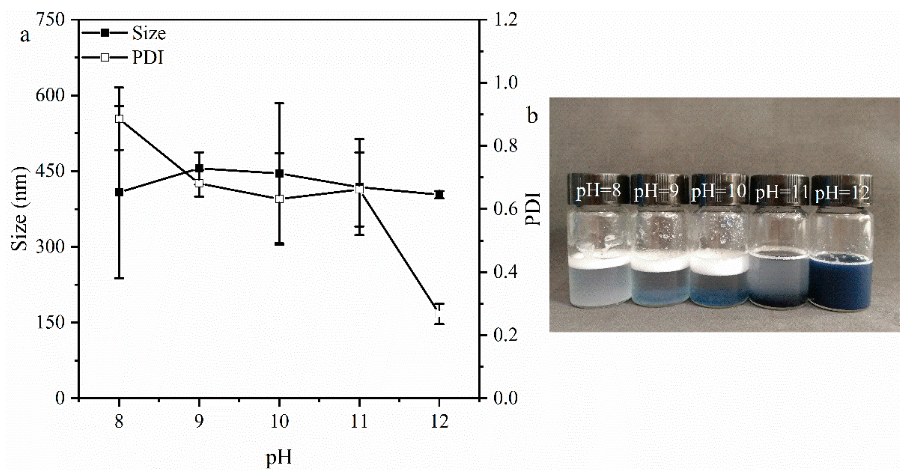
3.1. Effect of pH on Gelatin Particles
3.2. Effect of Genipin Dosages on GNPs
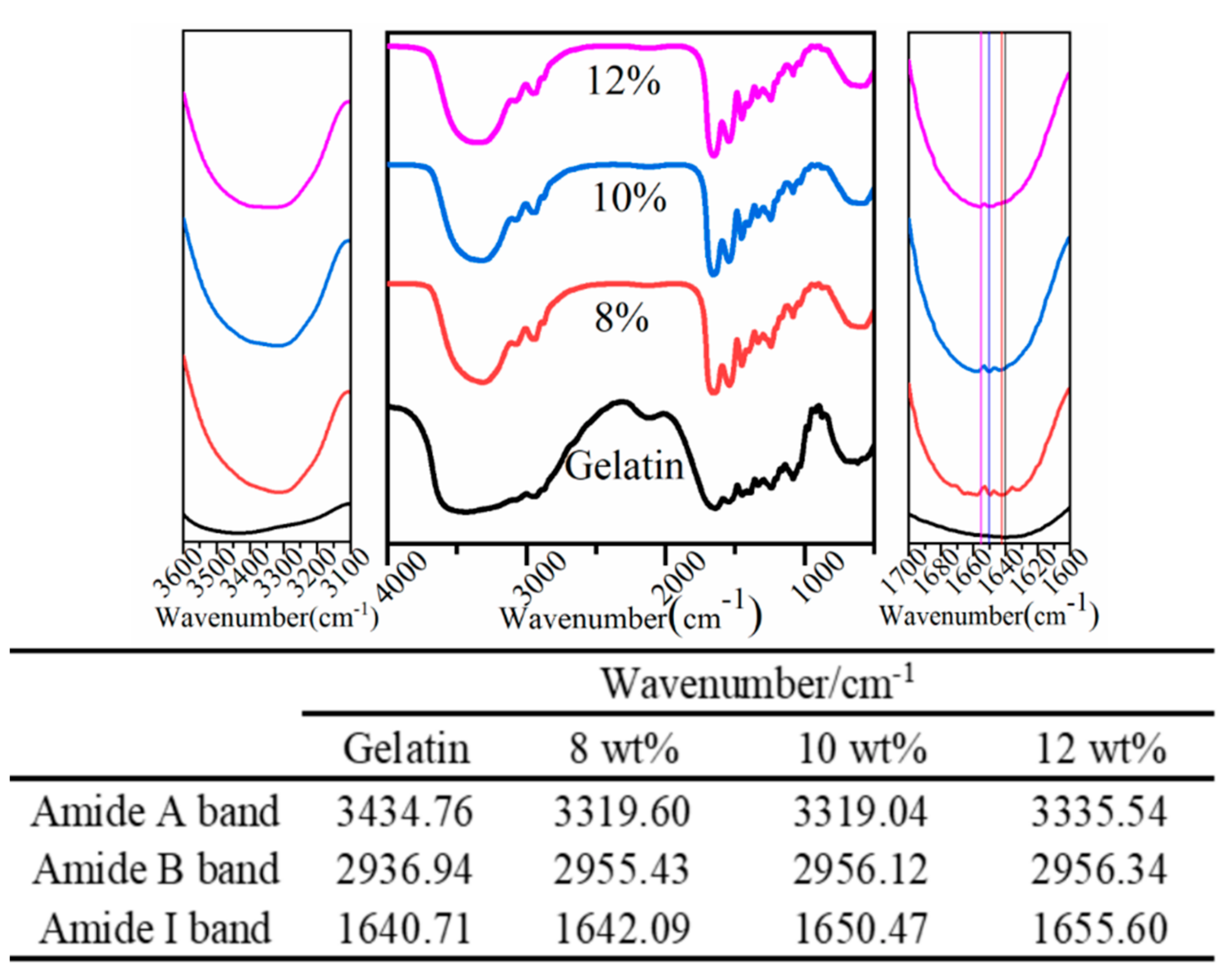
3.2.1. FTIR Analysis
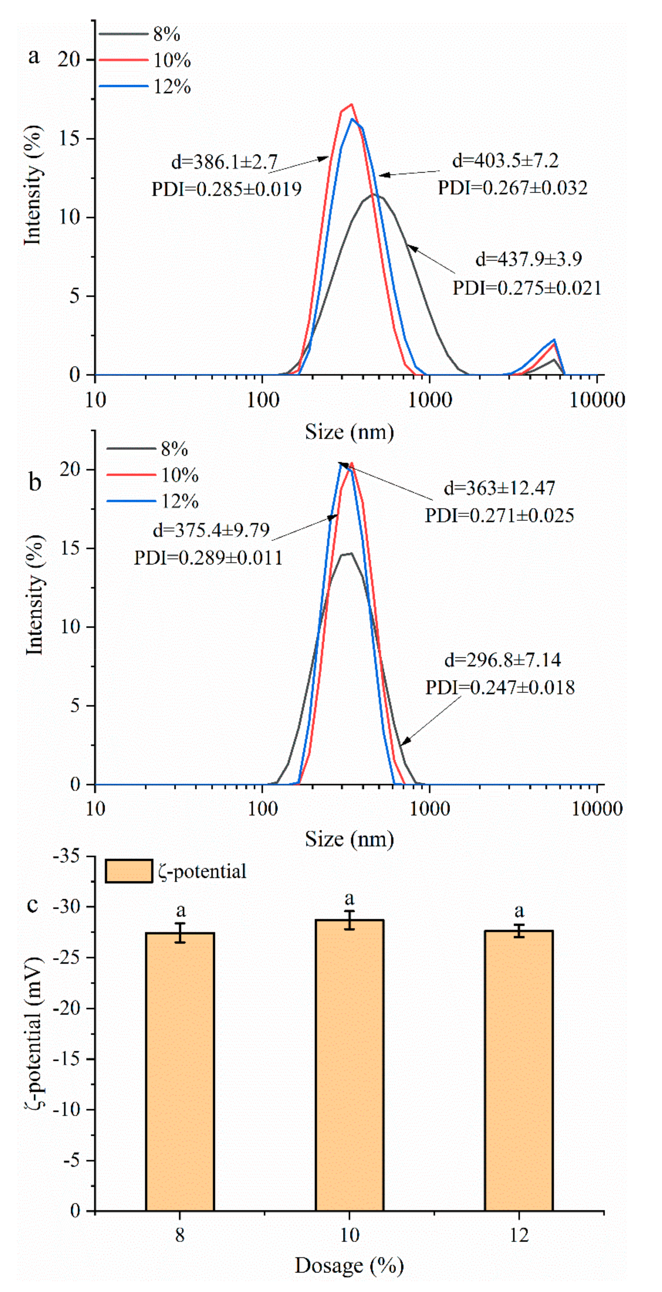
3.2.2. Particles Size and ζ-Potential Analysis
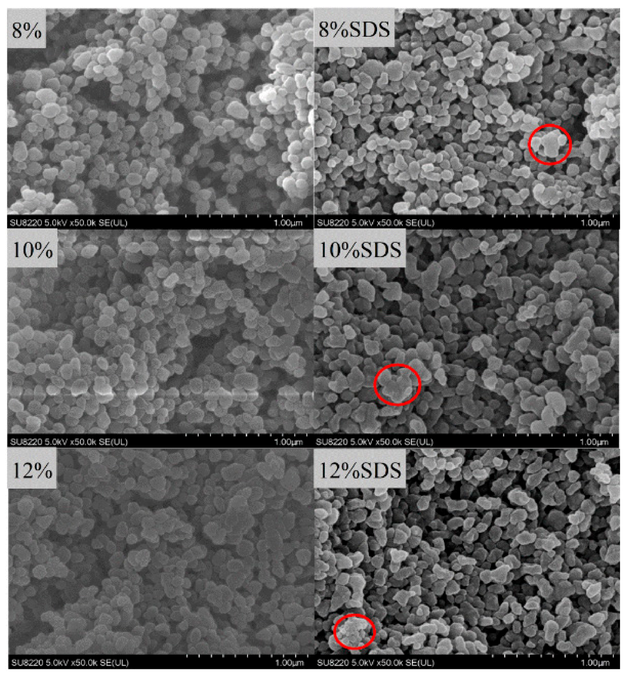
3.2.3. SEM Analysis
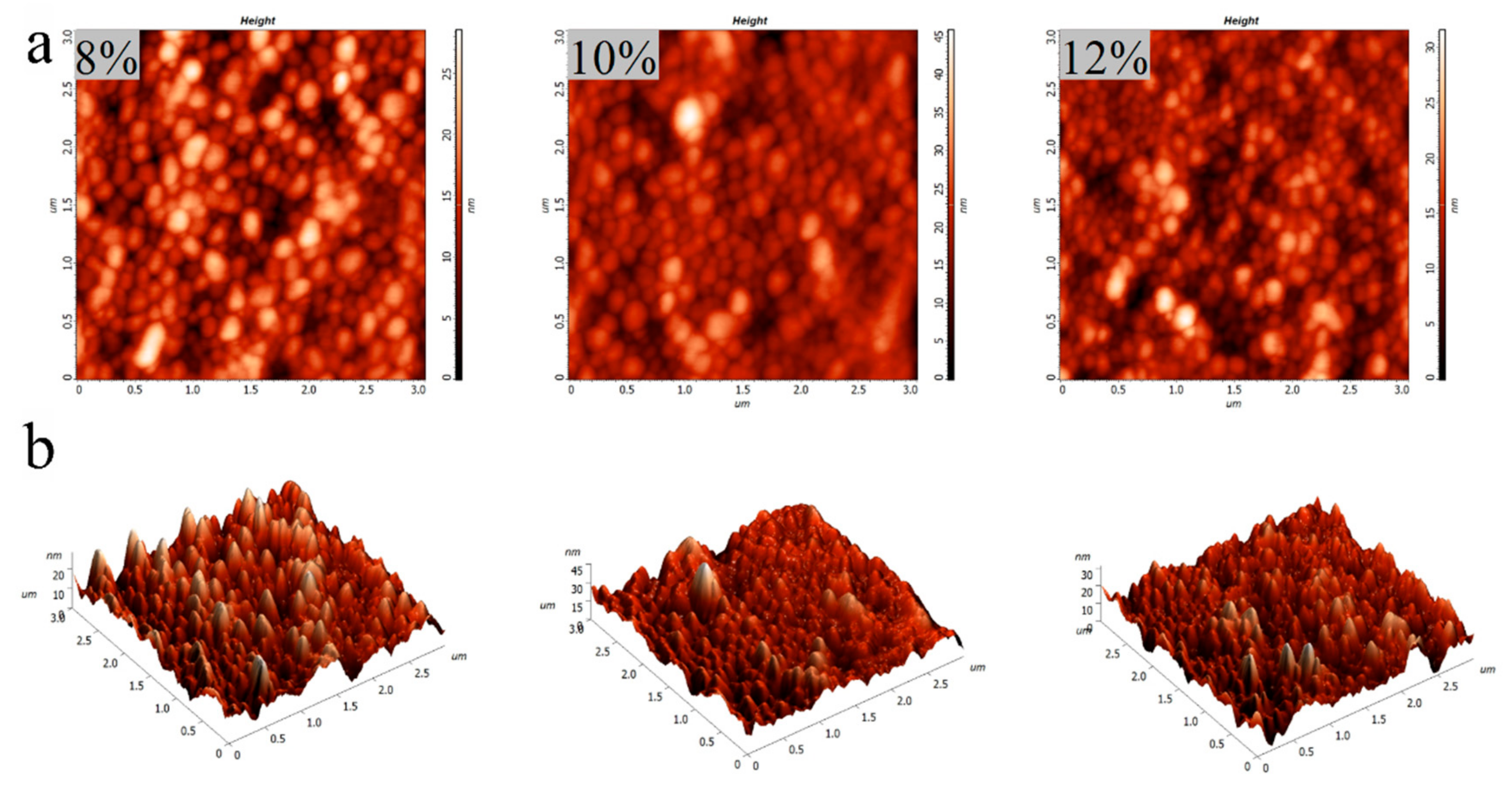
3.2.4. AFM Analysis
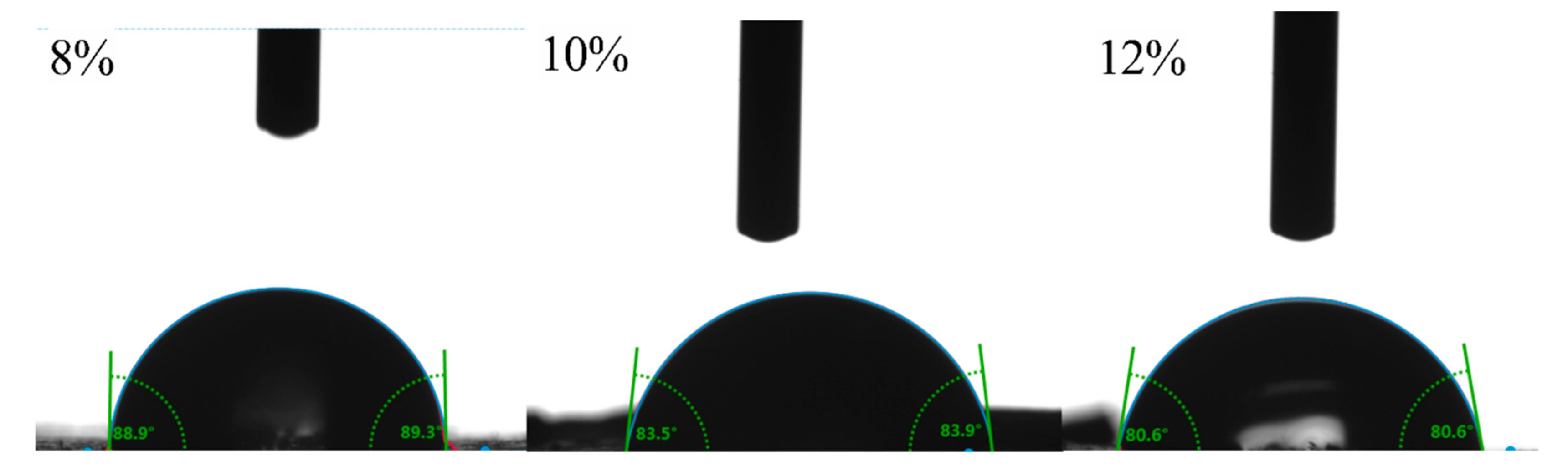
3.2.5. Wettability Analysis
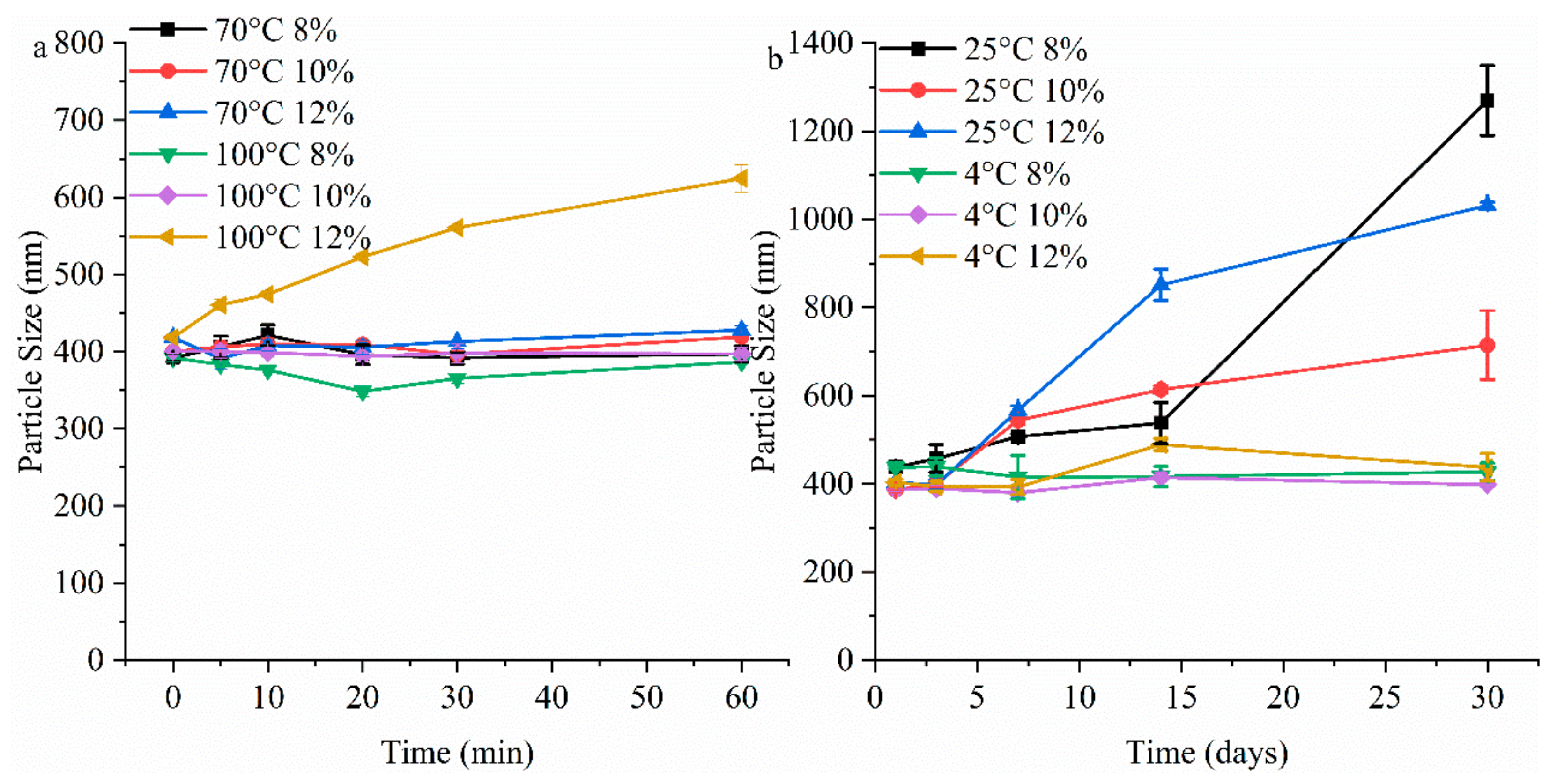
3.2.6. Thermal Stability and Storage Stability of Gelatin Particles Analysis
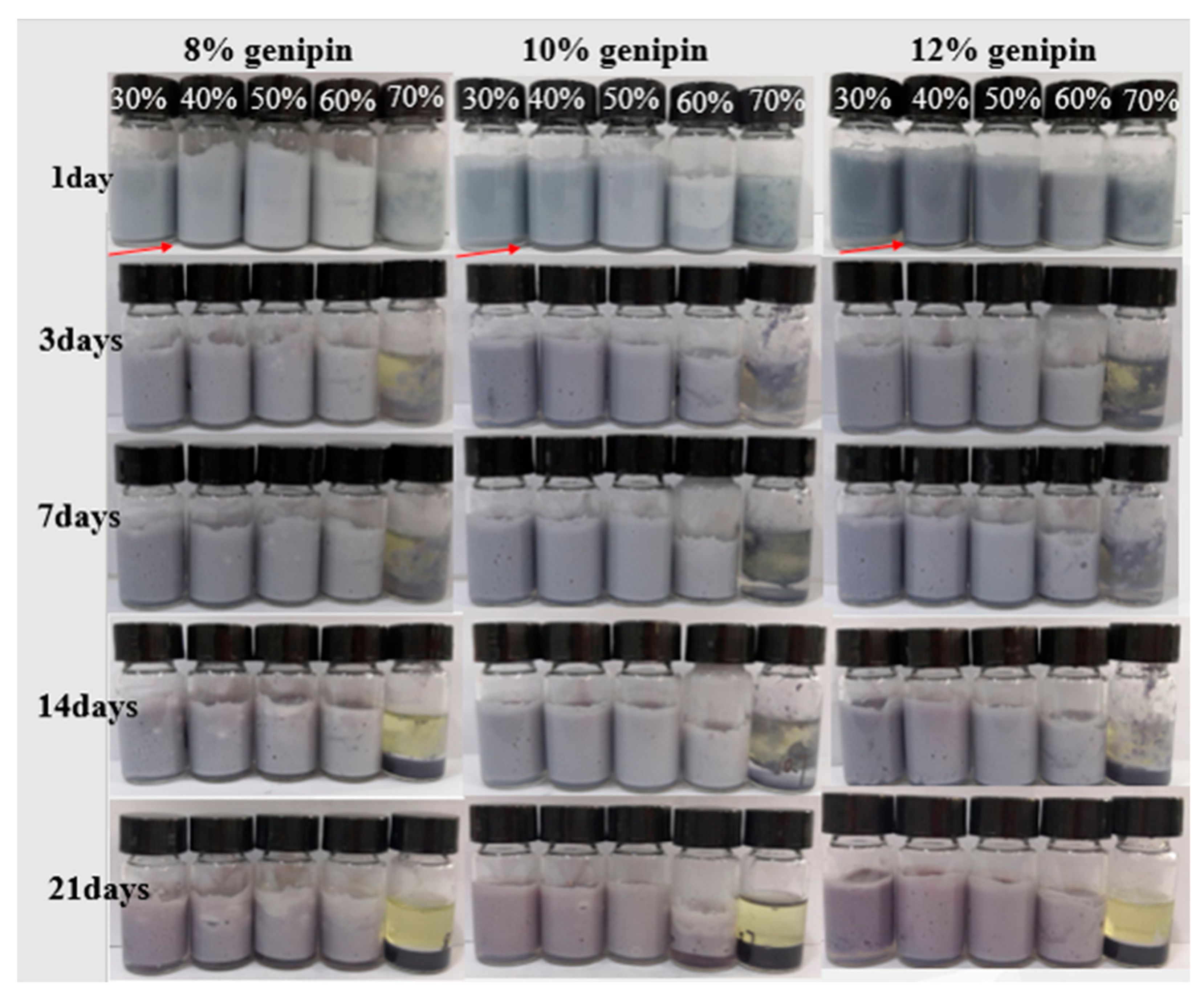
3.2.7. Gelatin Particles Stabilized Pickering Emulsion
4. Conclusions
Author Contributions
Funding
Conflicts of Interest
References
- Hoffmann, H.; Reger, M. Emulsions with unique properties from proteins as emulsifiers. Adv. Colloid Interface Sci. 2014, 205, 94–104. [Google Scholar] [CrossRef] [PubMed]
- Soriano-Ruiz, J.L.; Suñer-Carbó, J.; Calpena-Campmany, A.C.; Bozal-de Febrer, N.L.; Halbaut-Bellowa, A.; Boix-Montañés, E.B.; Souto, B. Clares-Naveros, Clotrimazole multiple W/O/W emulsion as anticandidal agent: Characterization and evaluation on skin and mucosae. Colloids Surf. B 2019, 175, 166–174. [Google Scholar] [CrossRef] [PubMed]
- Han, J.; Gu, L.; Zhang, R.; Shang, W.; Yan, J.; Wu, D.J.M.; Zhu, B.; Xiao, H. Bioaccessibility and cellular uptake of β-carotene in emulsion-based delivery systems using scallop (Patinopecten yessoensis) gonad protein isolates: Effects of carrier oil. Food Funct. 2019, 1, 49–60. [Google Scholar]
- Su, J.; Wang, X.; Li, W.; Chen, L.; Zeng, X.; Huang, Q.; Hu, B. Enhancing the Viability of Lactobacillus plantarum as Probiotics through Encapsulation with High Internal Phase Emulsions Stabilized with Whey Protein Isolate Microgels. J. Agric. Food Chem. 2018, 66, 12335–12343. [Google Scholar] [CrossRef] [PubMed]
- Zhou, F.; Yan, L.; Yin, S.; Tang, C.; Yang, X. Development of Pickering Emulsions Stabilized by Gliadin/Proanthocyanidins Hybrid Particles (GPHPs) and the Fate of Lipid Oxidation and Digestion. J. Agric. Food Chem. 2018, 66, 1461–1471. [Google Scholar] [CrossRef]
- Yang, T.; Hu, Y.; Wang, C.; Binks, B.P. Fabrication of Hierarchical Macroporous Biocompatible Scaffolds by Combining Pickering High Internal Phase Emulsion Templates with Three-Dimensional Printing. ACS Appl. Mater. Interfaces 2017, 9, 22950–22958. [Google Scholar] [CrossRef]
- Miao, C.; Tayebi, M.; Hamad, W.Y. Investigation of the formation mechanisms in high internal phase Pickering emulsions stabilized by cellulose nanocrystals. Philos. Trans. R. Soc. A Math. Phys. Eng. Sci. 2018, 376, 20170039. [Google Scholar] [CrossRef]
- Yao, C.; Liu, B.; Chang, C.; Hsu, S.; Chen, Y. Preparation of networks of gelatin and genipin as degradable biomaterials. Mater. Chem. Phys. 2004, 83, 204–208. [Google Scholar] [CrossRef]
- Tzoumaki, M.V.; Moschakis, T.; Kiosseoglou, V.; Biliaderis, C.G. Oil-in-water emulsions stabilized by chitin nanocrystal particles. Food Hydrocoll. 2011, 25, 1521–1529. [Google Scholar] [CrossRef]
- Wang, H.; Zhao, L.; Song, G.; Tang, G.; Shi, X. Organic-inorganic hybrid shell microencapsulated phase change materials prepared from SiO2/TiC-stabilized pickering emulsion polymerization. Sol. Energy Mater. Sol. Cells 2018, 175, 102–110. [Google Scholar] [CrossRef]
- Tzoumaki, M.V.; Moschakis, T.; Scholten, E.; Biliaderis, C.G. In vitro lipid digestion of chitin nanocrystal stabilized o/w emulsions. Food Funct. 2013, 4, 121–129. [Google Scholar] [CrossRef] [PubMed]
- Zhu, X.; Zheng, J.; Liu, F.; Qiu, C.; Lin, W.; Tang, C. Freeze-thaw stability of Pickering emulsions stabilized by soy protein nanoparticles. Influence of ionic strength before or after emulsification. Food Hydrocoll. 2018, 74, 37–45. [Google Scholar] [CrossRef]
- Yang, T.; Zheng, B.; Zheng, J.; Liu, F.; Wang, S.; Tang, C. High internal phase emulsions stabilized by starch nanocrystals. Food Hydrocoll. 2018, 82, 230–238. [Google Scholar] [CrossRef]
- Lu, X.; Zhang, H.; Li, Y.; Huang, Q. Fabrication of milled cellulose particles-stabilized Pickering emulsions. Food Hydrocoll. 2018, 77, 427–435. [Google Scholar] [CrossRef]
- Sarkar, A.; Murray, B.; Holmes, M.; Ettelaie, R.; Abdalla, A.; Yang, X.Y. In vitro digestion of Pickering emulsions stabilized by soft whey protein microgel particles: Influence of thermal treatment. Soft Matter 2016, 12, 3558–3569. [Google Scholar] [CrossRef]
- Liu, F.; Tang, C. Emulsifying properties of soy protein nanoparticles: Influence of the protein concentration and/or emulsification process. J. Agric. Food Chem. 2014, 62, 2644–2654. [Google Scholar] [CrossRef] [PubMed]
- De Folter, J.W.J.; Info, E.D.N.; van Ruijven, M.W.M.; Velikov, K. Oil-in-water Pickering emulsions stabilized by colloidal particles from the water-insoluble protein zein. Soft Matter 2012, 8, 6807. [Google Scholar] [CrossRef]
- Destribats, M.; Rouvet, M.; Gehin-Delval, C.; Schmitt, C.; Binks, B.P. Emulsions stabilised by whey protein microgel particles: Towards food-grade Pickering emulsions. Soft Matter 2014, 10, 6941–6954. [Google Scholar] [CrossRef]
- Ma, L.; Yang, H.; Ma, M.; Zhang, X.; Zhang, Y. Mechanical and structural properties of rabbit skin gelatin films. Int. J. Food Prop. 2018, 21, 1203–1218. [Google Scholar] [CrossRef]
- Li, Y.; Jia, H.; Cheng, Q.; Pan, F.; Jiang, Z. Sodium alginate–gelatin polyelectrolyte complex membranes with both high water vapor permeance and high permselectivity. J. Membr. Sci. 2011, 375, 304–312. [Google Scholar] [CrossRef]
- Tan, H.; Sun, G.; Lin, W.; Mu, C.; Ngai, T. Gelatin particle-stabilized high internal phase emulsions as nutraceutical containers. ACS Appl. Mater. Interfaces 2014, 6, 13977–13984. [Google Scholar] [CrossRef]
- Tan, H.; Zhao, L.; Tian, S.; Wen, H.; Gou, X.; Ngai, T. Gelatin Particle-Stabilized High-Internal Phase Emulsions for Use in Oral Delivery Systems: Protection Effect and in Vitro Digestion Study. J. Agric. Food Chem. 2017, 65, 900–907. [Google Scholar] [CrossRef] [PubMed]
- Heimbuck, A.M.; Priddy-Arrington, T.R.; Padgett, M.L.; Llamas, C.B.; Barnett, H.H.; Bunnell, B.A.; Caldorera-Moore, M.E. Development of Responsive Chitosan–Genipin Hydrogels for the Treatment of Wounds. ACS Appl. Bio Mater. 2019, 2, 2879–2888. [Google Scholar] [CrossRef]
- Lau, Y.; Kwok, L.; Tam, K.; Chan, Y.; Shum, D.K.; Shea, G.K. Genipin-treated chitosan nanofibers as a novel scaffold for nerve guidance channel design. Colloids Surf. B 2018, 162, 126–134. [Google Scholar] [CrossRef] [PubMed]
- Ramos-de-la-Peña, A.M.; Renard, C.M.G.C.; Montañez, J.; de la Luz Reyes-Vega, M.; Contreras-Esquivel, J.C. A review through recovery, purification and identification of genipin. Pharm. Res. 2016, 15, 37–49. [Google Scholar] [CrossRef]
- Semenova, Advances in molecular design of biopolymer-based delivery micro/nanovehicles for essential fatty acids. Food Hydrocoll. 2017, 68, 114–121. [CrossRef]
- Liang, H.C.; Chang, W.H.; Liang, H.F.; Lee, M.H.; Sung, H.W. Crosslinking structures of gelatin hydrogels crosslinked with genipin or a water-soluble carbodiimide. J. Appl. Polym. Sci. 2004, 91, 4017–4026. [Google Scholar] [CrossRef]
- Focaroli, S.; Teti, G.; Salvatore, V.; Durante, S.; Belmonte, M.M.; Giardino, R.; Mazzotti, A.; Bigi, A.; Falconi, M. Chondrogenic differentiation of human adipose mesenchimal stem cells: Influence of a biomimetic gelatin genipin crosslinked porous scaffold. Micros. Res. Tech. 2014, 77, 928–934. [Google Scholar] [CrossRef]
- Chen, B.Z.; Ashfaq, M.; Zhu, D.D.; Zhang, X.P.; Guo, X.D. Controlled Delivery of Insulin Using Rapidly Separating Microneedles Fabricated from Genipin-Crosslinked Gelatin. Macromol. Rapid Commun. 2018, 39, 1800075. [Google Scholar] [CrossRef]
- Gattazzo, F.; De Maria, C.; Rimessi, A.; Donà, S.; Braghetta, P.; Pinton, P.; Vozzi, G.; Bonaldo, P. Gelatin-genipin-based biomaterials for skeletal muscle tissue engineering. J. Biomed. Mater. Res. Part B 2018, 106, 2763–2777. [Google Scholar] [CrossRef]
- Coester, K.L.H.V.C.J. Gelatin nanoparticles by two step desolvation a new preparation method, surface modifications and cell uptake. J. Microencapsul. 2000, 17, 187–193. [Google Scholar] [PubMed]
- Dai, H.; Zhang, H.; Zhang, Y.; Ma, L.; Zhou, H.; Yu, Y.; Guo, T.; Huang, H. Green pH/magnetic sensitive hydrogels based on pineapple peel cellulose and polyvinyl alcohol: Synthesis, characterization and naringin prolonged release. Carbohydr. Polym. 2019, 209, 51–61. [Google Scholar] [CrossRef] [PubMed]
- Razi, M.A.; Wakabayashi, R.; Tahara, Y.; Goto, M.; Kamiya, N. Genipin-stabilized caseinate-chitosan nanoparticles for enhanced stability and anti-cancer activity of curcumin. Colloids Surf. B 2018, 164, 308–315. [Google Scholar] [CrossRef] [PubMed]
- Muyonga, J.H.; Cole, C.G.B.; Duodu, K.G. Characterisation of acid soluble collagen from skins of young and adult Nile perch (Lates niloticus). Food Chem. 2004, 85, 81–89. [Google Scholar] [CrossRef]
- Albu, M.G.; Ghica, M.V.; Leca, M.; Popa, L.; Borlescu, C.; Cremenescu, E.; Giurginca, M.; Trandafir, V. Doxycycline Delivery from Collagen Matrices Crosslinked With Tannic Acid. Mol. Cryst. Liq. Cryst. 2010, 523, 97–669. [Google Scholar] [CrossRef]
- He, L.; Mu, C.; Shi, J.; Shi, B.; Zhang, Q.; Lin, W. Modification of collagen with a natural cross-linker, procyanidin. Int. J. Biol. Macromol. 2011, 48, 354–359. [Google Scholar] [CrossRef]
- Ahmad, T.; Ahmad, S.A.; Ismail, A.; Khalil, K.A.; Awad, E.A.; Leo, T.K.; Imlan, J.C.; Sazili, A.Q. Characterization of gelatin from bovine skin extracted using ultrasound subsequent to bromelain pretreatment. Food Hydrocoll. 2018, 80, 264–273. [Google Scholar] [CrossRef]
- Teimouri, S.; Morrish, C.; Panyoyai, N.; Small, D.M.; Kasapis, S. Diffusion and relaxation contributions in the release of vitamin B6 from a moving boundary of genipin crosslinked gelatin matrices. Food Hydrocoll. 2019, 87, 839–846. [Google Scholar] [CrossRef]
- Yang, S.; Dai, L.; Sun, C.; Gao, Y. Characterization of curcumin loaded gliadin-lecithin composite nanoparticles fabricated by antisolvent precipitation in different blending sequences. Food Hydrocoll. 2018, 85, 185–194. [Google Scholar] [CrossRef]
- Khan, H.; Shukla, R.N.; Bajpai, A.K. Genipin-modified gelatin nanocarriers as swelling controlled drug delivery system for in vitro release of cytarabine. Mater. Sci. Eng. C 2016, 61, 457–465. [Google Scholar] [CrossRef]
- Zhao, Y.; Sun, Z. Effects of gelatin-polyphenol and gelatin-genipin cross-linking on the structure of gelatin hydrogels. Int. J. Food Prop. 2017, 20, S2822–CS2832. [Google Scholar] [CrossRef]
- Silva, N.N.; Bahri, A.; Guyomarc, H.; Beaucher, F.E.; Gaucheron, F. AFM study of casein micelles cross-linked by genipin: Effects of acid pH and citrate. Dairy Sci. Technol. 2015, 95, 75–86. [Google Scholar] [CrossRef]
- Wang, P.; Chen, C.; Guo, H.; Zhang, H.; Yang, Z.; Ren, F. Casein gel particles as novel soft Pickering stabilizers: The emulsifying property and packing behaviour at the oil-water interface. Food Hydrocoll. 2018, 77, 689–698. [Google Scholar] [CrossRef]
- Richtering, W. Responsive emulsions stabilized by stimuli-sensitive microgels: Emulsions with special non-Pickering properties. Langmuir 2012, 28, 17218–17229. [Google Scholar] [CrossRef] [PubMed]
- Deshmukh, O.S.; van den Ende, D.; Stuart, M.C.; Mugele, F.; Duits, M.H.G. Hard and soft colloids at fluid interfaces: Adsorption, interactions, assembly & rheology. Adv. Colloid Interface Sci. 2015, 222, 215–227. [Google Scholar] [PubMed]
- Kwok, D.Y.; Leung, A.; Lam, C.N.C.; Li, A.; Wu, R.; Neumann, A.W. Low-Rate Dynamic Contact Angles on Poly (methyl methacrylate) and the Determination of Solid Surface Tensions. J. Colloid Interface Sci. 1998, 206, 44–51. [Google Scholar] [CrossRef] [PubMed]
- Białopiotrowic, T.; Jańczuk, B. Surface Properties of Gelatin Films. Langmuir 2002, 18, 9462–9468. [Google Scholar] [CrossRef]
- Ma, L.; Ma, M.; Yu, W.; Zhang, X.; Zhang, Y.; Shen, Y. Research on rapid gelatinization of rabbit skin collagen as effect of acid treatment. Food Hydrocoll. 2018, 77, 945–951. [Google Scholar] [CrossRef]
- Migneault, I.; Dartiguenave, C.; Bertrand, M.J.; Waldron, K.C. Glutaraldehyde: Behavior in aqueous solution, reaction with proteins, and application to enzyme crosslinking. Biotechniques 2004, 37, 790–802. [Google Scholar] [CrossRef]
- Wu, W.; Kong, X.; Zhang, C.; Hua, Y.; Chen, Y. Improving the stability of wheat gliadin nanoparticles-Effect of gum arabic addition. Food Hydrocoll. 2018, 80, 78–87. [Google Scholar] [CrossRef]
- Wang, L.; Hu, Y.; Yin, S.; Yang, X.; Lai, F.; Wang, S. Fabrication and characterization of antioxidant pickering emulsions stabilized by zein/chitosan complex particles (ZCPs). J. Agric. Food Chem. 2015, 63, 2514–2524. [Google Scholar] [CrossRef] [PubMed]
- Jiao, B.; Shi, A.; Wang, Q.; Binks, B.P. High-Internal-Phase Pickering Emulsions Stabilized Solely by Peanut-Protein-Isolate Microgel Particles with Multiple Potential Applications. Angew. Chem. Int. Ed. 2018, 57, 9274–9278. [Google Scholar] [CrossRef] [PubMed]


© 2019 by the authors. Licensee MDPI, Basel, Switzerland. This article is an open access article distributed under the terms and conditions of the Creative Commons Attribution (CC BY) license (http://creativecommons.org/licenses/by/4.0/).
Share and Cite
Feng, X.; Dai, H.; Ma, L.; Yu, Y.; Tang, M.; Li, Y.; Hu, W.; Liu, T.; Zhang, Y. Food-Grade Gelatin Nanoparticles: Preparation, Characterization, and Preliminary Application for Stabilizing Pickering Emulsions. Foods 2019, 8, 479. https://doi.org/10.3390/foods8100479
Feng X, Dai H, Ma L, Yu Y, Tang M, Li Y, Hu W, Liu T, Zhang Y. Food-Grade Gelatin Nanoparticles: Preparation, Characterization, and Preliminary Application for Stabilizing Pickering Emulsions. Foods. 2019; 8(10):479. https://doi.org/10.3390/foods8100479
Chicago/Turabian StyleFeng, Xin, Hongjie Dai, Liang Ma, Yong Yu, Mi Tang, Yuan Li, Weijie Hu, Tingwei Liu, and Yuhao Zhang. 2019. "Food-Grade Gelatin Nanoparticles: Preparation, Characterization, and Preliminary Application for Stabilizing Pickering Emulsions" Foods 8, no. 10: 479. https://doi.org/10.3390/foods8100479