
A Colorimetric/Fluorescent Dual-Mode Aptasensor for Salmonella Based on the Magnetic Separation of Aptamers and a DNA-Nanotriangle Programmed Multivalent Aptamer
 and
and 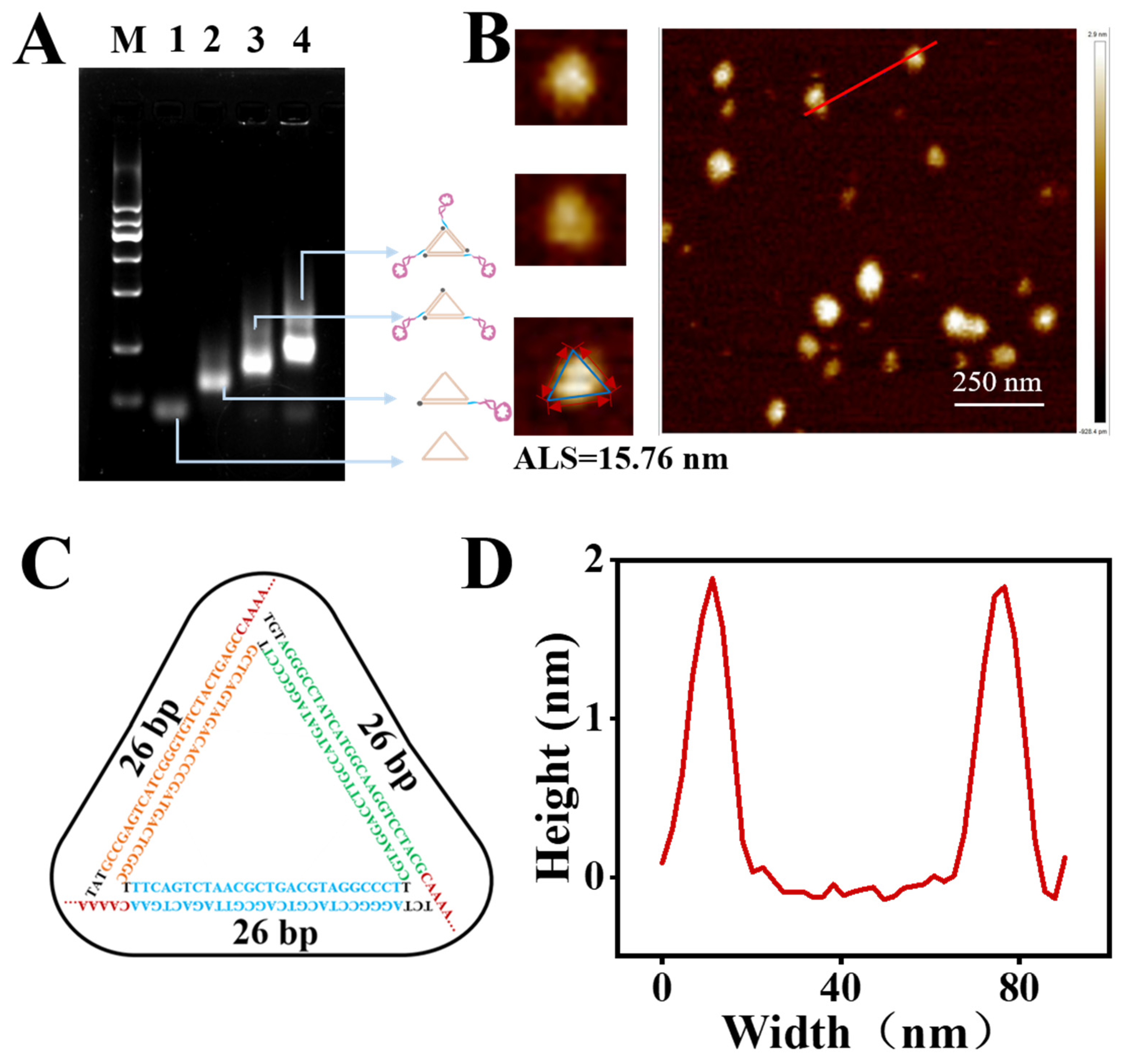
Abstract
:1. Introduction
2. Materials and Methods
2.1. Materials and Apparatuses
2.2. Bacterial Culture
2.3. The Preparation of Aptamer-Functionalized Magnetic Nanoparticles (MNP-Apts)
2.4. The Preparation and Characterization of the NTri-Multi-Apt
2.5. The Binding Affinity of the NTri-Multi-Apt
2.6. The Colorimetric/Fluorescent Dual-Mode Detection of Salmonella Based on the NTri-Multi-Apts
2.7. The Preparation of Spiked Samples
3. Results and Discussion
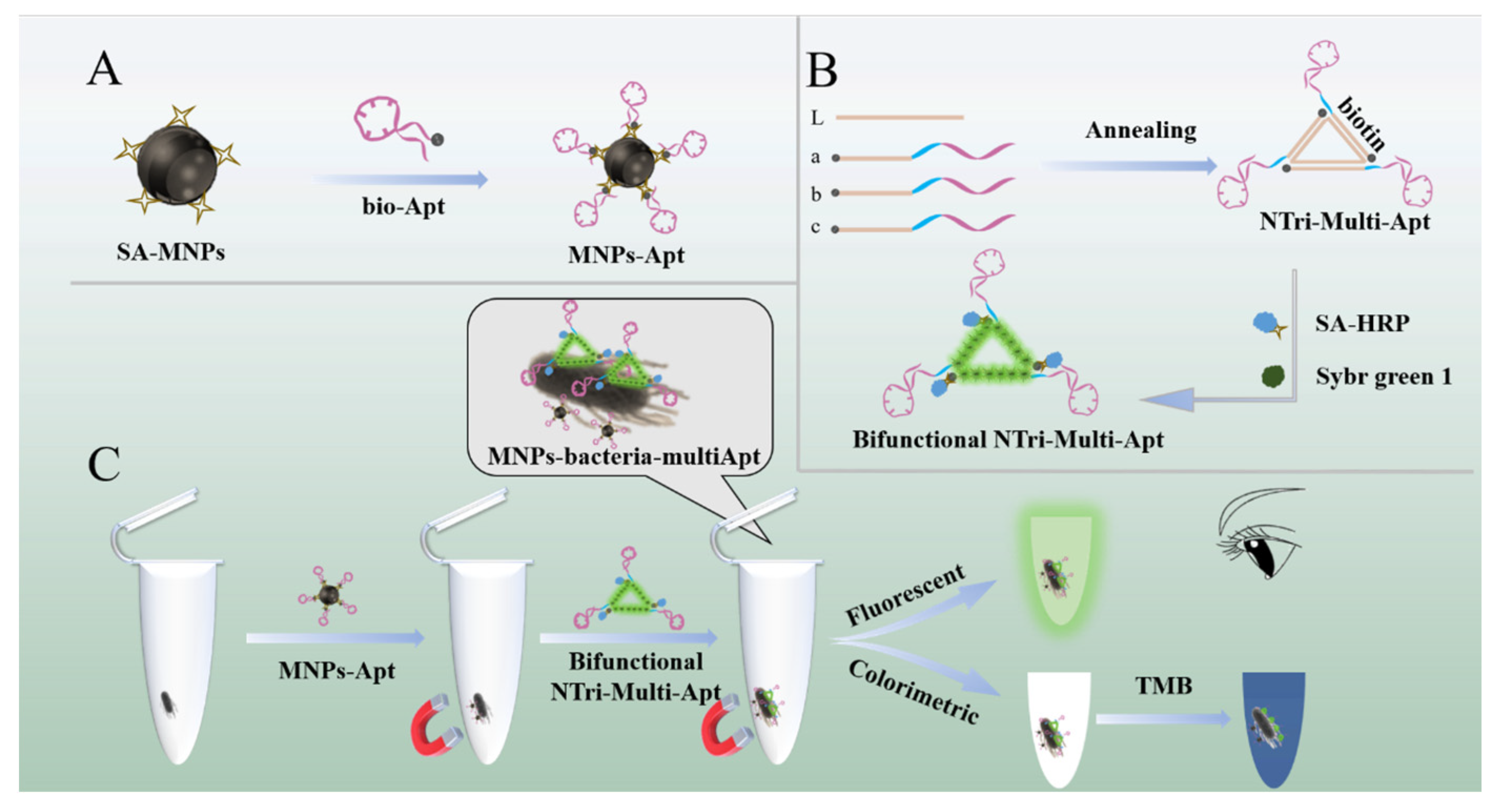
3.1. The Principle of Bacterial Detection
3.2. The Construction of the NTri-Multi-Apt
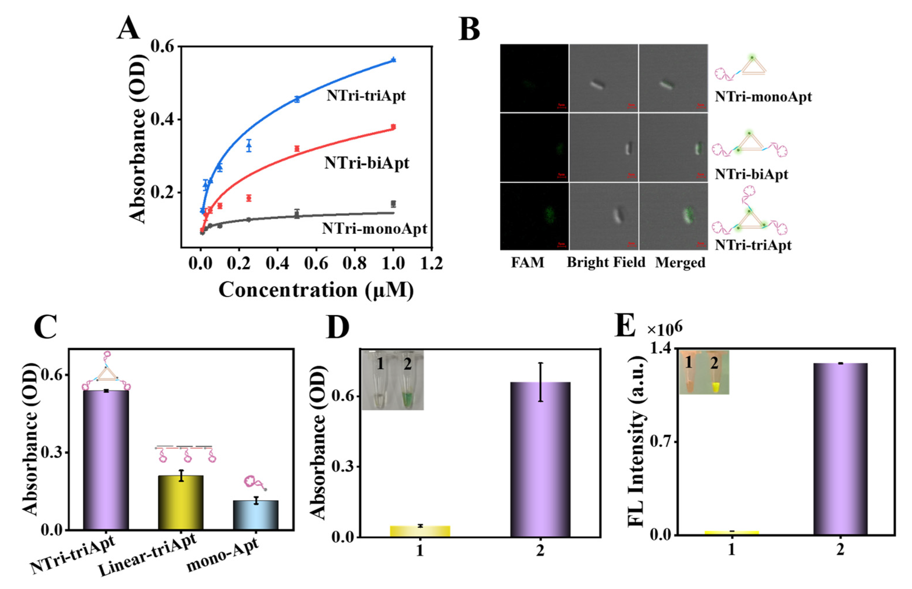
3.3. The Enhanced Binding Affinity of the NTri-Multi-Apt
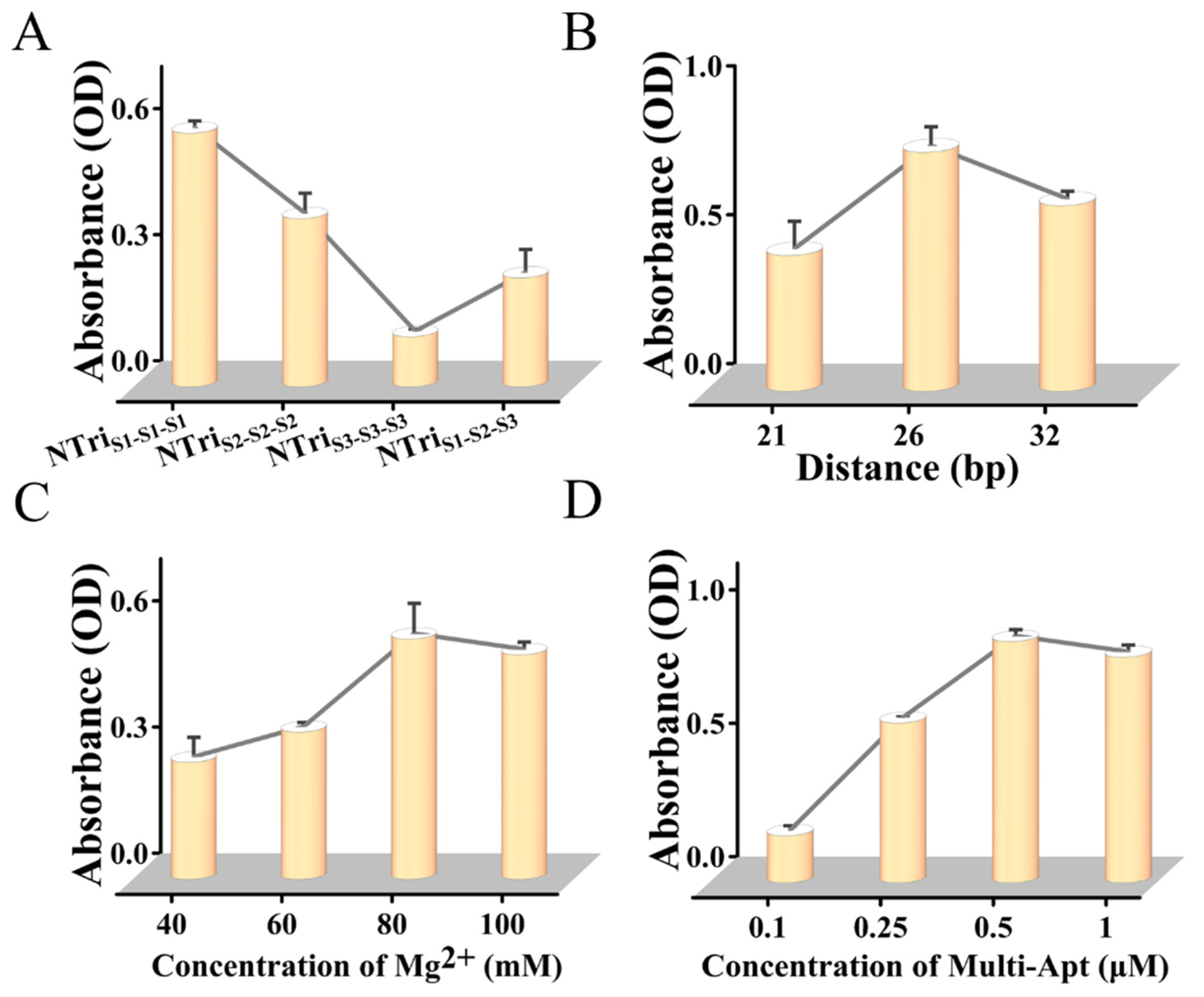
3.4. The Optimization of the Experimental Conditions
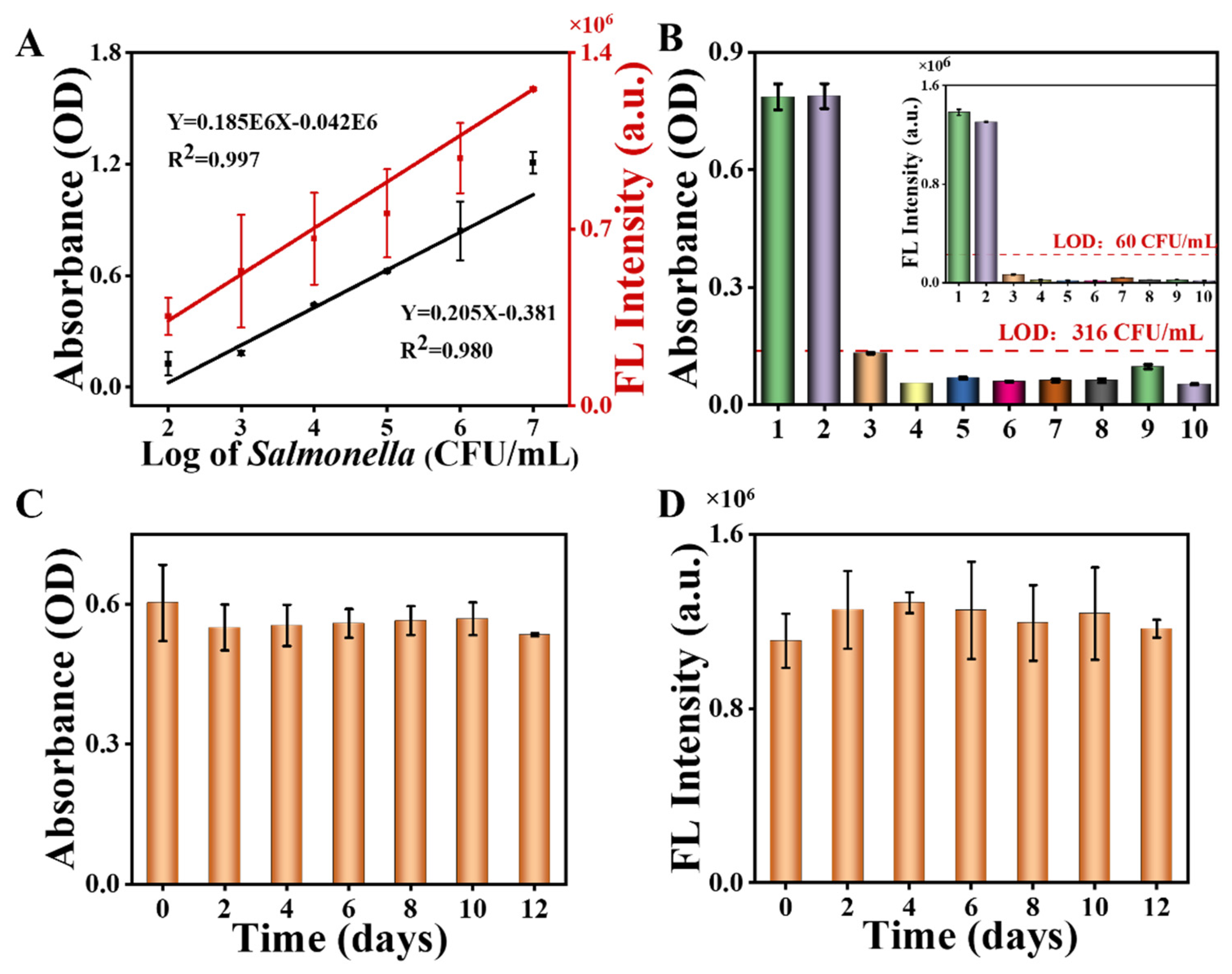
3.5. The Sensitivity of the Dual-Mode Detection System
3.6. The Specificity and Stability of the Colorimetric/Fluorescent Dual-Mode System
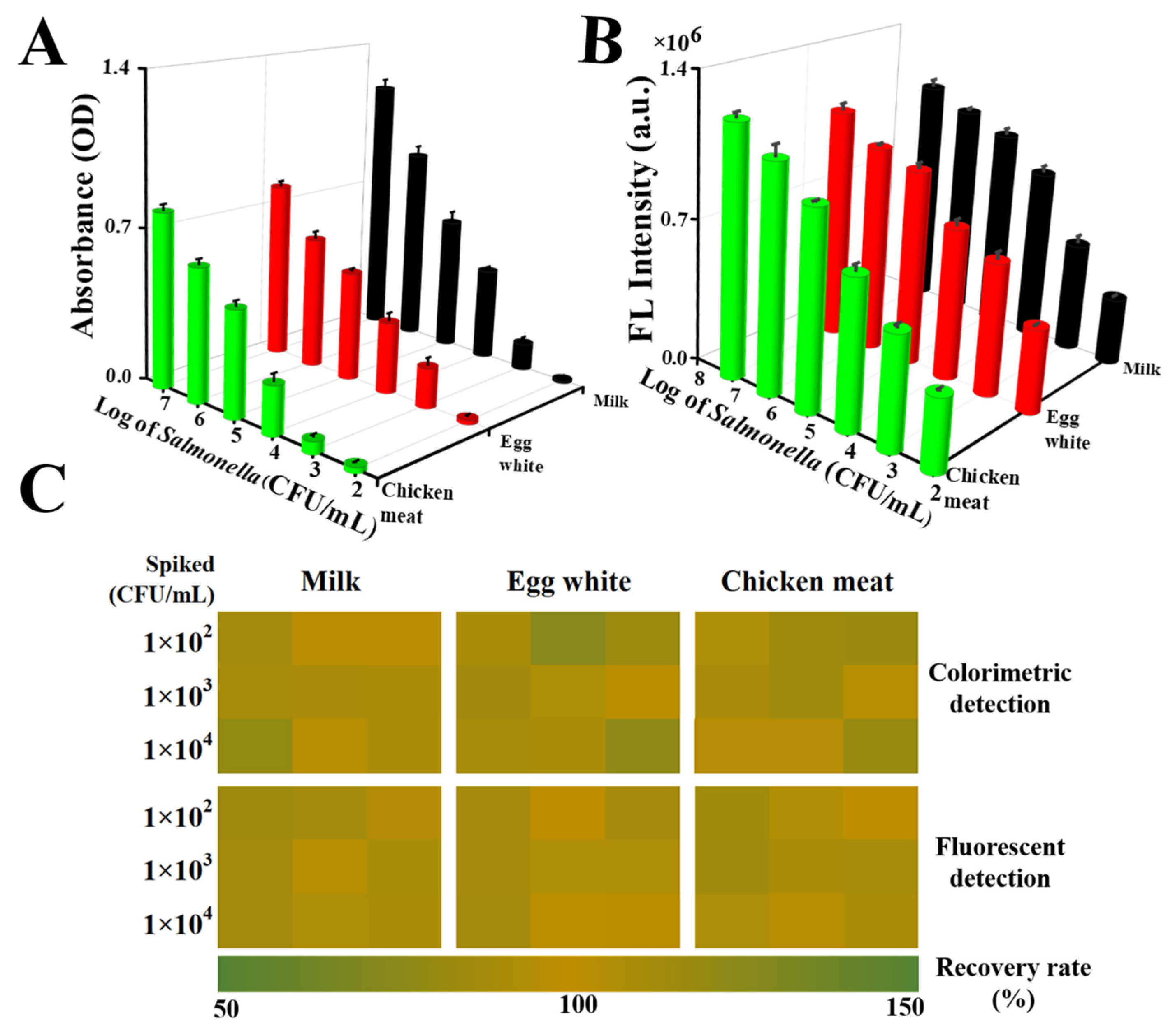
3.7. An Analysis of Salmonella Detection in Food Samples
4. Conclusions
Supplementary Materials
Author Contributions
Funding
Data Availability Statement
Acknowledgments
Conflicts of Interest
References
- Qiu, S.; Liu, B.; Leng, Y.; Fox, E.; Zhou, X.; Yan, B.; Sang, X.; Long, K.; Fu, Y.; He, X.; et al. A label-free fiber ring laser biosensor for ultrahigh sensitivity detection of Salmonella Typhimurium. Biosens. Bioelectron. 2023, 234, 115337. [Google Scholar] [CrossRef]
- Li, Z.; Hu, J.; Zhan, Y.; Shao, Z.; Gao, M.; Yao, Q.; Li, Z.; Sun, S.; Wang, L. Coupling bifunctional nanozyme-mediated catalytic signal amplification and label-free SERS with immunoassays for ultrasensitive detection of pathogens in milk samples. Anal. Chem. 2023, 95, 6417–6424. [Google Scholar] [CrossRef]
- Hice, S.A.; Clark, K.D.; Anderson, J.L.; Brehm-Stecher, B.F. Capture, concentration, and detection of Salmonella in foods using magnetic ionic liquids and recombinase polymerase amplification. Anal. Chem. 2019, 91, 1113–1120. [Google Scholar] [CrossRef]
- Sannigrahi, S.; Arumugasamy, S.K.; Mathiyarasu, J.; Suthindhiran, K. Magnetosome-anti-Salmonella antibody complex based biosensor for the detection of Salmonella typhimurium. Mater. Sci. Eng. C 2020, 114, 111071. [Google Scholar] [CrossRef]
- Guan, Z.; Sun, Y.; Ma, C.-B.; Lee, J.J.; Zhang, S.; Zhang, X.; Guo, Z.; Du, Y. Dual targets-induced specific hemin/G-quadruplex assemblies for label-free electrochemical detection capable of distinguishing Salmonella and its common serotype in food samples. Biosens. Bioelectron. 2023, 236, 115438. [Google Scholar] [CrossRef]
- Jiao, Y.; Zhang, Z.; Wang, K.; Zhang, H.; Gao, J. Rapid detection of Salmonella in food matrices by photonic PCR based on the photothermal effect of Fe3O4. Food Chem. X 2023, 19, 100798. [Google Scholar] [CrossRef] [PubMed]
- Jabbari, A.; Sameiyan, E.; Yaghoobi, E.; Ramezani, M.; Alibolandi, M.; Abnous, K.; Taghdisi, S.M. Aptamer-based targeted delivery systems for cancer treatment using DNA origami and DNA nanostructures. Int. J. Pharm. 2023, 646, 123448. [Google Scholar] [CrossRef]
- Lin, L.; Zheng, Q.; Lin, J.; Yuk, H.-G.; Guo, L. Immuno- and nucleic acid-based current technique for Salmonella detection in food. Eur. Food Res. Technol. 2020, 246, 373–395. [Google Scholar] [CrossRef]
- Khorshid, M.; Varshosaz, J.; Rostami, M.; Haghiralsadat, F.; Akbari, V.; Khorshid, P. Anti HER-2 aptamer functionalized gold nanoparticles of dasatinib for targeted chemo-radiotherapy in breast cancer cells. Biomater. Adv. 2023, 154, 213591. [Google Scholar] [CrossRef]
- He, Y.; Huang, Y.; Xu, H.; Yang, X.; Liu, N.; Xu, Y.; Ma, R.; Zhai, J.; Ma, Y.; Guan, S. Aptamer-modified M cell targeting liposomes for oral delivery of macromolecules. Colloids Surf. B Biointerfaces 2023, 222, 113109. [Google Scholar] [CrossRef]
- Majdinasab, M.; Hayat, A.; Marty, J.L. Aptamer-based assays and aptasensors for detection of pathogenic bacteria in food samples. TrAC Trends Anal. Chem. 2018, 107, 60–77. [Google Scholar] [CrossRef]
- Kim, E.; Yang, S.-M.; Kim, H.-Y. Development of rapid on-site detection of Salmonella Enteritidis, S. Typhimurium, and S. Thompson in food samples using an ultrafast PCR system. Food Biosci. 2023, 56, 103242. [Google Scholar] [CrossRef]
- Ding, S.; Hu, H.; Yue, X.; Feng, K.; Gao, X.; Dong, Q.; Yang, M.; Tamer, U.; Huang, G.; Zhang, J. A fluorescent biosensor based on quantum dot–labeled streptavidin and poly-l-lysine for the rapid detection of Salmonella in milk. J. Dairy Sci. 2022, 105, 2895–2907. [Google Scholar] [CrossRef]
- Yang, L.; Li, Y. Quantum dots as fluorescent labels for quantitative detection of Salmonella Typhimurium in chicken carcass wash water. J. Food Prot. 2005, 68, 1241–1245. [Google Scholar] [CrossRef]
- Wang, Q.-Y.; Kang, Y.-J. Bioprobes based on aptamer and silica fluorescent nanoparticles for bacteria Salmonella typhimurium detection. Nanoscale Res. Lett. 2016, 11, 150. [Google Scholar] [CrossRef]
- Zhang, X.; Peng, Y.; Yao, L.; Shang, H.; Zheng, Z.; Chen, W.; Xu, J. Self-assembly of multivalent aptamer-tethered DNA monolayers dedicated to a fluorescence polarization-responsive circular isothermal strand displacement amplification for Salmonella Assay. Anal. Chem. 2023, 95, 2570–2578. [Google Scholar] [CrossRef]
- Liu, Y.; Zhang, H.; Du, Y.; Zhu, Z.; Zhang, M.; Lv, Z.; Wu, L.; Yang, Y.; Li, A.; Yang, L.; et al. Highly sensitive minimal residual disease detection by biomimetic multivalent aptamer nanoclimber functionalized microfluidic chip. Small 2020, 16, 2000949. [Google Scholar] [CrossRef]
- Sun, M.; Ma, N.; Shi, H.; Cheong, L.-Z.; Yang, W.; Qiao, Z. A HCR based multivalent aptamer amplifier for ultrasensitive detection of Salmonella. Sens. Actuators B Chem. 2023, 375, 132860. [Google Scholar] [CrossRef]
- Lin, M.; Zhang, J.; Wan, H.; Yan, C.; Xia, F. Rationally designed multivalent aptamers targeting cell surface for biomedical applications. ACS Appl. Mater. Interfaces 2020, 13, 9369–9389. [Google Scholar] [CrossRef]
- Peng, J.; Liu, Y.; Su, R.; Zeng, L.; Huo, Z.; Peng, R.; Yu, X.; Zhang, H.; Yang, C.; Yang, L.; et al. DNA-programmed orientation-ordered multivalent microfluidic interface for liquid biopsy. Anal. Chem. 2022, 94, 8766–8773. [Google Scholar] [CrossRef]
- Kwon, P.S.; Ren, S.; Kwon, S.-J.; Kizer, M.E.; Kuo, L.; Xie, M.; Zhu, D.; Zhou, F.; Zhang, F.; Kim, D.; et al. Designer DNA architecture offers precise and multivalent spatial pattern-recognition for viral sensing and inhibition. Nat. Chem. 2019, 12, 26–35. [Google Scholar] [CrossRef]
- Xia, R.; Chai, H.; Jiao, J.; Miao, P. Assembly of DNA triangular pyramid frustum for ultrasensitive quantification of exosomal miRNA. Biosens. Bioelectron. 2023, 231, 115297. [Google Scholar] [CrossRef]
- Liu, J.; Yu, Z.; Chen, Q.; Jia, L. L-Tryptophan assisted construction of fluorescent and colorimetric dual-channel biosensor for detection of live Escherichia coli. Microchem. J. 2022, 174, 107085. [Google Scholar] [CrossRef]
- Wang, X.; Yang, F.; Deng, C.; Zhang, Y.; Yang, X.; Chen, X.; Huang, Y.; Ye, H.; Zhong, J.; Wang, Z. A dual-mode method based on aptamer recognition and time-resolved fluorescence resonance energy transfer for histamine detection in fish. Molecules 2022, 27, 8711. [Google Scholar] [CrossRef]
- Wang, C.; Gao, X.; Wang, S.; Liu, Y. A smartphone-integrated paper sensing system for fluorescent and colorimetric dual-channel detection of foodborne pathogenic bacteria. Anal. Bioanal. Chem. 2020, 412, 611–620. [Google Scholar] [CrossRef]
- Zhao, L.; Li, T.; Xu, X.; Xu, Y.; Li, D.; Song, W.; Zhan, T.; He, P.; Zhou, H.; Xu, J.-J.; et al. Polyhedral Au nanoparticle/MoO(x) heterojunction-enhanced ultrasensitive dual-mode biosensor for miRNA detection combined with a nonenzymatic cascade DNA amplification circuit. Anal. Chem. 2023, 95, 9271–9279. [Google Scholar] [CrossRef]
- Fan, P.; Qian, X.; Li, Q.; Jiang, P.; Wu, Q.; Huang, G.; Zhang, Z.; Li, L. A novel label-free dual-mode aptasensor based on the mutual regulation of silver nanoclusters and MoSe2 nanosheets for reliable detection of ampicillin. Anal. Chim. Acta 2023, 1251, 340997. [Google Scholar] [CrossRef]
- Liu, J.; Xie, G.; Xiong, Q.; Liang, T.; Xu, H. Sensitive dual readout assays based on rolling circle amplification for fluorescent and colorimetric detection of Cronobacter spp. in powdered infant formula. Food Control 2021, 124, 107840. [Google Scholar] [CrossRef]
- Bu, T.; Bai, F.; Zhao, S.; Cao, Y.; He, K.; Sun, X.; Wang, Q.; Jia, P.; Li, M.; Wang, X.; et al. Multifunctional bacteria-derived tags for advancing immunoassay analytical performance with dual-channel switching and antibodies bioactivity sustaining. Biosens. Bioelectron. 2021, 192, 113538. [Google Scholar] [CrossRef]
- Li, C.; Luo, M.; Wang, J.; Niu, H.; Shen, Z.; Wu, Z.-S. Rigidified DNA triangle-protected molecular beacon from endogenous nuclease digestion for monitoring microRNA expression in living cells. ACS Sens. 2020, 5, 2378–2387. [Google Scholar] [CrossRef] [PubMed]
- Mou, Y.; Yu, J.-Y.; Wannier, T.M.; Guo, C.-L.; Mayo, S.L. Computational design of co-assembling protein–DNA nanowires. Nature 2015, 525, 230–233. [Google Scholar] [CrossRef]
- Xue, C.; Zhang, S.; Li, C.; Yu, X.; Ouyang, C.; Lu, Y.; Wu, Z.-S. Y-Shaped backbone-rigidified triangular DNA scaffold-directed stepwise movement of a DNAzyme walker for sensitive microRNA imaging within living cells. Anal. Chem. 2019, 91, 15678–15685. [Google Scholar] [CrossRef] [PubMed]
- Zou, S.; Lei, Y.; Ma, W.; Chen, B.; Cheng, H.; Jia, R.; Li, Z.; He, X.; Wang, K. Extracellular pH-manipulated in situ reconfiguration of aptamer functionalized DNA monomer enables specifically improved affinity, detection and drug delivery. Analyst 2020, 145, 2562–2569. [Google Scholar] [CrossRef] [PubMed]
- Qi, L.; Tian, Y.; Li, N.; Mao, M.; Fang, X.; Han, D. Engineering circular aptamer assemblies with tunable selectivity to cell membrane antigens in vitro and in vivo. ACS Appl. Mater. Interfaces 2023, 15, 12822–12830. [Google Scholar] [CrossRef] [PubMed]
- Kolovskaya, O.S.; Savitskaya, A.G.; Zamay, T.N.; Reshetneva, I.T.; Zamay, G.S.; Erkaev, E.N.; Wang, X.; Wehbe, M.; Salmina, A.B.; Perianova, O.V.; et al. Development of bacteriostatic DNA aptamers for Salmonella. J. Med. Chem. 2013, 56, 1564–1572. [Google Scholar] [CrossRef] [PubMed]
- Kooshki, H.; Abbaszadeh, R.; Heidari, R.; Akbariqomi, M.; Mazloumi, M.; Shafei, S.; Absalan, M.; Tavoosidana, G. Developing a DNA aptamer-based approach for biosensing cystatin-c in serum: An alternative to antibody-based methods. Anal. Biochem. 2019, 584, 113386. [Google Scholar] [CrossRef] [PubMed]
- Dwivedi, H.P.; Smiley, R.D.; Jaykus, L.-A. Selection of DNA aptamers for capture and detection of Salmonella Typhimurium using a whole-cell SELEX approach in conjunction with cell sorting. Appl. Microbiol. Biotechnol. 2013, 97, 3677–3686. [Google Scholar] [CrossRef]
- Kim, Y.S.; Chung, J.; Song, M.Y.; Jurng, J.; Kim, B.C. Aptamer cocktails: Enhancement of sensing signals compared to single use of aptamers for detection of bacteria. Biosens. Bioelectron. 2014, 54, 195–198. [Google Scholar] [CrossRef]
- Zhang, Z.; Pandey, R.; Li, J.; Gu, J.; White, D.; Stacey, H.D.; Ang, J.C.; Steinberg, C.J.; Capretta, A.; Filipe, C.D.M.; et al. High-affinity dimeric aptamers enable the rapid electrochemical detection of wild-type and B.1.1.7 SARS-CoV-2 in unprocessed saliva. Angew. Chem. Int. Ed. 2021, 60, 24266–24274. [Google Scholar] [CrossRef]
- Rinker, S.; Ke, Y.; Liu, Y.; Chhabra, R.; Yan, H. Self-assembled DNA nanostructures for distance-dependent multivalent ligand–protein binding. Nat. Nanotechnol. 2008, 3, 418–422. [Google Scholar] [CrossRef]
- Samanta, D.; Iscen, A.; Laramy, C.R.; Ebrahimi, S.B.; Bujold, K.E.; Schatz, G.C.; Mirkin, C.A. multivalent cation-induced actuation of DNA-mediated colloidal superlattices. J. Am. Chem. Soc. 2019, 141, 19973–19977. [Google Scholar] [CrossRef]
- Xiong, J.; He, S.; Qin, L.; Zhang, S.; Shan, W.; Jiang, H. Aptasensor-based assay for dual-readout determination of aflatoxin B1 in corn and wheat via an electrostatic force–mediated FRET strategy. Microchim. Acta 2023, 190, 80. [Google Scholar] [CrossRef] [PubMed]
- Zhuang, Q.-Q.; He, S.-B.; Jiang, Y.-C.; Huang, K.-Y.; Xu, Y.-Y.; Peng, H.-P.; Deng, H.-H.; Chen, W. Immunofluorescent-aggregation assay based on anti-Salmonella typhimurium IgG-AuNCs, for rapid detection of Salmonella typhimurium. Mikrochim Acta 2022, 189, 160. [Google Scholar] [CrossRef] [PubMed]
- Srinivasan, S.; Ranganathan, V.; DeRosa, M.C.; Murari, B.M. Label-free aptasensors based on fluorescent screening assays for the detection of Salmonella typhimurium. Anal. Biochem. 2018, 559, 17–23. [Google Scholar] [CrossRef] [PubMed]
- Yang, X.; Wang, L.; Pang, L.; Fu, S.; Qin, X.; Chen, Q.; Man, C.; Jiang, Y. A novel fluorescent platform of DNA-stabilized silver nanoclusters based on exonuclease III amplification-assisted detection of Salmonella Typhimurium. Anal. Chim. Acta 2021, 1181, 338903. [Google Scholar] [CrossRef]
- Wu, W.; Li, J.; Pan, D.; Li, J.; Song, S.; Rong, M.; Li, Z.; Gao, J.; Lu, J. Gold nanoparticle-based enzyme-linked antibody-aptamer sandwich assay for detection of Salmonella Typhimurium. ACS Appl. Mater. Interfaces 2014, 6, 16974–16981. [Google Scholar] [CrossRef]
- Cai, G.; Wang, Y.; Zhang, Y.; Zheng, L.; Lin, J. Magnetorheological elastomer and smartphone enable microfluidic biosensing of foodborne pathogen. Chin Chem Lett. 2023, 34, 108059. [Google Scholar] [CrossRef]
- Srisa-Art, M.; Boehle, K.E.; Geiss, B.J.; Henry, C.S. Highly sensitive detection of Salmonella typhimurium using a colorimetric paper-based analytical device coupled with immunomagnetic separation. Anal. Chem. 2017, 90, 1035–1043. [Google Scholar] [CrossRef]
- Hu, J.; Jiang, Y.-Z.; Tang, M.; Wu, L.-L.; Xie, H.-y.; Zhang, Z.-L.; Pang, D.-W. Colorimetric-fluorescent-magnetic nanosphere-based multimodal assay platform for Salmonella Detection. Anal. Chem. 2018, 91, 1178–1184. [Google Scholar] [CrossRef]


Disclaimer/Publisher’s Note: The statements, opinions and data contained in all publications are solely those of the individual author(s) and contributor(s) and not of MDPI and/or the editor(s). MDPI and/or the editor(s) disclaim responsibility for any injury to people or property resulting from any ideas, methods, instructions or products referred to in the content. |
© 2023 by the authors. Licensee MDPI, Basel, Switzerland. This article is an open access article distributed under the terms and conditions of the Creative Commons Attribution (CC BY) license (https://creativecommons.org/licenses/by/4.0/).
Share and Cite
Ma, N.; Sun, M.; Shi, H.; Xue, L.; Zhang, M.; Yang, W.; Dang, Y.; Qiao, Z. A Colorimetric/Fluorescent Dual-Mode Aptasensor for Salmonella Based on the Magnetic Separation of Aptamers and a DNA-Nanotriangle Programmed Multivalent Aptamer. Foods 2023, 12, 3853. https://doi.org/10.3390/foods12203853
Ma N, Sun M, Shi H, Xue L, Zhang M, Yang W, Dang Y, Qiao Z. A Colorimetric/Fluorescent Dual-Mode Aptasensor for Salmonella Based on the Magnetic Separation of Aptamers and a DNA-Nanotriangle Programmed Multivalent Aptamer. Foods. 2023; 12(20):3853. https://doi.org/10.3390/foods12203853
Chicago/Turabian StyleMa, Na, Mengni Sun, Hanxing Shi, Liangliang Xue, Min Zhang, Wenge Yang, Yali Dang, and Zhaohui Qiao. 2023. "A Colorimetric/Fluorescent Dual-Mode Aptasensor for Salmonella Based on the Magnetic Separation of Aptamers and a DNA-Nanotriangle Programmed Multivalent Aptamer" Foods 12, no. 20: 3853. https://doi.org/10.3390/foods12203853