Article

Curcumin Suppresses Lead-Induced Inflammation and Memory Loss in Mouse Model and In Silico Molecular Docking

by
Suksan Changlek
1,2,
Mohammad Nasiruddin Rana
1,3,
Moe Pwint Phyu
1,
Naymul Karim
1,4,
Hideyuki J. Majima
1,2 and
Jitbanjong Tangpong
1,2,*
1
Biomedical Sciences, School of Allied Health Sciences, Walailak University, Thai Buri 80160, Thailand
2
Research Excellence Center for Innovation and Health Products (RECIHP), School of Allied Health Sciences, Walailak University, Thai Buri 80160, Thailand
3
Department of Pharmacology, School of Basic Medical Sciences, Zhejiang University, Hangzhou 310058, China
4
College of Biosystems Engineering and Food Science, Zhejiang University, Hangzhou 310058, China
*
Author to whom correspondence should be addressed.
Foods 2022, 11(6), 856; https://doi.org/10.3390/foods11060856
Submission received: 24 February 2022 / Revised: 12 March 2022 / Accepted: 15 March 2022 / Published: 17 March 2022
(This article belongs to the Section Food Nutrition)

Abstract

:
This study examined the efficacy of curcumin (Cur) against lead (Pb)-induced oxidative damage, inflammation, and cholinergic dysfunction. Institute for Cancer Research (ICR) mice received Pb (II) acetate in drinking water (1%) with or without Cur via oral gavage. Blood and brain tissues were collected for investigation. Pb increased the inflammatory markers and oxidative parameters, which were ameliorated by Cur administration. Cur treatment also improved memory loss, learning deficit, and cholinergic dysfunction via elevating acetylcholinesterase (AChE) enzymatic activity and protein expression. In silico molecular docking supported the results; Cur had a potent binding affinity for AChE receptors, tumor necrosis factor-α (TNF-α), cyclooxygenase-2 (COX-2), phosphorylations of IκB kinase (IKK), extracellular signal-regulated kinase (ERK), c-Jun N-terminal kinase (JNK), and p38 mitogen-activated protein kinase (p38). According to the chemical absorption, distribution, metabolism, excretion, and toxicity (ADMET) profile, Cur could serve as a potential candidate for Pb detoxication substance via exerting antioxidant activity. Taken together, our results suggest that Cur is a natural compound that could be used for the treatment of neurodegenerative disorders via suppressing lead-induced neurotoxicity.

1. Introduction

Humans are often exposed to lead (Pb) from water, food contamination, and air pollution caused by industrial emissions. Pb is a notorious xenobiotic that can affect all organs, including the brain [1,2]. In general, the toxic effect of Pb is related to the imbalance between oxidants and antioxidants, termed oxidative stress, which initiates the chain reactions of free radicals through lipid peroxidation [1,3,4]. Evidence suggests that Pb2+ and Ca2+ interactions in the mitochondria increase the reactive oxygen species (ROS) level, which further dysregulates the intracellular calcium ([Ca2+]i) homeostasis and affects Ca2+-dependent protein mechanisms, thereby causing protein dysfunction [5,6]. Besides this, it impairs the mitochondrial potential to trigger the cellular apoptosis via cytochrome c release and limits antioxidant activities [7,8]. A study by our lab revealed that Pb-induced neurotoxicity causes acetylcholinesterase (AChE) dysfunction related to memory loss and learning deficit [9]. AChE is a serine protease, which is responsible for the hydrolyses of the neurotransmitter acetylcholine (ACh) and is involved in cognitive processes. AChE is mainly available at neuromuscular junctions and cholinergic brain synapses [2,9]. Basha et al. reported that Pb exposure also causes monoamine oxidase (MAO) dysfunction-mediated cognitive impairments [10].
Curcumin (Cur) is a polyphenol primarily extracted from turmeric, and has drawn much attention in the field of natural drug discovery due to its excellent therapeutic effects, such as its antioxidant, anti-inflammation, anti-microbial, anti-arthritic, and anti-depressant activities [11]. In addition, Cur can modulate cognitive dysfunction and astrocyte proliferation [12,13]. Research revealed that Cur can suppress memory impairment [14] and can attenuate cognitive deficits [15]. In mercury chloride-treated offspring mice, Cur treatment improved memory and learning activity, antioxidant profile, and increased AChE, serotonin, and dopamine [16]. Cur also downregulated the AChE gene expressions [17,18]. Apart from Cur attention in the natural drug discovery field, Cur can also be used as an antioxidant supplement, flavoring agent (gives warm and bitter taste), and natural colorant (provides bright yellow color) in different foods or cuisines [19]. Though Cur has potential benefits, several limitations including low water solubility, degradation during processing, etc., reduce the scope of Cur application. Thus, many researchers are trying to increase the functionality of Cur in several foods, drugs, and cosmetic products after successful micro- and/or nano-encapsulation [20].
Interestingly, there is no existing report regarding the in silico molecular docking analysis of Cur against Pb-induced oxidative damage, inflammation, and cognitive dysfunctions in the brain. Therefore, this study aimed to investigate whether Cur could protect against Pb-treated neurotoxicity by alleviating the oxidative stress, inflammation, and AChE dysfunctions.

2. Materials and Methods

2.1. Chemicals and Reagent

Curcumin (Cur) was supplied by the Research Excellent Center for Innovation and Health Product (RECIHP), Walailak University, Nakhon Si Thammarat, Thailand. All analytical-grade chemicals were bought from Merck KGaA (Darmstadt, Germany) unless otherwise mentioned.

2.2. Animals Designing and Treatment

Five groups (n = 6) of 30 male ICR mice (31 ± 1 g, 8-week-old) were caged, nurtured, and acclimatized for 7 days in a 12 h:12 h light:dark cycle chamber at 23 ± 2 °C. The mice were handled following the guidelines approved by the Animal Care and Use Committee (ACUC) of Walailak University (No. 002/2013).
In this experiment, lead acetate (Pb, 1% w/v) was dissolved in drinking water, and sodium acetate (NaAc 1% w/v) was used to control lead acetate [9,21]. Treatment groups of mice received Cur 100 or 200 mg per kg body weight (BW) daily from 8:30 to 9:30 a.m. through oral gavages. The treatment was conducted for 38 days as follows:
  • Gr. i: Untreated control   NaAc (1%, w/v) + normal saline
  • Gr. ii: Pb (1%, w/v)   PbAc (1%, w/v) + normal saline
  • Gr. iii: Pb + Cur 100 mg/kgBW   PbAc (1%, w/v) + Cur 100 mg/kgBW in normal saline
  • Gr. iv: Pb + Cur 200 mg/kgBW   PbAc (1%, w/v) + Cur 200 mg/kgBW in normal saline
  • Gr. v: Cur 200 mg/kgBW   NaAc (1%, w/v) + Cur 200 mg/kgBW in normal saline

2.3. Water Maze Swimming Test

The water maze swimming task (WMST) was performed according to earlier methods [9] to understand memory and learning functions. In this study, each mouse was placed in a 190 × 45 cm circular swimming pool filled with water (25 ± 1 °C). A transparent plastic plinth (6 × 29 cm) was left in one of the pool’s quarters. Training was carried out for 120 s on day 1 in the absence of the plinth. The animals were given two daily trials on the following five days with a 30 min rest between tasks with the submerged plinth. When an animal found the plinth, it was left for 10 s. The mouse was left on the plinth for 10 s if it did not find the plinth within 120 s. The mouse was returned to the cage and patted dry with a paper towel. The time it took to find the submerged plinth in the final trial session, known as the latency time, was recorded.

2.4. Forced Swimming Test

A depressive-like behavior was determined by the forced swimming test (FST) following a previously published study [9]. Briefly, the mice were tested in individual 12 × 23 cm vertical glass cylinders. The pool was filled with water (25 ± 1 °C). The immobility times of 2 swimming sessions, including the first 15-min session followed by 24 h later a 6-min test, were recorded.

2.5. Blood Sampling and Tissue Harvesting

After the behavioral studies the animals were anesthetized using Nembutal/sodium (65 mg/kgBW), blood was obtained via left ventricle puncture, and perfusion followed using cold phosphate buffer saline at pH 7.4. The blood was added into an ethylenediaminetetraacetic acid (EDTA) tube and centrifuged at 2500× g for 15 min to separate plasma and red blood cells for analysis. The brain was removed and homogenized in cold phosphate buffer saline (PBS), containing a mixture of protease inhibitors (leupeptin, pepstatin, and aprotinin) prior to centrifugation at 15,000× g for 15 min. The resulting supernatant was collected and kept at −80 °C for further analysis. Brain tissues for immunohistochemistry were rolled on the surface of dry ice wrapped in aluminum foil and quickly put in liquid nitrogen; they were then kept at −80 °C until use.

2.6. Measurement of Lipid Peroxidation in Plasma, Red Blood Cell (RBC), and Brain Tissue

Malondialdehyde (MDA) was examined in the samples using a thiobarbituric acid-reactive substances (TBARS) assay [22]. Levels of MDA were compared with a standard 1,1,3,3-tetraethoxypropane. The result was calculated as nM of plasma, µM/g of Hb, and nM/g of protein.

2.7. Measurement of Acetylcholinesterase Activity in Plasma, RBC, and Brain Tissue

The acetylcholinesterase (AChE) activity was examined via the approach, with slight modifications, described by Ellman et al. [23]. Briefly, the plasma, RBC, or brain tissue homogenate was mixed with a reaction mixture in phosphate buffer (0.1 M, pH 7.4) containing 5,5-dithiobis-2 nitrobenzoate and acetylthiocholine iodide. The total mixture was then left at 25 °C for 30 min. The degradation of acetylthiocholine iodide was read at 412 nm using a UV–Vis spectrophotometer (Hitachi, Tokyo, Japan). The results were calculated and presented as U/mL for plasma sample, U/g hemoglobin for RBC sample, and U/g protein for brain tissue homogenates following the company protocol.

2.8. Enzyme-Linked Immunosorbent Assay (ELISA)

A mouse TNF-α colorimetric sandwich ELISA kit (R&D Systems, Minneapolis, MN, USA) was used to determine the circulating tumor necrosis factor alpha (TNF-α) following the company protocol.

2.9. Western Blot Assay

In brief, the brain protein levels were determined by Bradford assay [24], run via sodium dodecyl sulfate (SDS)-polyacrylamide gel electrophoresis (PAGE) at 100 V. The proteins were then transferred to a nitrocellulose membrane at 100 V and 4 °C for 2 h. The membrane was then inhibited for 1 h in Tris-buffered saline-Tween 20 (TBST) buffer, followed by immersion at 4 °C for 24 h with appropriate primary antibodies against AChE (Abcam PLC, Waltham, MA, USA), TNF-α, COX-2, iNOS (Cell Signaling Technology, Danvers, MA, USA) and β-actin (Sigma, Saint Louis, MO, USA). The membrane was then rinsed twice with TBST (7 min) and Tris-phosphate saline (TBS), and then immersed in horseradish peroxidase-conjugated secondary antibody at 25 °C. After rinsing, the protein bands were detected using an enhanced chemiluminescence detection kit (Bio-Rad Laboratories, Inc., Hercules, CA, USA). The results have been expressed as a ratio to the β-actin band using the Kodak GelQuant software.

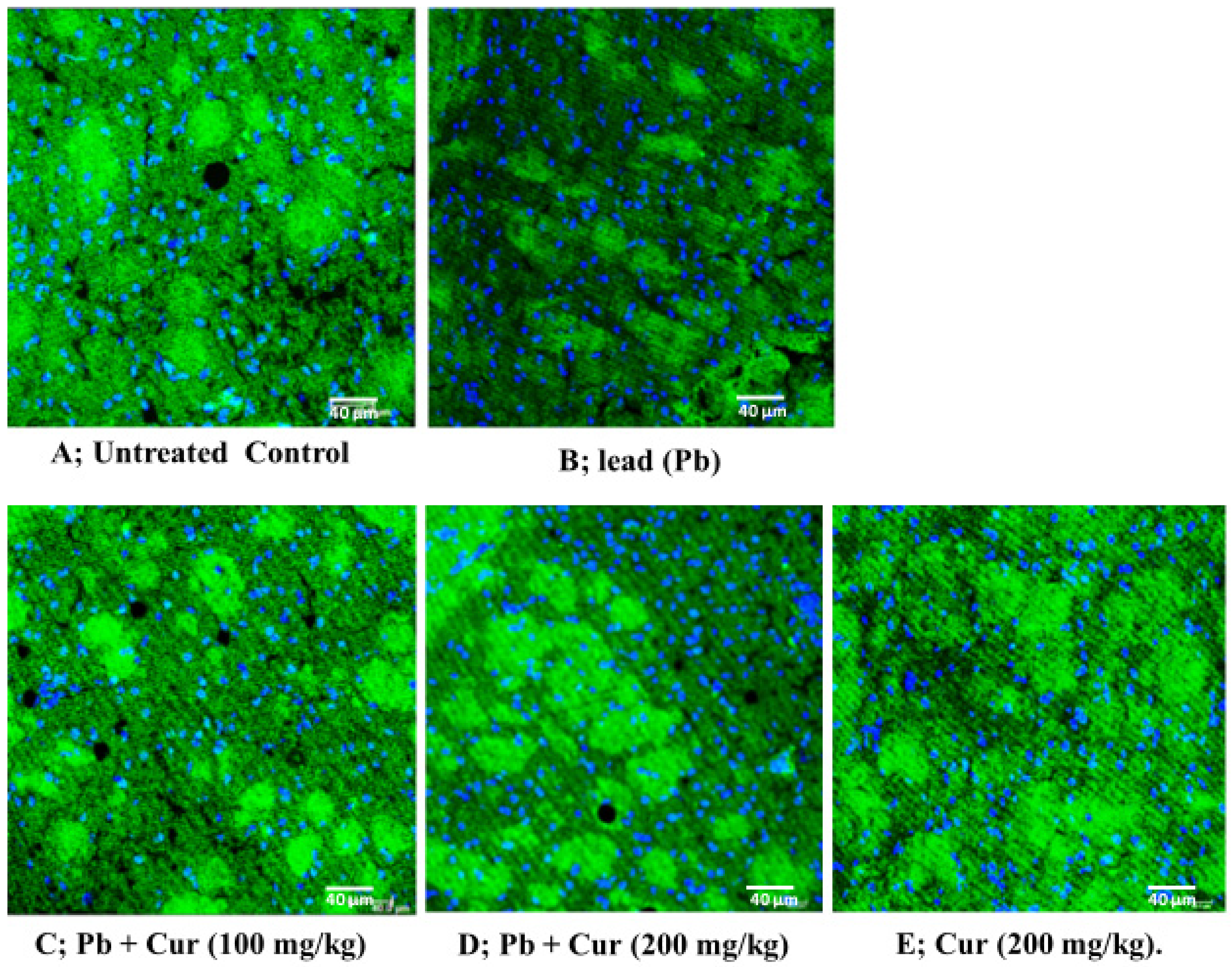
2.10. Immunohistochemistry Study

Brain tissue slices were fixed in 4% paraformaldehyde for 15 min, air-dried, and washed with PBS. Nonspecific proteins were blocked in the blocking serum, consisting of 3% normal donkey serum and 0.3% Triton X-100 in PBS, and incubated at room temperature for 30 min. After blocking, the slices were incubated with primary anti-AChE (Abcam PLC, Waltham, MA, USA). The sections were kept in a humidified box at 4 °C overnight. Tissues were washed three times with PBS and then were incubated for 1 h with donkey antibody-conjugated secondary antibodies conjugated with fluorescent dyes. Excess secondary antibodies were removed by washing three times in PBS and once with deionized H2O. Tissue slides were counter-stained with nuclear staining dye (DRAQ5) and mounted with a mounting medium (Vectashield, H-100, Vector Laboratories, Burlingame, CA, USA). Photomicrographs were obtained using a Leica confocal fluorescence microscope (Leica Microsystems Inc., Bannockburn, IL, USA).

2.11. In Silico Study

2.11.1. Molecular Docking Study

The required crystal structures were published in the PDB format, as follows: AChE (PDB ID: 4M0E), COX-2 (PDB ID: 5KIR), TNF-α (PDB ID: 2AZ5), IKK (PDB ID: 4KIK), ERK (PDB ID: 1TVO), JNK (PDB ID: 3OY1), and P38 (PDB ID: 5WJJ). These were analyzed using Maestro 11 software (Schrodinger, New York, NY, USA) for the molecular docking study. For protein docking, the protein preparations were allowed to preprocess by adding bond sequence, the H-bond, and SS-bond formation. After that, minimization was achieved using the OPLS5 force field. For ligand processing, the 2D structures of Cur (PubChem CID 969516) were converted into 3D structures using the ligprep software (Scodinger suite v11), with the OPLS5 force field at pH 7.0 ± 2.0 as an initial setting. The prepared proteins were then processed to form a receptor grid using the OPLS5 force field.

2.11.2. The Chemical Absorption, Distribution, Metabolism, Excretion, and Toxicity (ADMET) Analysis

The ADMET analysis and drug likeliness properties of Cur were identified using Schrodinger’s QikProp module. The drug likeliness properties were evaluated based on the rule of five. On the other hand, a candidate was regarded as drug-like if it displayed any three of the parameters’ molecular weight (MW) <500 Daltons, H-bond acceptor (HBA) ≤10, H-bond donor (HBD) ≤5, and predicted QPlogPo/w value (-) 2.0–6.5.

2.12. Statistical Analysis

The results were calculated as the mean ± SEM (standard error mean) of three independent experiments. The statistical analysis was conducted using one-way analysis of variance (ANOVA) followed by a post hoc test, Newman–Keuls, using GraphPad Prism v5 (San Diego, CA, USA). p < 0.05 was considered statistically significant.

3. Results

3.1. Effect of Curcumin on Lead-Induced Mice Body Weight Changes

The effects of Cur on animal body weight changes before and after Pb exposure are summarized in Table 1. According to Table 1, at the initial stage, there were no differences between the mice body weights in various treatment groups compared to untreated control. On the contrary, at the final stage, Cur with Pb treatment significantly (p < 0.05) improved body weight compared to the lead-treated group. In addition, mice treated with Pb showed less improvement of final body weight compared to Pb-treated initial body weight.

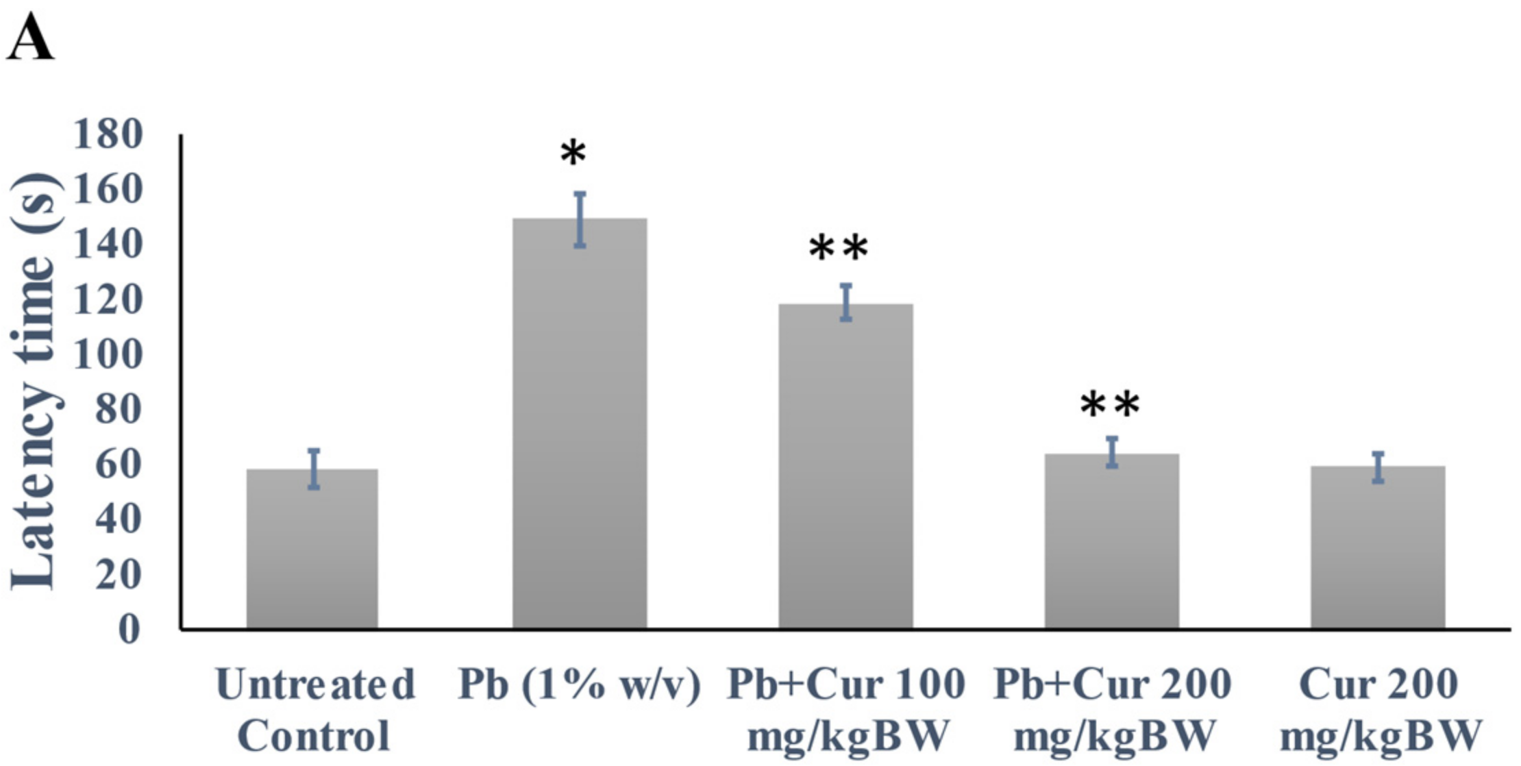
3.2. Validation of Curcumin’s Effect against Lead-Induced Cognitive Dysfunction

The Pb-induced cognitive function was evaluated via latency and immobility times. The data are presented in Figure 1. Our WMST revealed no significant differences between Pb-treated mice and others on the first day of the trials (data not shown). In contrast, Pb exposure significantly (p < 0.05) increased the latency time (approximately two-fold) on the fifth day of the trial. However, the increase of latency times was reduced significantly by Cur (Figure 1A). Furthermore, the latency time of Cur-treated mice was not significantly different compared to control on the fifth day of trial, which indicates the ameliorative effect of Cur.
The FST study was conducted to evaluate the immobility time of the normal, Pb-, and Cur co-treated mice, and the data are displayed in Figure 1B. Our results showed that co-treatment with Cur significantly (p < 0.05) increased the swimming time and reduced the immobility time compared to the lead-treated group. In contrast, Pb-treated mice showed a markedly (p < 0.05) prolonged immobility time compared to the control group. Furthermore, there was no significant difference between mice treated with Cur and the control group, indicating an improvement in memory function due to the Cur treatment. These studies confirm that latency and depressive-like behavior are markedly increased in the Pb-treated group.

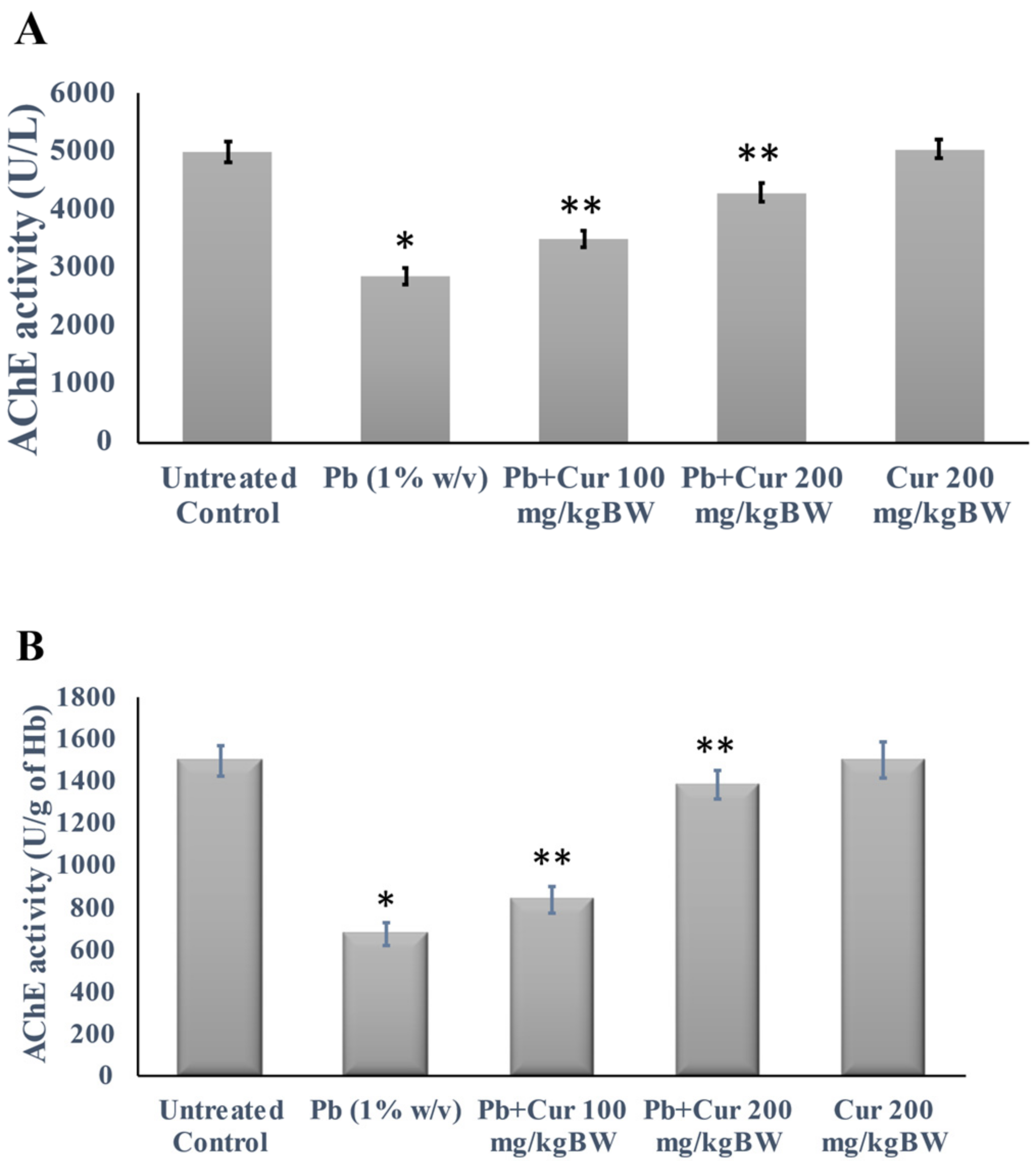
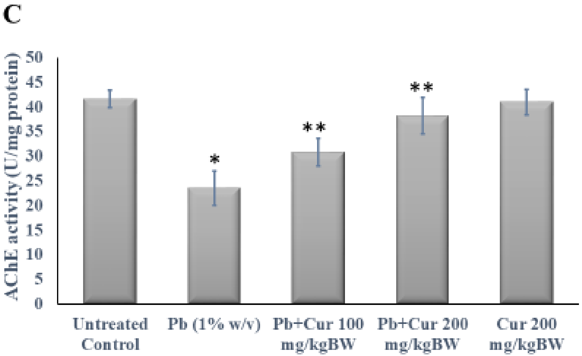
3.3. Protective Effect of Curcumin on Cholinergic Dysfunction

We investigated the acetylcholinesterase (AChE) activity in plasma, RBC, and brain homogenates to unveil Cur’s effect on Pb-treated neurotransmitter impairment. For further clarification, we determined the activity and expression of AChE in the brain tissue. The results regarding cholinesterase function are shown in Figure 2. Figure 2A–C represent the activities of AChE in plasma, RBC, and brain homogenate, respectively. Our study found that the AChE activities of Pb-treated mice were markedly (p < 0.05) diminished.
On the contrary, the Pb-treated mice with Cur showed significantly (p < 0.05) modulated AChE dysfunction in the plasma, RBC, and brain homogenates (Figure 2A–C). Moreover, the AChE values of Cur co-treatment groups were not significant compared to those of the control group. Afterward, to further confirm the effect of Cur, we determined the expression of AChE in the mouse brain (Figure 3). Figure 3 showed that the levels of AChE expression in the brains of Pb-treated mice were markedly (p < 0.05) reduced, while the expressions in Pb-treated mice with Cur were significantly (p < 0.05) ameliorated (Figure 3).
The Pb-induced lower AChE expression in brain tissues was confirmed by immunohistochemistry and was ameliorated in our Cur-co-treated group. Additionally, the statistical analysis has shown no significant differences in protein expression between the Cur-treated and the control group, highlighting the potential effect of Cur co-treatment on Pb-intoxicated mice (Figure 4).

3.4. Protective Effect of Curcumin on Oxidative Stress Parameters

Malonaldehyde (MDA) is a metabolite of oxidative stress, and is widely accepted as an indicator of lipid peroxidation. Therefore, the MDA levels in plasma, RBC, and brain homogenate were evaluated to understand the effect of Cur against Pb-induced neurotoxicity, and the data are presented in Table 2. As found in Table 2, the MDA in blood and brain of Pb-treated mice was markedly increased compared to the control group, whereas Cur co-treatment reduced the MDA in a concentration-dependent manner.

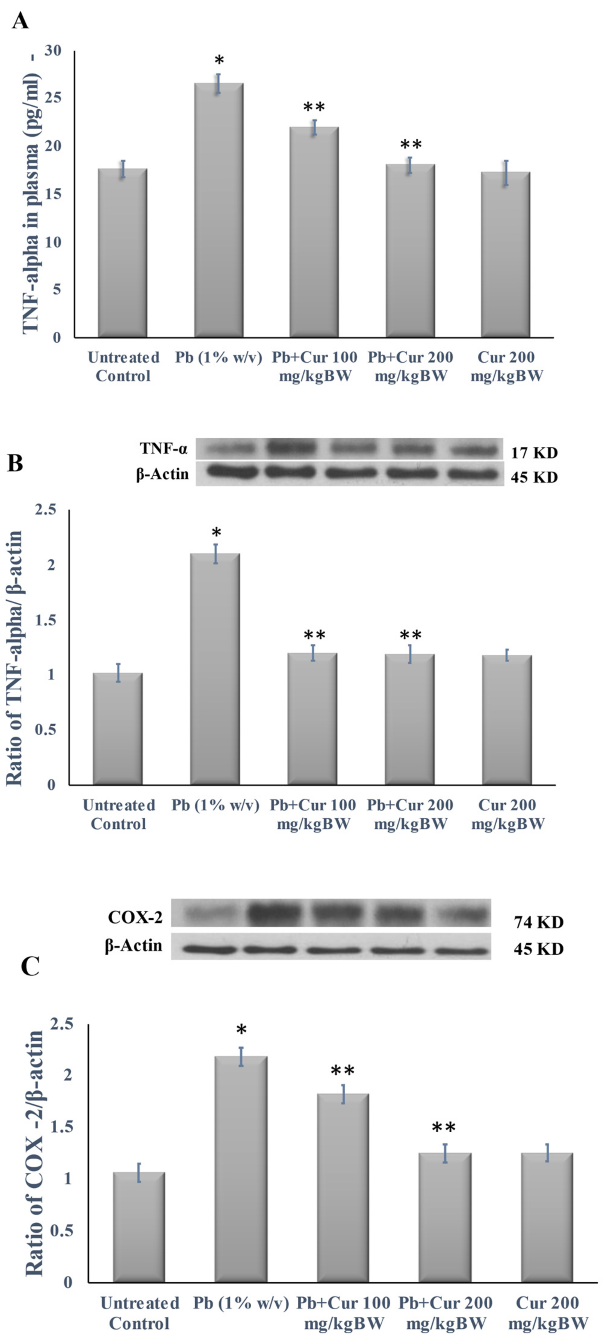
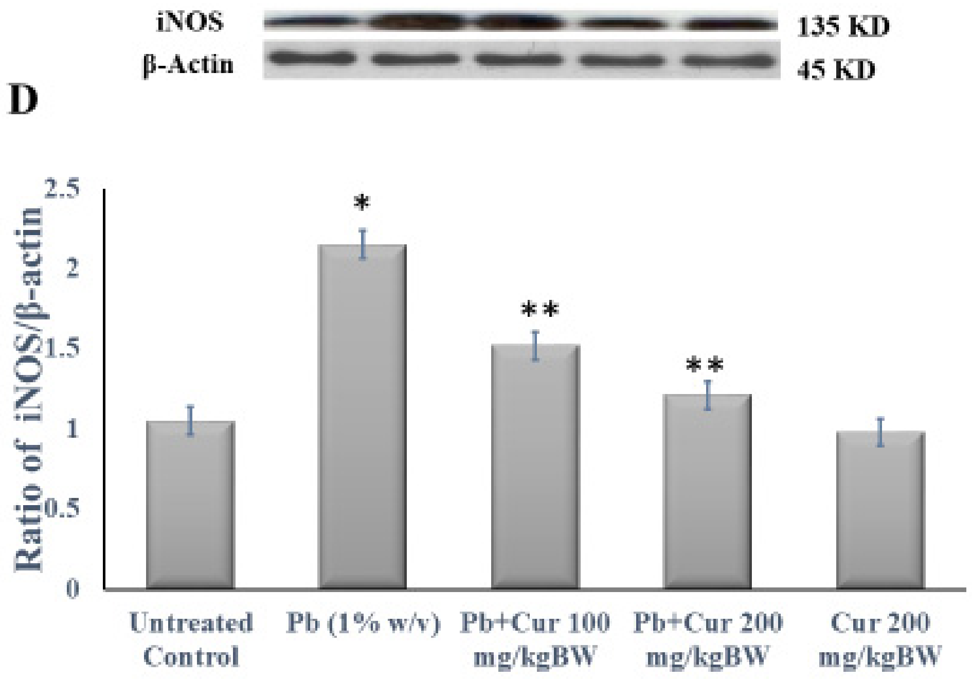
3.5. Protective Effect of Curcumin on Inflammatory Profile

The plasma TNF-α level, an expression of inflammatory markers in the brain homogenate, was estimated to confirm the anti-inflammatory effect of Cur against Pb-induced neurotoxicity. The results are shown in Figure 5. According to Figure 5A, the circulating TNF-α level was markedly (p < 0.05) increased in Pb-treated mice. On the flip side, Cur at a dose of 100 or 200 mg/kgBW to Pb-treated mice significantly (p < 0.05) reduced the plasma TNF-α levels (Figure 5A). Similarly, the expressions of inflammatory markers (TNF-α, COX-2, and iNOS) were higher in the brain tissue homogenate of Pb-treated brain (Figure 5B–D). Additionally, co-treatment with Cur significantly (p < 0.05) reduced the expressions of inflammatory markers (Figure 5B–D).

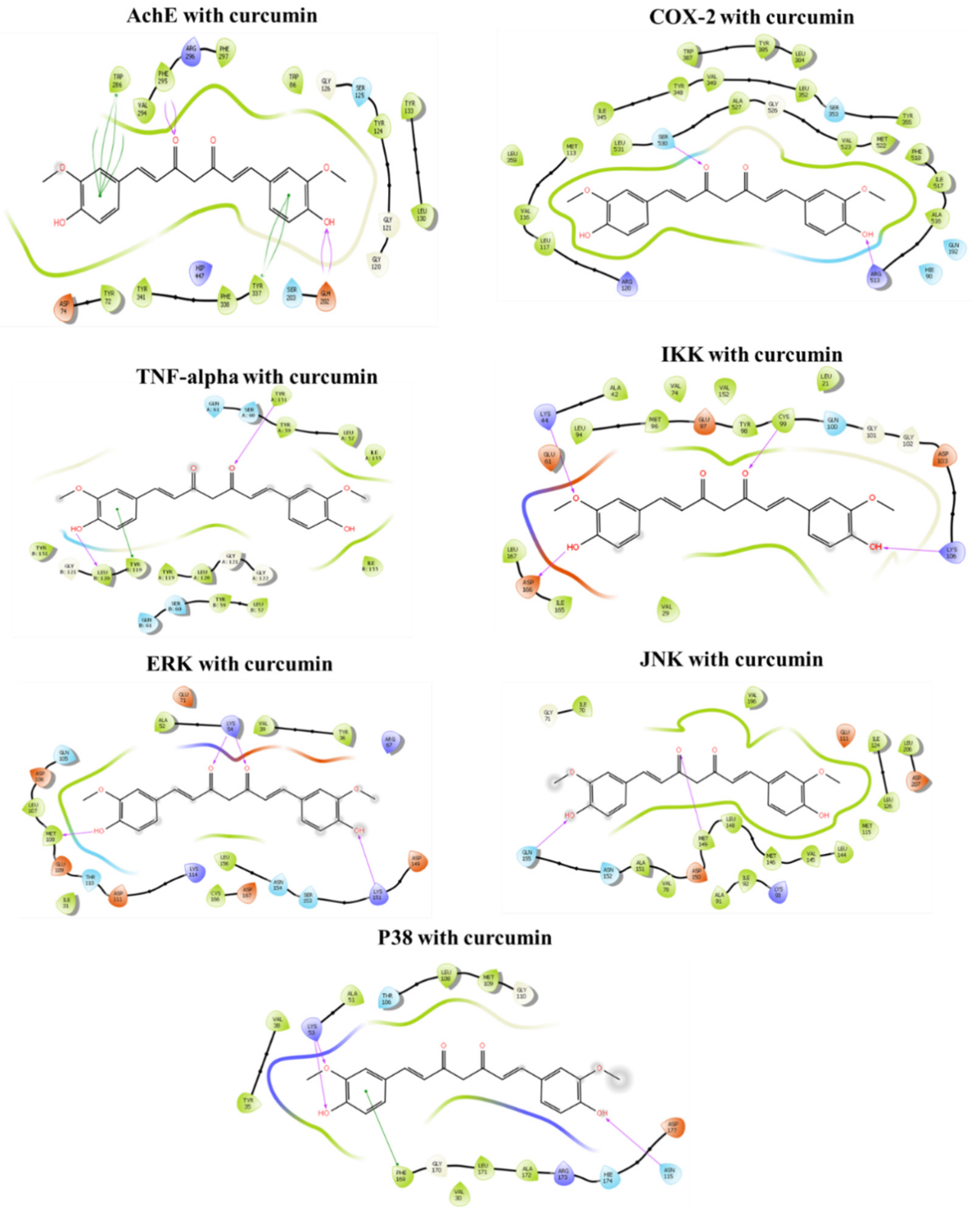
3.6. Molecular Docking In Silico

3.6.1. Binding Mechanism of Curcumin with Acetylcholinesterase (AChE)

In silico molecular docking showed Cur’s potent binding affinity for AChE receptors (PDB ID: 4M0E), COX-2 (PDB ID: 5KIR), TNF-α (PDB ID: 2AZ5), IKK (PDB ID: 4KIK), ERK (PDB ID: 1TVO), JNK (PDB ID: 3OY1), and P38 (PDB ID: 5WJJ) (Table 3 and Figure 6). According to Table 3, Cur showed a greater ability to dock with AChE, followed by IKK, COX-2, P38, JNK, TNF-α, and ERK. Thus, Cur was predicted to have neuroprotective effects by exerting its antioxidant and anti-inflammatory properties, and by inhibiting cholinergic alterations.

3.6.2. ADMET Analysis and Drug-Likeliness Properties of Curcumin

According to the ADMET analysis (Table 4), Cur could serve as a potential candidate, along with existing drugs. The ADMET profile of Cur meets the necessary criteria to become a drug candidate: a lower star (*:1), with moderate lipid solubility comprising characteristics such as FOSA (261.70), QPlogBB (−2.246), and QPPMDCK (65.952). Furthermore, to be an orally available drug candidate, the substance must follow Jorgensen’s rule of three and Lipinski’s rule of five. In addition, our QikProp module analysis suggests that Cur also has a modest ability to cross the gut barrier (QPPCaco 155.011) following oral intake. Therefore, Cur could be regarded as a potential candidate for further analysis in the clinical environment.
Here, a low star value indicates that a molecule is more drug-like than other molecules with many stars. The total solvent accessible surface area (SASA) in Å2 was measured using a probe with a 1.4 Å radius, the FOSA-hydrophobic component of the SASA (saturated C and attached H), and FISA-hydrophilic part of the SASA (SASA on N and O, and H on heteroatoms).
Here, Leu: leucine, Trp: tryptophan, Ala: alanine, Val: valine, Ile: isoleucine, Met: methionine, Cys: cysteine, Pro: proline, Tyr: tyrosine, Phe: phenylalanine, Lys: lysine, Arg: arginine, Thr: threonine, Gln: glutamine, Ser: serine, Asn: asparagine, Gly: glycine, His: crystal histidine. In addition, there are three residue templates for histidine such as Hie: histidine (default, H on the delta nitrogen), HID (H on the epsilon nitrogen), HIP (hydrogens on both nitrogens, this is positively charged).

4. Discussion

Lead (Pb) is a ubiquitous metal that causes decreases in body weight and has been implicated in many other organs; in particular, the nervous system is the primary target of Pb exposure, and the developing brain appears to be especially vulnerable to Pb neurotoxicity [25,26]. It has been observed that Pb exposure is associated with a decline in attention concentration, the inhibition of long-term potentiation (LTP), growth retardation, and learning and memory impairment [27,28]. Water maze swimming tests are related to spatial memory and are a popular method used to evaluate cognitive function in animal models, including Alzheimer’ disease (AD) model mice [29,30]. Acetylcholine exclusively encodes the regulation of spatial memory through theta rhythm oscillations. Besides this, acetylcholine processes and encodes novel information for short-term and long-term memory through persistent spiking (such as cortical neurons) [31,32]. Our current results show that Cur improved behavioral changes, including learning deficits, memory losses, and stress in mice. In addition, Cur has been reported to be able to repair learning and memory dysfunctions [33]. Similar studies have also confirmed Cur’s effect against learning and memory impairments [34,35].
The present study reports that the structure of AChE contains free SH groups, which have a high affinity for Pb. After binding Pb with the free SH groups of AChE, it can alter Ca2+ homeostasis and produce oxidative stress [2,36,37]. The reductions in AChE expression in Pb-treated brains were reversed via co-treatment with Cur, indicating improvements in cholinergic circuitry. At the cholinergic synapses, AChE hydrolyzes the neurotransmitter acetylcholine. Dysfunctions in AChE cause neuropsychiatric problems [17,34]. The over-activation of the cholinergic system hinders working memory activity in the primate prefrontal cortex [38]. Hence, the hippocampal–prefrontal axis assists in spatial encoding within the working memory, possibly via gamma oscillations synchrony [39]. On the contrary, reduced acetylcholine results in cognitive impairments. Thus, there should be a balance, as acetylcholine may be friendly or antagonistic in different contexts, as Baxter and Crimins discussed [40]. Therefore, the current study showed that Cur markedly restored AChE expression, which might help in establishing a balance in regular acetylcholine accumulation in Pb-intoxicated mice.
Studies have reported that Pb causes oxidative damage by generating free radicals, reducing the cellular antioxidant pool, and manipulating several pathways. It further diminishes the glutathione and protein-bound sulfhydryl group [1,9]. Cur is known to have a potent antioxidant activity due to its capacity for chain-breaking, as well as the hydrogen-donating phenolic groups in its structure [41]. Much research has also revealed the neuroprotective effects of Cur, which operate via exerting antioxidant effects and reducing oxidative damage [18,34,37,41,42]. Besides this, Pb accelerates caspase-3 activity via oxidative stress, lipid peroxidation, and apoptosis [7,43]. Curcumin exhibits great promise as a therapeutic agent for a variety of cancers, as well as for psoriasis, and Alzheimer’s disease [44]. It has been reported that Cur suppresses aluminum- and Pb-induced oxidative neurotoxicity, alterations in NMDA receptors that lead to decreased antioxidant enzyme activity, and AChE dysfunctions [45]. These two possible mechanisms (oxidative stress and cholinergic dysfunction) relate to Pb-induced neurotoxicity. Thus, the present study has indicated that Cur, a potent antioxidant compound, ameliorated lipid peroxidation and oxidative stress parameters in mice exposed to Pb, along with protecting against cholinergic dysfunction.
Accumulating evidence is suggesting that inflammation is the main outcome of Pb contamination. Pb intoxication causes ROS-mediated inflammation by activating the JNK–MAPK pathway. Furthermore, ROS-activated NF–κB is translocated to a nucleus in order to initiate inflammatory gene transcription [21,41,46]. Thus, Pb could induce neuroinflammation via regulating the NF–κB pathway, thereby increasing inflammatory gene expression and enzyme alterations [41,47,48,49], while Cur co-treatment effectively suppresses Pb-induced brain inflammation.
In silico molecular docking showed that Cur had a greater ability to dock with AchE, followed by IKK, COX-2, P38, JNK, TNF-α, and ERK. Thus, Cur may completely bind with AchE to increase enzymatic activity, which is predicted to have dose-dependent neuroprotective effects, at least in part via exerting antioxidant and anti-inflammatory effects, and reversing the Pb-induced alterations in transmitters and enzymes.

5. Conclusions

Our findings suggest that Pb intoxication causes memory and learning deficits associated with aberrant acetylcholine balance. However, co-treatment with Cur restored Pb-induced alterations in cognitive processes by maintaining the cholinergic system via rebalancing the acetylcholine level. Therefore, following our thorough investigation, the dietary intake of Cur could be suggested to prevent and cure heavy metal-associated memory loss, learning deficit, and depression-like behavior.

Author Contributions

Conceptualization, S.C., M.N.R., M.P.P., H.J.M. and J.T.; data curation, S.C., M.N.R., M.P.P. and N.K.; formal analysis, S.C., M.N.R. and M.P.P.; funding acquisition, J.T.; investigation, S.C., M.N.R. and M.P.P.; methodology, S.C., M.N.R. and M.P.P.; project administration, J.T.; resources, J.T.; software, S.C. and M.N.R.; supervision, H.J.M. and J.T.; validation, N.K., H.J.M. and J.T.; visualization, N.K. and J.T.; writing—original draft, S.C., M.N.R. and M.P.P.; writing—review and editing, N.K., H.J.M. and J.T. All authors have read and agreed to the published version of the manuscript.

Funding

This research was funded, in part, by The Thailand Research Fund (grant no. MRG5180200), and this research was financially supported by the new strategic research project (P2P), Walailak University, Thailand, and The Office of the Higher Education Commission (OHEC), Ministry of Higher Education, Science, Research and Innovation (MHESRI), Thailand. The APC was funded by Walailak University.

Institutional Review Board Statement

The animal study protocol was approved by the Animal Care and Use Committee (ACUC) of Walailak University (No. 002/2013).

Informed Consent Statement

Not applicable.

Data Availability Statement

The data presented in this study are available on request from the corresponding author.

Conflicts of Interest

The authors declare no conflict of interest.

References

  1. Rana, M.N.; Tangpong, J.; Rahman, M.M. Toxicodynamics of Lead, Cadmium, Mercury and Arsenic-induced kidney toxicity and treatment strategy: A mini-review. Toxicol. Rep. 2018, 5, 704–713. [Google Scholar] [CrossRef]
  2. Liu, C.M.; Zheng, G.H.; Ming, Q.L.; Sun, J.M.; Cheng, C. Protective effect of puerarin on lead-induced mouse cognitive im-pairment via altering activities of acetylcholinesterase, monoamine oxidase and nitric oxide synthase. Environ. Toxicol. Phar-macol. 2013, 35, 502–510. [Google Scholar] [CrossRef] [PubMed]
  3. Yang, X.; Wang, B.; Zeng, H.; Cai, C.; Hu, Q.; Cai, S.; Xu, L.; Meng, X.; Zou, F. Role of the mitochondrial Ca2+ uniporter in Pb2+-induced oxidative stress in human neuroblastoma cells. Brain Res. 2014, 1575, 12–21. [Google Scholar] [CrossRef]
  4. Indo, H.P.; Yen, H.C.; Nakanishi, I.; Matsumoto, K.; Tamura, M.; Nagano, Y.; Matsui, H.; Gusev, O.; Cornette, R.; Okuda, T.; et al. A mitochondrial super-oxide theory for oxidative stress diseases and aging. J. Clin. Biochem. Nutr. 2015, 56, 1–7. [Google Scholar] [CrossRef] [PubMed] [Green Version]
  5. Ma, L.; Liu, J.Y.; Dong, J.X.; Xiao, Q.; Zhao, J.; Jiang, F.L. Toxicity of Pb2+ on rat liver mitochondria induced by oxidative stress and mitochondrial permeability transition. Toxicol. Res. 2017, 6, 822–830. [Google Scholar] [CrossRef] [PubMed] [Green Version]
  6. Gleichmann, M.; Mattson, M.P. Neuronal calcium homeostasis and dysregulation. Antioxid. Redox Signal. 2011, 14, 1261–1273. [Google Scholar] [CrossRef] [PubMed] [Green Version]
  7. Tangpong, J.; Satarug, S. Alleviation of lead poisoning in the brain with aqueous leaf extract of the Thunbergia laurifolia (Linn.). Toxicol. Lett. 2010, 198, 83–88. [Google Scholar] [CrossRef]
  8. Indo, H.P.; Hawkins, C.L.; Nakanishi, I.; Matsumoto, K.; Matsui, H.; Suenaga, S.; Davies, M.J.; St Clair, D.K.; Ozawa, T.; Majima, H.J. Role of mitochondrial reactive oxygen species in the activation of cellular signals, molecules and function. Pharmacol. Mitochondria 2017, 240, 439–456. [Google Scholar] [CrossRef]
  9. Phyu, M.P.; Tangpong, J. Neuroprotective effects of xanthone derivative of Garcinia mangostana against lead-induced acetyl-cholinesterase dysfunction and cognitive impairment. Food Chem. Toxicol. 2014, 70, 151–156. [Google Scholar] [CrossRef]
  10. Basha, D.C.; Rani, M.U.; Devi, C.B.; Kumar, M.R.; Reddy, G.R. Perinatal lead exposure alters postnatal cholinergic and aminergic system in rat brain: Reversal effect of calcium co-administration. Int. J. Dev. Neurosci. 2012, 30, 343–350. [Google Scholar] [CrossRef] [PubMed]
  11. de Souza Tavares, W.; Akhtar, Y.; Gonçalves, G.L.P.; Zanuncio, J.C.; Isman, M.B. Turmeric powder and its derivatives from Curcuma longa rhizomes: Insecticidal effects on cabbage looper and the role of synergists. Sci. Rep. 2016, 6, 34093. [Google Scholar] [CrossRef] [PubMed] [Green Version]
  12. Esatbeyoglu, T.; Huebbe, P.; Ernst, I.M.; Chin, D.; Wagner, A.E.; Rimbach, G. Curcumin-from molecule to biological function. Angew. Chem. Int. Ed. Engl. 2012, 51, 5308–5332. [Google Scholar] [CrossRef] [PubMed]
  13. Gupta, S.C.; Patchva, S.; Koh, W.; Aggarwal, B.B. Discovery of Curcumin, a component of golden spice, and its miraculous biological activities. CEPP 2012, 39, 283–299. [Google Scholar] [CrossRef]
  14. Ng, T.P.; Chiam, P.C.; Lee, T.; Chua, H.C.; Lim, L.; Kua, E.H. Curry consumption and cognitive function in the elderly. Am. J. Epidemiol. 2006, 164, 898–906. [Google Scholar] [CrossRef] [PubMed] [Green Version]
  15. Cox, K.H.; Pipingas, A.; Scholey, A.B. Investigation of the effects of solid lipid curcumin on cognition and mood in a healthy older population. J. Psychopharmacol. 2015, 29, 642–651. [Google Scholar] [CrossRef]
  16. Abu-Taweel, G.M. Neurobehavioral protective properties of Curcumin against the mercury chloride treated mice offspring. Saudi J. Biol. Sci. 2019, 26, 736–743. [Google Scholar] [CrossRef] [PubMed]
  17. Akinyemi, A.J.; Oboh, G.; Fadaka, A.O.; Olatunji, B.P.; Akomolafe, S. Curcumin administration suppress acetylcholinesterase gene expression in cadmium treated rats. Neurotoxicology 2017, 62, 75–79. [Google Scholar] [CrossRef]
  18. Akinyemi, A.J.; Oboh, G.; Ogunsuyi, O.; Abolaji, A.O.; Udofia, A. Curcumin-supplemented diets improve antioxidant enzymes and alter acetylcholinesterase genes expression level in Drosophila melanogaster model. Metab. Brain Dis. 2018, 33, 369–375. [Google Scholar] [CrossRef]
  19. EFSA Panel on Food Additives and Nutrient Sources added to Food (ANS). Scientific Opinion on the re-evaluation of curcu-min (E 100) as a food additive. EFSA J. 2010, 8, 1679. [Google Scholar] [CrossRef]
  20. Rafiee, Z.; Nejatian, M.; Daeihamed, M.; Jafari, S.M. Application of curcumin-loaded nanocarriers for food, drug and cos-metic purposes. Trends Food Sci. Technol. 2019, 88, 445–458. [Google Scholar] [CrossRef]
  21. Rana, M.N.; Tangpong, J.; Rahman, M.A. Xanthones protects lead-induced chronic kidney disease (CKD) via activating Nrf-2 and modulating NF-kB, MAPK pathway. Biochem. Biophys. Rep. 2020, 21, 100718. [Google Scholar] [CrossRef] [PubMed]
  22. Meagher, E.A.; FitzGerald, G.A. Indices of lipid peroxidation in vivo: Strengths and limitations. Free Radic. Biol. Med. 2000, 28, 1745–1750. [Google Scholar] [CrossRef]
  23. Ellman, G.L.; Courtney, K.D.; Andres, V., Jr.; Feather-stone, R.M. A new and rapid colorimetric determination of acetylcholin-esterase activity. Biochem. Pharmacol. 1961, 7, 88–95. [Google Scholar] [CrossRef]
  24. Bradford, M.M. A rapid and sensitive for the quantitation of microgram quantities of protein utilizing the principle of protein-dye binding. Anal. Biochem. 1976, 72, 248–254. [Google Scholar] [CrossRef]
  25. Mason, L.H.; Harp, J.P.; Han, D.Y. Pb neurotoxicity: Neuropsychological effects of lead toxicity. Biomed. Res. Int. 2014, 2014, 840547. [Google Scholar] [CrossRef] [PubMed] [Green Version]
  26. Reddy, G.R.; Devi, B.C.; Chetty, C.S. Developmental lead neurotoxicity: Alterations in brain cholinergic system. Neurotoxicology 2007, 28, 402–407. [Google Scholar] [CrossRef] [PubMed]
  27. Toscano, C.D.; Guilarte, T.R. Lead neurotoxicity: From exposure to molecular effects. Brain Res. Rev. 2005, 49, 529–554. [Google Scholar] [CrossRef]
  28. Canfield, R.L.; Henderson, C.R.J.; Cory-Slechta, D.A.; Cox, C.; Jusko, T.A.; Lanphear, B.P. Intellectual impairment in children with blood lead concentrations below 10 μg per deciliter. N. Engl. J. 2003, 348, 1517–1526. [Google Scholar] [CrossRef] [Green Version]
  29. Vorhees, C.V.; Williams, M.T. Morris water maze: Procedures for assessing spatial and related forms of learning and memory. Nat. Protoc. 2006, 1, 848–858. [Google Scholar] [CrossRef] [Green Version]
  30. Tian, H.; Ding, N.; Guo, M.; Wang, S.; Wang, Z.; Liu, H.; Yang, J.; Li, Y.; Ren, J.; Jiang, J.; et al. Analysis of Learning and Memory Ability in an Alzheimer’s Disease Mouse Model using the Morris Water Maze. J. Vis. Exp. 2019, 29, e60055. [Google Scholar] [CrossRef]
  31. Hasselmo, M.E. The role of acetylcholine in learning and memory. Curr. Opin. Neurobiol. 2006, 16, 710–715. [Google Scholar] [CrossRef] [PubMed] [Green Version]
  32. Newman, E.L.; Gupta, K.; Climer, J.R.; Monaghan, C.K.; Hasselmo, M.E. Cholinergic modulation of cognitive processing: In-sights drawn from computational models. Front. Behav. Neurosci. 2012, 6, 24. [Google Scholar] [CrossRef] [PubMed] [Green Version]
  33. Liu, Y.H.; Lee, C.J.; Chen, L.C.; Lee, T.L.; Hsieh, Y.Y.; Han, C.H.; Yang, C.H.; Huang, W.J.; Hou, W.C. Acetylcholinesterase inhibitory activity and neuroprotection: In vitro, molecular docking, and improved learning and memory functions of deme-thylcurcumin in scopolamine-induced amnesia ICR mice. Food Funct. 2020, 11, 2328–2338. [Google Scholar] [CrossRef] [PubMed]
  34. Oz, M.; Atalik, K.E.N.; Yerlikaya, F.H.; Demir, E.A. Curcumin alleviates cisplatin-induced learning and memory impairments. Neurobiol. Learn. Mem. 2015, 123, 43–49. [Google Scholar] [CrossRef]
  35. Sarlak, Z.; Oryan, S.; Moghaddasi, M. Interaction between the antioxidant activity of curcumin and cholinergic system on memory retention in adult male Wistar rats. Iran. J. Basic Med. Sci. 2015, 18, 398–403. [Google Scholar]
  36. Mary, C.P.V.; Vijayakumar, S.; Shankar, R. Metal chelating ability and antioxidant properties of Curcumin-metal complexes—A DFT approach. J. Mol. Graph. Model. 2018, 79, 1–14. [Google Scholar] [CrossRef]
  37. Abubakar, K.; Muhammad Mailafiya, M.; Danmaigoro, A.; Musa Chiroma, S.; Abdul Rahim, E.B.; Abu Bakar Zakaria, M.Z. Curcumin attenuates lead-induced cerebellar toxicity in rats via chelating activity and inhibition of oxidative stress. Biomolecules 2019, 9, 453. [Google Scholar] [CrossRef] [Green Version]
  38. Vijayraghavan, S.; Major, A.J.; Everling, S. Muscarinic M1 receptor overstimulation disrupts working memory activity for rules in primate prefrontal cortex. Neuron 2018, 98, 1256–1268. [Google Scholar] [CrossRef] [Green Version]
  39. Spellman, T.; Rigotti, M.; Ahmari, S.E.; Fusi, S.; Gogos, J.A.; Gordon, J.A. Hippocampal–prefrontal input supports spatial en-coding in working memory. Nature 2015, 522, 309–314. [Google Scholar] [CrossRef] [Green Version]
  40. Baxter, M.G.; Crimins, J.L. Acetylcholine receptor stimulation for cognitive enhancement: Better the devil you know? Neuron 2018, 98, 1064–1066. [Google Scholar] [CrossRef] [Green Version]
  41. Kumar, A.; Dogra, S.; Prakash, A. Protective effect of Curcumin (Curcuma longa) against aluminum toxicity: Possible behav-ioral and biochemical alterations in rats. Behav. Brain Res. 2009, 205, 384–390. [Google Scholar] [CrossRef] [PubMed]
  42. Abolaji, A.O.; Fasae, K.D.; Iwezor, C.E.; Aschner, M.; Farombi, O.E. Curcumin attenuates copper-induced oxidative stress and neurotoxicity in Drosophila melanogaster. Toxicol. Rep. 2020, 7, 261–268. [Google Scholar] [CrossRef] [PubMed]
  43. Galal, M.K.; Elleithy, E.M.M.; Abdrabou, M.I.; Yasin, N.A.E.; Shaheen, Y.M. Modulation of caspase-3 gene expression and pro-tective effects of garlic and spirulina against CNS neurotoxicity induced by lead exposure in male rats. Neurotoxicology 2019, 72, 15–28. [Google Scholar] [CrossRef] [PubMed]
  44. Hatcher, H.; Planalp, R.; Cho, J.; Torti, F.M.; Torti, S.V. Curcumin: From ancient medicine to current clinical trials. Cell. Mol. Life Sci. 2008, 65, 1631–1652. [Google Scholar] [CrossRef] [PubMed]
  45. Tüzmen, M.N.; Yücel, N.C.; Kalburcu, T.; Demiryas, N. Effects of Curcumin and tannic acid on the aluminum- and lead-induced oxidative neurotoxicity and alterations in NMDA receptors. Toxicol. Mech. Methods 2015, 25, 120–127. [Google Scholar] [CrossRef] [PubMed]
  46. He, Y.; Yue, Y.; Zheng, X.; Zhang, K.; Chen, S.; Du, Z. Curcumin, inflammation, and chronic diseases: How are they linked? Molecules 2015, 20, 9183–9213. [Google Scholar] [CrossRef] [PubMed]
  47. Alqahtani, W.S.; Albasher, G. Moringa oleifera Lam. extract rescues lead-induced oxidative stress, inflammation, and apoptosis in the rat cerebral cortex. J. Food Biochem. 2021, 45, e13579. [Google Scholar] [CrossRef]
  48. Phyu, M.P.; Tangpong, J. Protective effect of Thunbergia laurifolia (Linn.) on lead induced acetylcholinesterase dysfunction and cognitive impairment in mice. BioMed Res. Int. 2013, 2013, 1–6. [Google Scholar] [CrossRef]
  49. Khalaf, A.A.; Moselhy, W.A.; Abdel-Hamed, M.I. The protective effect of green tea extract on lead induced oxidative and DNA damage on rat brain. Neurotoxicology 2012, 33, 280–289. [Google Scholar] [CrossRef]
Figure 1. Effect of curcumin on cognitive dysfunction caused by Pb. (A) Curcumin’s effect against memory loss and learning deficit of mice as related to latency time (s) using the Morris water maze swimming test. (B) Curcumin’s effect against depression-like behavior of mice as related to the immobility time (s) using the forced swimming Test. The results are displayed as mean ± SEM (n = 6). * p < 0.05 specifies a difference between control mice and Pb-treated mice. ** p < 0.05 compares Pb-treated mice and curcumin (Cur)-treated mice.
Foods 11 00856 g001aFoods 11 00856 g001b
Figure 2. Effect of curcumin on cholinergic dysfunction caused by lead. (AC) Acetylcholinesterase (AChE) activity in the plasma, RBC, and brain homogenate, respectively. The results are displayed as mean ± SEM (n = 6). * p < 0.05 specifies a difference between control mice and Pb-treated mice. ** p < 0.05 compares between Pb-treated mice and Curcumin (Cur)-treated mice.
Foods 11 00856 g002aFoods 11 00856 g002b
Figure 3. Effect of curcumin on the expression of acetylcholinesterase (AChE) in Pb-treated mouse brain. The internal control of protein bands was examined using β-actin in a relative density analysis. The results are displayed as mean ± SEM (n = 6). * p < 0.05 specifies difference between control mice and Pb-treated mice. ** p < 0.05 compares between Pb-treated mice and curcumin (Cur)-treated mice.
Foods 11 00856 g003
Figure 4. Immunohistochemistry—effect of curcumin on the acetylcholinesterase (AChE) expression in the brain tissue of lead (Pb)-exposed mice. (A) Group 1: Untreated control (sodium acetate, CH3COONa-3H2O) in drinking water. (B) Group 2: Pb (1%) in drinking water. (C) Group 3: Pb + Cur (100 mg/kg). (D) Group 4: Pb + Cur (200 mg/kg). (E) Group 5: Cur (200 mg/kg). In this figure, the green color represents AChE expression and blue color shows nucleus staining with DRAQ5.
Foods 11 00856 g004
Figure 5. Effect of curcumin on inflammation caused by lead. (A) TNF-α level in plasma. (BD) expressions of TNF-α, COX-2, and iNOS in brain homogenate, respectively. The results are displayed as mean ± SEM (n = 6). * p < 0.05 specifies the differences between control mice and Pb-treated mice. ** p < 0.05 compares between Pb-treated mice and curcumin (Cur)-treated mice.
Foods 11 00856 g005aFoods 11 00856 g005b
Figure 6. Molecular docking of curcumin with AChE (PDB ID: 4M0E), COX-2 (PDB ID: 5KIR), TNF-α (PDB ID: 2AZ5), IKK (PDB ID: 4KIK), ERK (PDB ID: 1TVO), JNK (PDB ID: 3OY1), and P38 (PDB ID: 5WJJ). Here, the colors indicate the residue: orange—acidic (Asp, Glu); green—hydrophobic (Leu, Trp, Ala, Val, Ile, Met, Cys, Pro, Tyr, Phe); purple—basic (Lys, His, Arg), blue—polar (Thr, Gln, Ser, Asn, Hie, His, Hid); grey (Gly and water). The line between receptor and ligand indicates the interaction: pink (H-bonds to the protein backbone); orange (π-cation interaction); green (π-π stacking interactions). The protein “pocket” is visualized with the colored line around the ligand.
Foods 11 00856 g006
Table 1. Effect of curcumin on mice body weight changes in lead-exposed mice.
GroupInitial Body Weight (g)Final Body Weight (g)
Untreated control29.53 ± 0.3537.16 ± 1.01
Pb (1%, w/v)30.01 ± 0.3732.80 ± 0.58 *
Pb + Cur 100 mg/kgBW30.78 ± 0.3736.50 ± 0.71 **
Pb + Cur 200 mg/kgBW30.25 ± 0.4638.69 ± 0.58 **
Cur 200 mg/kgBW30.22 ± 0.3837.50 ± 0.80 **
The results are shown as mean ± SEM (n = 6). * p < 0.05 specifies the differences between control mice and Pb-treated mice. ** p < 0.05 compared to Pb-treated mice vs. curcumin (Cur)-treated mice.
Table 2. The effects of curcumin treatment against lead-induced oxidative stress on malondialdehyde levels in plasma, RBC, and brain tissues after treatment 38 days.
GroupPlasma MDA
(nM)
RBC MDA
(µM/g Hb)
Brain MDA
(nM/g Protein)
Untreated control91.43 ± 3.423.72 ± 0.1837.67 ± 3.43
Pb (1%, w/v)178.74 ± 4.54 *6.98 ± 0.44 *50.80 ± 2.71 *
Pb + Cur 100 mg/kgBW125.78 ± 3.21 **4.14 ± 0.94 **42.08 ± 0.62 **
Pb + Cur 200 mg/kgBW103.94 ± 5.76 **3.65 ± 0.20 **39.02 ± 1.93 **
Cur 200 mg/kgBW87.83 ± 2.12 **3.89 ± 0.11 **38.88 ± 1.46 **
The results are shown as mean ± SEM (n = 6). * p < 0.05 specifies a difference between control mice and Pb-treated mice. ** p < 0.05 compares between Pb-treated mice and curcumin (Cur)-treated mice.
Table 3. Curcumin attenuates oxidative stress, inflammation, and acetylcholinesterase dysfunction via interacting with AChE, COX-2, TNF-α, IKK, ERK, p38, and JNK.
CompoundProteinPDB IDDocking ScoreGlide e-ModelGlide Energy
CurcuminAChE4M0E−9.251−75.443−52.127
COX-25KIR−8.113−55.858−42.837
TNF-α2AZ5−6.857−59.754−43.063
IKK4KIK−8.837−85.148−56.14
ERK1TVO−5.479−62.176−46.786
JNK3OY1−7.172−68.542−50.149
P385WJJ−7.739−70.85−49.057
AChE; acetylcholinesterase, COX-2; cyclooxygenase-2 (COX-2), TNF-α; tumor necrosis factor-α (TNF-α), phosphorylations of IκB kinase (IKK), extracellular signal-regulated kinase (ERK), p38 mito-gen-activated protein kinase (p38), and c-Jun N-terminal kinase (JNK).
Table 4. ADMET analysis of Curcumin.
CompoundCurcumin
Star1
SASA706.80
FOSA261.70
FISA190.40
QPPCaco155.01
QPlogBB−2.246
QPPMDCK65.96
Rule of three0
Rule of five0
SASA; total solvent accessible surface area, FOSA, hydrophobic SASA, FISA; hydrophilic SASA, QPPCaco; Caco-2 cell permeability, QPlogBB; predicted brain/blood partition coefficient, QPPMDCK; predicted apparent MDCK cell permeability.
Publisher’s Note: MDPI stays neutral with regard to jurisdictional claims in published maps and institutional affiliations.

