Communication

Improvements in Human Keratinocytes and Antimicrobial Effect Mediated by Cell-Free Supernatants Derived from Probiotics

1
MEDIOGEN, Co., Ltd., Biovalley 1-ro, Jecheon-si 27159, Korea
2
Department of Biotechnology, Korea National University of Transportation, 61 Daehak-ro, Jeungpyeong 27909, Korea
*
Authors to whom correspondence should be addressed.
Fermentation 2022, 8(7), 332; https://doi.org/10.3390/fermentation8070332
Submission received: 2 June 2022 / Revised: 11 July 2022 / Accepted: 12 July 2022 / Published: 15 July 2022
(This article belongs to the Special Issue Postbiotics from Production to Their Health-Promoting Aspects)

Abstract

:
The skin acts as a physical and physiological barrier, thereby protecting the body from various environmental components and stimuli. Cell-free supernatants (CFS) derived from probiotics can improve skin functions and retain moisture. In this study, to assess the efficacy of CFS derived from Ligilactobacillus salivarius and Limosilactobacillus fermentum, we investigated the barrier strengthening and moisturizing effects of CFS in keratinocytes along with their antibacterial effects. We also determined the adhesive effects of probiotics on colorectal cells. To confirm improvements in moisturization and barrier function mediated by CFS in keratinocytes, hyaluronic acid (HA) production, and mRNA expression of HA synthases (HAS)2, HAS3, and FLG were measured. The results showed that CFS from L. salivarius MG242 and L. fermentum MG901 increased the expression of these genes along with the production of HA (2.40- and 1.95-fold of control). Additionally, CFS derived from L. salivarius MG242 and L. fermentum MG901 inhibited the growth of S. aureus and E. coli, thereby demonstrating inhibitory effects against harmful pathogens observed on the skin. These results indicate that the use of CFS derived from L. salivarius MG242 and L. fermentum MG901 may increase moisturization in the skin and improve barrier function of keratinocytes along with elimination of potential pathogens.

1. Introduction

The skin is organized into three layers: epidermis, dermis, and hypodermis [1]. The epidermis, comprising the outermost layer of the skin, plays an important role in physiological immune reactions and as a physical barrier in the human body [2]. The epidermis prevents the absorption of chemicals released in various environments and protects against microorganisms and water loss [3]. The main constituents of the epidermis are keratinocytes (90–95%) [1,4]. Keratinocytes aid the immune system in the skin via the expression of toll-like receptors and nod-like receptors and maintain skin moisture by synthesizing hyaluronic acid (HA) [3,5]. Skin diseases, including atopic dermatitis, psoriasis, and skin inflammation, occur when the immune system, including immune reactions mediated by keratinocytes, is disrupted by pathogen infiltration [4].
According to the World Health Organization, probiotics are living microorganisms that provide benefits to the host when administered at a suitable dose [6]. The most common genera of probiotics are Lactobacillus and Bifidobacterium. Among members of the Lactobacillaceae family, Ligilactobacillus salivarius (Lactobacillus salivarius, L. salivarius) and Limosilactobacillus fermentum (Lactobacillus fermentum, L. fermentum) have the basic properties of Lactobacilli, such as inhibition of pathogen growth in the intestine, immunomodulatory activity, and intestinal barrier regulation, and are also known to have superior antibacterial effects against S. aureus compared to other species [7]. Additionally, they have demonstrated beneficial effects, such as protection from periodontitis, atopic dermatitis, intestinal inflammation, and obesity [8,9,10,11]. Probiotics produce bioactive metabolites and products, such as short-chain fatty acids (SCFA), organic acids and antimicrobial peptides [12]. In particular, cell-free supernatants from L. fermentum and L. salivarius contain SCFA, such as D-, and L-lactic acid, acetic acid, propionic acid and butyric acid [13,14]. These metabolites are characterized as postbiotics; cell-free supernatants (CFS) containing bioactive soluble factors produced during the fermentation of microorganisms demonstrate anti-inflammatory, anti-tumor, and anti-oxidant effects and aid in the treatment of diarrhea [15]. Probiotics produce metabolites, especially SCFA and organic acids, at a low pH [16]. The acidic pH (pH 4–6) of the skin modulates the skin barrier function by protecting against the invasion of various pathogens [17]. Therefore, we hypothesized that CFS with SCFA would improve and protect the skin.
Previously, we found that L. salivarius MG242 and L. fermentum MG901 effectively reduced bacterial vaginosis in mice models [13]. In this study, we investigated the skin improvement potential, including HA production in keratinocytes and anti-microbial effects, of CFS derived from the growth of two probiotics, L. salivarius and L. fermentum.

2. Materials and Methods

2.1. Preparation of CFS

All lactic acid bacteria (LAB), including L. salivarius MG242 (NCBI accession number, MN055708.1), MG4265 (NCBI accession number, MN060992.1), MG4227 (NCBI accession number, MF597747.1) and L. fermetum MG901 (NCBI accession number, MN055709.1), MG4231 (NCBI accession number, MW947163.1), MG4244 (NCBI accession number, MW947154.1) and CFS of LAB used in this study were supplied by MEDIOGEN Co., Ltd. (Jecheon, Korea). The CFS was collected by centrifugation (4000× g, 10 min at 4 °C) [18]. The supernatants were filtered and sterilized using a 0.22 μm polytetrafluoroethylene membrane filter (ADVANTEC, Tokyo, Japan). Filtered supernatants were kept at -80 ℃ after freeze-drying in vacuo following the study.

2.2. Cell Culture and Viability

HaCaT human keratinocytes were purchased from the Korean Cell Line Bank (Seoul, Korea). HaCaT keratinocytes were incubated in Dulbecco’s modified Eagle medium (DMEM; Gibco, NY, USA) containing 10% fetal bovine serum (Gibco) and 1% penicillin-streptomycin (Gibco) at 37 °C in an incubator containing 5% CO2. Keratinocytes were sub-cultured every two to three days.
Cell viability was measured using an MTT assay. HaCaT keratinocytes were seeded at 8 × 10⁴ cells/well in 96-well plates for 24 h. After removing the supernatants, keratinocytes were treated with CFS (200 and 400 μg/mL) in serum-free media for 24 h. The MTT stock (5 mg/mL, Sigma-Aldrich, St. Louis, MO, USA) was added to obtain a concentration of 0.1 mg/mL, followed by culturing of the cells for 2 h. Formazan crystals were dissolved in DMSO (Sigma-Aldrich). A microplate reader (EPOCH2, Biotek, Winooski, VT, USA) was used to evaluate the absorbance at 550 nm.

2.3. Quantification of HA Production

HA production was measured using a commercial ELISA-like assay kit (BioVision Inc., Milpitas, CA, USA). Briefly, the cells (2 × 10⁵ cells/mL) were incubated in the presence or absence of CFS in a 12-well plate for 24 h. After incubation, the absorbance of the supernatants was measured at 450 nm using a microplate reader according to the manufacturer’s instructions.

2.4. Quantitative Real Time-Polymerase Chain Reaction (qRT-PCR)

Total RNA from HaCaT keratinocytes was isolated using the NucleoZOL reagent (MACHEREY-NAGEL, Dueren, Germany) according to the manufacturer’s instructions. RNA was reverse-transcribed using the cDNA reverse transcriptase premix (iNtron, Gyeonggi-do, Korea). qRT-PCR (Bio-Rad, Hercules, CA, USA) was performed using the amfiSure qGreen Q-PCR Master Mix (GenDepot, Katy, TX, USA). The primers used for analyzing skin hydration are listed in Table 1. The glyceraldehyde 3-phosphate dehydrogenase (GAPDH) gene was used as a control for the normalization of gene expression. The mRNA expression level of each gene was calculated using the 2−ΔΔCt method.

2.5. Antimicrobial Effect against Escherichia coli and Staphylococcus aureus

To evaluate the inhibition of the growth of E. coli DH5α and S. aureus KCTC 3881, 1.0 × 10⁷ CFU/mL of pathogenic bacteria were inoculated in the LB and MRS broth, respectively and CFS (400 and 1000 μg/mL in peptone water, each) was added to the broths [19]. After incubation for 12 h at 37 °C, the viable cells were counted on LB and MRS agar, respectively, using the plate count method.

2.6. Adherence to HT-29 Colorectal Cells

Adherence of probiotics to HT-29 colorectal cells was analyzed according to the protocol reported in a previous study [20]. Briefly, 1 mL of 1 × 10⁶ HT-29 colorectal cells were grown in 12-well plates at 37 °C in an atmosphere containing 5% CO2 until a cell monolayer was formed. LAB cultured in MRS broth (Difco, MI, USA) were resuspended in DMEM at an optical density 600 of 1.0 in DMEM media and treated in 12-well plates inoculated with HT-29 colorectal cells. After 2 h, the non-adherent LAB was discarded, washed twice with PBS, and LAB adherents to colorectal cells were scraped. The LAB attached to the cells were counted using the plate count method on MRS agar.

2.7. Statistical Analysis

The results are expressed as mean ± standard deviation (SD) of three independent experiments. Statistical differences were evaluated using one-way ANOVA, and post hoc analysis was conducted using Duncan’s test at a value of p < 0.05. Statistical analyses were performed using the Statistical Package for the Social Sciences Statistics 21 (IBM Co., Armonk, NY, USA).

3. Results and Discussion

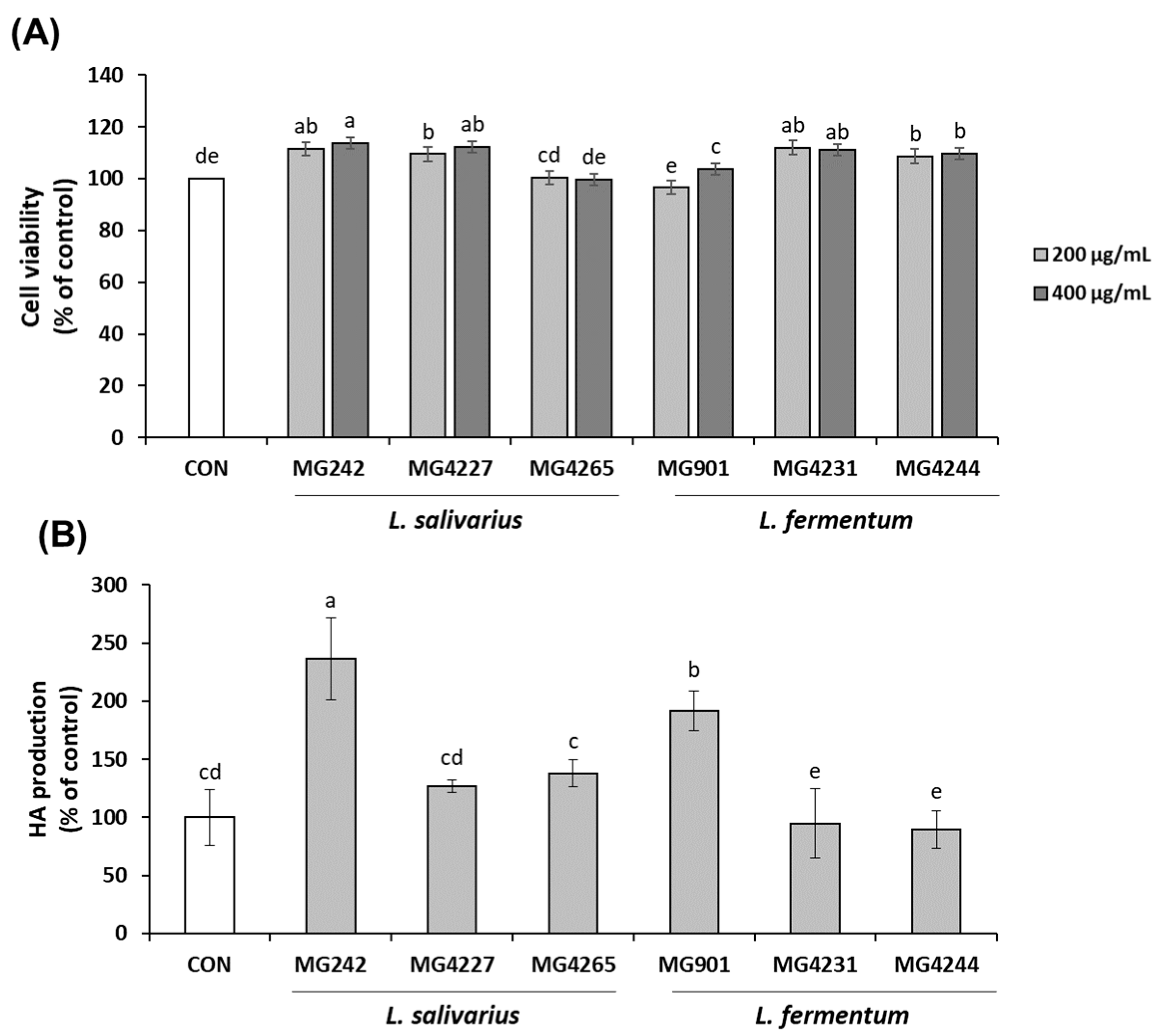
3.1. Effect of CFS Derived from L. salivarius and L. fermentum on Cell Viability and Hyaluronic Acid Production in HaCaT Keratinocytes

Various metabolites produced by probiotics have been retrieved from CFS [21]. An MTT assay was used to confirm the viability of HaCaT keratinocytes after treatment with CFS. CFS derived from L. salivarius and L. fermentum (200 and 400 μg/mL, respectively) showed no cytotoxicity against HaCaT keratinocytes (Figure 1A). Thus, further experiments were performed using 400 μg/mL of CFS.
Hyaluronic acid (HA), which is found in the extracellular matrix of epidermal keratinocytes, is the key component essential for skin moisturization. HA is a ubiquitous glycosaminoglycan with the unique ability to bind and retain water molecules [22]. In Figure 1B, HA production by HaCaT keratinocytes treated with CFS derived from L. salivarius and L. fermentum is shown. CFS derived from L. salivarius MG242 and L. fermentum MG901 significantly increased HA production by 2.40- and 1.95-fold, respectively, compared to that observed in the control.

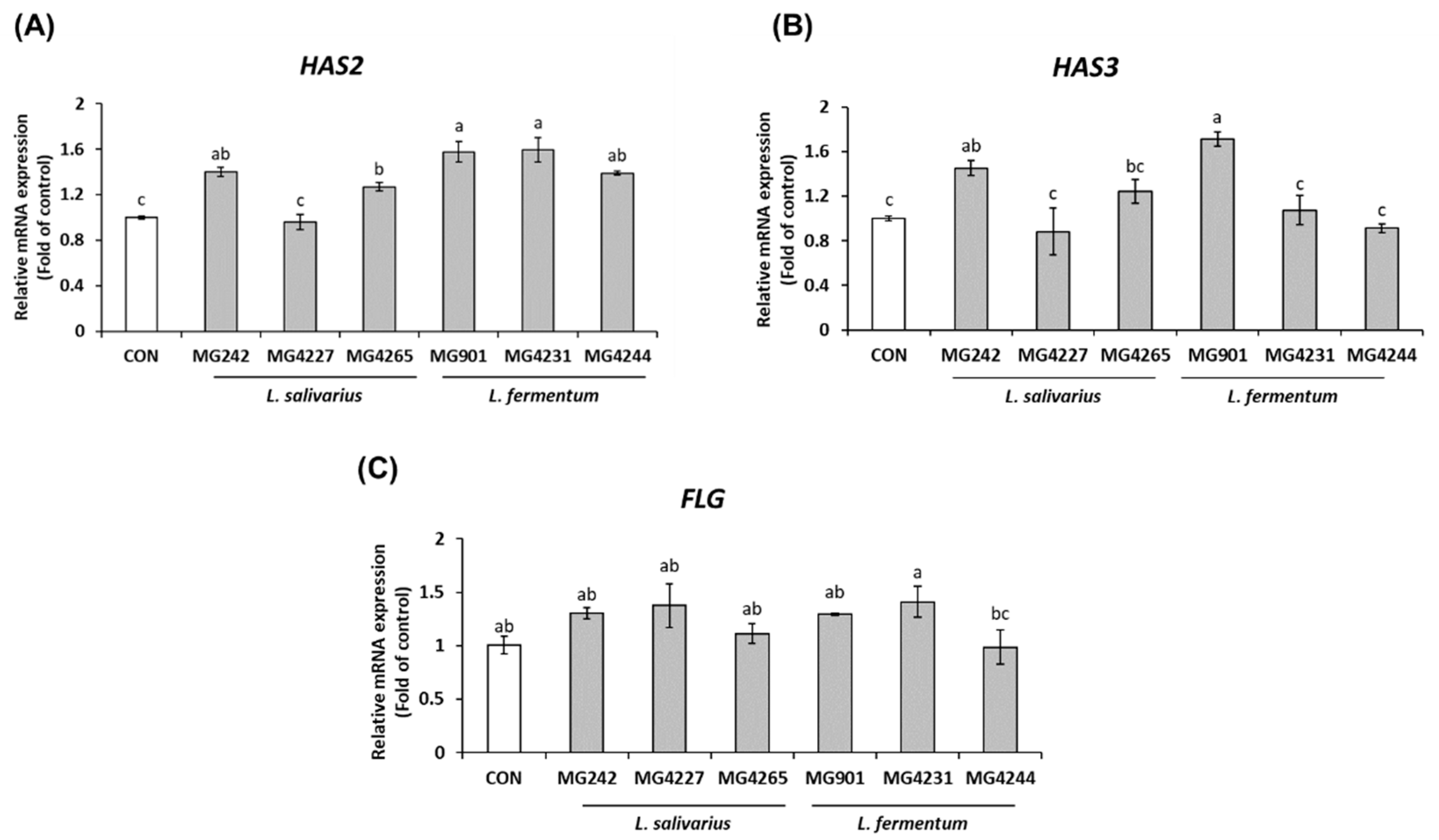
3.2. CFS Derived from L. salivarius and L. fermentum Affects mRNA Expression Associated with Skin Moisturization and Barrier Maintenance in HaCaT Keratinocytes

The polydispersity of HA determines its biological properties, and this polymer is formed by hyaluronic acid synthases (HAS) [23]. HAS comprise three isoenzymes, HAS1, HAS2, and HAS3. Each isozyme shows different properties. They produce HA of different sizes; HAS1 and HAS2 can synthesize HA polymers up to 2 × 10⁶ Da in length, and HAS3 synthesizes polymers with shorter chains than HAS1 and HAS2 [24]. The mRNA expression of HAS2 and HAS3 in MG242 (1.40- and 1.58-fold of control), and MG901 (1.45- and 1.71-fold of control) were higher than those of other CFSs (Figure 2A,B). Most HA was produced by CFSs of L. salivarius MG242 and L. fermentum MG901 (Figure 1). HA is primarily produced by HAS2 and HAS3. Intrinsically, HAS2 and HAS3 are more actively catalyzed than HAS1 [24]. Therefore, the increase in HA production observed upon treatment with CFSs of L. salivarius MG242 and L. fermentum MG901 can be attributed to increased expression of HAS2 and HAS3. The results also demonstrated that the influence of HAS3 on HA production was higher than that of HAS2.
A weakened skin barrier can lead to xerotic cutis. Filaggrin is a natural moisturizing agent that preserves moisture when combined with hydrophilic components in the skin [25]. FLG showed similar mRNA expression (1.40- and 1.58-fold of control) after treatment with all CFS, except with that derived from MG4244 (Figure 2C). Low-molecular-weight hyaluronan polymerized by HAS3 is involved in the production of FLG in the epidermis [26]. Thus, the effect of CFS derived from probiotics on FLG was mediated via modulation of the expression of HAS. In summary, the results confirmed that CFS derived from L. salivarius MG242 and L. fermentum MG901 improved skin hydration and barrier capacity.

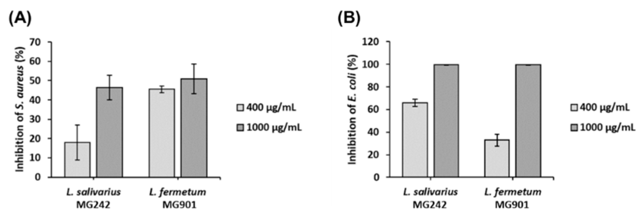
3.3. Antibacterial Effect of CFS Derived from L. salivarius MG242 and L. fermentum MG901

S. aureus, a gram-positive bacterium, is present in the skin, and skin infection by S. aureus can lead to severe conditions, such as abscesses and sepsis [27]. Common skin and soft tissue infections are mainly caused by gram-negative bacteria, such as E. coli [28]. The antibacterial effects of CFSs derived from L. salivarius MG242 and L. fermentum MG901 on S. aureus and E. coli are shown in Figure 3. The amount of S. aureus was reduced by up to 50% upon treatment with CFSs of L. salivarius MG242 and L. fermentum MG901. The CFS of L. salivarius MG242 and L. fermentum MG901 also decreased the amount of E. coli up to 99%. This antibacterial effect of CFS suggests that they can help improve skin health by reducing bacterial infections in the skin.

3.4. Adherence of Probiotics to HT-29 Colorectal Cells

Recently, studies examining the gut microbiome and gut-skin axis have increased. Findings on the gut-skin axis suggest that skin health may be affected by the gut immune system [29]. Some studies have reported that probiotics improve skin health [30]. Adherence of L. salivarius MG242 and L. fermentum MG901 to HT-29 colorectal cells was 85.79 and 82.69%, respectively (Table 2). Other probiotics have been analyzed, with L. fermentum and L. salivarious showing approximately 55 to 80% adherence. Pathogens present in colorectal cells produce inflammatory cytokines, which affect various organs, such as the oral cavity, skin, and respiratory tract. However, probiotics attach to colon cells and form biofilms to inhibit the invasion of pathogens, indirectly affecting skin health [31]. Metabolites produced by probiotics can be delivered directly to the skin through the bloodstream or can affect the skin microbiome [29]. Therefore, MG242 and MG901, which show remarkable adhesion to the intestine, produce metabolites that maintain skin health and protect against the invasion of pathogens by strengthening the skin barrier.

4. Conclusions

In this study, we confirmed the effects on skin of CFS, which contain various metabolites produced from probiotics. The CFS of L. salivarius MG242 and L. fermentum MG901 improved skin moisture and barrier function by modulating mRNA expression of hyaluronic acid synthase and filaggrin in keratinocytes and showed antimicrobial effects against S. aureus and E. coli. In addition, L. salivarius MG242 and L. fermentum MG901, which produced various metabolites with excellent intestinal wall adhesion, were demonstrated to have great potential for improving skin health. However, further research is needed to determine the components of these strains that are beneficial for skin health.

Author Contributions

Conceptualization, C.-H.K.; methodology, J.Y.L., Y.K., J.-I.K. and H.-Y.L.; investigation, J.Y.L., Y.K. and J.-I.K.; resources, C.-H.K.; data curation, J.Y.L. and Y.K.; writing—original draft preparation, J.Y.L.; writing—review and editing, J.Y.L.; visualization, J.Y.L.; supervision, G.-S.M. and C.-H.K.; project administration, Y.K. and C.-H.K.; funding acquisition, G.-S.M. All authors have read and agreed to the published version of the manuscript.

Funding

This research was funded by the Regional Innovation Strategy (RIS) through the National Research Foundation of Korea (NRF), funded by the Ministry of Education (MOE), grant number 1345329680. This research was also supported by the Basic Science Research Program through the National Research Foundation of Korea (NRF), funded by the Ministry of Education (MOE) (No. 2021R1A6A1A03046418).

Institutional Review Board Statement

Not applicable.

Informed Consent Statement

Not applicable.

Data Availability Statement

Data are available in publicly accessible repository/contained within the article.

Conflicts of Interest

The authors declare no conflict of interest.

References

  1. Kanitakis, J. Anatomy, histology and immunohistochemistry of normal human skin. Eur. J. Dermatol. 2002, 12, 390–399. [Google Scholar] [PubMed]
  2. Coates, M.; Blanchard, S.; MacLeod, A.S. Innate antimicrobial immunity in the skin: A protective barrier against bacteria, viruses, and fungi. PLoS Pathog. 2018, 14, e1007353. [Google Scholar] [CrossRef] [PubMed]
  3. Jiang, Y.; Tsoi, L.C.; Billi, A.C.; Ward, N.L.; Harms, P.W.; Zeng, C.; Maverakis, E.; Kahlenberg, J.M.; Gudjonsson, J.E. Cytokinocytes: The diverse contribution of keratinocytes to immune responses in skin. JCI Insight 2020, 5, e142067. [Google Scholar] [CrossRef] [PubMed]
  4. Bitschar, K.; Wolz, C.; Krismer, B.; Peschel, A.; Schittek, B. Keratinocytes as sensors and central players in the immune defense against Staphylococcus aureus in the skin. J. Dermatol. Sci. 2017, 87, 215–220. [Google Scholar] [CrossRef] [Green Version]
  5. Kakehi, K.; Kinoshita, M.; Yasueda, S.-i. Hyaluronic acid: Separation and biological implications. J. Chromatogr. B 2003, 797, 347–355. [Google Scholar] [CrossRef]
  6. Sanders, M.E. Probiotics: Definition, sources, selection, and uses. Clin. Infect. Dis. 2008, 46, S58–S61. [Google Scholar] [CrossRef] [Green Version]
  7. Kang, M.-S.; Lim, H.-S.; Oh, J.-S.; Lim, Y.-j.; Wuertz-Kozak, K.; Harro, J.M.; Shirtliff, M.E.; Achermann, Y. Antimicrobial activity of Lactobacillus salivarius and Lactobacillus fermentum against Staphylococcus aureus. Pathog. Dis. 2017, 75, ftx009. [Google Scholar] [CrossRef]
  8. Higuchi, T.; Suzuki, N.; Nakaya, S.; Omagari, S.; Yoneda, M.; Hanioka, T.; Hirofuji, T. Effects of Lactobacillus salivarius WB21 combined with green tea catechins on dental caries, periodontitis, and oral malodor. Arch. Oral Biol. 2019, 98, 243–247. [Google Scholar] [CrossRef]
  9. Niccoli, A.A.; Artesi, A.L.; Candio, F.; Ceccarelli, S.; Cozzali, R.; Ferraro, L.; Fiumana, D.; Mencacci, M.; Morlupo, M.; Pazzelli, P. Preliminary results on clinical effects of probiotic Lactobacillus salivarius LS01 in children affected by atopic dermatitis. J. Clin. Gastroenterol. 2014, 48, S34–S36. [Google Scholar] [CrossRef]
  10. Rodríguez-Nogales, A.; Algieri, F.; Garrido-Mesa, J.; Vezza, T.; Utrilla, M.P.; Chueca, N.; Garcia, F.; Olivares, M.; Rodríguez-Cabezas, M.E.; Gálvez, J. Differential intestinal anti-inflammatory effects of Lactobacillus fermentum and Lactobacillus salivarius in DSS mouse colitis: Impact on microRNAs expression and microbiota composition. Mol. Nutr. Food Res. 2017, 61, 1700144. [Google Scholar] [CrossRef]
  11. Kim, S.; Choi, S.-I.; Jang, M.; Jeong, Y.; Kang, C.-H.; Kim, G.-H. Anti-adipogenic effect of Lactobacillus fermentum MG4231 and MG4244 through AMPK pathway in 3T3-L1 preadipocytes. Food Sci. Biotechnol. 2020, 29, 1541–1551. [Google Scholar] [CrossRef] [PubMed]
  12. Moradi, M.; Kousheh, S.A.; Almasi, H.; Alizadeh, A.; Guimarães, J.T.; Yılmaz, N.; Lotfi, A. Postbiotics produced by lactic acid bacteria: The next frontier in food safety. Compr. Rev. Food Sci. Food Saf. 2020, 19, 3390–3415. [Google Scholar] [CrossRef] [PubMed]
  13. Kim, H.; Kim, Y.; Kang, C.-H. In vivo confirmation of the antimicrobial effect of probiotic candidates against Gardnerella vaginalis. Microorganisms 2021, 9, 1690. [Google Scholar] [CrossRef] [PubMed]
  14. Kang, C.-H.; Kim, J.-S.; Park, H.M.; Kim, S.; Paek, N.-S. Antioxidant activity and short-chain fatty acid production of lactic acid bacteria isolated from Korean individuals and fermented foods. 3 Biotech 2021, 11, 217. [Google Scholar] [CrossRef]
  15. Żółkiewicz, J.; Marzec, A.; Ruszczyński, M.; Feleszko, W. Postbiotics—a step beyond pre-and probiotics. Nutrients 2020, 12, 2189. [Google Scholar] [CrossRef]
  16. Kim, D.-W.; Chae, S.-J.; Kim, Y.-H.; Jung, H.-J.; Lee, S.-D.; Park, J.-C.; Cho, K.-H.; Sa, S.-J.; Kim, I.-C.; Kim, I.-H. Effects of prebiotics and probiotics on swine intestinal microflora and fermentation products in vitro fermentation. Korean J. Microbiol. 2013, 49, 24–29. [Google Scholar] [CrossRef] [Green Version]
  17. Ali, S.M.; Yosipovitch, G. Skin pH: From basic science to basic skin care. Acta Derm. Vener. 2013, 93, 261–269. [Google Scholar] [CrossRef] [Green Version]
  18. Lee, J.Y.; Kang, C.-H. Probiotics alleviate oxidative stress in H2O2-exposed hepatocytes and t-BHP-induced C57BL/6 mice. Microorganisms 2022, 10, 234. [Google Scholar] [CrossRef]
  19. Kim, S.-G.; Lee, Y.-D.; Park, J.-H.; Moon, G.-S. Synergistic inhibition by bacteriocin and bacteriophage against Staphylococcus aureus. Food Sci. Anim. Resour. 2019, 39, 1015. [Google Scholar] [CrossRef] [Green Version]
  20. Kim, S.; Lee, J.Y.; Jeong, Y.; Kang, C.-H. Antioxidant activity and probiotic properties of lactic acid bacteria. Fermentation 2022, 8, 29. [Google Scholar] [CrossRef]
  21. Parolin, C.; Abruzzo, A.; Giordani, B.; Oliver, J.C.; Marangoni, A.; Luppi, B.; Vitali, B. Anti-candida activity of hyaluronic acid combined with Lactobacillus crispatus lyophilised supernatant: A new antifungal strategy. Antibiotics 2021, 10, 628. [Google Scholar] [CrossRef] [PubMed]
  22. Papakonstantinou, E.; Roth, M.; Karakiulakis, G. Hyaluronic acid: A key molecule in skin aging. Derm.-Endocrinol. 2012, 4, 253–258. [Google Scholar] [CrossRef] [PubMed] [Green Version]
  23. Agarwal, G.; KV, K.; Prasad, S.B.; Bhaduri, A.; Jayaraman, G. Biosynthesis of hyaluronic acid polymer: Dissecting the role of sub structural elements of hyaluronan synthase. Sci. Rep. 2019, 9, 12510. [Google Scholar] [CrossRef] [PubMed] [Green Version]
  24. Spicer, A.P.; McDonald, J. Eukaryotic Hyaluronan Synthases. Available online: http://www.glycoforum.gr.jp/science/hyaluronan/HA07/HA07E.html (accessed on 20 May 2022).
  25. Kim, Y.; Lim, K.-M. Skin barrier dysfunction and filaggrin. Arch. Pharm. Res. 2021, 44, 36–48. [Google Scholar] [CrossRef] [PubMed]
  26. Hashimoto, M.; Maeda, K. New functions of low-molecular-weight hyaluronic acid on epidermis filaggrin production and degradation. Cosmetics 2021, 8, 118. [Google Scholar] [CrossRef]
  27. Ryu, S.; Song, P.I.; Seo, C.H.; Cheong, H.; Park, Y. Colonization and infection of the skin by S. aureus: Immune system evasion and the response to cationic antimicrobial peptides. Int. J. Mol. Sci. 2014, 15, 8753–8772. [Google Scholar] [CrossRef] [Green Version]
  28. Pfalzgraff, A.; Brandenburg, K.; Weindl, G. Antimicrobial peptides and their therapeutic potential for bacterial skin infections and wounds. Front. Pharmacol. 2018, 9, 281. [Google Scholar] [CrossRef]
  29. Pessemier, B.D.; Grine, L.; Debaere, M.; Maes, A.; Paetzold, B.; Callewaert, C. Gut–skin axis: Current knowledge of the interrelationship between microbial dysbiosis and skin conditions. Microorganisms 2021, 9, 353. [Google Scholar] [CrossRef]
  30. Sinha, S.; Lin, G.; Ferenczi, K. The skin microbiome and the gut-skin axis. Clin. Dermatol. 2021, 39, 829–839. [Google Scholar] [CrossRef]
  31. Singh, Y.; Ahmad, J.; Musarrat, J.; Ehtesham, N.Z.; Hasnain, S.E. Emerging importance of holobionts in evolution and in probiotics. Gut Pathog. 2013, 5, 12. [Google Scholar] [CrossRef] [Green Version]
Figure 1. Effect of CFS derived from probiotic bacterial strains on (A) cell viability, and (B) hyaluronic acid (HA) production in HaCaT keratinocytes. Data are represented as mean ± SD (n = 3). Statistical significance is indicated by different letters over the column (p < 0.05).
Fermentation 08 00332 g001
Figure 2. Effect of CFS derived from probiotics on (A) HAS2, (B) HAS3, and (C) FLG mRNA expression in HaCaT keratinocytes. Data are shown as mean ± SD (n = 3). Statistical significance is indicated by different letters on the column (p < 0.05).
Fermentation 08 00332 g002
Figure 3. Effect of CFS derived from L. salivarius MG242 and L. fermentum MG901 on the growth of (A) S. aureus and (B) E. coli in the media. Data are shown as mean ± SD (n = 3).
Fermentation 08 00332 g003
Table 1. Primer sequences of human origin used for qRT-PCR.
Gene 1PrimerSequence (5′→3′)
HAS2ForwardATTACCCAGTCCTGGCTTCG
ReverseCCTGTGGAAGACTCAGCAGAA
HAS3ForwardTGTCCAGATCCTCAACAAGTACGA
ReverseCTGGAGGAGGCTGTTGC
FLGForwardGGCTAAGTGAAAGACTTGAAGAGA
ReverseAATAGACTATCAGTGGTGTCATAGG
GAPDHForwardGTCTTCACCACCATGGAGAA
ReverseAGGAGGCATTGCTGATGAT
1HAS2, hyaluronan synthase 2; HAS3, hyaluronan synthase 3; FLG, filaggrin; GAPDH, glyceraldehyde 3-phosphate dehydrogenase.
Table 2. Adhesion of L. salivarius MG242 and L. fermentum MG901 to HT-29 colorectal cells.
StrainsInitial Counts
(CFU/mL)
Adhesion Counts
(CFU/mL)
Adherence
(%)
L. salivarius MG2428.54 ± 0.057.41 ± 0.0785.79 ± 1.55%
L. fermentum MG9018.62 ± 0.017.15 ± 0.0282.69 ± 0.41%
Data are represented as mean ± SD (n = 3).
Publisher’s Note: MDPI stays neutral with regard to jurisdictional claims in published maps and institutional affiliations.

