1. Introduction
Among tangible cultural heritage, wooden artworks are very common due to the availability and workability of this material and its suitability for generating tools or decorative elements.
Wood conservation represents an open task for scientists and conservators to work synergistically to properly solve a specific issue. An advanced physico-chemical approach can be very helpful in the evaluation of the conservation state [
1,
2,
3,
4,
5], as well as to formulate new materials used in restoration protocols that, nowadays, are tailored to specific artworks [
6,
7,
8,
9,
10,
11,
12,
13].
Knowledge of the physico-chemical state of the object, and particularly of any visible alteration products that form during display or in storage environments, is also essential to preventive conservation.
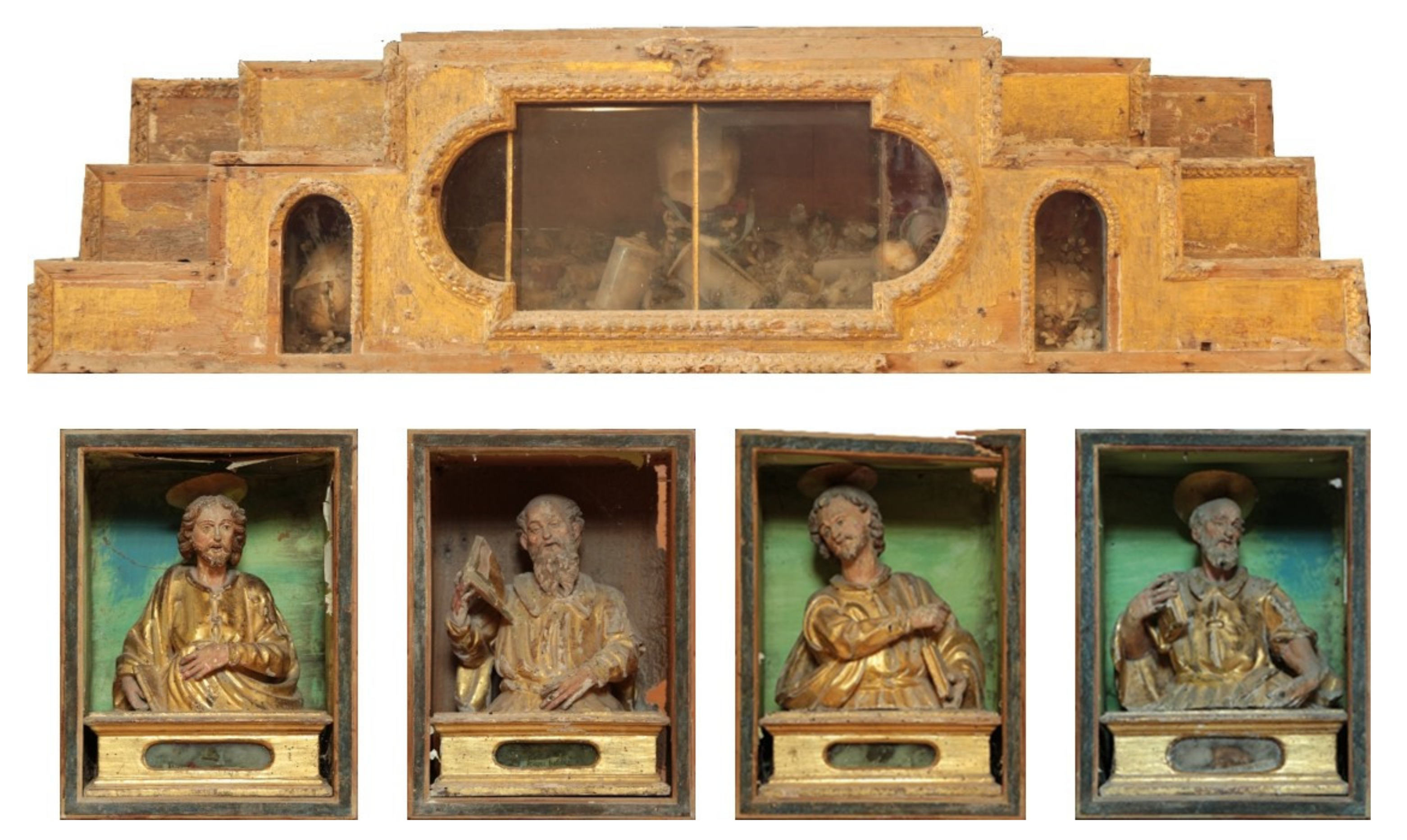
This article is about the restoration work of a XVII century
predella reliquary and of four reliquary busts that date back to the XVII century from an unknown artist (
Figure 1). These goods belong to the church of “Madre Maria SS. Assunta”, in Polizzi Generosa in Sicily (Italy).
The exact year of the predella reliquary’s completion is not known. However, the priest Giovanni Malatacca prepared a detailed drawing showing the complete reliquary setup, including this predella, before his death in 1722. Thus, it is likely this artwork was completed no later than the end of the 1600s. The work is a Baroque ornament sumptuously carved with flowers, leaves and repetitive geometric patterns, typical of this period.
With respect to the four busts, their creation dates, their artists and their original positions are unknown. The four busts are also contained in some wooden shrines. According to their technique with which they were made, these shrines were probably realized for a larger structure, such as, for example, a wall reliquary, into which every shrine was a kind of moving drawer.
However, they should be dated back to the XVII century, which is the same period of predella reliquary.
Here, we report the identification of wood species, as well as a detailed view of the surface treatment and materials used for decoration. Electron microscopy with elemental mapping (energy-dispersive X-ray; EDX) will be used for the investigation of the layered structure in the gilded surface while hyperspectral imaging will provide an objective view of the conservation state and execution technique, providing the necessary information for a conservation action and for a correct historical interpretation of this artwork. Microscopy is a powerful tool in cultural heritage and historical studies, allowing for investigation of minute features [
14]. In addition, image processing software was employed for the enhancement of historical drawings and engravings [
15].
Moreover, we investigate the application of an advanced nanocomposite glue system based on Halloysite nanotubes and Funori. Both components can be considered sustainable materials, bringing a novel approach to the conservation protocol. Halloysite is a nanotubular clay with the chemical structure of kaolinite and a rolled-up morphology that is considered an emerging material in a wide range of application [
16,
17,
18,
19,
20,
21,
22,
23,
24,
25]. It should be noted that halloysite exhibits low toxicity as proved by in vitro and in vivo tests [
26,
27]. The characteristic sizes are 20–100 nm for diameter and up to 2 µm for the length and depend on the deposit [
28,
29]. The anisotropic shape typically enhances the mechanical performances of bionanocomposites and it can also function as a smart container for bioactive species [
30,
31,
32,
33,
34]. Funori is a polysaccharide mucilage from algae, extracted from seaweed, composed primarily of galactose, used as a weak water-soluble adhesive [
35]. Although the adhesive strength of funori is far below that for synthetic adhesives, its use is justified in conservation protocols due to the reversibility of the application. We believe that the combination of the polysaccharide and halloysite nanotubes can show synergistic effects in mechanical materials, as well as glue composite materials, similar to other nanocomposite systems [
36,
37]. With this in mind, tensile measurements have been carried out to evaluate the best biopolymer/halloysite nanotubes ratio based on the adhesive strength. It should also be noted that natural product-based binders, such as polysaccharides, could be subjected to biological attack; therefore, the addition of hollow nanotubular fillers can be further evaluated for biocide vehiculation with slow release.
2. Materials and Methods
Halloysite nanotubes were purchased from Sigma-Aldrich (Italy). Funori was obtained from CTS srl (Italy).
Tensile Analysis: Tensile properties of glued wood samples were determined by means of a DMA Q800 instrument (TA Instruments, Milan, Italy). Tensile tests were performed on rectangular samples obtained by gluing two identical pieces (length × width × thickness, ca. 10 × 5 × 3 mm; glued surface: 5 × 3 mm) under a stress ramp of 1 MPa min−1 at 25.0 ± 0.5 °C. We determined the stress at which the material fractures (σr) and the corresponding maximum deformation from the stress vs. strain curves. The reproducibility was assessed by repeating the experiment at least three times.
Identification of wooden species: wood species were identified by means of a dichotomous keys method, by observing the microscopic anatomical features of the samples using a Leica DMLM stereoscopic microscope equipped with a Dinolite DinoEye camera using Dinoscope software. Wood sections were prepared by cutting wood slides directly from the artworks by hand, using a razor blade after the surface had been softened by wetting (see figure in supporting information). This was carried out in order to minimize the amount of material required for identification.
SEM-EDX: Scanning electron microscopy (SEM) was performed using an Auriga CrossBeam microscope (Carl Zeiss, Jena, Germany), as described elsewhere [
14]. The specimens were coated with ca. 20 nm carbon layer using a Quorum Q150T ES sputter-coater. SEM images were obtained in secondary electron imaging mode (20 kV, 700 pA). For energy-dispersive X-ray (EDX) spectroscopy data and elemental surface mapping, an INCA X-Max EDS-spectrometer (Oxford Instruments, High Wycombe, UK) was used.
Hyperspectral images: Bright-field reflected light microscopy images and reflected light spectra in visible-near infrared range were obtained using an Olympus BX51 (Olympus) upright microscope equipped with a 150 W Fibre-Lite DC-950 halogen light source (Dolan-Jener) and a ProScan III motorized stage (JH Technologies, Fremont, CA, US) [
38]. Optical images were obtained using Exponent 7 (Dage-MTI) image acquisition software [
39]. Spectra were recorded using a Specim V10E spectrometer (Spectral Imaging LTD) and a Pixelfy.usb CCD camera (PCO) in 400–1000 nm range. Hyperspectral data were analyzed using ENVI v. 4.8 software (Harris Geospatial Solutions, Broomfield, CO, USA) [
40]. All HSI data obtained were corrected for the spectral contribution of the light source.
Preparation of nanocomposite glue: The funori solution was prepared by adding ca. 40 g of Funori to 500 g of water and was kept at 80 °C under magnetic stirring for 2 h. A yellowish viscous solution was obtained that was vacuum filtered through a 0.45 µm cellulosic filter. The final funori concentration in the stock solution was 7.3 ± 0.2 wt% (estimated from gravimetric method after solvent evaporation at 120 °C).
The funori/halloysite nanotube (HNT) composite glue was prepared by adding varying amounts of HNTs to 10 g of funori stock solution. The glue composition is expressed in terms of halloysite mass percentage referring to the funori + halloysite total dried mass.
3. Results and Discussion
This work reports on the characterization of the XVII century predella reliquary and four reliquary busts. Firstly, the identification of wood taxon and the finishing layers are reported. The rigorous physico-chemical characterization of the artefacts helps to define the degradation level, design the conservation protocol, but also contributes to the understanding of the early technologies. Secondly, the conservation action, with a particular focus on the use of an advanced nanocomposite glue that was designed in our laboratories, is described.
3.1. Identification of the Wood Species and Microscopy Analysis
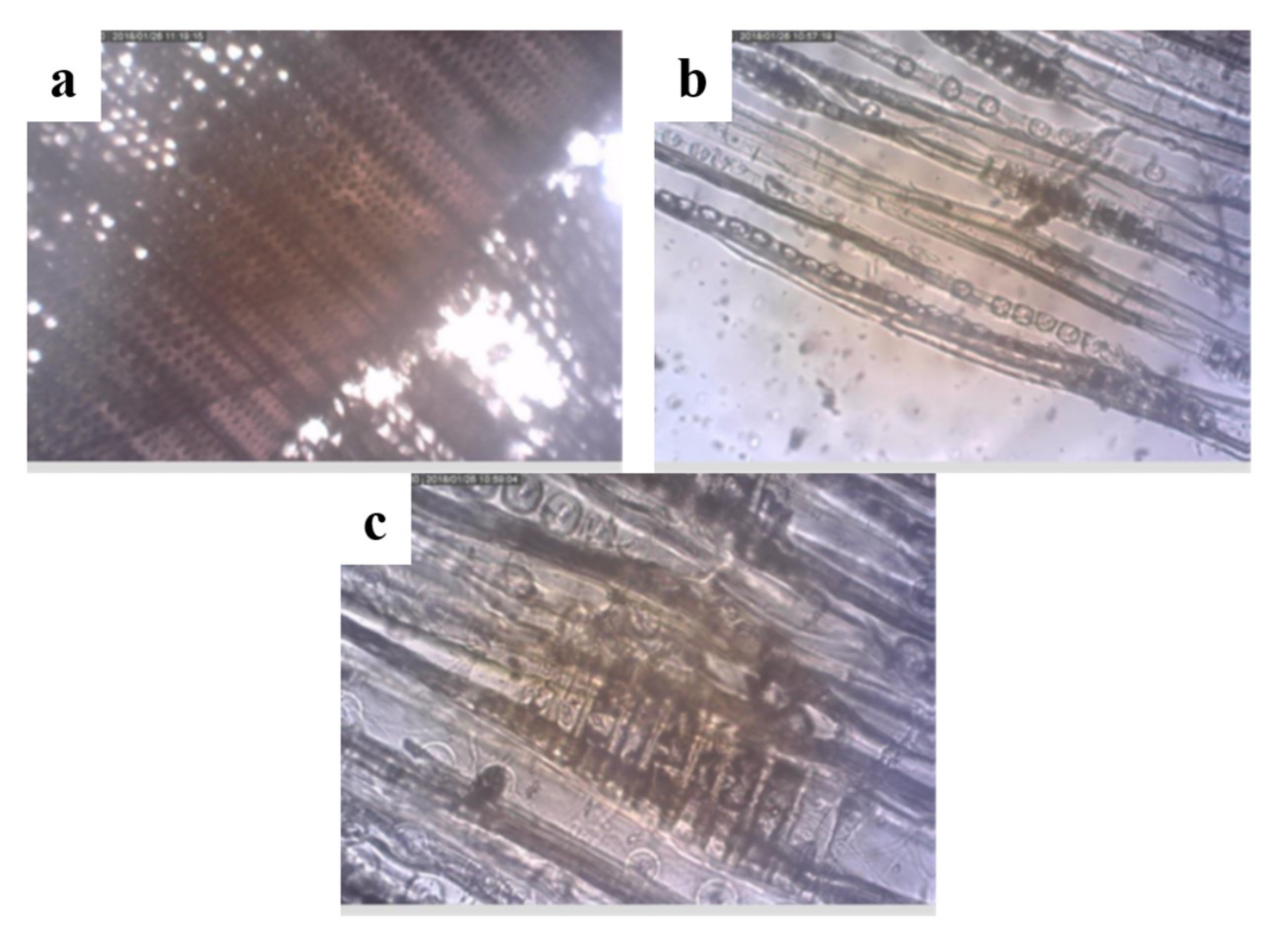
The external predella wooden sample was observed by optical microscopy (
Figure 2). The images indicate a coniferous wood without resin canals. Tangential and radial sections highlight that the tracheid walls have no spiral thickenings. The rays are homogeneous with small pits in the cross-fields, and are without tracheids. Based on the observed characteristics, one can identify the wood species belonging to the Abies genus. According to the literature, it is impossible to distinguish between the different species of this genus [
41]. The identification of this genus could indicate the use of the species
Abies nebrodensis. Nowadays, this is classified as critically endangered by the International Union for Conservation of Nature, but was widespread in Sicily in the past as an endemic species [
42].
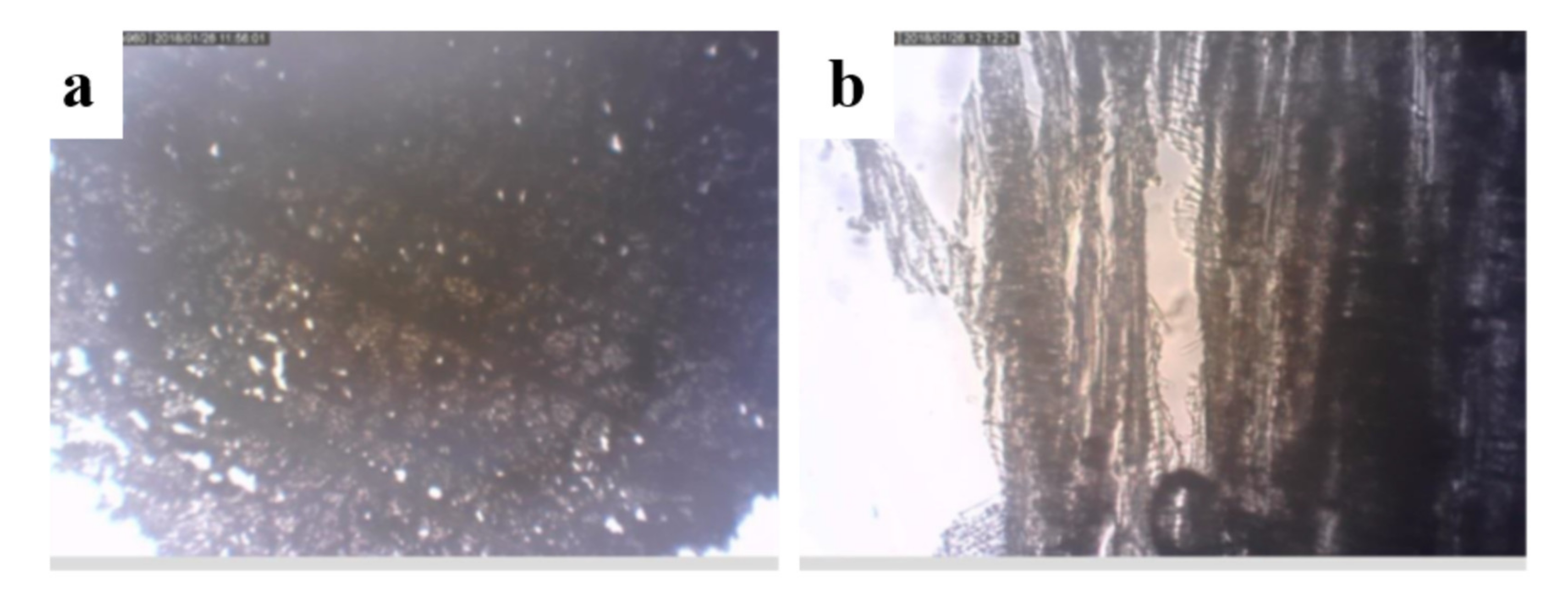
The little frame structure is a hardwood with diffuse porosity and multiseriate homogeneous rays (
Figure 3), two to four cells wide, with vessels showing spiral thickenings. Rays flare along growth ring boundaries. It can be concluded that it is lime wood, specifically Tilia cordata Mill.
As the busts are fully gilded, only tangential sections have been observed due to the sampling. The observable features—spiral thickening of the vessels, mutiseriate homogeneous rays—further indicate that the wooden species is likely to be lime wood.
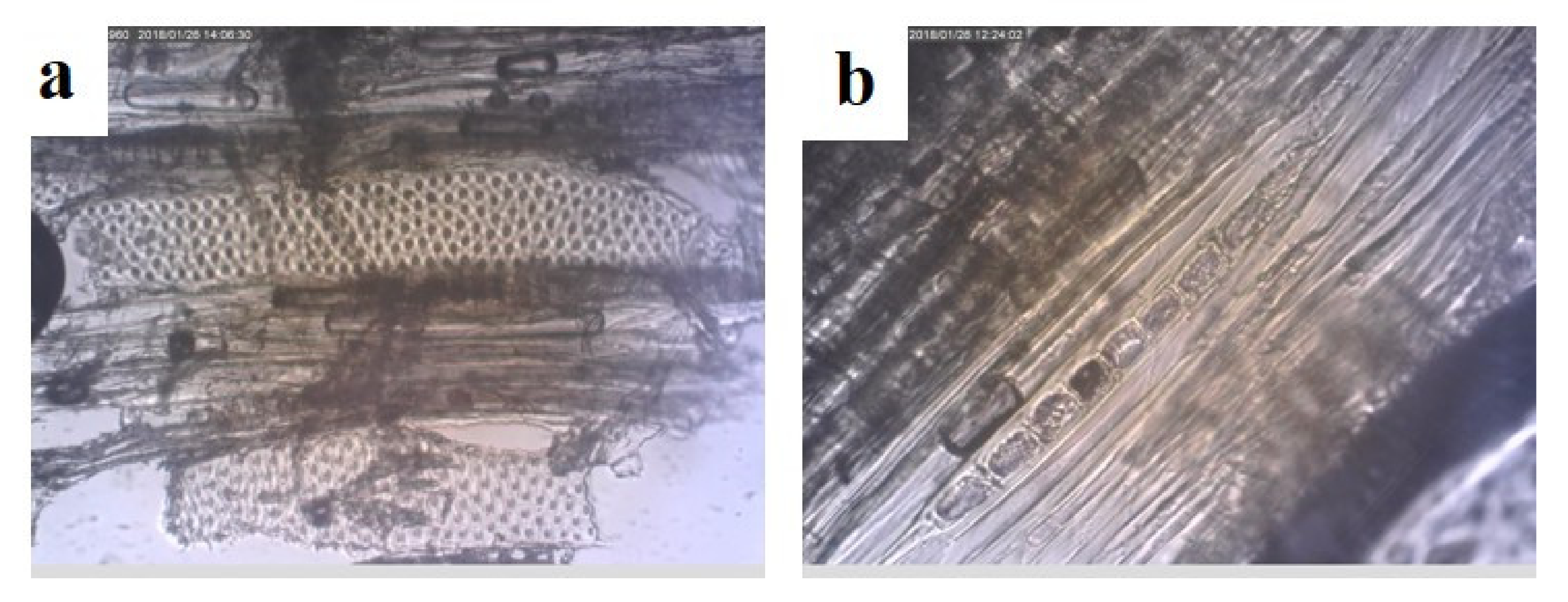
The bases of the busts consist of different wood species. Microscopic observation of tangential sections, the only available for sampling, revealed homogeneous uniseriate rays, vessels with simple perforation and without spiral thickenings (
Figure 4). According to these features, the most likely wood species is poplar,
Populus nigra L.
Mixing several wood species in a single work is not peculiar. In fact, the carvers knew very well the materials they used and the selection was based on the physico-chemical characteristics. For example, lime wood, having a fine texture and a low degree of hardness, was more suitable for carving as an excellent degree of detail could be achieved. Poplar has a strong tendency to chip, but being available at low cost, it is more suitable for parts that do not require carving.
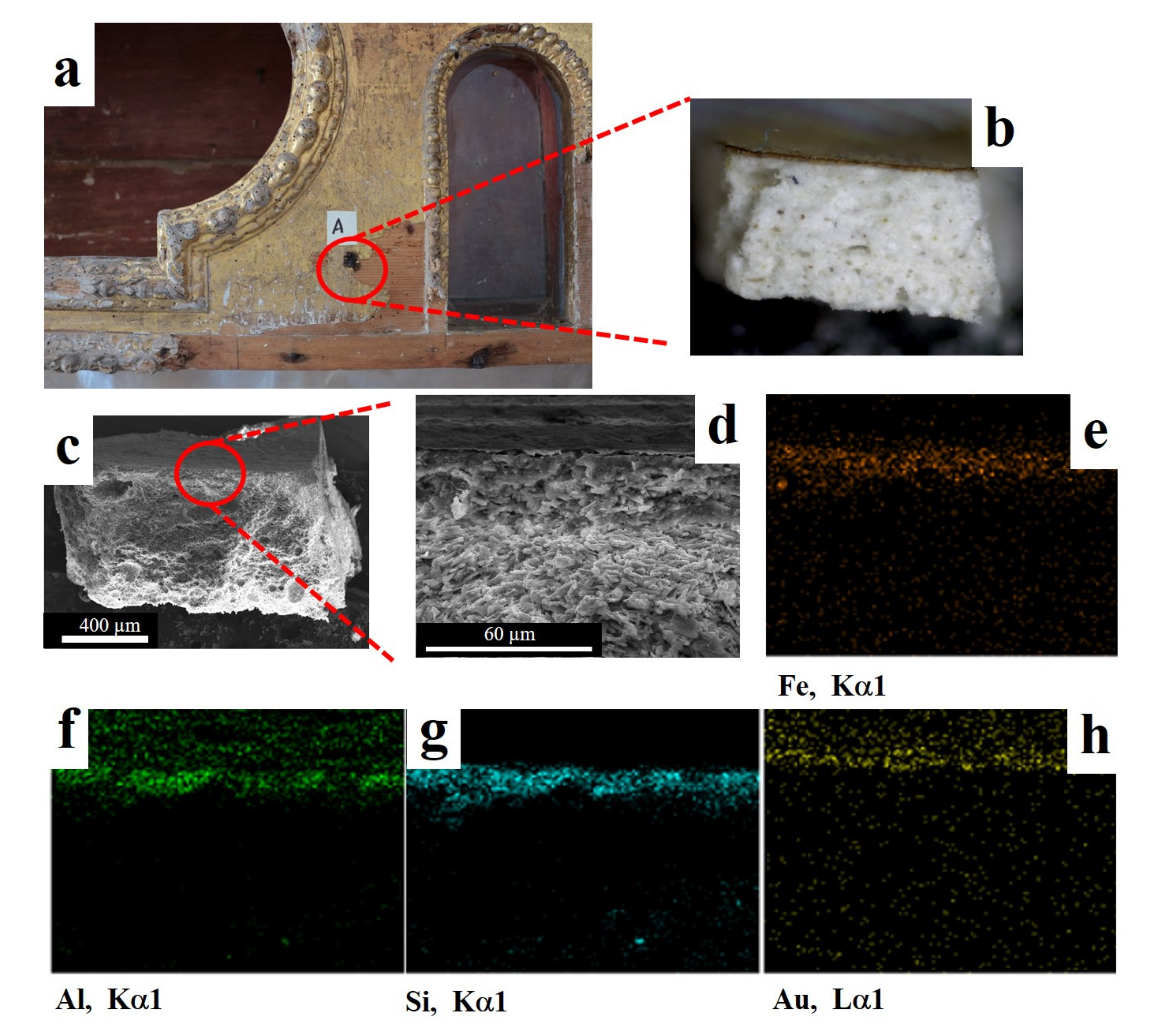
Concerning the preparation layer and decorative surface, from the optical micrographs one can already see evidence of red bole (a soft fine clay typically with a reddish-brown color also used as a pigment) layers appearing below a gilded surface. To shed more light on this aspect, SEM imaging and EDX mapping have been carried out on the
predella reliquary (
Figure 5). The elemental analysis clearly identified the gold layer of ca. 7 µm (
Figure 5d,h). It should be noted that modern gold leaf is very thin (below 200 nm) [
43] and therefore we might conclude that this is an original gilded surface. The preparation layer located between the gold foil and the wood has a thickness of ca. 40 µm and it is composed of O (34%), Si (23%), Fe (17%), Al (16%) and Ca (10%). The obtained composition is consistent with red bole clay [
44]. The use of clay in the preparation layer to paste a gold leaf was largely used by Italian artists and it is also reported for gilded wooden artworks from Giotto [
43].
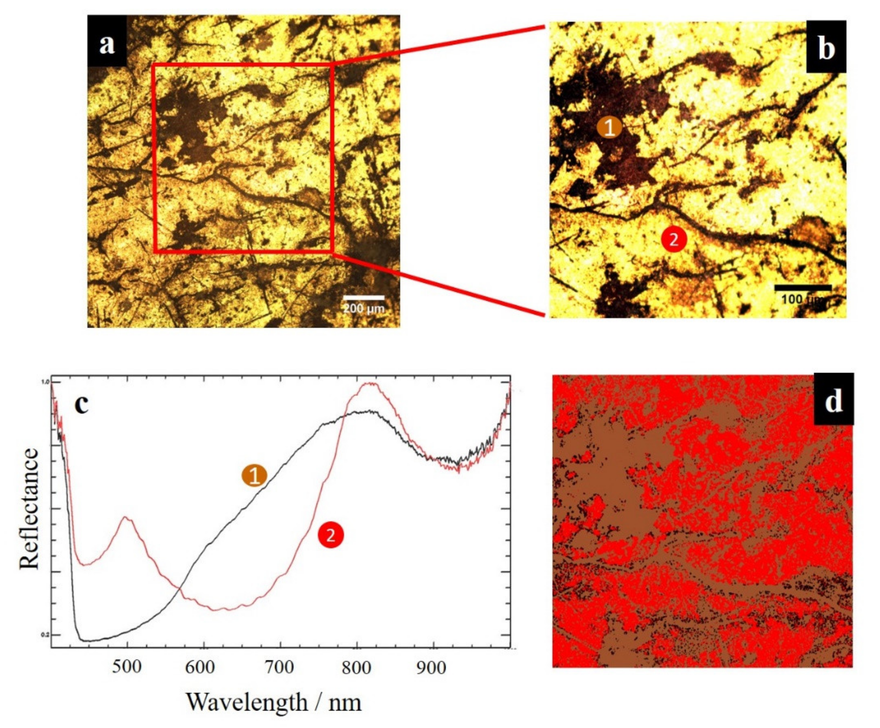
The decorative surface was also imaged by means of hyperspectral microscopy. This methodology is based on full spectrum collection from the specimen surface, providing more detailed chemical information on the surface composition. The reflected light microscopy images and spectra are shown in
Figure 6. The spectra obtained from the “lacuna” and the gilded surface are very different. In particular, the surface plasmon resonance (SPR) peaks at the characteristic wavelengths for gold thin layers are evidenced [
45]. On this basis, the reflected light hyperspectral mapping can be used as a valuable tool to clearly identify the gold lacuna on an analytical basis. Moreover, as the surface plasmon resonance peaks is sensitive to the gold layer thickness, it should be efficient in evidencing modern gold leaf from recent restorations.
3.2. Application of an Advanced Nanocomposite Glue
The restoration was carried out with a conservation approach aimed at restoring the cultural aesthetic harmony. The treatments were based on the principle of minimal intervention not to predominate with respect to “normal” aging. Diagnostic investigations, with the aim to recognizing the woody species of the support, the materials of the preparatory and pictorial layers and the stratigraphy have allowed us to study the cultural asset in its totality. Although a full description is out of the scope of this manuscript, herein we provide a brief outline of the work. The restoration was carried out in several steps: dusting with the aid of microaspirator and brushes with soft bristles; disinfestation of the support with an insecticide based on permethrin; consolidation of the support was carried out with an acrylic resin; restoration of the mechanical capacities of the structure; restoration of wooden inserts using Balsa wood; grouting of erosions with cellulose pulp and cellulose ether; treatment of metal parts, with rust converter. After this, we moved on to the process of the preparatory layers and gilding consolidation, a thermoplastic polymer (poly (2-ethyl-2-Oxazoline), Mw = 500 kg mol−1), applied punctually and sintered with the aid of slight heating. Then, we progressed to cleaning with a water in oil emulsion at neutral pH and to the reconstruction of the missing frames, with two-component epoxy-based putty after making the casts with silicone rubber. We then proceeded to the intermediate painting with retouching paint using watercolors.
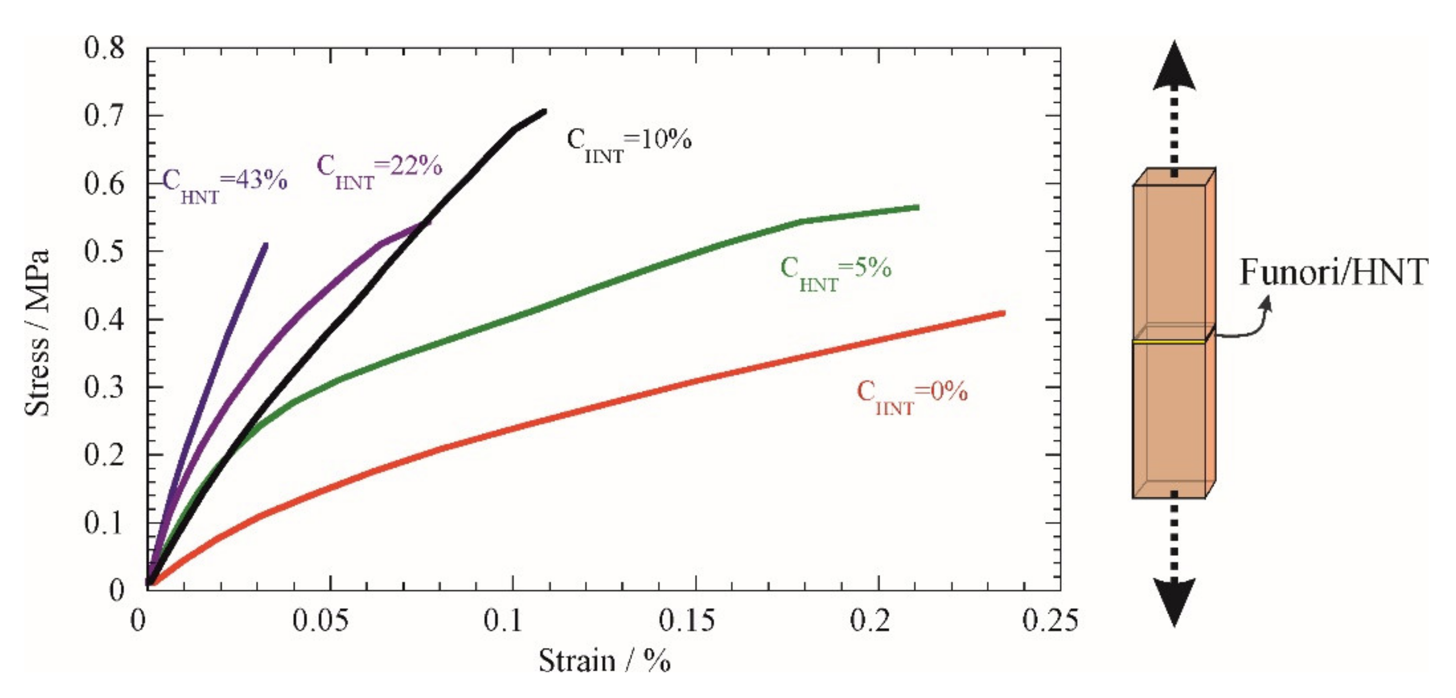
During the restoration it was necessary to fix some fragments that had become detached. Within this issue, we investigated the possibility of using a weak water-based glue (based on funori) in combination with natural nanotubes (halloysite HNTs). Therefore, the adhesion was tested by measuring the stress vs. strain curves of glued wooden samples (
Figure 7). A systematic trend was observed by increasing the percentage of halloysite nanotubes in the HNT/funori mixture (C
HNT) and, in particular, the response appears more rigid to deformation in the geometry that is sketched in
Figure 7.
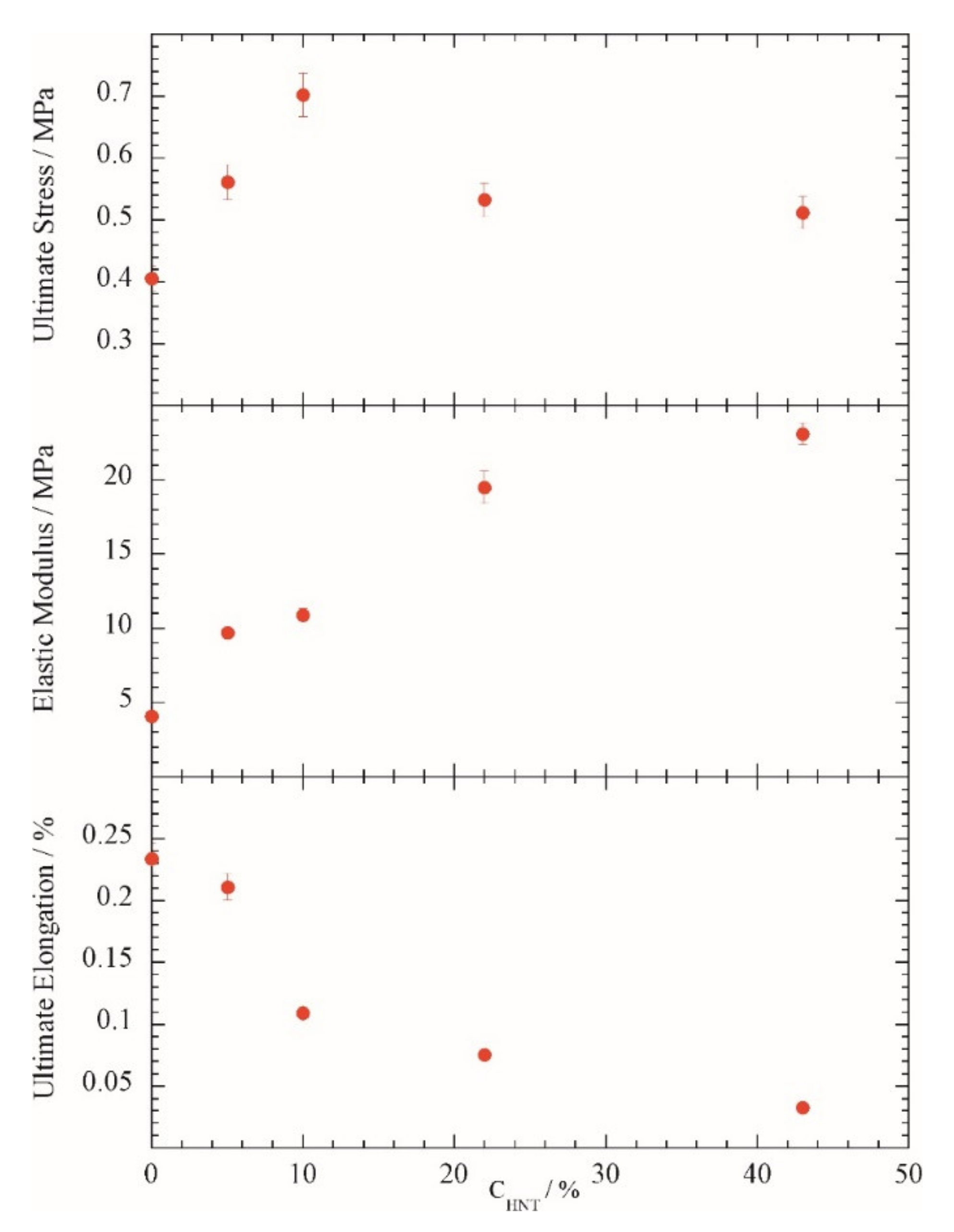
For a quantitative assessment, the ultimate stress and deformation at detaching, as well as the slope of the linear deformation region (Young’s modulus) were calculated and are provided in
Figure 8 for the wood glued with pure funori and funori with increasing percentages of HNTs (C
HNT). The ultimate stress was improved by halloysite nanotubes up to a concentration of 10%; additional nanotubes did not further improve this parameter. This behavior could be explained considering the percolation threshold that predicts clustering effects above 10% of halloysite in polymeric matrices [
32]—namely, at ca. 10% the contact distance between nanotubes is approached and clustering effects might reduce the efficacy as reinforcing agents. Furthermore, the ultimate elongation and elastic modulus reports a monotonic change with the increase in halloysite nanotube contents. In particular, the glue appears more rigid (increase in the modulus and decrease in the ultimate deformation) in the presence of halloysite. Based on the above arguments, we considered the mixture at C
HNT = 10% as the best compromise for the actual application.
4. Conclusions
In this work, we briefly report on the characterization and restoration of a number of wooden artifacts. We used advanced imaging methods for the evaluation of the conservation conditions as a preliminary step toward the restoration protocol and for a comprehensive understanding of the ancient methodology and material selection.
Finally, a nanocomposite glue has been designed by using natural nanotubular particles and a biopolymer extract from algae, funori. We, therefore, report a sustainable approach for the preparation of consolidant materials with a functional nanoarchitecture that can be further developed to target specific needs such as biocide or smart responsive features by filling the nanotubular lumen with proper active molecules.
Author Contributions
Conceptualization, G.L. and G.C.; methodology, G.F.B. and B.G.; investigation, R.F. (Ramil Fakhrullin), F.A. and R.F. (Rawil Fakhrullin); data curation, G.F.B. and B.G.; writing—original draft preparation, G.C., G.L. and B.M.; funding acquisition, G.L. and R.F. (Rawil Fakhrullin). All authors have read and agreed to the published version of the manuscript.
Funding
G.C., G.L. and B.M. thank University of Palermo for financial support. This work was funded by the subsidy allocated to Kazan Federal University for the state assignment in the sphere of scientific activities (0671-2020-0058). Rawil Fakhrullin acknowledges funding by Russian Federation presidential grant MD-2153.2020.3.
Institutional Review Board Statement
Not applicable.
Informed Consent Statement
Not applicable.
Data Availability Statement
Data are available upon request to corresponding authors.
Acknowledgments
We thank Ilvina Safina for technical help with SEM. We thank Giovanni Silvestri as priest of the church of “Madre Maria SS. Assunta” in Polizzi Generosa (Italy).
Conflicts of Interest
The authors declare no conflict of interest.
References
- Cavallaro, G.; Agliolo Gallitto, A.; Lisuzzo, L.; Lazzara, G. Comparative Study of Historical Woods from XIX Century by Thermogravimetry Coupled with FTIR Spectroscopy. Cellulose 2019, 26, 8853–8865. [Google Scholar] [CrossRef]
- Bernabei, M.; Macchioni, N.; Pizzo, B.; Sozzi, L.; Lazzeri, S.; Fiorentino, L.; Pecoraro, E.; Quarta, G.; Calcagnile, L. The Wooden Foundations of Rialto Bridge (Ponte Di Rialto) in Venice: Technological Characterisation and Dating. J. Cult. Herit. 2019, 36, 85–93. [Google Scholar] [CrossRef]
- Babiński, L.; Izdebska-Mucha, D.; Waliszewska, B. Evaluation of the State of Preservation of Waterlogged Archaeological Wood Based on Its Physical Properties: Basic Density vs. Wood Substance Density. J. Archaeol. Sci. 2014, 46, 372–383. [Google Scholar] [CrossRef] [Green Version]
- Čufar, K.; Merela, M.; Erič, M. A Roman Barge in the Ljubljanica River (Slovenia): Wood Identification, Dendrochronological Dating and Wood Preservation Research. J. Archaeol. Sci. 2014, 44, 128–135. [Google Scholar] [CrossRef]
- Guo, J.; Zhang, M.; Liu, J.; Luo, R.; Yan, T.; Yang, T.; Jiang, X.; Dong, M.; Yin, Y. Evaluation of the Deterioration State of Archaeological Wooden Artifacts: A Nondestructive Protocol Based on Direct Analysis in Real Time—Mass Spectrometry (DART-MS) Coupled to Chemometrics. Anal. Chem. 2020, 92, 9908–9915. [Google Scholar] [CrossRef]
- Cavallaro, G.; Lazzara, G.; Milioto, S.; Parisi, F.; Ruisi, F. Nanocomposites Based on Esterified Colophony and Halloysite Clay Nanotubes as Consolidants for Waterlogged Archaeological Woods. Cellulose 2017, 24, 3367–3376. [Google Scholar] [CrossRef]
- Cavallaro, G.; Milioto, S.; Parisi, F.; Lazzara, G. Halloysite Nanotubes Loaded with Calcium Hydroxide: Alkaline Fillers for the Deacidification of Waterlogged Archeological Woods. ACS Appl. Mater. Interfaces 2018, 10, 27355–27364. [Google Scholar] [CrossRef]
- Broda, M.; Dąbek, I.; Dutkiewicz, A.; Dutkiewicz, M.; Popescu, C.-M.; Mazela, B.; Maciejewski, H. Organosilicons of Different Molecular Size and Chemical Structure as Consolidants for Waterlogged Archaeological Wood—A New Reversible and Retreatable Method. Sci. Rep. 2020, 10, 2188. [Google Scholar] [CrossRef] [Green Version]
- Broda, M.; Mazela, B.; Dutkiewicz, A. Organosilicon Compounds with Various Active Groups as Consolidants for the Preservation of Waterlogged Archaeological Wood. J. Cult. Herit. 2019, 35, 123–128. [Google Scholar] [CrossRef]
- Antonelli, F.; Galotta, G.; Sidoti, G.; Zikeli, F.; Nisi, R.; Davidde Petriaggi, B.; Romagnoli, M. Cellulose and Lignin Nano-Scale Consolidants for Waterlogged Archaeological Wood. Front. Chem. 2020, 8, 32. [Google Scholar] [CrossRef]
- Poggi, G.; Toccafondi, N.; Chelazzi, D.; Canton, P.; Giorgi, R.; Baglioni, P. Calcium Hydroxide Nanoparticles from Solvothermal Reaction for the Deacidification of Degraded Waterlogged Wood. J. Colloid Interface Sci. 2016, 473, 1–8. [Google Scholar] [CrossRef] [PubMed]
- Broda, M. Natural Compounds for Wood Protection against Fungi—A Review. Molecules 2020, 25, 3538. [Google Scholar] [CrossRef] [PubMed]
- Lisuzzo, L.; Hueckel, T.; Cavallaro, G.; Sacanna, S.; Lazzara, G. Pickering Emulsions Based on Wax and Halloysite Nanotubes: An Ecofriendly Protocol for the Treatment of Archeological Woods. ACS Appl. Mater. Interfaces 2021, 13, 1651–1661. [Google Scholar] [CrossRef]
- Khramchenkova, R.; Safina, I.; Drobyshev, S.; Batasheva, S.; Nuzhdin, E.; Fakhrullin, R. Advanced Microscopy Techniques for Nanoscale Diagnostic of Cultural Heritage. In Nanotechnologies and Nanomaterials for Diagnostic, Conservation and Restoration of Cultural Heritage; Elsevier: Amsterdam, The Netherlands, 2019; pp. 1–23. ISBN 978-0-12-813910-3. [Google Scholar]
- Aidarova-Volkova, G.N.; Fakhrullin, R.F.; Kees, B. Historical-Architectural Analysis of the Panoramic Image of Kazan by Cornelis de Bruijn. Vestn. Spbsu. Hist. 2020, 65, 566–583. [Google Scholar] [CrossRef]
- Papoulis, D. Halloysite Based Nanocomposites and Photocatalysis: A Review. Appl. Clay Sci. 2019, 168, 164–174. [Google Scholar] [CrossRef]
- Lvov, Y.M.; Shchukin, D.G.; Mohwald, H.; Price, R.R. Halloysite Clay Nanotubes for Controlled Release of Protective Agents. ACS Nano 2008, 2, 814–820. [Google Scholar] [CrossRef]
- Yendluri, R.; Otto, D.P.; De Villiers, M.M.; Vinokurov, V.; Lvov, Y.M. Application of Halloysite Clay Nanotubes as a Pharmaceutical Excipient. Int. J. Pharm. 2017, 521, 267–273. [Google Scholar] [CrossRef]
- Joussein, E.; Petit, S.; Churchman, G.J.; Theng, B.; Righi, D.; Delvaux, B. Halloysite Clay Minerals—A Review. Clay Miner. 2005, 40, 383–426. [Google Scholar] [CrossRef]
- Panchal, A.; Fakhrullina, G.; Fakhrullin, R.; Lvov, Y. Self-Assembly of Clay Nanotubes on Hair Surface for Medical and Cosmetic Formulations. Nanoscale 2018, 10, 18205–18216. [Google Scholar] [CrossRef] [PubMed]
- Cavallaro, G.; Milioto, S.; Lazzara, G. Halloysite Nanotubes: Interfacial Properties and Applications in Cultural Heritage. Langmuir 2020, 36, 3677–3689. [Google Scholar] [CrossRef]
- Zhang, Y.; Bai, L.; Cheng, C.; Zhou, Q.; Zhang, Z.; Wu, Y.; Zhang, H. A Novel Surface Modification Method upon Halloysite Nanotubes: A Desirable Cross-Linking Agent to Construct Hydrogels. Appl. Clay Sci. 2019, 182, 105259. [Google Scholar] [CrossRef]
- Cheng, C.; Gao, Y.; Song, W.; Zhao, Q.; Zhang, H.; Zhang, H. Halloysite Nanotube-Based H2O2-Responsive Drug Delivery System with a Turn on Effect on Fluorescence for Real-Time Monitoring. Chem. Eng. J. 2020, 380, 122474. [Google Scholar] [CrossRef]
- Taroni, T.; Cauteruccio, S.; Vago, R.; Franchi, S.; Barbero, N.; Licandro, E.; Ardizzone, S.; Meroni, D. Thiahelicene-Grafted Halloysite Nanotubes: Characterization, Biological Studies and PH Triggered Release. Appl. Surf. Sci. 2020, 520, 146351. [Google Scholar] [CrossRef]
- Barman, M.; Mahmood, S.; Augustine, R.; Hasan, A.; Thomas, S.; Ghosal, K. Natural Halloysite Nanotubes /Chitosan Based Bio-Nanocomposite for Delivering Norfloxacin, an Anti-Microbial Agent in Sustained Release Manner. Int. J. Biol. Macromol. 2020, 162, 1849–1861. [Google Scholar] [CrossRef] [PubMed]
- Long, Z.; Wu, Y.-P.; Gao, H.-Y.; Zhang, J.; Ou, X.; He, R.-R.; Liu, M. In Vitro and in Vivo Toxicity Evaluation of Halloysite Nanotubes. J. Mater. Chem. B 2018, 6, 7204–7216. [Google Scholar] [CrossRef]
- Wang, X.; Gong, J.; Gui, Z.; Hu, T.; Xu, X. Halloysite Nanotubes-Induced Al Accumulation and Oxidative Damage in Liver of Mice after 30-Day Repeated Oral Administration. Environ. Toxicol. 2018, 33, 623–630. [Google Scholar] [CrossRef] [PubMed]
- Cavallaro, G.; Chiappisi, L.; Pasbakhsh, P.; Gradzielski, M.; Lazzara, G. A Structural Comparison of Halloysite Nanotubes of Different Origin by Small-Angle Neutron Scattering (SANS) and Electric Birefringence. Appl. Clay Sci. 2018, 160, 71–80. [Google Scholar] [CrossRef]
- Pasbakhsh, P.; Churchman, G.J.; Keeling, J.L. Characterisation of Properties of Various Halloysites Relevant to Their Use as Nanotubes and Microfibre Fillers. Appl. Clay Sci. 2013, 74, 47–57. [Google Scholar] [CrossRef]
- Bugatti, V.; Brachi, P.; Viscusi, G.; Gorrasi, G. Valorization of Tomato Processing Residues Through the Production of Active Bio-Composites for Packaging Applications. Front. Mater. 2019, 6, 34. [Google Scholar] [CrossRef] [Green Version]
- Gorrasi, G. Dispersion of Halloysite Loaded with Natural Antimicrobials into Pectins: Characterization and Controlled Release Analysis. Carbohydr. Polym. 2015, 127, 47–53. [Google Scholar] [CrossRef] [PubMed]
- Lisuzzo, L.; Cavallaro, G.; Milioto, S.; Lazzara, G. Effects of Halloysite Content on the Thermo-Mechanical Performances of Composite Bioplastics. Appl. Clay Sci. 2020, 185, 105416. [Google Scholar] [CrossRef] [Green Version]
- Lisuzzo, L.; Cavallaro, G.; Milioto, S.; Lazzara, G. Layered Composite Based on Halloysite and Natural Polymers: A Carrier for the PH Controlled Release of Drugs. New J. Chem. 2019, 43, 10887–10893. [Google Scholar] [CrossRef] [Green Version]
- Tarasova, E.; Naumenko, E.; Rozhina, E.; Akhatova, F.; Fakhrullin, R. Cytocompatibility and Uptake of Polycations-Modified Halloysite Clay Nanotubes. Appl. Clay Sci. 2019, 169, 21–30. [Google Scholar] [CrossRef]
- Swider, J.R.; Smith, M. Funori: Overview of a 300-Year-Old Consolidant. J. Am. Inst. Conserv. 2005, 44, 117–126. [Google Scholar] [CrossRef]
- Zhao, X.; Zhou, C.; Liu, M. Self-Assembled Structures of Halloysite Nanotubes: Towards the Development of High-Performance Biomedical Materials. J. Mater. Chem. B 2020, 8, 838–851. [Google Scholar] [CrossRef]
- Bertolino, V.; Cavallaro, G.; Milioto, S.; Lazzara, G. Polysaccharides/Halloysite Nanotubes for Smart Bionanocomposite Materials. Carbohydr. Polym. 2020, 245, 116502. [Google Scholar] [CrossRef]
- Fakhrullin, R.; Nigamatzyanova, L.; Fakhrullina, G. Dark-Field/Hyperspectral Microscopy for Detecting Nanoscale Particles in Environmental Nanotoxicology Research. Sci. Total Environ. 2021, 772, 145478. [Google Scholar] [CrossRef] [PubMed]
- Nigamatzyanova, L.; Fakhrullin, R. Dark-Field Hyperspectral Microscopy for Label-Free Microplastics and Nanoplastics Detection and Identification in Vivo: A Caenorhabditis Elegans Study. Environ. Pollut. 2021, 271, 116337. [Google Scholar] [CrossRef] [PubMed]
- Akhatova, F.; Danilushkina, A.; Kuku, G.; Saricam, M.; Culha, M.; Fakhrullin, R. Simultaneous Intracellular Detection of Plasmonic and Non-Plasmonic Nanoparticles Using Dark-Field Hyperspectral Microscopy. Bull. Chem. Soc. Jpn. 2018, 91, 1640–1645. [Google Scholar] [CrossRef]
- Schweingruber, F.H. Microscopic Wood Anatomy; Fluck-Wirth: Teufen, Switzerland, 1982. [Google Scholar]
- Rivers, M. European Red List of Trees; IUCN, International Union for Conservation of Nature: Grand, Switzerland, 2019; ISBN 978-2-8317-1985-6. [Google Scholar]
- Eveno, M.; Ravaud, E.; Calligaro, T.; Pichon, L.; Laval, E. The Louvre Crucifix by Giotto—Unveiling the Original Decoration by 2D-XRF, X-Ray Radiography, Emissiography and SEM-EDX Analysis. Herit. Sci. 2014, 2, 17. [Google Scholar] [CrossRef] [Green Version]
- Barata, C.; Rocha, F.; Cruz, A.J.; Andrejkovičová, S.; Reguer, S. Synchrotron X-ray Diffraction of Bole Layers from Portuguese Gilded Baroque Retables. Appl. Clay Sci. 2015, 116–117, 39–45. [Google Scholar] [CrossRef]
- Rai, V.N.; Srivastava, A.K.; Mukherjee, C.; Deb, S.K. Surface Enhanced Absorption and Transmission from Dye Coated Gold Nanoparticles in Thin Films. Appl. Opt. 2012, 51, 2606. [Google Scholar] [CrossRef] [PubMed]
| Publisher’s Note: MDPI stays neutral with regard to jurisdictional claims in published maps and institutional affiliations. |
© 2021 by the authors. Licensee MDPI, Basel, Switzerland. This article is an open access article distributed under the terms and conditions of the Creative Commons Attribution (CC BY) license (http://creativecommons.org/licenses/by/4.0/).


 ,
, 


