

An Innovative Analysis of Time Series-Based Detection Models for Improved Cancer Detection in Modern Healthcare Environments †
Abstract
:1. Introduction
- Automated detection of early-onset cancer is possible by observing current and past cancer patterns, obtaining maximum accuracy in classifying tumor subtypes through time series analysis.
- Improved understanding of the complex progression patterns of tumors by combining clinical and molecular data, predicting diagnosis and survival rates based on temporal changes in tumor sizes.
- Improved accuracy of imaging modalities for diagnosis by including temporal information and intelligently identifying new patterns in tumor characteristics that could indicate prognosis or treatment outcomes.
2. Materials and Methods
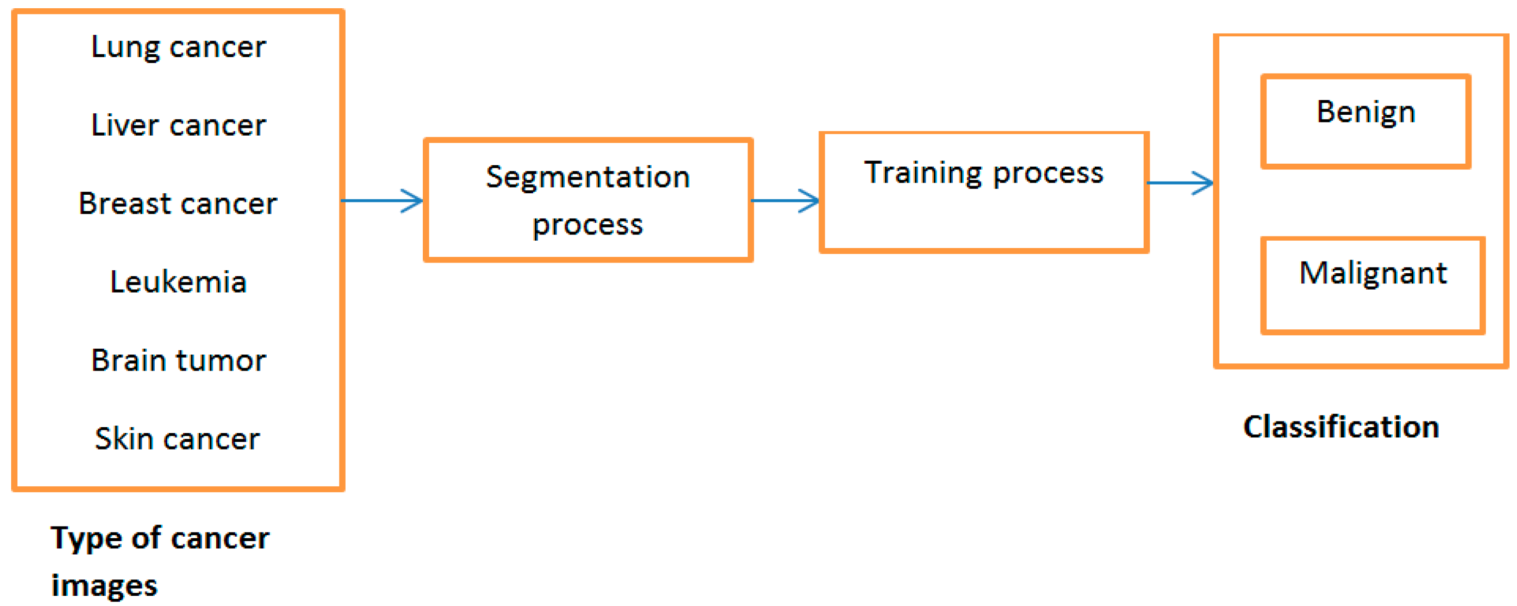
2.1. Proposed Model
2.2. Dataset Description
2.3. Pre-Processing
2.4. Feature Extraction
2.5. Segmentation
2.6. Classification
2.7. Proposed Algorithm
3. Results and Discussion
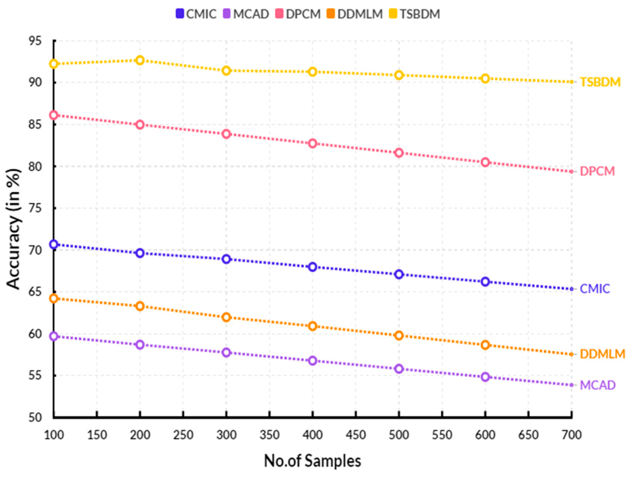
3.1. Computation of Accuracy
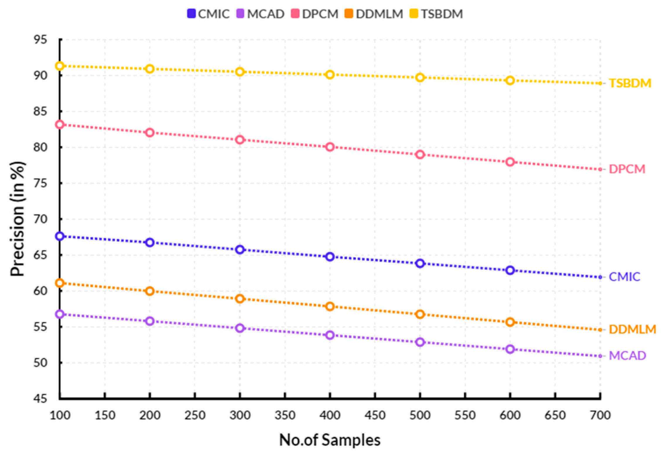
3.2. Precision
3.3. Recall
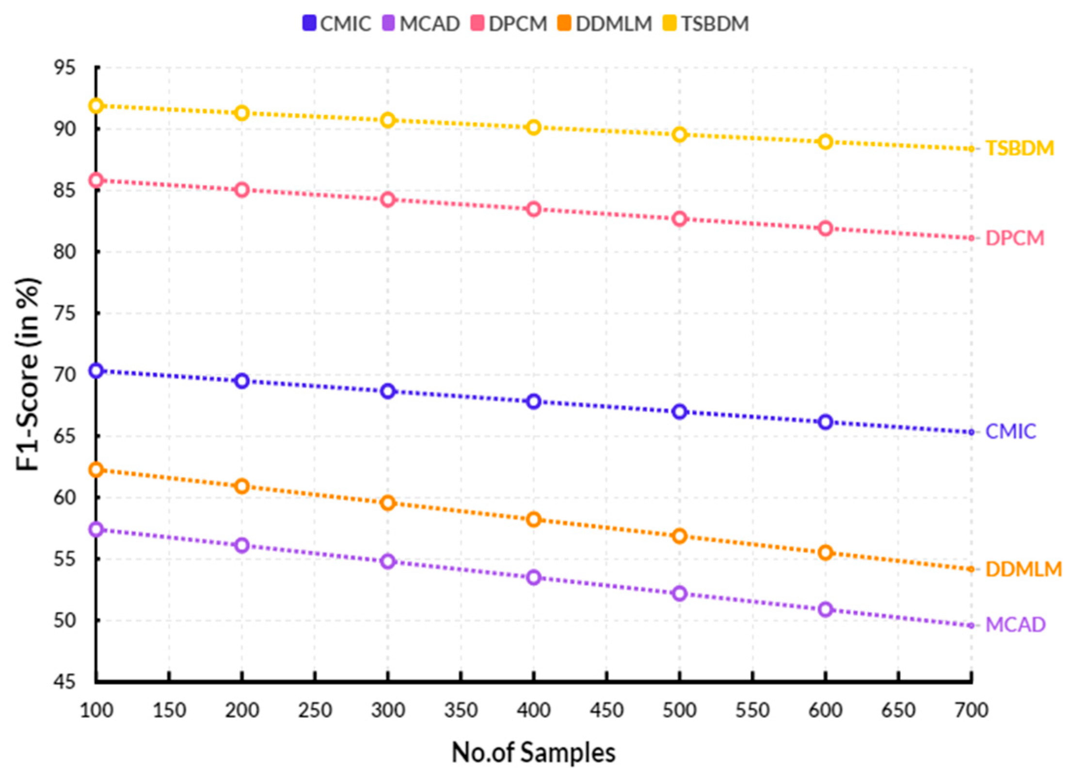
3.4. F1-Score
- Limited application: Time series-based detection models are limited in their ability to detect cancers effectively in all parts of the body, as their detection methodology is largely reliant on the type of physiological signal being monitored.
- Sensitivity: Time series-based detection models are limited in their sensitivity, as most of the current models only detect changes that occur over long time periods. This means that early-stage cancers may be missed.
- Invasive: Time series-based detection models rely on invasive techniques for some of their measurements, which can be uncomfortable and even painful for some patients.
4. Conclusions
Author Contributions
Funding
Institutional Review Board Statement
Informed Consent Statement
Data Availability Statement
Conflicts of Interest
References
- Li, C.; Liu, Y.; Xue, D.; Chan, C.W. Effects of nurse-led interventions on early detection of cancer: A systematic review and meta-analysis. Int. J. Nurs. Stud. 2020, 110, 103684. [Google Scholar] [CrossRef] [PubMed]
- Upadhyaya, A.M.; Srivastava, M.C.; Sharan, P. Integrated MOEMS based cantilever sensor for early detection of cancer. Optik 2021, 227, 165321. [Google Scholar] [CrossRef]
- Froelich, M.F.; Capoluongo, E.; Kovacs, Z.; Patton, S.J.; Lianidou, E.S.; Haselmann, V. The value proposition of integrative diagnostics for (early) detection of cancer. On behalf of the EFLM interdisciplinary Task and Finish Group “CNAPS/CTC for early detection of cancer”. Clin. Chem. Lab. Med. (CCLM) 2022, 60, 821–829. [Google Scholar] [CrossRef] [PubMed]
- Ullah, M.; Akbar, A.; Yannarelli, G. Applications of artificial intelligence in, early detection of cancer, clinical diagnosis and personalized medicine. Artif. Intell. Cancer 2020, 1, 39–44. [Google Scholar] [CrossRef]
- O’Connell, T.M.; Golzarri-Arroyo, L.; Pin, F.; Barreto, R.; Dickinson, S.L.; Couch, M.E.; Bonetto, A. Metabolic biomarkers for the early detection of cancer cachexia. Front. Cell Dev. Biol. 2021, 9, 720096. [Google Scholar] [CrossRef] [PubMed]
- Hamadani, J.D.; Hasan, M.I.; Baldi, A.J.; Hossain, S.J.; Shiraji, S.; Bhuiyan, M.S.A.; Pasricha, S.R. Immediate impact of stay-at-home orders to control COVID-19 transmission on socioeconomic conditions, food insecurity, mental health, and intimate partner violence in Bangladeshi women and their families: An interrupted time series. Lancet Glob. Health 2020, 8, e1380–e1389. [Google Scholar] [CrossRef] [PubMed]
- Marcuello, M.; Vymetalkova, V.; Neves, R.P.; Duran-Sanchon, S.; Vedeld, H.M.; Tham, E.; Gironella, M. Circulating biomarkers for early detection and clinical management of colorectal cancer. Mol. Asp. Med. 2019, 69, 107–122. [Google Scholar] [CrossRef] [PubMed]
- Ireland, A.S.; Micinski, A.M.; Kastner, D.W.; Guo, B.; Wait, S.J.; Spainhower, K.B.; Oliver, T.G. MYC drives temporal evolution of small cell lung cancer subtypes by reprogramming neuroendocrine fate. Cancer Cell 2020, 38, 60–78. [Google Scholar] [CrossRef]
- Yang, G.; Xiao, Z.; Tang, C.; Deng, Y.; Huang, H.; He, Z. Recent advances in biosensor for detection of lung cancer biomarkers. Biosens. Bioelectron. 2019, 141, 111416. [Google Scholar] [CrossRef]
- Zhang, Y.; Li, M.; Gao, X.; Chen, Y.; Liu, T. Nanotechnology in cancer diagnosis: Progress, challenges and opportunities. J. Hematol. Oncol. 2019, 12, 137. [Google Scholar] [CrossRef]
- Pashayan, N.; Antoniou, A.C.; Ivanus, U.; Esserman, L.J.; Easton, D.F.; French, D.; Widschwendter, M. Personalized early detection and prevention of breast cancer: ENVISION consensus statement. Nat. Rev. Clin. Oncol. 2020, 17, 687–705. [Google Scholar] [CrossRef] [PubMed]
- Venugopalan, J.; Tong, L.; Hassanzadeh, H.R.; Wang, M.D. Multimodal deep learning models for early detection of Alzheimer’s disease stage. Sci. Rep. 2021, 11, 3254. [Google Scholar] [CrossRef] [PubMed]
- Klein, E.A.; Richards, D.; Cohn, A.; Tummala, M.; Lapham, R.; Cosgrove, D.; Liu, M.C. Clinical validation of a targeted methylation-based multi-cancer early detection test using an independent validation set. Ann. Oncol. 2021, 32, 1167–1177. [Google Scholar] [CrossRef] [PubMed]
- King, A.; Woo, J.K.S.; Ai, Q.Y.; Mo, F.K.F.; So, T.Y.; Lam, W.K.J.; Chan, K.C.A. Early detection of cancer: Evaluation of MR imaging grading systems in patients with suspected nasopharyngeal carcinoma. Am. J. Neuroradiol. 2020, 41, 515–521. [Google Scholar] [CrossRef] [PubMed]
- Crosby, D.; Bhatia, S.; Brindle, K.M.; Coussens, L.M.; Dive, C.; Emberton, M.; Balasubramanian, S. Early detection of cancer. Science 2022, 375, eaay9040. [Google Scholar] [CrossRef] [PubMed]
- Van Der Pol, Y.; Mouliere, F. Toward the early detection of cancer by decoding the epigenetic and environmental fingerprints of cell-free DNA. Cancer Cell 2019, 36, 350–368. [Google Scholar] [CrossRef] [PubMed]
- Roy, D.; Tiirikainen, M. Diagnostic power of DNA methylation classifiers for early detection of cancer. Trends Cancer 2020, 6, 78–81. [Google Scholar] [CrossRef] [PubMed]
- Chen, X.; Gole, J.; Gore, A.; He, Q.; Lu, M.; Min, J.; Jin, L. Non-invasive early detection of cancer four years before conventional diagnosis using a blood test. Nat. Commun. 2020, 11, 3475. [Google Scholar] [CrossRef]
- Islam, N.; Shkolnikov, V.M.; Acosta, R.J.; Klimkin, I.; Kawachi, I.; Irizarry, R.A.; Lacey, B. Excess deaths associated with covid-19 pandemic in 2020: Age and sex disaggregated time series analysis in 29 high income countries. BMJ 2021, 373, n1137. [Google Scholar] [CrossRef]
- Jeyaraj, P.R.; Samuel Nadar, E.R. Computer-assisted medical image classification for early diagnosis of oral cancer employing deep learning algorithm. J. Cancer Res. Clin. Oncol. 2019, 145, 829–837. [Google Scholar] [CrossRef]
- Yao, Q.; Wang, R.; Fan, X.; Liu, J.; Li, Y. Multi-class arrhythmia detection from 12-lead varied-length ECG using attention-based time-incremental convolutional neural network. Inf. Fusion 2020, 53, 174–182. [Google Scholar] [CrossRef]
- Ginsburg, O.; Yip, C.H.; Brooks, A.; Cabanes, A.; Caleffi, M.; Dunstan Yataco, J.A.; Anderson, B.O. Breast cancer early detection: A phased approach to implementation. Cancer 2020, 126, 2379–2393. [Google Scholar] [CrossRef]
- Liu, C.; Zhao, J.; Tian, F.; Cai, L.; Zhang, W.; Feng, Q.; Tan, W. Low-cost thermophoretic profiling of extracellular-vesicle surface proteins for the early detection and classification of cancers. Nat. Biomed. Eng. 2019, 3, 183–193. [Google Scholar] [CrossRef]
- Cai, J.; Chen, L.; Zhang, Z.; Zhang, X.; Lu, X.; Liu, W.; Fan, J. Genome-wide mapping of 5-hydroxymethylcytosines in circulating cell-free DNA as a non-invasive approach for early detection of hepatocellular carcinoma. Gut 2019, 68, 2195–2205. [Google Scholar] [CrossRef]


| Dataset | Description |
|---|---|
| Lung Tumor | No.of Samples: 6125 Training Samples: 75% (4778 Samples) Testing Samples: 25% (1347 Samples) |
| Liver Tumor | |
| Breast Tumor | |
| Leukemia Tumor | |
| Brain Tumor | |
| Skin Tumor |
Disclaimer/Publisher’s Note: The statements, opinions and data contained in all publications are solely those of the individual author(s) and contributor(s) and not of MDPI and/or the editor(s). MDPI and/or the editor(s) disclaim responsibility for any injury to people or property resulting from any ideas, methods, instructions or products referred to in the content. |
© 2023 by the authors. Licensee MDPI, Basel, Switzerland. This article is an open access article distributed under the terms and conditions of the Creative Commons Attribution (CC BY) license (https://creativecommons.org/licenses/by/4.0/).
Share and Cite
Srinivasan, U.S.; Pavithra, V.; Sutha, K.; Ramachandiran, S.; Indumathi, N. An Innovative Analysis of Time Series-Based Detection Models for Improved Cancer Detection in Modern Healthcare Environments. Eng. Proc. 2023, 59, 114. https://doi.org/10.3390/engproc2023059114
Srinivasan US, Pavithra V, Sutha K, Ramachandiran S, Indumathi N. An Innovative Analysis of Time Series-Based Detection Models for Improved Cancer Detection in Modern Healthcare Environments. Engineering Proceedings. 2023; 59(1):114. https://doi.org/10.3390/engproc2023059114
Chicago/Turabian StyleSrinivasan, Uma Shankari, Venkat Pavithra, Kaliappan Sutha, Sridevi Ramachandiran, and Nallathambi Indumathi. 2023. "An Innovative Analysis of Time Series-Based Detection Models for Improved Cancer Detection in Modern Healthcare Environments" Engineering Proceedings 59, no. 1: 114. https://doi.org/10.3390/engproc2023059114