
Automatic Malignant and Benign Skin Cancer Classification Using a Hybrid Deep Learning Approach

 , and
, and 
Abstract
:1. Introduction
Related Work
2. Materials and Methods
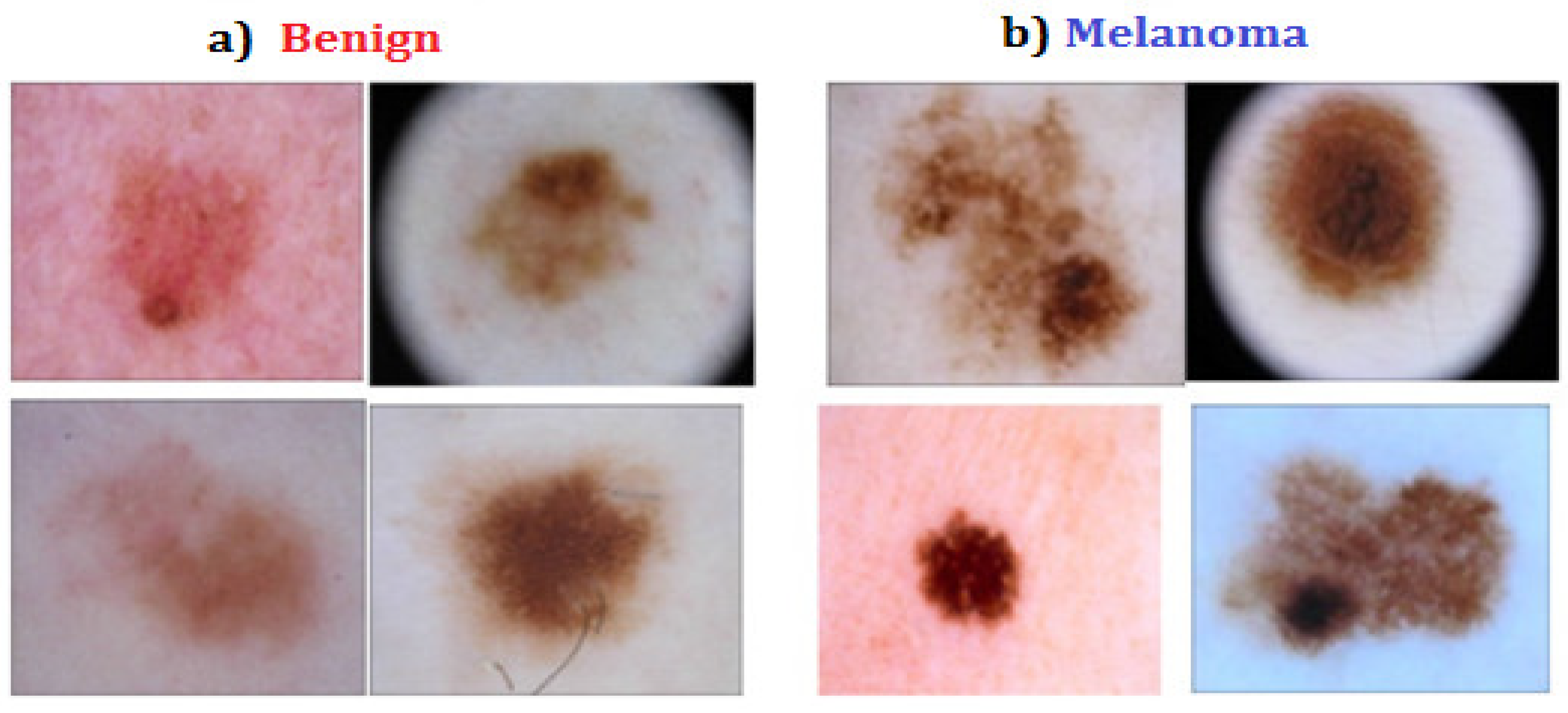
2.1. Dataset for Research
2.2. Methods for Implementation
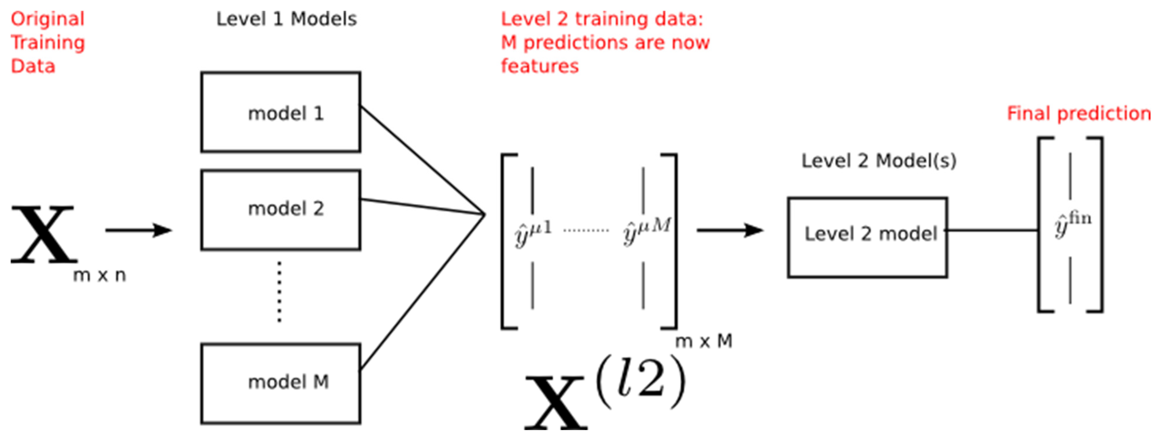
2.2.1. Proposed Method

2.2.2. Feature Extraction
2.3. Classification Approach
2.4. Performance Evaluation Metrics
3. Experimental Analysis
4. Conclusions
Author Contributions
Funding
Institutional Review Board Statement
Informed Consent Statement
Data Availability Statement
Conflicts of Interest
References
- Chaturvedi, S.S.; Gupta, K.; Prasad, P.S. Skin lesion analyzer: An efficient seven-way multi-class skin cancer classification using MobileNet. In Proceedings of the International Conference on Advanced Machine Learning Technologies and Applications, Cairo, Egypt, 20–22 March 2020; Springer: Singapore, 2020. [Google Scholar]
- Cancer Facts and Figures 2019. American Cancer Society. 2019. Available online: https://www.cancer.org/content/dam/cancer-org/research/cancer-facts-and-statistics/annual-cancerfacts-and-figures/2019/cancer-facts-and-figures-2019.pdf (accessed on 22 June 2020).
- Zghal, N.S.; Derbel, N. Melanoma Skin Cancer Detection based on Image Processing. Curr. Med. Imaging 2020, 16, 50–58. [Google Scholar] [CrossRef] [PubMed]
- Polat, K.; Koc, K.O. Detection of skin diseases from dermoscopy image using the combination of convolutional neural network and one-versus-all. J. Artif. Intell. Syst. 2020, 2, 80–97. [Google Scholar] [CrossRef]
- Wei, L.; Ding, K.; Hu, H. Automatic Skin Cancer Detection in Dermoscopy Images based on Ensemble Lightweight Deep Learning Network. IEEE Access 2020, 8, 99633–99647. [Google Scholar] [CrossRef]
- Giuffrida, R.; Conforti, C.; Di Meo, N.; Deinlein, T.; Guida, S.; Zalaudek, I.; Giuffrida, R.; Conforti, C.; Di Meo, N.; Deinlein, T.; et al. Use of noninvasive imaging in the management of skin cancer. Curr. Opin. Oncol. 2020, 32, 98–105. [Google Scholar] [CrossRef] [PubMed]
- Ech-Cherif, A.; Misbhauddin, M.; Ech-Cherif, M. Deep Neural Network-based mobile dermoscopy application for triaging skin cancer detection. In Proceedings of the 2019 2nd International Conference on Computer Applications & Information Security (ICCAIS), Riyadh, Saudi Arabia, 1–3 May 2019. [Google Scholar]
- Milton, M.A.A. Automated skin lesion classification using an ensemble of deep neural networks in ISIC 2018: Skin lesion analysis towards melanoma detection challenge. arXiv 2019, arXiv:1901.10802. [Google Scholar]
- Nasif, A.; Othman, Z.A.; Sani, N.S. The deep learning solutions on lossless compression methods for alleviating data load on IoT nodes in smart cities. Sensors 2021, 21, 4223. [Google Scholar] [CrossRef]
- Yélamos, O.; Braun, R.P.; Liopyris, K.; Wolner, Z.J.; Kerl, K.; Gerami, P.; Marghoob, A.A. Usefulness of dermoscopy to improve the clinical and histopathologic diagnosis of skin cancers. J. Am. Acad. Dermatol. 2019, 80, 365–377. [Google Scholar] [CrossRef] [PubMed] [Green Version]
- Holliday, J.; Sani, N.; Willett, P. Ligand-based virtual screening using a genetic algorithm with data fusion. Match Commun. Math. Comput. Chem. 2018, 80, 623–638. [Google Scholar]
- Othman, Z.A.; Bakar, A.A.; Sani, N.S.; Sallim, J. Household Overspending Model Amongst B40, M40 and T20 using Classification Algorithm. Int. J. Adv. Comput. Sci. Appl. 2020, 11, 392–399. [Google Scholar] [CrossRef]
- Wolner, Z.J.; Yélamos, O.; Liopyris, K.; Rogers, T.; Marchetti, M.A.; Marghoob, A.A. Enhancing skin cancer diagnosis with dermoscopy. Dermatol. Clin. 2017, 35, 417–437. [Google Scholar] [CrossRef] [PubMed]
- Dascalu, A.; David, E.O. Skin cancer detection by deep learning and sound analysis algorithms: A prospective clinical study of an elementary dermoscopy. EBioMedicine 2019, 43, 107–113. [Google Scholar] [CrossRef] [PubMed] [Green Version]
- Kassani, S.H.; Kassani, P.H. A comparative study of deep learning architectures on melanoma detection. Tissue Cell 2019, 58, 76–83. [Google Scholar] [CrossRef] [PubMed]
- Rajasekhar, K.S.; Babu, T.R. Analysis and Classification of Dermoscopic Images Using Spectral Graph Wavelet Transform. Period. Polytech. Electr. Eng. Comput. Sci. 2020, 64, 313–323. [Google Scholar] [CrossRef]
- Murugan, A.; Nair, S.H.; Kumar, K.P.S. Detection of skin cancer using SVM, Random Forest, and kNN classifiers. J. Med. Syst. 2019, 43, 269. [Google Scholar] [CrossRef]
- Seeja, R.D.; Suresh, A. Deep learning-based skin lesion segmentation and classification of melanoma using support vector machine (SVM). Asian Pac. J. Cancer Prev. APJCP 2019, 20, 1555–1561. [Google Scholar]
- Goyal, M.; Oakley, A.; Bansal, P.; Dancey, D.; Yap, M.H. Skin lesion segmentation in dermoscopic images with ensemble deep learning methods. IEEE Access 2019, 8, 4171–4181. [Google Scholar] [CrossRef]
- Taghanaki, S.A.; Abhishek, K.; Cohen, J.P.; Cohen-Adad, J.; Hamarneh, G. Deep semantic segmentation of natural and medical images: A review. Artif. Intell. Rev. 2020, 54, 137–178. [Google Scholar] [CrossRef]
- Hasan, K.; Dahal, L.; Samarakoon, P.N.; Tushar, F.I.; Martí, R. DSNet: Automatic dermoscopic skin lesion segmentation. Comput. Biol. Med. 2020, 120, 103738. [Google Scholar] [CrossRef] [Green Version]
- Munir, K.; Elahi, H.; Ayub, A.; Frezza, F.; Rizzi, A. Cancer diagnosis using deep learning: A bibliographic review. Cancers 2019, 11, 1235. [Google Scholar] [CrossRef] [Green Version]
- Jianu, S.R.S.; Ichim, L.; Popescu, D. Automatic diagnosis of skin cancer using neural networks. In Proceedings of the 2019 11th International Symposium on Advanced Topics in Electrical Engineering (ATEE), Bucharest, Romania, 28–30 March 2019. [Google Scholar]
- Garg, N.; Sharma, V.; Kaur, P. Melanoma skin cancer detection using image processing. In Sensors and Image Processing; Springer: Singapore, 2018; pp. 111–119. [Google Scholar]
- Nafea, A.A.; Omar, N.; Al-Ani, M.M. Adverse Drug Reaction Detection Using Latent Semantic Analysis. J. Comput. Sci. 2021, 17, 960–970. [Google Scholar] [CrossRef]
- AL-Ani, M.M.; Omar, N.; Nafea, A.A. A Hybrid Method of Long Short-Term Memory and Auto-Encoder Architectures for Sarcasm Detection. J. Comput. Sci. 2021, 17, 1093–1098. [Google Scholar] [CrossRef]
- Jamal, N.; Mohd, M.; Noah, S.A. Poetry classification using support vector machines. J. Comput. Sci. 2012, 8, 1441–1446. [Google Scholar]
- Kassem, M.A.; Hosny, K.M.; Fouad, M.M. Skin lesions classification into eight classes for ISIC 2019 using deep convolutional neural network and transfer learning. IEEE Access 2020, 8, 114822–114832. [Google Scholar] [CrossRef]
- Chaturvedi, S.S.; Tembhurne, J.V.; Diwan, T. A multi-class skin Cancer classification using deep convolution neural networks. Multimed. Tools Appl. 2020, 79, 28477–28498. [Google Scholar] [CrossRef]
- Abadi, M.; Barham, P.; Chen, J.; Chen, Z.; Davis, A.; Dean, J.; Devin, M.; Ghemawat, S.; Irving, G.; Isard, M.; et al. Tensorflow: A system for large-scale machine learning. In Proceedings of the 12th USENIX Symposium on Operating Systems Design and Implementation (OSDI 16), Savannah, GA, USA, 2–4 November 2016. [Google Scholar]
- Dorj, U.-O.; Lee, K.-K.; Choi, J.-Y.; Lee, M. The skin cancer classification using deep convolution neural network. Multimed. Tools Appl. 2018, 77, 9909–9924. [Google Scholar] [CrossRef]
- Kucharski, D.; Kleczek, P.; Jaworek-Korjakowska, J.; Dyduch, G.; Gorgon, M. Semi-Supervised Nests of Melanocytes Segmentation Method Using Convolutional Autoencoders. Sensors 2020, 20, 1546. [Google Scholar] [CrossRef] [Green Version]
- Kim, M.; Lee, M.; An, M.; Lee, H. Effective automatic defect classification process based on CNN with stacking ensemble model for TFT-LCD panel. J. Intell. Manuf. 2020, 31, 1165–1174. [Google Scholar] [CrossRef]
- Sánchez-Morales, A.; Sancho-Gómez, J.L.; Figueiras-Vidal, A.R. Complete auto encoders for classification with missing values. Neural Comput. Appl. 2021, 33, 1951–1957. [Google Scholar] [CrossRef]
- Kadam, V.J.; Jadhav, S.M.; Kurdukar, A.A.; Shirsath, M.R. Arrhythmia Classification using Feature Ensemble Learning based on Stacked Sparse Autoencoders with GA-SVM Guided Features. In Proceedings of the 2020 International Conference on Industry 4.0 Technology (I4Tech), Pune, India, 13–15 February 2020. [Google Scholar]
- Chen, M.; Chen, W.; Chen, W.; Cai, L.; Chai, G. Skin Cancer Classification with Deep Convolution Neural Networks. J. Med. Imaging Health Inform. 2020, 10, 1707–1713. [Google Scholar] [CrossRef]
- Nahata, H.; Singh, S.P. Deep Learning Solutions for Skin Cancer Detection and Diagnosis. In Machine Learning with Health Care Perspective; Springer: Cham, Switzerland, 2020; pp. 159–182. [Google Scholar]
- Tr, G.B. An Efficient Skin Cancer Diagnostic System Using Bendlet Transform and Support Vector Machine. An. Acad. Bras. Ciências 2020, 92, e20190554. [Google Scholar]
- Abdulkareem, A.B.; Sani, N.S.; Sahran, S.; Alyessari, Z.A.A.; Adam, A.; Rahman, A.H.A.; Abdulkarem, A.B. Predicting COVID-19 based on environmental factors with machine learning. Intell. Autom. Soft Comput. 2021, 28, 305–320. [Google Scholar] [CrossRef]
- Khamparia, A.; Singh, P.K.; Rani, P.; Samanta, D.; Khanna, A.; Bhushan, B. An internet of health things-driven deep learning framework for detection and classification of skin cancer using transfer learning. Trans. Emerg. Telecommun. Technol. 2020, 32, e3963. [Google Scholar] [CrossRef]
- Alameri, S.A.; Mohd, M. Comparison of fake news detection using machine learning and deep learning techniques. In Proceedings of the 2021 3rd International Cyber Resilience Conference (CRC), Langkawi Island, Malaysia, 29–31 January 2021; pp. 1–6. [Google Scholar]


| Skin Cancer Class | Number of Images |
|---|---|
| Benign | 1440 + 360 |
| Malignant | 1197 + 300 |
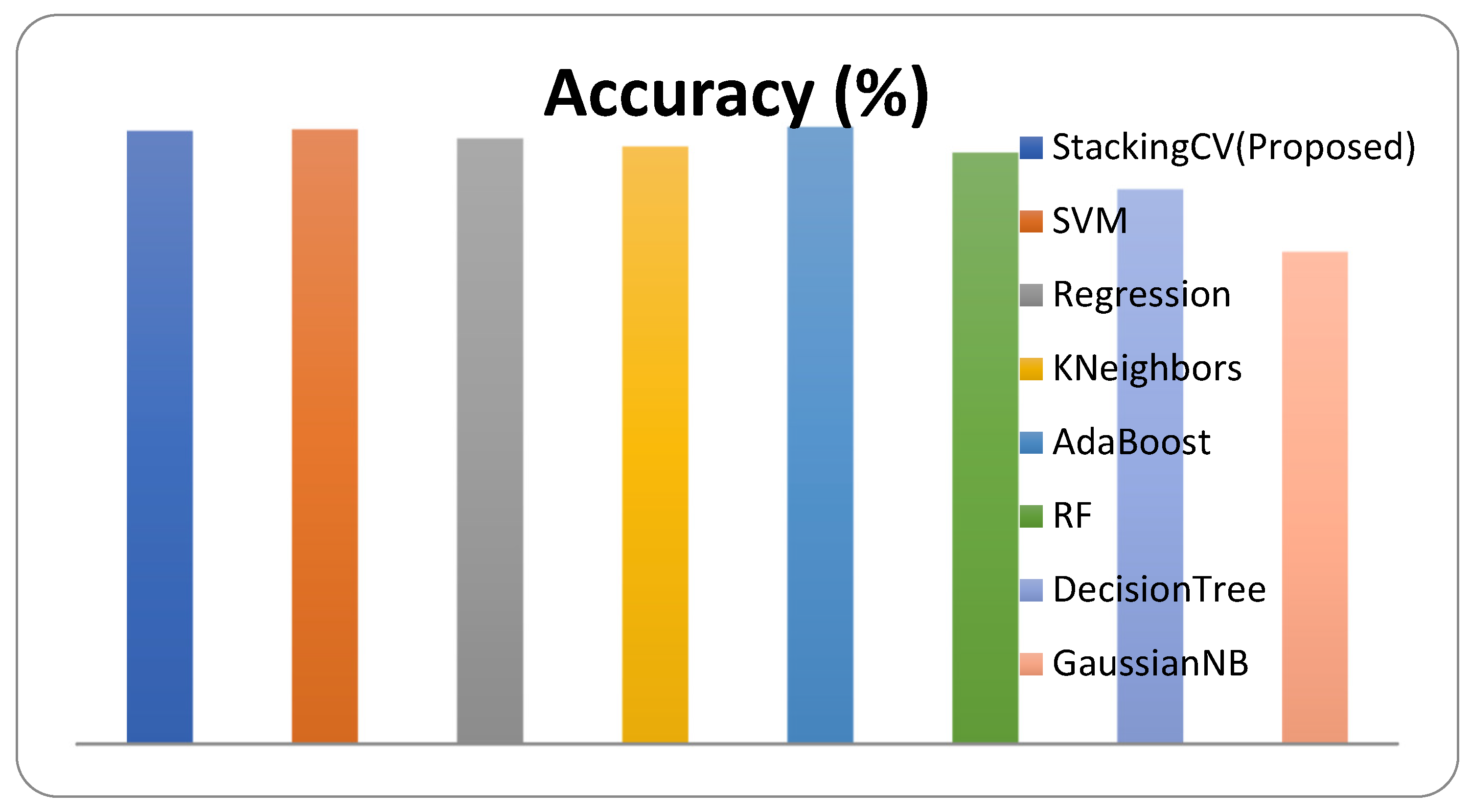
| Classifier | Accuracy (%) | F1-Score | Sensitivity | AUC |
|---|---|---|---|---|
| StackingCV (Proposed) | 81.6 | 0.788 | 0.821 | 0.818 |
| SVM | 81.8 | 0.787 | 0.816 | 0.817 |
| Regression | 80.6 | 0.762 | 0.752 | 0.798 |
| KNN | 79.5 | 0.754 | 0.761 | 0.790 |
| AdaBoost | 82.1 | 0.792 | 0.825 | 0.822 |
| RF | 78.7 | 0.735 | 0.715 | 0.777 |
| DecisionTree | 73.8 | 0.666 | 0.633 | 0.722 |
| GaussianNB | 65.5 | 0.363 | 0.238 | 0.593 |
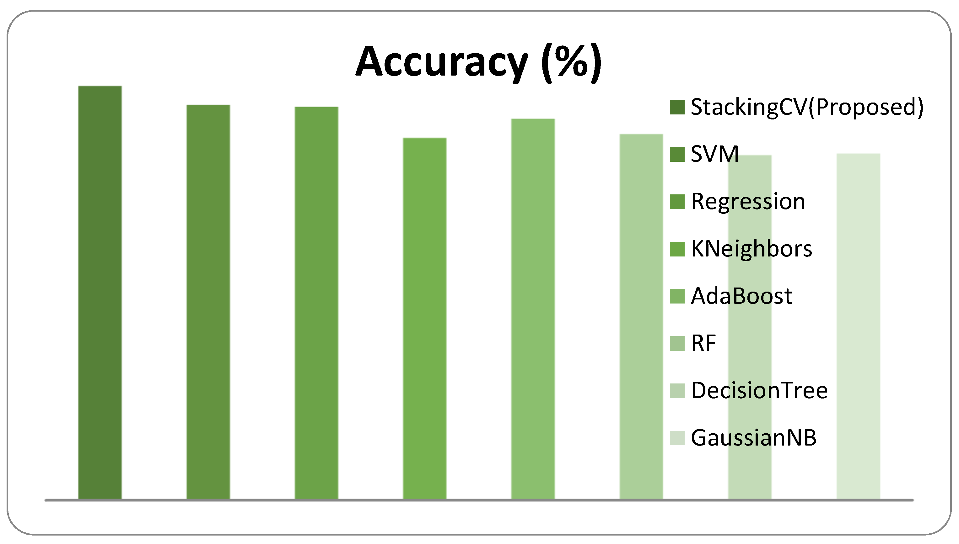
| Classifier | Accuracy (%) | F1-Score | Sensitivity | AUC |
|---|---|---|---|---|
| StackingCV (Proposed) | 90.9 | 0.896 | 0.886 | 0.917 |
| SVM | 86.7 | 0.838 | 0.834 | 0.862 |
| Regression | 86.3 | 0.837 | 0.853 | 0.862 |
| KNN | 79.5 | 0.732 | 0.678 | 0.778 |
| AdaBoost | 83.7 | 0.801 | 0.798 | 0.831 |
| RF | 80.3 | 0.739 | 0.678 | 0.784 |
| DecisionTree | 75.7 | 0.676 | 0.614 | 0.736 |
| GaussianNB | 76.1 | 0.731 | 0.788 | 0.765 |
| Classifier | Accuracy (%) | F1-Score | Sensitivity | AUC |
|---|---|---|---|---|
| StackingCV (Proposed) | 86.5 | 0.842 | 0.804 | 0.843 |
| SVM | 86.7 | 0.835 | 0.810 | 0.859 |
| Regression | 87.5 | 0.847 | 0.844 | 0.870 |
| KNN | 81 | 0.761 | 0.733 | 0.799 |
| AdaBoost | 79.9 | 0.766 | 0.798 | 0.799 |
| RF | 84 | 0.805 | 0.798 | 0.834 |
| DecisionTree | 76.1 | 0.701 | 0.678 | 0.749 |
| GaussianNB | 77.6 | 0.723 | 0.706 | 0.766 |
| Classifier | Resnet50 Features | Xception Features | VGG16 Features |
|---|---|---|---|
| StackingCV (Proposed) | 81.6 | 90.9 | 86.5 |
| SVM | 81.8 | 86.7 | 86.7 |
| Regression | 80.6 | 86.3 | 87.5 |
| KNeighbors | 79.5 | 79.5 | 81 |
| AdaBoost | 82.1 | 83.7 | 79.9 |
| RF | 78.7 | 80.3 | 84 |
| Decision Tree | 73.8 | 75.7 | 76.1 |
| GaussianNB | 65.5 | 76.1 | 77.6 |
Publisher’s Note: MDPI stays neutral with regard to jurisdictional claims in published maps and institutional affiliations. |
© 2022 by the authors. Licensee MDPI, Basel, Switzerland. This article is an open access article distributed under the terms and conditions of the Creative Commons Attribution (CC BY) license (https://creativecommons.org/licenses/by/4.0/).
Share and Cite
Bassel, A.; Abdulkareem, A.B.; Alyasseri, Z.A.A.; Sani, N.S.; Mohammed, H.J. Automatic Malignant and Benign Skin Cancer Classification Using a Hybrid Deep Learning Approach. Diagnostics 2022, 12, 2472. https://doi.org/10.3390/diagnostics12102472
Bassel A, Abdulkareem AB, Alyasseri ZAA, Sani NS, Mohammed HJ. Automatic Malignant and Benign Skin Cancer Classification Using a Hybrid Deep Learning Approach. Diagnostics. 2022; 12(10):2472. https://doi.org/10.3390/diagnostics12102472
Chicago/Turabian StyleBassel, Atheer, Amjed Basil Abdulkareem, Zaid Abdi Alkareem Alyasseri, Nor Samsiah Sani, and Husam Jasim Mohammed. 2022. "Automatic Malignant and Benign Skin Cancer Classification Using a Hybrid Deep Learning Approach" Diagnostics 12, no. 10: 2472. https://doi.org/10.3390/diagnostics12102472