
Radiomics in Oncological PET Imaging: A Systematic Review—Part 1, Supradiaphragmatic Cancers

 , , , and
Abstract
:1. Introduction
2. Materials and Methods
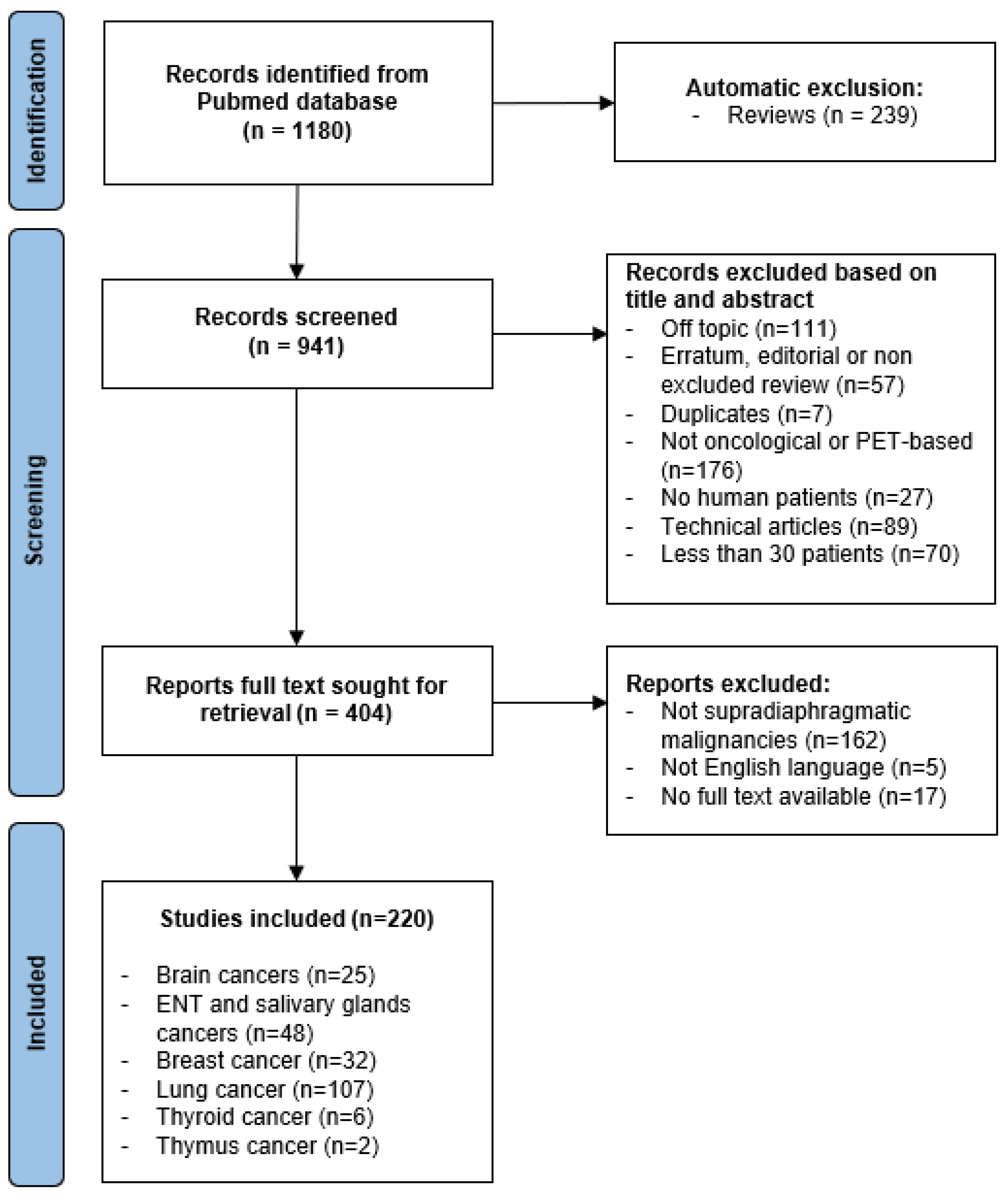
2.1. Search Strategy, Inclusion and Exclusion Criteria
2.2. Quality Assessment
- The number of patients, estimating the risk of bias and overfitting: fewer than 50 patients (score 0), 50 to 100 patients (score 1), more than 100 patients (score 2);
- The retrospective (score 0) or prospective (score 2) nature of the collection of data;
- The use of a completely independent cohort for validation: no or k-folding—as it can expose to data leakage—(score 0), partition of the cohort between completely separated training and test set (score 1), external validation cohort (score 2).
2.3. Textural Parameters Used
- Shape features: it is a purely geometric description of the segmented volume (metabolic tumoral volume, sphericity).
- First order features (also called histogram-based features): these parameters are based on the value of each voxel included in the segmented region without taking into account their spatial inter-relationships (maximum, minimum, average, standard deviation, etc.).
- Second order features: these parameters take into account the spatial interrelations between groups of pixels and are computed from texture matrices, calculated from the segmented region of interest [9]. Let us mention as an example Gray-level co-occurrence matrix (GLCM), which represents the frequency of occurrence of two intensity levels in neighboring voxels within a specific distance along a fixed direction or Gray-level run-length matrix (GLRLM), which encodes the size of homogenous runs for each image intensity.
- Higher order features: higher-order statistics features are computed after the application of specific mathematical transformations or filters [8].
2.4. Data Collection and Review
- PMID, first author, year of publication;
- Organ/type of cancer;
- Quality data: number of patients, retrospective or prospective nature, validation, quality score;
- Objective of the study;
- Maximal order of textural parameter (shape only, first order, second order, higher order).
3. Results
3.1. Discrepancies between the Two Reading Sessions
3.2. Searching Results
3.3. Quality Assessments
3.4. Textural Parameters Used
3.5. Brain Cancers
3.6. Head and Neck Cancer including Salivary Gland Cancers
3.7. Lung Cancer
3.8. Breast Cancer
3.9. Thyroid and Thymus
4. Discussion
4.1. Quality Assessment and Textural Parameters Used
4.2. Trends and Topics
4.3. Limitations
5. Conclusions
Supplementary Materials
Author Contributions
Funding
Institutional Review Board Statement
Informed Consent Statement
Data Availability Statement
Conflicts of Interest
References
- Micheel, C.M.; Institute of Medicine (Eds.) Omics-Based Clinical Discovery: Science, Technology, and Applications. In Evolution of Translational Omics: Lessons Learned and the Path Forward; National Academies Press: Washington, DC, USA, 2012; pp. 33–63. ISBN 978-0-309-22418-5. [Google Scholar]
- van Timmeren, J.E.; Cester, D.; Tanadini-Lang, S.; Alkadhi, H.; Baessler, B. Radiomics in Medical Imaging—“How-to” Guide and Critical Reflection. Insights Imaging 2020, 11, 91. [Google Scholar] [CrossRef] [PubMed]
- Park, J.E.; Kim, D.; Kim, H.S.; Park, S.Y.; Kim, J.Y.; Cho, S.J.; Shin, J.H.; Kim, J.H. Quality of Science and Reporting of Radiomics in Oncologic Studies: Room for Improvement According to Radiomics Quality Score and TRIPOD Statement. Eur. Radiol. 2020, 30, 523–536. [Google Scholar] [CrossRef] [PubMed]
- Shiyam Sundar, L.K.; Muzik, O.; Buvat, I.; Bidaut, L.; Beyer, T. Potentials and Caveats of AI in Hybrid Imaging. Methods 2021, 188, 4–19. [Google Scholar] [CrossRef] [PubMed]
- Hatt, M.; Cheze Le Rest, C.; Antonorsi, N.; Tixier, F.; Tankyevych, O.; Jaouen, V.; Lucia, F.; Bourbonne, V.; Schick, U.; Badic, B.; et al. Radiomics in PET/CT: Current Status and Future AI-Based Evolutions. Semin. Nucl. Med. 2021, 51, 126–133. [Google Scholar] [CrossRef]
- PRISMA-P Group; Moher, D.; Shamseer, L.; Clarke, M.; Ghersi, D.; Liberati, A.; Petticrew, M.; Shekelle, P.; Stewart, L.A. Preferred Reporting Items for Systematic Review and Meta-Analysis Protocols (PRISMA-P) 2015 Statement. Syst. Rev. 2015, 4, 1. [Google Scholar] [CrossRef] [Green Version]
- Papadimitroulas, P.; Brocki, L.; Christopher Chung, N.; Marchadour, W.; Vermet, F.; Gaubert, L.; Eleftheriadis, V.; Plachouris, D.; Visvikis, D.; Kagadis, G.C.; et al. Artificial Intelligence: Deep Learning in Oncological Radiomics and Challenges of Interpretability and Data Harmonization. Phys. Med. 2021, 83, 108–121. [Google Scholar] [CrossRef]
- Lohmann, P.; Bousabarah, K.; Hoevels, M.; Treuer, H. Radiomics in Radiation Oncology—Basics, Methods, and Limitations. Strahlenther. Onkol. 2020, 196, 848–855. [Google Scholar] [CrossRef]
- Haralick, R.M.; Shanmugam, K.; Dinstein, I. Textural Features for Image Classification. IEEE Trans. Syst. Man Cybern. 1973, SMC-3, 610–621. [Google Scholar] [CrossRef] [Green Version]
- Zhao, K.; Yu, P.; Xue, Z.; Liu, J.; Yao, A.; Zhao, Y.; Yang, F.; Tian, J.; Xu, B. (11)C-Methionine Integrated PET/MRI-Based Texture Analysis Features May Have a Potential Ability to Distinguish Oligodendroglioma (IDH-Mutant and 1p/19q-Codeleted) from Varied Gliomas. Acad. Radiol. 2020, 27, e159–e167. [Google Scholar] [CrossRef]
- Kong, Z.; Jiang, C.; Zhu, R.; Feng, S.; Wang, Y.; Li, J.; Chen, W.; Liu, P.; Zhao, D.; Ma, W.; et al. (18)F-FDG-PET-Based Radiomics Features to Distinguish Primary Central Nervous System Lymphoma from Glioblastoma. NeuroImage Clin. 2019, 23, 101912. [Google Scholar] [CrossRef]
- Kong, Z.; Lin, Y.; Jiang, C.; Li, L.; Liu, Z.; Wang, Y.; Dai, C.; Liu, D.; Qin, X.; Wang, Y.; et al. (18)F-FDG-PET-Based Radiomics Signature Predicts MGMT Promoter Methylation Status in Primary Diffuse Glioma. Cancer Imaging Off. Publ. Int. Cancer Imaging Soc. 2019, 19, 58. [Google Scholar] [CrossRef] [Green Version]
- Zaragori, T.; Oster, J.; Roch, V.; Hossu, G.; Chawki, M.B.; Grignon, R.; Pouget, C.; Gauchotte, G.; Rech, F.; Blonski, M.; et al. (18)F-FDOPA PET for the Noninvasive Prediction of Glioma Molecular Parameters: A Radiomics Study. J. Nucl. Med. Off. Publ. Soc. Nucl. Med. 2022, 63, 147–157. [Google Scholar] [CrossRef]
- Hotta, M.; Minamimoto, R.; Miwa, K. 11C-Methionine-PET for Differentiating Recurrent Brain Tumor from Radiation Necrosis: Radiomics Approach with Random Forest Classifier. Sci. Rep. 2019, 9, 15666. [Google Scholar] [CrossRef]
- Li, L.; Mu, W.; Wang, Y.; Liu, Z.; Liu, Z.; Wang, Y.; Ma, W.; Kong, Z.; Wang, S.; Zhou, X.; et al. A Non-Invasive Radiomic Method Using (18)F-FDG PET Predicts Isocitrate Dehydrogenase Genotype and Prognosis in Patients With Glioma. Front. Oncol. 2019, 9, 1183. [Google Scholar] [CrossRef]
- Muzi, M.; Wolsztynski, E.; Fink, J.R.; O’Sullivan, J.N.; O’Sullivan, F.; Krohn, K.A.; Mankoff, D.A. Assessment of the Prognostic Value of Radiomic Features in (18)F-FMISO PET Imaging of Hypoxia in Postsurgery Brain Cancer Patients: Secondary Analysis of Imaging Data from a Single-Center Study and the Multicenter ACRIN 6684 Trial. Tomogr. Ann. Arbor. Mich. 2020, 6, 14–22. [Google Scholar] [CrossRef]
- Russo, G.; Stefano, A.; Alongi, P.; Comelli, A.; Catalfamo, B.; Mantarro, C.; Longo, C.; Altieri, R.; Certo, F.; Cosentino, S.; et al. Feasibility on the Use of Radiomics Features of 11[C]-MET PET/CT in Central Nervous System Tumours: Preliminary Results on Potential Grading Discrimination Using a Machine Learning Model. Curr. Oncol. Tor. Ont. 2021, 28, 5318–5331. [Google Scholar] [CrossRef]
- Lohmann, P.; Elahmadawy, M.A.; Gutsche, R.; Werner, J.-M.; Bauer, E.K.; Ceccon, G.; Kocher, M.; Lerche, C.W.; Rapp, M.; Fink, G.R.; et al. FET PET Radiomics for Differentiating Pseudoprogression from Early Tumor Progression in Glioma Patients Post-Chemoradiation. Cancers 2020, 12, 3835. [Google Scholar] [CrossRef]
- Carles, M.; Popp, I.; Starke, M.M.; Mix, M.; Urbach, H.; Schimek-Jasch, T.; Eckert, F.; Niyazi, M.; Baltas, D.; Grosu, A.L. FET-PET Radiomics in Recurrent Glioblastoma: Prognostic Value for Outcome after Re-Irradiation? Radiat. Oncol. 2021, 16, 46. [Google Scholar] [CrossRef]
- Papp, L.; Pötsch, N.; Grahovac, M.; Schmidbauer, V.; Woehrer, A.; Preusser, M.; Mitterhauser, M.; Kiesel, B.; Wadsak, W.; Beyer, T.; et al. Glioma Survival Prediction with Combined Analysis of In Vivo (11)C-MET PET Features, Ex Vivo Features, and Patient Features by Supervised Machine Learning. J. Nucl. Med. Off. Publ. Soc. Nucl. Med. 2018, 59, 892–899. [Google Scholar] [CrossRef] [Green Version]
- Yu, P.; Ning, J.; Xu, B.; Liu, J.; Dang, H.; Lin, M.; Feng, X.; Grimm, R.; Tian, J. Histogram Analysis of 11C-Methionine Integrated PET/MRI May Facilitate to Determine the O6-Methylguanylmethyltransferase Methylation Status in Gliomas. Nucl. Med. Commun. 2019, 40, 850–856. [Google Scholar] [CrossRef]
- Wang, K.; Qiao, Z.; Zhao, X.; Li, X.; Wang, X.; Wu, T.; Chen, Z.; Fan, D.; Chen, Q.; Ai, L. Individualized Discrimination of Tumor Recurrence from Radiation Necrosis in Glioma Patients Using an Integrated Radiomics-Based Model. Eur. J. Nucl. Med. Mol. Imaging 2020, 47, 1400–1411. [Google Scholar] [CrossRef] [Green Version]
- Zhou, W.; Huang, Q.; Wen, J.; Li, M.; Zhu, Y.; Liu, Y.; Dai, Y.; Guan, Y.; Zhou, Z.; Hua, T. Integrated CT Radiomics Features Could Enhance the Efficacy of (18)F-FET PET for Non-Invasive Isocitrate Dehydrogenase Genotype Prediction in Adult Untreated Gliomas: A Retrospective Cohort Study. Front. Oncol. 2021, 11, 772703. [Google Scholar] [CrossRef]
- Mitamura, K.; Yamamoto, Y.; Kudomi, N.; Maeda, Y.; Norikane, T.; Miyake, K.; Nishiyama, Y. Intratumoral Heterogeneity of (18)F-FLT Uptake Predicts Proliferation and Survival in Patients with Newly Diagnosed Gliomas. Ann. Nucl. Med. 2017, 31, 46–52. [Google Scholar] [CrossRef]
- Haubold, J.; Demircioglu, A.; Gratz, M.; Glas, M.; Wrede, K.; Sure, U.; Antoch, G.; Keyvani, K.; Nittka, M.; Kannengiesser, S.; et al. Non-Invasive Tumor Decoding and Phenotyping of Cerebral Gliomas Utilizing Multiparametric (18)F-FET PET-MRI and MR Fingerprinting. Eur. J. Nucl. Med. Mol. Imaging 2020, 47, 1435–1445. [Google Scholar] [CrossRef]
- Lohmann, P.; Lerche, C.; Bauer, E.K.; Steger, J.; Stoffels, G.; Blau, T.; Dunkl, V.; Kocher, M.; Viswanathan, S.; Filss, C.P.; et al. Predicting IDH Genotype in Gliomas Using FET PET Radiomics. Sci. Rep. 2018, 8, 13328. [Google Scholar] [CrossRef]
- Qian, J.; Herman, M.G.; Brinkmann, D.H.; Laack, N.N.; Kemp, B.J.; Hunt, C.H.; Lowe, V.; Pafundi, D.H. Prediction of MGMT Status for Glioblastoma Patients Using Radiomics Feature Extraction from (18)F-DOPA-PET Imaging. Int. J. Radiat. Oncol. Biol. Phys. 2020, 108, 1339–1346. [Google Scholar] [CrossRef]
- Li, Z.; Kaiser, L.; Holzgreve, A.; Ruf, V.C.; Suchorska, B.; Wenter, V.; Quach, S.; Herms, J.; Bartenstein, P.; Tonn, J.-C.; et al. Prediction of TERTp-Mutation Status in IDH-Wildtype High-Grade Gliomas Using Pre-Treatment Dynamic [(18)F]FET PET Radiomics. Eur. J. Nucl. Med. Mol. Imaging 2021, 48, 4415–4425. [Google Scholar] [CrossRef]
- Manabe, O.; Yamaguchi, S.; Hirata, K.; Kobayashi, K.; Kobayashi, H.; Terasaka, S.; Toyonaga, T.; Magota, K.; Kuge, Y.; Tamaki, N.; et al. Preoperative Texture Analysis Using (11)C-Methionine Positron Emission Tomography Predicts Survival after Surgery for Glioma. Diagnostics 2021, 11, 189. [Google Scholar] [CrossRef]
- Kong, Z.; Li, J.; Liu, Z.; Liu, Z.; Zhao, D.; Cheng, X.; Li, L.; Lin, Y.; Wang, Y.; Tian, J.; et al. Radiomics Signature Based on FDG-PET Predicts Proliferative Activity in Primary Glioma. Clin. Radiol. 2019, 74, 815.e15–815.e23. [Google Scholar] [CrossRef]
- Ahrari, S.; Zaragori, T.; Rozenblum, L.; Oster, J.; Imbert, L.; Kas, A.; Verger, A. Relevance of Dynamic (18)F-DOPA PET Radiomics for Differentiation of High-Grade Glioma Progression from Treatment-Related Changes. Biomedicines 2021, 9, 1924. [Google Scholar] [CrossRef]
- Pyka, T.; Gempt, J.; Hiob, D.; Ringel, F.; Schlegel, J.; Bette, S.; Wester, H.-J.; Meyer, B.; Förster, S. Textural Analysis of Pre-Therapeutic [18F]-FET-PET and Its Correlation with Tumor Grade and Patient Survival in High-Grade Gliomas. Eur. J. Nucl. Med. Mol. Imaging 2016, 43, 133–141. [Google Scholar] [CrossRef] [PubMed]
- Lohmann, P.; Kocher, M.; Ceccon, G.; Bauer, E.K.; Stoffels, G.; Viswanathan, S.; Ruge, M.I.; Neumaier, B.; Shah, N.J.; Fink, G.R.; et al. Combined FET PET/MRI Radiomics Differentiates Radiation Injury from Recurrent Brain Metastasis. NeuroImage Clin. 2018, 20, 537–542. [Google Scholar] [CrossRef] [PubMed]
- Lohmann, P.; Stoffels, G.; Ceccon, G.; Rapp, M.; Sabel, M.; Filss, C.P.; Kamp, M.A.; Stegmayr, C.; Neumaier, B.; Shah, N.J.; et al. Radiation Injury vs. Recurrent Brain Metastasis: Combining Textural Feature Radiomics Analysis and Standard Parameters May Increase (18)F-FET PET Accuracy without Dynamic Scans. Eur. Radiol. 2017, 27, 2916–2927. [Google Scholar] [CrossRef] [PubMed]
- van Dijk, L.V.; Noordzij, W.; Brouwer, C.L.; Boellaard, R.; Burgerhof, J.G.M.; Langendijk, J.A.; Sijtsema, N.M.; Steenbakkers, R.J.H.M. (18)F-FDG PET Image Biomarkers Improve Prediction of Late Radiation-Induced Xerostomia. Radiother. Oncol. J. Eur. Soc. Ther. Radiol. Oncol. 2018, 126, 89–95. [Google Scholar] [CrossRef] [PubMed] [Green Version]
- Carles, M.; Fechter, T.; Grosu, A.L.; Sörensen, A.; Thomann, B.; Stoian, R.G.; Wiedenmann, N.; Rühle, A.; Zamboglou, C.; Ruf, J.; et al. (18)F-FMISO-PET Hypoxia Monitoring for Head-and-Neck Cancer Patients: Radiomics Analyses Predict the Outcome of Chemo-Radiotherapy. Cancers 2021, 13, 3449. [Google Scholar] [CrossRef]
- Wang, K.; Zhou, Z.; Wang, R.; Chen, L.; Zhang, Q.; Sher, D.; Wang, J. A Multi-Objective Radiomics Model for the Prediction of Locoregional Recurrence in Head and Neck Squamous Cell Cancer. Med. Phys. 2020, 47, 5392–5400. [Google Scholar] [CrossRef]
- Chen, R.-Y.; Lin, Y.-C.; Shen, W.-C.; Hsieh, T.-C.; Yen, K.-Y.; Chen, S.-W.; Kao, C.-H. Associations of Tumor PD-1 Ligands, Immunohistochemical Studies, and Textural Features in (18)F-FDG PET in Squamous Cell Carcinoma of the Head and Neck. Sci. Rep. 2018, 8, 105. [Google Scholar] [CrossRef]
- Chen, L.; Zhou, Z.; Sher, D.; Zhang, Q.; Shah, J.; Pham, N.-L.; Jiang, S.; Wang, J. Combining Many-Objective Radiomics and 3D Convolutional Neural Network through Evidential Reasoning to Predict Lymph Node Metastasis in Head and Neck Cancer. Phys. Med. Biol. 2019, 64, 075011. [Google Scholar] [CrossRef]
- Bogowicz, M.; Riesterer, O.; Stark, L.S.; Studer, G.; Unkelbach, J.; Guckenberger, M.; Tanadini-Lang, S. Comparison of PET and CT Radiomics for Prediction of Local Tumor Control in Head and Neck Squamous Cell Carcinoma. Acta Oncol. 2017, 56, 1531–1536. [Google Scholar] [CrossRef] [Green Version]
- Chen, S.-W.; Shen, W.-C.; Lin, Y.-C.; Chen, R.-Y.; Hsieh, T.-C.; Yen, K.-Y.; Kao, C.-H. Correlation of Pretreatment (18)F-FDG PET Tumor Textural Features with Gene Expression in Pharyngeal Cancer and Implications for Radiotherapy-Based Treatment Outcomes. Eur. J. Nucl. Med. Mol. Imaging 2017, 44, 567–580. [Google Scholar] [CrossRef]
- Ulrich, E.J.; Menda, Y.; Boles Ponto, L.L.; Anderson, C.M.; Smith, B.J.; Sunderland, J.J.; Graham, M.M.; Buatti, J.M.; Beichel, R.R. FLT PET Radiomics for Response Prediction to Chemoradiation Therapy in Head and Neck Squamous Cell Cancer. Tomography 2019, 5, 161–169. [Google Scholar] [CrossRef]
- Shiri, I.; Arabi, H.; Sanaat, A.; Jenabi, E.; Becker, M.; Zaidi, H. Fully Automated Gross Tumor Volume Delineation From PET in Head and Neck Cancer Using Deep Learning Algorithms. Clin. Nucl. Med. 2021, 46, 872–883. [Google Scholar] [CrossRef]
- Fujima, N.; Hirata, K.; Shiga, T.; Li, R.; Yasuda, K.; Onimaru, R.; Tsuchiya, K.; Kano, S.; Mizumachi, T.; Homma, A.; et al. Integrating Quantitative Morphological and Intratumoural Textural Characteristics in FDG-PET for the Prediction of Prognosis in Pharynx Squamous Cell Carcinoma Patients. Clin. Radiol. 2018, 73, 1059.e1–1059.e8. [Google Scholar] [CrossRef] [Green Version]
- Choi, J.W.; Lee, D.; Hyun, S.H.; Han, M.; Kim, J.-H.; Lee, S.J. Intratumoural Heterogeneity Measured Using FDG PET and MRI Is Associated with Tumour-Stroma Ratio and Clinical Outcome in Head and Neck Squamous Cell Carcinoma. Clin. Radiol. 2017, 72, 482–489. [Google Scholar] [CrossRef]
- Lafata, K.J.; Chang, Y.; Wang, C.; Mowery, Y.M.; Vergalasova, I.; Niedzwiecki, D.; Yoo, D.S.; Liu, J.-G.; Brizel, D.M.; Yin, F.-F. Intrinsic Radiomic Expression Patterns after 20 Gy Demonstrate Early Metabolic Response of Oropharyngeal Cancers. Med. Phys. 2021, 48, 3767–3777. [Google Scholar] [CrossRef]
- Du, D.; Feng, H.; Lv, W.; Ashrafinia, S.; Yuan, Q.; Wang, Q.; Yang, W.; Feng, Q.; Chen, W.; Rahmim, A.; et al. Machine Learning Methods for Optimal Radiomics-Based Differentiation Between Recurrence and Inflammation: Application to Nasopharyngeal Carcinoma Post-Therapy PET/CT Images. Mol. Imaging Biol. 2020, 22, 730–738. [Google Scholar] [CrossRef]
- Zhong, J.; Frood, R.; Brown, P.; Nelstrop, H.; Prestwich, R.; McDermott, G.; Currie, S.; Vaidyanathan, S.; Scarsbrook, A.F. Machine Learning-Based FDG PET-CT Radiomics for Outcome Prediction in Larynx and Hypopharynx Squamous Cell Carcinoma. Clin. Radiol. 2021, 76, 78.e9–78.e17. [Google Scholar] [CrossRef]
- Lv, W.; Ashrafinia, S.; Ma, J.; Lu, L.; Rahmim, A. Multi-Level Multi-Modality Fusion Radiomics: Application to PET and CT Imaging for Prognostication of Head and Neck Cancer. IEEE J. Biomed. Health Inform. 2020, 24, 2268–2277. [Google Scholar] [CrossRef]
- Haider, S.P.; Mahajan, A.; Zeevi, T.; Baumeister, P.; Reichel, C.; Sharaf, K.; Forghani, R.; Kucukkaya, A.S.; Kann, B.H.; Judson, B.L.; et al. PET/CT Radiomics Signature of Human Papilloma Virus Association in Oropharyngeal Squamous Cell Carcinoma. Eur. J. Nucl. Med. Mol. Imaging 2020, 47, 2978–2991. [Google Scholar] [CrossRef]
- Bogowicz, M.; Leijenaar, R.T.H.; Tanadini-Lang, S.; Riesterer, O.; Pruschy, M.; Studer, G.; Unkelbach, J.; Guckenberger, M.; Konukoglu, E.; Lambin, P. Post-Radiochemotherapy PET Radiomics in Head and Neck Cancer—The Influence of Radiomics Implementation on the Reproducibility of Local Control Tumor Models. Radiother. Oncol. J. Eur. Soc. Ther. Radiol. Oncol. 2017, 125, 385–391. [Google Scholar] [CrossRef] [Green Version]
- Haider, S.P.; Zeevi, T.; Baumeister, P.; Reichel, C.; Sharaf, K.; Forghani, R.; Kann, B.H.; Judson, B.L.; Prasad, M.L.; Burtness, B.; et al. Potential Added Value of PET/CT Radiomics for Survival Prognostication beyond AJCC 8th Edition Staging in Oropharyngeal Squamous Cell Carcinoma. Cancers 2020, 12, 1778. [Google Scholar] [CrossRef]
- Crispin-Ortuzar, M.; Apte, A.; Grkovski, M.; Oh, J.H.; Lee, N.Y.; Schöder, H.; Humm, J.L.; Deasy, J.O. Predicting Hypoxia Status Using a Combination of Contrast-Enhanced Computed Tomography and [(18)F]-Fluorodeoxyglucose Positron Emission Tomography Radiomics Features. Radiother. Oncol. J. Eur. Soc. Ther. Radiol. Oncol. 2018, 127, 36–42. [Google Scholar] [CrossRef]
- Peng, L.; Hong, X.; Yuan, Q.; Lu, L.; Wang, Q.; Chen, W. Prediction of Local Recurrence and Distant Metastasis Using Radiomics Analysis of Pretreatment Nasopharyngeal [18F]FDG PET/CT Images. Ann. Nucl. Med. 2021, 35, 458–468. [Google Scholar] [CrossRef]
- Haider, S.P.; Sharaf, K.; Zeevi, T.; Baumeister, P.; Reichel, C.; Forghani, R.; Kann, B.H.; Petukhova, A.; Judson, B.L.; Prasad, M.L.; et al. Prediction of Post-Radiotherapy Locoregional Progression in HPV-Associated Oropharyngeal Squamous Cell Carcinoma Using Machine-Learning Analysis of Baseline PET/CT Radiomics. Transl. Oncol. 2021, 14, 100906. [Google Scholar] [CrossRef]
- Ghosh, S.; Maulik, S.; Chatterjee, S.; Mallick, I.; Chakravorty, N.; Mukherjee, J. Prediction of Survival Outcome Based on Clinical Features and Pretreatment (18)FDG-PET/CT for HNSCC Patients. Comput. Methods Programs Biomed. 2020, 195, 105669. [Google Scholar] [CrossRef]
- Fujima, N.; Andreu-Arasa, V.C.; Meibom, S.K.; Mercier, G.A.; Salama, A.R.; Truong, M.T.; Sakai, O. Prediction of the Treatment Outcome Using Machine Learning with FDG-PET Image-Based Multiparametric Approach in Patients with Oral Cavity Squamous Cell Carcinoma. Clin. Radiol. 2021, 76, 711.e1–711.e7. [Google Scholar] [CrossRef]
- Folkert, M.R.; Setton, J.; Apte, A.P.; Grkovski, M.; Young, R.J.; Schöder, H.; Thorstad, W.L.; Lee, N.Y.; Deasy, J.O.; Oh, J.H. Predictive Modeling of Outcomes Following Definitive Chemoradiotherapy for Oropharyngeal Cancer Based on FDG-PET Image Characteristics. Phys. Med. Biol. 2017, 62, 5327–5343. [Google Scholar] [CrossRef]
- Martens, R.M.; Koopman, T.; Noij, D.P.; Pfaehler, E.; Übelhör, C.; Sharma, S.; Vergeer, M.R.; Leemans, C.R.; Hoekstra, O.S.; Yaqub, M.; et al. Predictive Value of Quantitative (18)F-FDG-PET Radiomics Analysis in Patients with Head and Neck Squamous Cell Carcinoma. EJNMMI Res. 2020, 10, 102. [Google Scholar] [CrossRef]
- Lin, H.-C.; Chan, S.-C.; Cheng, N.-M.; Liao, C.-T.; Hsu, C.-L.; Wang, H.-M.; Lin, C.-Y.; Chang, J.T.-C.; Ng, S.-H.; Yang, L.-Y.; et al. Pretreatment (18)F-FDG PET/CT Texture Parameters Provide Complementary Information to Epstein-Barr Virus DNA Titers in Patients with Metastatic Nasopharyngeal Carcinoma. Oral Oncol. 2020, 104, 104628. [Google Scholar] [CrossRef]
- Peng, H.; Dong, D.; Fang, M.-J.; Li, L.; Tang, L.-L.; Chen, L.; Li, W.-F.; Mao, Y.-P.; Fan, W.; Liu, L.-Z.; et al. Prognostic Value of Deep Learning PET/CT-Based Radiomics: Potential Role for Future Individual Induction Chemotherapy in Advanced Nasopharyngeal Carcinoma. Clin. Cancer Res. Off. J. Am. Assoc. Cancer Res. 2019, 25, 4271–4279. [Google Scholar] [CrossRef] [Green Version]
- Guezennec, C.; Robin, P.; Orlhac, F.; Bourhis, D.; Delcroix, O.; Gobel, Y.; Rousset, J.; Schick, U.; Salaün, P.-Y.; Abgral, R. Prognostic Value of Textural Indices Extracted from Pretherapeutic 18-F FDG-PET/CT in Head and Neck Squamous Cell Carcinoma. Head Neck 2019, 41, 495–502. [Google Scholar] [CrossRef] [PubMed]
- Yoon, H.; Ha, S.; Kwon, S.J.; Park, S.Y.; Kim, J.; Yoo, I.R. Prognostic Value of Tumor Metabolic Imaging Phenotype by FDG PET Radiomics in HNSCC. Ann. Nucl. Med. 2021, 35, 370–377. [Google Scholar] [CrossRef] [PubMed]
- Feliciani, G.; Fioroni, F.; Grassi, E.; Bertolini, M.; Rosca, A.; Timon, G.; Galaverni, M.; Iotti, C.; Versari, A.; Iori, M.; et al. Radiomic Profiling of Head and Neck Cancer: (18)F-FDG PET Texture Analysis as Predictor of Patient Survival. Contrast Media Mol. Imaging 2018, 2018, 3574310. [Google Scholar] [CrossRef] [PubMed] [Green Version]
- Feng, Q.; Liang, J.; Wang, L.; Niu, J.; Ge, X.; Pang, P.; Ding, Z. Radiomics Analysis and Correlation With Metabolic Parameters in Nasopharyngeal Carcinoma Based on PET/MR Imaging. Front. Oncol. 2020, 10, 1619. [Google Scholar] [CrossRef] [PubMed]
- Lv, W.; Yuan, Q.; Wang, Q.; Ma, J.; Feng, Q.; Chen, W.; Rahmim, A.; Lu, L. Radiomics Analysis of PET and CT Components of PET/CT Imaging Integrated with Clinical Parameters: Application to Prognosis for Nasopharyngeal Carcinoma. Mol. Imaging Biol. 2019, 21, 954–964. [Google Scholar] [CrossRef] [PubMed]
- Liao, K.Y.-K.; Chiu, C.-C.; Chiang, W.-C.; Chiou, Y.-R.; Zhang, G.; Yang, S.-N.; Huang, T.-C. Radiomics Features Analysis of PET Images in Oropharyngeal and Hypopharyngeal Cancer. Medicine 2019, 98, e15446. [Google Scholar] [CrossRef] [PubMed]
- Ger, R.B.; Zhou, S.; Elgohari, B.; Elhalawani, H.; Mackin, D.M.; Meier, J.G.; Nguyen, C.M.; Anderson, B.M.; Gay, C.; Ning, J.; et al. Radiomics Features of the Primary Tumor Fail to Improve Prediction of Overall Survival in Large Cohorts of CT- and PET-Imaged Head and Neck Cancer Patients. PLoS ONE 2019, 14, e0222509. [Google Scholar] [CrossRef]
- Vallières, M.; Kay-Rivest, E.; Perrin, L.J.; Liem, X.; Furstoss, C.; Aerts, H.J.W.L.; Khaouam, N.; Nguyen-Tan, P.F.; Wang, C.-S.; Sultanem, K.; et al. Radiomics Strategies for Risk Assessment of Tumour Failure in Head-and-Neck Cancer. Sci. Rep. 2017, 7, 10117. [Google Scholar] [CrossRef] [Green Version]
- Liu, Z.; Cao, Y.; Diao, W.; Cheng, Y.; Jia, Z.; Peng, X. Radiomics-Based Prediction of Survival in Patients with Head and Neck Squamous Cell Carcinoma Based on Pre- and Post-Treatment (18)F-PET/CT. Aging 2020, 12, 14593–14619. [Google Scholar] [CrossRef]
- Lv, W.; Yuan, Q.; Wang, Q.; Ma, J.; Jiang, J.; Yang, W.; Feng, Q.; Chen, W.; Rahmim, A.; Lu, L. Robustness versus Disease Differentiation When Varying Parameter Settings in Radiomics Features: Application to Nasopharyngeal PET/CT. Eur. Radiol. 2018, 28, 3245–3254. [Google Scholar] [CrossRef]
- Xu, H.; Lv, W.; Feng, H.; Du, D.; Yuan, Q.; Wang, Q.; Dai, Z.; Yang, W.; Feng, Q.; Ma, J.; et al. Subregional Radiomics Analysis of PET/CT Imaging with Intratumor Partitioning: Application to Prognosis for Nasopharyngeal Carcinoma. Mol. Imaging Biol. 2020, 22, 1414–1426. [Google Scholar] [CrossRef]
- Chen, S.-W.; Shen, W.-C.; Hsieh, T.-C.; Liang, J.-A.; Hung, Y.-C.; Yeh, L.-S.; Chang, W.-C.; Lin, W.-C.; Yen, K.-Y.; Kao, C.-H. Textural Features of Cervical Cancers on FDG-PET/CT Associate with Survival and Local Relapse in Patients Treated with Definitive Chemoradiotherapy. Sci. Rep. 2018, 8, 11859. [Google Scholar] [CrossRef] [Green Version]
- Cheng, N.-M.; Fang, Y.-H.D.; Chang, J.T.-C.; Huang, C.-G.; Tsan, D.-L.; Ng, S.-H.; Wang, H.-M.; Lin, C.-Y.; Liao, C.-T.; Yen, T.-C. Textural Features of Pretreatment 18F-FDG PET/CT Images: Prognostic Significance in Patients with Advanced T-Stage Oropharyngeal Squamous Cell Carcinoma. J. Nucl. Med. Off. Publ. Soc. Nucl. Med. 2013, 54, 1703–1709. [Google Scholar] [CrossRef] [Green Version]
- Wong, C.-K.; Chan, S.-C.; Ng, S.-H.; Hsieh, C.-H.; Cheng, N.-M.; Yen, T.-C.; Liao, C.-T. Textural Features on 18F-FDG PET/CT and Dynamic Contrast-Enhanced MR Imaging for Predicting Treatment Response and Survival of Patients with Hypopharyngeal Carcinoma. Medicine 2019, 98, e16608. [Google Scholar] [CrossRef]
- Kimura, M.; Kato, I.; Ishibashi, K.; Sone, Y.; Nagao, T.; Umemura, M. Texture Analysis Using Preoperative Positron Emission Tomography Images May Predict the Prognosis of Patients With Resectable Oral Squamous Cell Carcinoma. J. Oral Maxillofac. Surg. Off. J. Am. Assoc. Oral Maxillofac. Surg. 2021, 79, 1168–1176. [Google Scholar] [CrossRef]
- Tixier, F.; Cheze-le-Rest, C.; Schick, U.; Simon, B.; Dufour, X.; Key, S.; Pradier, O.; Aubry, M.; Hatt, M.; Corcos, L.; et al. Transcriptomics in Cancer Revealed by Positron Emission Tomography Radiomics. Sci. Rep. 2020, 10, 5660. [Google Scholar] [CrossRef]
- Chan, S.-C.; Chang, K.-P.; Fang, Y.-H.D.; Tsang, N.-M.; Ng, S.-H.; Hsu, C.-L.; Liao, C.-T.; Yen, T.-C. Tumor Heterogeneity Measured on F-18 Fluorodeoxyglucose Positron Emission Tomography/Computed Tomography Combined with Plasma Epstein-Barr Virus Load Predicts Prognosis in Patients with Primary Nasopharyngeal Carcinoma. Laryngoscope 2017, 127, E22–E28. [Google Scholar] [CrossRef]
- Cheng, N.-M.; Fang, Y.-H.D.; Lee, L.; Chang, J.T.-C.; Tsan, D.-L.; Ng, S.-H.; Wang, H.-M.; Liao, C.-T.; Yang, L.-Y.; Hsu, C.-H.; et al. Zone-Size Nonuniformity of 18F-FDG PET Regional Textural Features Predicts Survival in Patients with Oropharyngeal Cancer. Eur. J. Nucl. Med. Mol. Imaging 2015, 42, 419–428. [Google Scholar] [CrossRef]
- Cheng, N.-M.; Hsieh, C.-E.; Fang, Y.-H.D.; Liao, C.-T.; Ng, S.-H.; Wang, H.-M.; Chou, W.-C.; Lin, C.-Y.; Yen, T.-C. Development and Validation of a Prognostic Model Incorporating [(18)F]FDG PET/CT Radiomics for Patients with Minor Salivary Gland Carcinoma. EJNMMI Res. 2020, 10, 74. [Google Scholar] [CrossRef]
- Cheng, N.-M.; Hsieh, C.-E.; Liao, C.-T.; Ng, S.-H.; Wang, H.-M.; Fang, Y.-H.D.; Chou, W.-C.; Lin, C.-Y.; Yen, T.-C. Prognostic Value of Tumor Heterogeneity and SUVmax of Pretreatment 18F-FDG PET/CT for Salivary Gland Carcinoma With High-Risk Histology. Clin. Nucl. Med. 2019, 44, 351–358. [Google Scholar] [CrossRef]
- Wu, W.-J.; Li, Z.-Y.; Dong, S.; Liu, S.-M.; Zheng, L.; Huang, M.-W.; Zhang, J.-G. Texture Analysis of Pretreatment [(18)F]FDG PET/CT for the Prognostic Prediction of Locally Advanced Salivary Gland Carcinoma Treated with Interstitial Brachytherapy. EJNMMI Res. 2019, 9, 89. [Google Scholar] [CrossRef] [Green Version]
- Polverari, G.; Ceci, F.; Bertaglia, V.; Reale, M.L.; Rampado, O.; Gallio, E.; Passera, R.; Liberini, V.; Scapoli, P.; Arena, V.; et al. (18)F-FDG Pet Parameters and Radiomics Features Analysis in Advanced Nsclc Treated with Immunotherapy as Predictors of Therapy Response and Survival. Cancers 2020, 12, 1163. [Google Scholar] [CrossRef]
- Wang, L.; Li, T.; Hong, J.; Zhang, M.; Ouyang, M.; Zheng, X.; Tang, K. (18)F-FDG PET-Based Radiomics Model for Predicting Occult Lymph Node Metastasis in Clinical N0 Solid Lung Adenocarcinoma. Quant. Imaging Med. Surg. 2021, 11, 215–225. [Google Scholar] [CrossRef]
- Yang, B.; Ji, H.-S.; Zhou, C.-S.; Dong, H.; Ma, L.; Ge, Y.-Q.; Zhu, C.-H.; Tian, J.-H.; Zhang, L.-J.; Zhu, H.; et al. (18)F-Fluorodeoxyglucose Positron Emission Tomography/Computed Tomography-Based Radiomic Features for Prediction of Epidermal Growth Factor Receptor Mutation Status and Prognosis in Patients with Lung Adenocarcinoma. Transl. Lung Cancer Res. 2020, 9, 563–574. [Google Scholar] [CrossRef]
- Orlhac, F.; Soussan, M.; Chouahnia, K.; Martinod, E.; Buvat, I. 18F-FDG PET-Derived Textural Indices Reflect Tissue-Specific Uptake Pattern in Non-Small Cell Lung Cancer. PLoS ONE 2015, 10, e0145063. [Google Scholar] [CrossRef]
- Carvalho, S.; Leijenaar, R.T.H.; Troost, E.G.C.; van Timmeren, J.E.; Oberije, C.; van Elmpt, W.; de Geus-Oei, L.-F.; Bussink, J.; Lambin, P. 18F-Fluorodeoxyglucose Positron-Emission Tomography (FDG-PET)-Radiomics of Metastatic Lymph Nodes and Primary Tumor in Non-Small Cell Lung Cancer (NSCLC)—A Prospective Externally Validated Study. PLoS ONE 2018, 13, e0192859. [Google Scholar] [CrossRef] [Green Version]
- Mattonen, S.A.; Davidzon, G.A.; Bakr, S.; Echegaray, S.; Leung, A.N.C.; Vasanawala, M.; Horng, G.; Napel, S.; Nair, V.S. [18F] FDG Positron Emission Tomography (PET) Tumor and Penumbra Imaging Features Predict Recurrence in Non-Small Cell Lung Cancer. Tomogr. Ann. Arbor. Mich. 2019, 5, 145–153. [Google Scholar] [CrossRef]
- Valentinuzzi, D.; Vrankar, M.; Boc, N.; Ahac, V.; Zupancic, Z.; Unk, M.; Skalic, K.; Zagar, I.; Studen, A.; Simoncic, U.; et al. [18F]FDG PET Immunotherapy Radiomics Signature (IRADIOMICS) Predicts Response of Non-Small-Cell Lung Cancer Patients Treated with Pembrolizumab. Radiol. Oncol. 2020, 54, 285–294. [Google Scholar] [CrossRef]
- Chang, C.; Zhou, S.; Yu, H.; Zhao, W.; Ge, Y.; Duan, S.; Wang, R.; Qian, X.; Lei, B.; Wang, L.; et al. A Clinically Practical Radiomics-Clinical Combined Model Based on PET/CT Data and Nomogram Predicts EGFR Mutation in Lung Adenocarcinoma. Eur. Radiol. 2021, 31, 6259–6268. [Google Scholar] [CrossRef]
- Chang, C.; Sun, X.; Wang, G.; Yu, H.; Zhao, W.; Ge, Y.; Duan, S.; Qian, X.; Wang, R.; Lei, B.; et al. A Machine Learning Model Based on PET/CT Radiomics and Clinical Characteristics Predicts ALK Rearrangement Status in Lung Adenocarcinoma. Front. Oncol. 2021, 11, 603882. [Google Scholar] [CrossRef]
- Hyun, S.H.; Ahn, M.S.; Koh, Y.W.; Lee, S.J. A Machine-Learning Approach Using PET-Based Radiomics to Predict the Histological Subtypes of Lung Cancer. Clin. Nucl. Med. 2019, 44, 956–960. [Google Scholar] [CrossRef] [PubMed]
- Luo, Y.; McShan, D.L.; Matuszak, M.M.; Ray, D.; Lawrence, T.S.; Jolly, S.; Kong, F.-M.; Ten Haken, R.K.; El Naqa, I. A Multiobjective Bayesian Networks Approach for Joint Prediction of Tumor Local Control and Radiation Pneumonitis in Nonsmall-Cell Lung Cancer (NSCLC) for Response-Adapted Radiotherapy. Med. Phys. 2018, 45, 3980–3995. [Google Scholar] [CrossRef] [PubMed] [Green Version]
- Zhou, J.; Zou, S.; Kuang, D.; Yan, J.; Zhao, J.; Zhu, X. A Novel Approach Using FDG-PET/CT-Based Radiomics to Assess Tumor Immune Phenotypes in Patients with Non-Small Cell Lung Cancer. Front. Oncol. 2021, 11, 769272. [Google Scholar] [CrossRef] [PubMed]
- Nakajo, M.; Jinguji, M.; Shinaji, T.; Aoki, M.; Tani, A.; Nakabeppu, Y.; Nakajo, M.; Sato, M.; Yoshiura, T. A Pilot Study of Texture Analysis of Primary Tumor [(18)F]FDG Uptake to Predict Recurrence in Surgically Treated Patients with Non-Small Cell Lung Cancer. Mol. Imaging Biol. 2019, 21, 771–780. [Google Scholar] [CrossRef]
- Li, S.; Yang, N.; Li, B.; Zhou, Z.; Hao, H.; Folkert, M.R.; Iyengar, P.; Westover, K.; Choy, H.; Timmerman, R.; et al. A Pilot Study Using Kernelled Support Tensor Machine for Distant Failure Prediction in Lung SBRT. Med. Image Anal. 2018, 50, 106–116. [Google Scholar] [CrossRef]
- Shen, H.; Chen, L.; Liu, K.; Zhao, K.; Li, J.; Yu, L.; Ye, H.; Zhu, W. A Subregion-Based Positron Emission Tomography/Computed Tomography (PET/CT) Radiomics Model for the Classification of Non-Small Cell Lung Cancer Histopathological Subtypes. Quant. Imaging Med. Surg. 2021, 11, 2918–2932. [Google Scholar] [CrossRef]
- Kirienko, M.; Cozzi, L.; Rossi, A.; Voulaz, E.; Antunovic, L.; Fogliata, A.; Chiti, A.; Sollini, M. Ability of FDG PET and CT Radiomics Features to Differentiate between Primary and Metastatic Lung Lesions. Eur. J. Nucl. Med. Mol. Imaging 2018, 45, 1649–1660. [Google Scholar] [CrossRef]
- Nie, P.; Yang, G.; Wang, N.; Yan, L.; Miao, W.; Duan, Y.; Wang, Y.; Gong, A.; Zhao, Y.; Wu, J.; et al. Additional Value of Metabolic Parameters to PET/CT-Based Radiomics Nomogram in Predicting Lymphovascular Invasion and Outcome in Lung Adenocarcinoma. Eur. J. Nucl. Med. Mol. Imaging 2021, 48, 217–230. [Google Scholar] [CrossRef]
- Cook, G.J.R.; Yip, C.; Siddique, M.; Goh, V.; Chicklore, S.; Roy, A.; Marsden, P.; Ahmad, S.; Landau, D. Are Pretreatment 18F-FDG PET Tumor Textural Features in Non-Small Cell Lung Cancer Associated with Response and Survival after Chemoradiotherapy? J. Nucl. Med. Off. Publ. Soc. Nucl. Med. 2013, 54, 19–26. [Google Scholar] [CrossRef] [Green Version]
- Jiang, M.; Zhang, Y.; Xu, J.; Ji, M.; Guo, Y.; Guo, Y.; Xiao, J.; Yao, X.; Shi, H.; Zeng, M. Assessing EGFR Gene Mutation Status in Non-Small Cell Lung Cancer with Imaging Features from PET/CT. Nucl. Med. Commun. 2019, 40, 842–849. [Google Scholar] [CrossRef]
- Kim, B.S.; Kang, J.; Jun, S.; Kim, H.; Pak, K.; Kim, G.H.; Heo, H.J.; Kim, Y.H. Association between Immunotherapy Biomarkers and Glucose Metabolism from F-18 FDG PET. Eur. Rev. Med. Pharmacol. Sci. 2020, 24, 8288–8295. [Google Scholar] [CrossRef]
- Koh, Y.W.; Park, S.Y.; Hyun, S.H.; Lee, S.J. Associations Between PET Textural Features and GLUT1 Expression, and the Prognostic Significance of Textural Features in Lung Adenocarcinoma. Anticancer Res. 2018, 38, 1067–1071. [Google Scholar] [CrossRef]
- Yip, S.S.F.; Kim, J.; Coroller, T.P.; Parmar, C.; Velazquez, E.R.; Huynh, E.; Mak, R.H.; Aerts, H.J.W.L. Associations Between Somatic Mutations and Metabolic Imaging Phenotypes in Non-Small Cell Lung Cancer. J. Nucl. Med. Off. Publ. Soc. Nucl. Med. 2017, 58, 569–576. [Google Scholar] [CrossRef]
- Ha, S.; Choi, H.; Cheon, G.J.; Kang, K.W.; Chung, J.-K.; Kim, E.E.; Lee, D.S. Autoclustering of Non-Small Cell Lung Carcinoma Subtypes on (18)F-FDG PET Using Texture Analysis: A Preliminary Result. Nucl. Med. Mol. Imaging 2014, 48, 278–286. [Google Scholar] [CrossRef] [Green Version]
- Mattonen, S.A.; Davidzon, G.A.; Benson, J.; Leung, A.N.C.; Vasanawala, M.; Horng, G.; Shrager, J.B.; Napel, S.; Nair, V.S. Bone Marrow and Tumor Radiomics at (18)F-FDG PET/CT: Impact on Outcome Prediction in Non-Small Cell Lung Cancer. Radiology 2019, 293, 451–459. [Google Scholar] [CrossRef]
- Karacavus, S.; Yılmaz, B.; Tasdemir, A.; Kayaaltı, Ö.; Kaya, E.; İçer, S.; Ayyıldız, O. Can Laws Be a Potential PET Image Texture Analysis Approach for Evaluation of Tumor Heterogeneity and Histopathological Characteristics in NSCLC? J. Digit. Imaging 2018, 31, 210–223. [Google Scholar] [CrossRef]
- Wolsztynski, E.; O’Sullivan, J.; Hughes, N.M.; Mou, T.; Murphy, P.; O’Sullivan, F.; O’Regan, K. Combining Structural and Textural Assessments of Volumetric FDG-PET Uptake in NSCLC. IEEE Trans. Radiat. Plasma Med. Sci. 2019, 3, 421–433. [Google Scholar] [CrossRef]
- Moon, S.H.; Kim, J.; Joung, J.-G.; Cha, H.; Park, W.-Y.; Ahn, J.S.; Ahn, M.-J.; Park, K.; Choi, J.Y.; Lee, K.-H.; et al. Correlations between Metabolic Texture Features, Genetic Heterogeneity, and Mutation Burden in Patients with Lung Cancer. Eur. J. Nucl. Med. Mol. Imaging 2019, 46, 446–454. [Google Scholar] [CrossRef]
- Ouyang, M.-L.; Wang, Y.-R.; Deng, Q.-S.; Zhu, Y.-F.; Zhao, Z.-H.; Wang, L.; Wang, L.-X.; Tang, K. Development and Validation of a (18)F-FDG PET-Based Radiomic Model for Evaluating Hypermetabolic Mediastinal-Hilar Lymph Nodes in Non-Small-Cell Lung Cancer. Front. Oncol. 2021, 11, 710909. [Google Scholar] [CrossRef]
- Yang, B.; Zhong, J.; Zhong, J.; Ma, L.; Li, A.; Ji, H.; Zhou, C.; Duan, S.; Wang, Q.; Zhu, C.; et al. Development and Validation of a Radiomics Nomogram Based on (18)F-Fluorodeoxyglucose Positron Emission Tomography/Computed Tomography and Clinicopathological Factors to Predict the Survival Outcomes of Patients With Non-Small Cell Lung Cancer. Front. Oncol. 2020, 10, 1042. [Google Scholar] [CrossRef]
- Desseroit, M.-C.; Visvikis, D.; Tixier, F.; Majdoub, M.; Perdrisot, R.; Guillevin, R.; Cheze Le Rest, C.; Hatt, M. Development of a Nomogram Combining Clinical Staging with (18)F-FDG PET/CT Image Features in Non-Small-Cell Lung Cancer Stage I–III. Eur. J. Nucl. Med. Mol. Imaging 2016, 43, 1477–1485. [Google Scholar] [CrossRef] [PubMed] [Green Version]
- Yan, M.; Wang, W. Development of a Radiomics Prediction Model for Histological Type Diagnosis in Solitary Pulmonary Nodules: The Combination of CT and FDG PET. Front. Oncol. 2020, 10, 555514. [Google Scholar] [CrossRef] [PubMed]
- Chen, S.; Harmon, S.; Perk, T.; Li, X.; Chen, M.; Li, Y.; Jeraj, R. Diagnostic Classification of Solitary Pulmonary Nodules Using Dual Time (18)F-FDG PET/CT Image Texture Features in Granuloma-Endemic Regions. Sci. Rep. 2017, 7, 9370. [Google Scholar] [CrossRef] [PubMed] [Green Version]
- Zhang, J.; Ma, G.; Cheng, J.; Song, S.; Zhang, Y.; Shi, L.Q. Diagnostic Classification of Solitary Pulmonary Nodules Using Support Vector Machine Model Based on 2-[18F]Fluoro-2-Deoxy-D-Glucose PET/Computed Tomography Texture Features. Nucl. Med. Commun. 2020, 41, 560–566. [Google Scholar] [CrossRef]
- Suga, M.; Nishii, R.; Miwa, K.; Kamitaka, Y.; Yamazaki, K.; Tamura, K.; Yamamoto, N.; Kohno, R.; Kobayashi, M.; Tanimoto, K.; et al. Differentiation between Non-Small Cell Lung Cancer and Radiation Pneumonitis after Carbon-Ion Radiotherapy by (18)F-FDG PET/CT Texture Analysis. Sci. Rep. 2021, 11, 11509. [Google Scholar] [CrossRef]
- Arshad, M.A.; Thornton, A.; Lu, H.; Tam, H.; Wallitt, K.; Rodgers, N.; Scarsbrook, A.; McDermott, G.; Cook, G.J.; Landau, D.; et al. Discovery of Pre-Therapy 2-Deoxy-2-(18)F-Fluoro-D-Glucose Positron Emission Tomography-Based Radiomics Classifiers of Survival Outcome in Non-Small-Cell Lung Cancer Patients. Eur. J. Nucl. Med. Mol. Imaging 2019, 46, 455–466. [Google Scholar] [CrossRef] [Green Version]
- Dong, X.; Sun, X.; Sun, L.; Maxim, P.G.; Xing, L.; Huang, Y.; Li, W.; Wan, H.; Zhao, X.; Xing, L.; et al. Early Change in Metabolic Tumor Heterogeneity during Chemoradiotherapy and Its Prognostic Value for Patients with Locally Advanced Non-Small Cell Lung Cancer. PLoS ONE 2016, 11, e0157836. [Google Scholar] [CrossRef]
- Zhang, N.; Liang, R.; Gensheimer, M.F.; Guo, M.; Zhu, H.; Yu, J.; Diehn, M.; Loo, B.W.J.; Li, R.; Wu, J. Early Response Evaluation Using Primary Tumor and Nodal Imaging Features to Predict Progression-Free Survival of Locally Advanced Non-Small Cell Lung Cancer. Theranostics 2020, 10, 11707–11718. [Google Scholar] [CrossRef]
- Astaraki, M.; Wang, C.; Buizza, G.; Toma-Dasu, I.; Lazzeroni, M.; Smedby, Ö. Early Survival Prediction in Non-Small Cell Lung Cancer from PET/CT Images Using an Intra-Tumor Partitioning Method. Phys. Medica PM Int. J. Devoted Appl. Phys. Med. Biol. Off. J. Ital. Assoc. Biomed. Phys. AIFB 2019, 60, 58–65. [Google Scholar] [CrossRef]
- Buizza, G.; Toma-Dasu, I.; Lazzeroni, M.; Paganelli, C.; Riboldi, M.; Chang, Y.; Smedby, Ö.; Wang, C. Early Tumor Response Prediction for Lung Cancer Patients Using Novel Longitudinal Pattern Features from Sequential PET/CT Image Scans. Phys. Medica PM Int. J. Devoted Appl. Phys. Med. Biol. Off. J. Ital. Assoc. Biomed. Phys. AIFB 2018, 54, 21–29. [Google Scholar] [CrossRef]
- Wu, J.; Aguilera, T.; Shultz, D.; Gudur, M.; Rubin, D.L.; Loo, B.W.J.; Diehn, M.; Li, R. Early-Stage Non-Small Cell Lung Cancer: Quantitative Imaging Characteristics of (18)F Fluorodeoxyglucose PET/CT Allow Prediction of Distant Metastasis. Radiology 2016, 281, 270–278. [Google Scholar] [CrossRef] [Green Version]
- Li, J.; Ge, S.; Sang, S.; Hu, C.; Deng, S. Evaluation of PD-L1 Expression Level in Patients With Non-Small Cell Lung Cancer by (18)F-FDG PET/CT Radiomics and Clinicopathological Characteristics. Front. Oncol. 2021, 11, 789014. [Google Scholar] [CrossRef]
- Lovinfosse, P.; Janvary, Z.L.; Coucke, P.; Jodogne, S.; Bernard, C.; Hatt, M.; Visvikis, D.; Jansen, N.; Duysinx, B.; Hustinx, R. FDG PET/CT Texture Analysis for Predicting the Outcome of Lung Cancer Treated by Stereotactic Body Radiation Therapy. Eur. J. Nucl. Med. Mol. Imaging 2016, 43, 1453–1460. [Google Scholar] [CrossRef]
- Miwa, K.; Inubushi, M.; Wagatsuma, K.; Nagao, M.; Murata, T.; Koyama, M.; Koizumi, M.; Sasaki, M. FDG Uptake Heterogeneity Evaluated by Fractal Analysis Improves the Differential Diagnosis of Pulmonary Nodules. Eur. J. Radiol. 2014, 83, 715–719. [Google Scholar] [CrossRef]
- Carles, M.; Fechter, T.; Radicioni, G.; Schimek-Jasch, T.; Adebahr, S.; Zamboglou, C.; Nicolay, N.H.; Martí-Bonmatí, L.; Nestle, U.; Grosu, A.L.; et al. FDG-PET Radiomics for Response Monitoring in Non-Small-Cell Lung Cancer Treated with Radiation Therapy. Cancers 2021, 13, 814. [Google Scholar] [CrossRef]
- Afshar, P.; Mohammadi, A.; Tyrrell, P.N.; Cheung, P.; Sigiuk, A.; Plataniotis, K.N.; Nguyen, E.T.; Oikonomou, A. [Formula: See Text]: Deep Learning-Based Radiomics for the Time-to-Event Outcome Prediction in Lung Cancer. Sci. Rep. 2020, 10, 12366. [Google Scholar] [CrossRef]
- Chen, L.; Liu, K.; Zhao, X.; Shen, H.; Zhao, K.; Zhu, W. Habitat Imaging-Based (18)F-FDG PET/CT Radiomics for the Preoperative Discrimination of Non-Small Cell Lung Cancer and Benign Inflammatory Diseases. Front. Oncol. 2021, 11, 759897. [Google Scholar] [CrossRef]
- van Gómez López, O.; García Vicente, A.M.; Honguero Martínez, A.F.; Soriano Castrejón, A.M.; Jiménez Londoño, G.A.; Udias, J.M.; León Atance, P. Heterogeneity in [18F]Fluorodeoxyglucose Positron Emission Tomography/Computed Tomography of Non-Small Cell Lung Carcinoma and Its Relationship to Metabolic Parameters and Pathologic Staging. Mol. Imaging 2014, 13, 11073. [Google Scholar] [CrossRef]
- Krarup, M.M.K.; Nygård, L.; Vogelius, I.R.; Andersen, F.L.; Cook, G.; Goh, V.; Fischer, B.M. Heterogeneity in Tumours: Validating the Use of Radiomic Features on (18)F-FDG PET/CT Scans of Lung Cancer Patients as a Prognostic Tool. Radiother. Oncol. J. Eur. Soc. Ther. Radiol. Oncol. 2020, 144, 72–78. [Google Scholar] [CrossRef]
- Han, Y.; Ma, Y.; Wu, Z.; Zhang, F.; Zheng, D.; Liu, X.; Tao, L.; Liang, Z.; Yang, Z.; Li, X.; et al. Histologic Subtype Classification of Non-Small Cell Lung Cancer Using PET/CT Images. Eur. J. Nucl. Med. Mol. Imaging 2021, 48, 350–360. [Google Scholar] [CrossRef]
- Ninomiya, K.; Arimura, H. Homological Radiomics Analysis for Prognostic Prediction in Lung Cancer Patients. Phys. Medica PM Int. J. Devoted Appl. Phys. Med. Biol. Off. J. Ital. Assoc. Biomed. Phys. AIFB 2020, 69, 90–100. [Google Scholar] [CrossRef] [Green Version]
- Shao, D.; Du, D.; Liu, H.; Lv, J.; Cheng, Y.; Zhang, H.; Lv, W.; Wang, S.; Lu, L. Identification of Stage IIIC/IV EGFR-Mutated Non-Small Cell Lung Cancer Populations Sensitive to Targeted Therapy Based on a PET/CT Radiomics Risk Model. Front. Oncol. 2021, 11, 721318. [Google Scholar] [CrossRef]
- Sha, X.; Gong, G.; Qiu, Q.; Duan, J.; Li, D.; Yin, Y. Identifying Pathological Subtypes of Non-Small-Cell Lung Cancer by Using the Radiomic Features of (18)F-Fluorodeoxyglucose Positron Emission Computed Tomography. Transl. Cancer Res. 2019, 8, 1741–1749. [Google Scholar] [CrossRef]
- Lv, J.; Chen, X.; Liu, X.; Du, D.; Lv, W.; Lu, L.; Wu, H. Imbalanced Data Correction Based PET/CT Radiomics Model for Predicting Lymph Node Metastasis in Clinical Stage T1 Lung Adenocarcinoma. Front. Oncol. 2022, 12, 788968. [Google Scholar] [CrossRef]
- Chen, Y.-H.; Wang, T.-F.; Chu, S.-C.; Lin, C.-B.; Wang, L.-Y.; Lue, K.-H.; Liu, S.-H.; Chan, S.-C. Incorporating Radiomic Feature of Pretreatment 18F-FDG PET Improves Survival Stratification in Patients with EGFR-Mutated Lung Adenocarcinoma. PLoS ONE 2020, 15, e0244502. [Google Scholar] [CrossRef]
- Liu, W.; Sun, X.; Qi, Y.; Jia, X.; Huang, Y.; Liu, N.; Chen, J.; Yuan, S. Integrated Texture Parameter of 18F-FDG PET May Be a Stratification Factor for the Survival of Nonoperative Patients with Locally Advanced Non-Small-Cell Lung Cancer. Nucl. Med. Commun. 2018, 39, 732–740. [Google Scholar] [CrossRef]
- Kang, F.; Mu, W.; Gong, J.; Wang, S.; Li, G.; Li, G.; Qin, W.; Tian, J.; Wang, J. Integrating Manual Diagnosis into Radiomics for Reducing the False Positive Rate of (18)F-FDG PET/CT Diagnosis in Patients with Suspected Lung Cancer. Eur. J. Nucl. Med. Mol. Imaging 2019, 46, 2770–2779. [Google Scholar] [CrossRef]
- Cui, S.; Ten Haken, R.K.; El Naqa, I. Integrating Multiomics Information in Deep Learning Architectures for Joint Actuarial Outcome Prediction in Non-Small Cell Lung Cancer Patients After Radiation Therapy. Int. J. Radiat. Oncol. Biol. Phys. 2021, 110, 893–904. [Google Scholar] [CrossRef]
- Du, D.; Gu, J.; Chen, X.; Lv, W.; Feng, Q.; Rahmim, A.; Wu, H.; Lu, L. Integration of PET/CT Radiomics and Semantic Features for Differentiation between Active Pulmonary Tuberculosis and Lung Cancer. Mol. Imaging Biol. 2021, 23, 287–298. [Google Scholar] [CrossRef]
- Koh, Y.W.; Lee, D.; Lee, S.J. Intratumoral Heterogeneity as Measured Using the Tumor-Stroma Ratio and PET Texture Analyses in Females with Lung Adenocarcinomas Differs from That of Males with Lung Adenocarcinomas or Squamous Cell Carcinomas. Medicine 2019, 98, e14876. [Google Scholar] [CrossRef]
- Park, S.; Ha, S.; Lee, S.-H.; Paeng, J.C.; Keam, B.; Kim, T.M.; Kim, D.-W.; Heo, D.S. Intratumoral Heterogeneity Characterized by Pretreatment PET in Non-Small Cell Lung Cancer Patients Predicts Progression-Free Survival on EGFR Tyrosine Kinase Inhibitor. PLoS ONE 2018, 13, e0189766. [Google Scholar] [CrossRef] [PubMed]
- Piñeiro-Fiel, M.; Moscoso, A.; Lado-Cacheiro, L.; Pombo-Pasín, M.; Rey-Bretal, D.; Gómez-Lado, N.; Mondelo-García, C.; Silva-Rodríguez, J.; Pubul, V.; Sánchez, M.; et al. Is FDG-PET Texture Analysis Related to Intratumor Biological Heterogeneity in Lung Cancer? Eur. Radiol. 2021, 31, 4156–4165. [Google Scholar] [CrossRef] [PubMed]
- Ren, C.; Zhang, J.; Qi, M.; Zhang, J.; Zhang, Y.; Song, S.; Sun, Y.; Cheng, J. Machine Learning Based on Clinico-Biological Features Integrated (18)F-FDG PET/CT Radiomics for Distinguishing Squamous Cell Carcinoma from Adenocarcinoma of Lung. Eur. J. Nucl. Med. Mol. Imaging 2021, 48, 1538–1549. [Google Scholar] [CrossRef] [PubMed]
- Lee, S.H.; Kao, G.D.; Feigenberg, S.J.; Dorsey, J.F.; Frick, M.A.; Jean-Baptiste, S.; Uche, C.Z.; Cengel, K.A.; Levin, W.P.; Berman, A.T.; et al. Multiblock Discriminant Analysis of Integrative (18)F-FDG-PET/CT Radiomics for Predicting Circulating Tumor Cells in Early-Stage Non-Small Cell Lung Cancer Treated With Stereotactic Body Radiation Therapy. Int. J. Radiat. Oncol. Biol. Phys. 2021, 110, 1451–1465. [Google Scholar] [CrossRef]
- Shiri, I.; Maleki, H.; Hajianfar, G.; Abdollahi, H.; Ashrafinia, S.; Hatt, M.; Zaidi, H.; Oveisi, M.; Rahmim, A. Next-Generation Radiogenomics Sequencing for Prediction of EGFR and KRAS Mutation Status in NSCLC Patients Using Multimodal Imaging and Machine Learning Algorithms. Mol. Imaging Biol. 2020, 22, 1132–1148. [Google Scholar] [CrossRef] [Green Version]
- Mu, W.; Jiang, L.; Zhang, J.; Shi, Y.; Gray, J.E.; Tunali, I.; Gao, C.; Sun, Y.; Tian, J.; Zhao, X.; et al. Non-Invasive Decision Support for NSCLC Treatment Using PET/CT Radiomics. Nat. Commun. 2020, 11, 5228. [Google Scholar] [CrossRef]
- Mu, W.; Jiang, L.; Shi, Y.; Tunali, I.; Gray, J.E.; Katsoulakis, E.; Tian, J.; Gillies, R.J.; Schabath, M.B. Non-Invasive Measurement of PD-L1 Status and Prediction of Immunotherapy Response Using Deep Learning of PET/CT Images. J. Immunother. Cancer 2021, 9. [Google Scholar] [CrossRef]
- Cook, G.J.R.; O’Brien, M.E.; Siddique, M.; Chicklore, S.; Loi, H.Y.; Sharma, B.; Punwani, R.; Bassett, P.; Goh, V.; Chua, S. Non-Small Cell Lung Cancer Treated with Erlotinib: Heterogeneity of (18)F-FDG Uptake at PET-Association with Treatment Response and Prognosis. Radiology 2015, 276, 883–893. [Google Scholar] [CrossRef]
- Amini, M.; Hajianfar, G.; Hadadi Avval, A.; Nazari, M.; Deevband, M.R.; Oveisi, M.; Shiri, I.; Zaidi, H. Overall Survival Prognostic Modelling of Non-Small Cell Lung Cancer Patients Using Positron Emission Tomography/Computed Tomography Harmonised Radiomics Features: The Quest for the Optimal Machine Learning Algorithm. Clin. Oncol. R. Coll. Radiol. 2022, 34, 114–127. [Google Scholar] [CrossRef]
- Zhang, M.; Bao, Y.; Rui, W.; Shangguan, C.; Liu, J.; Xu, J.; Lin, X.; Zhang, M.; Huang, X.; Zhou, Y.; et al. Performance of (18)F-FDG PET/CT Radiomics for Predicting EGFR Mutation Status in Patients With Non-Small Cell Lung Cancer. Front. Oncol. 2020, 10, 568857. [Google Scholar] [CrossRef]
- Kim, C.; Cho, H.-H.; Choi, J.Y.; Franks, T.J.; Han, J.; Choi, Y.; Lee, S.-H.; Park, H.; Lee, K.S. Pleomorphic Carcinoma of the Lung: Prognostic Models of Semantic, Radiomics and Combined Features from CT and PET/CT in 85 Patients. Eur. J. Radiol. Open 2021, 8, 100351. [Google Scholar] [CrossRef]
- Zhang, R.; Zhu, L.; Cai, Z.; Jiang, W.; Li, J.; Yang, C.; Yu, C.; Jiang, B.; Wang, W.; Xu, W.; et al. Potential Feature Exploration and Model Development Based on 18F-FDG PET/CT Images for Differentiating Benign and Malignant Lung Lesions. Eur. J. Radiol. 2019, 121, 108735. [Google Scholar] [CrossRef]
- Zheng, K.; Wang, X.; Jiang, C.; Tang, Y.; Fang, Z.; Hou, J.; Zhu, Z.; Hu, S. Pre-Operative Prediction of Mediastinal Node Metastasis Using Radiomics Model Based on (18)F-FDG PET/CT of the Primary Tumor in Non-Small Cell Lung Cancer Patients. Front. Med. 2021, 8, 673876. [Google Scholar] [CrossRef]
- Ahn, H.K.; Lee, H.; Kim, S.G.; Hyun, S.H. Pre-Treatment (18)F-FDG PET-Based Radiomics Predict Survival in Resected Non-Small Cell Lung Cancer. Clin. Radiol. 2019, 74, 467–473. [Google Scholar] [CrossRef]
- Liu, Q.; Sun, D.; Li, N.; Kim, J.; Feng, D.; Huang, G.; Wang, L.; Song, S. Predicting EGFR Mutation Subtypes in Lung Adenocarcinoma Using (18)F-FDG PET/CT Radiomic Features. Transl. Lung Cancer Res. 2020, 9, 549–562. [Google Scholar] [CrossRef]
- Kirienko, M.; Cozzi, L.; Antunovic, L.; Lozza, L.; Fogliata, A.; Voulaz, E.; Rossi, A.; Chiti, A.; Sollini, M. Prediction of Disease-Free Survival by the PET/CT Radiomic Signature in Non-Small Cell Lung Cancer Patients Undergoing Surgery. Eur. J. Nucl. Med. Mol. Imaging 2018, 45, 207–217. [Google Scholar] [CrossRef]
- Li, X.; Yin, G.; Zhang, Y.; Dai, D.; Liu, J.; Chen, P.; Zhu, L.; Ma, W.; Xu, W. Predictive Power of a Radiomic Signature Based on (18)F-FDG PET/CT Images for EGFR Mutational Status in NSCLC. Front. Oncol. 2019, 9, 1062. [Google Scholar] [CrossRef] [Green Version]
- Dissaux, G.; Visvikis, D.; Da-Ano, R.; Pradier, O.; Chajon, E.; Barillot, I.; Duvergé, L.; Masson, I.; Abgral, R.; Santiago Ribeiro, M.-J.; et al. Pretreatment (18)F-FDG PET/CT Radiomics Predict Local Recurrence in Patients Treated with Stereotactic Body Radiotherapy for Early-Stage Non-Small Cell Lung Cancer: A Multicentric Study. J. Nucl. Med. Off. Publ. Soc. Nucl. Med. 2020, 61, 814–820. [Google Scholar] [CrossRef]
- Ohri, N.; Duan, F.; Snyder, B.S.; Wei, B.; Machtay, M.; Alavi, A.; Siegel, B.A.; Johnson, D.W.; Bradley, J.D.; DeNittis, A.; et al. Pretreatment 18F-FDG PET Textural Features in Locally Advanced Non-Small Cell Lung Cancer: Secondary Analysis of ACRIN 6668/RTOG 0235. J. Nucl. Med. Off. Publ. Soc. Nucl. Med. 2016, 57, 842–848. [Google Scholar] [CrossRef] [Green Version]
- Jensen, G.L.; Yost, C.M.; Mackin, D.S.; Fried, D.V.; Zhou, S.; Court, L.E.; Gomez, D.R. Prognostic Value of Combining a Quantitative Image Feature from Positron Emission Tomography with Clinical Factors in Oligometastatic Non-Small Cell Lung Cancer. Radiother. Oncol. J. Eur. Soc. Ther. Radiol. Oncol. 2018, 126, 362–367. [Google Scholar] [CrossRef]
- Moran, A.; Wang, Y.; Dyer, B.A.; Yip, S.S.F.; Daly, M.E.; Yamamoto, T. Prognostic Value of Computed Tomography and/or (18)F-Fluorodeoxyglucose Positron Emission Tomography Radiomics Features in Locally Advanced Non-Small Cell Lung Cancer. Clin. Lung Cancer 2021, 22, 461–468. [Google Scholar] [CrossRef]
- Sharma, A.; Pandey, A.K.; Sharma, A.; Arora, G.; Mohan, A.; Bhalla, A.S.; Gupta, L.; Biswal, S.K.; Kumar, R. Prognostication Based on Texture Analysis of Baseline (18)F Fluorodeoxyglucose Positron Emission Tomography/Computed Tomography in Nonsmall-Cell Lung Carcinoma Patients Who Underwent Platinum-Based Chemotherapy as First-Line Treatment. Indian J. Nucl. Med. IJNM Off. J. Soc. Nucl. Med. India 2021, 36, 252–260. [Google Scholar] [CrossRef]
- Harmon, S.; Seder, C.W.; Chen, S.; Traynor, A.; Jeraj, R.; Blasberg, J.D. Quantitative FDG PET/CT May Help Risk-Stratify Early-Stage Non-Small Cell Lung Cancer Patients at Risk for Recurrence Following Anatomic Resection. J. Thorac. Dis. 2019, 11, 1106–1116. [Google Scholar] [CrossRef]
- Nair, J.K.R.; Saeed, U.A.; McDougall, C.C.; Sabri, A.; Kovacina, B.; Raidu, B.V.S.; Khokhar, R.A.; Probst, S.; Hirsh, V.; Chankowsky, J.; et al. Radiogenomic Models Using Machine Learning Techniques to Predict EGFR Mutations in Non-Small Cell Lung Cancer. Can. Assoc. Radiol. J. J. Assoc. Can. Radiol. 2021, 72, 109–119. [Google Scholar] [CrossRef]
- Oikonomou, A.; Khalvati, F.; Tyrrell, P.N.; Haider, M.A.; Tarique, U.; Jimenez-Juan, L.; Tjong, M.C.; Poon, I.; Eilaghi, A.; Ehrlich, L.; et al. Radiomics Analysis at PET/CT Contributes to Prognosis of Recurrence and Survival in Lung Cancer Treated with Stereotactic Body Radiotherapy. Sci. Rep. 2018, 8, 4003. [Google Scholar] [CrossRef]
- Kirienko, M.; Sollini, M.; Corbetta, M.; Voulaz, E.; Gozzi, N.; Interlenghi, M.; Gallivanone, F.; Castiglioni, I.; Asselta, R.; Duga, S.; et al. Radiomics and Gene Expression Profile to Characterise the Disease and Predict Outcome in Patients with Lung Cancer. Eur. J. Nucl. Med. Mol. Imaging 2021, 48, 3643–3655. [Google Scholar] [CrossRef]
- Mu, W.; Tunali, I.; Qi, J.; Schabath, M.B.; Gillies, R.J. Radiomics of (18)F Fluorodeoxyglucose PET/CT Images Predicts Severe Immune-Related Adverse Events in Patients with NSCLC. Radiol. Artif. Intell. 2020, 2, e190063. [Google Scholar] [CrossRef]
- Mu, W.; Tunali, I.; Gray, J.E.; Qi, J.; Schabath, M.B.; Gillies, R.J. Radiomics of (18)F-FDG PET/CT Images Predicts Clinical Benefit of Advanced NSCLC Patients to Checkpoint Blockade Immunotherapy. Eur. J. Nucl. Med. Mol. Imaging 2020, 47, 1168–1182. [Google Scholar] [CrossRef]
- Mu, W.; Katsoulakis, E.; Whelan, C.J.; Gage, K.L.; Schabath, M.B.; Gillies, R.J. Radiomics Predicts Risk of Cachexia in Advanced NSCLC Patients Treated with Immune Checkpoint Inhibitors. Br. J. Cancer 2021, 125, 229–239. [Google Scholar] [CrossRef]
- Whi, W.; Ha, S.; Bae, S.; Choi, H.; Paeng, J.C.; Cheon, G.J.; Kang, K.W.; Lee, D.S. Relationship of EGFR Mutation to Glucose Metabolic Activity and Asphericity of Metabolic Tumor Volume in Lung Adenocarcinoma. Nucl. Med. Mol. Imaging 2020, 54, 175–182. [Google Scholar] [CrossRef]
- Konert, T.; Everitt, S.; La Fontaine, M.D.; van de Kamer, J.B.; MacManus, M.P.; Vogel, W.V.; Callahan, J.; Sonke, J.-J. Robust, Independent and Relevant Prognostic 18F-Fluorodeoxyglucose Positron Emission Tomography Radiomics Features in Non-Small Cell Lung Cancer: Are There Any? PLoS ONE 2020, 15, e0228793. [Google Scholar] [CrossRef] [PubMed]
- Albano, D.; Gatta, R.; Marini, M.; Rodella, C.; Camoni, L.; Dondi, F.; Giubbini, R.; Bertagna, F. Role of (18)F-FDG PET/CT Radiomics Features in the Differential Diagnosis of Solitary Pulmonary Nodules: Diagnostic Accuracy and Comparison between Two Different PET/CT Scanners. J. Clin. Med. 2021, 10, 5064. [Google Scholar] [CrossRef] [PubMed]
- Ji, Y.; Qiu, Q.; Fu, J.; Cui, K.; Chen, X.; Xing, L.; Sun, X. Stage-Specific PET Radiomic Prediction Model for the Histological Subtype Classification of Non-Small-Cell Lung Cancer. Cancer Manag. Res. 2021, 13, 307–317. [Google Scholar] [CrossRef] [PubMed]
- Pyka, T.; Bundschuh, R.A.; Andratschke, N.; Mayer, B.; Specht, H.M.; Papp, L.; Zsótér, N.; Essler, M. Textural Features in Pre-Treatment [F18]-FDG-PET/CT Are Correlated with Risk of Local Recurrence and Disease-Specific Survival in Early Stage NSCLC Patients Receiving Primary Stereotactic Radiation Therapy. Radiat. Oncol. 2015, 10, 100. [Google Scholar] [CrossRef] [Green Version]
- Bianconi, F.; Palumbo, I.; Fravolini, M.L.; Chiari, R.; Minestrini, M.; Brunese, L.; Palumbo, B. Texture Analysis on [(18)F]FDG PET/CT in Non-Small-Cell Lung Cancer: Correlations Between PET Features, CT Features, and Histological Types. Mol. Imaging Biol. 2019, 21, 1200–1209. [Google Scholar] [CrossRef]
- Bashir, U.; Azad, G.; Siddique, M.M.; Dhillon, S.; Patel, N.; Bassett, P.; Landau, D.; Goh, V.; Cook, G. The Effects of Segmentation Algorithms on the Measurement of (18)F-FDG PET Texture Parameters in Non-Small Cell Lung Cancer. EJNMMI Res. 2017, 7, 60. [Google Scholar] [CrossRef] [Green Version]
- Önner, H.; Coşkun, N.; Erol, M.; Eren Karanis, M.İ. The Role of Histogram-Based Textural Analysis of (18)F-FDG PET/CT in Evaluating Tumor Heterogeneity and Predicting the Prognosis of Invasive Lung Adenocarcinoma. Mol. Imaging Radionucl. Ther. 2022, 31, 33–41. [Google Scholar] [CrossRef]
- Lue, K.-H.; Chu, S.-C.; Wang, L.-Y.; Chen, Y.-C.; Li, M.-H.; Chang, B.-S.; Chan, S.-C.; Chen, Y.-H.; Lin, C.-B.; Liu, S.-H. Tumor Glycolytic Heterogeneity Improves Detection of Regional Nodal Metastasis in Patients with Lung Adenocarcinoma. Ann. Nucl. Med. 2021, 36, 256–266. [Google Scholar] [CrossRef]
- Li, H.; Galperin-Aizenberg, M.; Pryma, D.; Simone, C.B., II; Fan, Y. Unsupervised Machine Learning of Radiomic Features for Predicting Treatment Response and Overall Survival of Early Stage Non-Small Cell Lung Cancer Patients Treated with Stereotactic Body Radiation Therapy. Radiother. Oncol. J. Eur. Soc. Ther. Radiol. Oncol. 2018, 129, 218–226. [Google Scholar] [CrossRef]
- Zhou, Y.; Ma, X.-L.; Zhang, T.; Wang, J.; Zhang, T.; Tian, R. Use of Radiomics Based on (18)F-FDG PET/CT and Machine Learning Methods to Aid Clinical Decision-Making in the Classification of Solitary Pulmonary Lesions: An Innovative Approach. Eur. J. Nucl. Med. Mol. Imaging 2021, 48, 2904–2913. [Google Scholar] [CrossRef]
- Koyasu, S.; Nishio, M.; Isoda, H.; Nakamoto, Y.; Togashi, K. Usefulness of Gradient Tree Boosting for Predicting Histological Subtype and EGFR Mutation Status of Non-Small Cell Lung Cancer on (18)F FDG-PET/CT. Ann. Nucl. Med. 2020, 34, 49–57. [Google Scholar] [CrossRef]
- Chen, S.; Harmon, S.; Perk, T.; Li, X.; Chen, M.; Li, Y.; Jeraj, R. Using Neighborhood Gray Tone Difference Matrix Texture Features on Dual Time Point PET/CT Images to Differentiate Malignant from Benign FDG-Avid Solitary Pulmonary Nodules. Cancer Imaging Off. Publ. Int. Cancer Imaging Soc. 2019, 19, 56. [Google Scholar] [CrossRef]
- Hu, Y.; Zhao, X.; Zhang, J.; Han, J.; Dai, M. Value of (18)F-FDG PET/CT Radiomic Features to Distinguish Solitary Lung Adenocarcinoma from Tuberculosis. Eur. J. Nucl. Med. Mol. Imaging 2021, 48, 231–240. [Google Scholar] [CrossRef]
- Shao, X.; Niu, R.; Shao, X.; Jiang, Z.; Wang, Y. Value of (18)F-FDG PET/CT-Based Radiomics Model to Distinguish the Growth Patterns of Early Invasive Lung Adenocarcinoma Manifesting as Ground-Glass Opacity Nodules. EJNMMI Res. 2020, 10, 80. [Google Scholar] [CrossRef]
- Yang, B.; Ji, H.; Zhong, J.; Ma, L.; Zhong, J.; Dong, H.; Zhou, C.; Duan, S.; Zhu, C.; Tian, J.; et al. Value of (18)F-FDG PET/CT-Based Radiomics Nomogram to Predict Survival Outcomes and Guide Personalized Targeted Therapy in Lung Adenocarcinoma with EGFR Mutations. Front. Oncol. 2020, 10, 567160. [Google Scholar] [CrossRef]
- Zhang, J.; Zhao, X.; Zhao, Y.; Zhang, J.; Zhang, Z.; Wang, J.; Wang, Y.; Dai, M.; Han, J. Value of Pre-Therapy (18)F-FDG PET/CT Radiomics in Predicting EGFR Mutation Status in Patients with Non-Small Cell Lung Cancer. Eur. J. Nucl. Med. Mol. Imaging 2020, 47, 1137–1146. [Google Scholar] [CrossRef]
- Palumbo, B.; Bianconi, F.; Palumbo, I.; Fravolini, M.L.; Minestrini, M.; Nuvoli, S.; Stazza, M.L.; Rondini, M.; Spanu, A. Value of Shape and Texture Features from (18)F-FDG PET/CT to Discriminate between Benign and Malignant Solitary Pulmonary Nodules: An Experimental Evaluation. Diagnostics 2020, 10, 696. [Google Scholar] [CrossRef]
- Tixier, F.; Hatt, M.; Valla, C.; Fleury, V.; Lamour, C.; Ezzouhri, S.; Ingrand, P.; Perdrisot, R.; Visvikis, D.; Le Rest, C.C. Visual versus Quantitative Assessment of Intratumor 18F-FDG PET Uptake Heterogeneity: Prognostic Value in Non-Small Cell Lung Cancer. J. Nucl. Med. Off. Publ. Soc. Nucl. Med. 2014, 55, 1235–1241. [Google Scholar] [CrossRef] [Green Version]
- Li, P.; Wang, X.; Xu, C.; Liu, C.; Zheng, C.; Fulham, M.J.; Feng, D.; Wang, L.; Song, S.; Huang, G. (18)F-FDG PET/CT Radiomic Predictors of Pathologic Complete Response (PCR) to Neoadjuvant Chemotherapy in Breast Cancer Patients. Eur. J. Nucl. Med. Mol. Imaging 2020, 47, 1116–1126. [Google Scholar] [CrossRef]
- Antunovic, L.; Gallivanone, F.; Sollini, M.; Sagona, A.; Invento, A.; Manfrinato, G.; Kirienko, M.; Tinterri, C.; Chiti, A.; Castiglioni, I. [(18)F]FDG PET/CT Features for the Molecular Characterization of Primary Breast Tumors. Eur. J. Nucl. Med. Mol. Imaging 2017, 44, 1945–1954. [Google Scholar] [CrossRef]
- Groheux, D.; Martineau, A.; Teixeira, L.; Espié, M.; de Cremoux, P.; Bertheau, P.; Merlet, P.; Lemarignier, C. (18)FDG-PET/CT for Predicting the Outcome in ER+/HER2- Breast Cancer Patients: Comparison of Clinicopathological Parameters and PET Image-Derived Indices Including Tumor Texture Analysis. Breast Cancer Res. BCR 2017, 19, 3. [Google Scholar] [CrossRef] [Green Version]
- Song, B.-I. A Machine Learning-Based Radiomics Model for the Prediction of Axillary Lymph-Node Metastasis in Breast Cancer. Breast Cancer 2021, 28, 664–671. [Google Scholar] [CrossRef]
- Ou, X.; Wang, J.; Zhou, R.; Zhu, S.; Pang, F.; Zhou, Y.; Tian, R.; Ma, X. Ability of (18)F-FDG PET/CT Radiomic Features to Distinguish Breast Carcinoma from Breast Lymphoma. Contrast Media Mol. Imaging 2019, 2019, 4507694. [Google Scholar] [CrossRef] [Green Version]
- Romeo, V.; Clauser, P.; Rasul, S.; Kapetas, P.; Gibbs, P.; Baltzer, P.A.T.; Hacker, M.; Woitek, R.; Helbich, T.H.; Pinker, K. AI-Enhanced Simultaneous Multiparametric (18)F-FDG PET/MRI for Accurate Breast Cancer Diagnosis. Eur. J. Nucl. Med. Mol. Imaging 2022, 49, 596–608. [Google Scholar] [CrossRef]
- Payan, N.; Presles, B.; Brunotte, F.; Coutant, C.; Desmoulins, I.; Vrigneaud, J.-M.; Cochet, A. Biological Correlates of Tumor Perfusion and Its Heterogeneity in Newly Diagnosed Breast Cancer Using Dynamic First-Pass (18)F-FDG PET/CT. Eur. J. Nucl. Med. Mol. Imaging 2020, 47, 1103–1115. [Google Scholar] [CrossRef]
- Krajnc, D.; Papp, L.; Nakuz, T.S.; Magometschnigg, H.F.; Grahovac, M.; Spielvogel, C.P.; Ecsedi, B.; Bago-Horvath, Z.; Haug, A.; Karanikas, G.; et al. Breast Tumor Characterization Using [(18)F]FDG-PET/CT Imaging Combined with Data Preprocessing and Radiomics. Cancers 2021, 13, 1249. [Google Scholar] [CrossRef]
- Araz, M.; Soydal, Ç.; Gündüz, P.; Kırmızı, A.; Bakırarar, B.; Dizbay Sak, S.; Özkan, E. Can Radiomics Analyses in (18)F-FDG PET/CT Images of Primary Breast Carcinoma Predict Hormone Receptor Status? Mol. Imaging Radionucl. Ther. 2022, 31, 49–56. [Google Scholar] [CrossRef]
- Lee, J.W.; Kim, S.Y.; Han, S.W.; Lee, J.E.; Hong, S.H.; Lee, S.M.; Jo, I.Y. Clinical Significance of Peritumoral Adipose Tissue PET/CT Imaging Features for Predicting Axillary Lymph Node Metastasis in Patients with Breast Cancer. J. Pers. Med. 2021, 11, 1029. [Google Scholar] [CrossRef]
- Acar, E.; Turgut, B.; Yiğit, S.; Kaya, G. Comparison of the Volumetric and Radiomics Findings of 18F-FDG PET/CT Images with Immunohistochemical Prognostic Factors in Local/Locally Advanced Breast Cancer. Nucl. Med. Commun. 2019, 40, 764–772. [Google Scholar] [CrossRef]
- Lemarignier, C.; Martineau, A.; Teixeira, L.; Vercellino, L.; Espié, M.; Merlet, P.; Groheux, D. Correlation between Tumour Characteristics, SUV Measurements, Metabolic Tumour Volume, TLG and Textural Features Assessed with (18)F-FDG PET in a Large Cohort of Oestrogen Receptor-Positive Breast Cancer Patients. Eur. J. Nucl. Med. Mol. Imaging 2017, 44, 1145–1154. [Google Scholar] [CrossRef]
- Groheux, D.; Majdoub, M.; Tixier, F.; Le Rest, C.C.; Martineau, A.; Merlet, P.; Espié, M.; de Roquancourt, A.; Hindié, E.; Hatt, M.; et al. Do Clinical, Histological or Immunohistochemical Primary Tumour Characteristics Translate into Different (18)F-FDG PET/CT Volumetric and Heterogeneity Features in Stage II/III Breast Cancer? Eur. J. Nucl. Med. Mol. Imaging 2015, 42, 1682–1691. [Google Scholar] [CrossRef] [PubMed] [Green Version]
- Huang, S.-Y.; Franc, B.L.; Harnish, R.J.; Liu, G.; Mitra, D.; Copeland, T.P.; Arasu, V.A.; Kornak, J.; Jones, E.F.; Behr, S.C.; et al. Exploration of PET and MRI Radiomic Features for Decoding Breast Cancer Phenotypes and Prognosis. NPJ Breast Cancer 2018, 4, 24. [Google Scholar] [CrossRef] [PubMed] [Green Version]
- Aide, N.; Elie, N.; Blanc-Fournier, C.; Levy, C.; Salomon, T.; Lasnon, C. Hormonal Receptor Immunochemistry Heterogeneity and (18)F-FDG Metabolic Heterogeneity: Preliminary Results of Their Relationship and Prognostic Value in Luminal Non-Metastatic Breast Cancers. Front. Oncol. 2020, 10, 599050. [Google Scholar] [CrossRef] [PubMed]
- Schiano, C.; Franzese, M.; Pane, K.; Garbino, N.; Soricelli, A.; Salvatore, M.; de Nigris, F.; Napoli, C. Hybrid (18)F-FDG-PET/MRI Measurement of Standardized Uptake Value Coupled with Yin Yang 1 Signature in Metastatic Breast Cancer. A Preliminary Study. Cancers 2019, 11, 1444. [Google Scholar] [CrossRef] [Green Version]
- Boughdad, S.; Nioche, C.; Orlhac, F.; Jehl, L.; Champion, L.; Buvat, I. Influence of Age on Radiomic Features in (18)F-FDG PET in Normal Breast Tissue and in Breast Cancer Tumors. Oncotarget 2018, 9, 30855–30868. [Google Scholar] [CrossRef] [Green Version]
- Molina-García, D.; García-Vicente, A.M.; Pérez-Beteta, J.; Amo-Salas, M.; Martínez-González, A.; Tello-Galán, M.J.; Soriano-Castrejón, Á.; Pérez-García, V.M. Intratumoral Heterogeneity in (18)F-FDG PET/CT by Textural Analysis in Breast Cancer as a Predictive and Prognostic Subrogate. Ann. Nucl. Med. 2018, 32, 379–388. [Google Scholar] [CrossRef]
- Yoon, H.-J.; Kim, Y.; Kim, B.S. Intratumoral Metabolic Heterogeneity Predicts Invasive Components in Breast Ductal Carcinoma in Situ. Eur. Radiol. 2015, 25, 3648–3658. [Google Scholar] [CrossRef]
- Ha, S.; Park, S.; Bang, J.-I.; Kim, E.-K.; Lee, H.-Y. Metabolic Radiomics for Pretreatment (18)F-FDG PET/CT to Characterize Locally Advanced Breast Cancer: Histopathologic Characteristics, Response to Neoadjuvant Chemotherapy, and Prognosis. Sci. Rep. 2017, 7, 1556. [Google Scholar] [CrossRef]
- Liu, J.; Bian, H.; Zhang, Y.; Gao, Y.; Yin, G.; Wang, Z.; Li, X.; Ma, W.; Xu, W. Molecular Subtype Classification of Breast Cancer Using Established Radiomic Signature Models Based on (18)F-FDG PET/CT Images. Front. Biosci. Landmark Ed. 2021, 26, 475–484. [Google Scholar] [CrossRef]
- Umutlu, L.; Kirchner, J.; Bruckmann, N.M.; Morawitz, J.; Antoch, G.; Ingenwerth, M.; Bittner, A.-K.; Hoffmann, O.; Haubold, J.; Grueneisen, J.; et al. Multiparametric Integrated (18)F-FDG PET/MRI-Based Radiomics for Breast Cancer Phenotyping and Tumor Decoding. Cancers 2021, 13, 2928. [Google Scholar] [CrossRef]
- Antunovic, L.; De Sanctis, R.; Cozzi, L.; Kirienko, M.; Sagona, A.; Torrisi, R.; Tinterri, C.; Santoro, A.; Chiti, A.; Zelic, R.; et al. PET/CT Radiomics in Breast Cancer: Promising Tool for Prediction of Pathological Response to Neoadjuvant Chemotherapy. Eur. J. Nucl. Med. Mol. Imaging 2019, 46, 1468–1477. [Google Scholar] [CrossRef]
- Yoon, H.-J.; Kim, Y.; Chung, J.; Kim, B.S. Predicting Neo-Adjuvant Chemotherapy Response and Progression-Free Survival of Locally Advanced Breast Cancer Using Textural Features of Intratumoral Heterogeneity on F-18 FDG PET/CT and Diffusion-Weighted MR Imaging. Breast J. 2019, 25, 373–380. [Google Scholar] [CrossRef]
- Lee, H.; Lee, D.-E.; Park, S.; Kim, T.S.; Jung, S.-Y.; Lee, S.; Kang, H.S.; Lee, E.S.; Sim, S.H.; Park, I.H.; et al. Predicting Response to Neoadjuvant Chemotherapy in Patients With Breast Cancer: Combined Statistical Modeling Using Clinicopathological Factors and FDG PET/CT Texture Parameters. Clin. Nucl. Med. 2019, 44, 21–29. [Google Scholar] [CrossRef]
- Chen, Y.; Wang, Z.; Yin, G.; Sui, C.; Liu, Z.; Li, X.; Chen, W. Prediction of HER2 Expression in Breast Cancer by Combining PET/CT Radiomic Analysis and Machine Learning. Ann. Nucl. Med. 2022, 36, 172–182. [Google Scholar] [CrossRef]
- Chang, C.-C.; Chen, C.-J.; Hsu, W.-L.; Chang, S.-M.; Huang, Y.-F.; Tyan, Y.-C. Prognostic Significance of Metabolic Parameters and Textural Features on (18)F-FDG PET/CT in Invasive Ductal Carcinoma of Breast. Sci. Rep. 2019, 9, 10946. [Google Scholar] [CrossRef] [Green Version]
- Bouron, C.; Mathie, C.; Seegers, V.; Morel, O.; Jézéquel, P.; Lasla, H.; Guillerminet, C.; Girault, S.; Lacombe, M.; Sher, A.; et al. Prognostic Value of Metabolic, Volumetric and Textural Parameters of Baseline [(18)F]FDG PET/CT in Early Triple-Negative Breast Cancer. Cancers 2022, 14, 637. [Google Scholar] [CrossRef]
- Ou, X.; Zhang, J.; Wang, J.; Pang, F.; Wang, Y.; Wei, X.; Ma, X. Radiomics Based on (18) F-FDG PET/CT Could Differentiate Breast Carcinoma from Breast Lymphoma Using Machine-Learning Approach: A Preliminary Study. Cancer Med. 2020, 9, 496–506. [Google Scholar] [CrossRef] [Green Version]
- Soussan, M.; Orlhac, F.; Boubaya, M.; Zelek, L.; Ziol, M.; Eder, V.; Buvat, I. Relationship between Tumor Heterogeneity Measured on FDG-PET/CT and Pathological Prognostic Factors in Invasive Breast Cancer. PLoS ONE 2014, 9, e94017. [Google Scholar] [CrossRef]
- Cheng, L.; Zhang, J.; Wang, Y.; Xu, X.; Zhang, Y.; Zhang, Y.; Liu, G.; Cheng, J. Textural Features of (18)F-FDG PET after Two Cycles of Neoadjuvant Chemotherapy Can Predict PCR in Patients with Locally Advanced Breast Cancer. Ann. Nucl. Med. 2017, 31, 544–552. [Google Scholar] [CrossRef]
- Moscoso, A.; Ruibal, Á.; Domínguez-Prado, I.; Fernández-Ferreiro, A.; Herranz, M.; Albaina, L.; Argibay, S.; Silva-Rodríguez, J.; Pardo-Montero, J.; Aguiar, P. Texture Analysis of High-Resolution Dedicated Breast (18) F-FDG PET Images Correlates with Immunohistochemical Factors and Subtype of Breast Cancer. Eur. J. Nucl. Med. Mol. Imaging 2018, 45, 196–206. [Google Scholar] [CrossRef]
- Nakajo, M.; Jinguji, M.; Shinaji, T.; Tani, A.; Nakabeppu, Y.; Nakajo, M.; Nakajo, A.; Natsugoe, S.; Yoshiura, T. (18)F-FDG-PET/CT Features of Primary Tumours for Predicting the Risk of Recurrence in Thyroid Cancer after Total Thyroidectomy: Potential Usefulness of Combination of the SUV-Related, Volumetric, and Heterogeneous Texture Parameters. Br. J. Radiol. 2019, 92, 20180620. [Google Scholar] [CrossRef]
- Sollini, M.; Cozzi, L.; Pepe, G.; Antunovic, L.; Lania, A.; Di Tommaso, L.; Magnoni, P.; Erba, P.A.; Kirienko, M. [(18)F]FDG-PET/CT Texture Analysis in Thyroid Incidentalomas: Preliminary Results. Eur. J. Hybrid Imaging 2017, 1, 3. [Google Scholar] [CrossRef] [Green Version]
- Aksu, A.; Karahan Şen, N.P.; Acar, E.; Çapa Kaya, G. Evaluating Focal (18)F-FDG Uptake in Thyroid Gland with Radiomics. Nucl. Med. Mol. Imaging 2020, 54, 241–248. [Google Scholar] [CrossRef]
- de Koster, E.J.; Noortman, W.A.; Mostert, J.M.; Booij, J.; Brouwer, C.B.; de Keizer, B.; de Klerk, J.M.H.; Oyen, W.J.G.; van Velden, F.H.P.; de Geus-Oei, L.-F.; et al. Quantitative Classification and Radiomics of [(18)F]FDG-PET/CT in Indeterminate Thyroid Nodules. Eur. J. Nucl. Med. Mol. Imaging 2022, 49, 2174–2188. [Google Scholar] [CrossRef] [PubMed]
- Giovanella, L.; Milan, L.; Piccardo, A.; Bottoni, G.; Cuzzocrea, M.; Paone, G.; Ceriani, L. Radiomics Analysis Improves (18)FDG PET/CT-Based Risk Stratification of Cytologically Indeterminate Thyroid Nodules. Endocrine 2022, 75, 202–210. [Google Scholar] [CrossRef] [PubMed]
- Ceriani, L.; Milan, L.; Virili, C.; Cascione, L.; Paone, G.; Trimboli, P.; Giovanella, L. Radiomics Analysis of [(18)F]-Fluorodeoxyglucose-Avid Thyroid Incidentalomas Improves Risk Stratification and Selection for Clinical Assessment. Thyroid Off. J. Am. Thyroid Assoc. 2021, 31, 88–95. [Google Scholar] [CrossRef] [PubMed]
- Lee, H.S.; Oh, J.S.; Park, Y.S.; Jang, S.J.; Choi, I.S.; Ryu, J.-S. Differentiating the Grades of Thymic Epithelial Tumor Malignancy Using Textural Features of Intratumoral Heterogeneity via (18)F-FDG PET/CT. Ann. Nucl. Med. 2016, 30, 309–319. [Google Scholar] [CrossRef] [PubMed]
- Nakajo, M.; Jinguji, M.; Shinaji, T.; Nakajo, M.; Aoki, M.; Tani, A.; Sato, M.; Yoshiura, T. Texture Analysis of (18)F-FDG PET/CT for Grading Thymic Epithelial Tumours: Usefulness of Combining SUV and Texture Parameters. Br. J. Radiol. 2018, 91, 20170546. [Google Scholar] [CrossRef]
- Gillies, R.J.; Kinahan, P.E.; Hricak, H. Radiomics: Images Are More than Pictures, They Are Data. Radiology 2016, 278, 563–577. [Google Scholar] [CrossRef] [Green Version]
- Zwanenburg, A.; Vallières, M.; Abdalah, M.A.; Aerts, H.J.W.L.; Andrearczyk, V.; Apte, A.; Ashrafinia, S.; Bakas, S.; Beukinga, R.J.; Boellaard, R.; et al. The Image Biomarker Standardization Initiative: Standardized Quantitative Radiomics for High-Throughput Image-Based Phenotyping. Radiology 2020, 295, 328–338. [Google Scholar] [CrossRef] [Green Version]
- Lambin, P.; Leijenaar, R.T.H.; Deist, T.M.; Peerlings, J.; de Jong, E.E.C.; van Timmeren, J.; Sanduleanu, S.; Larue, R.T.H.M.; Even, A.J.G.; Jochems, A.; et al. Radiomics: The Bridge between Medical Imaging and Personalized Medicine. Nat. Rev. Clin. Oncol. 2017, 14, 749–762. [Google Scholar] [CrossRef]
- Piñeiro-Fiel, M.; Moscoso, A.; Pubul, V.; Ruibal, Á.; Silva-Rodríguez, J.; Aguiar, P. A Systematic Review of PET Textural Analysis and Radiomics in Cancer. Diagnostics 2021, 11, 380. [Google Scholar] [CrossRef]


| Parameter | Inclusion Criteria | Exclusion Criteria |
|---|---|---|
| Patients | Patients with any supradiaphragmatic cancer | Patients with a not strictly supradiaphragmatic malignancy |
| Intervention | Radiomics analysis on PET studies | |
| Comparator | Diagnostic performances | |
| Outcome | Primary outcome measures, diagnostic accuracy, area under curve | |
| Study design | Any trials, retrospective, prospective or concurrent cohort studies. At least 30 patients. Published in English. | Reviews, expert opinions, comments, letters to editor, case reports, studies on animals or phantoms, conference reports. Fewer than 30 patients. Studies with no outcomes reported. Published in any language other than English |
| Year | Quality Score (Mean-95%CI) | Number of Publications |
|---|---|---|
| 2013 | 1 [-] | 2 |
| 2014 | 0.80 [0;2.44] | 5 |
| 2015 | 1.33 [0;4.28] | 6 |
| 2016 | 1.86 [0;4.49] | 7 |
| 2017 | 1.72 [0;4.48] | 18 |
| 2018 | 1.96 [0;4.71] | 28 |
| 2019 | 1.88 [0;4.29] | 42 |
| 2020 | 2.31 [0.34;4.27] | 50 |
| 2021 | 2.33 [0.11;4.55] | 52 |
| 2022 * | 2.40 [0.29;4.51] * | 10 * |
Publisher’s Note: MDPI stays neutral with regard to jurisdictional claims in published maps and institutional affiliations. |
© 2022 by the authors. Licensee MDPI, Basel, Switzerland. This article is an open access article distributed under the terms and conditions of the Creative Commons Attribution (CC BY) license (https://creativecommons.org/licenses/by/4.0/).
Share and Cite
Morland, D.; Triumbari, E.K.A.; Boldrini, L.; Gatta, R.; Pizzuto, D.; Annunziata, S. Radiomics in Oncological PET Imaging: A Systematic Review—Part 1, Supradiaphragmatic Cancers. Diagnostics 2022, 12, 1329. https://doi.org/10.3390/diagnostics12061329
Morland D, Triumbari EKA, Boldrini L, Gatta R, Pizzuto D, Annunziata S. Radiomics in Oncological PET Imaging: A Systematic Review—Part 1, Supradiaphragmatic Cancers. Diagnostics. 2022; 12(6):1329. https://doi.org/10.3390/diagnostics12061329
Chicago/Turabian StyleMorland, David, Elizabeth Katherine Anna Triumbari, Luca Boldrini, Roberto Gatta, Daniele Pizzuto, and Salvatore Annunziata. 2022. "Radiomics in Oncological PET Imaging: A Systematic Review—Part 1, Supradiaphragmatic Cancers" Diagnostics 12, no. 6: 1329. https://doi.org/10.3390/diagnostics12061329