
Concentrated Growth Factors (CGF) Combined with Melatonin in Guided Bone Regeneration (GBR): A Case Report

 , , and
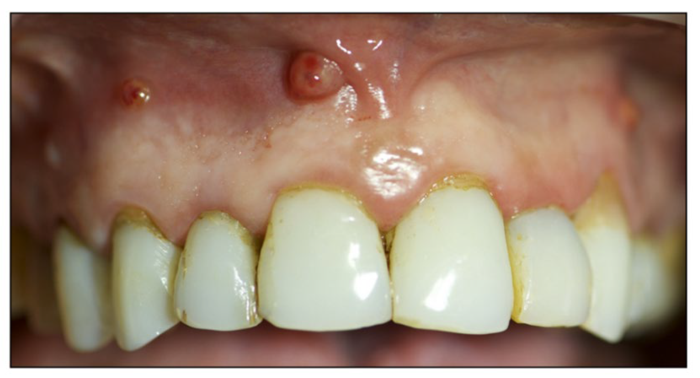
Abstract
:1. Introduction
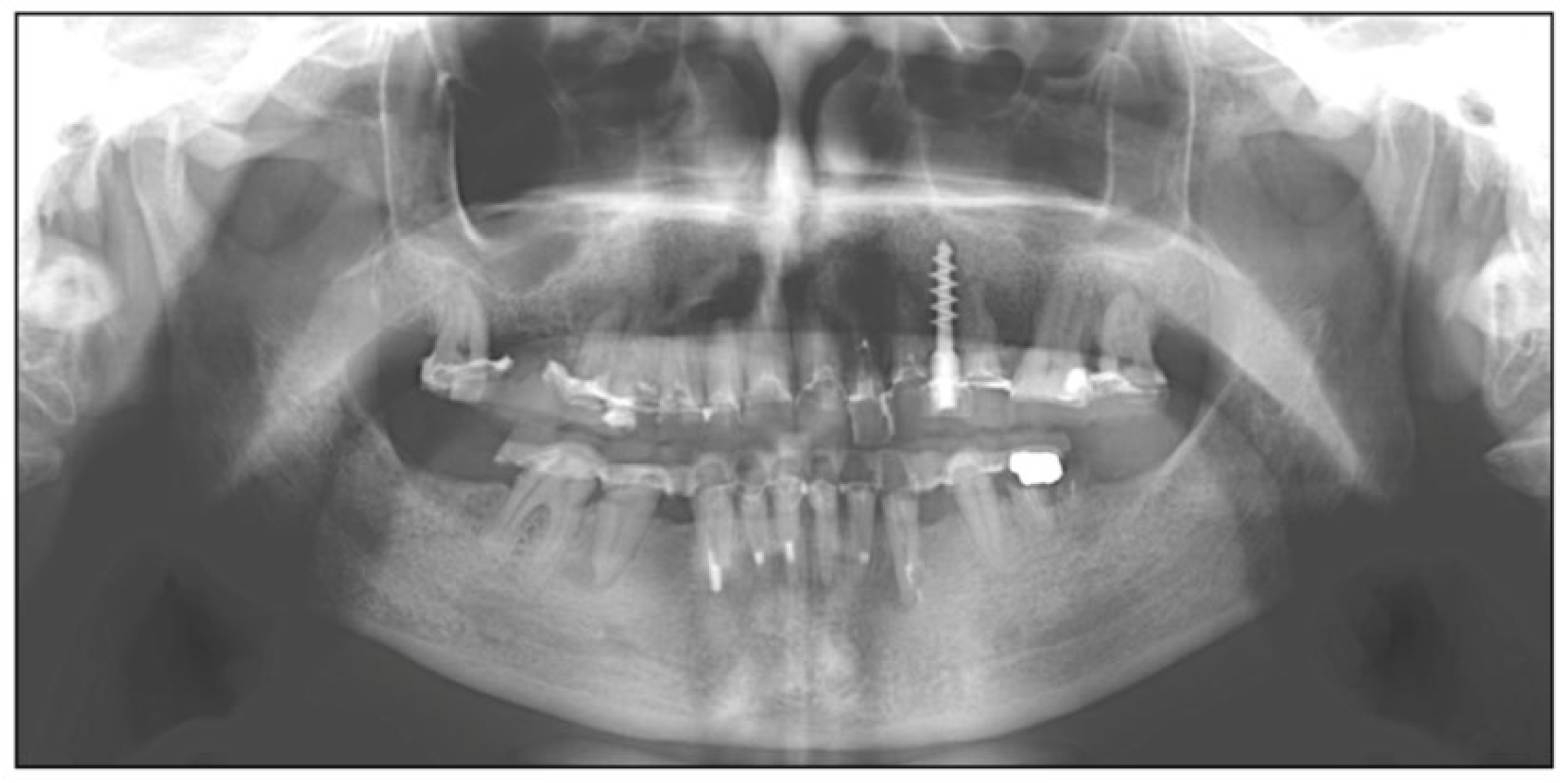
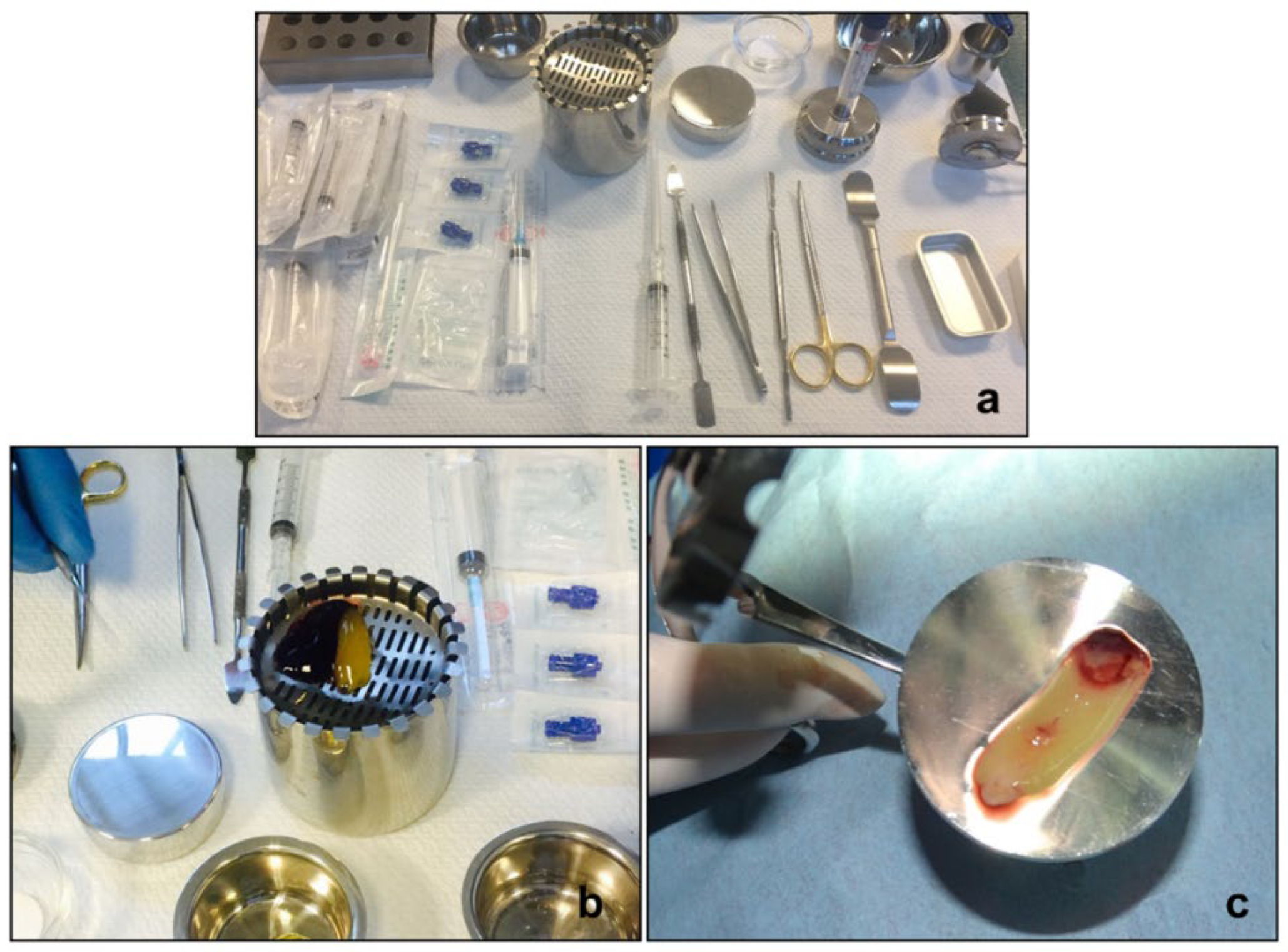
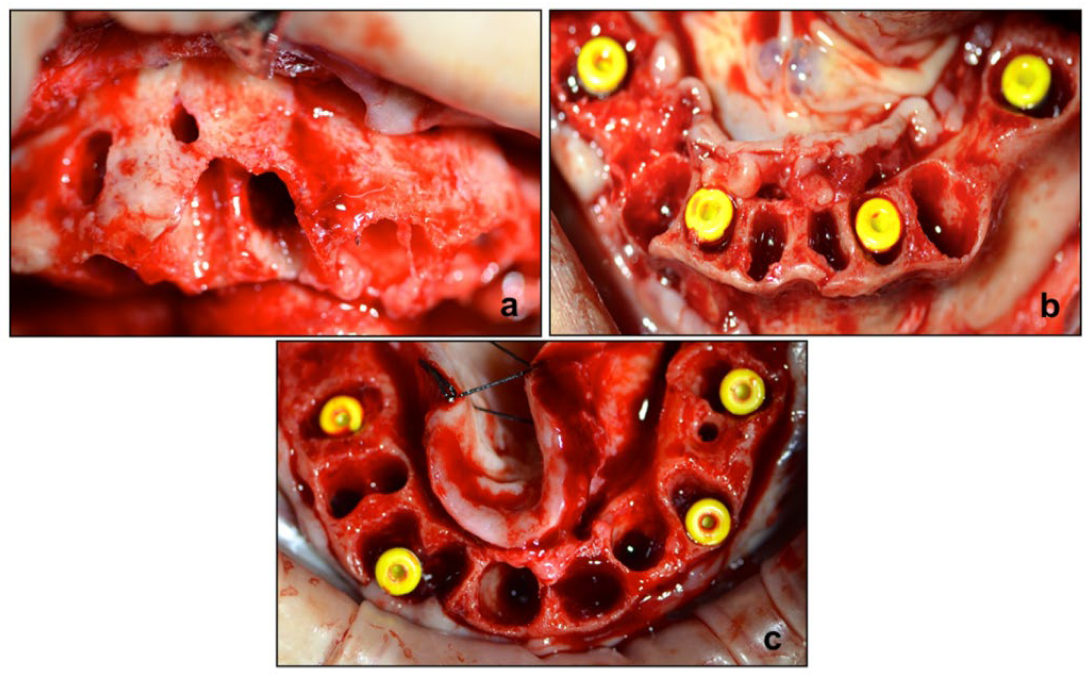
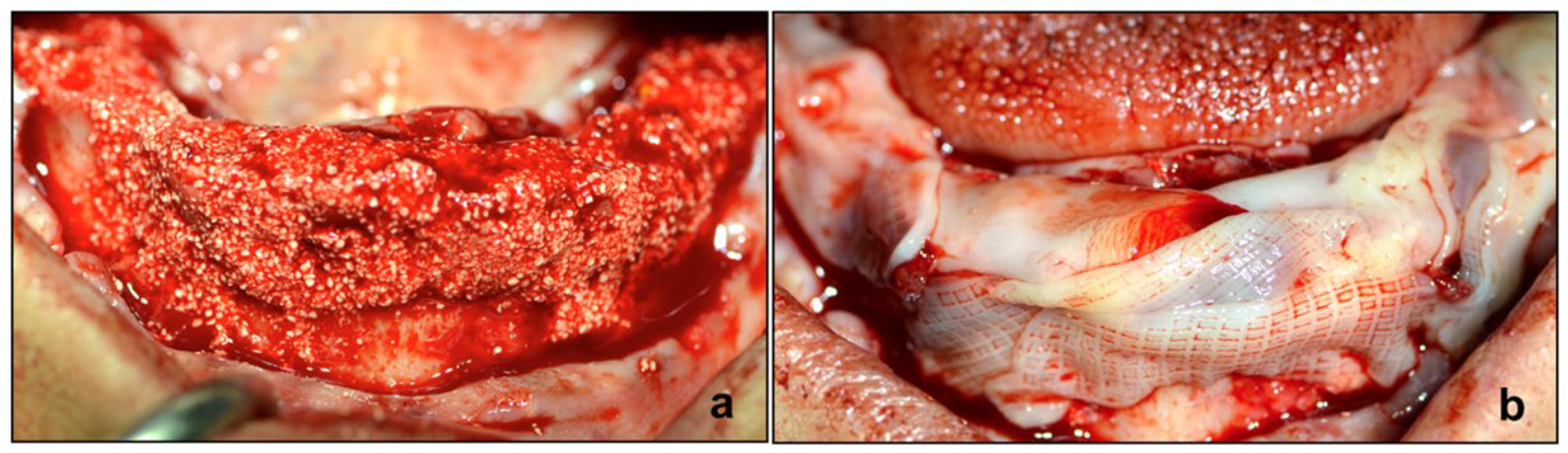
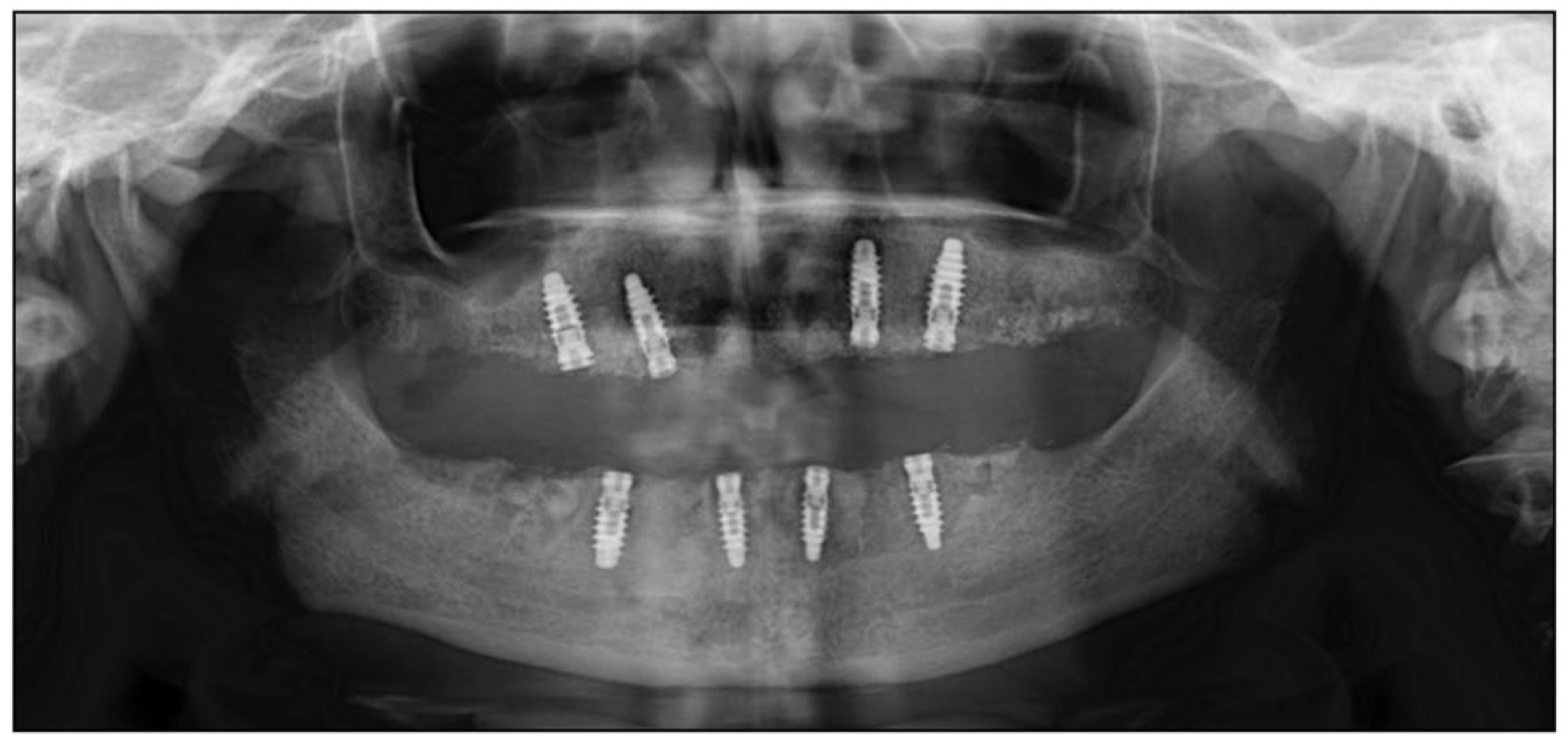
2. Case Report
3. Discussion
4. Conclusions
Author Contributions
Funding
Institutional Review Board Statement
Informed Consent Statement
Conflicts of Interest
References
- Ra, G.; Wo, Q. Bone regeneration in dentistry: An overview. J. Biol. Regul. Homeost. Agents 2021, 35, 37–46. [Google Scholar] [PubMed]
- Roca-Millan, E.; Jané-Salas, E.; Estrugo-Devesa, A.; López-López, J. Evaluation of bone gain and complication rates after guided bone regeneration with titanium foils: A systematic review. Materials 2020, 13, 5346. [Google Scholar] [CrossRef] [PubMed]
- Rodella, L.F.; Favero, G.; Labanca, M. Biomaterials in maxillofacial surgery: Membranes and grafts. Int. J. Biomed. Sci. 2011, 7, 81–88. [Google Scholar] [PubMed]
- Toledano, M.; Vallecillo-Rivas, M.; Osorio, M.T.; Muñoz-Soto, E.; Toledano-Osorio, M.; Vallecillo, C.; Toledano, R.; Lynch, C.D.; Serrera-Figallo, M.A.; Osorio, R. Zn-containing membranes for guided bone regeneration in dentistry. Polymers 2021, 13, 1797. [Google Scholar] [CrossRef] [PubMed]
- Park, W.B.; Kim, Y.J.; Han, J.Y.; Kang, P. Successful management of dental implants in postoperative maxillary cyst: A case report with a 13-year follow-up. J. Oral Implantol. 2020, 46, 133–138. [Google Scholar] [CrossRef]
- Payer, M.; Tan, W.C.; Han, J.; Ivanovski, S.; Mattheos, N.; Pjetursson, B.E.; Zhuang, L.; Fokas, G.; Wong, M.; Acham, S.; et al. The effect of systemic antibiotics on clinical and patient-reported outcome measures of oral implant therapy with simultaneous guided bone regeneration. Clin. Oral Implants Res. 2020, 31, 442–451. [Google Scholar] [CrossRef] [Green Version]
- Sanz-Sánchez, I.; Sanz-Martín, I.; Ortiz-Vigón, A.; Molina, A.; Sanz, M. Complications in bone-grafting procedures: Classification and management. Periodontology 2022, 88, 86–102. [Google Scholar] [CrossRef]
- Dragonas, P.; Schiavo, J.H.; Avila-Ortiz, G.; Palaiologou, A.; Katsaros, T. Plasma rich in growth factors (PRGF) in intraoral bone grafting procedures: A systematic review. J. Craniomaxillofac. Surg. 2019, 47, 443–453. [Google Scholar] [CrossRef]
- Rossi, R., Jr.; Garg, A.K.; Kurtzman, G.M. Novel protocols for the production of autologous blood concentrates with high platelet volume. Compend. Contin. Educ. Dent. 2022, 43, 140–145, quiz 146. [Google Scholar]
- Solakoglu, Ö.; Heydecke, G.; Amiri, N.; Anitua, E. The use of plasma rich in growth factors (PRGF) in guided tissue regeneration and guided bone regeneration. A review of histological, immunohistochemical, histomorphometrical, radiological and clinical results in humans. Ann. Anat. 2020, 231, 151528. [Google Scholar] [CrossRef]
- Arshad, S.; Tehreem, F.; Rehab Khan, M.; Ahmed, F.; Marya, A.; Karobari, M.I. Platelet-rich fibrin used in regenerative endodontics and dentistry: Current uses, limitations, and future recommendations for application. Int. J. Dent. 2021, 2021, 4514598. [Google Scholar] [CrossRef]
- Gelpi, F.; De Santis, D.; Luciano, U.; Bertajola, A.; Bernardello, F.; Zambotti, T.; Causarano, G.; Zarantonello, M.; Iurlaro, A.; Poscolere, A.; et al. Platelet rich plasma grafting technique combined with trans-sinusal post-extractive implants placement in the posterior maxilla: A technical report and brief literature review. J. Biol. Regul. Homeost. Agents 2020, 34, 9–20. [Google Scholar]
- Ulusoy, A.T.; Turedi, I.; Cimen, M.; Cehreli, Z.C. Evaluation of blood clot, platelet-rich plasma, platelet-rich fibrin, and platelet pellet as scaffolds in regenerative endodontic treatment: A prospective randomized trial. J. Endod. 2019, 45, 560–566. [Google Scholar] [CrossRef]
- Zotti, F.; Albanese, M.; Rodella, L.F.; Nocini, P.F. Platelet-rich plasma in treatment of temporomandibular joint dysfunctions: Narrative review. Int. J. Mol. Sci. 2019, 20, 227. [Google Scholar] [CrossRef] [Green Version]
- Borsani, E.; Bonazza, V.; Buffoli, B.; Cocchi, M.A.; Castrezzati, S.; Scarì, G.; Baldi, F.; Pandini, S.; Licenziati, S.; Parolini, S.; et al. Biological characterization and in vitro effect of human concentrated growth factor preparation: An innovative approach to tissue regeneration. Biol. Med. 2015, 7, 5. [Google Scholar] [CrossRef] [Green Version]
- Chen, J.; Wan, Y.; Lin, Y.; Jiang, H. Considerations for clinical use of concentrated growth factor in maxillofacial regenerative medicine. J. Craniofac. Surg. 2021, 132, 1316–1321. [Google Scholar] [CrossRef]
- Liao, Y.; Fang, Y.; Zhu, H.; Huang, Y.; Zou, G.; Dai, B.; Rausch, M.A.; Shi, B. Concentrated growth factors promote hBMSCs osteogenic differentiation in a co-culture system with HUVECs. Front. Bioeng. Biotechnol. 2021, 10, 837295. [Google Scholar] [CrossRef]
- Nivedhitha, M.S.; Jacob, B.; Ranganath, A. Concentrated growth factor: A novel platelet concentrate for revascularization of immature permanent teeth-A report of two cases. Case Rep. Dent. 2020, 2020, 1329145. [Google Scholar] [CrossRef]
- Bonazza, V.; Borsani, E.; Buffoli, B.; Parolini, S.; Inchingolo, F.; Rezzani, R.; Rodella, L.F. In vitro treatment with concentrated growth factors (CGF) and sodium orthosilicate positively affects cell renewal in three different human cell lines. Cell Biol. Int. 2018, 42, 353–364. [Google Scholar] [CrossRef]
- Chen, J.; Jiang, H. A Comprehensive review of concentrated growth factors and their novel applications in facial reconstructive and regenerative medicine. Aesthetic Plast. Surg. 2020, 44, 1047–1057. [Google Scholar] [CrossRef]
- Rodella, L.F.; Favero, G.; Boninsegna, R.; Buffoli, B.; Labanca, M.; Scarì, G.; Sacco, L.; Batani, T.; Rezzani, R. Growth factors, CD34 positive cells, and fibrin network analysis in concentrated growth factors fraction. Microsc. Res. Tech. 2011, 74, 772–777. [Google Scholar] [CrossRef]
- Da, W.; Tao, L.; Wen, K.; Tao, Z.; Wang, S.; Zhu, Y. Protective role of melatonin against postmenopausal bone loss via enhancement of citrate secretion from osteoblasts. Front. Pharmacol. 2020, 11, 667. [Google Scholar] [CrossRef] [PubMed]
- Najeeb, S.; Khurshid, Z.; Zohaib, S.; Zafar, M.S. Therapeutic potential of melatonin in oral medicine and periodontology. Kaohsiung J. Med. Sci. 2016, 32, 391–396. [Google Scholar] [CrossRef] [PubMed]
- Reiter, R.J.; Rosales-Corral, S.A.; Liu, X.Y.; Acuna-Castroviejo, D.; Escames, G.; Tan, D.X. Melatonin in the oral cavity: Physiological and pathological implications. J. Periodontal. Res. 2015, 50, 9–17. [Google Scholar] [CrossRef]
- Favero, G.; Franceschetti, L.; Bonomini, F.; Rodella, L.F.; Rezzani, R. Melatonin as an anti-inflammatory agent modulating inflammasome activation. Int. J. Endocrinol. 2017, 2017, 1835195. [Google Scholar] [CrossRef] [Green Version]
- Moretti, R.; Zanin, A.; Pansiot, J.; Spiri, D.; Manganozzi, L.; Kratzer, I.; Favero, G.; Vasiljevic, A.; Rinaldi, V.E.; Pic, I.; et al. Melatonin reduces excitotoxic blood-brain barrier breakdown in neonatal rats. Neuroscience 2015, 311, 382–397. [Google Scholar] [CrossRef]
- Wongchitrat, P.; Shukla, M.; Sharma, R.; Govitrapong, P.; Reiter, R.J. Role of melatonin on virus-induced neuropathogenesis-A concomitant therapeutic strategy to understand SARS-CoV-2 infection. Antioxidants 2021, 10, 47. [Google Scholar] [CrossRef]
- Bonazza, V.; Hajistilly, C.; Patel, D.; Patel, J.; Woo, R.; Cocchi, M.A.; Buffoli, B.; Lancini, D.; Gheno, E.; Rezzani, R.; et al. Growth factors release from concentrated growth factors: Effect of β-tricalcium phosphate addition. J. Craniofac. Surg. 2018, 29, 2291–2295. [Google Scholar] [CrossRef]
- Bonazza, V.; Borsani, E.; Buffoli, B.; Castrezzati, S.; Rezzani, R.; Rodella, L.F. How the different material and shape of the blood collection tube influences the Concentrated Growth Factors production. Microsc. Res. Tech. 2016, 79, 1173–1178. [Google Scholar] [CrossRef]
- Ranganathan, A.T.; Chandran, C.R. Platelet-rich fibrin in the treatment of periodontal bone defects. J. Contemp. Dent. Pract. 2014, 15, 372–375. [Google Scholar] [CrossRef]
- Lei, L.; Yu, Y.; Han, J.; Shi, D.; Sun, W.; Zhang, D.; Chen, L. Quantification of growth factors in advanced platelet-rich fibrin and concentrated growth factors and their clinical efficacy as adjunctive to the GTR procedure in periodontal intrabony defects. J. Periodontol. 2020, 91, 462–472. [Google Scholar] [CrossRef]
- Dai, Y.; Han, X.H.; Hu, L.H.; Wu, H.W.; Huang, S.Y.; Lü, Y.P. Efficacy of concentrated growth factors combined with mineralized collagen on quality of life and bone reconstruction of guided bone regeneration. Regen. Biomater. 2020, 7, 313–320. [Google Scholar] [CrossRef]
- Malli Sureshbabu, N.; Selvarasu, K.; Nandakumar, M.; Selvam, D. Concentrated growth factors as an ingenious biomaterial in regeneration of bony defects after periapical surgery: A report of two cases. Case Rep. Dent. 2019, 2019, 7046203. [Google Scholar] [CrossRef] [Green Version]
- Qiao, J.; An, N.; Ouyang, X. Quantification of growth factors in different platelet concentrates. Platelets 2017, 28, 774–778. [Google Scholar] [CrossRef]
- Mirković, S.; Djurdjević-Mirković, T.; Pugkar, T. Application of concentrated growth factors in reconstruction of bone defects after removal of large jaw cysts—The two cases report. Vojnosanit. Pregl. 2015, 72, 368–371. [Google Scholar] [CrossRef]
- Durmuşlar, M.C.; Balli, U.; Dede, F.Ö.; Misir, A.F.; Bariş, E.; Kürkçü, M.; Kahraman, S.A. Histological evaluation of the effect of concentrated growth factor on bone healing. J. Craniofac. Surg. 2016, 27, 1494–1497. [Google Scholar] [CrossRef]
- Yu, T.T.; Liu, J.; Yin, J.J.; Xu, X.N.; Yan, S.J.; Lan, J. Effects of concentrated growth factors on relieving postoperative reaction of guided bone regeneration in esthetic zone. Hua Xi Kou Qiang Yi Xue Za Zhi. 2019, 37, 398–402. [Google Scholar] [CrossRef]
- Taschieri, S.; Khijmatgar, S.; Corbella, S.; Francetti, L.; Parrini, M.; Corradini, C.; Del Fabbro, M. Effect of concentrated growth factors on quality of life of patients undergoing implant therapy: A cohort study. J. Biol. Regul. Homeost. Agents 2021, 35, 147–154. [Google Scholar] [CrossRef]
- Li, C.; Chen, X.; Qiao, S.; Liu, X.; Liu, C.; Zhu, D.; Su, J.; Wang, Z. Melatonin lowers edema after spinal cord injury. Neural. Regen. Res. 2014, 9, 2205–2210. [Google Scholar] [CrossRef]
- Liu, X.; Wang, Y.; Yang, J.; Liu, Y.; Zhou, D.; Hou, M.; Xiang, L. Anti-edema effect of melatonin on spinal cord injury in rats. Biomed. Pap. Med. Fac. Univ. Palacky Olomouc. Czech. Repub. 2015, 159, 220–226. [Google Scholar] [CrossRef] [Green Version]
- Cobo-Vázquez, C.; Fernández-Tresguerres, I.; Ortega-Aranegui, R.; López-Quiles, J. Effects of local melatonin application on post-extraction sockets after third molar surgery. A pilot study. Med. Oral Patol. Oral Cir. Bucal. 2014, 19, e628–e633. [Google Scholar] [CrossRef] [PubMed]
- Andersen, L.P.; Gögenur, I.; Rosenberg, J.; Reiter, R.J. The safety of melatonin in humans. Clin. Drug Investig. 2016, 36, 169–175. [Google Scholar] [CrossRef] [PubMed]


Publisher’s Note: MDPI stays neutral with regard to jurisdictional claims in published maps and institutional affiliations. |
© 2022 by the authors. Licensee MDPI, Basel, Switzerland. This article is an open access article distributed under the terms and conditions of the Creative Commons Attribution (CC BY) license (https://creativecommons.org/licenses/by/4.0/).
Share and Cite
Leonida, A.; Favero, G.; Caccianiga, P.; Ceraulo, S.; Rodella, L.F.; Rezzani, R.; Caccianiga, G. Concentrated Growth Factors (CGF) Combined with Melatonin in Guided Bone Regeneration (GBR): A Case Report. Diagnostics 2022, 12, 1257. https://doi.org/10.3390/diagnostics12051257
Leonida A, Favero G, Caccianiga P, Ceraulo S, Rodella LF, Rezzani R, Caccianiga G. Concentrated Growth Factors (CGF) Combined with Melatonin in Guided Bone Regeneration (GBR): A Case Report. Diagnostics. 2022; 12(5):1257. https://doi.org/10.3390/diagnostics12051257
Chicago/Turabian StyleLeonida, Alessandro, Gaia Favero, Paolo Caccianiga, Saverio Ceraulo, Luigi Fabrizio Rodella, Rita Rezzani, and Gianluigi Caccianiga. 2022. "Concentrated Growth Factors (CGF) Combined with Melatonin in Guided Bone Regeneration (GBR): A Case Report" Diagnostics 12, no. 5: 1257. https://doi.org/10.3390/diagnostics12051257