
Three-Dimensional Echocardiography Assessment of Right Ventricular Volumes and Function: Technological Perspective and Clinical Application
Abstract
:1. Introduction
2. Right Ventricular Anatomy
3. Right Ventricular Mode of Contractility
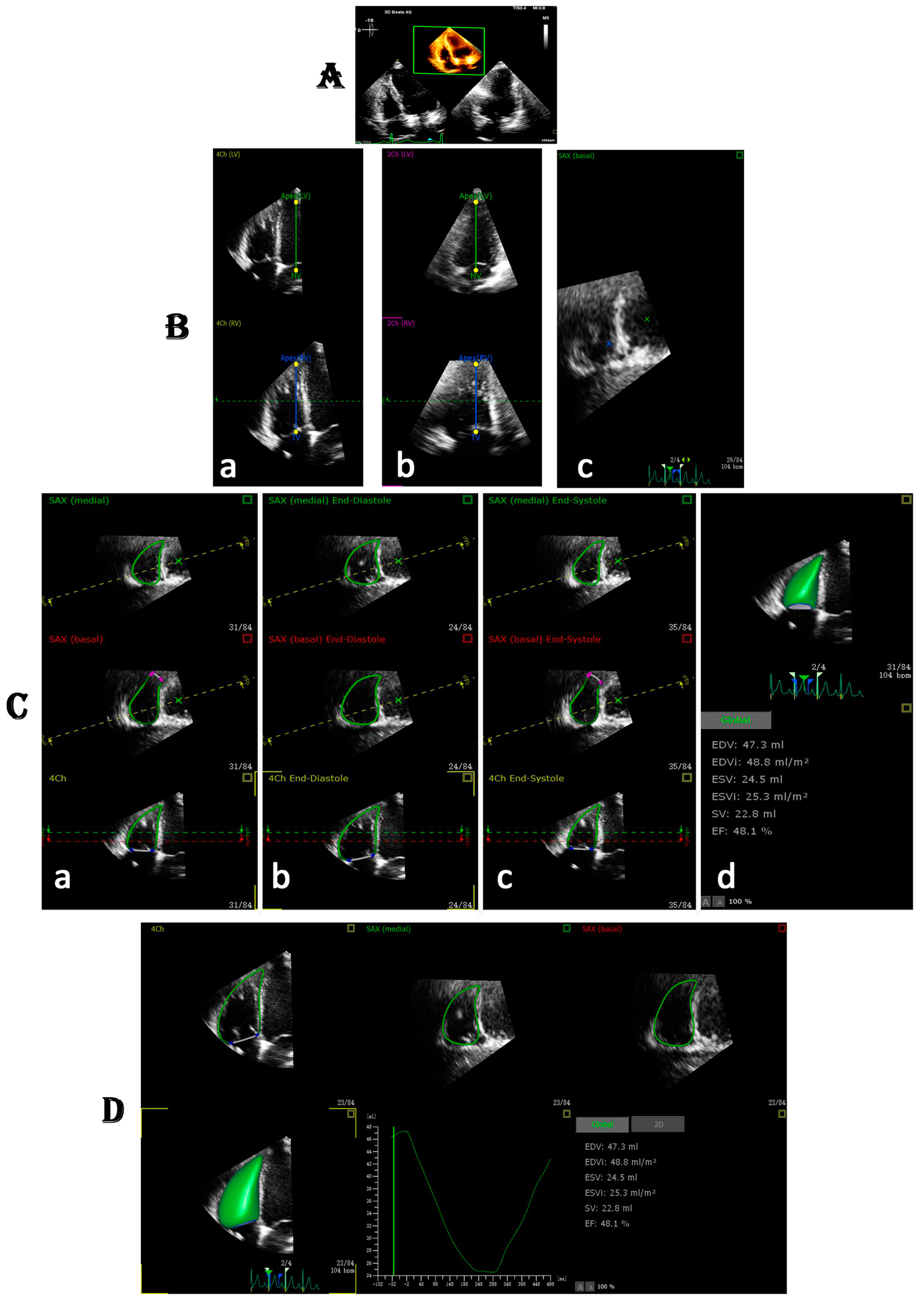
4. Acquisition of RV Dataset for 3D Images
5. Reliability, Feasibility, and Reproducibility of RV Volumes and Function with Three-Dimensional Echocardiography
6. Impact of RV Function and Frame Rate on 3DE Software
7. Clinical Application
7.1. Prognostic Value of RV Function in Patients with Various Cardiovascular Diseases
7.2. Pulmonary Hypertension
7.3. Heart Failure
7.4. Congenital Heart Disease
7.5. Valvular Heart Disease
7.6. Myocardial Infarction
7.7. Post Cardiac Surgery

| References | Disease | Sample Size (n) | Age (Years) | Men, n (%) | RVEF (%) | 3D-STE Parameters | Main Findings |
|---|---|---|---|---|---|---|---|
| Lu, K.J. et al. [15] | HF | 60 | 45 ± 10 | 60% | 53 ± 8 | RVGLS | RV GLS best predicted the presence of RV dysfunction, |
| Seckin Gobut, O. et al. [44] | MS | 20 C 20 † 20 # | 46.9 ± 11.6 46.9 ± 10.4 49.6 ± 11.8 | 13 (65%) 13 (65%) 13 (65%) | – | RV-FWLS | RV deformation indices showed significant decrease in correlation with the severity of the mitral stenosis |
| Jone, P.N. et al. [59] | PHT | 96 | 8.1 ± 5.2 | 53 (55%) * | 46 ± 5 | RV LS free wall and septum | PH patients have impaired RV function compared with normal children. 3D RV EF, volumes, FAC, and free wall RV strain serve as outcome predictors for PH patients. |
| Moceri, P. et al. [60] | PHT | 104 | 65.9 [62.0–68.8] | 58 (55.8%) * | 35.6 ± 9.7 | RVGLS, RVCS, RVAS | RV strain patterns gradually worsen in PH patients and provide independent prognostic information. This technique could help better stratify the risk in PH patients. |
| Vitarelli, A. et al. [61] | PHT | 73 | 53 ± 11 | 44% | 35.5 ± 7.6 | RVGLS, RV-GFW, RV-FWAS | In PH patients, the quantitative assessment of global and regional RV function by 3D and STE provides useful hemodynamic and prognostic information. |
| Vitarelli, A. et al. [62] | PE | 66 66 c | 53 ± 11 | 32 | 37 ± 8 | MFW RVLS | Decreases in MFW RVLS and 3D RVEF may persist during short-term and long-term follow-up and correlate with unfavorable outcomes |
| Smith, B.C.F. et al. [64] | PHT | 97 | 60.6 ± 15.3 | 34 (35) | 31.4 ± 9.6 | RVGLS, RVCS, RVAS | AS best correlated with RVEF and provides prognostic information independent of other variables. |
| Meng, Y. et al. [74] | HFpEF | 81 (n = 42) a (n = 39) b | 61 ± 12 a 63 ± 13 b | 27 (64%) a 26 (67%) b | 47 ± 4 a 44 ± 5 b | 3D-RVFWLS | 3D-STE parameters were powerful predictors of poor outcomes, providing similar predictive values as 2D-STE indices in patients with HFpEF. |
| Tian, F. et al. [75] | HF | 105 | 44 ± 16 | 17 (16%) * | 26.89 ± 8.09 | 3D-RVFWLS | 3D-RVFWLS could be a promising noninvasive parameter in identifying severe MF in patients with end-stage HF |
| Sato, T. et al. [80] | CHD | 64 | 10.6 (2.4–18.4) | 28 (43.8%) * | 51.2 (22.9–64.2) | GLS, GPS, GCS | Analysis of a single 3D-STE clip of the cardiac cycle provides useful information regarding both volume and the functional status of HLHS, which can be useful during longitudinal follow-up as outpatients. |
| Ishizu, T. et al. [81] | WPW | 38 | 42 ± 21 | 22 (57%) | – | 3DSTE (AI) | Isochrone AI with 3D-STE may be a promising noninvasive imaging tool to assess cardiac synchronized activation in normal hearts and detect abnormal breakthrough of mechanical activation from both atrioventricular annuli in Wolff–Parkinson–White syndrome. |
| Cui, C. et al. [82] | TOF | 20 22 c | 24.7 ± 8.6 20.6 ± 7.0 c | 12 11 | 28.1 ± 64.4 31.9 ± 63.8 c | RV-GAS | progressive RV dysfunction in patients with repaired TOF. |
| Vitarelli, A. et al. [95] | MVD | 32 | 79.4 ± 5.5 | 18 (56.2%) | 53.6 ± 7.2 c 42.5 ± 7.6 β 52.2 ± 7.9 Ω | GLS, FWLS | 3D-STE showed overall biventricular strain improvement after clip implantation and lower post procedural LV strain in patients with worse preexisting RV function. |
8. Conclusions
Author Contributions
Funding
Institutional Review Board Statement
Informed Consent Statement
Data Availability Statement
Conflicts of Interest
References
- Li, Y.; Li, H.; Zhu, S.; Xie, Y.; Wang, B.; He, L.; Zhang, D.; Zhang, Y.; Yuan, H.; Wu, C.; et al. Prognostic Value of Right Ventricular Longitudinal Strain in Patients With COVID. JACC Cardiovasc. Imaging 2020, 13, 2287–2299. [Google Scholar] [CrossRef] [PubMed]
- Seo, J.; Jung, I.H.; Park, J.H.; Kim, G.S.; Lee, H.Y.; Byun, Y.S.; Kim, B.O.; Rhee, K.J. The prognostic value of 2D strain in assessment of the right ventricle in patients with dilated cardiomyopathy. Eur. Heart J. Cardiovasc. Imaging 2019, 20, 1043–1050. [Google Scholar] [CrossRef]
- Rigolin, V.H.; Robiolio, P.A.; Wilson, J.S.; Harrison, J.K.; Bashore, T.M. The forgotten chamber: The importance of the right ventricle. Cathet. Cardiovasc. Diagn. 1995, 35, 18–28. [Google Scholar] [CrossRef] [PubMed]
- Kawut, S.M.; Barr, R.G.; Lima, J.A.C.; Praestgaard, A.; Johnson, W.C.; Chahal, H.; Ogunyankin, K.O.; Bristow, M.R.; Kizer, J.R.; Tandri, H.; et al. Right ventricular structure is associated with the risk of heart failure and cardiovascular death: The multi-ethnic study of atherosclerosis (MESA)-right ventricle study. Circulation 2012, 126, 1681–1688. [Google Scholar] [CrossRef] [PubMed] [Green Version]
- DeFaria Yeh D, Foster EIs MRI the preferred method for evaluating right ventricular size and function in patients with congenital heart disease?: MRI is not the preferred method for evaluating right ventricular size and function in patients with congenital heart disease. Circ. Cardiovasc. Imaging 2014, 7, 198–205. [CrossRef] [Green Version]
- Kawel-Boehm, N.; Hetzel, S.J.; Ambale-Venkatesh, B.; Captur, G.; Francois, C.J.; Jerosch-Herold, M.; Salerno, M.; Teague, S.D.; Valsangiacomo-Buechel, E.; van der Geest, R.J.; et al. Reference ranges (“normal values”) for cardiovascular magnetic resonance (CMR) in adults and children: 2020 update. J. Cardiovasc. Magn. Reson. 2020, 22, 87. [Google Scholar] [CrossRef]
- Kawel-Boehm, N.; Maceira, A.; Valsangiacomo-Buechel, E.R.; Vogel-Claussen, J.; Turkbey, E.B.; Williams, R.; Plein, S.; Tee, M.; Eng, J.; Bluemke, D.A. Normal values for cardiovascular magnetic resonance in adults and children. J. Cardiovasc. Magn. Reson. 2015, 17, 29. [Google Scholar] [CrossRef] [Green Version]
- Maffei, E.; Messalli, G.; Martini, C.; Nieman, K.; Catalano, O.; Rossi, A.; Seitun, S.; Guaricci, A.I.; Tedeschi, C.; Mollet, N.R.; et al. Left and right ventricle assessment with Cardiac CT: Validation study vs. Cardiac MR. Eur. Radiol. 2012, 22, 1041–1049. [Google Scholar] [CrossRef] [Green Version]
- Rudski, L.G.; Lai, W.W.; Afilalo, J.; Hua, L.; Handschumacher, M.D.; Chandrasekaran, K.; Solomon, S.D.; Louie, E.K.; Schiller, N.B. Guidelines for the Echocardiographic Assessment of the Right Heart in Adults: A Report from the American Society of Echocardiography. Endorsed by the European Association of Echocardiography, a registered branch of the European Society of Cardiology, and the Canadian Society of Echocardiography. J. Am. Soc. Echocardiogr. 2010, 23, 685–713. [Google Scholar] [CrossRef]
- Fernández-Golfín, C.; Zamorano, J.L. Three-Dimensional Echocardiography and Right Ventricular Function. Circ. Cardiovasc. Imaging 2017, 10, e006099. [Google Scholar] [CrossRef] [Green Version]
- Cosyns, B.; Le Tourneau, T.; Rudski, L. Right here, right now! Eur. Heat. J. Cardiovasc. Imaging 2020, 21, 22–23. [Google Scholar] [CrossRef] [PubMed]
- Lang, R.M.; Badano, L.P.; Mor-Avi, V.; Afilalo, J.; Armstrong, A.; Ernande, L.; Flachskampf, F.A.; Foster, E.; Goldstein, S.A.; Kuznetsova, T.; et al. Recommendations for cardiac chamber quantification by echocardiography in adults: An update from the American society of echocardiography and the European association of cardiovascular imaging. J. Am. Soc. Echocardiogr. 2015, 16, 233–271. [Google Scholar] [CrossRef]
- Leibundgut, G.; Rohner, A.; Grize, L.; Bernheim, A.; Kessel-Schaefer, A.; Bremerich, J.; Zellweger, M.; Buser, P.; Handke, M. Dynamic Assessment of Right Ventricular Volumes and Function by Real-Time Three-Dimensional Echocardiography: A Comparison Study With Magnetic Resonance Imaging in 100 Adult Patients. J. Am. Soc. Echocardiogr. 2010, 23, 116–126. [Google Scholar] [CrossRef] [PubMed]
- Park, J.H.; Negishi, K.; Kwon, D.H.; Popovic, Z.B.; Grimm, R.A.; Marwick, T.H. Validation of global longitudinal strain and strain rate as reliable markers of right ventricular dysfunction: Comparison with cardiac magnetic resonance and outcome. J. Cardiovasc. Ultrasound 2014, 22, 113–120. [Google Scholar] [CrossRef] [Green Version]
- Lu, K.J.; Chen, J.X.; Profitis, K.; Kearney, L.G.; DeSilva, D.; Smith, G.; Ord, M.; Harberts, S.; Calafiore, P.; Jones, E.; et al. Right ventricular global longitudinal strain is an independent predictor of right ventricular function: A multimodality study of cardiac magnetic resonance imaging, real time three-dimensional echocardiography and speckle tracking echocardiography. Echocardiography 2015, 32, 966–974. [Google Scholar] [CrossRef]
- Medvedofsky, D.; Addetia, K.; Patel, A.R.; Sedlmeier, A.; Baumann, R.; Mor-Avi, V.; Lang, R.M. Novel Approach to Three-Dimensional Echocardiographic Quantification of Right Ventricular Volumes and Function from Focused Views. J. Am. Soc. Echocardiogr. 2015, 28, 1222–1231. [Google Scholar] [CrossRef]
- Grewal, J.; Majdalany, D.; Syed, I.; Pellikka, P.; Warnes, C.A. Three-Dimensional Echocardiographic Assessment of Right Ventricular Volume and Function in Adult Patients With Congenital Heart Disease: Comparison With Magnetic Resonance Imaging. J. Am. Soc. Echocardiogr. 2010, 23, 127–133. [Google Scholar] [CrossRef]
- Park, J.B.; Lee, S.P.; Lee, J.H.; Yoon, Y.E.; Park, E.A.; Kim, H.K.; Lee, W.; Kim, Y.J.; Cho, G.Y.; Sohn, D.W. Quantification of Right Ventricular Volume and Function Using Single-Beat Three-Dimensional Echocardiography: A Validation Study with Cardiac Magnetic Resonance. J. Am. Soc. Echocardiogr. 2016, 29, 392–401. [Google Scholar] [CrossRef]
- Muraru, D.; Spadotto, V.; Cecchetto, A.; Romeo, G.; Aruta, P.; Ermacora, D.; Jenei, C.; Cucchini, U.; Iliceto, S.; Badano, L.P. New speckle-tracking algorithm for right ventricular volume analysis from three-dimensional echocardiographic data sets: Validation with cardiac magnetic resonance and comparison with the previous analysis tool. Eur. Heart J. Cardiovasc. Imaging 2016, 17, 1279–1289. [Google Scholar] [CrossRef]
- Nagata, Y.; Wu, V.C.C.; Kado, Y.; Otani, K.; Lin, F.C.; Otsuji, Y.; Negishi, K.; Takeuchi, M. Prognostic Value of Right Ventricular Ejection Fraction Assessed by Transthoracic 3D Echocardiography. Circ. Cardiovasc. Imaging 2017, 10, e005384. [Google Scholar] [CrossRef] [Green Version]
- Murata, M.; Tsugu, T.; Kawakami, T.; Kataoka, M.; Minakata, Y.; Endo, J.; Tsuruta, H.; Itabashi, Y.; Maekawa, Y.; Murata, M.; et al. Prognostic value of three-dimensional echocardiographic right ventricular ejection fraction in patients with pulmonary arterial hypertension. Oncotarget 2016, 7, 86781–86790. [Google Scholar] [CrossRef] [PubMed] [Green Version]
- Surkova, E.; Muraru, D.; Genovese, D.; Aruta, P.; Palermo, C.; Badano, L.P. Relative Prognostic Importance of Left and Right Ventricular Ejection Fraction in Patients With Cardiac Diseases. J. Am. Soc. Echocardiogr. 2019, 32, 1407–1415. [Google Scholar] [CrossRef] [PubMed]
- Tamborini, G.; Marsan, N.A.; Gripari, P.; Maffessanti, F.; Brusoni, D.; Muratori, M.; Caiani, E.G.; Fiorentini, C.; Pepi, M. Reference Values for Right Ventricular Volumes and Ejection Fraction With Real-Time Three-Dimensional Echocardiography: Evaluation in a Large Series of Normal Subjects. J. Am. Soc. Echocardiogr. 2010, 23, 109–115. [Google Scholar] [CrossRef] [PubMed]
- Sánchez-Quintana, D.; Doblado-Calatrava, M.; Cabrera, J.A.; Macías, Y.; Saremi, F. Anatomical Basis for the Cardiac Interventional Electrophysiologist. Biomed Res. Int. 2015, 2015, 547364. [Google Scholar] [CrossRef]
- Ho, S.Y.; Nihoyannopoulos, P. Anatomy, echocardiography, and normal right ventricular dimensions. Heart 2006, 92 (Suppl. 1), i2–i13. [Google Scholar] [CrossRef] [Green Version]
- Dell’Italia, L.J. The right ventricle: Anatomy, physiology, and clinical importance. Curr. Probl. Cardiol. 1991, 16, 658–720. [Google Scholar] [CrossRef]
- Atsumi, A.; Seo, Y.; Ishizu, T.; Nakamura, A.; Enomoto, Y.; Harimura, Y.; Okazaki, T.; Abe, Y.; Aonuma, K. Right Ventricular Deformation Analyses Using a Three-Dimensional Speckle-Tracking Echocardiographic System Specialized for the Right Ventricle. J. Am. Soc. Echocardiogr. 2016, 29, 402–411. [Google Scholar] [CrossRef]
- Ishizu, T.; Seo, Y.; Atsumi, A.; Tanaka, Y.O.; Yamamoto, M.; Machino-Ohtsuka, T.; Horigome, H.; Aonuma, K.; Kawakami, Y. Global and Regional Right Ventricular Function Assessed by Novel Three-Dimensional Speckle-Tracking Echocardiography. J. Am. Soc. Echocardiogr. 2017, 30, 1203–1213. [Google Scholar] [CrossRef] [PubMed]
- Leather, H.A.; Ama’, R.; Missant, C.; Rex, S.; Rademakers, F.E.; Wouters, P.F. Longitudinal but not circumferential deformation reflects global contractile function in the right ventricle with open pericardium. Am. J. Physiol. Heart Circ. Physiol. 2006, 290. [Google Scholar] [CrossRef]
- Meier, G.D.; Bove, A.A.; Santamore, W.P.; Lynch, P.R. Contractile function in canine right ventricle. Am. J. Physiol. Circ. Physiol. 1980, 239, 794–804. [Google Scholar] [CrossRef]
- Santamore, W.P.; Dell’Italia, L.J. Ventricular interdependence: Significant left ventricular contributions to right ventricular systolic function. Prog. Cardiovasc. Dis. 1998, 40, 289–308. [Google Scholar] [CrossRef]
- Haddad, F.; Hunt, S.A.; Rosenthal, D.N.; Murphy, D.J. Right ventricular function in cardiovascular disease, part I: Anatomy, physiology, aging, and functional assessment of the right ventricle. Circulation 2008, 117, 1436–1448. [Google Scholar] [CrossRef] [PubMed]
- Ostenfeld, E.; Carlsson, M.; Shahgaldi, K.; Roijer, A.; Holm, J. Manual correction of semi-automatic three-dimensional echocardiography is needed for right ventricular assessment in adults; Validation with cardiac magnetic resonance. Cardiovasc. Ultrasound 2012, 10, 1. [Google Scholar] [CrossRef] [Green Version]
- Badano, L.P.; Kolias, T.J.; Muraru, D.; Abraham, T.P.; Aurigemma, G.; Edvardsen, T.; D’Hooge, J.; Donal, E.; Fraser, A.G.; Marwick, T.; et al. Standardization of left atrial, right ventricular, and right atrial deformation imaging using two-dimensional speckle tracking echocardiography: A consensus document of the EACVI/ASE/Industry Task Force to standardize deformation imaging. Eur. Heart J. Cardiovasc. Imaging 2018, 19, 591–600. [Google Scholar] [CrossRef]
- Seo, Y.; Ishizu, T.; Ieda, M.; Ohte, N. Right ventricular three-dimensional echocardiography: The current status and future perspectives. J. Echocardiogr. 2020, 18, 149–159. [Google Scholar] [CrossRef]
- Medvedofsky, D.; Mor-Avi, V.; Kruse, E.; Guile, B.; Ciszek, B.; Weinert, L.; Yamat, M.; Volpato, V.; Addetia, K.; Patel, A.R.; et al. Quantification of Right Ventricular Size and Function from Contrast-Enhanced Three-Dimensional Echocardiographic Images. J. Am. Soc. Echocardiogr. 2017, 30, 1193–1202. [Google Scholar] [CrossRef]
- Genovese, D.; Rashedi, N.; Weinert, L.; Narang, A.; Addetia, K.; Patel, A.R.; Prater, D.; Gonçalves, A.; Mor-Avi, V.; Lang, R.M. Machine Learning–Based Three-Dimensional Echocardiographic Quantification of Right Ventricular Size and Function: Validation Against Cardiac Magnetic Resonance. J. Am. Soc. Echocardiogr. 2019, 32, 969–977. [Google Scholar] [CrossRef]
- Li, Y.; Zhang, L.; Gao, Y.; Wan, X.; Xiao, Q.; Zhang, Y.; Sun, W.; Xie, Y.; Zeng, Q.; Chen, Y.; et al. Comprehensive Assessment of Right Ventricular Function by Three-Dimensional Speckle-Tracking Echocardiography: Comparisons with Cardiac Magnetic Resonance Imaging. J. Am. Soc. Echocardiogr. 2021, 34, 472–482. [Google Scholar] [CrossRef]
- De Potter, T.; Weytjens, C.; Motoc, A.; Luchian, M.L.; Scheirlynck, E.; Roosens, B.; Tanaka, K.; Houard, L.; Droogmans, S.; Cosyns, B. Feasibility, reproducibility and validation of right ventricular volume and function assessment using three-dimensional echocardiography. Diagnostics 2021, 11, 699. [Google Scholar] [CrossRef]
- Laser, K.T.; Horst, J.P.; Barth, P.; Kelter-Klöpping, A.; Haas, N.A.; Burchert, W.; Kececioglu, D.; Körperich, H. Knowledge-based reconstruction of right ventricular volumes using real-time three-dimensional echocardiographic as well as cardiac magnetic resonance images: Comparison with a cardiac magnetic resonance standard. J. Am. Soc. Echocardiogr. 2014, 27, 1087–1097. [Google Scholar] [CrossRef]
- van der Zwaan, H.B.; Helbing, W.A.; McGhie, J.S.; Geleijnse, M.L.; Luijnenburg, S.E.; Roos-Hesselink, J.W.; Meijboom, F.J. Clinical Value of Real-Time Three-Dimensional Echocardiography for Right Ventricular Quantification in Congenital Heart Disease: Validation With Cardiac Magnetic Resonance Imaging. J. Am. Soc. Echocardiogr. 2010, 23, 134–140. [Google Scholar] [CrossRef] [PubMed]
- Sugeng, L.; Nesser, H.J.; Weinert, L.; Niel, J.; Ebner, C.; Steringer-Mascherbauer, R.; Bartolles, R.; Baumann, R.; Schummers, G.; Lang, R.M.; et al. Multimodality comparison of quantitative volumetric analysis of the right ventricle. Comput. Cardiol. 2009, 36, 29–32. [Google Scholar] [CrossRef] [PubMed] [Green Version]
- Otani, K.; Nabeshima, Y.; Kitano, T.; Takeuchi, M. Accuracy of fully automated right ventricular quantification software with 3D echocardiography: Direct comparison with cardiac magnetic resonance and semi-automated quantification software. Eur. Heart J. Cardiovasc. Imaging 2020, 21, 787–795. [Google Scholar] [CrossRef] [PubMed]
- Seçkin Göbüt, Ö.; Ünlü, S.; Taçoy, G. Evaluation of left and right ventricular functions with three-dimensional speckle tracking in patients with mitral stenosis. Echocardiography 2021, 38, 289–295. [Google Scholar] [CrossRef] [PubMed]
- Tamborini, G.; Cefalù, C.; Celeste, F.; Fusini, L.; Garlaschè, A.; Muratori, M.; Ghulam Ali, S.; Gripari, P.; Berna, G.; Pepi, M. Multi-parametric “on board” evaluation of right ventricular function using three-dimensional echocardiography: Feasibility and comparison to traditional two-and three dimensional echocardiographic measurements. Int. J. Cardiovasc. Imaging 2019, 35, 275–284. [Google Scholar] [CrossRef]
- Knight, D.S.; Grasso, A.E.; Quail, M.A.; Muthurangu, V.; Taylor, A.M.; Toumpanakis, C.; Caplin, M.E.; Coghlan, J.G.; Davar, J. Accuracy and reproducibility of right ventricular quantification in patients with pressure and volume overload using single-beat three-dimensional echocardiography. J. Am. Soc. Echocardiogr. 2015, 28, 363–374. [Google Scholar] [CrossRef] [Green Version]
- Narang, A.; Mor-Avi, V.; Prado, A.; Volpato, V.; Prater, D.; Tamborini, G.; Fusini, L.; Pepi, M.; Goyal, N.; Addetia, K.; et al. Machine learning based automated dynamic quantification of left heart chamber volumes. Eur. Heart J. Cardiovasc. Imaging 2019, 20, 541–549. [Google Scholar] [CrossRef]
- Namisaki, H.; Nabeshima, Y.; Kitano, T.; Otani, K.; Takeuchi, M. Prognostic Value of the Right Ventricular Ejection Fraction, Assessed by Fully Automated Three-Dimensional Echocardiography: A Direct Comparison of Analyses Using Right Ventricular–Focused Views versus Apical Four-Chamber Views. J. Am. Soc. Echocardiogr. 2021, 34, 117–126. [Google Scholar] [CrossRef]
- van der Zwaan, H.B.; Helbing, W.A.; Boersma, E.; Geleijnse, M.L.; McGhie, J.S.; Soliman, O.I.; Roos-Hesselink, J.W.; Meijboom, F.J. Usefulness of real-time three-dimensional echocardiography to identify right ventricular dysfunction in patients with congenital heart disease. Am. J. Cardiol. 2010, 106, 843–850. [Google Scholar] [CrossRef]
- Li, Y.; Wang, T.; Haines, P.; Li, M.; Wu, W.; Liu, M.; Chen, Y.; Jin, Q.; Xie, Y.; Wang, J.; et al. Prognostic Value of Right Ventricular Two-Dimensional and Three-Dimensional Speckle-Tracking Strain in Pulmonary Arterial Hypertension: Superiority of Longitudinal Strain over Circumferential and Radial Strain. J. Am. Soc. Echocardiogr. 2020, 33, 985–994. [Google Scholar] [CrossRef]
- Johnson, T.R.; Hoch, M.; Huber, A.; Römer, U.; Reiser, M.F.; Schönberg, S.O.; Netz, H. Quantification of right ventricular function in congenital heart disease: Correlation of 3D echocardiography and MRI as complementary methods. RöFo. Fortschritte auf dem Gebiet der Röntgenstrahlen und der Bildgeb. Verfahren 2006, 178, 1014–1021. [Google Scholar] [CrossRef] [PubMed]
- Ahmad, A.; Li, H.; Wan, X.; Zhong, Y.; Zhang, Y.; Liu, J.; Gao, Y.; Qian, M.; Lin, Y.; Yi, L.; et al. Feasibility and Accuracy of a Fully Automated Right Ventricular Quantification Software With Three-Dimensional Echocardiography: Comparison With Cardiac Magnetic Resonance. Front. Cardiovasc. Med. 2021, 8, 732893. [Google Scholar] [CrossRef] [PubMed]
- Maffessanti, F.; Muraru, D.; Esposito, R.; Gripari, P.; Ermacora, D.; Santoro, C.; Tamborini, G.; Galderisi, M.; Pepi, M.; Badano, L.P. Age-, Body Size-, and Sex-Specific Reference Values for Right Ventricular Volumes and Ejection Fraction by Three-Dimensional Echocardiography. Circ. Cardiovasc. Imaging 2013, 6, 700–710. [Google Scholar] [CrossRef] [Green Version]
- Gopal, A.S.; Chukwu, E.O.; Iwuchukwu, C.J.; Katz, A.S.; Toole, R.S.; Schapiro, W.; Reichek, N. Normal Values of Right Ventricular Size and Function by Real-time 3-Dimensional Echocardiography: Comparison with Cardiac Magnetic Resonance Imaging. J. Am. Soc. Echocardiogr. 2007, 20, 445–455. [Google Scholar] [CrossRef] [PubMed]
- Tamborini, G.; Brusoni, D.; Torres Molina, J.E.; Galli, C.A.; Maltagliati, A.; Muratori, M.; Susini, F.; Colombo, C.; Maffessanti, F.; Pepi, M. Feasibility of a New Generation Three-Dimensional Echocardiography for Right Ventricular Volumetric and Functional Measurements. Am. J. Cardiol. 2008, 102, 499–505. [Google Scholar] [CrossRef] [PubMed]
- Trzebiatowska-Krzynska, A.; Swahn, E.; Wallby, L.; Nielsen, N.E.; Carlhäll, C.J.; Engvall, J. Three-dimensional echocardiography to identify right ventricular dilatation in patients with corrected Fallot anomaly or pulmonary stenosis. Clin. Physiol. Funct. Imaging 2021, 41, 51–61. [Google Scholar] [CrossRef]
- Tsang, W.; Salgo, I.S.; Medvedofsky, D.; Takeuchi, M.; Prater, D.; Weinert, L.; Yamat, M.; Mor-Avi, V.; Patel, A.R.; Lang, R.M. Transthoracic 3D Echocardiographic Left Heart Chamber Quantification Using an Automated Adaptive Analytics Algorithm. JACC Cardiovasc. Imaging 2016, 9, 769–782. [Google Scholar] [CrossRef]
- Muraru, D.; Badano, L.P.; Nagata, Y.; Surkova, E.; Nabeshima, Y.; Genovese, D.; Otsuji, Y.; Guida, V.; Azzolina, D.; Palermo, C.; et al. Development and prognostic validation of partition values to grade right ventricular dysfunction severity using 3D echocardiography. Eur. Heart J. Cardiovasc. Imaging 2020, 21, 10–21. [Google Scholar] [CrossRef]
- Jone, P.N.; Schäfer, M.; Pan, Z.; Bremen, C.; Ivy, D.D. 3D echocardiographic evaluation of right ventricular function and strain: A prognostic study in paediatric pulmonary hypertension. Eur. Heart J. Cardiovasc. Imaging 2018, 19, 1026–1033. [Google Scholar] [CrossRef] [Green Version]
- Moceri, P.; Duchateau, N.; Baudouy, D.; Schouver, E.D.; Leroy, S.; Squara, F.; Ferrari, E.; Sermesant, M. Three-dimensional right-ventricular regional deformation and survival in pulmonary hypertension. Eur. Heart J. Cardiovasc. Imaging 2018, 19, 450–458. [Google Scholar] [CrossRef] [Green Version]
- Vitarelli, A.; Mangieri, E.; Terzano, C.; Gaudio, C.; Salsano, F.; Rosato, E.; Capotosto, L.; D’Orazio, S.; Azzano, A.; Truscelli, G.; et al. Three-dimensional echocardiography and 2D-3D speckle-tracking imaging in chronic pulmonary hypertension: Diagnostic accuracy in detecting hemodynamic signs of right ventricular (RV) failure. J. Am. Heart Assoc. 2015, 4, 1–14. [Google Scholar] [CrossRef] [PubMed] [Green Version]
- Vitarelli, A.; Barillà, F.; Capotosto, L.; D’Angeli, I.; Truscelli, G.; De Maio, M.; Ashurov, R. Right ventricular function in acute pulmonary embolism: A combined assessment by three-dimensional and speckle-tracking echocardiography. J. Am. Soc. Echocardiogr. 2014, 27, 329–338. [Google Scholar] [CrossRef] [PubMed]
- Kong, D.; Shu, X.; Pan, C.; Cheng, L.; Dong, L.; Yao, H.; Zhou, D. Evaluation of Right Ventricular Regional Volume and Systolic Function in Patients with Pulmonary Arterial Hypertension Using Three-Dimensional Echocardiography. Echocardiography 2012, 29, 706–712. [Google Scholar] [CrossRef] [PubMed]
- Smith, B.C.F.; Dobson, G.; Dawson, D.; Charalampopoulos, A.; Grapsa, J.; Nihoyannopoulos, P. Three-Dimensional Speckle Tracking of the Right Ventricle. J. Am. Coll. Cardiol. 2014, 64, 41–51. [Google Scholar] [CrossRef] [Green Version]
- Owan, T.E.; Hodge, D.O.; Herges, R.M.; Jacobsen, S.J.; Roger, V.L.; Redfield, M.M. Trends in Prevalence and Outcome of Heart Failure with Preserved Ejection Fraction. N. Engl. J. Med. 2006, 355, 251–259. [Google Scholar] [CrossRef] [Green Version]
- Shah, A.M.; Claggett, B.; Sweitzer, N.K.; Shah, S.J.; Anand, I.S.; O’Meara, E.; Desai, A.S.; Heitner, J.F.; Li, G.; Fang, J.; et al. Cardiac structure and function and prognosis in heart failure with preserved ejection fraction: Findings from the echocardiographic study of the Treatment of Preserved Cardiac Function Heart Failure with an Aldosterone Antagonist (TOPCAT) Trial. Circ. Heart Fail. 2014, 7, 740–751. [Google Scholar] [CrossRef] [Green Version]
- Mohammed, S.F.; Hussain, I.; Abou Ezzeddine, O.F.; Takahama, H.; Kwon, S.H.; Forfia, P.; Roger, V.L.; Redfield, M.M. Right ventricular function in heart failure with preserved ejection fraction: A community-based study. Circulation 2014, 130, 2310–2320. [Google Scholar] [CrossRef] [Green Version]
- Melenovsky, V.; Hwang, S.J.; Lin, G.; Redfield, M.M.; Borlaug, B.A. Right heart dysfunction in heart failure with preserved ejection fraction. Eur. Heart J. 2014, 35, 3452–3462. [Google Scholar] [CrossRef] [Green Version]
- Bosch, L.; Lam, C.S.P.; Gong, L.; Chan, S.P.; Sim, D.; Yeo, D.; Jaufeerally, F.; Leong, K.T.G.; Ong, H.Y.; Ng, T.P.; et al. Right ventricular dysfunction in left-sided heart failure with preserved versus reduced ejection fraction. Eur. J. Heart Fail. 2017, 19, 1664–1671. [Google Scholar] [CrossRef] [Green Version]
- Xie, M.; Li, Y.; Cheng, T.O.; Wang, X.; Dong, N.; Nie, X.; Lu, Q.; Yang, Y.; He, L.; Li, L.; et al. The effect of right ventricular myocardial remodeling on ventricular function as assessed by two-dimensional speckle tracking echocardiography in patients with tetralogy of Fallot: A single center experience from China. Int. J. Cardiol. 2015, 178, 300–307. [Google Scholar] [CrossRef]
- Pirat, B.; McCulloch, M.L.; Zoghbi, W.A. Evaluation of Global and Regional Right Ventricular Systolic Function in Patients With Pulmonary Hypertension Using a Novel Speckle Tracking Method. Am. J. Cardiol. 2006, 98, 699–704. [Google Scholar] [CrossRef] [PubMed]
- Zhang, L.; Xie, M.; Fu, M.; Wang, X.; Lü, Q.; Han, W.; Xiang, F. Assessment of age-related changes in left ventricular twist by two-dimensional ultrasound speckle tracking imaging. J. Huazhong Univ. Sci. Technol. Med. Sci. 2008, 27, 691–695. [Google Scholar] [CrossRef] [PubMed]
- Houard, L.; Benaets, M.B.; de Meester de Ravenstein, C.; Rousseau, M.F.; Ahn, S.A.; Amzulescu, M.S.; Roy, C.; Slimani, A.; Vancraeynest, D.; Pasquet, A.; et al. Additional Prognostic Value of 2D Right Ventricular Speckle-Tracking Strain for Prediction of Survival in Heart Failure and Reduced Ejection Fraction: A Comparative Study With Cardiac Magnetic Resonance. JACC Cardiovasc. Imaging 2019, 12, 2373–2385. [Google Scholar] [CrossRef] [PubMed]
- Meng, Y.; Zhu, S.; Xie, Y.; Zhang, Y.; Qian, M.; Gao, L.; Li, M.; Lin, Y.; Wu, W.; Wang, J.; et al. Prognostic Value of Right Ventricular 3D Speckle-Tracking Strain and Ejection Fraction in Patients With HFpEF. Front. Cardiovasc. Med. 2021, 8, 694365. [Google Scholar] [CrossRef]
- Tian, F.; Zhang, L.; Xie, Y.; Zhang, Y.; Zhu, S.; Wu, C.; Sun, W.; Li, M.; Gao, Y.; Wang, B.; et al. 3-Dimensional Versus 2-Dimensional STE for Right Ventricular Myocardial Fibrosis in Patients With End-Stage Heart Failure. JACC Cardiovasc. Imaging 2021, 14, 1309–1320. [Google Scholar] [CrossRef]
- Li, C.; Li, C.; Bai, W.; Zhang, X.; Tang, H.; Qing, Z.; Li, R. Value of Three-Dimensional Speckle-Tracking in Detecting Left Ventricular Dysfunction in Patients with Aortic Valvular Diseases. J. Am. Soc. Echocardiogr. 2013, 26, 1245–1252. [Google Scholar] [CrossRef]
- Yang, H.S.; Bansal, R.C.; Mookadam, F.; Khandheria, B.K.; Tajik, A.J.; Chandrasekaran, K. Practical Guide for Three-Dimensional Transthoracic Echocardiography Using a Fully Sampled Matrix Array Transducer. J. Am. Soc. Echocardiogr. 2008, 21, 979–989. [Google Scholar] [CrossRef]
- Vitarelli, A.; Sardella, G.; Roma, A.D.; Capotosto, L.; De Curtis, G.; D’Orazio, S.; Cicconetti, P.; Battaglia, D.; Caranci, F.; De Maio, M.; et al. Assessment of right ventricular function by three-dimensional echocardiography and myocardial strain imaging in adult atrial septal defect before and after percutaneous closure. Int. J. Cardiovasc. Imaging 2012, 28, 1905–1916. [Google Scholar] [CrossRef]
- Kong, D.; Cheng, L.; Dong, L.; Pan, C.; Yao, H.; Zhou, D.; Shu, X. Three-Dimensional Echocardiography in the Evaluation of Right Ventricular Global and Regional Systolic Function in Patients with Atrial Septal Defect before and after Percutaneous Closure. Echocardiography 2016, 33, 596–605. [Google Scholar] [CrossRef]
- Sato, T.; Calderon, R.J.C.; Klas, B.; Pedrizzetti, G.; Banerjee, A. Simultaneous volumetric and functional assessment of the right ventricle in hypoplastic left heart syndrome after Fontan palliation, utilizing 3-dimensional speckle-tracking echocardiography. Circ. J. 2020, 84, 235–244. [Google Scholar] [CrossRef] [Green Version]
- Ishizu, T.; Seo, Y.; Igarashi, M.; Sekiguchi, Y.; Machino-Ohtsuka, T.; Ogawa, K.; Kuroki, K.; Yamamoto, M.; Nogami, A.; Kawakami, Y.; et al. Noninvasive Localization of Accessory Pathways in Wolff–Parkinson–White Syndrome by Three-Dimensional Speckle Tracking Echocardiography. Circ. Cardiovasc. Imaging 2016, 9, e004532. [Google Scholar] [CrossRef] [PubMed] [Green Version]
- Cui, C.; Liu, L.; Fan, T.; Peng, B.; Cheng, Z.; Ge, Z.; Li, Y.; Liu, Y.; Zhang, Y.; Ai, F.; et al. Application of real-time three-dimensional echocardiography to evaluate the pre- and postoperative right ventricular systolic function of patients with tetralogy of fallot. Acta Cardiol. Sin. 2015, 31, 345–352. [Google Scholar] [CrossRef] [PubMed]
- van der Hulst, A.E.; Roest, A.A.W.; Holman, E.R.; de Roos, A.; Blom, N.A.; Bax, J.J.; Delgado, V. Real-Time Three-Dimensional Echocardiography: Segmental Analysis of the Right Ventricle in Patients with Repaired Tetralogy of Fallot. J. Am. Soc. Echocardiogr. 2011, 24, 1183–1190. [Google Scholar] [CrossRef]
- Hozumi, T.; Yoshikawa, J.; Yoshida, K.; Akasaka, T.; Takagi, T.; Yamamuro, A. Assessment of Flail Mitral Leaflets by Dynamic Three-Dimensional Echocardiographic Imaging. Am. J. Cardiol. 1997, 79, 223–225. [Google Scholar] [CrossRef]
- Chauvel, C.; Bogino, E.; Clerc, P.; Fernandez, G.; Vernhet, J.C.; Becat, A.; Dehant, P. Usefulness of three-dimensional echocardiography for the evaluation of mitral valve prolapse: An intraoperative study. J. Heart Valve Dis. 2000, 9, 341–349. [Google Scholar]
- Macnab, A.; Jenkins, N.P.; Bridgewater, B.J.; Hooper, T.L.; Greenhalgh, D.L.; Patrick, M.R.; Ray, S.G. Three-dimensional echocardiography is superior to multiplane transoesophageal echo in the assessment of regurgitant mitral valve morphology. Eur. J. Echocardiogr. 2004, 5, 212–222. [Google Scholar] [CrossRef]
- Macnab, A. A method for the morphological analysis of the regurgitant mitral valve using three dimensional echocardiography. Heart 2004, 90, 771–776. [Google Scholar] [CrossRef]
- Delabays, A.; Jeanrenaud, X.; Chassot, P.; Vonsegesser, L.; Kappenberger, L. Localization and quantification of mitral valve prolapse using three-dimensional echocardiography. Eur. J. Echocardiogr. 2004, 5, 422–429. [Google Scholar] [CrossRef] [Green Version]
- Sugeng, L.; Coon, P.; Weinert, L.; Jolly, N.; Lammertin, G.; Bednarz, J.E.; Thiele, K.; Lang, R.M. Use of Real-time 3-dimensional Transthoracic Echocardiography in the Evaluation of Mitral Valve Disease. J. Am. Soc. Echocardiogr. 2006, 19, 413–421. [Google Scholar] [CrossRef]
- Pepi, M.; Tamborini, G.; Maltagliati, A.; Galli, C.A.; Sisillo, E.; Salvi, L.; Naliato, M.; Porqueddu, M.; Parolari, A.; Zanobini, M.; et al. Head-to-Head Comparison of Two- and Three-Dimensional Transthoracic and Transesophageal Echocardiography in the Localization of Mitral Valve Prolapse. J. Am. Coll. Cardiol. 2006, 48, 2524–2530. [Google Scholar] [CrossRef] [Green Version]
- Hirata, K.; Pulerwitz, T.; Sciacca, R.; Otsuka, R.; Oe, Y.; Fujikura, K.; Oe, H.; Hozumi, T.; Yoshiyama, M.; Yoshikawa, J.; et al. Clinical Utility of New Real Time Three-Dimensional Transthoracic Echocardiography in Assessment of Mitral Valve Prolapse. Echocardiography 2008, 25, 482–488. [Google Scholar] [CrossRef] [PubMed]
- Kim, J.; Alakbarli, J.; Yum, B.; Tehrani, N.H.; Pollie, M.P.; Abouzeid, C.; Di Franco, A.; Ratcliffe, M.B.; Poppas, A.; Levine, R.A.; et al. Tissue-based markers of right ventricular dysfunction in ischemic mitral regurgitation assessed via stress cardiac magnetic resonance and three-dimensional echocardiography. Int. J. Cardiovasc. Imaging 2019, 35, 683–693. [Google Scholar] [CrossRef] [PubMed]
- Sauter, R.J.; Patzelt, J.; Mezger, M.; Nording, H.; Reil, J.-C.; Saad, M.; Seizer, P.; Schreieck, J.; Rosenberger, P.; Langer, H.F.; et al. Conventional echocardiographic parameters or three-dimensional echocardiography to evaluate right ventricular function in percutaneous edge-to-edge mitral valve repair (PMVR). IJC Heart Vasc. 2019, 24, 100413. [Google Scholar] [CrossRef]
- Maffessanti, F.; Gripari, P.; Tamborini, G.; Muratori, M.; Fusini, L.; Alamanni, F.; Zanobini, M.; Fiorentini, C.; Caiani, E.G.; Pepi, M. Evaluation of Right Ventricular Systolic Function after Mitral Valve Repair: A Two-Dimensional Doppler, Speckle-Tracking, and Three-Dimensional Echocardiographic Study. J. Am. Soc. Echocardiogr. 2012, 25, 701–708. [Google Scholar] [CrossRef] [PubMed]
- Vitarelli, A.; Mangieri, E.; Capotosto, L.; Tanzilli, G.; D’Angeli, I.; Viceconte, N.; Placanica, A.; Placanica, G.; Cocco, N.; Ashurov, R.; et al. Assessment of Biventricular Function by Three-Dimensional Speckle-Tracking Echocardiography in Secondary Mitral Regurgitation after Repair with the MitraClip System. J. Am. Soc. Echocardiogr. 2015, 28, 1070–1082. [Google Scholar] [CrossRef] [PubMed]
- Vengala, S.; Nanda, N.C.; Dod, H.S.; Singh, V.; Agrawal, G.; Sinha, A.; Khanna, D.; Upendram, S.K.; Chockalingam, A.; McGiffin, D.C.; et al. Usefulness of Live Three-Dimensional Transthoracic Echocardiography in Aortic Valve Stenosis Evaluation. Am. J. Geriatr. Cardiol. 2004, 13, 279–284. [Google Scholar] [CrossRef]
- Fang, L.; Hsiung, M.C.; Miller, A.P.; Nanda, N.C.; Yin, W.H.; Young, M.S.; Velayudhan, D.E. Assessment of Aortic Regurgitation by Live Three-Dimensional Transthoracic Echocardiographic Measurements of Vena Contracta Area: Usefulness and Validation. Echocardiography 2005, 22, 775–781. [Google Scholar] [CrossRef]
- Goland, S.; Trento, A.; Iida, K.; Czer, L.S.C.; De Robertis, M.; Naqvi, T.Z.; Tolstrup, K.; Akima, T.; Luo, H.; Siegel, R.J. Assessment of aortic stenosis by three-dimensional echocardiography: An accurate and novel approach. Heart 2007, 93, 801–807. [Google Scholar] [CrossRef]
- Gutiérrez-Chico, J.L.; Zamorano, J.L.; Prieto-Moriche, E.; Hernández-Antolín, R.A.; Bravo-Amaro, M.; Pérez de Isla, L.; Sanmartín-Fernández, M.; Baz-Alonso, J.A.; Íñiguez-Romo, A. Real-time three-dimensional echocardiography in aortic stenosis: A novel, simple, and reliable method to improve accuracy in area calculation. Eur. Heart J. 2008, 29, 1296–1306. [Google Scholar] [CrossRef]
- Galli, E.; Guirette, Y.; Feneon, D.; Daudin, M.; Fournet, M.; Leguerrier, A.; Flecher, E.; Mabo, P.; Donal, E. Prevalence and prognostic value of right ventricular dysfunction in severe aortic stenosis. Eur. Heart J. Cardiovasc. Imaging 2015, 16, 531–538. [Google Scholar] [CrossRef] [Green Version]
- Généreux, P.; Pibarot, P.; Redfors, B.; Mack, M.J.; Makkar, R.R.; Jaber, W.A.; Svensson, L.G.; Kapadia, S.; Tuzcu, E.M.; Thourani, V.H.; et al. Staging classification of aortic stenosis based on the extent of cardiac damage. Eur. Heart J. 2017, 38, 3351–3358. [Google Scholar] [CrossRef] [PubMed] [Green Version]
- Nabeshima, Y.; Kitano, T.; Takeuchi, M. Prognostic Value of the Three-Dimensional Right Ventricular Ejection Fraction in Patients with Asymptomatic Aortic Stenosis. Front. Cardiovasc. Med. 2021, 8, 795016. [Google Scholar] [CrossRef] [PubMed]
- Bahit, M.C.; Kochar, A.; Granger, C.B. Post-Myocardial Infarction Heart Failure. JACC Heart Fail. 2018, 6, 179–186. [Google Scholar] [CrossRef]
- Risum, N.; Valeur, N.; Søgaard, P.; Hassager, C.; Køber, L.; Ersbøll, M. Right ventricular function assessed by 2D strain analysis predicts ventricular arrhythmias and sudden cardiac death in patients after acute myocardial infarction. Eur. Heart J. Cardiovasc. Imaging 2018, 19, 800–807. [Google Scholar] [CrossRef]
- Zamfir, D.; Pitic, D.; Tamaşescu, G.; Onciul, S.; Tăutu, O.; Angelescu, C.; Onuţ, R.; Stoian, M.; Dorobanţu, M. Prognostic Value of Right Ventricular Function Assessed by Echocardiography in Patients Presenting With a First Acute ST Elevation Myocardial Infarction Treated By Primary PCI. Rev. Med. Chir. Soc. Med. Nat. Iasi 2016, 120, 824–833. [Google Scholar] [PubMed]
- Sun, J.P.; James, K.B.; Sheng Yang, X.; Solankhi, N.; Shah, M.S.; Arheart, K.L.; Thomas, J.D.; Stewart, W.J. Comparison of Mortality Rates and Progression of Left Ventricular Dysfunction in Patients With Idiopathic Dilated Cardiomyopathy and Dilated Versus Nondilated Right Ventricular Cavities. Am. J. Cardiol. 1997, 80, 1583–1587. [Google Scholar] [CrossRef]
- Kidawa, M.; Chizynski, K.; Zielinska, M.; Kasprzak, J.D.; Krzeminska-Pakula, M. Real-time 3D echocardiography and tissue Doppler echocardiography in the assessment of right ventricle systolic function in patients with right ventricular myocardial infarction. Eur. Heart J. Cardiovasc. Imaging 2013, 14, 1002–1009. [Google Scholar] [CrossRef] [Green Version]
- Patel, A.R.; Dubrey, S.W.; Mendes, L.A.; Skinner, M.; Cupples, A.; Falk, R.H.; Davidoff, R. Right Ventricular Dilation in Primary Amyloidosis. Am. J. Cardiol. 1997, 80, 486–492. [Google Scholar] [CrossRef]
- de Groote, P.; Millaire, A.; Foucher-Hossein, C.; Nugue, O.; Marchandise, X.; Ducloux, G.; Lablanche, J.-M. Right ventricular ejection fraction is an independent predictor of survival in patients with moderate heart failure. J. Am. Coll. Cardiol. 1998, 32, 948–954. [Google Scholar] [CrossRef] [Green Version]
- Tamborini, G.; Muratori, M.; Brusoni, D.; Celeste, F.; Maffessanti, F.; Caiani, E.G.; Alamanni, F.; Pepi, M. Is right ventricular systolic function reduced after cardiac surgery? A two- and three-dimensional echocardiographic study. Eur. J. Echocardiogr. 2009, 10, 630–634. [Google Scholar] [CrossRef] [Green Version]
- Lv, Q.; Li, M.; Li, H.; Wu, C.; Dong, N.; Li, Y.; Zhang, L.; Xie, M. Assessment of biventricular function by three-dimensional speckle-tracking echocardiography in clinically well pediatric heart transplantation patients. Echocardiography 2020, 37, 2107–2115. [Google Scholar] [CrossRef] [PubMed]
- Keyl, C.; Schneider, J.; Beyersdorf, F.; Ruile, P.; Siepe, M.; Pioch, K.; Schneider, R.; Jander, N. Right ventricular function after aortic valve replacement: A pilot study comparing surgical and transcatheter procedures using 3D echocardiography. Eur. J. Cardio-Thoracic Surg. 2016, 49, 966–971. [Google Scholar] [CrossRef] [PubMed] [Green Version]
- Cronin, B.; O’Brien, E.O.; Gu, W.; Banks, D.; Maus, T. Intraoperative 3-Dimensional Echocardiography–Derived Right Ventricular Volumetric Analysis in Chronic Thromboembolic Pulmonary Hypertension Patients Before and After Pulmonary Thromboendarterectomy. J. Cardiothorac. Vasc. Anesth. 2019, 33, 1498–1503. [Google Scholar] [CrossRef] [PubMed]

| References | Sample (n) | 3DE Tool | RVEDV | RVESV | RVEF | ||||||||||||
|---|---|---|---|---|---|---|---|---|---|---|---|---|---|---|---|---|---|
| CMR RV-EDV | 3DE RV-EDV | r | Bias LOA | p Value | CMR RV-ESV | 3DE RV-ESV | r | Bias LOA | p Value | CMR RV-EF | 3DE RV-EF | r | Bias LOA | p Value | |||
| Leibundgut, G. et al. [13] | 100 | RT 3DE | 134.2 ± 39.2 | 124.0 ± 34.4 | 0.84 | 10.2 (−31.3–51.7) | <0.001 | 69.7 ± 25.5 | 65.2 ± 23.5 | 0.83 | 4.5 (−23.8–32.9) | <0.02 | 48.2 ± 10.8% | 47.8 ± 8.5% | 0.72 | 0.4 (−14.2–15.1) | 0.57 |
| Lu, K.J. et al. [15] | 60 | RT3DE | 188 ± 69 | 171 ± 48 | 0.74 | 23 (−65–111) | <0.001 | 91 ± 47 | 85 ± 36 | 0.85 | 11 (−33–55) | <0.001 | 53 ± 8 | 53 ± 8 | 0.56 | −0.1 (−14.1–14.1) | >0.05 |
| Medvedofsky, D. et al. [16] | 147 | novel 3DE | 183 ± 66 | 172 ± 61 | 0.95 | –11± 20 | 0.17 | 102 ± 57 | 101 ± 55 | 0.96 | −0.3 ± 15.3 | 0.96 | 47 ± 13 | 44 ± 13 | 0.83 | −3.3 ± 7.6 | – |
| Muraru, D. et al. [19] | 47 | 4D RV fn STA-3DE (auto) | – | – | 0.89 | –27 ± 54 | <0.0001 | – | – | 0.82 | 10 ± 40 | <0.0001 | – | – | 0.36 | −17.0 ± 19.0 | 0.021 |
| Manual | – | – | 0.92 | –15 ± 45 | <0.0001 | – | – | 0.93 | −4 ± 28 | <0.0001 | – | – | 0.86 | 1.4 ± 9.7% | <0.0001 | ||
| Ishizu, T. et al. [28] | 75 | 3D STE System | 127 ± 69 | 118 ± 71 | 0.88 | −9.1 (−56–38.7) | <0.001 | 84 ± 54 | 81 ± 55 | 0.88 | −1.7 (−39.6–33.3) | <0.001 | 35 ± 12 | 32 ± 11 | 0.71 | −2.3 (−14.7–9.9) | <0.001 |
| Medvedofsky, D. et al. [36] | 30 | 4D-RV Contrast | 192 ± 56 | 176 ± 46 | 0.92 | −16 ± 23 | 0.00 | 103 ± 44 | 92 ± 36 | 0.92 | −10 ± 16 | 0.00 | 47.7 ± 6.10 | 48.4 ± 11 | 0.87 | 0.7 ± 5.5 | 0.25 |
| without Contrast | 192 ± 56 | 156 ± 49 | 0.90 | −36 ± 25 | 0.00 | 103 ± 44 | 79 ± 35 | 0.92 | −23 ± 18 | 0.00 | 47.7 ± 6.10 | 50.5 ± 11 | 0.70 | 2.7 ± 8.1 | 0.25 | ||
| Genovase, D. et al. [37] | 56 | MLA 3DE | 176.6 ± 50.3 | 151.0 ± 50.0 | 0.91 | −25.6 (−66.9–15.6) | <0.001 | 88.0 ± 38.5 | 80.5 ± 37.4 | 0.92 | −7.4 (23.8–38.6) | <0.001 | 51.3 ± 10.1 | 48.0 ± 7.8 | 0.87 | −3.3 (6.9––13.4) | <0.001 |
| Li, Y. et al. [38] | 195 | 3D-STE | 140.9 ± 76.9 | 134.4 ± 68.3 | 0.94 | −6.4 {51.2 (−57.6, 44.8)} | <0.001 | 102.6 ± 76.2 | 92.0 ± 60.7 | 0.96 | −10.6 {50.3 (−60.9, 39.7)} | <0.001 | 32.4 ± 15.5 | 35.5 ± 13.1 | 0.91 | 3.1 {12.6 (−9.5, 15.7)} | <0.001 |
| De Potter, T. et al. [39] | 36 + 30 | Multi beat 3DE | 144.3 ± 43.0 | 91.1 ± 24.4 | 0.65 | −53 ± 32.8 | <0.0001 | 60.4 ± 21.2 | 48.1 ± 16.4 | 0.53 | −12.3 ± 18.7 | 0.003 | 58.2 ± 5.4 | 47.5 ± 7.4 | 0.1 | −10.7 ± 8.7 | <0.001 |
| Laser, K.T. et al. [40] | 60 (20 + 40CHD) | CMR (KBR) vs. RT3DE | 134.4 ± 73.3 | 127.5 ± 58.0 | 0.98 | 2.7 ± 9.5% | – | 63.0 ± 48.4 | 58.0 ± 33.1 | 0.97 | 2.2 ± 13.7% | – | 55.4 ± 9.4 | 55.6 ± 8.5 | 0.82 | 0.1 ± 9.5% | – |
| CMR (MOD) vs. RT3DE | 131.9 ± 68.7 | 127.5 ± 58.0 | 0.99 | 1.1 ± 7.4% | – | 61.0 ± 45.4 | 58.0 ± 33.1 | 0.97 | −1.5 ± 13.3% | – | 56.1 ± 10.7 | 55.6 ± 8.5 | 0.87 | 0.8 ± 9.2% | – | ||
| van der Zwaan, H.B. et al. [41] | 62 | RT 3DE | 219 ± 86 | 185 ± 71 | 0.93 | 34 (−32–99.0) | <0.001 | 114 ± 62 | 103 ± 48 | 0.91 | 11 (−43–66) | <0.001 | 49 ± 10 | 46 ± 08 | 0.74 | 4 (−10–17) | <0.001 |
| Otani, K. et al. [43] | 100 | Fully Auto 3DE | 105 (88–132) | 93 (75–113) | 0.82 | −12.6 | <0.001 | 57 (45–83) | 51 (39–72) | 0.82 | –7.5 | <0.001 | 43.4 (35.8–51.5) | 44.1 (34.2–49.4) | 0.72 | −0.3 | 1.00 |
| Manual | 105 (88–132) | 102 (84–121) | 0.83 | –2.9 | 0.77 | 57 (45–83) | 56 (44–72) | 0.87 | –2.4 | 1.00 | 43.4 (35.8–51.5) | 45.6 (36.1–51.6) | 0.87 | 0.8 | 0.79 | ||
| Knight, D.S. et al. [46] | 100 | 3DE Single beat | – | – | 0.97 | −2.3 ± 27.4 | <0.0001 | – | – | 0.98 | 5.2 ± 19.5 | <0.0001 | – | – | 0.91 | −4.6 ± 13.8 | <0.0001 |
| Namisaki, H. et al. [48] | 174 | Fully Automated 3D (RVFV-Auto) | 103 (87–130) | 93 (74–120) | – | – | <0.001 | 56 (45–83) | 53 (39–72) | – | – | – | 43 (36–51) | 43 (34–49) | – | – | <0.001 |
| (RVFV-Manual edit) | 103 (87–130) | 105 (85–135) | – | – | <0.005 | 56 (45–83) | 57 (44–78) | – | – | – | 43 (36–51) | 45 (36–51) | – | – | – | ||
| (4CV-Automated) | 103 (87–130) | 93 (70–120) | – | – | <0.001 | 56 (45–83) | 53 (390–74) | – | – | – | 43 (36–51) | 42 (34–48) | – | – | – | ||
| (4CV-Manual edit) | 103 (87–130) | 103 (82–132) | – | – | <0.001 | 56 (45–83) | 58 (42–82) | – | – | – | 43 (36–51) | 44 (37–50) | – | – | 0.001 | ||
| Van der Zwaan, H.B. et al. [49] | 41 100 | RT 3DE (Control) | 158 ± 32 | 127 ± 32 | – | 34 ± 65 | <0.001 | 65 ± 18 | 58 ± 16 | – | 11 ± 55 | <0.05 | 60 ± 6 | 55 ± 5 | – | 4 ± 13% | <0.001 |
| Case (CHD) | 193 ± 72 | 170 ± 21 | – | – | <0.001 | 94 ± 47 | 96 ± 44 | – | – | <0.001 | 53 ± 9 | 48 ± 9 | – | – | <0.001 | ||
| Ahmad, A. et al. [52] | 170 | 3DE auto RV | 119.8 (91.1–175.8) | 112.9 (84.6–150.0) | 0.79 | –17.8 (−112.6–77.0) | <0.0001 | 78.1 (51.7–147.7) | 64.7 (42.9–110.3) | 0.85 | −23.6 (−117.2–70.0) | <0.0001 | 34.0 (17.5–44.5) | 38.9 (27.6–50.1) | 0.78 | 6.8 (−12.4–26.0) | <0.0001 |
| Manual Edit | 119.8 (91.1–175.8) | 116.9 (88.6–148.9) | 0.92 | −12 (−79.1–54.5) | <0.0001 | 78.1 (51.7–147.7) | 73.6 (48.1–113.7) | 0.95 | −13.8 (−73.7–46.1) | <0.0001 | 34.0 (17.5–44.5) | 35.6 (22.9–45.6) | 0.94 | 2.6 (−7.6–12.8) | <0.0001 | ||
| Trzebiatowska-K, A. et al. [56] | 36 | 3DE | 197 ± 59 | 188 ± 53 | 0.82 | 8.46 (−55.8–72.7) | <0.001 | 114 ± 41 | 100 ± 30 | 0.72 | 13.2 ± 29 | <0.001 | 43 ± 8 | 46 ± 8 | – | −3.29 (−19.7–13.1) | – |
Publisher’s Note: MDPI stays neutral with regard to jurisdictional claims in published maps and institutional affiliations. |
© 2022 by the authors. Licensee MDPI, Basel, Switzerland. This article is an open access article distributed under the terms and conditions of the Creative Commons Attribution (CC BY) license (https://creativecommons.org/licenses/by/4.0/).
Share and Cite
Ahmad, A.; Li, H.; Zhang, Y.; Liu, J.; Gao, Y.; Qian, M.; Lin, Y.; Yi, L.; Zhang, L.; Li, Y.; et al. Three-Dimensional Echocardiography Assessment of Right Ventricular Volumes and Function: Technological Perspective and Clinical Application. Diagnostics 2022, 12, 806. https://doi.org/10.3390/diagnostics12040806
Ahmad A, Li H, Zhang Y, Liu J, Gao Y, Qian M, Lin Y, Yi L, Zhang L, Li Y, et al. Three-Dimensional Echocardiography Assessment of Right Ventricular Volumes and Function: Technological Perspective and Clinical Application. Diagnostics. 2022; 12(4):806. https://doi.org/10.3390/diagnostics12040806
Chicago/Turabian StyleAhmad, Ashfaq, He Li, Yanting Zhang, Juanjuan Liu, Ying Gao, Mingzhu Qian, Yixia Lin, Luyang Yi, Li Zhang, Yuman Li, and et al. 2022. "Three-Dimensional Echocardiography Assessment of Right Ventricular Volumes and Function: Technological Perspective and Clinical Application" Diagnostics 12, no. 4: 806. https://doi.org/10.3390/diagnostics12040806