
Isolated Splenic Metastasis of Primary Lung Cancer Presented as Metachronous Oligometastatic Disease—A Case Report

 , , ,
, , , 
Abstract
:1. Introduction
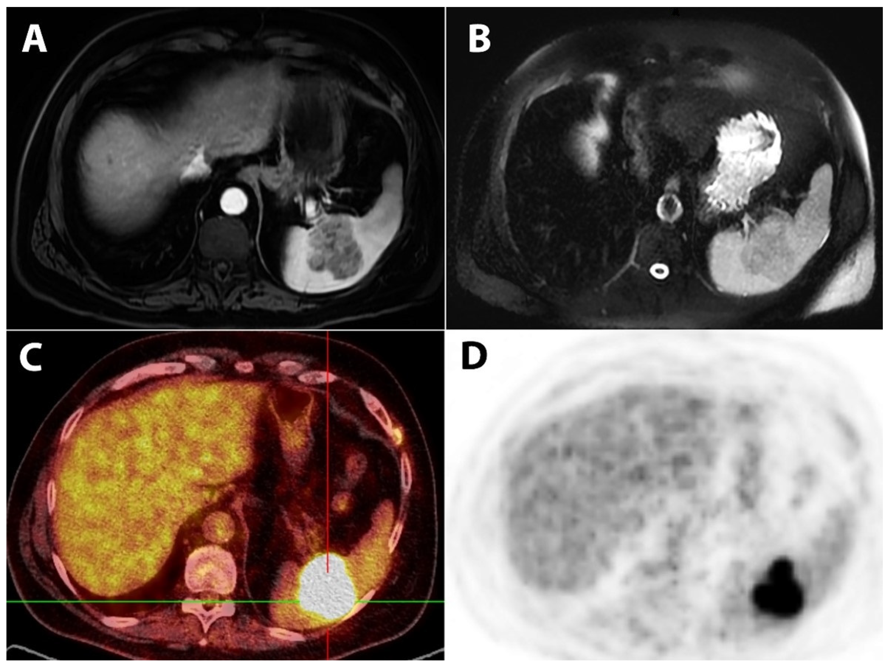
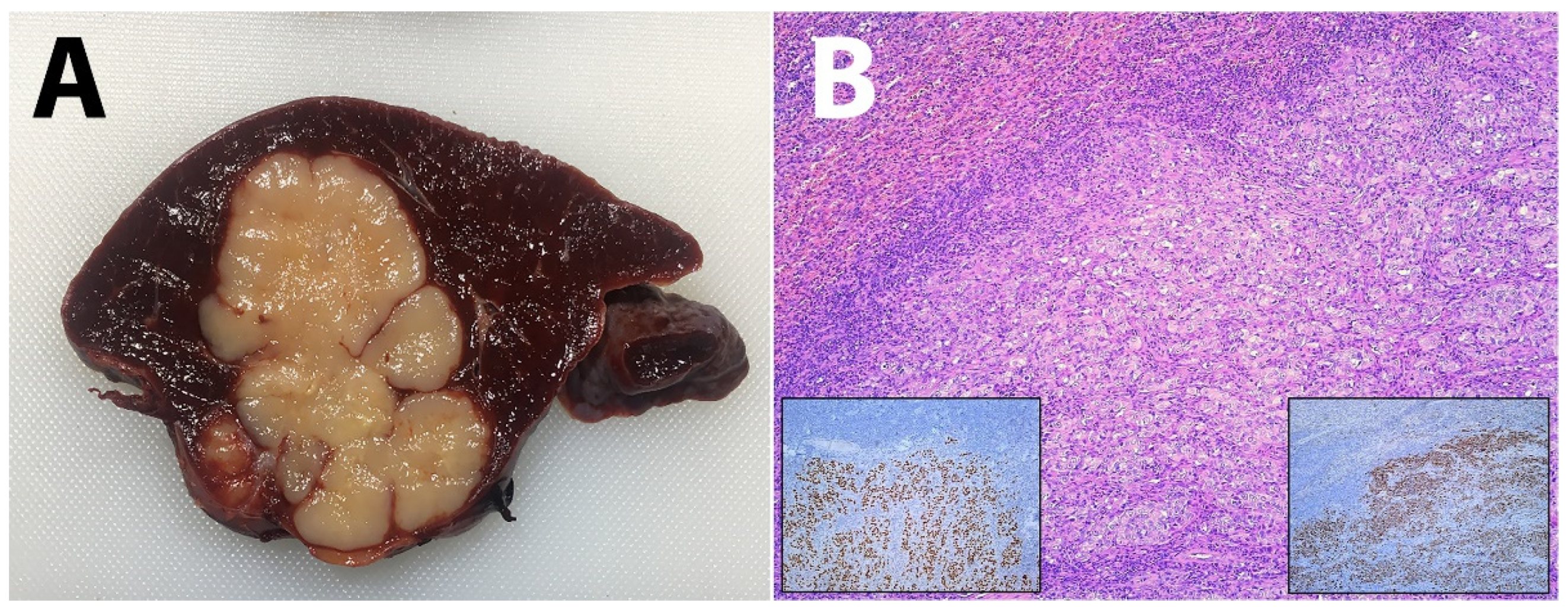
2. Case Report
3. Discussion
4. Conclusions
Author Contributions
Funding
Informed Consent Statement
Conflicts of Interest
References
- Berge, T. Splenic metastases. Frequencies and patterns. Acta Pathol. Microbiol. Scand. A 1974, 82, 499–506. [Google Scholar] [PubMed]
- Lam, K.Y.; Tang, V. Metastatic tumors to the spleen: A 25-year clinicopathologic study. Arch. Pathol. Lab. Med. 2000, 124, 526–530. [Google Scholar] [CrossRef]
- Comperat, E.; Bardier-Dupas, A.; Camparo, P.; Capron, F.; Charlotte, F. Splenic metastases: Clinicopathologic presentation, differential diagnosis, and pathogenesis. Arch. Pathol. Lab. Med. 2007, 131, 965–969. [Google Scholar] [CrossRef]
- Romano, S.; Scaglione, M.; Gatta, G.; Lombardo, P.; Stavolo, C.; Romano, L.; Grassi, R. Association of splenic and renal infarctions in acute abdominal emergencies. Eur. J. Radiol. 2004, 50, 48–58. [Google Scholar] [CrossRef]
- Quint, L.E.; Tummala, S.; Brisson, L.J.; Francis, I.R.; Krupnick, A.S.; Kazerooni, E.A.; Iannettoni, M.D.; Whyte, R.I.; Orringer, M.B. Distribution of distant metastases from newly diagnosed non-small cell lung cancer. Ann. Thorac. Surg. 1996, 62, 246–250. [Google Scholar] [CrossRef]
- Perisano, C.; Spinelli, M.S.; Graci, C.; Scaramuzzo, L.; Marzetti, E.; Barone, C.; Fabbriciani, C.; Maccauro, G. Soft tissue metastases in lung cancer: A review of the literature. Eur. Rev. Med. Pharmacol. Sci. 2012, 16, 1908–1914. [Google Scholar]
- Satoh, H.; Watanabe, K.; Ishikawa, H.; Yamashita, Y.T.; Ohtsuka, M.; Sekizawa, K. Splenic metastasis of lung cancer. Oncol. Rep. 2001, 8, 1239–1241. [Google Scholar] [CrossRef] [PubMed]
- Schon, C.A.; Gorg, C.; Ramaswamy, A.; Barth, P.J. Splenic metastases in a large unselected autopsy series. Pathol. Res. Pract. 2006, 202, 351–356. [Google Scholar] [CrossRef]
- Kinoshita, A.; Nakano, M.; Fukuda, M.; Kasai, T.; Suyama, N.; Inoue, K.; Nakata, T.; Shigematsu, K.; Oka, M.; Hara, K. Splenic metastasis from lung cancer. Neth. J. Med. 1995, 47, 219–223. [Google Scholar] [CrossRef]
- Sappington, S. Carcinoma of the spleen: Its microscopic frequency: A possible etiologic factor. J. Am. Med. Assoc. 1922, 78, 953–955. [Google Scholar]
- Kettle, E. Carcinomatous metastases in the spleen. J. Pathol. Bacteriol. 1912, 17, 40–46. [Google Scholar] [CrossRef]
- Klein, B.; Stein, M.; Kuten, A.; Steiner, M.; Barshalom, D.; Robinson, E.; Gal, D. Splenomegaly and solitary spleen metastasis in solid tumors. Cancer 1987, 60, 100–102. [Google Scholar] [CrossRef]
- Place, R.J. Isolated colon cancer metastasis to the spleen. Am. Surg. 2001, 67, 454–457. [Google Scholar]
- Takada, T.; Takami, H. Solitary splenic metastasis of a carcinoid tumor of the lung eight years postoperatively. J. Surg. Oncol. 1998, 67, 47–48. [Google Scholar] [CrossRef]
- Gupta, P.B.; Harvey, L. Spontaneous rupture of the spleen secondary to metastatic carcinoma. Br. J. Surg. 1993, 80, 613. [Google Scholar] [CrossRef] [PubMed]
- Massarweh, S.; Dhingra, H. Unusual sites of malignancy: Case 3. Solitary splenic metastasis in lung cancer with spontaneous rupture. J. Clin. Oncol. 2001, 19, 1574–1575. [Google Scholar] [CrossRef] [PubMed]
- Lachachi, F.; Abita, T.; Fontanier, S.D.; Maisonnette, F.; Descottes, B. Spontaneous splenic rupture due to splenic metastasis of lung cancer. Ann. Chir. 2004, 129, 521–522. [Google Scholar] [CrossRef]
- Metser, U.; Even-Sapir, E. The role of 18F-FDG PET/CT in the evaluation of solid splenic masses. Semin. Ultrasound CT MR 2006, 27, 420–425. [Google Scholar] [CrossRef]
- Patel, N.; Dawe, G.; Tung, K. Ultrasound-guided percutaneous splenic biopsy using an 18-G core biopsy needle: Our experience with 52 cases. Br. J. Radiol. 2015, 88, 20150400. [Google Scholar] [CrossRef] [Green Version]
- Rana, S.S.; Sharma, V.; Sharma, R.; Srinivasan, R.; Gupta, R. Safety and utility of endoscopic ultrasound-guided fine-needle aspiration of focal splenic lesions: A retrospective analysis. Ann. Gastroenterol. 2017, 30, 559–563. [Google Scholar] [CrossRef]
- Makrin, V.; Avital, S.; White, I.; Sagie, B.; Szold, A. Laparoscopic splenectomy for solitary splenic tumors. Surg. Endosc. 2008, 22, 2009–2012. [Google Scholar] [CrossRef]
- Yano, H.; Nakano, Y.; Tono, T.; Ohnishi, T.; Iwazawa, T.; Kimura, Y.; Kanoh, T.; Monden, T. Hand-assisted laparoscopic splenectomy for splenic tumors. Dig. Surg. 2004, 21, 215–222. [Google Scholar] [CrossRef]
- Milosavljevic, V.; Tadic, B.; Grubor, N.; Eric, D.; Reljic, M.; Matic, S. Analysis of the surgical treatment of the patients operated on by using laparoscopic and classic splenectomy due to benign disorders of the spleen. Turk. J. Surg. 2019, 35, 111–116. [Google Scholar] [CrossRef] [PubMed]
- Detterbeck, F.C.; Nicholson, A.G.; Franklin, W.A.; Marom, E.M.; Travis, W.D.; Girard, N.; Arenberg, D.A.; Bolejack, V.; Donington, J.S.; Mazzone, P.J.; et al. The IASLC Lung Cancer Staging Project: Summary of Proposals for Revisions of the Classification of Lung Cancers with Multiple Pulmonary Sites of Involvement in the Forthcoming Eighth Edition of the TNM Classification. J. Thorac. Oncol. 2016, 11, 639–650. [Google Scholar] [CrossRef] [PubMed] [Green Version]
- Hellman, S.; Weichselbaum, R.R. Oligometastases. J. Clin. Oncol. 1995, 13, 8–10. [Google Scholar] [CrossRef] [PubMed]
- Moran, A.; Daly, M.E. Surveillance imaging for non-small cell lung cancer: Mounting evidence that less is more. Transl. Lung Cancer Res. 2019, 8 (Suppl. 4), S343–S346. [Google Scholar] [CrossRef]
- Edelman, A.S.; Rotterdam, H. Solitary splenic metastasis of an adenocarcinoma of the lung. Am. J. Clin. Pathol. 1990, 94, 326–328. [Google Scholar] [CrossRef] [Green Version]
- Macheers, S.K.; Mansour, K.A. Management of isolated splenic metastases from carcinoma of the lung: A case report and review of the literature. Am. Surg. 1992, 58, 683–685. [Google Scholar]
- Tomaszewski, D.; Bereza, S.; Sternau, A. Solitary splenic metastases from lung cancer—One-time surgical procedure. Pneumonol. Alergol. Pol. 2003, 71, 533–537. [Google Scholar]
- Schmidt, B.J.; Smith, S.L. Isolated splenic metastasis from primary lung adenocarcinoma. South Med. J. 2004, 97, 298–300. [Google Scholar] [CrossRef]
- Pramesh, C.S.; Sg, P.; As, P. Isolated splenic metastasis from non small cell lung cancer. Ann. Thorac. Cardiovasc. Surg. 2004, 10, 247–248. [Google Scholar] [PubMed]
- Sanchez-Romero, A.; Oliver, I.; Costa, D.; Orduna, A.; Lacueva, J.; Perez-Vicente, F.; Arroyo, A.; Calpena, R. Giant splenic metastasis due to lung adenocarcinoma. Clin. Transl. Oncol. 2006, 8, 294–295. [Google Scholar] [CrossRef]
- Van Hul, I.; Cools, P.; Rutsaert, R. Solitary splenic metastasis of an adenocarcinoma of the lung 2 years postoperatively. Acta Chir. Belg. 2008, 108, 462–463. [Google Scholar] [CrossRef]
- Ando, K.; Kaneko, N.; Yi, L.; Sato, C.; Yasui, D.; Inoue, K.; Misawa, M.; Ohkuni, Y. Splenic metastasis of lung cancer. Nihon Kokyuki Gakkai Zasshi 2009, 47, 581–584. [Google Scholar] [PubMed]
- Chloros, D.; Bitzikas, G.; Kakoura, M.; Chatzikostas, G.; Makridis, C.; Tsitouridis, I. Solitary splenic metastasis of squamous lung cancer: A case report. Cases J. 2009, 2, 9091. [Google Scholar] [CrossRef] [Green Version]
- Tang, H.; Huang, H.; Xiu, Q.; Shi, Z. Isolated splenic metastasis from lung cancer: Ringleader of continuous fever. Eur. Respir. Rev. 2010, 19, 253–256. [Google Scholar] [CrossRef] [Green Version]
- Scintu, F.; Carta, M.; Frau, G.; Marongiu, L.; Pipia, G.; Casula, G. Splenic metastases of pulmonary carcinoma. Apropos of a clinical case. Minerva Chir. 1991, 46, 1277–1280. [Google Scholar]
- Yen, R.F.; Wu, Y.W.; Pan, M.H.; Tzen, K.Y. Early detection of splenic metastasis of lung cancer by 18F-2-fluoro-2-deoxyglucose positron emission tomography. J. Formos. Med. Assoc. 2005, 104, 674–676. [Google Scholar] [PubMed]
- Fujii, M.; Tanaka, H.; Sawazumi, T.; Nakamura, N.; Takahashi, M.; Inomata, S.; Chiba, H.; Takahashi, H. A case of solitary splenic metastasis following operation for pulmonary pleomorphic carcinoma: Detected at an early stage by FDG-PET. Nihon Kokyuki Gakkai Zasshi 2008, 46, 950–954. [Google Scholar]
- Assouline, P.; Leger-Ravet, M.B.; Paquet, J.C.; Kardache, M.; Decoux, L.; Kettaneh, L.; Faucher, J.N.; Oliviero, G. Splenic metastasis from a bronchial carcinoma. Rev. Mal. Respir. 2006, 23 Pt 1, 265–268. [Google Scholar] [CrossRef]
- Eisa, N.; Alhafez, B.; Alraiyes, A.H.; Alraies, M.C. Abdominal pain as initial presentation of lung cancer. BMJ Case Rep. 2014, 2014, bcr2013200613. [Google Scholar] [CrossRef] [Green Version]
- Belli, A.; De Luca, G.; Bianco, F.; De Franciscis, S.; Tatangelo, F.; Romano, G.M.; Rocco, G. An Unusual Metastatic Site for Primary Lung Cancer: The Spleen. J. Thorac. Oncol. 2016, 11, 128–129. [Google Scholar] [CrossRef] [Green Version]
- Sardenberg, R.A.; Pinto, C.; Bueno, C.A.; Younes, R.N. Non-small cell lung cancer stage IV long-term survival with isolated spleen metastasis. Ann. Thorac. Surg. 2013, 95, 1432–1434. [Google Scholar] [CrossRef]
- Dias, A.R.; Pinto, R.A.; Ravanini, J.N.; Lupinacci, R.M.; Cecconello, I.; Ribeiro, U., Jr. Isolated splenic metastasis from lung squamous cell carcinoma. World J. Surg. Oncol. 2012, 10, 24. [Google Scholar] [CrossRef] [PubMed] [Green Version]
- Cai, Q.; Kragel, P. Isolated splenic metastasis in a patient with lung carcinoma: Case report and review of the literature. J. Clin. Exp. Pathol. 2015, 5, 2161-0681. [Google Scholar] [CrossRef]
- Soussan, M.; Pop, G.; Ouvrier, M.J.; Neuman, A.; Weinmann, P. Diagnosis of synchronous isolated splenic metastasis from lung adenocarcinoma: Complementary role of FDG PET/CT and diffusion-weighted MRI. Clin. Nucl. Med. 2011, 36, 707–709. [Google Scholar] [CrossRef]
- Iguchi, K.; Ishibashi, O.; Kondo, T.; Kagohashi, K.; Takayashiki, N.; Satoh, H. Isolated spleen recurrence in a patient with lung adenocarcinoma: A case report. Exp. Ther. Med. 2015, 10, 733–736. [Google Scholar] [CrossRef] [PubMed] [Green Version]
- Mitsimponas, N.; Mitsogianni, M.; Crespo, F.; Hartmann, K.A.; Diederich, S.; Klosterhalfen, B.; Giagounidis, A. Isolated Splenic Metastasis from Non-Small-Cell Lung Cancer: A Case Report and Review of the Literature. Case Rep. Oncol. 2017, 10, 638–643. [Google Scholar] [CrossRef]
- Hara, K.; Izumi, N.; Tsukioka, T.; Komatsu, H.; Toda, M.; Miyamoto, H.; Suzuki, S.; Kimura, T.; Shibata, T.; Nishiyama, N. Solitary splenic metastasis from lung adenocarcinoma: A case report. Thorac. Cancer 2017, 8, 539–542. [Google Scholar] [CrossRef] [Green Version]
- Zeng, Z.; Chen, N.; Zhu, Y.; Lin, F. Isolated splenic metastasis from pulmonary adenoid cystic carcinoma. QJM 2018, 111, 405–406. [Google Scholar] [CrossRef]
- Lopera, C.A.; Vergnaud, J.P.; Matute-Turizo, G.; Pereira-Warr, S. Laparoscopic Splenectomy for Splenic Metastasis from Primary Lung Carcinoma. Case Rep. Surg. 2018, 2018, 2620301. [Google Scholar] [CrossRef] [PubMed] [Green Version]
- Tanaka, K.; Iwata, T.; Yoshida, S.; Nishii, K.; Matsui, Y.; Sugiyama, T.; Itami, M.; Iizasa, T. A surgical case of synchronous solitary splenic metastasis from lung squamous cell carcinoma: Report of a case and review of the literature. Gen. Thorac. Cardiovasc. Surg. 2020, 68, 866–870. [Google Scholar] [CrossRef]
- Oussama, B.; Makrem, M.; Neji, F.M.; Amine, L.; Brahim, K.; Karim, S.; Sami, B. Non small cell lung cancer revealed by a solitary splenic metastasis of lung cancer. Tunis. Med. 2013, 91, 484–485. [Google Scholar] [PubMed]
- Grant-Freemantle, M.C.; Bass, G.A.; Butt, W.T.; Gillis, A.E. Splenectomy for isolated splenic metastasis from primary lung adenocarcinoma. BMJ Case Rep. 2020, 13, e233256. [Google Scholar] [CrossRef] [PubMed]


| No. | First Author/Year | Histology (Primary Lung Lesion) | Lung Lesion Side | Time to Splenic Metastasis | Sex | Age | Metastasis Symptoms | Treatment of Primary Tumor | Treatment of Splenic Metastasis | Follow-up at the Time of the Report |
|---|---|---|---|---|---|---|---|---|---|---|
| 1. | Klein/1987 [12] | Bronchioalveolar carcinoma | Right | 20 months | F | 57 | Abdominal pain | Right lower and middle lobectomy | Splenectomy | Died 49 months after splenectomy |
| 2. | Edelman/1990 [27] | Poorly differentiated adenocarcinoma | Left | 0 months | F | 63 | Asymptomatic | n.a | n.a | n.a |
| 3. | Macheers/1992 [28] | Large-cell undifferentiated carcinoma | Left | 0 months | n.a. | Asymptomatic | n.a. | Splenectomy | Died 1 month after splenectomy | |
| 4. | Gupta/1993 [15] | Squamous cell carcinoma | Right | 0 months | n.a. | Splenic rupture | n.a. | Splenectomy | Died 8 weeks after splenectomy | |
| 5. | Kinoshita/1995 [9] | Squamous cell carcinoma | Left | 14 months | M | 72 | Asymptomatic | Surgical removal of primary tumor | Splenectomy | Died 27 months after splenectomy |
| 6. | Takada/1998 [14] | Bronchopulmonary carcinoid tumor | Right | 96 months | M | 49 | Abdominal pain | Right upper lobectomy | Splenectomy | Disease free after 8 years |
| 7. | Tomaszewski/2003 [29] | Lung cancer | Left | 0 months | M | 68 | Asymptomatic | Upper left lobectomy | Splenectomy | n.a. |
| 8. | Massarweh/2001 [16] | Poorly differentiated adenocarcinoma | Left | 0 months | M | 68 | Splenic rupture | Palliative chemotherapy | Splenectomy | n.a. |
| 9. | Schmidt/2004 [30] | Moderately differentiated adenocarcinoma | Left | 25 months | M | 72 | Asymptomatic | Surgical removal of primary tumor | n.a. | Disease free after 2 years |
| 10. | Pramesh/2004 [31] | Squamous cell carcinoma | Left | 2 months | M | 55 | Asymptomatic | Combined radiochemotherapy | chemotherapy | n.a. |
| 11. | Lachachi/2004 [17] | Poorly differentiated carcinoma | Right | 0 months | n.a. | Splenic rupture | n.a. | Splenectomy | n.a. | |
| 12. | Sánchez-Romor/2006 [32] | Adenocarcinoma | Left | 0 months | M | 73 | Abdominal pain | Left lung resection | Splenectomy | n.a. |
| 13. | Van Hul/2008 [33] | Adenocarcinoma | Left | 24 months | M | 67 | Asymptomatic | Surgical removal of primary tumor | Splenectomy | n.a. |
| 14. | Ando/2009 [34] | Squamous cellcarcinma | Right | 10 months | M | 71 | Asymptomatic | Combined radiochemotherapy | Splenectomy | n.a. |
| 15. | Chloros/2009 [35] | Squamous cellcarcinma | Right | 0 months | M | 59 | Asymptomatic | Surgical removal of primary tumor | Splenectomy | n.a. |
| 16. | Tang/2010 [36] | Large-cell undifferentiated carcinoma | Right | 4 months | F | 49 | Fever | Lobectomy of the right middle and lower lobe | Splenectomy | n.a. |
| 17. | Scintu/1991 [37] | Large-cell anaplastic carcinoma | n.a. | 0 months | n.a. | Asymptomatic | Pulmonary lobectomy | Splenectomy | Disease free after 41 months | |
| 18. | Yen/2005 [38] | Adenocarcinoma | Left | 24 months | M | 56 | Asymptomatic | Left pneumonectomy | Splenectomy | n.a. |
| 19. | Fujii/2008 [39] | Poorly differentiated adenocarcinoma | Left | 3 months | M | 58 | Asymptomatic | Left upper lobectomy | Splenectomy | n.a. |
| 20. | Assouline/2006 [40] | Large-cell undifferentiated carcinoma | Right | 21 months | M | 77 | Abdominal pain | Right pneumonectomy | Splenectomy | Disease free after 2 years |
| 21. | Eisa/2014 [41] | Adenocarcinoma | Right | 0 months | F | 53 | Abdominal pain | Surgical removal of primary tumor | Splenectomy | Disease free at the time of the report |
| 22. | Belli/2016 [42] | Large-cell carcinoma | Right | 60 months | M | 65 | Asymptomatic | Right pneumonectomy | n.a. | n.a. |
| 23. | Sardenberg/2013 [43] | Adenocarcinoma | Right | 7 months | F | 49 | Abdominal pain | Right upper lobectomy | Splenectomy | Disease free after 96 months |
| 24. | Dias/2012 [44] | Squamous cell carcinoma | Right | 16 months | M | 82 | Asymptomatic | Right bilobectomy | Splenectomy | Disease free after 12 months |
| 25. | Cai/2015 [45] | Adenocarcinoma | Right | 17 months | F | 56 | Asymptomatic | Right lower lobectomy | Splenectomy | n.a. |
| 26. | Soussan/2011 [46] | Adenocarcinoma | n.a. | 0 months | M | 52 | Asymptomatic | n.a. | n.a. | n.a. |
| 27. | Iguchi/2015 [47] | Adenocarcinoma | Left | 12 months | F | 63 | Asymptomatic | Left lower lobectomy | Splenectomy | n.a. |
| 28. | Mitsimponas/[48] | Adenocarcinoma | Right | 0 months | F | 66 | Asymptomatic | Radiochemotherapy | Chemotherapy | Alive at the time of the report |
| 29. | Hara/2017 [49] | Poorly differentiate adenocarcinoma | Right | 0 months | F | 81 | Asymptomatic | Right upper lobactomy | Lap. splenectomy | n.a. |
| 30. | Zeng/2018 [50] | Adenoid cystic carcinoma | Right | 48 months | F | 38 | Abdominal pain | Right middle lobectomy | Splenectomy | n.a |
| 31. | Lopera/2018 [51] | Large cell carcinoma | Right | n.a. | F | 69 | Abdominal pain | Right upper lobactomy | Lap. Splenectomy | n.a |
| 32. | Tanaka/2020 [52] | Squamous cell carcinoma | Righ | 0 months | M | 78 | Abdominal pain | Surgery | Splenectomy | n.a. |
| 33. | Ousama/2001 [53] | Non-small-cell lung cancer | Left | 0 months | M | 58 | Abdominal pain | Chemotherapy | Splenectomy | n.a. |
| 34. | Grant/2020 [54] | Adenocarcinoma | Right | n.a. | F | 73 | Asymptomatic | Right lower lobe lobectomy | Splenectomy | Alive at the time of the report |
| 35. | Present case | Adenosquamos carcinoma | Left | 144 months | M | 56 | Asymptomatic | Left upper lobectomy | Splenectomy | Disease free after 24 months |
Publisher’s Note: MDPI stays neutral with regard to jurisdictional claims in published maps and institutional affiliations. |
© 2022 by the authors. Licensee MDPI, Basel, Switzerland. This article is an open access article distributed under the terms and conditions of the Creative Commons Attribution (CC BY) license (https://creativecommons.org/licenses/by/4.0/).
Share and Cite
Reljic, M.; Tadic, B.; Stosic, K.; Mitrovic, M.; Grubor, N.; Kmezic, S.; Ceranic, M.; Milosavljevic, V. Isolated Splenic Metastasis of Primary Lung Cancer Presented as Metachronous Oligometastatic Disease—A Case Report. Diagnostics 2022, 12, 209. https://doi.org/10.3390/diagnostics12010209
Reljic M, Tadic B, Stosic K, Mitrovic M, Grubor N, Kmezic S, Ceranic M, Milosavljevic V. Isolated Splenic Metastasis of Primary Lung Cancer Presented as Metachronous Oligometastatic Disease—A Case Report. Diagnostics. 2022; 12(1):209. https://doi.org/10.3390/diagnostics12010209
Chicago/Turabian StyleReljic, Milorad, Boris Tadic, Katarina Stosic, Milica Mitrovic, Nikola Grubor, Stefan Kmezic, Miljan Ceranic, and Vladimir Milosavljevic. 2022. "Isolated Splenic Metastasis of Primary Lung Cancer Presented as Metachronous Oligometastatic Disease—A Case Report" Diagnostics 12, no. 1: 209. https://doi.org/10.3390/diagnostics12010209