Next Article in Journal
Rapid Diagnosis of Pseudomonas aeruginosa in Wounds with Point-Of-Care Fluorescence Imaing
Previous Article in Journal
Salivary Inflammatory Molecules as Biomarkers of Sleep Alterations: A Scoping Review
 
 
Correction published on 18 July 2022, see Diagnostics 2022, 12(7), 1741.
Article

Impact of [18F]FDG-PET and [18F]FLT-PET-Parameters in Patients with Suspected Relapse of Irradiated Lung Cancer

by
Tine N. Christensen
1,2,*,
Seppo W. Langer
3,4,
Gitte Persson
4,5,
Klaus Richter Larsen
6,
Annemarie G. Amtoft
1,
Sune H. Keller
1,
Andreas Kjaer
1,2 and
Barbara Malene Fischer
1,4,7
1
Department of Clinical Physiology, Nuclear Medicine & PET, Copenhagen University Hospital, Rigshospitalet, 2100 Copenhagen Ø, Denmark
2
Cluster for Molecular Imaging, University of Copenhagen, 2200 Copenhagen N, Denmark
3
Department of Oncology, Copenhagen University Hospital, Rigshospitalet, 2100 Copenhagen Ø, Denmark
4
Department of Clinical Medicine, University of Copenhagen, 2100 Copenhagen Ø, Denmark
5
Department of Oncology, Herlev-Gentofte Hospital, University of Copenhagen, 2730 Herlev, Denmark
6
Department of Pulmonary Medicine, Bispebjerg University Hospital, 2400 Copenhagen NV, Denmark
7
The PET Centre, School of Biomedical Engineering and Imaging Science, King’s College London, London SE1 7EH, UK
*
Author to whom correspondence should be addressed.
Diagnostics 2021, 11(2), 279; https://doi.org/10.3390/diagnostics11020279
Submission received: 21 December 2020 / Revised: 26 January 2021 / Accepted: 7 February 2021 / Published: 11 February 2021 / Corrected: 18 July 2022
(This article belongs to the Section Medical Imaging and Theranostics)

Abstract

:
Radiation-induced changes may cause a non-malignant high 2-deoxy-2-[18F]fluoro-d-glucose (FDG)-uptake. The 3′-deoxy-3′-[18F]fluorothymidine (FLT)-PET/CT performs better in the differential diagnosis of inflammatory changes and lung lesions with a higher specificity than FDG-PET/CT. We investigated the association between post-radiotherapy FDG-PET-parameters, FLT-PET-parameters, and outcome. Sixty-one patients suspected for having a relapse after definitive radiotherapy for lung cancer were included. All the patients had FDG-PET/CT and FLT-PET/CT. FDG-PET- and FLT-PET-parameters were collected from within the irradiated high-dose volume (HDV) and from recurrent pulmonary lesions. For associations between PET-parameters and relapse status, respectively, the overall survival was analyzed. Thirty patients had a relapse, of these, 16 patients had a relapse within the HDV. FDG-SUVmax and FLT-SUVmax were higher in relapsed HDVs compared with non-relapsed HDVs (median FDG-SUVmax: 12.8 vs. 4.2; p < 0.001; median FLT-SUVmax 3.9 vs. 2.2; p < 0.001). A relapse within HDV had higher FDG-SUVpeak (median FDG-SUVpeak: 7.1 vs. 3.5; p = 0.014) and was larger (median metabolic tumor volume (MTV50%): 2.5 vs. 0.7; 0.014) than the relapsed lesions outside of HDV. The proliferative tumor volume (PTV50%) was prognostic for the overall survival (hazard ratio: 1.07 pr cm3 [1.01–1.13]; p = 0.014) in the univariate analysis, but not in the multivariate analysis. FDG-SUVmax and FLT-SUVmax may be helpful tools for differentiating the relapse from radiation-induced changes, however, they should not be used definitively for relapse detection.

1. Introduction

The early and precise diagnosis in patients suspected for lung cancer relapse is essential, as a curative intended treatment might be feasible [1]. The 2-deoxy-2-[18F]fluoro-d-glucose (FDG)-PET/CT is recommended in patients with a clinical suspicion of recurrence of lung cancer after treatment [2,3]. However, a pathological confirmation is not always feasible. Concerns of risks associated with performing a biopsy and possibilities of sampling errors in small tumors rule out a biopsy in some patients. Therefore, a better accuracy with non-invasive procedures is warranted. Recently, we have shown that 3′-deoxy-3′-[18F]fluorothymidine (FLT)-PET/CT adds a diagnostic value in patients who have been treated with definitive radiotherapy [4].
The value of semi-quantitative analysis of FDG-PET/CT after radiotherapy has been sparsely investigated [5,6,7,8]. FDG-SUVmax is higher in relapses than in radiation-induced changes [5,6]. However, even a moderate to intense FDG-uptake may be caused by radiation-induced changes rather than recurrence [7], and SUVmax ≥ 2.5 has been described 2 years after radiotherapy without evidence of recurrence [8].
During and after radiotherapy, the FLT-uptake decreased more rapidly and more pronounced than the FDG-uptake in several cancers [9,10,11,12], including lung cancer [13,14,15,16]. With a higher specificity and better discrimination between inflammatory lesions and lung cancer [17], the FLT-uptake may reflect a relapse-status better than FDG-uptake. To our knowledge, post-radiotherapy FLT-PET-parameters have only been investigated in two studies: (1) After stereotactic body radiotherapy (SBRT), FLT-SUVmax was higher in recurrent lesions than in non-recurrent lesions in a study with 10 patients [18]. (2) After carbon-ion radiotherapy, FLT-SUVmax was not significantly different in recurrent and non-recurrent lesions in a study of 19 patients [19].
The prognostic values of FDG-PET-parameters after radiotherapy are not in agreement. Lopez Guerra et al. [20] showed a prognostic value of post-radiotherapy FDG-SUVmax for overall survival, and Bollineni et al. [21] showed a trend towards a prognostic value of survival of FDG-SUVmax 3 months after SBRT. Other studies did not find any prognostic value of FDG-PET neither in non-small cell lung cancer (NSCLC) [22] nor small cell lung cancer (SCLC) [23].
In this study, we explore the associations between FDG-PET- and FLT-PET-parameters after radiotherapy and patient outcome, to assess if the quantitative analysis can add value to the diagnosis of relapse after radiotherapy and to evaluate the prognostic value of FDG- and FLT-PET-parameters.

2. Materials and Methods

2.1. Patients

Patients were eligible if they met the following inclusion criteria: Histologically confirmed lung cancer, i.e., NSCLC or SCLC; definitive radiotherapy within 2 years and current suspicion of local relapse based on symptoms and/or CT- or FDG-PET/CT warranting (further) FDG-PET/CT for relapse diagnosis.
Patients were recruited prospectively from three university hospitals in the Capital Region of Denmark during January 2015 to 2019. The follow-up ended in May 2020. The study was approved by the local ethics committee (approval number H-4-2014-060) and by institutional review boards. All the patients signed a written informed consent. We have previously published data from this cohort concerning the diagnostic value of visual analysis of PET scans [4].

2.2. Imaging

All the patients went through an FLT-PET/CT as well as an FDG-PET/CT within 4 weeks of each other.
FLT-PET/CT was performed at Rigshospitalet on a Siemens Biograph TruePoint TrueV 40 or 64 PET/CT-scanner. FLT (5 mg/kg, max 350 MBq) was injected 60 ± 10 min prior to scanning, according to the study guidelines. Patients were not subject to any restrictions regarding fasting or resting before FLT-PET/CT. FLT-PET were reconstructed using an ordered subset expectation maximization (OSEM) with point spread function modelling (PSF), three iterations, and 21 subsets with a 2 mm full width half maximum (FWHM) Gaussian post-reconstruction filter.
FDG-PET/CT was performed as a routine clinical investigation, according to the local procedures at the referring hospital. Patients fasted at least 4 h before the injection of FDG and rested approximately 60 min between the injection and scan. Images were reconstructed following clinical guidelines for FDG-PET/CT. Details are available in Supplemental Table S1.

2.3. Image Analyses

PET/CT-scans were reviewed and analyzed on a Mirada Medical Ltd. XD 3.6 workstation by TNC, AGA, and BMF. The two latter readers being a board certified radiologist and nuclear medicine physician, respectively with more than 10 years of experience.
From the radiotherapy-plan, a high-dose irradiated volume (HDV) was defined within the 50% isodose curve (Figure 1 and Figure 2) for each patient. Radiation-induced changes may occur in larger areas than, e.g., the planning target volume, thus to be able to evaluate the potential of FDG-PET/CT and FLT-PET/CT to differentiate radiation-induced changes from relapse, HDV was selected to be more representative of the clinically challenging area. Within HDV, the tracer uptake was quantified by a standardized uptake value (SUV) measuring SUVmax and SUVpeak. SUVmax was defined as an SUV in the voxel with the highest SUV in the region of interest. SUVpeak was defined as the highest average of SUV in a 1 cm3 sphere within the region of interest.
In patients with intrapulmonary relapse, SUVmax, SUVpeak, and the functional tumor volume were measured in the relapses. Functional tumor volumes were delineated in three different ways: (1) by 50% of SUVmax (MTV50 from FDG-PET/CT (metabolic tumor volume) and PTV50 from FLT-PET/CT (proliferative tumor volume), as recommended for FDG-PET by the European Association of Nuclear Medicine (EANM) procedure guidelines [24]; (2) by 80% of SUVmax (MTV80 and PTV80) as suggested for post-radiotherapy delineation for better discriminating from the adjacent tissue [25]; and (3) by SUVmax > 3.0 (MTV3.0 and PTV3.0) as this threshold previously has shown promise in the post-treatment setting [26]. In patients with more than one intrapulmonary malignant lesions, the highest SUVmax and SUVpeak from the lesions were used, and the functional tumor volume was calculated as the sum of all malignant lesions.

2.4. Outcome

The endpoints were (1) relapse and (2) overall survival. Relapses were categorized as (1) within HDV, (2) intrapulmonary, and (3) extra-pulmonary. The relapse status was defined retrospectively as the presence or absence of relapse within 6 months after inclusion. The relapse status was preferably confirmed by histology. If histology was not available or inconclusive, the relapse status was judged by a clinical oncologist, based on subsequent imaging procedures, invasive procedures, histology, and conference decisions within 6 months. The clinical oncologist was blinded for the name and age of the patient, names of the involved physicians, and dates of investigations.
The overall survival was calculated from the day of relapse suspicion to death by any cause.

2.5. Statistics

The correlation analyses were performed using Spearman’s correlation as the PET-parameters were not normally distributed. Differences of PET-parameters in HDVs with relapse and non-relapsed HDVs, and differences of PET-parameters in relapsed lesions within vs. outside of HDV were estimated with independent t-tests or the Welch test, if assumptions for the t-test were not fulfilled. Receiver operating characteristic (ROC) curves was created to explore the diagnostic accuracy of FDG-SUVmax and FLT-SUVmax. The sensitivity and specificity for relapse diagnosis within HDV with variant SUVmax cutoffs were estimated. The diagnostic value of combining FDG-SUVmax and FLT-SUVmax was explored with different combinations of FDG-SUVmax- and FLT-SUVmax-cutoffs.
Overall survival was calculated by the Kaplan-Meier method. The univariate and multivariate survival analysis including clinical parameters and PET-parameters from relapsed lesions and HDV were performed by Cox regression. PET-parameters were included both as continuous variables and dichotomized by the median cut-off. The number of included covariates in the multivariate analysis was restricted due to the limited number of patients. PET-parameters with p < 0.150 in the univariate analysis were included in addition to preselected clinical covariates: Sex, age (at time of suspicion), and time from the end of radiotherapy to the suspicion of relapse. The correlation analyses were performed separately on FDG-PET and FLT-PET-parameters to avoid including highly correlated parameters. Statistical analyses were performed in SPSS, version 25.

3. Results

A total of 63 patients were enrolled in this study. In two patients, the relapse status after 6 months of follow-up was non-confirmed, and they were excluded from further analysis. Accordingly, 61 patients were included in the final analysis. Patients were included based on the relapse suspicion raised on surveillance CT (n = 51) or surveillance FDG-PET/CT (n = 9). Four patients had symptoms of relapse when included, of these, one patient was included without prior CT. Fifty-seven patients had NSCLC, two patients SCLC, and two patients mixed NSCLC/SCLC. Patients were included a median of 7 months (interquartile range (IQ): 5–12) after the end of radiotherapy.
FDG-PET/CT was performed a median of 21 days [IQ: 15–27] after relapse was suspected, and FLT-PET/CT was performed a median of 23 days [IQ: 21–29] after relapse was suspected. FDG-PET/CT and FLT-PET/CT were conducted a median of 6 days apart (IQ: 3–9 days). Patient characteristics are presented in Table 1.
Thirty patients were diagnosed with intrapulmonary relapse within 6 months after inclusion. In 16 patients, the relapse was located within the HDV. Figure 1 and Figure 2 illustrate the radiotherapy plan and FDG-PET/CT and FLT-PET/CT from two patients with relapse within HDV and without relapse, respectively. The relapse was confirmed by histology in eight patients (four HDVs) and by subsequent growth in 11 patients (six HDVs). In twelve patients, the relapse diagnosis was based solely on FDG-PET/CT as decided by a multi-disciplinary conference, of these, six patients had the disseminated disease. Two patients were diagnosed with extra-pulmonary relapse only.

3.1. FDG-SUVmax and FLT-SUVmax

SUVmax and SUVpeak in HDV were strongly correlated for both FDG and FLT (FDG-SUV: R = 0.958; p < 0.001; FLT-SUV: R = 0.958; p < 0.001). FDG-SUV and FLT-SUV in HDV had a positive and strong correlation (SUVmax: R = 0.804; p < 0.001; SUVpeak: R = 0.770; p < 0.001), as shown in Figure 3.
FDG-SUVmax and FLT-SUVmax both decreased with time since radiotherapy in HDV without relapse (negative correlation, FDG-SUVmax: R = −0.349; p = 0.019; FLT-SUVmax: R = −0.401; p = 0.006), and increased with time in HDV with relapse (FDG-SUVmax: R = 0.510; p = 0.044; FLT-SUVmax: R = 0.567; p = 0.022). As illustrated in Figure 4, the difference of SUVmax in HDV with relapse vs. no-relapse increases with time from the end of radiotherapy.
Similar results were obtained when the analyses were restricted to include only patients with NSCLC (Supplemental Tables S2 and S3).
There was no significant correlation between the time from relapse suspicion to FDG-PET/CT and FDG-SUVmax in recurrent pulmonary lesions (p = 0.512), nor from the suspicion of relapse to FLT-PET/CT and FLT-SUVmax in recurrent lesions (p = 0.670), implying that SUV-estimates were not biased from the time delay between the relapse suspicion and conducting the PET scans.

3.2. Relapse Detection

The HDV with relapse had a significantly higher SUVmax compared with non-relapsed HDV (median FDG-SUVmax: 12.8 vs. 4.2; p < 0.001; median FLT-SUVmax 3.9 vs. 2.2; p = 0.005), as illustrated in Figure 5. FDG-SUVpeak and FLT-SUVpeak showed similar results (median FDG-SUVpeak: 7.1 vs. 3.2; p < 0.001; median FLT-SUVpeak: 2.5 vs. 1.6; p = 0.005).
PET-parameters from pulmonary recurrent lesions are presented in Table 2. Recurrent lesions within HDV had higher SUV and larger functional tumor volumes than lesions with recurrence outside of HDV. However, only significantly so for FDG-PET-parameters (FDG-SUVpeak, MTV3.0, and MTV50%).

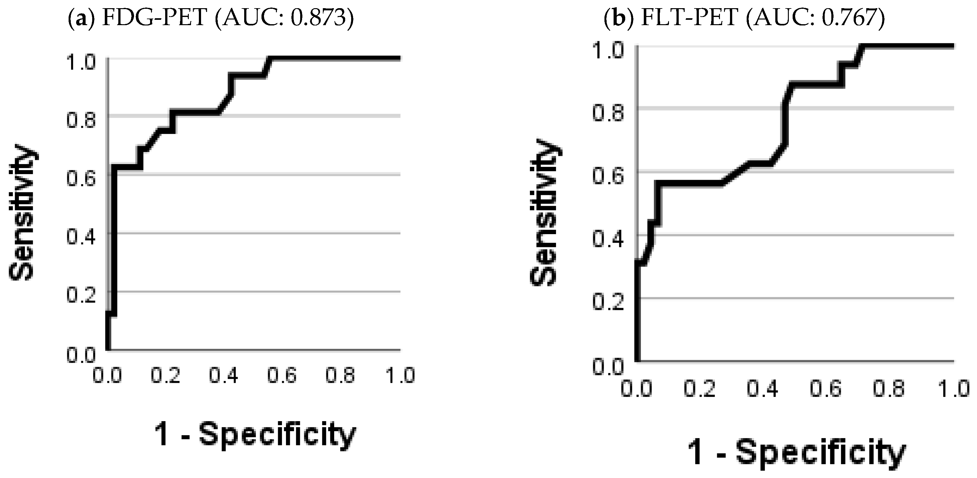
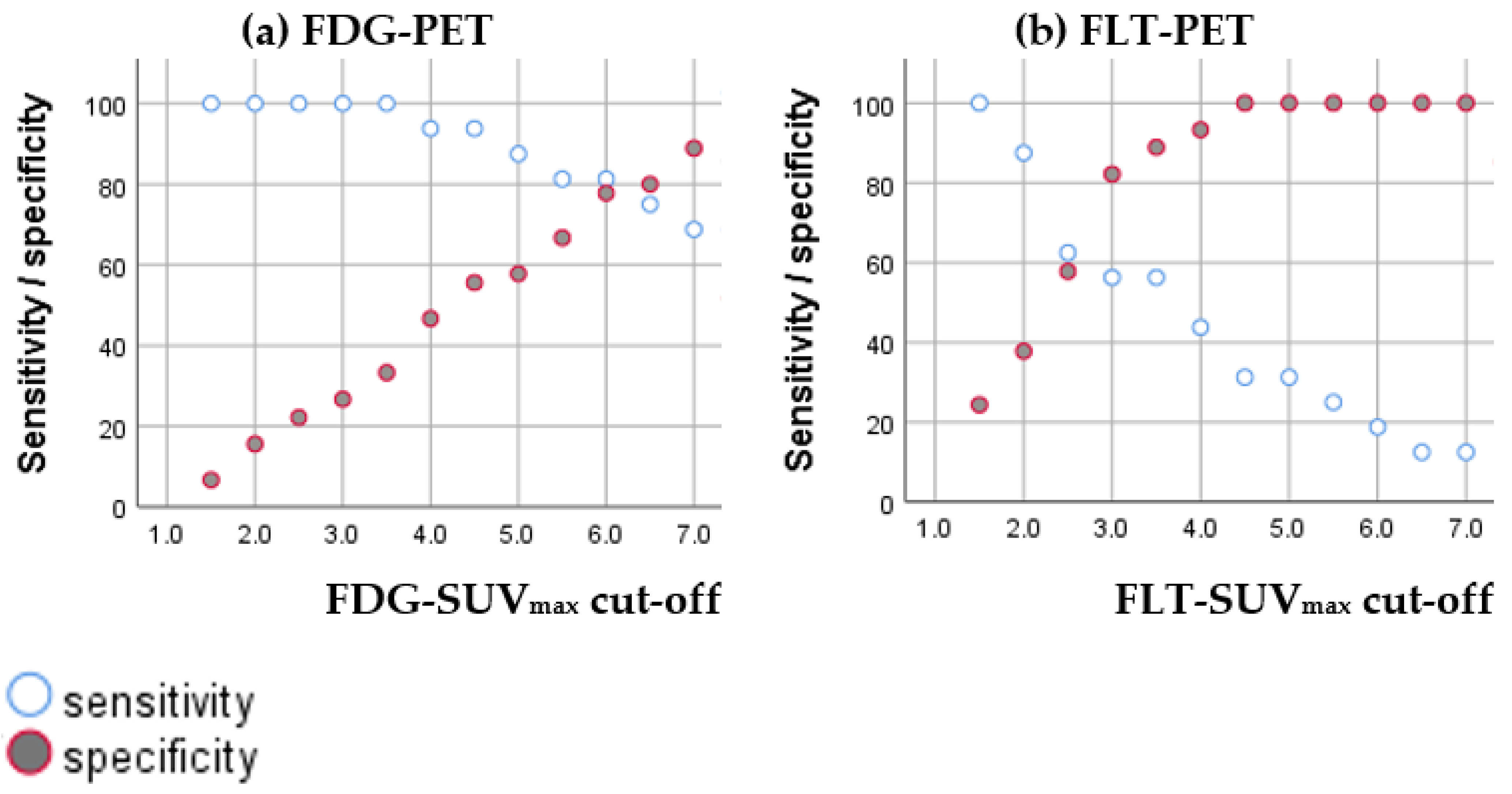
3.2.1. Diagnostic Accuracy of FDG-SUVmax and FLT-SUVmax

ROC-curves showed a moderate diagnostic accuracy for both FDG-SUVmax (AUC: 0.873) and FLT-SUVmax (AUC: 0.767), as shown in Figure 6. It was not possible from the ROC curves to establish cutoffs with a reasonable balance between sensitivity and specificity for neither FDG-SUVmax nor FLT-SUVmax. The sensitivity and specificity for variant cutoffs of SUVmax are presented in Figure 7.

3.2.2. Diagnostic Value of Combining FDG-SUVmax and FLT-SUVmax

A superior diagnostic value of combining FDG-SUVmax and FLT-SUVmax was not obvious due to the high correlation of FDG-SUVmax and FLT-SUVmax. Combinations of FDG-SUVmax and FLT-SUVmax with variant cutoffs were explored without an additional diagnostic value. In the subgroup of HDVs with high FDG-SUVmax (variant cutoffs), no optimal cutoff could be identified by the ROC-curves for FLT-SUVmax and the diagnostic accuracy was low (e.g., HDV with FDG-SUVmax ≥ 6.0: AUC: 0.654).

3.3. Prognosis

At the time of analysis, 33 patients had died. The median overall survival from relapse suspicion was 41 months. The median survival of patients who had intra-pulmonary relapse was 42 months. The two patients with extra-pulmonary relapse only died 7 and 10 months after relapse suspicion. The median overall survival of patients without relapse could not be estimated due to too few events (n = 12), and was at least 41 months.
In the univariate analysis, PET-parameters in HDV did not show a significant prognostic value of overall survival in the entire population (FDG-SUVmax: p = 0.154; FLT-SUVmax: p = 0.856), in patients with intrapulmonary relapse (n = 30; FDG-SUVmax: p = 0.431; FLT-SUVmax: p = 0.782), patients with HDV-relapse (n = 14; FDG-SUVmax: p = 0.391; FLT-SUVmax: p = 0.423), nor in patients without relapse (n = 29; FDG-SUVmax: p = 0.314; FLT-SUVmax: p = 0.862).
The univariate survival analysis performed on the patients with intrapulmonary relapse, appointed PTV50% as the only significant prognostic factor for overall survival (HR: 1.07 pr cm3 [1.01–1.13]; p = 0.014). MTV50%, MTV80%, PTV80%, and sex were borderline significant. Results from the univariate analyses are shown in Table 3.
MTV50% and MTV80% were highly correlated (R = 0.785, p < 0.001), as were PTV50% and PTV80% (R = 0.531, p = 0.003). Therefore, only MTV50% and PTV50% were included in the multivariate analysis. Male sex was independently prognostic for a poorer overall survival. No PET-parameter had an independent prognostic value. Larger MTV50% and a higher age were borderline significant for poorer overall survival. Results from the multivariate analysis are shown in Table 4. The subgroup analysis on patients with NSCLC yielded similar results (Supplemental Tables S4 and S5).

4. Discussion

We investigated the impact of semi-quantitative parameters of FDG-PET and FLT-PET in patients suspected for having a relapse of lung cancer after treatment with definitive radiotherapy. The FDG- and FLT-uptake was significantly higher in relapsed HDVs than HDVs without relapse. However, with a considerable overlap of SUVs in relapsed vs. non-relapsed HDVs, neither FDG-SUVmax nor FLT-SUVmax can be used definitively for distinguishing the relapse from radiation-induced changes. However, the longer after radiotherapy the PET scan was performed, the difference between SUVmax in HDVs with relapse vs. HDVs without relapse increased. The prognostic value of FDG- and FLT-PET-parameters after radiotherapy was limited.
Results from the few studies addressing PET-parameters in relapses after radiotherapy were similar to ours. Nakajima et al. [6] showed that patients with recurrences after SBRT had a significantly higher FDG-SUVmax than patients without relapse. Saga et al. [18] reported that after SBRT, FLT-SUVmax in relapsed lesions (range: 2.0–5.9; n = 5) were higher than in non-relapsed lesions (range: 1.3–2.1, n = 3; no statistics performed).
In our data, FDG- and FLT-uptakes in non-relapsed HDVs were lower the longer after radiotherapy PET/CT was performed, whereas FDG- and FLT-uptakes were higher in HDVs with relapse the longer after radiotherapy. PET-parameters may have a higher impact late after radiotherapy, as the difference of tracer-uptake in relapsed vs. non-relapsed lesions increased with time. In FDG-PET, a reduced FDG-uptake with time was expected following the resolution of radiation-induced changes [7,27]. FLT-PET is less likely to have an unspecific uptake in inflammation [28,29,30]. However, a moderate FLT-uptake the first 9 months after radiotherapy, as seen in our study, implies that some degree of non-specific FLT-uptake may be present.
It has previously been shown that the diagnostic accuracy of FDG-SUVmax was higher when FDG-PET/CT was performed more than 12 months after SBRT [6]. The sensitivity and specificity were 100% for SUVmax > 4.5 for patients who had FDG-PET/CT conducted more than 12 months after SBRT, compared with 87% and 98% for their entire cohort. Hoopes et al. [8] did not support a specificity of 100% of SUVmax > 4.5, as FDG-SUVmax was 5.1 and 5.9 in two patients without evidence of disease 23 and 26 months after SBRT, respectively [8]. A direct comparison of SUVmax from different studies should be taken with precaution. Particular precautions when comparing our study with the study of Nakajima et al. [6] are the different reconstruction methods (OSEM with and without PSF point-spread function, respectively) and different FDG-uptake times (60 vs. 90 min). PSF reconstructions lead on average to higher SUVmax than non-PSF reconstructions [31,32]. However, in our study, two patients without relapse had a high FDG-uptake on FDG-PET/CT more than 12 months after the end of radiotherapy: In one patient who had ended SBRT 14 months prior to FDG-PET/CT, FDG-SUVmax was 5.9. In another patient who had ended normo-fractionated radiotherapy 21 months prior to FDG-PET/CT, FDG-SUVmax was 6.0. FLT-SUVmax in the two patients were 2.5 and 2.2, respectively. In both patients, the biopsy was negative and there was no evidence of progression the following 6 months. Accordingly, from our data, the specificity of SUVmax > 4.5 was 75% in patients with relapse suspicion more than 12 months after the end of radiotherapy. The diagnostic value of the visual interpretation of FDG-PET/CT after treatment with a curative intent, surgery, and radiotherapy has been addressed in several papers with an estimated sensitivity between 82% and 100%, and specificity between 82% and 98% [2]. A meta-analysis showed a pooled sensitivity and specificity of 90% [33]. After radiotherapy, we have previously demonstrated a higher sensitivity and specificity with visual analysis of FDG-PET/CT (94%, and 71%, respectively) and FLT-PET/CT (69% and 90%, respectively) than any SUVmax cut-off [4]. From our data, we cannot support a fixed threshold of neither FDG-SUVmax nor FLT-SUVmax for the relapse diagnosis.
In this study, we also observed that relapses within HDV had higher SUVmax, SUVpeak, and larger tumor volumes than relapsed lesions that had not been irradiated previously, particularly for FDG-parameters. A non-malignant FDG-uptake caused by radiation-induced changes [6] could attribute to the differences, though also difficulties in the diagnostic process leading to later diagnosis of relapses within HDVs may cause larger and more aggressive tumor relapses in HDVs.
A limitation to our study was that the pathological confirmation of relapse was not always obtained. To minimize the effect of this limitation, a retrospective compound reference standard for the relapse status was applied, based on the patients’ records within 6 months of follow-up. In most patients, the relapse was confirmed by histology or follow-up with subsequent progression, and non-relapse by follow-up without progression. However, in 13/61 patients (six HDVs), the relapse was decided based on FDG-PET/CT by a multi-disciplinary conference, and the clinical decision warranted the start of treatment rather than further confirmation, either due to an obvious outcome of FDG-PET/CT and/or due to fact that the patient was unfit for invasive procedures. FDG-PET/CT is recommended as a second step test when the relapse is suspected after radiotherapy [3], thus excluding patients with an obvious relapse on FDG-PET/CT would have caused the study to be less representative.
Our survival analysis showed that FLT-PET (PTV50%) had a prognostic value in patients with relapsed lung cancer after radiotherapy. However, when adjusting for sex, age and time since RT, PTV50% was not independently prognostic for overall survival. MTV50% was borderline significant in both univariate and multivariate analysis. Therefore, MTV50% might be a better prognosticator than PTV50%, but needs further confirmation.
Previous studies exploring the prognostic value of post-radiotherapy PET-parameters are limited, and most studies have focused on an FDG-change rather than the absolute value of PET-parameters [2]. In contrast to our results, Lopez Guerra et al. [20] showed a prognostic value of FDG-SUVmax after normo-fractionated radiotherapy for overall survival with HR 1.27. In addition, Bollineni et al. [21] showed a trend for the prognostic value of survival after SBRT [21]. In studies including lung cancer patients treated with surgery [2], and in studies including cohorts mixed of patients treated with a curative intent, the post-treatment FDG-SUVmax also showed a prognostic value [2,34]. FLT-SUVmax after the end of carbon-ion radiotherapy has not shown a prognostic value [19]. The above studies address different populations with different stages. However, they have in common that all patients have been treated with a curative intent. Baseline studies have previously shown a prognostic value of FDG-SUVmax and MTV [35,36]. Results from baseline FLT-PET studies are not in agreement [16,19,37], and Scheffler et al. [37] showed an independent prognostic value of FDG-SUVmax, but not FLT-SUVmax. The prognostic value of PET-parameters measured before the treatment may, however, not be applicable on PET-parameters measured after radiotherapy, as the more complex biological tumor characteristics may affect the tracer-uptake. In addition, the lesser treatment options for relapsed cancers affect the survival compared with survival in patients with treatment naïve tumors. The survival analysis in our study was limited by the sample size. Our cohort consisted of patients with different stages, different histologies, different treatment schemes, and accordingly a different prognosis, although all the patients were initially treated with a curative intent. A prognostic value of PET-parameters may be affected by other prognostic factors. However, the small patient number limited possibilities for adjustment. The overall survival in our study was long, also for patients with relapse. PET-parameters measured several years prior to death probably contain a less prognostic value. Approximately half of the patients with relapse were treated with a curative intent. However, the intent or relapse treatment did not have a prognostic value. Therefore, in this study, the overall survival may not be useful as an endpoint. The progression free survival was not selected as an endpoint, as patients were included due to a suspicion of relapse, and therefore, many patients were confirmed with relapse shortly after inclusion.
An early diagnosis of lung cancer relapse is essential, as in some cases, a curative intended treatment might be feasible [1]. In this study, relapsed lesions within HDV had higher FDG-SUVpeak and larger MTV, than the relapsed lesions outside of HDV. The same characteristics were applied for the FLT-uptake and PTV, though not statistically significant. The cause is unknown, however, it is possible that radiation-induced changes in the HDV challenge and thus delay the diagnostic decision making. FDG-PET-SUVmax and FLT-PET-SUVmax can be supportive for distinguishing between radiation-induced changes and relapse, but should not be used definitively. Over time, the differences in FDG-SUVmax and FLT-SUVmax, respectively in relapsed vs. non-relapsed HDVs increased, thus SUVmax may have a higher impact later after radiotherapy.

5. Conclusions

The FDG- and FLT-uptake was significantly higher in relapsed HDV than HDV without relapse. However, caution should be taken if using SUV for the relapse diagnosis due to a considerable overlap of FDG-SUVmax, respectively, FLT-SUVmax in relapsed vs. non-relapsed HDVs.
PTV50% was prognostic for the overall survival in patients with relapse, however, PTV50% had no independently prognostic value.

Supplementary Materials

The following are available online at https://www.mdpi.com/2075-4418/11/2/279/s1. Table S1: Data on the PET model and reconstruction used for FDG-PET/CT; Table S2: PET-parameters in intrapulmonary malignant lesions in patients with NSCLC; Table S3: FDG-SUVmax and FLT-SUVmax in relapsed vs. non-relapsed HDV in patients with NSCLC; Table S4: Prognostic value of PET-parameters and clinical variables in patients with intrapulmonary relapse of irradiated NSCLC; Table S5: multivariate survival analysis in patients with relapsed NSCLC.

Author Contributions

Conceptualization, T.N.C., S.W.L., G.P., A.K. and B.M.F.; methodology, T.N.C., S.W.L., G.P., A.K. and B.M.F.; software, S.H.K.; formal analysis, T.N.C.; investigation, T.N.C., S.W.L., G.P., K.R.L., A.G.A., S.H.K. and B.M.F.; resources, A.K.; writing—original draft preparation, T.N.C.; writing—review and editing, T.N.C., S.W.L., G.P., K.R.L., A.G.A., S.H.K., A.K. and B.M.F.; visualization, T.N.C.; supervision, S.W.L., A.K. and B.M.F.; project administration, T.N.C.; funding acquisition, A.K. and B.M.F. All authors have read and agreed to the published version of the manuscript.

Funding

This project received funding from the Danish Cancer Society (grant no. R134-A8543-15-S42) and Department of Clinical Physiology, Nuclear Medicine and PET, Rigshospitalet, University of Copenhagen, Denmark.

Institutional Review Board Statement

The study was conducted according to the guidelines of the Declaration of Helsinki, and approved by the Ethics Committee of the Capital Region of Denmark (approval number H-4-2014-060, 27 June 2014).

Informed Consent Statement

Informed consent was obtained from all subjects involved in the study.

Data Availability Statement

The data presented in this study are available on reasonable request from the corresponding author.

Acknowledgments

The authors thank all the patients who participated in the study and staff at the involved departments for supporting the study.

Conflicts of Interest

The authors declare no conflict of interest.

References

  1. Matsuo, Y. A Systematic Literature Review on Salvage Radiotherapy for Local or Regional Recurrence After Previous Stereotactic Body Radiotherapy for Lung Cancer. Technol. Cancer Res. Treat. 2018, 17, 1533033818798633. [Google Scholar] [CrossRef]
  2. Sheikhbahaei, S.; Mena, E.; Yanamadala, A.; Reddy, S.; Solnes, L.B.; Wachsmann, J.; Subramaniam, R.M. The Value of FDG PET/CT in Treatment Response Assessment, Follow-Up, and Surveillance of Lung Cancer. AJR Am. J. Roentgenol. 2017, 208, 420–433. [Google Scholar] [CrossRef] [PubMed]
  3. Postmus, P.E.; Kerr, K.M.; Oudkerk, M.; Senan, S.; Waller, D.A.; Vansteenkiste, J.; Escriu, C.; Peters, S.; Committee, E.G. Early and locally advanced non-small-cell lung cancer (NSCLC): ESMO Clinical Practice Guidelines for diagnosis, treatment and follow-up. Ann. Oncol. 2017, 28 (Suppl. 4), iv1–iv21. [Google Scholar] [CrossRef]
  4. Christensen, T.N.; Langer, S.W.; Persson, G.F.; Larsen, K.R.; Loft, A.; Amtoft, A.G.; Berthelsen, A.K.; Johannesen, H.H.; Keller, S.H.; Kjaer, A.; et al. (18)F-FLT-PET/CT adds value to (18)F-FDG-PET/CT for diagnosing relapse after definitive radiotherapy in patients with lung cancer. Results of a prospective clinical trial. J. Nucl. Med. 2020. [Google Scholar] [CrossRef]
  5. Inoue, T.; Kim, E.E.; Komaki, R.; Wong, F.C.; Bassa, P.; Wong, W.H.; Yang, D.J.; Endo, K.; Podoloff, D.A. Detecting recurrent or residual lung cancer with FDG-PET. J. Nucl. Med. 1995, 36, 788–793. [Google Scholar] [PubMed]
  6. Nakajima, N.; Sugawara, Y.; Kataoka, M.; Hamamoto, Y.; Ochi, T.; Sakai, S.; Takahashi, T.; Kajihara, M.; Teramoto, N.; Yamashita, M.; et al. Differentiation of tumor recurrence from radiation-induced pulmonary fibrosis after stereotactic ablative radiotherapy for lung cancer: Characterization of 18F-FDG PET/CT findings. Ann. Nucl. Med. 2013, 27, 261–270. [Google Scholar] [CrossRef]
  7. Matsuo, Y.; Nakamoto, Y.; Nagata, Y.; Shibuya, K.; Takayama, K.; Norihisa, Y.; Narabayashi, M.; Mizowaki, T.; Saga, T.; Higashi, T.; et al. Characterization of FDG-PET images after stereotactic body radiation therapy for lung cancer. Radiother. Oncol. 2010, 97, 200–204. [Google Scholar] [CrossRef] [PubMed] [Green Version]
  8. Hoopes, D.J.; Tann, M.; Fletcher, J.W.; Forquer, J.A.; Lin, P.F.; Lo, S.S.; Timmerman, R.D.; McGarry, R.C. FDG-PET and stereotactic body radiotherapy (SBRT) for stage I non-small-cell lung cancer. Lung Cancer 2007, 56, 229–234. [Google Scholar] [CrossRef]
  9. Hoeben, B.A.; Troost, E.G.; Bussink, J.; van Herpen, C.M.; Oyen, W.J.; Kaanders, J.H. 18F-FLT PET changes during radiotherapy combined with cetuximab in head and neck squamous cell carcinoma patients. Nukl. Nucl. Med. 2014, 53. [Google Scholar] [CrossRef]
  10. Hoshikawa, H.; Mori, T.; Kishino, T.; Yamamoto, Y.; Inamoto, R.; Akiyama, K.; Mori, N.; Nishiyama, Y. Changes in (18)F-fluorothymidine and (18)F-fluorodeoxyglucose positron emission tomography imaging in patients with head and neck cancer treated with chemoradiotherapy. Ann. Nucl. Med. 2013, 27, 363–370. [Google Scholar] [CrossRef]
  11. Hoeben, B.A.; Troost, E.G.; Span, P.N.; van Herpen, C.M.; Bussink, J.; Oyen, W.J.; Kaanders, J.H. 18F-FLT PET during radiotherapy or chemoradiotherapy in head and neck squamous cell carcinoma is an early predictor of outcome. J. Nucl. Med. Off. Publ. Soc. Nucl. Med. 2013, 54, 532–540. [Google Scholar] [CrossRef] [PubMed] [Green Version]
  12. Cho, L.P.; Kim, C.K.; Viswanathan, A.N. Pilot study assessing (18)F-fluorothymidine PET/CT in cervical and vaginal cancers before and after external beam radiation. Gynecol. Oncol. Rep. 2015, 14, 34–37. [Google Scholar] [CrossRef] [Green Version]
  13. Everitt, S.J.; Ball, D.L.; Hicks, R.J.; Callahan, J.; Plumridge, N.; Collins, M.; Herschtal, A.; Binns, D.; Kron, T.; Schneider, M.; et al. Differential (18)F-FDG and (18)F-FLT Uptake on Serial PET/CT Imaging Before and During Definitive Chemoradiation for Non-Small Cell Lung Cancer. J. Nucl. Med. 2014, 55, 1069–1074. [Google Scholar] [CrossRef] [Green Version]
  14. Vera, P.; Bohn, P.; Edet-Sanson, A.; Salles, A.; Hapdey, S.; Gardin, I.; Menard, J.F.; Modzelewski, R.; Thiberville, L.; Dubray, B. Simultaneous positron emission tomography (PET) assessment of metabolism with (1)(8)F-fluoro-2-deoxy-d-glucose (FDG), proliferation with (1)(8)F-fluoro-thymidine (FLT), and hypoxia with (1)(8)fluoro-misonidazole (F-miso) before and during radiotherapy in patients with non-small-cell lung cancer (NSCLC): A pilot study. Radiother. Oncol. 2011, 98, 109–116. [Google Scholar] [CrossRef] [PubMed]
  15. Everitt, S.; Hicks, R.J.; Ball, D.; Kron, T.; Schneider-Kolsky, M.; Walter, T.; Binns, D.; Mac Manus, M. Imaging cellular proliferation during chemo-radiotherapy: A pilot study of serial 18F-FLT positron emission tomography/computed tomography imaging for non-small-cell lung cancer. Int. J. Radiat. Oncol. Biol. Phys. 2009, 75, 1098–1104. [Google Scholar] [CrossRef] [PubMed]
  16. Trigonis, I.; Koh, P.K.; Taylor, B.; Tamal, M.; Ryder, D.; Earl, M.; Anton-Rodriguez, J.; Haslett, K.; Young, H.; Faivre-Finn, C.; et al. Early reduction in tumour [18F]fluorothymidine (FLT) uptake in patients with non-small cell lung cancer (NSCLC) treated with radiotherapy alone. Eur. J. Nucl. Med. Mol. Imaging 2014, 41, 682–693. [Google Scholar] [CrossRef] [Green Version]
  17. Wang, Z.; Wang, Y.; Sui, X.; Zhang, W.; Shi, R.; Zhang, Y.; Dang, Y.; Qiao, Z.; Zhang, B.; Song, W.; et al. Performance of FLT-PET for pulmonary lesion diagnosis compared with traditional FDG-PET: A meta-analysis. Eur. J. Radiol. 2015, 84, 1371–1377. [Google Scholar] [CrossRef]
  18. Hiniker, S.M.; Sodji, Q.; Quon, A.; Gutkin, P.M.; Arksey, N.; Graves, E.E.; Chin, F.T.; Maxim, P.G.; Diehn, M.; Loo, B.W., Jr. FLT-PET-CT for the Detection of Disease Recurrence After Stereotactic Ablative Radiotherapy or Hyperfractionation for Thoracic Malignancy: A Prospective Pilot Study. Front. Oncol. 2019, 9, 467. [Google Scholar] [CrossRef]
  19. Saga, T.; Koizumi, M.; Inubushi, M.; Yoshikawa, K.; Tanimoto, K.; Fukumura, T.; Miyamoto, T.; Nakajima, M.; Yamamoto, N.; Baba, M. PET/CT with 3’-deoxy-3’-[18F]fluorothymidine for lung cancer patients receiving carbon-ion radiotherapy. Nucl. Med. Commun. 2011, 32, 348–355. [Google Scholar] [CrossRef]
  20. Lopez Guerra, J.L.; Gladish, G.; Komaki, R.; Gomez, D.; Zhuang, Y.; Liao, Z. Large decreases in standardized uptake values after definitive radiation are associated with better survival of patients with locally advanced non-small cell lung cancer. J. Nucl. Med. 2012, 53, 225–233. [Google Scholar] [CrossRef] [Green Version]
  21. Bollineni, V.R.; Widder, J.; Pruim, J.; Langendijk, J.A.; Wiegman, E.M. Residual (1)(8)F-FDG-PET uptake 12 weeks after stereotactic ablative radiotherapy for stage I non-small-cell lung cancer predicts local control. Int. J. Radiat. Oncol. Biol. Phys. 2012, 83, e551-5. [Google Scholar] [CrossRef]
  22. Pierson, C.; Grinchak, T.; Sokolovic, C.; Holland, B.; Parent, T.; Bowling, M.; Arastu, H.; Walker, P.; Ju, A. Response criteria in solid tumors (PERCIST/RECIST) and SUVmax in early-stage non-small cell lung cancer patients treated with stereotactic body radiotherapy. Radiat. Oncol. 2018, 13, 34. [Google Scholar] [CrossRef] [PubMed]
  23. Lee, J.; Kim, J.O.; Jung, C.K.; Kim, Y.S.; Yoo Ie, R.; Choi, W.H.; Jeon, E.K.; Hong, S.H.; Chun, S.H.; Kim, S.J.; et al. Metabolic activity on [18f]-fluorodeoxyglucose-positron emission tomography/computed tomography and glucose transporter-1 expression might predict clinical outcomes in patients with limited disease small-cell lung cancer who receive concurrent chemoradiation. Clin. Lung Cancer 2014, 15, e13-21. [Google Scholar] [CrossRef]
  24. Boellaard, R.; Delgado-Bolton, R.; Oyen, W.J.; Giammarile, F.; Tatsch, K.; Eschner, W.; Verzijlbergen, F.J.; Barrington, S.F.; Pike, L.C.; Weber, W.A.; et al. European Association of Nuclear M. FDG PET/CT: EANM procedure guidelines for tumour imaging: Version 2.0. Eur. J. Nucl. Med. Mol. Imaging 2015, 42, 328–354. [Google Scholar] [CrossRef] [PubMed]
  25. Shusharina, N.; Cho, J.; Sharp, G.C.; Choi, N.C. Correlation of (18)F-FDG avid volumes on pre-radiation therapy and post-radiation therapy FDG PET scans in recurrent lung cancer. Int. J. Radiat. Oncol. Biol. Phys. 2014, 89, 137–144. [Google Scholar] [CrossRef] [PubMed] [Green Version]
  26. Jones, M.P.; Hruby, G.; Metser, U.; Sridharan, S.; Capp, A.; Kumar, M.; Gallagher, S.; Rutherford, N.; Holder, C.; Oldmeadow, C.; et al. FDG-PET parameters predict for recurrence in anal cancer-results from a prospective, multicentre clinical trial. Radiat. Oncol. 2019, 14, 140. [Google Scholar] [CrossRef] [Green Version]
  27. Boellaard, R.; O’Doherty, M.J.; Weber, W.A.; Mottaghy, F.M.; Lonsdale, M.N.; Stroobants, S.G.; Oyen, W.J.; Kotzerke, J.; Hoekstra, O.S.; Pruim, J.; et al. FDG PET and PET/CT: EANM procedure guidelines for tumour PET imaging: Version 1.0. Eur. J. Nucl. Med. Mol. Imaging 2010, 37, 181–200. [Google Scholar] [CrossRef] [Green Version]
  28. Van Waarde, A.; Cobben, D.C.; Suurmeijer, A.J.; Maas, B.; Vaalburg, W.; de Vries, E.F.; Jager, P.L.; Hoekstra, H.J.; Elsinga, P.H. Selectivity of 18F-FLT and 18F-FDG for differentiating tumor from inflammation in a rodent model. J. Nucl. Med. 2004, 45, 695–700. [Google Scholar]
  29. Lee, T.S.; Ahn, S.H.; Moon, B.S.; Chun, K.S.; Kang, J.H.; Cheon, G.J.; Choi, C.W.; Lim, S.M. Comparison of 18F-FDG, 18F-FET and 18F-FLT for differentiation between tumor and inflammation in rats. Nucl. Med. Biol. 2009, 36, 681–686. [Google Scholar] [CrossRef]
  30. Yue, J.; Chen, L.; Cabrera, A.R.; Sun, X.; Zhao, S.; Zheng, F.; Han, A.; Zheng, J.; Teng, X.; Ma, L.; et al. Measuring tumor cell proliferation with 18F-FLT PET during radiotherapy of esophageal squamous cell carcinoma: A pilot clinical study. J. Nucl. Med. 2010, 51, 528–534. [Google Scholar] [CrossRef] [Green Version]
  31. Andersen, F.L.; Klausen, T.L.; Loft, A.; Beyer, T.; Holm, S. Clinical evaluation of PET image reconstruction using a spatial resolution model. Eur. J. Radiol. 2013, 82, 862–869. [Google Scholar] [CrossRef]
  32. Riegler, G.; Karanikas, G.; Rausch, I.; Hirtl, A.; El-Rabadi, K.; Marik, W.; Pivec, C.; Weber, M.; Prosch, H.; Mayerhoefer, M. Influence of PET reconstruction technique and matrix size on qualitative and quantitative assessment of lung lesions on [18F]-FDG-PET: A prospective study in 37 cancer patients. Eur. J. Radiol. 2017, 90, 20–26. [Google Scholar] [CrossRef]
  33. He, Y.Q.; Gong, H.L.; Deng, Y.F.; Li, W.M. Diagnostic efficacy of PET and PET/CT for recurrent lung cancer: A meta-analysis. Acta Radiol. 2014, 55, 309–317. [Google Scholar] [CrossRef] [PubMed]
  34. Hellwig, D.; Groschel, A.; Graeter, T.P.; Hellwig, A.P.; Nestle, U.; Schafers, H.J.; Sybrecht, G.W.; Kirsch, C.M. Diagnostic performance and prognostic impact of FDG-PET in suspected recurrence of surgically treated non-small cell lung cancer. Eur. J. Nucl. Med. Mol. Imaging 2006, 33, 13–21. [Google Scholar] [CrossRef] [PubMed]
  35. Im, H.J.; Pak, K.; Cheon, G.J.; Kang, K.W.; Kim, S.J.; Kim, I.J.; Chung, J.K.; Kim, E.E.; Lee, D.S. Prognostic value of volumetric parameters of (18)F-FDG PET in non-small-cell lung cancer: A meta-analysis. Eur. J. Nucl. Med. Mol. Imaging 2015, 42, 241–251. [Google Scholar] [CrossRef] [PubMed]
  36. Nygard, L.; Vogelius, I.R.; Fischer, B.M.; Kjaer, A.; Langer, S.W.; Aznar, M.C.; Persson, G.F.; Bentzen, S.M. A Competing Risk Model of First Failure Site after Definitive Chemoradiation Therapy for Locally Advanced Non-Small Cell Lung Cancer. J. Thorac. Oncol. 2018, 13, 559–567. [Google Scholar] [CrossRef] [PubMed] [Green Version]
  37. Scheffler, M.; Zander, T.; Nogova, L.; Kobe, C.; Kahraman, D.; Dietlein, M.; Papachristou, I.; Heukamp, L.; Buttner, R.; Boellaard, R.; et al. Prognostic impact of [18F]fluorothymidine and [18F]fluoro-D-glucose baseline uptakes in patients with lung cancer treated first-line with erlotinib. PLoS ONE 2013, 8, e53081. [Google Scholar] [CrossRef] [PubMed] [Green Version]
Figure 1. Patient with relapse in the irradiated high-dose volume (HDV). (a) HDV defined as the volume irradiated with >50% of max from the radiotherapy plan. The patient received two Gy × 33 for a T3N1M0 tumor in the left upper lobe. FDG-PET/CT (b) and FLT-PET/CT (c) 7 months after the end of radiotherapy both showed a high tracer uptake. SUV: Standardized uptake value.
Diagnostics 11 00279 g001
Figure 2. Patient with radiation-induced changes in HDV. (a) HDV defined as volume irradiated with >50% of max from the radiotherapy plan. The patient received SBRT (22 Gy × 3) for a T1N0M0 tumor in the right lower lobe. (b) FDG-PET/CT and (c) FLT-PET/CT 13 months after the end of radiotherapy showed only a faint shadow of FDG- and FLT-uptake in the HDV.
Diagnostics 11 00279 g002
Figure 3. Correlation of (a) FDG-SUVmax and FLT-SUVmax with linear fit (R = 0.804; p < 0.001; spearman’s correlation), and (b) FDG-SUVpeak and FLT-SUVpeak with linear fit (R = 0.770; p < 0.001; spearman’s correlation).
Diagnostics 11 00279 g003
Figure 4. Scatter plot of SUVmax in HDV vs. time from the end of radiotherapy to the suspicion of relapse with a linear fit stratified by the relapse status. (a) FDG-SUVmax; (b) FLT-SUVmax.
Diagnostics 11 00279 g004
Figure 5. Boxplots of FDG- and FLT-SUVmax in HDV with relapse vs. HDV without relapse in patients with irradiated lung cancer.
Diagnostics 11 00279 g005
Figure 6. Receiver operating characteristic (ROC) curves for the relapse diagnosis with FDG-SUVmax (a) and FLT-SUVmax (b).
Diagnostics 11 00279 g006
Figure 7. Scatter plot of sensitivity and specificity of FDG-SUVmax (a) and FLT-SUVmax (b) in HDV with variable cutoffs.
Diagnostics 11 00279 g007
Table 1. Patient characteristics, time logistics, and outcome (n = 61).
Patient CharacteristicsNumber/Median
Age, at day of suspicion (median (IQ)) 70 (65–77)
Sex (male/female) 34/27
HistologyAdenocarcinoma29
Squamous cell carcinoma25
SCLC2
Not otherwise specified (NOS)3
Mixed NSCLC/SCLC2
Stadium: IA12
Ib6
IIa2
IIb5
IIIa14
IIIb16
IV6
Chemotherapy (yes/no) 34/27
Radiotherapy: SBRT (45-72 Gy)28
Normofractionated radiotherapy (66 Gy)29
Hyperfractionated radiotherapy (45-60 Gy)3
Normofractionated radiotherapy (66 Gy) and SBRT (45 Gy)1
Timing between scans
Time from the end of radiotherapy to relapse suspicion; in months (median (IQ)) 7 (5–12)
Time from relapse suspicion to FDG-PET/CT; in days (median (IQ)) 21 (15–27)
Time from relapse suspicion to FLT-PET/CT; in days (median (IQ)) 23 (21–29)
Time between FLT-PET/CT and FDG-PET/CT; in days (median (IQ)) 6 (3–9)
Outcome
Overall relapse (yes/no) 32/29
HDV relapse (yes/no) 16/45
Intra-pulmonary relapse (yes/no) 30/31
Extra-pulmonary relapse (yes/no) 7/54
Deceased (yes/no) 33/28
Follow-up (from suspicion of relapse); in months (median (IQ)) 25 (16–44)
IQ: Interquartile range; SCLC: Small cell lung cancer; NSCLC: Non-small cell lung cancer; SBRT: Stereotactic body radiotherapy; HDV: High-dose irradiated volume (>50% of prescribed dose).
Table 2. PET-parameters in intrapulmonary malignant lesions in patients with relapsed lung cancer.
FDG-PET-ParametersAll Lesion
Median [IQ] (n = 30)
Lesions within HDV
Median [IQ] (n = 16)
Lesions Outside of HDV
Median [IQ] (n = 14)
p-Value
SUVmax8.6 [5.1–16.0]12.8 [6.4–16.2]6.2 [3.3–13.7]0.095
SUVpeak4.7 [3.1–9.0]7.1 [4.1–10.0]3.5 [1.9–5.7]0.014 *
MTV3.01.7 [0.5–10.5]6.6 [1.6–52.2]0.9 [0.2–2.0]0.016 *
MTV80%0.1 [0.1–0.5]0.2 [0.1–0.6]0.1 [0.1–0.2]0.113
MTV50%1.2 [0.5–3.6]2.5 [1.2–7.9]0.7 [0.4–1.2]0.014 *
FLT-PET-parameters
SUVmax3.7 [2.0–5.1]3.9 [1.2–7.9]3.3 [1.4–4.4]0.188
SUVpeak2.3 [1.3–3.3]2.5 [1.6–3.5]2.0 [1.0–2.9]0.145
PTV3.00.2 [0.0–2.0]0.3 [0.0–4.0]0.1 [0.0–0.4]0.128
PTV80%0.2 [0.1–0.3]0.3 [0.1–0.5]0.1 [0.1–0.2]0.311
PTV50%1.3 [0.8–4.3]2.7 [0.8–10.1]1.2 [0.5–1.5]0.066
MTV: Metabolic tumor volume; PTV: Proliferative tumor volume. * Statistically significant.
Table 3. Prognostic value of PET-parameters and clinical variables in patients with relapsed lung cancer (n = 30). Univariate survival analysis.
FDG-PET-Parameters in Recurrent Lesions, Continuous VariableHazard Ratio [95% CI]p-Value
SUVmax (per unit)1.02 [0.94–1.10]0.675
SUVpeak (per unit)1.07 [0.94–1.21]0.335
MTV3.0 (per cm3)1.01 [0.99–1.02]0.485
MTV80% (per cm3)3.63 [0.95–13.91]0.060
MTV50% (per cm3)1.13 [1.00–1.27]0.054
FDG-PET-parameters in recurrent lesions, dichotomized variable
SUVmax (>8.6)1.09 [0.43–2.76]0.853
SUVpeak (>4.7)1.17 [0.46–2.96]0.741
MTV3.0 (>1.7 cm3)0.81 [0.32–2.07]0.665
MTV80% (>0.1 cm3)1.43 [0.56–3.64]0.454
MTV50% (>1.2 cm3)1.56 [0.61–3.97]0.355
FLT-PET-parameters in recurrent lesions, continuous variable
SUVmax (per unit)0.93 [0.75–1.15]0.496
SUVpeak (per unit)0.92 [0.64–1.31]0.635
PTV3.0 (per cm3)1.05 [0.94–1.17]0.411
PTV80% (per cm3)4.13 [0.85–20.16]0.080
PTV50% (per cm3)1.07 [1.01–1.13]0.014 *
FLT-PET-parameters in recurrent lesions, dichotomized variable
SUVmax (>3.7)0.80 [0.31–2.01]0.644
SUVpeak (>2.3)0.96 [0.38–2.47]0.939
PTV3.0 (>0.2 cm3)0.78 [0.30–2.07]0.619
PTV80% (>0.2 cm3)1.67 [0.65–4.26]0.285
PTV50% (>1.3 cm3)1.42 [0.56–3.61]0.465
Clinical parameters
Age (at suspicion) (per year)1.04 [0.97–1.11]0.268
Sex (male)2.74 [0.98–7.67]0.055
Stadium (III vs. I–II)1.18 [0.45–3.14]0.736
(IV vs. I–II)0.59 [0.07–4.83]0.622
Radiotherapy (Conventionally fractionated radiotherapy vs. SBRT)0.97 [0.38–2.52]0.956
Histology (Squamous cell carcinoma vs. adenocarcinoma)1.24 [0.48–3.20]0.659
Time since the end of radiotherapy (per month)0.99 [0.90–1.09]0.837
Site of relapse (within HDV vs. outside HDV)1.53 [0.59–3.94]0.381
Intention for the relapse treatment (palliation vs. curative)1.33 [0.51–3.46]0.564
Extra-pulmonary metastases (present)2.18 [0.70–6.77]0.180
* Statistically significant.
Table 4. Multivariate survival analysis in patients with relapsed lung cancer.
CovariateHazard Ratio [95% CI]p-Value
MTV50% (per cm3)1.19 [0.99–1.44]0.063
PTV50% (per cm3)1.02 [0.95–1.09]0.650
Sex (male)4.32 [1.34–13.92]0.014 *
Age (at suspicion) (per year)1.08 [1.00–1.17]0.054
Time since the end of radiotherapy (per month)0.96 [0.88–1.05]0.349
* Statistically significant.
Publisher’s Note: MDPI stays neutral with regard to jurisdictional claims in published maps and institutional affiliations.

