Article

Morphological and Molecular Characterization of Trichuris sp. (Nematoda: Trichuridae) in Crested Porcupines (Hystrix cristata; Rodentia: Hystricidae) from Italy

by
Serena Cavallero
1,†,
Margherita Montalbano Di Filippo
2,†,
Emiliano Mori
3,*,
Andrea Viviano
3,4,
Claudio De Liberato
5,
Andrea Sforzi
6,
Stefano D’Amelio
1 and
Federica Berrilli
2
1
Department of Public Health and Infectious Diseases, Sapienza University of Rome, Viale del Policlinico 155, 00161 Rome, Italy
2
Department of Clinical Sciences and Translational Medicine, University of Rome “Tor Vergata”, Via Montpellier 1, 00133 Rome, Italy
3
Consiglio Nazionale delle Ricerche, Istituto di Ricerca sugli Ecosistemi Terrestri, Via Madonna del Piano 10, 50019 Sesto Fiorentino, Italy
4
Department of Agriculture, Food and Environment (DAFE), Università degli Studi di Pisa, Via del Borghetto 80, 56124 Pisa, Italy
5
Istituto Zooprofilattico Sperimentale del Lazio e della Toscana “M. Aleandri”, Via Appia Nuova 1411, 00178 Rome, Italy
6
Maremma Natural History Museum, Strada Corsini 5, 58100 Grosseto, Italy
*
Author to whom correspondence should be addressed.
These authors contribute equally to this work.
Diversity 2021, 13(12), 628; https://doi.org/10.3390/d13120628
Submission received: 8 November 2021 / Revised: 24 November 2021 / Accepted: 25 November 2021 / Published: 30 November 2021

Abstract

:
Adult specimens of Trichuris sp. collected from crested porcupines (Hystrix cristata) from Italy were characterized using an integrative taxonomic approach involving morphological and molecular tools. The morphological features of this Trichuris sp. were compared to data already available for Trichuris spp. from Hystrix sp., revealing diagnostic traits, such as spicule length in males or vulva shape in females, which distinguish this Trichuris sp. from the other species. Evidence from sequences analysis of the partial mitochondrial COX1 region indicated that the taxon under study is a distinct lineage. Biometrical and genetic data suggested this Trichuris sp. to be a valid and separated taxon. However, since molecular data from other Trichuris spp. infecting Hystrix, such as T. infundibulus, T. hystricis, T. javanica, T. landak and T. lenkorani, are missing in public repositories, the number and identity of distinct lineages able to infect porcupines remain only partially defined.

1. Introduction

Whipworms of the genus Trichuris Roederer, 1761 (Nematoda: Trichuridae) parasitize a broad range of mammalian hosts including ruminants, marsupials, rodents and primates, thus showing a wide geographic distribution [1]. Each Trichuris species usually displays a restricted range of hosts, being limited to a particular host species or a group of related species. Several morphological traits with high discriminatory values are traditionally used to identify Trichuris species, such as the presence/absence of the spicular tube, the shape and distribution of the spines of the spicular sheath, the length of the spicule and the morphology of the vulva, along with classic morphometric characteristics [2,3,4]. Morphological convergence and the overlapping of morphometric features may call into question a correct taxonomical definition [5,6]. In recent years, the designation of parasitic species within the Trichuris genus has often been determined by the use of an integrative approach based on the combination of morphological and molecular methodologies. The sequences of the nuclear ribosomal ITS and of the mitochondrial cytochrome oxidase gene (COX1) have commonly been used to investigate the phylogeny, molecular systematics and population genetics of Trichuris spp. from rodents [7,8].
When considering the suborder Hystricomorpha, the taxonomy of Trichuris spp. from the genus Hystrix remains undefined. Few morphological studies without molecular support are available: the first report described Trichuris infundibulus Hall, 1916 [9] from Hystrix cristata L. 1758, and this was followed by the description of Trichuris hystricis Kreis, 1938 [10], from a captive porcupine from the Basel Zoo, Switzerland, with both species previously named in synonymy as Trichocephalus [11,12]. Trichuris hystricis was then reported from Hystrix indica Kerr, 1792, in Iran [13]. Trichuris lenkorani Petriov and Sadikhov, 1961 [14], was described from the intestine of H. indica (named as H. hirsutirostris Brandt, 1835) in Azerbaijan [14]. Finally, Trichuris landak Purwaningsih, 2013, was recently described from the Javan porcupine Hystrix javanica (Cuvier, 1823) in Indonesia [15].
The aim of the present paper is to identify adults of Trichuris sp. infecting Hystrix cristata collected during two independent samplings carried out in central Italy (Lazio and Tuscany) using an integrated approach, including a morphological comparative evaluation of diagnostic features and a molecular characterization based on sequence analysis of both the partial COX1 and phylogenetic inference.

2. Materials and Methods

2.1. Collection of Material

We collected Trichuris samples from crested porcupines during two sampling periods. The first sampling, carried out in 2015–2017, included six individual Trichuris-positive crested porcupines H. cristata (Rodentia: Hystricidae), which had been brought to a recovery center for injured wildlife or illegally traded animals in Rome (Italy). The second sampling was carried out in 2018–2019 in Southern Tuscany (provinces of Grosseto and Siena) and included the intestines of five Trichuris-positive roadkill crested porcupines. Overall, 77 adult specimens belonging to the Trichuris genus were collected during necropsies. Analyses included all Trichuris samples pooled in the same dataset. Nematodes were washed in a saline solution and stored in glycerol–ethanol 70% until morphological and molecular analyses.

2.2. Morphological and Morphometric Analysis

The best preserved specimens (seven males and 23 females) were selected for morphometric analyses and, after the measurement of their total length, the anterior and posterior parts of the body were removed from each one and cleared in lactic acid-phenol (1:1) for the purpose of morphological study. The remaining parts of the specimens were used for genetic purposes.
Metrical features, indicated both as raw measurements and as ratios, were obtained from fully grown specimens to avoid bias due to unequal growth during the development of adult worms.
The morphological traits used for measurements and comparisons in males (M) and females (F) were: total length of body (M1-F1), length of esophageal region of body (F2-M6), length of posterior region of body (F8-M9), body width at the junction of esophagus and intestine (M2-F3), maximum width of posterior region of body (thickness) (M3-F4), length of spicule (M4), spicule width (M7), width of spicule sheath at the tail end of body (M5), length and width of eggs (F5-F6), terminal distance uterus–tail (F7). These also included the ratios of total length of body and length of posterior region of body (R1), total length of body and length of spicule (R2), length of posterior region of body and length of spicule (R3) and anterior and posterior body length (R4).
These measurements, along with those of other related Trichuris species from rodents of suborders Hystricomorpha infecting Hystrix, were selected for morphological comparison and are reported in Table 1. All measurements are given in millimeters (mm).
Hierarchical Clustering on Principal Components (HCPC) using R software v.3.3.2 “FactoMineR” and “missMDA” packages [16] was used for morphometric analyses. Analyses were intended to (i) confirm that Trichuris specimens from the two distinct samplings of the present study (TIS and TIM) were grouped together (dataset I) and (ii) compare the specimens recovered in this study to other related species of Trichuris from Hystrix, such as T. landak from H. javanica, T. hystricis from H. indica and H. cristata (dataset II). Bearing in mind that a complete set of measurements was not available for all the species used for comparison (dataset II), we imputed missing values in continuous datasets using the PCA model and the package “MissMDA”, before then performing the HCPC analysis on this “new dataset” characterized by nine complete variables (male traits: M1-M5; female traits: F1-F2, F5-F6).

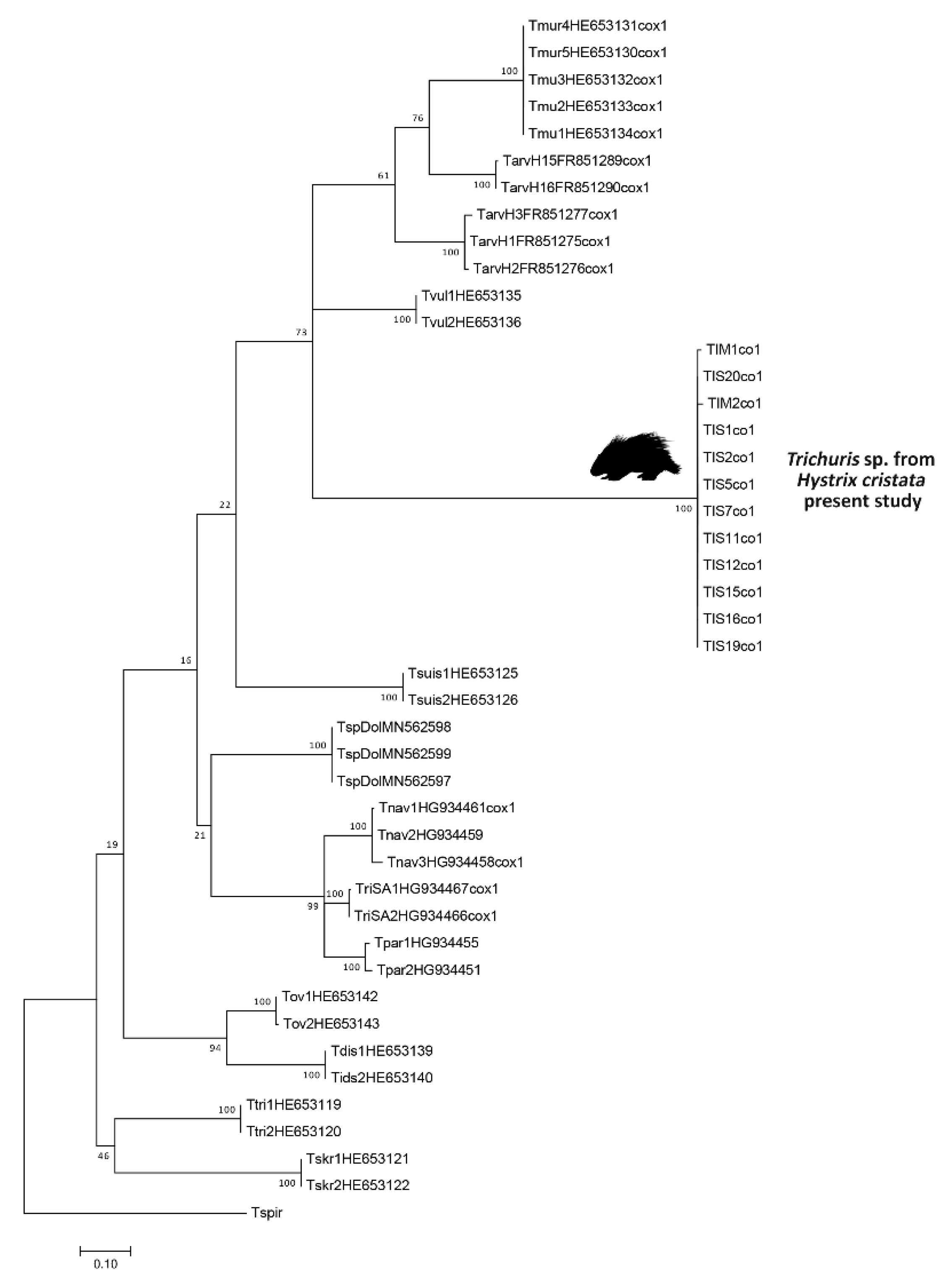
2.3. Genetic and Comparative Phylogenetic Analyses

Genomic DNA was isolated using the Wizard GenomicDNA purification kit (Promega) according to the manufacturer’s protocol. Genetic characterization was performed on 17 whipworms (15 from Lazio, 2 from Tuscany) by sequence analysis of the partial mitochondrial region cytochrome c oxidase subunit I (COX1) [17]. The primers used were CORA 5′-ACYACATAGTAGGTRTCATG-3′ and HC02198F 5′-TGATTTTTTGGTCACCCTGAAGTTTA-3′. Positive amplicons were purified using SureClean (Bioline) and shipped to an external service for sequencing (MWG Eurofins Operon). Electropherograms obtained were manually checked using Trace implemented in MEGA7 [18]. The best-fit model and parameters determined for the dataset were used for Maximum Likelihood (ML) statistical analysis, performed using the TN model+G+I (as selected by ModelTest, AIC criterion) using MEGA7, and the statistical reliability of nodes was evaluated using 1000 bootstrap pseudoreplications [18].
The retrieved sequences used for comparative purposes, including species from other rodents as well as other hosts such as canids, pigs, ruminants and primates, are listed in Table 1. Estimates of evolutionary divergence between pairs of sequences were obtained using TN as modelled by MEGA7. Two representative sequences are available in GenBank under the following accession numbers: MK779003 and OK489802 (Table 1).
Table 1. List of materials analyzed in the present study for polymorphism analyses and phylogenetic inferences based on partial COX1 mitochondrial region. Information on parasite species as defined by authors, host species, GenBank accession numbers, Specimen codes and references are reported.
Parasite Species Host SpeciesGenBank Accession NumberSpecimen CodeReferences
Trichuris sp.Hystrix cristataTIS: MK779003
TIM: OK489802
TIS and TIMPresent study
Trichuris sp.Dolichotis patagonumMN562597-99TspDol[19]
T. colobaeColobus guerezaHE653119-20Ttri1-2[17]
T. suisSus scrofaHE653125-26S1-2Zhang et al. (2016) (unpublished)
T. ovisCapra hircusHE653142-43Tov1-2[17]
T. skrjabiniCapra hircusHE653121-22Tskr1-2[17]
T. discolorBos taurusHE653139-40Tdis1-2[17]
T. pardinasiPhyllotis xanthopygusHG934451,55Tpar1-2[8]
T. navonaeAkodon montensisHG934458,59,61Tnav1-3[8]
Trichuris sp.Sooretamys angouyaHG934465-66Sa1-2[8]
T. murisMus domesticus/musculusHE653130-34M1-5[17]
T. arvicolaeMicrotus agrestisFR851275-77,89-90TarvH1-3, H15-16[17]
T. vulpisCanis (lupus) familiarisHE653135-36Tvul1-2[17]
Outgroup species
Trichinella spiralis AF293969Tspir[20]

3. Results

3.1. Morphological and Morphometric Analysis

The Trichuris sp. specimens analyzed showed the anterior part of the body to be long, narrow, tapered and whip-like, the posterior part of body to be broad and handle-like, the anterior esophageal part to be elongated and filiform and the posterior part to be large and cylindrical. The ratios between the total body length and the posterior body length (R1) were equal to 1:2.36–1:2.76 for males and 1:2.00–1:2.66 for females Meanwhile, the ratios between the anterior and the posterior body length (R4) were equal to 1:1.36–1:1.73 for males and to 1:1–1:1.67 for females.
Cuticles with transverse striae or annularity were present on the entire length of the body, except for the most anterior portion, corresponding to around 150 μm and in proximity to the vulvar region and excretory pore (cloaca). The maximum width of the body was at the junction of the esophagus and the intestine. The caudal end of the anus subterminal showed a terminal torsion. Details on the morphological data of adult specimens of Trichuris sp. from H. cristata and of other related species infecting Hystrix spp. are reported in Table 2.
The specimens analyzed resemble T. hystricis and T. landak in general morphology, especially when considering the shape of the spicular structure and vulvar region (Figure 1). Despite the ranges of several metric characters mostly overlapping, the specimens under study differed from other described species in terms of total body length and spicule length. HCPC analysis of dataset I confirmed that Trichuris specimens from the two distinct samplings of the present study are grouped together (TIS and TIM specimens—Figure S1 in the Supplementary) and are separated from all other species used for comparison (dataset II—Figure 2). The comparison of ratio values between total body length and spicule length (R2) gave distinct rather than overlapping values. In fact, the specimens studied showed values of 1:7.53–1:9.36, while T. hystricis sensu Youssefi [13] showed an R2 of 13.3, T. hystricis sensu Kreis [10] showed an R2 of 40.53, T. infundibulus an R2 was of 22.98 and, for T. lenkorani, the R2 was of 5.99.
HCPC analysis was not carried out for vulvar shape, as this is a qualitative trait. Descriptions of the vulvar region are only available for a limited number of species: in T. landak, the surface of the vulva is not densely covered with spines and the cuticle appears rugose at the anterior lip [15], while in T. hystricis sensu Kreis [10] the vulva does not protrude appreciably and is described as “extremely large but not very long” with highly developed muscles. In the present study, the vulvar region appears slightly protruded, showing a visible bulge with a trilobated muscular bore and an internal curved surface densely covered with spines (Figure 1c–e).

3.2. Molecular Analyses

All of the 17 Trichuris specimens analyzed gave a positive fragment of around 450 bp at COX1 PCR amplification. Eleven of fifteen individuals were successfully sequenced, giving a partial COX1 alignment of 305 bp after manual trimming at the ends of sequences following a comparison with GenBank retrieved sequences. Few polymorphic nucleotide sites were detected among these eleven sequences (three SNPs), which is consistent with an intraspecific polymorphism. A BLAST search revealed the identity of 87% of the specimens to be T. arvicolae (accession number FR851280, performed on specimens included in the phylogenetic comparison, coded as TriH6). However, the percentage of query coverage was 65% (174/199 nucleotide matching, on a total of 305 bp), which is not enough to refer these sequences to any taxa reporting available sequences in GenBank.
The phylogenetic ML tree with the highest log likelihood (Figure 3) describes the existence of distinct taxonomic entities supported by a maximum bootstrap value of 100% for all species included in the analysis (T. muris, T. navonae, T. pardinasi, T. vulpis, T. suis, T. trichiura, T. skrjabini, T. ovis and T. discolor). Likewise, Trichuris sp. from H. cristata was analyzed and identified as a separated branch compared to all other taxa. The genetic distance values between pairs of sequences indicate that the Trichuris we describe here was highly divergent from other Trichuris lineages, including: T. trichiura (0.35, SE = 0.03–0.04), T. ovis and T. pardinasi (0.32–0.33, SE = 0.02–0.04), T. vulpis (0.29–0.35, SE = 0.03–0.04), T. suis (0.30 SE = 0.03–0.04), T. navonae (0.29, SE = 0.03–0.04), T. muris (0.28, SE = 0) and T. arvicolae (0.26–0.29, SE = 0.01–0.03).

4. Discussion

The crested porcupine H. cristata Linnaeus, 1758 is a large, nocturnal rodent which is widespread in North and sub-Saharan Africa [21,22,23]. It is the only porcupine species occurring in Europe, with a geographical distribution restricted to mainland Italy and Sicily, most probably due to its introduction from North Africa in the early Medieval period [24,25,26]. In the last 40 years, the distribution range of the crested porcupine has increased in Italy [27,28,29], mostly because of the introduction of legal protections (National Italian Law n. 968/1977, Annex II of the Bern Convention in 1979, Annex IV of the Habitat Directive 1992/43/EEC and National Italian Law n. 157/1992). This increased distribution may potentially increase the risk of parasite infections for this species.
Despite this, the available information on the parasites of crested porcupines, both ectoparasites [30,31,32,33,34] and endoparasites [35,36,37,38], is scant. Among nematodes, both the genetic characteristics of Trichuris and the evolutionary relationships between Trichuris and members of the suborder hystricomorpha—crested porcupines in particular—have so far been poorly investigated and understood.
To date, few authors have carried out morphological studies on Trichuris from Hystrix spp. and no molecular data are available for comparisons, generating controversial and limited outcomes. The lack of clear diagnostic morphological features for Trichuris taxonomy and phenotypic plasticity observed in parasitic nematodes [2,6,39], together with the occurrence of coinfections [40], demands the mandatory use of integrative taxonomy for a more accurate characterization of species.
Four Trichuris species have so far been described, from at least three host species belonging to the genus Hystrix: T. hystricis from H. cristata and H. javanica, T. infundibulus from H. cristata, T. lenkorani and T. landak from H. indica [13,14,15]. The validity of T. infundibulus as a separate taxon from T. hystricis is unclear, despite the differences in morphological traits, given the lack of information on the locality in the original description of T. infundibulus, as shown by [41]. Spicule length measurements of T. landak reported by [15] were likely wrongly expressed, generating errors not only in its description but also in the interpretation of results and any comparisons to the present data. After a doubtful first description of T. hystricis based on specimens from captive porcupines from Basel Zoo (Basel, Switzerland) [11], a subsequent independent study on H. cristata from Tunis Zoo described whipworms putatively referable to the original T. hystricis description [42] without reporting any measurements or metrical data. Indeed, the author emphasizes this doubtful observation and host origin, declaring that it is not clear whether T. hystricis is circulating in Tunisia. In addition, previous studies have reported the presence of T. ovis in porcupines from Italy [43], advocating for the occurrence of more than one Trichuris species potentially infective to porcupines. Regarding Trichuris circulating in rodents in Italy, only T. muris and T. arvicolae have been reported so far [44,45].
Although deposited nucleotide sequences from the same genomic region are not available for Trichuris from porcupines, the morphometric differences evidenced support the status of the specimens under study as representatives of a separate taxon. This is also supported by HCPC analysis and specifically by striking differences in spicule length (3.18–4.08) and body length/spicule length ratios. Here, spicules are markedly longer than those described in T. hystricis sensu Youssefi (1.87–1.98 mm: [13]), T. hystricis sensu Kreis (0.89–1.21 mm, [10]) and T. infundibulus (1.94 mm), thus indicating that the length of spicule is a significant characteristic for the identification of the taxon under study. The taxonomic relevance of spicule length and its utility as a marker has also been demonstrated in other parasitic nematodes [46,47].
Regarding molecular characterization, the specimens of Trichuris analyzed here are confirmed as a separate lineage by phylogenetic evidence and genetic distance, with them being sufficiently differentiated from all other species included in the comparison. Assuming genetic distance is related to species separation and validity (sensu [48]), the values obtained for recognized distinct species in the genus Trichuris ranged from 0.20 to 0.22 (SE 0.03–0.04) between T. muris and T. arvicolae and from 0.20 to 0.24 (SE 0.03) between T. trichiura and T. suis. Here, Trichuris specimens from H. cristata showed even higher values of genetic distance (range of 0.26–0.35), helping to unambiguously distinguish the Trichuris sp. analyzed here from others infecting rodents.
The evolutionary relationships between taxonomic entities are not fully resolved in phylogeny by means of a single molecular marker. The monophyly of Trichuris species from rodents is not confirmed in the present tree given the very low reliability of the values at relative nodes and the position of T. vulpis as an internal branch of a group including Trichuris from H. cristata, T. muris and T. arvicolae. Few Trichuris clades were uniformly resolved across separated analyses of individual genes, as suggested by [49], and the use of additional molecular markers other than COX1 is desirable to address evolutionary-related hypotheses.

5. Conclusions

Although the genetic and morphological description in this study of the particular Trichuris that infects porcupines in Italy could justify the existence of an undescribed taxon, a formal description is not advisable. In fact, morphological data retrievable from the literature are limited (i.e., the whole set of measurements is not available uniformly among all the species studied (see Table 1), and the mitochondrial sequences in GenBank from previously described Trichuris species infecting the same host are lacking. However, zoogeographical considerations may support the status of the Trichuris sp. detected in the present study as a new, undescribed species. In fact, T. hystricis described by [13] was recovered in a single host specimen of H. indica from Iran, and the origin of the captive porcupine in which Kreis [10] obtained the first description of T. hystricis is unknown. The present study represents the first integrated morphometric and molecular investigation on Trichuris samplings carried out directly in the European geographical area of H. cristata.

Supplementary Materials

The following are available online at https://www.mdpi.com/article/10.3390/d13120628/s1: Figure S1: PCA and HCPC graphics for males (a) and females (b) morphological traits of Trichuris sp. collected from Hystrix cristata in the present study (TIS and TIM).

Author Contributions

Conceptualization, S.D., F.B. and C.D.L.; sample and data collection, C.D.L., F.B, E.M., A.V., A.S., S.C. and M.M.D.F.; software and data analysis, M.M.D.F. and S.C.; validation, S.D. and F.B.; formal analysis, S.C., C.D.L. and M.M.D.F.; investigation, S.C., E.M., A.V.; writing—original draft preparation, S.C., M.M.D.F., E.M. and S.D.; writing—review and editing, S.C., F.B., C.D.L., E.M.; supervision, S.D. and F.B. All authors have read and agreed to the published version of the manuscript.

Funding

The present research was funded by the Italian Society of Parasitology SOIPA (Starting young researcher grant 2016) and was also partially funded by Sapienza, University of Rome (Starting young researcher grant 2015—prot. C26N159B8C), with both grants going to to SC. EM and AV were supported by the MUR-FOE-Project Capitale Naturale-Task “Biodiversità”.

Institutional Review Board Statement

Not Applicable.

Data Availability Statement

All data are available via the first author. Genetic sequences have been submitted to GenBank.

Acknowledgments

Authors thank Carlo Taccari for graphical support and Silvia Rondon for inspection of animals. Edoardo Basset (University of Birmingham, UK) kindly took the time to improve the English language of our first draft. Two anonymous reviewers and the Associate Editor Gary Walker kindly improved our first MS with useful comments.

Conflicts of Interest

On behalf of all authors, the corresponding author states that there is no conflict of interest.

References

  1. Anderson, R.C. Nematode Parasites of Vertebrates: Their Development and Transmission, 2nd ed.; CAB International: Wallingford, UK, 2000. [Google Scholar]
  2. Špakulová, M. Discriminant analysis as a method for the numerical evaluation of taxonomic characters in male trichurid nematodes. System. Parasitol. 1994, 29, 113–119. [Google Scholar] [CrossRef]
  3. Robles, M.D.R.; Cutillas, C.; Panei, C.J.; Callejòn, R. Morphological and molecular characterization of a new Trichuris species (Nematoda- Trichuridae), and phylogenetic relationships of Trichuris species of Cricetid rodents from Argentina. PLoS ONE 2014, 9, e112069. [Google Scholar]
  4. Tenora, F.; Kamiya, M.; Spakulová, M.; Stanek, M.; Ooi, H.K. Scanning electron microscopy of Trichuris suis and Trichuris vulpis from Slovakia and Japan. Helminthologia 1993, 30, 93–98. [Google Scholar]
  5. Cutillas, C.; German, P.; Arias, P.; Guevara, D. Trichuris ovis and Trichuris globulosa: Morphological, biometrical, and genetic studies. Exp. Parasitol. 1995, 81, 621–625. [Google Scholar] [CrossRef] [PubMed]
  6. Feliu, C.; Spakulová, M.; Casanova, J.C.; Renaud, F.; Morand, S.; Hugot, J.P.; Santalla, F.; Durand, P. Genetic and morphological heterogeneity in small rodent whipworms in south western Europe: Characterization of Trichuris muris and description of Trichuris arvicolae n. sp. (Nematoda: Trichuridae). J. Parasitol. 2000, 86, 442–449. [Google Scholar] [CrossRef]
  7. Cutillas, C.; Oliveros, R.; de Rojas, M.; Guevara, D.C. Determination of Trichuris skrjabini by sequencing of the ITS1-5.8S-ITS2 segment of the ribosomal DNA: Comparative molecular study of different species of trichurids. J. Parasitol. 2004, 90, 648–652. [Google Scholar] [CrossRef]
  8. Callejón, R.; Robles, M.D.R.; Panei, C.J.; Cutillas, C. Molecular diversification of Trichuris spp. from Sigmodontinae (Cricetidae) rodents from Argentina based on mitochondrial DNA sequences. Parasitol. Res. 2016, 115, 2933–2945. [Google Scholar] [CrossRef]
  9. Hall, M.C. Nematode Parasites of Mammals of the Orders Rodentia, Lagomorpha, and Hyracoidea; US Government Printing Office: New York, NY, USA, 1916; Volume 50.
  10. Kreis, H.A. Beitrage zur Kenntuis parasitischer Nematoden VII. Neue parasitische Nematoden aus dem Naturhistorischen Museum Basel. Zentralbl. Bakt. Abt. Orig. 1938, 142, 329–352. [Google Scholar]
  11. Skrjabin, K.I.; Shikhobalova, N.P.; Orlov, I.V. Trichocephalidae and diseases caused by them. In Trichocephalidae and Capillariidae of Animals and Man and the Diseases Caused by Them; Skrjabin, K.I., Shikhobalova, N.P., Orlov, I.V., Eds.; Academy of Sciences of the USSR: Moscow, Russia, 1957; pp. 26–223. [Google Scholar]
  12. Yamaguti, S. The Nematodes of Vertebrates; Systema helminthum; Interscience Publishers: New York, NY, USA, 1961; Volume III. [Google Scholar]
  13. Youssefi, M.R.; Hoseini, S.H.; Rahimi, M.T.; Esfandiari, B. Trichuris hystricis, a whipworm from Hystrix indica in Iran. World J. Zool. 2010, 5, 244–245. [Google Scholar]
  14. Petriov, A.M.; Sadikhov, I.A. Trichuris lenkorani n. sp. from the intestine of Hystrix hirsutirostris in Azerbaidzhan. Dokl. Akad. Nauk. Azerb. SSR 1961, 7, 631–634. [Google Scholar]
  15. Purwaningsih, E. The first report of new species: Trichuris landak n. sp. Asian Pac. J. Trop. Biomed. 2013, 3, 85–88. [Google Scholar] [CrossRef] [Green Version]
  16. Lê, S.; Josse, J.; Husson, F. FactoMineR: An R package for multivariate analysis. J. Stat. Softw. 2008, 25, 1–18. [Google Scholar] [CrossRef] [Green Version]
  17. Callejón, R.; Nadler, S.; De Rojas, M.; Zurita, A.; Petrášová, J.; Cutillas, C. Molecular characterization and phylogeny of whipworm nematodes inferred from DNA sequences of COX1 mtDNA and 18S rDNA. Parasitol. Res. 2013, 112, 3933–3949. [Google Scholar] [CrossRef] [PubMed]
  18. Kumar, S.; Stecher, G.; Tamura, K. MEGA7: Molecular Evolutionary Genetics Analysis Version 7.0 for Bigger Datasets. Mol. Biol. Evol. 2016, 33, 1870–1874. [Google Scholar] [CrossRef] [Green Version]
  19. Di Filippo, M.M.; Berrilli, F.; De Liberato, C.; Di Giovanni, V.; D’Amelio, S.; Friedrich, K.G.; Cavallero, S. Molecular characterization of Trichuris spp. from captive animals based on mitochondrial markers. Parasitol. Int. 2020, 75, 102043. [Google Scholar] [CrossRef]
  20. Lavrov, D.V.; Brown, W.M. Trichinella spiralis mtDNA: A nematode mitochondrial genome that encodes a putative ATP8 and normally structured tRNAs and has a gene arrangement relatable to those of coelomate metazoans. Genetics 2001, 157, 621–637. [Google Scholar] [CrossRef] [PubMed]
  21. Monetti, L.; Massolo, A.; Sforzi, A.; Lovari, S. Site selection and fidelity by crested porcupine for denning. Ethol. Ecol. Evol. 2005, 17, 149–159. [Google Scholar] [CrossRef]
  22. Mori, E.; Lovari, S. Sexual size monomorphism in the crested porcupine (Hystrix cristata). Mamm. Biol. 2014, 79, 157–160. [Google Scholar] [CrossRef]
  23. Viviano, A.; Amori, G.; Luiselli, L.; Oebel, H.; Bahleman, F.; Mori, E. Blessing the rains down in Africa: Spatiotemporal behaviour of the crested porcupine Hystrix cristata (Mammalia: Rodentia) in the rainy and dry seasons, in the African savannah. Trop. Zool. 2020, 33, 113–124. [Google Scholar] [CrossRef]
  24. Amori, G.; De Smet, K. Hystrix cristata. The IUCN Red List of Threatened Species 2016: E.T10746A22232484. 2016. Available online: http://dx.doi.org/10.2305/IUCN.UK.2016-2.RLTS.T10746A22232484.en (accessed on 25 October 2021).
  25. Masseti, M.; Albarella, U.; De Grossi Mazzorin, J. The crested porcupine, Hystrix cristata L., 1758, in Italy. Anthropozoologica 2010, 45, 27–42. [Google Scholar] [CrossRef]
  26. Trucchi, E.; Sbordoni, V. Unveiling an ancient biological invasion: Molecular analysis of an old European alien, the crested porcupine (Hystrix cristata). BMC Evol. Biol. 2009, 9, 109. [Google Scholar] [CrossRef] [Green Version]
  27. Mori, E.; Ficetola, G.F.; Bartolomei, R.; Capobianco, G.; Varuzza, P.; Falaschi, M. How the South was won: Current and potential range expansion of the crested porcupine in Southern Italy. Mamm. Biol. 2021, 101, 11–19. [Google Scholar] [CrossRef]
  28. Mori, E.; Sforzi, A.; Bogliani, G.; Milanesi, P. Range expansion and redefinition of a crop-raiding rodent associated with global warming and temperature increase. Clim. Chang. 2018, 150, 319–331. [Google Scholar] [CrossRef]
  29. Torretta, E.; Orioli, V.; Bani, L.; Mantovani, S.; Dondina, O. En route to the North: Modelling crested porcupine habitat suitability and dispersal flows across a highly anthropized area in northern Italy. Mamm. Biol. 2021, 1–11. [Google Scholar] [CrossRef]
  30. Bailly-Choumaira, H.; Morel, P.C.; Rageau, J. Sommaire des donnees actuelles sur les tiques du Maroc (Acari, Ixodoidea). Bull. Inst. Sci. 1976, 1, 102–117. [Google Scholar]
  31. Beaucournu, J.C.; Launay, H. Les Puces de France et du Bassin Méditerranéen Occidental. Faune de France 76; Fédération Française des Sociétés de Sciences Naturelles: Paris, France, 1990. [Google Scholar]
  32. Kolonin, G.V. Haemaphysalis lobachovi sp. n. (Acharina: Ixodidae) from porcupine (Hystrix cristata) from Ethiopia. Folia Parasitol. 1995, 42, 239. [Google Scholar]
  33. Mori, E.; Sforzi, A.; Menchetti, M.; Mazza, G.; Lovari, S.; Pisanu, B. Ectoparasite load in the crested porcupine Hystrix cristata Linnaeus, 1758 in Central Italy. Parasitol. Res. 2015, 114, 2223–2229. [Google Scholar] [CrossRef]
  34. Scaravelli, D.; Senini, C.; Bonacci, T. First case of traumatic myiasis caused by Calliphora vicina in a crested porcupine Hystrix cristata L. in Italy. J. Entomol. Acarol. Res. 2017, 49, 6823. [Google Scholar] [CrossRef] [Green Version]
  35. Cilia, G.; Bertelloni, F.; Coppola, F.; Turchi, B.; Biliotti, C.; Poli, A.; Parisi, F.; Felicioli, A.; Cerri, D.; Fratini, F. Isolation of Leptospira serovar Pomona from a crested porcupine (Hystrix cristata, L., 1758). Vet. Med. Sci. 2020, 6, 985–991. [Google Scholar] [CrossRef] [PubMed]
  36. Coppola, F.; Maestrini, M.; Berrilli, F.; Procesi, I.G.; Felicioli, A.; Perrucci, S. First report of Giardia duodenalis infection in the crested porcupine (Hystrix cristata L., 1758). Int. J. Parasitol. Parasites Wildl. 2020, 11, 108–113. [Google Scholar] [CrossRef] [PubMed]
  37. Coppola, F.; Cilia, G.; Bertelloni, F.; Casini, L.; D’Addio, E.; Fratini, F.; Cerri, D.; Felicioli, A. Crested porcupine (Hystrix cristata L.): A new potential host for pathogenic Leptospira among semi-fossorial mammals. Comp. Immunol. Microbiol. Infect. Dis. 2020, 70, 101472. [Google Scholar] [CrossRef]
  38. Harrison, T.M.; Moorman, J.B.; Bolin, S.R.; Grosjean, N.L.; Lim, A.; Fitzgerald, S.D. Toxoplasma gondii in an African porcupine. J. Vet. Diagn. Investig. 2007, 19, 191–194. [Google Scholar] [CrossRef] [Green Version]
  39. Robles, M.D.R.; Navone, G.T. Redescription of Trichuris laevitestis (Nematoda: Trichuridae) from Akodon azarae and Scapteromys aquaticus (Sigmodontinae: Cricetidae) in Buenos Aires province, Argentina. J. Parasitol. 2006, 92, 1053–1057. [Google Scholar] [CrossRef]
  40. Vejl, P.; Nechybová, S.; Peřinková, P.; Melounová, M.; Sedláková, V.; Vašek, J.; Čílová, D.; Rylková, K.; Jankovská, I.; Vadlejch, J.; et al. Reliable molecular differentiation of Trichuris ovis and Trichuris discolor from sheep (Ovis orientalis aries) and roe deer (Capreolus capreolus) and morphological characterisation of their females: Morphology does not work sufficiently. Parasitol. Res. 2017, 116, 2199–2210. [Google Scholar] [CrossRef] [PubMed]
  41. Ribas, A.; López, S.; Makundi, R.H.; Leirs, H.; de Bellocq, J.G. Trichuris spp. (Nematoda: Trichuridae) from two rodents, Mastomys natalensis and Gerbilliscus vicinus in Tanzania. J. Parasitol. 2013, 99, 868–875. [Google Scholar] [CrossRef] [PubMed]
  42. Bernard, J. Faune des nematodes parasites des mammiferes de Tunisie et des contrees voisines. Arch. L’inst. Pasteur Tunis 1987, 64, 265–319. [Google Scholar]
  43. Poglayen, G.; Scaravelli, D.; Tampieri, M.P.; Galuppi, R.; Nuti, C.; Gaglio, G.; Abbene, S. Fauna parassitaria dell’istrice Hystrix cristata in Italia. Hystrix J. Mammal. 2005, 1, 106. [Google Scholar]
  44. Callejón, R.; De Rojas, M.; Feliú, C.; Balao, F.; Marrugal, A.; Henttonen, H.; Guevara, D.; Cutillas, C. Phylogeography of Trichuris populations isolated from different Cricetidae rodents. Parasitology 2012, 39, 1795–1812. [Google Scholar] [CrossRef] [Green Version]
  45. Milazzo, C.; de Bellocq, J.G.; Cagnin, M.; Casanova, J.C.; di Bella, C.; Feliu, C.; Morand, S.; Santalla, F. Helminths and ectoparasites of Rattus rattus and Mus musculus from Sicily, Italy. Comp. Parasitol. 2003, 70, 199–204. [Google Scholar] [CrossRef]
  46. Iglesias, R.; D’Amelio, S.; Ingrosso, S.; Farjallah, S.; Martínez-Cedeira, J.A.; García-Estévez, J.M. Molecular and morphological evidence for the occurrence of Anisakis sp. A (Nematoda, Anisakidae) in the Blainville’s beaked whale Mesoplodon densirostris. J. Helminthol. 2008, 82, 305–308. [Google Scholar] [CrossRef]
  47. Mattiucci, S.; Paoletti, M.; Webb, S.C. Anisakis nascettii n. sp. (Nematoda: Anisakidae) from beaked whales of the southern hemisphere: Morphological description, genetic relationships between congeners and ecological data. Syst. Parasitol. 2009, 74, 199–217. [Google Scholar] [CrossRef] [PubMed]
  48. Blouin, M.S. Molecular prospecting for cryptic species of nematodes: Mitochondrial DNA versus internal transcribed spacer. Int. J. Parasitol. 2002, 32, 527–531. [Google Scholar] [CrossRef]
  49. Callejón, R.; Cutillas, C.; Nadler, S.A. Nuclear and mitochondrial genes for inferring Trichuris phylogeny. Parasitol. Res. 2015, 114, 4591–4599. [Google Scholar] [CrossRef] [PubMed]
Figure 1. Optical microscopy pictures of Trichuris sp. adult specimens of males (a,b) and females (ce). In detail: (a): detail of the spines of distal spicular sheath, lateral view; (b) entire spicule, with portion of spiny spicular sheath, similar size and morphology of spines, and spicule; (c,e) lateral view of protrusive vagina tulip-shaped uterus with eggs; (d) detail on frontal vagina view. (fh): measurements of eggs parameters.
Diversity 13 00628 g001
Figure 2. (A) Principal component analysis and (B) HCPC cluster dendrogram based on Trichuris sp. morphological traits (dataset II), alongside with other Trichuris spp. used for comparative purposes.
Diversity 13 00628 g002
Figure 3. Maximum Likelihood tree based on the analysis of the partial COX1 mitochondrial region, including sequences retrieved from GenBank (cf. Table 1) and sequences of Trichuris sp. analyzed in the present study (TIS and TIM codes). Statistical support values at nodes are reported as bootstrap percentages. Scale bars indicate evolutionary distance.
Diversity 13 00628 g003
Table 2. Morphological data of male (M) and female (F) specimens of Trichuris from H. cristata analyzed in the present study and of other Trichuris species from Hystrix spp. (M1-F1: total length of body; M6-F2: length of esophageal part; M2-F3: width of body at level of esophagus end; M3-F4: width of posterior part; M4: length of spicule; M5: length of spicular sheat; F5: length of eggs; F6: width of eggs; F7: uterus–tail distance). All measurements are in mm. Arithmetic mean of measurements are indicated, with minimum and maximum in parenthesis and ± standard deviation (SD).
Trichuris sp.
from
Hystrix cristata
Present Study
Trichuris
landak
from
Hystrix javanica [15]
Trichuris
hystricis
from
Hystrix indica
[11]
Trichuris
hystricis
from
Hystrix cristata
[10]
Trichuris
infundibulus
from
Hystrix cristata
[9]
Trichuris
lenkorani
from
Hystrix indica
[14]
M130.70
(25–35) ± 5.0
39.34
(36.05–42.64)
25.60
(23.76–27.45)
42.56
(39.42–45.70)
44.6014.21–16.72
M20.20
(0.08–0.31) ± 0.1
0.286
(0.282–0.29)
-0.37
(0.34–0.41)
-
M30.56
(0.46–0.63) ± 0.08
0.74
(0.71–0.77)
-0.20
(0.19–0.21)
-
M43.6
(3.18–4.08) ± 0.45
-1.92
(1.87–1.98)
1.05
(0.89–1.21)
1.942.42–2.74
M50.07
(0.06–0.09) ± 0.01
0.04
(0.03–0.05)
0.000760.08
M618.80
(15.0–21.0) ± 3
----
M70.04
(0.03–0.04) ± 0.005
----
M911.97
(10.0–14.8) ± 2.4
M1018.80
(15.0–21.0) ± 3
F142.34
(30.00–49.30) ± 9.65
36.40
(36.00–41.90)
34.40
(32.90–35.90)
48.40
(47.20–49.50)
52.1020.00–27.70
F225.00
(20–28) ± 4
23.09
(22.20–23.80)
13.80
(12.32–15.21)
--
F30.15
(0.13–0.19) ± 0.03
0.30
(0.30–0.31)
---
F40.80
(0.44–1.03) ± 0.3
0.80
(0.80–0.81)
---
F50.06
(0.06–0.07) ± 0.04
0.05
(0.05–0.06)
0.05
(0.04–0.06)
0.06
F60.03
(0.02–0.03) ± 0.02
0.02
(0.02–0.03)
0.03
(0.02–0.03)
0.03
F70.42
(0.26–0.58) ± 0.16
----
Publisher’s Note: MDPI stays neutral with regard to jurisdictional claims in published maps and institutional affiliations.

