Article

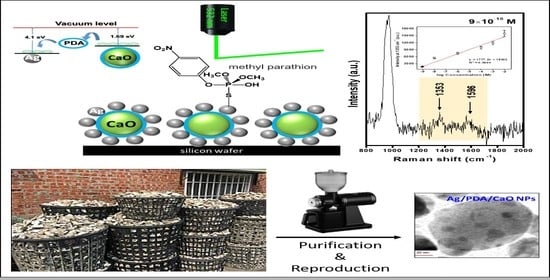
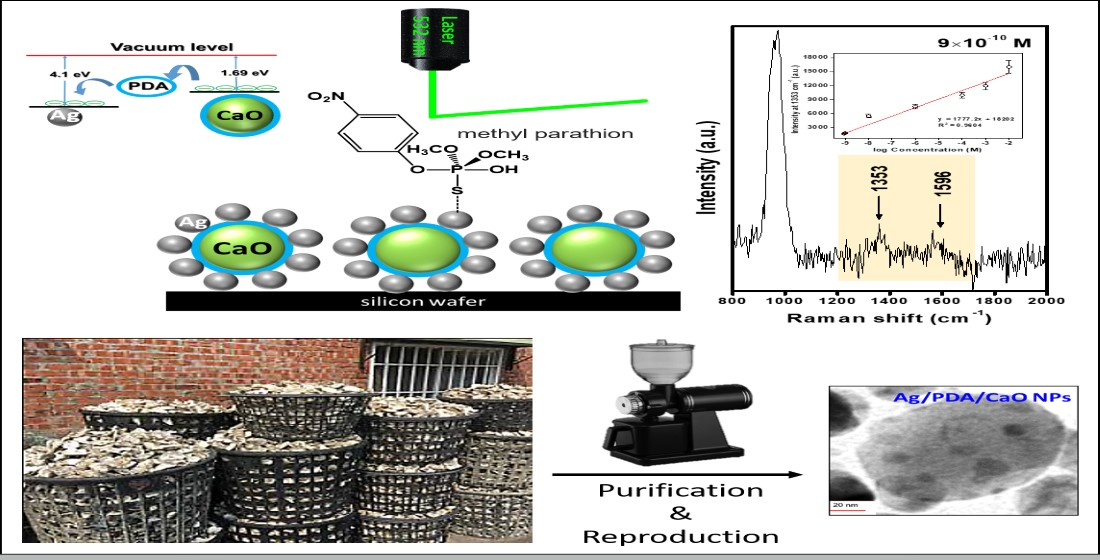
A Novel SERS Substrate Based on Discarded Oyster Shells for Rapid Detection of Organophosphorus Pesticide

1
Department of Pathology, Kaohsiung Armed Forces General Hospital, Kaohsiung 80284, Taiwan
2
Department of Chemistry, National Sun Yat-Sen University, Kaohsiung 80424, Taiwan
3
Department of Surgery, Division of Neurosurgery, Kaohsiung Armed Forces General Hospital, Kaohsiung 80284, Taiwan
4
Institute of Medical Science and Technology, National Sun Yat-Sen University, Kaohsiung 80424, Taiwan
5
Department of Marine Biotechnology and Resources, National Sun Yat-Sen University, Kaohsiung 80424, Taiwan
6
Center for Stem Cell Research, Kaohsiung Medical University, Kaohsiung 80708, Taiwan
7
School of Pharmacy, College of Pharmacy, Kaohsiung Medical University, Kaohsiung 80708, Taiwan
*
Author to whom correspondence should be addressed.
Coatings 2022, 12(4), 506; https://doi.org/10.3390/coatings12040506
Submission received: 14 March 2022 / Revised: 2 April 2022 / Accepted: 6 April 2022 / Published: 8 April 2022

Abstract

:
Over the past few years, the concern for green chemistry and sustainable development has risen dramatically. Researchers make an effort to find solutions to difficult challenges using green chemical processes. In this study, we use oyster shells as a green chemical source to prepare calcium oxide nanoparticles (CaO-NPs). Transmission electron microscopy (TEM) results showed the CaO-NPs morphology, which was spherical in shape, 40 ± 5 nm in diameter, with uniform dispersion. We further prepared silver/polydopamine/calcium-oxide (Ag/PDA/CaO) nanocomposites as the surface-enhanced Raman scattering (SERS) substrates and evaluated their enhancement effect using the methyl parathion pesticide. The effective SERS detection limit of this method is 0.9 nM methyl parathion, which is much lower than the safety limits set by the Collaborative International Pesticides Analytical Council for insecticide in fruits. This novel green material is an excellent SERS substrate for future applications and meets the goal of green chemistry and sustainable development.

Graphical Abstract

1. Introduction

The great advantages that surface-enhanced Raman scattering (SERS) spectroscopy offer in detection applications include fingerprint recognition of chemical molecules, non-destructive testing, high sensitivity, rapid detection, quantification, etc. [1]. Therefore, SERS can be applied to a wide range of applications, such as the detection of antibiotic and pesticide residues in agricultural products [2], pathogenic bacteria testing in aquatic environments [3], and identification of plastic particles released from consumer products [4], environmental exhaust fumes, etc. [5]. The significant impact of SERS techniques on molecular detection and plasmon catalysis are quite promising and due to the fact that these materials are of significant interest for a wide range of applications [6]. Since SERS-based techniques rely on the well-organized nanostructures, capable of generating plasmonic events, thus, SERS substrates based on the newly designed and more controlled nanostructures are highly demanded for further advancements. For the efficacy and to obtain the enhanced signals, simultaneous occurrence of a long-range electromagnetic effect (EM) and a short-range chemical effect, i.e., charge transfer (CT), are well needed during SERS measurements [7]. In this context, simultaneous CT can be seen by the use of semiconductor materials, which predominantly include metal oxides [8]. Therefore, the incorporation of semiconductor materials into SERS substrates would be able to generate a CT mechanism as a dominant contribution to the surface-enhanced Raman signal. To this end, efforts have been made with semiconductor oxide materials, such as ZnO, TiO2 and Cu2O, which are reported to be capable of generating weak and/or strong SERS signals via chemical enhancement [4,9,10]. The versatile application of metal oxides in various areas are well known. As a major drawback, after use, these materials are released to soil and surface water that passes over lakes, rivers, and seas. Nanomaterials, due to their shape and size, are prominent to bind to a wide variety of biomolecules, including proteins, drugs and nucleotides. Therefore, humans become exposed to nanoparticles (NPs) in medical and industrial settings quite commonly. The existence of metal nanoparticles (MNPs) in the ecological system lead to bioaccumulations, causing potential effects on human health and living organisms. Especially the induced toxicity of CuO, ZnO, TiO2, and other metal oxides, which are well known and their adverse effects have been well studied in the past few decades [11,12,13,14,15]. Therefore, it is urgently required to incorporate some non-toxic, biocompatible and ecological friendly metal oxides for the construction of SERS substrates for societal welfare and care.
In our previous work, we reported a simultaneous occurrence of CT and EM mechanisms into a Ag/CuO nanocomposite system at the metal–semiconductor interface [16]. However, realizing the scenario of toxicity, we further present the utilization of non-toxic CaO MNPs into SERS substrates; i.e., a metal-molecular-semiconductor composite system consisting of Ag NPs, PDA and CaO NPs to construct a silver/polydopamine/calcium-oxide (Ag/PDA/CaO) system. For the generation of the SERS substrate, CaO NPs were mainly obtained from a green resource (oyster shells) via a simple chemical process. The strong adhesion of the PDA layer provided a uniform and well-packed composite system [17]. Simultaneously, in-situ preparation of Ag NPs and, thus, effective charge redistributions among Ag NPs and CaO, via a conducting PDA layer to induce a chemical effect during the SERS measurement, was obtained [18]. In our previous work, the Ag/CuO nanocomposite demonstrated an approximately 105 times SERS signal enhancement, mainly through the combination of CT and EM mechanisms [19]. In the present study, such an enhancement factor (4.7 × 10⁵) in 10 h with LOD in a nano-regime for 4-aminothiophenol (4-ATP) was obtained for the Ag/CaO nanocomposite and, thus, the incorporation of CaO can be validated. To date, there are no reports demonstrating the incorporation of CaO into a SERS substrate to obtain enhanced SERS signals. Therefore, we believe our study would be able to introduce a newer and non-toxic metal oxide into the process of advancement for newer ecologically friendly SERS substrates and nanomaterials.

2. Materials and Methods

Oyster shells, having a high CaCO3 content, were used as the green source of CaO in our study. After calcination, the CaO content was highest followed by CO2; the percentages of Na2O, MgO, SO3, SiO2, SrO, and H2O were very low [20,21]. The shells were scrubbed to remove sand and salt from the surface and were then placed in an oven to remove moisture and thus to reduce impurities. The agents used in this experiment include dopamine hydrochloride (Sigma-Aldrich, Saint Louis, MO, USA), tris buffer (Sigma-Aldrich, Saint Louis, MO, USA), silver nitrate (Sigma-Aldrich, Saint Louis, MO, USA), sodium hydroxide (Sigma-Aldrich, Saint Louis, MO, USA), ethanol (ECHO Chemical CO., LTD, Kaohsiung, Taiwan), hydrochloric acid (J.T. Baker, Radnor, PA, USA), sodium carbonate (Sigma-Aldrich, Saint Louis, MO, USA), 4-aminothiophenol and methyl parathion (Sigma-Aldrich, Saint Louis, MO, USA). All agents were used directly in the experiment without further purification. The pure water required for samples synthesized and dispensed was obtained from pure water (18.2 MΩ-cm at 25 °C) Milli-Q reagent-grade (type I).
Scheme 1 shows the Ag/PDA/CaO NPs preparation process. After the oyster shells were ground at room temperature, 1 M NaOH solution was added and stirred for 2 h. It was then centrifuged and washed with deionized water until neutral. Hydrochloric acid was added, and the mixture was stirred for 6 h. The filtrate was filtered to obtain a CaCl2 solution. CaCl2 and sodium carbonate were mixed into the water-heating device to heat to 100 °C. After filtering, the powder was placed into a high-temperature furnace heated to 900 °C for 2 h, and spherical calcium oxide nanoparticles (CaO NPs) were synthesized. The reaction mechanism is given by Equation (1):
CaCO 3 s   +   2 HCl aq     CaCl 2 aq   +   CO 2 g   +   H 2 O l
CaCl 2 aq   +   NaCO 3 s     CaCO 3 s   +   2 NaCl aq
CaCO 3 s   Δ   CaO s   +   CO 2 g
For the PDA and Ag NPs surface modification method of Ag/PDA/CaO NPs, refer to Lin et al. [5]. Utilizing the catechol-amine chemistry of dopamine (DA) as an adhesive layer between CaO NPs and Ag NPs, PDA coating layers were formed on the surface of CaO NPs by oxidative polymerization in an alkaline environment [22]. Surface-exposed PDA functional groups, including catechol, amine, and imine [23] can be used as reducing agents for Ag NPs. We prepared a solution of synthetic PDA, the tris buffer (C8H11NO2•HCl) adjusted to pH 8.5 as a solvent and configured the 10 mM dopamine solution. We soaked the synthesized CaO NPs in the dopamine solution and stirred for 12 h. Then, we soaked the PDA-coated CaO NPs in a silver amine solution (Tollen’s reagent), dried it in the oven overnight, and obtained the Ag/PDA/CaO NPs.

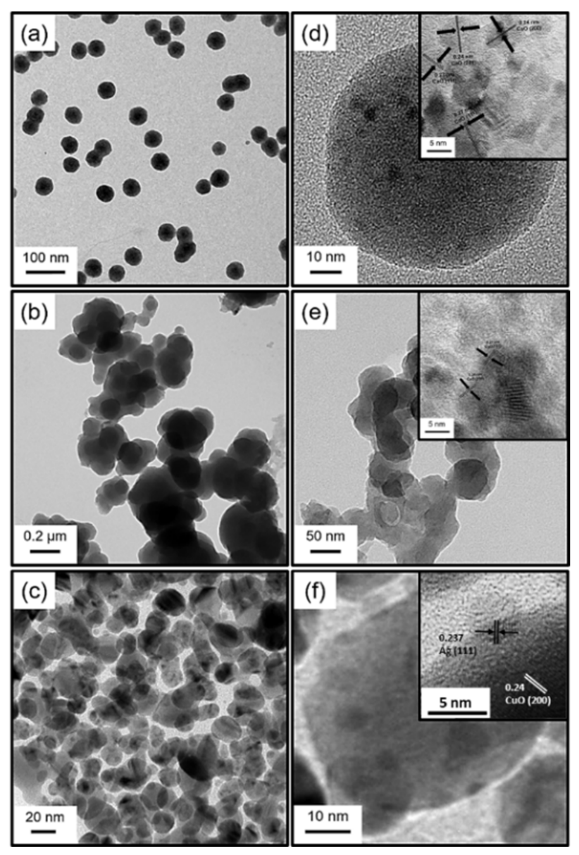
2.1. Transmission Electron Microscopy (TEM)

After the nanoparticle powder was dissolved in alcohol, 30 μL of the sample was dripped onto a carbonized copper mesh sheet and dried to complete the test substrate, shooting the image with a transmission electron microscope (JEOL, JEM-2100, Tokyo, Japan). Magnification: 2000–1,500,000×; Resolution: Point image: 0.27 nm and Lattice image: 0.14 nm; Dual tilt bases: X-axis ± 60°; Electron gun: field emission acceleration voltage of 200 kv.

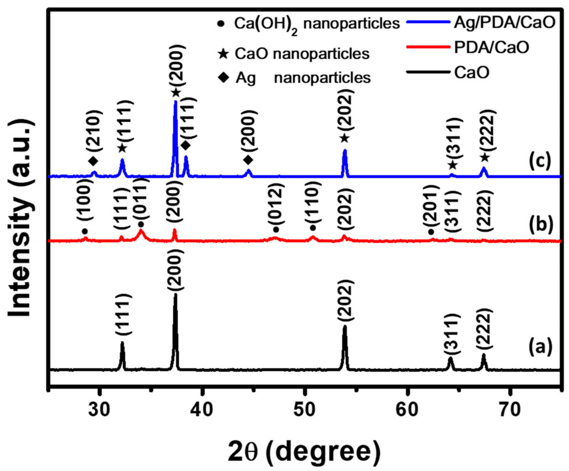
2.2. X-ray Diffraction (XRD)

The samples were prepared by drying and grinding calcium oxide nanoparticles into a uniform powder. The powder was flattened on a test substrate for X-ray diffraction. The diffraction pattern of the calcium oxide nanoparticles was used to identify the crystal grid parameters. We measured calcium oxide and calcium carbonate powder samples at room temperature using an X-ray powder diffractometer (Bruker D8 Advance, Karlsruhe, Germany). The Cu Kα1 with a wavelength of 1.5418 Å and energy of 8.05 KeV was used as the X-ray source and the X-ray tube was a Cu target. The operating voltage was 40 kV and the operating current was 40 mA. The detector is a high-speed, high-sensitivity PSD detector with the best resolution of ~0.03° (FWHM). The 2θ angle was scanned at a scanning speed of 0.5 s at each pitch from 20 °C to 80 °C, and the spacing was 0.1°.

2.3. X-ray Photoelectron Spectroscopy (XPS)

Samples were prepared for analysis using X-ray photoelectron spectroscopy (XPS) by depositing an aliquot of the sample solution onto clean silicon wafer at room temperature and drying in air. XPS spectra were acquired using a JAMP-9500F (JEOL, Tokyo, Japan) equipped with a monochromatic Mg Kα X-ray (1253.6 eV) radiation source. The spectra were calibrated using the C 1s peak at 285 eV from adventitious surface carbon as an internal reference. The Cu and Ag peak curve fitting was performed with a Gaussian/Lorentzian ratio of 0.7 using peak fitting software by JEOL SpecSurf XPS (version 1.7.3.9), after a Shirley-type background subtraction.

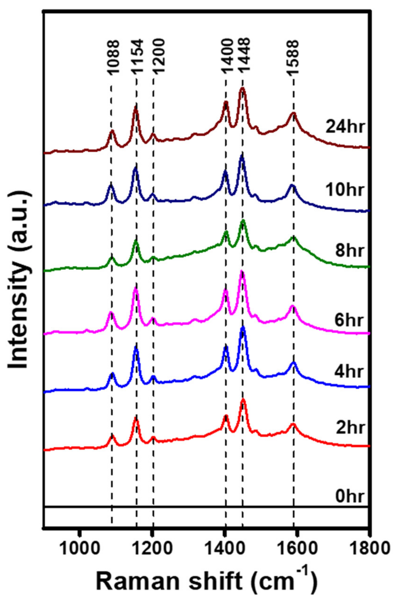
2.4. Raman Spectroscopy

The modified metal/polymer/CaO NPs were deposited on a silicon substrate (1 cm × 1 cm) as a SERS substrate. Next, 5 µL 4-ATP and methyl parathion at different concentrations were dropped on the surface of SERS substrate, and its SERS enhancement effect was detected by Raman spectroscopy (WITec alpha300R, WITec, Ulm, Germany) at a laser wavelength of 532 nm and an intensity of 30.5 μW, and further detected the representative pesticide in water.

3. Results and Discussion

The surface morphologies of the composites were observed using transmission electron microscopy (TEM), as shown in Figure 1a,d, showing that CaO NPs, 40 ± 5 nm in diameter, were synthesized successfully with a spherical shape. Figure 1b and e demonstrate that after synthesizing the PDA onto CaO NPs, its surface became rough, and a coating was observed. The inset in Figure 1d and e shows high-resolution TEM (HRTEM) images of CaO NPs and PDA/CaO nanocomposites. The lattice spacings were 0.24 and 0.27 nm through calculation, corresponding to the crystal plane of (200) and (111), which proves a calcium oxide composition [24]. Figure 1c,f show that Ag NPs were successfully deposited on each calcium oxide nanoparticle. The inset in Figure 1f shows the HRTEM result. The spacing of Ag (111) was 0.237 nm [25], which confirms that Ag NPs were reduced on the surface of the CaO NPs by about 12 ± 5 nm.
The X-ray diffraction (XRD) results of the synthesized composites are shown in Figure 2. In addition to the CaO signal, the Ag/PDA/CaO (blue line) also had Ag (111) and (200) signals, in accordance with the standard diffraction pattern of Ag NPs (JCPDS: 04-0783) [26]. These results prove that we successfully synthesized Ag NPs on CaO NPs.
We analyzed each step of the Ag/PDA/CaO SERS substrate formation by using high-resolution X-ray photo-electron spectroscopy (XPS). The Ag 3D XPS spectra in Figure 3a for Ag NPs exhibited peaks at 368.1 eV and 371.2 eV, which are assigned to Ag 3d5/2 and Ag 3d3/2, respectively. As shown in Figure 3b, the high-resolution XPS of the Ca 2p revealed a signal of calcium oxide at 350.8 eV and 354.4 eV. Moreover, dopamine molecules were polymerized on the CaO NP surfaces, and the Ca 2p signal shifted 0.4 eV from 350.8 eV to the lower binding energy of 350.4 eV. These results indicate that the push electron effects of PDA resulted in a shift in Ca 2p signal to a lower binding energy because the chemical environment around Ca changed. After the PDA reduced the Ag NPs on the calcium oxide, the signal of Ca 2p shifted to a lower binding energy, from 350.4 eV to 350.1 eV. According to the literature, when semiconductors combine with metals, electrons flow from higher to lower Fermi levels, allowing both Fermi levels to reach a new equilibrium [27,28,29]. Because the work function is 1.69 eV of calcium oxide and 4.1 eV of silver [30], the electrons transferred from calcium oxide to silver when they were combined, as shown in Figure 3c.
To evaluate the SERS activity of the Ag/PDA/CaO substrate, we selected 4-ATP as model probe molecules and configured 4-ATP using ethanol to 10−3 M. We examined pure 4-ATP 10−2 M on a silicon substrate as normal Raman spectra (Figure 4a). The labeled peaks in the normal Raman spectra correspond well to those reported for pure 4-ATP in earlier reports. The peaks at 1154, 1200, and 1448 cm−1 are assigned to the δ(C−H) stretching vibration. The peaks at 1088, 1154, 1400, and 1588 cm−1 are assigned to the ν(C−S), δ(C−H), [ν(C−C) + δ(C−H)], and ν(C−C) stretching vibration, respectively. We used different Ag NP deposition times of 2, 4, 6, 8, 10, and 24 h to prepare the SERS substrates and analyzed 4-ATP, in order to find the most suitable preparation condition. Figure A1 in the Appendix A shows significant enhancement in intensity, beginning 2 h after deposition. We calculated the effective enhancement factors (EFs) by comparing the SERS peak intensity at 1448 cm−1 of 4-ATP on Ag/PDA/CaO nanocomposites to that of 4-ATP on CaO NPs, according to the formula ISERS/Isubstrate × (concentration factor) [31,32] ISERS is the Raman intensity of the probe molecule on the Ag/PDA/CaO substrate, Isubstrate is the Raman intensity of the probe molecule on the silicon substrate. The concentration factor is the corresponding concentration of the different concentrations of the probe molecule in ethanol. The calculated EFs were 3.8 × 10⁵, 5.1 × 10⁵, 5.2 × 10⁵, 3.2 × 10⁵, 4.7 × 10⁵, and 4.6 × 10⁵ at 2, 4, 6, 8, 10, and 24 h respectively. The results indicate that EF reached 5.1 × 10⁵ after 4 h of deposition. Therefore, we found that 4 h of deposition time for Ag NPs is the most appropriate for preparing a Ag/PDA/CaO nanocomposite as the SERS substrate for detecting 4-ATP. Under these conditions, less deposition time was needed for the best EF result.
We further investigated the detection limit of 4-ATP with the Ag/PDA/CaO-4h substrate. We formulated 4-ATP to 10−3, 10−4, 10−5, 10−6, 10−7 and 10−8 M in ethanol. As shown in Figure 4, CaO and Ag/PDA materials showed no SERS effects; however, after detection of the Ag/PDA/CaO-4 h substrates, an obvious SERS effect was observed. The calculated EFs were 1.1 × 10⁶, 7.8 × 10⁶, 3.6 × 10⁷, 1.0 × 10⁸, 9.6 × 10⁸, and 6.5 × 10⁹, for 10−3, 10−4, 10−5, 10−6, 10−7 and 10−8 M, respectively, and the detection limit reached ~10−8 M.
We compared these results with those reported in other studies. Sun et al. [33] used a silver nanoparticle colloidal solution to enhance the 4-ATP molecular signal, and the detection limit was ~10−7 M. Recently, scientists have used different substrates to reduce the silver nanoparticles to further enhance the 4-ATP signal, with a detection limit of ~10−7 M for a single molecule [34,35,36]. In our study, Ag/PDA/CaO nanocomposites synthesized through simple steps successfully enhanced the SERS effect and improved the detection limit to ~10−8 M for 4-ATP single-molecule detection. The SERS intensity is determined by several factors, such as particle size and shape and the nature of the aggregate [37,38]. In our results, the self-assembled Ag/PDA/CaO nanocomposites had uniform particle sizes and shapes of Ag NPs. This characteristic, resulting in the formation of numerous “hot spots”, offers great signal improvement in SERS through strong electromagnetic enhancement.
The spectra of methyl parathion of differing concentrations (5 μL of 10 mM, 1 mM, 0.1 mM, 0.01 mM, 1 μM, 0.01 μM, 1 nM, and 0.9 nM) on the Ag/PDA/CaO nanocomposites are shown in Figure 5. The major characteristic bands in the SERS spectra of the methyl parathion could be identified at 1353 and 1596 cm−1, which correspond with the variation in the C–H bending and phenyl stretching, respectively [39]. The calibration curve of 1353 cm−1 peak intensity and the corresponding concentration displayed a good linear relationship, ranging from 0.01 M to 0.9 nM, showing a linear correlation coefficient R2 of 0.96 for methyl parathion (Figure 5c). The detection limit was 0.9 nM, which is much lower than the safety limits set by the Collaborative International Pesticides Analytical Council (CIPAC) for insecticide in fruits.
In addition, Table 1 compares the analytical performances of various SERS nanocomposites for the detection of methyl parathion, which is comparable or even superior to the other SERS nanocomposites reported in the literature. The above results demonstrate that Ag/PDA/CaO nanocomposites have the potential for the highly sensitive detection and quantitative analysis of methyl parathion residue.

4. Conclusions

A SERS substrate was successfully designed, constructed, and established by utilizing non-toxic CaO as a semiconductor material for the first time, over other toxic, traditionally used metal oxides (CuO, Cu2O, ZnO, Fe2O3, etc.). The construction process involved the construction of CaO NPs from green resource oyster shells, followed by PDA coating on CaO NPs and further in-situ construction of Ag NPs onto a PDA/CaO support. The obtained LOD in the nanomolar regime and significant enhancement factor in SERS signals for sensing methyl parathion with the constructed substrate demonstrate the effectiveness of the SERS-constructed substrate by using CaO.

Author Contributions

C.-Y.C., P.-Y.L. and J.-S.L. contributed equally to this work. C.-Y.C.: funding acquisition, project administration, and supervision. P.-Y.L.: methodology, data curation, formal analysis, original draft writing. J.-S.L.: methodology, formal analysis, and original draft writing. R.K.: original draft writing. C.-Y.T.: methodology and data curation. N.-F.C. and Z.-H.W.: resources. S.H.: conceptualization, funding acquisition, project administration and supervision. All authors have read and agreed to the published version of the manuscript.

Funding

This work was financially supported by grants from Kaohsiung Armed Forces General Hospital (KAFGH-A-111007), the Ministry of Science and Technology of Taiwan (MOST 109-2113-M-110-001 and 110-2113-M-110-018) and the National Sun Yat-Sen University Center for Nanoscience and Nanotechnology.

Institutional Review Board Statement

Not applicable.

Informed Consent Statement

Not applicable.

Data Availability Statement

No data availability.

Acknowledgments

We would like to thank Hsien-Tsan Lin of the Regional Instruments Center at National Sun Yat-Sen University for his help in TEM experiments.

Conflicts of Interest

The authors declare no conflict of interest.

Appendix A

Figure A1. SERS spectra of 4-ATP acquired on Ag/PDA/CaO substrates prepared under different Ag-NPs deposition times (0, 2, 4, 6, 8, 10 and 24 h).
Coatings 12 00506 g0a1

References

  1. Han, X.X.; Rodriguez, R.S.; Haynes, C.L.; Ozaki, Y.; Zhao, B. Surface-enhanced Raman spectroscopy. Nat. Rev. Methods Primers 2022, 1, 87. [Google Scholar] [CrossRef]
  2. Mikac, L.; Kovacevic, E.; Ukic, S.; Raic, M.; Jurkin, T.; Maric, I.; Gotic, M.; Ivanda, M. Detection of multi-class pesticide residues with surface-enhanced Raman spectroscopy. Spectrochim. Acta A Mol. Biomol. Spectrosc. 2021, 252, 119478. [Google Scholar] [CrossRef] [PubMed]
  3. Ho, C.S.; Jean, N.; Hogan, C.A.; Blackmon, L.; Jeffrey, S.S.; Holodniy, M.; Banaei, N.; Saleh, A.A.E.; Ermon, S.; Dionne, J. Rapid identification of pathogenic bacteria using Raman spectroscopy and deep learning. Nat. Commun. 2019, 10, 4927. [Google Scholar] [CrossRef] [PubMed]
  4. Lin, P.-Y.; Wu, I.H.; Tsai, C.-Y.; Kirankumar, R.; Hsieh, S. Detecting the release of plastic particles in packaged drinking water under simulated light irradiation using surface-enhanced Raman spectroscopy. Anal. Chim. Acta 2022, 1198, 339516. [Google Scholar] [CrossRef]
  5. Lin, P.-Y.; He, G.; Chen, J.; Dwivedi, A.K.; Hsieh, S. Monitoring the photoinduced surface catalytic coupling reaction and environmental exhaust fumes with an Ag/PDA/CuO modified 3D glass microfiber platform. J. Ind. Eng. Chem. 2020, 82, 424–432. [Google Scholar] [CrossRef]
  6. Koo, K.M.; Wang, J.; Richards, R.S.; Farrell, A.; Yaxley, J.W.; Samaratunga, H.; Teloken, P.E.; Roberts, M.J.; Coughlin, G.D.; Lavin, M.F.; et al. Design and Clinical Verification of Surface-Enhanced Raman Spectroscopy Diagnostic Technology for Individual Cancer Risk Prediction. ACS Nano 2018, 12, 8362–8371. [Google Scholar] [CrossRef] [PubMed] [Green Version]
  7. Rajput, V.; Gupta, R.K.; Prakash, J. Engineering metal oxide semiconductor nanostructures for enhanced charge transfer: Fundamentals and emerging SERS applications. J. Mater. Chem. C 2022, 10, 73–95. [Google Scholar] [CrossRef]
  8. Cui, Y.; Nilius, N.; Freund, H.-J.; Prada, S.; Giordano, L.; Pacchioni, G. Controlling the charge state of single Mo dopants in a CaO film. Phys. Rev. B 2013, 88, 205421. [Google Scholar] [CrossRef] [Green Version]
  9. Li, L.; Hutter, T.; Finnemore, A.S.; Huang, F.M.; Baumberg, J.J.; Elliott, S.R.; Steiner, U.; Mahajan, S. Metal Oxide Nanoparticle Mediated Enhanced Raman Scattering and Its Use in Direct Monitoring of Interfacial Chemical Reactions. Nano Lett. 2012, 12, 4242–4246. [Google Scholar] [CrossRef] [Green Version]
  10. Rhodes, C.; Franzen, S.; Maria, J.-P.; Losego, M.; Leonard, D.N.; Laughlin, B.; Duscher, G.; Weibel, S. Surface plasmon resonance in conducting metal oxides. J. Appl. Phys. 2006, 100, 054905. [Google Scholar] [CrossRef] [Green Version]
  11. Ates, M.; Arslan, Z.; Demir, V.; Daniels, J.; Farah, I.O. Accumulation and toxicity of CuO and ZnO nanoparticles through waterborne and dietary exposure of goldfish (Carassius auratus). Environ. Toxicol. 2015, 30, 119–128. [Google Scholar] [CrossRef] [PubMed] [Green Version]
  12. Hua, J.; Vijver, M.G.; Ahmad, F.; Richardson, M.K.; Peijnenburg, W.J.G.M. Toxicity of different-sized copper nano- and submicron particles and their shed copper ions to zebrafish embryos. Environ. Toxicol. Chem. 2014, 33, 1774–1782. [Google Scholar] [CrossRef] [PubMed]
  13. Jia, X.; Wang, S.; Zhou, L.; Sun, L. The Potential Liver, Brain, and Embryo Toxicity of Titanium Dioxide Nanoparticles on Mice. Nanoscale Res. Lett. 2017, 12, 478. [Google Scholar] [CrossRef] [PubMed]
  14. Rizk, M.Z.; Ali, S.A.; Hamed, M.A.; El-Rigal, N.S.; Aly, H.F.; Salah, H.H. Toxicity of titanium dioxide nanoparticles: Effect of dose and time on biochemical disturbance, oxidative stress and genotoxicity in mice. Biomed. Pharmacother. 2017, 90, 466–472. [Google Scholar] [CrossRef] [PubMed]
  15. Das, S.; Thiagarajan, V.; Chandrasekaran, N.; Ravindran, B.; Mukherjee, A. Nanoplastics enhance the toxic effects of titanium dioxide nanoparticle in freshwater algae Scenedesmus obliquus. Comp. Biochem. Physiol. Part C Toxicol. Pharmacol. 2022, 256, 109305. [Google Scholar] [CrossRef]
  16. Hsieh, S.; Lin, P.-Y.; Chu, L.-Y. Improved Performance of Solution-Phase Surface-Enhanced Raman Scattering at Ag/CuO Nanocomposite Surfaces. J. Phys. Chem. C 2014, 118, 12500–12505. [Google Scholar] [CrossRef]
  17. Huang, Z.-H.; Peng, S.-W.; Hsieh, S.-L.; Kirankumar, R.; Huang, P.-F.; Chang, T.-M.; Dwivedi, A.K.; Chen, N.-F.; Wu, H.-M.; Hsieh, S. Polydopamine Ultrathin Film Growth on Mica via In-Situ Polymerization of Dopamine with Applications for Silver-Based Antimicrobial Coatings. Materials 2021, 14, 671. [Google Scholar] [CrossRef]
  18. Cheng, M.; Li, C.; Li, W.; Liu, Y. Trace Cd(2+) Ions Detection on the Flower-Like Ag@CuO Substrate. Nanomaterials 2020, 10, 1664. [Google Scholar] [CrossRef]
  19. Ye, F.; Ju, S.; Liu, Y.; Jiang, Y.; Chen, H.; Ge, L.; Yan, C.; Yuan, A. Ag-CuO Nanocomposites: Surface-Enhanced Raman Scattering Substrate and Photocatalytic Performance. Cryst. Res. Technol. 2019, 54, 1800257. [Google Scholar] [CrossRef]
  20. Alvarenga, R.A.F.d.; Galindro, B.M.; Helpa, C.d.F.; Soares, S.R. The recycling of oyster shells: An environmental analysis using Life Cycle Assessment. J. Environ. Manag. 2012, 106, 102–109. [Google Scholar] [CrossRef]
  21. Smith, R.A.; Wright, E.R. Elemental composition of oyster shell. Tex. J. Sci. 1962, 14, 222–224. [Google Scholar]
  22. Ryu, J.H.; Messersmith, P.B.; Lee, H. Polydopamine Surface Chemistry: A Decade of Discovery. ACS Appl. Mater. Interfaces 2018, 10, 7523–7540. [Google Scholar] [CrossRef] [PubMed]
  23. Lee, H.; Dellatore, S.M.; Miller, W.M.; Messersmith, P.B. Mussel-Inspired Surface Chemistry for Multifunctional Coatings. Science 2007, 318, 426–430. [Google Scholar] [CrossRef] [PubMed] [Green Version]
  24. Prathap, A.; Shaijumon, M.M.; Sureshan, K.M. CaO nanocrystals grown over SiO2 microtubes for efficient CO2 capture: Organogel sets the platform. Chem. Commun. 2016, 52, 1342–1345. [Google Scholar] [CrossRef] [PubMed]
  25. Gusev, A.; Sadovnikov, S. Acanthite–argentite transformation in nanocrystalline silver sulfide and the Ag2S/Ag nanoheterostructure. Semiconductors 2016, 50, 682–687. [Google Scholar] [CrossRef]
  26. Park, H.-H.; Zhang, X.; Choi, Y.-J.; Park, H.-H.; Hill, R.H. Synthesis of Ag Nanostructures by Photochemical Reduction Using Citrate-Capped Pt Seeds. J. Nanomater. 2011, 2011, 265287. [Google Scholar] [CrossRef]
  27. Taherpour, A.A.; Rizehbandi, M.; Jahanian, F.; Naghibi, E.; Mahdizadeh, N.-A. Theoretical study of electron transfer process between fullerenes and neurotransmitters; acetylcholine, dopamine, serotonin and epinephrine in nanostructures [neurotransmitters].C n complexes. J. Chem. Biol. 2016, 9, 19–29. [Google Scholar] [CrossRef] [Green Version]
  28. Feng, J.-J.; Zhang, P.-P.; Wang, A.-J.; Liao, Q.-C.; Xi, J.-L.; Chen, J.-R. One-step synthesis of monodisperse polydopamine-coated silver core-shell nanostructures for enhanced photocatalysis. New J. Chem. 2012, 36, 148–154. [Google Scholar] [CrossRef]
  29. Akin, M.S.; Yilmaz, M.; Babur, E.; Ozdemir, B.; Erdogan, H.; Tamer, U.; Demirel, G. Large area uniform deposition of silver nanoparticles through bio-inspired polydopamine coating on silicon nanowire arrays for practical SERS applications. J. Mater. Chem. B 2014, 2, 4894–4900. [Google Scholar] [CrossRef] [Green Version]
  30. Hopkins, B.J.; Vick, F.A. Thermionic and Related Properties of Calcium Oxide. Br. J. Appl. Phys. 1958, 9, 257–264. [Google Scholar] [CrossRef]
  31. Hsieh, C.-W.; Lin, P.-Y.; Hsieh, S. Improved surface-enhanced Raman scattering of insulin fibril templated colloidal gold nanoparticles on silicon. J. NanoPhotonics 2012, 6, 063501. [Google Scholar] [CrossRef]
  32. Le Ru, E.C.; Blackie, E.; Meyer, M.; Etchegoin, P.G. Surface Enhanced Raman Scattering Enhancement Factors: A Comprehensive Study. J. Phys. Chem. C 2007, 111, 13794–13803. [Google Scholar] [CrossRef]
  33. Sun, L.; Song, Y.; Wang, L.; Guo, C.; Sun, Y.; Liu, Z.; Li, Z. Ethanol-Induced Formation of Silver Nanoparticle Aggregates for Highly Active SERS Substrates and Application in DNA Detection. J. Phys. Chem. C 2008, 112, 1415–1422. [Google Scholar] [CrossRef]
  34. Cha, M.G.; Kim, H.M.; Kang, Y.L.; Lee, M.; Kang, H.; Kim, J.; Pham, X.H.; Kim, T.H.; Hahm, E.; Lee, Y.S.; et al. Thin silica shell coated Ag assembled nanostructures for expanding generality of SERS analytes. PLoS ONE 2017, 12, e0178651. [Google Scholar] [CrossRef] [Green Version]
  35. Huang, J.; Ma, D.; Chen, F.; Chen, D.; Bai, M.; Xu, K.; Zhao, Y. Green in Situ Synthesis of Clean 3D Chestnutlike Ag/WO3−x Nanostructures for Highly Efficient, Recyclable and Sensitive SERS Sensing. ACS Appl. Mater. Interfaces 2017, 9, 7436–7446. [Google Scholar] [CrossRef]
  36. Yang, L.; Bao, Z.; Wu, Y.; Liu, J. Clean and reproducible SERS substrates for high sensitive detection by solid phase synthesis and fabrication of Ag-coated Fe3O4 microspheres. J. Raman Spectrosc. 2012, 43, 848–856. [Google Scholar] [CrossRef]
  37. Pal, J.; Ganguly, M.; Dutta, S.; Mondal, C.; Negishi, Y.; Pal, T. Hierarchical Au-CuO nanocomposite from redox transformation reaction for surface enhanced Raman scattering and clock reaction. CrystEngComm 2014, 16, 883–893. [Google Scholar] [CrossRef]
  38. Hu, J.-W.; Li, J.-F.; Ren, B.; Wu, D.-Y.; Sun, S.-G.; Tian, Z.-Q. Palladium-Coated Gold Nanoparticles with a Controlled Shell Thickness Used as Surface-Enhanced Raman Scattering Substrate. J. Phys. Chem. C 2007, 111, 1105–1112. [Google Scholar] [CrossRef]
  39. Liu, J.; Meng, G.; Li, Z.; Huang, Z.; Li, X. Ag-NP@Ge-nanotaper/Si-micropillar ordered arrays as ultrasensitive and uniform surface enhanced Raman scattering substrates. Nanoscale 2015, 7, 18218–18224. [Google Scholar] [CrossRef]
  40. Zhu, C.; Meng, G.; Zheng, P.; Huang, Q.; Li, Z.; Hu, X.; Wang, X.; Huang, Z.; Li, F.; Wu, N. A Hierarchically Ordered Array of Silver-Nanorod Bundles for Surface-Enhanced Raman Scattering Detection of Phenolic Pollutants. Adv. Mater. 2016, 28, 4871–4876. [Google Scholar] [CrossRef]
  41. Hu, X.; Zheng, P.; Meng, G.; Huang, Q.; Zhu, C.; Han, F.; Huang, Z.; Li, Z.; Wang, Z.; Wu, N. An ordered array of hierarchical spheres for surface-enhanced Raman scattering detection of traces of pesticide. Nanotechnology 2016, 27, 384001. [Google Scholar] [CrossRef] [PubMed]
  42. Wang, P.; Wu, L.; Lu, Z.; Li, Q.; Yin, W.; Ding, F.; Han, H. Gecko-Inspired Nanotentacle Surface-Enhanced Raman Spectroscopy Substrate for Sampling and Reliable Detection of Pesticide Residues in Fruits and Vegetables. Anal. Chem. 2017, 89, 2424–2431. [Google Scholar] [CrossRef] [PubMed]
  43. Wang, X.; Zhu, C.; Hu, X.; Xu, Q.; Zhao, H.; Meng, G.; Lei, Y. Highly sensitive surface-enhanced Raman scattering detection of organic pesticides based on Ag-nanoplate decorated graphene-sheets. Appl. Surf. Sci. 2019, 486, 405–410. [Google Scholar] [CrossRef]
  44. Ma, Y.; Du, Y.; Chen, Y.; Gu, C.; Jiang, T.; Wei, G.; Zhou, J. Intrinsic Raman signal of polymer matrix induced quantitative multiphase SERS analysis based on stretched PDMS film with anchored Ag nanoparticles/Au nanowires. Chem. Eng. J. 2020, 381, 122710. [Google Scholar] [CrossRef]
  45. Liang, Y.; Wang, H.; Xu, Y.; Pan, H.; Guo, K.; Zhang, Y.; Chen, Y.; Liu, D.; Zhang, Y.; Yao, C.; et al. A novel molecularly imprinted polymer composite based on polyaniline nanoparticles as sensitive sensors for parathion detection in the field. Food Control 2022, 133, 108638. [Google Scholar] [CrossRef]
  46. Wu, H.; Chen, S.; Wang, Y.; Tan, J.; Feng, Y.; Hou, L.; Wang, H. Spiny gold nanoparticles colloids as substrate for sensing of methyl parathion based on surfaced-enhanced Raman scattering. Mater. Lett. 2022, 313, 131687. [Google Scholar] [CrossRef]
Scheme 1. Preparation process of Ag/PDA/CaO nanoparticles.
Coatings 12 00506 sch001
Figure 1. TEM images of various nanoparticles: (a,d) CaO NPs at different magnifications; (b,e) PDA/CaO nanocomposites at different magnifications; the insets in (d) and (e) are HRTEM images of random CaO nanoparticles and PDA/CaO nanocomposites, showing lattice spacing between CaO (111) and (200) planes. (c,f) Ag/PDA/CaO nanocomposites at different magnifications; the inset in (f) is an HRTEM image of a random Ag/PDA/CaO nanocomposite showing lattice spacing between Ag (111) and CaO (200) planes as well.
Coatings 12 00506 g001
Figure 2. XRD patterns of synthesized (a) CaO NPs, (b) PDA/CaO and (c) Ag/PDA/CaO nanocomposites.
Coatings 12 00506 g002
Figure 3. XPS spectra of (a) Ag 3d and (b) Ca 2p peak regions of the CaO NPs, PDA/CaO and Ag/PDA/CaO nanocomposites. (c) Scheme of charge transfer from CaO NPs to Ag.
Coatings 12 00506 g003
Figure 4. (a) Raman spectra of pure 4-ATP 10−2 M. (b) SERS spectra as a function of 4-ATP concentration while being adsorbed on Ag/PDA/CaO-4h nanocomposites.
Coatings 12 00506 g004
Figure 5. (a) SERS spectra of methyl parathion at different concentrations. (b) A zoomed-in view of the rectangle in (a), showing the intensity signal of 9 × 10⁻¹⁰ M methyl parathion. (c) Linear calibration plot between the SERS intensity (peak at 1353 cm−1) and methyl parathion concentration.
Coatings 12 00506 g005
Table 1. Comparison of various SERS substrates in methyl parathion detection.
MaterialsMethodChemicalsDetection Limit (nM)References
AgNPs-Ge/SiRamanMethyl Parathion100[39]
Au-Ag alloyRamanMethyl Parathion5[40]
Au/Ag-nanosphere/Al2O3-layer/Ag-nanoparticlesRamanMethyl Parathion1[41]
Ag NPs on 3D PDMS nanotentacle arrayRamanMethyl Parathion78[42]
Silver nanoparticles/grapheneRamanMethyl Parathion600[43]
Ag/AuNWs/PDMSRamanMethyl Parathion3.8[44]
polyaniline nanoparticlesRamanMethyl Parathion11.3[45]
triangular gold nanoparticlesRamanMethyl Parathion500[46]
Ag/PDA/CaORaman4-ATP10This work
Methyl Parathion0.9This work
Publisher’s Note: MDPI stays neutral with regard to jurisdictional claims in published maps and institutional affiliations.

