Article

Physicochemical Investigation of Biosynthesis of a Protein Coating on Glass That Promotes Mammalian Cell Growth Using Lactobacillus rhamnosus GG Bacteria

Faculty of Chemistry, Jagiellonian University, Gronostajowa 2 Street, 30-387 Krakow, Poland
*
Author to whom correspondence should be addressed.
Coatings 2021, 11(11), 1410; https://doi.org/10.3390/coatings11111410
Submission received: 29 October 2021 / Revised: 12 November 2021 / Accepted: 18 November 2021 / Published: 19 November 2021
(This article belongs to the Section Bioactive Coatings and Biointerfaces)

Abstract

:
Glass surfaces, although the first to be used for culturing ex vivo adherent cells, are not the perfect substrates for this purpose. Today, plastics dominate these applications, but in light of the global trend to reduce the use of synthetic polymers, it is reasonable to consider a return to glass vessels with coatings for these purposes. The ideal surface for cell growth is one that simulates the composition and structure of the mainly protein-based intercellular matrix. The work presented here shows a new idea of preparing porous protein coatings on glass using biosynthesis. The process utilizes the colonization of the gold nanoparticle-coated glass surface with Lactobacillus rhamnosus GG bacteria, followed by permeabilization (using ethanol) of their membrane and partial thermal degradation (at 160 °C in vacuum) of the surface-bound protein components of these microorganisms. It results in a development of coating on the glass that promotes mammalian cell growth, which has been preliminary confirmed using Vero cells. Subsequent steps in the formation of coating components were documented by reflectance ultraviolet and visible spectra and infrared spectroscopy. The presence of microorganisms and mammalian cells was confirmed using scanning electron and optical microscopy and crystalline violet staining.

Graphical Abstract

1. Introduction

Cell line cultures are one of the most important tools of modern biological and medical sciences [1,2]. In laboratory practice, typically, adherent cells are cultured requiring an optimal growth substrate [3,4]. Initially, glass surfaces were used, but mainly due to some difficulties in cleaning and sterilization, they were replaced by disposable plastic vessels [5]. Polystyrene and its modifications are the main material used today to produce cell culture dish surfaces [5]. The optimal surface for growth of cells would be one that chemically resembles the intercellular matrix composed of glycosaminoglycans and proteins mainly collagen [6]. Therefore, for more demanding cell lines, modified polystyrene is not sufficient and additional coatings of collagen, gelatin or polylysine are used [7]. These solutions are effective, but usually labor intensive and use perishable protein materials.
At this point, moving away from fossil-based plastics in all areas of life is becoming a common trend. It is also to be expected that this trend applies to cell culture vessel materials [8]. And here, as a starting point in the search for such materials, one could use glass which has already been used in these applications and additionally exploited for years in biological and medical sciences [9] due to its chemical neutrality, among other aspects. However, the biggest limitation here is that compared to modified polystyrene, adherent cells grow on glass less efficiently and their adhesion is weaker [5,10]. As in the case of plastic, or more precisely, polystyrene (oxidized surface [5]), where the surface coating improved the properties of vessels, a similar effect can be expected for glass. Such coatings would have to be relatively easy and “green” to produce and, at the same time, stable and facile to sterilize.
Our strategy for making coatings that stimulate cell growth on glass is based on the well-known and successful practice used for coating plasticware with proteins, but the source and method of their deposition on the surface is completely new. In our case, we do not use proteins or their lysates of animal origin, such as collagen or gelatin or their synthetic analogs including very expensive polylysine. The source of protein is the bacteria, which has the added benefit of distributing the protein material on the surface in a manner inherited from the structure formed by the self-organization of the bacteria during growth.
Central to the proposed new strategy is our observation described in earlier publications regarding the phenomenon of Lactobacillus rhamnosus GG bacterial growth on gold surfaces [11,12]. This is an unusual observation, since gold surfaces are considered to be universally bacteriostatic [13], and we showed that there are some exceptions to this rule. In the work presented here, we show that these observations can also be generalized to surfaces only partially covered with gold, even in the form of adsorbed nanoparticles of this metal. Usually, gold in the form of nanoparticles [14] is more antimicrobial than flat surfaces, but in this case, the presence of nanoparticles does not adversely affect the growth of this bacterial strain.
Lactobacillus rhamnosus GG is a strain of lactic acid bacteria used in medicine and biotechnology [15,16]. It is one of the better-known strains of this family with many beneficial properties for humans, used as a prebiotic during antibiotic therapy and in other disorders of the digestive system [17]. Recent years have also brought information about the influence of gastrointestinal flora on the functioning of other aspects of the human body even the nervous [18] or immune system [19]. In this field, we also see examples of the use of this strain, for instance, in the practice of treatment of depression or autism.
We show in this work that even a small modification, in terms of the amount of material, caused by adsorption of gold nanoparticles via –SH groups [20,21] to the glass surface, is enough to make this substrate susceptible to colonization by Lactobacillus rhamnosus GG bacteria. The final coating based on these bacteria is made by treating the glutaldehyde-fixed microorganisms present on the glass surface with ethanol [11,12] and then heating at 160 °C. These procedures ensure the removal of all undesirable components (residual biological membrane and non-protein bacterial components) and, at the same time, sterilization of the glass surface [22]. The thermal treatment should also give the coating a long shelf life if stored.
Vero cell line (ATCC CCL-81), one of the more commonly utilized cell lines in laboratories, is cultured on a large-scale and used mostly for infectious disease research and vaccine production [23,24]. This large-scale use made us choose this line as a model to test new coatings that would promote cell growth. The obtained results, although they should be considered as preliminary because the study was performed on only one line, are very promising and show that the new coating favors the growth of cells on the surface compared to glass and does not affect their morphology.

2. Materials and Methods

2.1. General Instrumentation and Materials

(3-mercaptopropyl)trimethoxysilane (95%), gold nanoparticles (5 nm diameter, OD 1, stabilized suspension in a citrate buffer), MRS Broth, phosphate-buffered saline tablets (PBS), hexamethyldisilazane, crystal violet, DMEM, Fetal Bovine Serum, and antibiotic penicillin–streptomycin solution (10,000 units penicillin and 10 mg streptomycin/mL) were purchased from Sigma-Aldrich (St. Louis, MO, USA). The following commercially available organisms were used in this publication: Lactobacillus rhamnosus GG (Dicoflor® 60, Bayer, Leverkusen, Germany) and Vero cell lines (ATCC CCL-81). Acetone, ethanol, methanol, sodium citrate, citric acid were obtained from POCh (Gliwice, Poland). Glutaraldehyde (25%) was obtained from Acros Organics (Geel, Geel, Blgium). Microscope slides were purchased from Heinz Herenz (Hamburg, Germany). IR spectra were measured using an FT-IR instrument (Nicolet iS10, SMART iTX, Thermo Scientific, Waltham, MA, USA). Zeta potential measurements were performed by using the Zetasizer Nano ZS (Malvern Panalytical, Malvern, UK). UV-VIS spectra were measured using Epoch—2 Microplate Spectrophotometer (Biotek Instruments, Inc., Winooski, VT, USA). The UV-Vis diffuse reflectance spectra were measured using the Lambda 650 spectrophotometer (Perkin-Elmer, Waltham, MA, USA) equipped with integrating sphere and reference material Spectralon (SRS-99-010). Microphotographs of the glass surface with bacteria were obtained by using scanning electron microscopy (SEM, Hitachi S-4700, Hitachi High-Tech Corporation, Tokyo, Japan or Phenom Pro phenomworld, Thermo Scientific, Waltham, MA, USA). The Nikon eclipse LV 100 optical microscope (Nikon, Tokyo, Japan) was used for observation of the samples.

2.2. Coating Glass with Gold Nanoparticles

Microscope slides (G) were cut into coupons, afterward cleaned and degreased by immersion in acetone and then ethanol for 10 min. The surface of the glass was dried with a stream of argon and then the slide was immersed for 10 min in (3-mercaptopropyl)trimethoxysilane. After that, the slides were rinsed with ethanol 3 times, then water, and immersed in the PBS solution for 1 h. The prepared surface (GSH) was used for the next stages of investigation and processing. In the next step of the surface preparation, the previously prepared GSH glass was immersed in the solution obtained by mixing commercially available gold nanoparticles and isotonic PBS with pH = 7.4 in the proportion of 1:3 (v/v) for 10 min. After this time, the glass was rinsed with PBS 3 times and then immersed in the PBS solution for 1 h. The prepared surface (GSHAu) was also used for further studies.

2.3. Surface Inoculation with Lactobacillus rhamnosus GG Bacteria

The gold nanoparticle-coated surfaces were colonized with bacteria as described in our earlier work [12]. The culture medium used in this study was MRS Broth prepared without any modification to the commercial protocol. An inoculate was obtained by culturing bacteria starting with a single dose of lyophilisate (6 × 10⁹ CFU) in the medium for 24 h at 20 °C. In order to estimate a repetitive start-point for colonization of surfaces, the inoculate was diluted with a fresh culture medium in such a way as to obtain the optical density of 1 at 600 nm (OD600). The diluted inoculate was transferred to a Falcon tube with glass substrates (GSH or GSHAu) attached to the inner side of the cap (substrate was placed below the level of the liquid and more than 2 cm above the bottom of the vessel). A stirring bar, placed at the vessel bottom, ensures homogenous mixing of the inoculate. The substrates were left in a diluted suspension for 30 min allowing bacteria to adhere. Then, the glass slides were transferred to a new Falcon tube with a fresh medium, where bacteria were cultured for 24 h without stirring. All procedures were performed in sterile conditions.

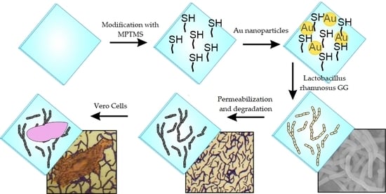
2.4. Formation of the Final Coating

After culturing, bacteria were fixed on the substrates using buffered glutataldehyde (1:7 v/v in PBS) by immersing the colonized glass surface for 15 min. The material was then immersed in successively increasing concentrations of ethanol (60, 70, 80, 90 and 100%) for 10 min for each concentration. The samples thus prepared were dried with a stream of nitrogen to remove most of the ethanol. Next, it was placed in a vacuum dryer and dried under the reduced pressure (100 hPa) at 100 °C for 1 h. Then, the temperature of the dryer was raised to 160 °C for 1 h and after this time, the heating element was turned off without unsealing the chamber, which allows the sample to reach room temperature under vacuum. The obtained sample (GSHAuGG) was used for further studies.

2.5. Microscopic Imaging of Surfaces Obtained at Various Stages of the Synthesis

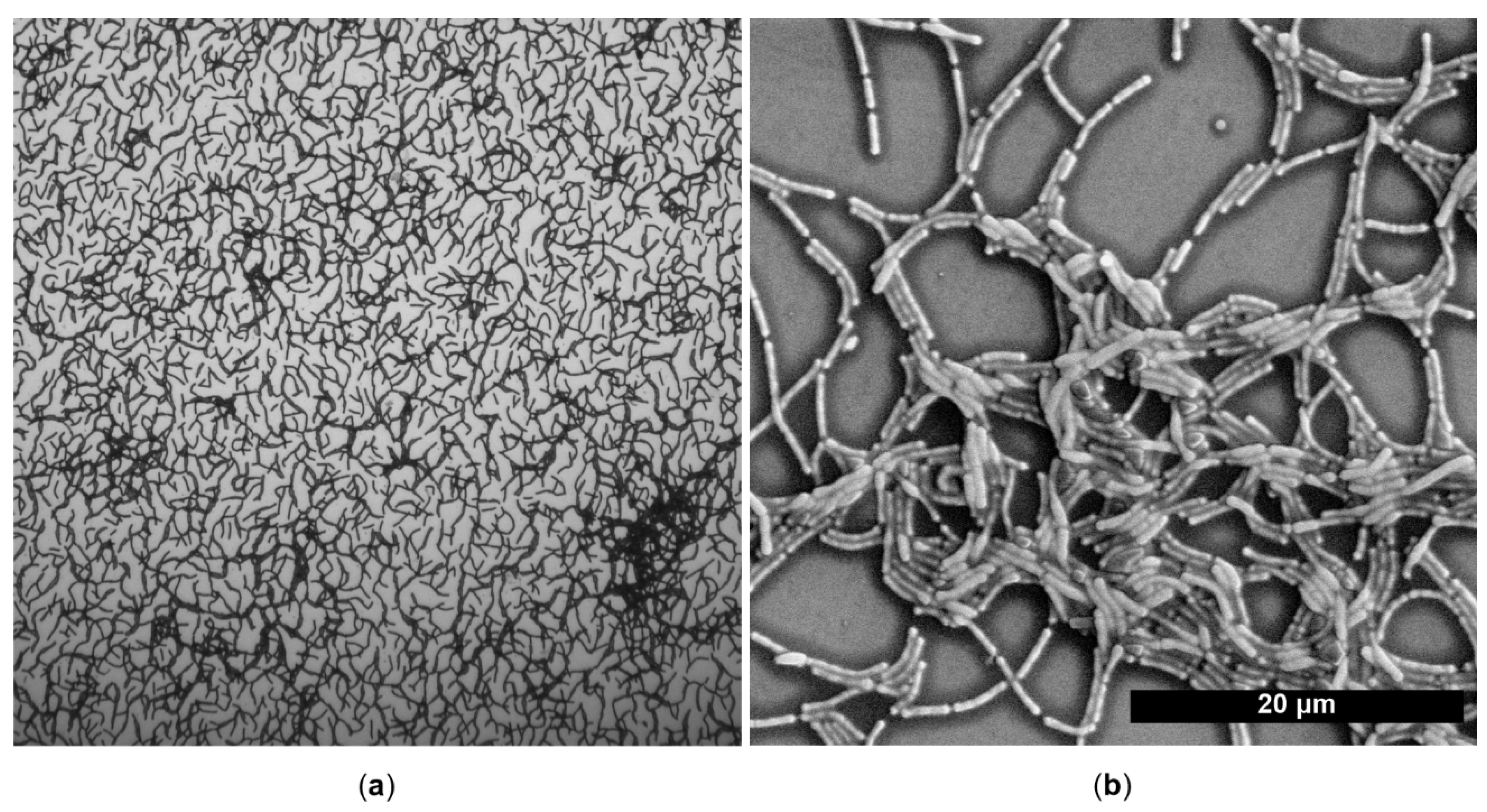
2.5.1. Scanning Electron Microscopy (SEM)

SEM imaging of the samples with microorganisms was also carried out, preceded by the fixing procedure described elsewhere [12]. Briefly, the samples were fixed in glutaraldehyde in the PBS solution (1:7 v/v) for 10 min, and dehydrated in ethanol solution series with increasing the ethanol concentration from 60 up to 100% (in increments of 10). Finally, the samples were rinsed with hexamethyldisilazane and left to dry. Prior to microscopic observations, the samples were sputter-coated with a thin gold layer (approximately 15 nm). For the coating obtained from dehydrated and annealed microorganisms, the samples (GSHAuGG) were only sputter-coated with a thin gold layer (approximately 15 nm) before measurement.

2.5.2. Optical Microscopy and Counting Mammalian Cells

Final coatings on glass GSHAuGG were examined under the optical microscope without additional preparation. After cell experiments, the materials were stained using crystal violet assay, which ensured good visualization of the cells. The number of cells per unit area regularly coated with GSHAuGG was estimated from 20 images for each tested sample taken at 5× magnification.

2.6. DLS and Zeta Potential Measurements

Commercially available gold nanoparticles were mixed in the proportion of 1:3 (v/v) using isotonic PBS with pH = 7.4. For the obtained solution, zeta potential and particle size measurements were carried out using the dynamic light scattering method. For zeta potential measurements, the obtain result was an average of three measurements while the measurement uncertainty was the standard deviation. The distribution diagram of the particle size, obtained by dynamic light scattering, represents the average of 18 measurements.

2.7. Evaluation of Bacterial Counts by Crystal Violet Staining

First, after bacteria cultivation, microorganisms were fixed according to the procedure for SEM measurements. The materials were then immersed in a crystal violet (CV) solution (0.1% v/v in 70% v/v ethanol in water) for 10 min. After this time, the unbound dye was rinsed with distilled water, and the surface was dried with a stream of argon. Next, the bacteria-bounded dye was dissolved in 1 mL of the solution obtained by dissolving citric acid (13.22 g) and sodium citrate (10.88 g) in a mixture of 500 mL distilled water and 500 mL methanol, and the absorbance at 540 nm was measured using the microplate spectrophotometer. The obtained absorbance values were optimized per slide area, which was estimated using a caliper (accuracy of 0.05 mm).

2.8. Study of Cell Cultures Growth on the New Coating

Vero cell lines were used to study how the formed coating affects mammalian cell growth. Cultures were conducted in a DMEM medium with the addition of 10% Fetal Bovine Serum (FBS) and 1% v/v antibiotics mix. A day after seeding, the cells were washed with PBS (at 37 °C) and fixed using a 1:10 mixture of formaldehyde (38%) and PBS. The cells were then stained using a crystal violet assay. Samples were immersed in the CV solution (0.1% in 70% ethanol in water) for 10 min, rinsed with distilled water, and dried with a stream of argon. Due to that, the morphology of cells and their number per unit area could be assessed by using microscopic methods.

3. Results and Discussion

3.1. Formation of the Gold Nanoparticle Layer on Glass

To confirm the presence of gold nanoparticles in prepared mixtures of the commercial gold suspension and PBS solution, UV-Vis spectra of mixtures were recorded. On the other hand, dynamic light scattering (DLS) experiments were performed to determine the mean size of nanoparticles. Both methods confirmed the presence of Au nanoparticles (Figure 1a, DLS diagram in Supplementary Figure S1). The UV-Vis spectrum of the prepared mixture with dispersed Au nanoparticles (Figure 1a) exhibits a typical surface plasmon resonance band at 520 nm [25]. The average diameter of Au nanoparticles estimated from DLS measurements was 15 nm. The zeta potential of nanoparticles in this system was also measured and was negative (−8.7 ± 2.0 mV). In practice, it means that a spontaneous deposition of gold nanoparticles on glass with a negative surface charge is impossible due to electrostatic repulsion. Therefore, a new procedure was proposed in order to chemically introduce -SH groups to the glass surface, which after modification will be able to bond gold. The effectiveness of bonding of gold nanoparticles to the glass surface was confirmed by diffuse reflectance UV-Vis spectroscopy. The obtained results (Figure 1b), i.e., the position and shape of the maximum in the spectrum, satisfactorily coincide with the spectrum of Au nanoparticles in the solution (Figure 1a) [25]. These results confirm a successful deposition of Au nanoparticles on the prepared glass substrate.

3.2. Lactobacillus rhamnosus GG Bacteria on the Obtained Surface

In our previous publications [11,12], we have shown a phenomenon concerning the ability of Lactobacillus rhamnosus GG strain bacteria to grow on a gold surface. Additionally, we have shown that the roughness of such a surface has also a positive influence on the growth of this strain. These facts made it possible to hypothesize that L. rhamnosuus GG could also grow on surfaces covered only with a layer of gold nanoparticles, as in our case. We have verified this assumption by observation of successful colonization of the tested surfaces by the strain as confirmed by SEM images (Figure 2) and quantitative evaluation of the amount of bacteria using crystal violet staining (Supplementary Materials, Table S1). As a negative control, glass with –SH groups (GSH) was used as a substrate for the reaction that produces the finished surface. The number of bacteria per cm2 of surface area, expressed as the amount of bound dye, was 186 (±10) times higher on the gold nanoparticle surface compared to surfaces containing only -SH groups (accurate absorbance values are given in the Supplementary Materials, Table S1). These results are in agreement with the surface colonization observed in SEM microphotographs (Figure 2), where the amount of bacteria is negligibly small for the control surface compared to the nanoparticle-coated surface. In the latter case, we observe the characteristic lace-like structures of interconnected bacteria described by us in our previous papers for all-gold surfaces [11,12].

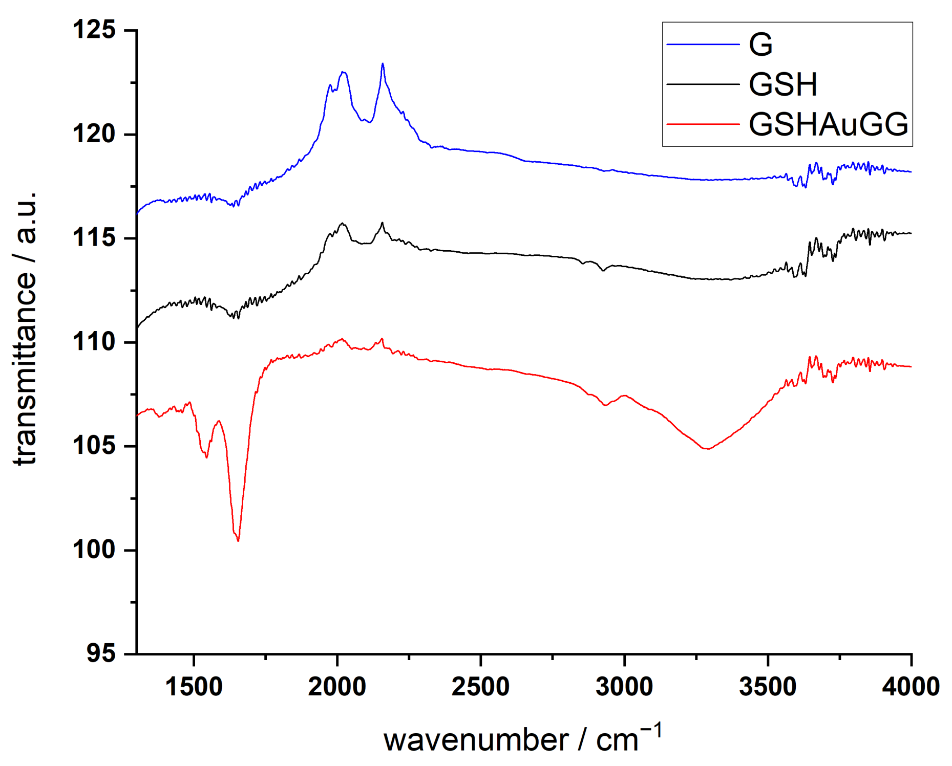
3.3. Chemical Analysis and Imaging of the Final Coating

The process of chemical dehydration of the fixed bacteria followed by further annealing at 160 °C in vacuum resulted in the formation of a discontinuous coating on the glass surface. The structure of this material resembles the structure of the lace in which the living bacteria organized themselves, which can be seen in SEM and optical microscopy images (Figure 3).
Spectroscopic methods show that the obtained coatings consist as expected mainly of proteins. Other components of the dead bacteria were almost all removed during ethanol treatment and heating. The proteins were bound to the surface mainly during the fixation of the bacteria using the crosslinking reagent glutaraldehyde. The IR spectra of final coatings (Figure 4) are similar to pure bovine serum albumin BSA, which is considered as a model protein [26,27]. Signals at 3430, 3062, 1652 and 1531 cm1 may be assigned to the stretching vibration of –OH and amide bands (mainly –NH or C=O stretching vibrations, and the coupling of N–H bending with C–N stretching vibration), respectively.

3.4. Aplication of Obtained Coatings for Cell Cultures

In laboratory cell culture practice, coatings that simulate the intercellular matrix are often used to improve adhesion and growth of mammalian cells. It includes complex products such as commercially available (MaxGel, ECM Gel or matrigel, all available from Sigma Aldrich) or simpler protein-only systems formed from degraded collagen, i.e., gelatin gels. Although their use usually requires only spreading them before use on a substrate (completely ready-made systems on dishes and bottles are also available), these products are not stable, expensive and used exclusively for specific cells requiring it. Hence, no early consideration was given to using them only to improve the properties of glass as a culture substrate in order to remove plastics from these applications. Our proposed method, based on green biosynthesis without direct usage of animal substrates, may provide a fresh starting point for the search for these kinds of new materials. Preliminary results obtained with Vero cells are very promising. The increase in cell number relative to the unmodified glass substrate, based on direct cell counting on a representative group of microscope images, was approximately 50% (Figure 5a). In addition, the morphology of the cells in comparison to that observed for the unmodified glass was similar, which is confirmed in the images (Figure 5b,c) taken at high magnifications (100×).

4. Conclusions

Changes in the public’s perception of fossil-based plastics mean that cell culture vessel materials are expected to change as well. Initially, vessels used for such applications were made of glass. Therefore, it is reasonable to consider glass once again as a starting material for culture vessels. Crucial, as in the case of plastic vessels, will be the modification of the surface or, in practice, development of new coatings, which can be used at glass. It is appropriate to use coatings that replicate the protein-rich composition of the intercellular matrix in which cells are embedded in tissues. Our proposed approach to these fossil-based plastics in culture vessel problem, presented in this work, is based on the use of green synthesis exploiting the self-organization of Lactobacillus rhamnosus GG bacteria on thin gold surfaces. The technically simple steps of surface preparation consist of introducing -SH groups to commercially available glass, then attaching gold nanoparticles to these groups. As demonstrated, such surfaces can be easily colonized by bacteria, which served as a source of protein after permeabilization of the bacteria membrane with ethanol and heat treatment. This method preserves the characteristic discontinuous primary structure created during bacterial growth and allows the microorganisms to be transformed into a protein coating that significantly improves the surface properties when it comes to Vero cell growth. The observed increase in a relative number of Vero cells is about 50%. These promising results suggest that this material should be further examined using other cell lines. The results described here represent a first step in the development of a new strategy for obtaining protein coatings using Lactobacillus rhamnosus GG bacterial cultures. There are other works in the literature showing the formation of protein coatings on glass [28] or non-organic biomaterials [29,30], but our proposed approach based on biosynthesis using bacteria in such a way is a completely new strategy. Our system has, despite all these arguments, limitations and obvious drawbacks due to the fact that it is dedicated and restricted to cell line cultures. In practice, it will be applicable ex vivo either for diagnostics or for biosynthesis in cell culture-based bioreactors. Glass is not a material, which can be directly applied in vivo in clinical settings, for example, in regenerative medicine.

Supplementary Materials

The following are available online at https://www.mdpi.com/article/10.3390/coatings11111410/s1, Figure S1: Diameter of commercial gold nanoparticles (Sigma) after 4-fold dilution with physiological PBS 7.4 based on DLS measurements.; Table S1: Absorbance of crystal violet in destaining solutions after an experiment to assess the amount of bacteria on the tested surfaces.

Author Contributions

Conceptualization, K.K. and K.S.; methodology, K.K.; software, J.G.; investigation, K.K., M.O., K.S., M.A., J.G.; resources, K.K.; writing—original draft preparation, K.K.; writing—review and editing, G.D.S., K.S., M.O., J.G.; visualization, J.G.; supervision, G.D.S.; funding acquisition, K.K. All authors have read and agreed to the published version of the manuscript.

Funding

This research received no external funding.

Institutional Review Board Statement

Not applicable.

Informed Consent Statement

Not applicable.

Data Availability Statement

No new data were created or analyzed in this study. Data sharing is not applicable to this article.

Acknowledgments

The SEM imaging was performed in the Laboratory of Field Emission Scanning Electron Microscopy and Microanalysis at the Institute of Geological Sciences, Jagiellonian University, Poland.

Conflicts of Interest

The author Joanna Grudzień is an employee of MDPI; however, she does not work for the journal Coatings at the time of submission and publication. All remaining authors declare no conflict of interest.

References

  1. Risbridger, G.P. Human Cell Lines as Tools of Our Trade: “Laying It on the (Cell) Line”. Mol. Endocrinol. 2015, 29, 1–2. [Google Scholar] [CrossRef] [PubMed]
  2. Mirabelli, P.; Coppola, L.; Salvatore, M. Cancer Cell Lines Are Useful Model Systems for Medical Research. Cancers 2019, 11, 1098. [Google Scholar] [CrossRef] [PubMed] [Green Version]
  3. Segeritz, C.-P.; Vallier, L. Basic Science Methods for Clinical Researchers Chapter 9—Cell Culture: Growing Cells as Model Systems In Vitro; Academic Press: Cambridge, MA, USA, 2017; pp. 151–172. ISBN 9780128030776. [Google Scholar]
  4. Verma, A.; Verma, M.; Singh, A. Animal tissue culture principles and applications. Anim. Biotechnol. 2020, 269–293. [Google Scholar] [CrossRef]
  5. Lerman, M.J.; Lembong, J.; Muramoto, S.; Gillen, G.; Fisher, J.P. The Evolution of Polystyrene as a Cell Culture Material. Tissue Eng. Part B Rev. 2018, 24, 359–372. [Google Scholar] [CrossRef] [Green Version]
  6. Cooke, M.J.; Phillips, S.R.; Shah, D.S.; Athey, D.; Lakey, J.H.; Przyborski, S.A. Enhanced cell attachment using a novel cell culture surface presenting functional domains from extracellular matrix proteins. Cytotechnology 2008, 56, 71–79. [Google Scholar] [CrossRef] [Green Version]
  7. McKeehan, W.L.; Ham, R.G. Stimulation of clonal growth of normal fibroblasts with substrata coated with basic polymers. J. Cell Biol. 1976, 71, 727–734. [Google Scholar] [CrossRef] [PubMed] [Green Version]
  8. Joseph, B.; James, J.; Kalarikkal, N.; Thomas, S. Recycling of medical plastics. Adv. Ind. Eng. Polym. Res. 2021, 4, 199–208. [Google Scholar] [CrossRef]
  9. Vallet-Regi, M.; Ragel, C.V.; Salinas, A.J. Glasses with Medical Applications. Eur. J. Inorg. Chem. 2003, 2003, 1029–1042. [Google Scholar] [CrossRef]
  10. Iijima, K.; Suzuki, R.; Iizuka, A.; Ueno-Yokohata, H.; Kiyokawa, N.; Hashizume, M. Surface functionalization of tissue culture polystyrene plates with hydroxyapatite under body fluid conditions and its effect on differentiation behaviors of mesenchymal stem cells. Colloids Surf. B Biointerfaces 2016, 147, 351–359. [Google Scholar] [CrossRef] [PubMed]
  11. Grudzien, J.; Jarosz, M.; Kaminski, K.; Kobasa, M.; Wolski, K.; Koziel, M.; Pisarek, M.; Sulka, G.D. Growth of Lactic Acid Bacteria on Gold-Influence of Surface Roughness and Chemical Composition. Nanomaterials 2020, 10, 2499. [Google Scholar] [CrossRef]
  12. Jarosz, M.; Grudzien, J.; Kaminski, K.; Gawlak, K.; Wolski, K.; Nowakowska, M.; Sulka, G.D. Novel bioelectrodes based on polysaccharide modified gold surfaces and electrochemically active Lactobacillus rhamnosus GG biofilms. Electrochim. Acta 2019, 296, 999–1008. [Google Scholar] [CrossRef]
  13. Gu, X.; Xu, Z.; Gu, L.; Xu, H.; Han, F.; Chen, B.; Pan, X. Preparation and antibacterial properties of gold nanoparticles: A review. Environ. Chem. Lett. 2021, 19, 167–187. [Google Scholar] [CrossRef]
  14. Mohamed, M.M.; Fouad, S.A.; Elshoky, H.A.; Mohammed, G.M.; Salaheldin, T.A. Antibacterial effect of gold nanoparticles against Corynebacterium pseudotuberculosis. Int. J. Vet. Sci. Med. 2017, 5, 23–29. [Google Scholar] [CrossRef] [PubMed] [Green Version]
  15. Roselli, M.; Finamore, A.; Britti, M.; Mengheri, E. Probiotic bacteria Bifidobacterium animalis MB5 and Lactobacillus rhamnosus GG protect intestinal Caco-2 cells from the inflammation-associated response induced by enterotoxigenic Escherichia coli K88. Br. J. Nutr. 2006, 95, 1177–1184. [Google Scholar] [CrossRef] [PubMed]
  16. Jia, R.; Chen, H.; Chen, H.; Ding, W. Effects of fermentation with Lactobacillus rhamnosus GG on product quality and fatty acids of goat milk yogurt. J. Dairy Sci. 2016, 99, 221–227. [Google Scholar] [CrossRef] [PubMed]
  17. Canani, R.B.; Sangwan, N.; Stefka, A.T.; Nocerino, R.; Paparo, L.; Aitoro, R.; Calignano, A.; Khan, A.A.; Gilbert, J.A.; Nagler, C.R. Lactobacillus rhamnosus GG-supplemented formula expands butyrate-producing bacterial strains in food allergic infants. ISME. J. 2016, 10, 742–750. [Google Scholar] [CrossRef]
  18. Sarkar, A.; Lehto, S.M.; Harty, S.; Dinan, T.G.; Cryan, J.F.; Burnet, P.W.J. Psychobiotics and the Manipulation of Bacteria–Gut–Brain Signals. Trends Neurosci. 2016, 39, 763–781. [Google Scholar] [CrossRef] [Green Version]
  19. Shi, C.W.; Cheng, M.Y.; Yang, X.; Lu, Y.Y.; Yin, H.D.; Zeng, Y.; Wang, R.Y.; Jiang, Y.L.; Yang, W.T.; Wang, J.Z.; et al. Probiotic Lactobacillus rhamnosus GG Promotes Mouse Gut Microbiota Diversity and T Cell Differentiation. Front. Microbiol. 2020, 11, 607735. [Google Scholar] [CrossRef]
  20. Jazayeri, M.H.; Amani, H.; Pourfatollah, A.A.; Pazoki-Toroudi, H.; Sedighimoghaddam, B. Various methods of gold nanoparticles (GNPs) conjugation to antibodies. Sens. Bio-Sens. Res. 2016, 9, 17–22. [Google Scholar] [CrossRef] [Green Version]
  21. Chen, J.-J.; Struk, K.N.; Brennan, A.B. Surface Modification of Silicate Glass Using 3-(Mercaptopropyl)trimethoxysilane for Thiol-Ene Polymerization. Langmuir 2011, 27, 13754–13761. [Google Scholar] [CrossRef]
  22. Darmady, E.M.; Hughes, K.E.A.; Jones, J.D.; Prince, D.; Tuke, W. Sterilization by dry heat. J. Clin. Path. 1961, 14, 38. [Google Scholar] [CrossRef] [PubMed] [Green Version]
  23. Ammerman, N.C.; Beier-Sexton, M.; Azad, A.F. Growth and Maintenance of Vero Cell Lines. Curr. Protoc. Microbiol. 2008, 11, A-4E. [Google Scholar] [CrossRef] [PubMed] [Green Version]
  24. Sène, M.A.; Kiesslich, S.; Djambazian, H.; Ragoussis, J.; Xia, Y.; Kamen, A.A. Haplotype-resolved de novo assembly of the Vero cell line genome. npj Vaccines 2021, 6, 106. [Google Scholar] [CrossRef] [PubMed]
  25. Link, S.; El-Sayed, M.A. Size and Temperature Dependence of the Plasmon Absorption of Colloidal Gold Nanoparticles. J. Phys. Chem. B 1999, 103, 4212–4217. [Google Scholar] [CrossRef]
  26. Huang, P.; Li, Z.; Hu, H.; Cui, D. Synthesis and Characterization of Bovine Serum Albumin-Conjugated Copper Sulfide Nanocomposites. J. Nanomater. 2010, 2010, 641545. [Google Scholar] [CrossRef]
  27. Jokar, S.; Pourjavadia, A.; Adeli, M. Albumin–graphene oxide conjugates; carriers for anticancer drugs. RSC Adv. 2014, 4, 33001–33006. [Google Scholar] [CrossRef]
  28. Acuña-Nelson, S.M.; Bastías-Montes, J.M.; Cerda-Leal, F.R.; Parra-Flores, J.E.; Aguirre-García, J.S.; Toledo, P.G. Nanocoatings of Bovine Serum Albumin on Glass: Effects of pH and Temperature. J. Nanomater. 2020, 2020, 11. [Google Scholar] [CrossRef]
  29. Raphel, J.; Karlsson, J.; Galli, S.; Wennerberg, A.; Lindsay, C.; Haugh, M.G.; Pajarinen, J.; Goodman, S.B.; Jimbo, R.; Andersson, M.; et al. Engineered protein coatings to improve the osseointegration of dental and orthopaedic implants. Biomaterials 2016, 83, 269–282. [Google Scholar] [CrossRef] [PubMed] [Green Version]
  30. Van Gaal, R.C.; Vrehen, A.F.; Van Sprang, J.F.; Fransen, P.P.; Van Turnhout, M.C.; Dankers, P.Y. Biomaterial screening of protein coatings and peptide additives: Towards a simple synthetic mimic of a complex natural coating for a bio-artificial kidney. Biomater. Sci. 2021, 9, 2209–2220. [Google Scholar] [CrossRef]
Figure 1. (a) Ultraviolet and visible spectrum of gold nanoparticles in the mixture composed of the commercial gold suspension and saline phosphate buffer solution. (b) Ultraviolet and visible diffuse reflectance spectra of glass containing -SH groups (GSH) and additionally covalently attached gold nanoparticles substrates (GSHAu).
Coatings 11 01410 g001
Figure 2. Scanning electron microscope images of Lactobacillus rhamnosus GG bacteria on the surface of (a) glass containing -SH groups (GSH) and (b) additionally covalently attached gold nanoparticles (GSHAu).
Coatings 11 01410 g002
Figure 3. Final coating (GSHAuGG) visualized with (a) optical microscope (objective magnification 100×) and (b) scanning electron microscope. In the latter case, the surfaces were sputter deposited with a thin gold layer before imaging.
Coatings 11 01410 g003
Figure 4. Infrared spectroscopy spectra of glass (G), glass modified with –SH groups (GSH), and the final coating on glass (GSHAuGG).
Coatings 11 01410 g004
Figure 5. (a) Relative number of cells counted for glass (G), glass modified with –SH groups (GSH), and the final coating on the glass (GSHAuGG) substrates after crystal violet staining. Optical microscope images at 100× magnification of (b) Vero cells on the glass substrate (G) and (c) and on the GHSAuGG sample.
Coatings 11 01410 g005
Publisher’s Note: MDPI stays neutral with regard to jurisdictional claims in published maps and institutional affiliations.

