
Revisiting Tetra-p-Sulphonated Porphyrin as Antimicrobial Photodynamic Therapy Agent


1
Materials Engineering Department, Valahia University, 13000 Targoviste, Romania
2
Nanomedicine Research Group, National Institute of R&D for Chemistry and Petrochemistry—ICECHIM, 060021 Bucharest, Romania
Coatings 2021, 11(4), 393; https://doi.org/10.3390/coatings11040393
Submission received: 25 February 2021
/
Revised: 18 March 2021
/
Accepted: 23 March 2021
/
Published: 30 March 2021
(This article belongs to the Special Issue Advanced Coatings for Biomedical Applications)
Abstract
:Photodynamic inactivation is known as a new antimicrobial photodynamic therapy (aPDT). It is based on the administration of a photosensitizer located in the bacterial/viral cell followed by exposure to light radiations (with a proper wavelength corresponding with the maximum value of absorption of the photosensitizer) that generate singlet oxygen or reactive oxygen species, which lead to the death of different microorganisms. This review will present an overview beyond the state-of-the-art of the photosensitizer types (based on tetra-p-sulphonated-phenyl porphyrin—TSPP, which is able to form cationic and J-aggregates forms at different pH values ((1–4) and concentrations around 10−5 M) and their applications of PDT for viruses, especially. The mechanism of dicationic and J-aggregates formation is presented in this paper, and the photophysical parameters have been collected and harmonized to support their behaviours. Studies on Herpes Simplex virus type 1 (HSV-1) are useful, because without the help of HSV-1, the COVID-19 virus may not be able to cause serious illness or death in humans. This method could be a new direction for COVID treatment and immunization, either to prevent infections or to develop photoactive fabrics (e.g., masks, suits, gloves) to disinfect surfaces, under artificial light and/or natural sunlight. The use of photodynamic therapy (PDT) can be an alternative approach against SARS-CoV-2 that deserves to be explored.
1. Introduction
Antimicrobial photodynamic therapy (aPDT) consists of the selective uptake of a photosensitizing dye, often a cationic dye by bacterial/viral cell, and subsequent irradiation of the tumor with a light flux of an appropriate wavelength matched to the absorption spectrum of the photosensitizing dye. This class of molecules is capable of using the energy used for excitation in order to produce reactive oxygen species (singlet oxygen, superoxide anion, hydroxyl radicals, so on). The action mechanism involves the generation of singlet oxygen and other free radicals when the light-excited sensitizer loses or accepts an electron [1].
The present status of clinical PDT is discussed along with the new photosensitizers being used and their clinical roles. The development of new photosensitizers for the localization and treatment of tumors is a research area of current interest: photosensitizer dose (mg/kg of body weight), rate uptake of the sensitizer and its form (monomer and/or aggregated), cytotoxicity, and the balance between the concentration of the drug in the tumor tissue (mg/g tissue) and in normal tissues at the time of light irradiation [2].
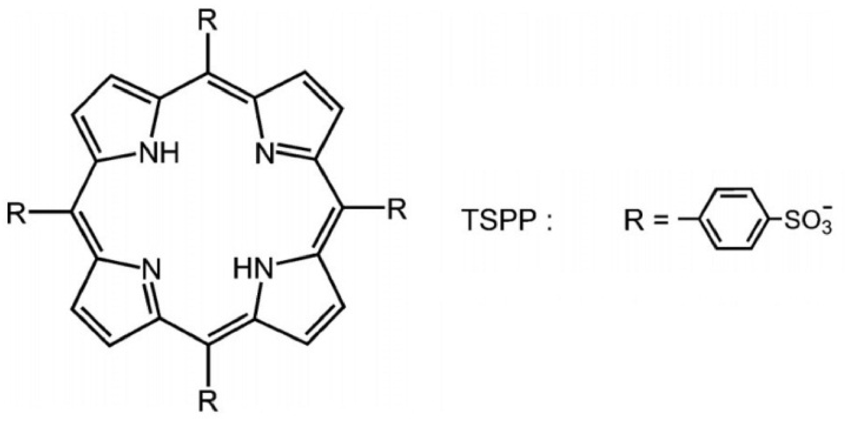
In recent years, many improvements have been achieved in developing new photosensitizers and light sources suitable for antimicrobial photodynamic treatment (aPDT) used to kill protozoa, bacteria, and viruses. One of the most used porphyrins, tetra-p-sulphonated-phenyl porphyrin (TSPP), as it is shown in Figure 1, was investigated as an anti-cancer photosensitizer some years ago [3,4,5], but it was abandoned when the scientists reported its neurotoxic effects in experimental animals even in the dark [6]. Due to its versatility, this PS started to be reconsidered [7,8].
This review will present an overview of this anionic photosensitizer; the chemical structure at different pH and temperatures; photodynamic mechanism and photophysical properties (absorption/emission and its excited states with lifetimes and decay); the effect of wavelength of excitation on aPDT efficacy; and the application of TSPP in aPDT, including HSV-1 photodynamic inactivation for disinfection of surfaces. Some aspects in relevant microbiological cultures (viruses) are mentioned in order to investigate the phototoxicity of photoactive TSPP. aPDT can be potentiated by the use of TSPP as anionic porphyrin, and the conditions to transform it into dicationic form, and J-aggregate, too.
2. Historical Considerations of aPDT
The civilizations based in China, India, Egypt, and Greece used the exposure to the sun to restore health. The ancient Indian civilizations (1000 BC) discovered that administration of psoralens when combined with careful exposure to sunlight could be used to treat the congenital vitiligo. The modern era of photodynamic therapy was established by Dr. T.J. Dougherty, who reported that a systematically injected porphyrin on activation with red light caused complete eradication of transplanted experimental tumours [9,10].
Photodynamic therapy (PDT) is a potentially effective treatment approach for superficial human cancers and selected benign conditions. PDT is used mainly for superficial types of skin cancer: actinic keratoses and superficial basal cell carcinomas, nodular basal cell carcinomas, oesophageal, and lung carcinomas, but also in non-oncological situations like age-related macular degeneration (AMD). Additionally, in the last few years, the approach has been shown to be highly effective against all types of microorganisms, such as Gram-positive and Gram-negative bacteria, fungi, viruses, and parasites [11].
Photodynamic inactivation has been known since Raab, at the beginning of the 20th century, observed that acridine was harmless to Paramecium caudatum, Proflavine, acridine orange in the dark, and lethal when the organisms were exposed to visible light [12]. Also, some blue dyes such as methylene blue and toluidine blue O have been the main used photosensitizers.
In 1958, Yamamoto [13] reported the first quantitative studies on photodynamic inactivation of bacterial virus. Neutral red was the first dye used in photodynamic inactivation of herpes [14,15]. Also, proflavine, methylene blue was used with good results for herpes simplex inactivation [16,17].
Methylene blue and toluidine blue O have been widely used in microorganism’s reduction for over a century without causing human toxicity [8]. Several groups of researchers used methylene blue or toluidine blue O for lethal photosensitizing of Staphylococcus aureus and Helicobacter pylori, Porphyromonas gingivalis, Streptococcus mutans, Streptococcus sobrinus, and Lactobacillus acidophilus involved in periodontal infection [18].
The aPDT has been applied to viruses through the same mechanism: formation of radicals, anions and, in general, ROS (via Type I and Type II mechanisms), which damage the target cells or entities (e.g., bacteria, viruses, or more recently, prions) [19,20,21,22,23]. The target structures for this damage (e.g., membrane structures of tumor cells and bacteria, lipid structures or proteins of the viral envelope, or nucleic acids) are affected during aPDT. Some studies have reported a reactivation of viruses (e.g., herpes simplex virus, HSV) by photodynamic treatments [24]. PDT has also been used to augment the efficacy of oncolytic vaccinia viruses in metastatic tumors in vivo [25].
The DNA and RNA of viruses can be affected by aPDT. PS can bind or intercalate with nucleic acids, but this is not always sufficient for effective photosensitization. An important role is played by cationic sensitizers, such as methylene blue, which can pass through the outer cover of viruses and intercalate into their DNA/RNA [26]. For cationic PS, electrostatic interaction should allow direct PS–DNA interaction. For photosensitized oxidation reactions of type II, which is the most common mechanism, 1O2 dominates [27]. Such oxidative transformations destroy DNA, leading to fragmentation, single chain breaks, and protein crosslinking. Prevention of viral replication and reduction of infectivity after DNA damage by ROS has been demonstrated, and guanine residues seem to be susceptible to oxidative damage, producing 8-oxo-7,8-dihydroguanine 2 as main products (photosensitized oxidation type I) [28,29].
ROS are able to react with RNA/DNA, proteins, and lipids alike. The viral life cycle includes the general stages of attachment of the virus to the host cell, penetration of the virus into the cell (fusion of cell and viral membranes), failure to cover viral RNA/DNA, replication of the viral genome, and assembly of new virions from nucleic acids and proteins. These processes have as result newly synthesized viral and eventually the release of new virions from the host cell [30,31]. Some proteins play an important role in attaching viruses to the surface of the host cell. Lenard et al. showed that hypericin, or Rose Bengal, were able to inhibit the fusion of vesicular stomatitis virus (VSV) virus, influenza virus, and Sendai virus (all enveloped viruses) by cross-linking viral proteins [32]. Harmful effect of PDI on important viral proteins for viral cell fusion has also been demonstrated for specific phthalocyanines and HSV-1, as well as for other PS and viruses [33,34,35,36,37,38,39].
This broad-spectrum activity of some sensitizers will clearly be useful in the treatment of emerging infectious diseases, such as COVID-19 [40,41,42]. The use of photodynamic therapy (PDT) can be an alternative approach against SARS-CoV-2 that deserves to be explored. Based on previous studies about Herpes simplex virus type 1 (HSV-1), it has been shown that it can inactivate most of the elements in the immune system and damage the blood brain barrier (BBB) [43,44,45,46]. Without the help of HSV-1, the COVID-19 virus may not be able to cause serious illness or death in humans. The discovery or reconsidering of new photosensitizers [47] is a necessity.
3. Mechanism of aPDT
PDT requires the use of a photosensitizer (PS), a molecule that, after being excited by visible light, can react with dioxygen (3O2, the atmospheric oxygen), producing reactive oxygen species (ROS) such as singlet oxygen (1O2) and/or superoxide anion, hydroxyl radicals, and hydrogen peroxide. These ROS can react with biological molecules (e.g., proteins, lipids and nucleic acids), causing their oxidation and, consequently, damage to cells and tissues [48,49].
The antimicrobial photodynamic therapy is based on the photooxidation of biological matter in which three essential constituents are involved: photosensitizer, light radiation (with the wavelength corresponding to the maximum absorption of the photosensitive substance) and oxygen [50].
Two main phases are important inside of this mechanism:
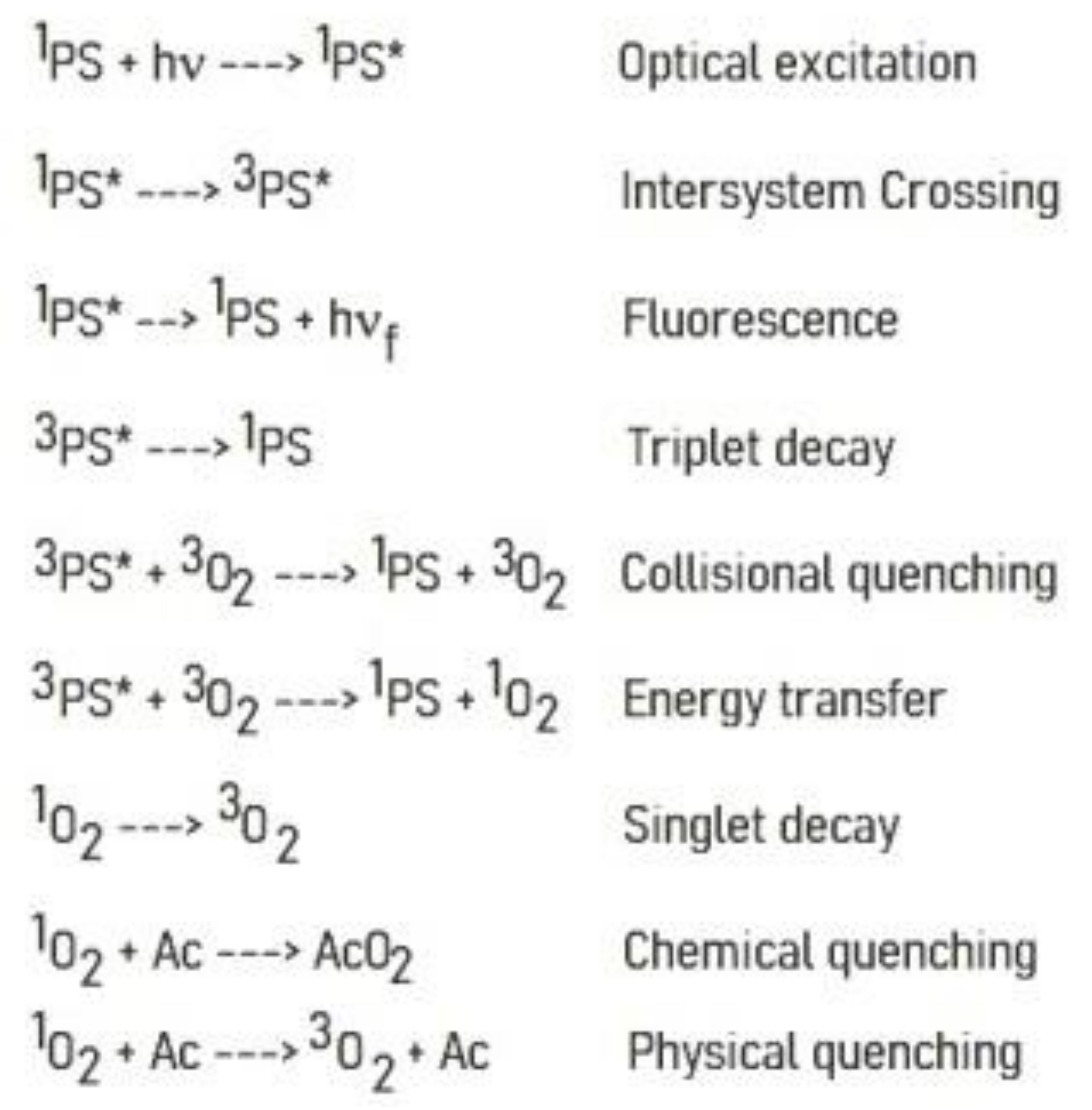
- Light excites the ground state photosensitizer to an excited singlet state.
- The formation of triplets of excited sensitizer molecule (intersystem crossing).
- However, this state is short lived and can decay to the ground state by radiative or non-radiative transition directly emitting light as fluorescence.
- From this excited state, the photosensitive substance can then return to the ground state by phosphorescence.
- The triplet excited state of the photosensitizer is able to react with oxygen in its triplet state, generating singlet oxygen (type II reaction) or initiating free radical chain reactions with superoxide and hydrogen peroxide ions as well as hydroxyl radicals (type I reaction).
The photosensitive substance in the triplet state 3S is very reactive due to its long-life time (10−3–100) s. The lifetime of the singlet state 1S is small (10−9–10−7) so that the photosensitive substance in this state cannot interact with other molecules before returning to the ground state.
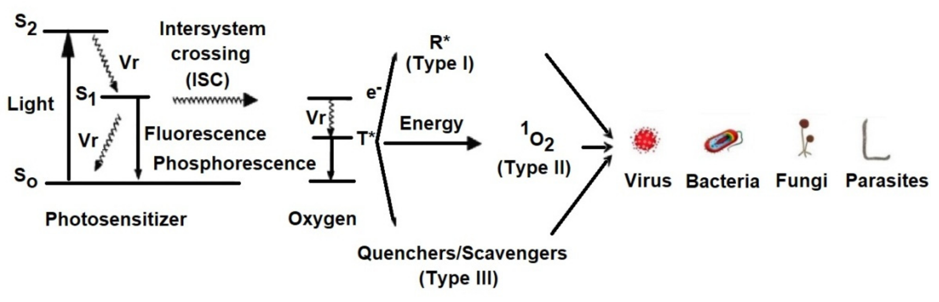
The Jablonski diagram (Figure 2) demonstrates the reactions involved in the process.
Vr = Vibrational relaxation; T* = Oxygen in triplet state; R* = Free radical; S0 = Photosensitizer in ground state; S1 = Photosensitizer in singlet state
The main mechanisms by which the photosensitive substance, in the excited state of triplet, can react chemically with the biological environment are:
3.1. Type I Mechanism
The photosensitive substance, in a 3S* triplet state, can react with a target molecule, other than oxygen, by exchanging hydrogen or electron:
- hydrogen transfer: 3S * + RH → SH + R˙
- electron transfer: 3S * + RH → S− + RH+
where: S: photosensitive substance, RH: H-linked substrate,
Furthermore, it can react with triplet oxygen resulting in hydrogen dioxide or superoxide anion:
- formation of hydrogen dioxide
- formation of superoxide anion
SH˙ + 3O2 → 1S + HO2˙
S˙− + 3O2 → 1S + O2˙
3.2. Type II Mechanism
The photosensitive substance in the excited state of triplet 3S* can transfer its excitation energy to molecular oxygen (which is triplet in its ground state), which passes to its lowest excited singlet state. Molecular oxygen in the excited state of singlet 1O2* is strongly electrophilic and interacts strongly with biomolecules:
- intermolecular exchange
- cellular oxidation
3S * + 3O2 → 1S + 1O2*
1O2 * + Cell → Cellox
Photodynamic therapy is mainly based on the type II reaction mechanism for generating singlet oxygen that can react quickly with various biological targets (cell wall/membranes, peptides, lipids, DNA, RNA), and further reactive substances including organic peroxides and sulfoxides are responsible for photooxidative stress in microorganisms. Singlet oxygen has a short lifetime (0.01–0.04 μs) and a limited diffusion distance in the biological environment (0.01–0.02 μm) due to its reactivity, so that cell damage occurs, mainly in the immediate vicinity of the location of the photosensitive substance in the cell depending on the cell type and the type of photosensitizer used (structure, concentration, photochemical characteristics, and its ability to bind the cellular wall).
The complex structure of the cell wall of different microorganisms causes a difficult penetration of photosensitization into the cell and a low production of singlet oxygen. However, singlet oxygen generated by the type II reaction mechanism can react with cytoplasmic membrane components leading to the generation of peroxide reaction products that can cause lethal damage to vital targets. Some of the singlet oxygen that penetrates the cytoplasmic membrane can reach the outer membrane and can react with unsaturated fatty acids and proteins in the outer membrane. The reaction products of singlet oxygen with the components of the outer membrane may be able to undergo reactions that cause cell death. The singlet oxygen can react only with the components of the cytoplasmic membrane and the peptidoglycan layer. Regardless of the situation, the lethal effects on bacterial/viral cells depend on the location of the photosensitizer on/or in the cell, the photodynamic efficiency of the photosensitizer in that environment, laser irradiation parameters, and oxygen transport. Major biological targets are membranes that undergo rupture and the cells are destroyed. It has been recently demonstrated that most damage is to the membranes around the mitochondria and the lysosomes. These organelles liberate destructive proteins that induce subsequent cellular destruction. Photosensitizers that target the outer plasma membrane are less effective. It is important again to emphasize that a critical level is required since the cells have developed mechanisms to withstand this oxidative damage, responsible for necrosis or apoptosis [51].
4. Light Sources
Similar with PDT, the aPDT requires sources of light to activate the photosensitizer by exposure to low-power visible light at a specific wavelength. Most photosensitizers are activated by red light between 630–700 nm, corresponding to a light penetration depth from 0.5–1.5 cm [52].
Different light sources are applied now in photodynamic therapy as follows: helium-neon lasers (633 nm), gallium-aluminum-arsenide diode lasers (630–690, 830 or 906 nm), and argon laser (488–514 nm) [53].
Recently, non-laser light sources, such as light-emitting diodes (LED), have been used as new light activators in PDT. LED devices are more compact, portable, and cost effective compared to traditional lasers [54].
It is a fundamental principle of PDT that the wavelength of the light source should in general be tuned to the absorption maximum of the PS. In general, the wavelengths that have been used for aPDT include ultraviolet A (UVA) (330–400 nm), blue (400–490 nm), green (490–550 nm), yellow (550–600 nm), red (600–700 nm), and near infrared (NIR) (700–810 nm).
The porphyrins have relatively large Soret bands (around 420 nm) and four small Q bands in the region 500–700 nm. These long wavelengths display much better tissue penetration than shorter wavelengths. For an efficient aPDT, the porphyrin should be excited with blue light (400 nm) or with red light (630 nm), to produce highly active antimicrobial PS. Many in vitro studies have used relatively simple broadband white light (400–700 nm) from an incandescent lamp. All the wavelengths mentioned above are based on single-photon absorption by the PS. Even so, in recent years non-linear processes (two-photon absorption) have been involved in aPDT [55,56]. If two long-wavelength photons 750–1000 nm arrive at the PS molecule at virtually the same time (within 1 ps), they will both be absorbed and it will be as if a single photon with half the wavelength was absorbed instead. The advantage is that the long-wavelength photons pass much better through tissue than the equivalent (1/2 λ) photons with a shorter wavelength [57].
5. Photosensitizers Used for Photodynamic Inactivation of Microorganisms
A large number of photosensitizers have been tested during last 10 years for the photodynamic inactivation of various microorganisms. Many of these photosensitizers have been tested to evaluate their antimicrobial efficiency in correlation with the main factors, which define the antimicrobial efficiency of the photosensitizer, as: chemical structure of the photosensitizer, the intracellular localization and binding site of the photosensitizer into the cell. Therefore, the studies demonstrated the DNA and RNA binding of the photosensitizer and selective uptake of the photosensitizer by the cellular organelles (bacteria and yeast cellular membrane, viral envelope, algae chloroplasts) are highly dependent on the physico-chemical photosensitizer properties. The photosensitizers must be aromatic molecules that can form long-lived triplet excited states and must have high quantum yield of singlet oxygen production. Also, the absorption properties of the photosensitizer should have a potential impact on the efficacy of photosensitization. The absorption maxims of the photosensitizer used till now are in UV and VIS spectral range. The range of absorption wavelength for psoralen (furocoumarin) and for relative photosensitizers are shown in Table 1 [58].
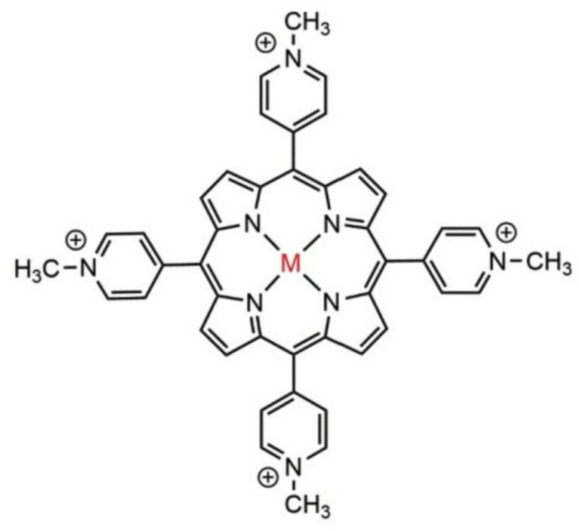
For the photodynamic inactivation of various microorganisms, a large variety of photosensitizer substances have been used [59]. The most used are the macrocyclic photosensitizers as cationic porphyrins and phthalocyanines, an example being 5,10,15,20-tetra-p-methyl-pyrydil porphyrin (TMPyP), Figure 3.
Also, some natural photosensitizers, are present in plants or in fungi that have been used in this area, such as: psoralen (furanocoumarins), perylenequinonoid pigments, hypericin, and hypocrellin. Nowadays, many photosensitizer types with different physicochemical and optical properties are available for photodynamic inactivation of a wide range of microorganisms.
As a rule, the chemical purity, the selective uptake and the localization inside the microorganism, the high antimicrobial efficiency, and the lack of mutagenic activity or genotoxicity are the important characteristics of an ideal photosensitizer [51]. Any type of these sensitizers should meet several criteria: chemical purity, tumor selectivity, fast accumulation in target tissues and rapid clearance, proper wavelengths and deeper penetration, and no dark toxicity.
In terms of solubility, photosensitizers can be classified into three main groups:
- hydrophobic photosensitizers without peripheral substituents with electric charge and being slightly soluble in water or alcohol (phthalocyanines and naphthalocyanines, hematoporphyrin, hematoporphyrin derivative (HpD), porfimer sodium, and porphyrin precursors)
- hydrophilic photosensitizers that contain three or more peripheral substituents with electric charge and have a high solubility in water at physiological pH.
- amphiphilic photosensitizers that contain one or two peripheral substituents with electric charges, soluble in water or alcohol, at physiological pH. In their structure, there are always two regions, one hydrophobic (represented by porphyrin with electrically charged groups) and another hydrophilic [60].
6. Anionic Photosensitizers as Anti-Viral Agent for aPDT
In recent years, many achievements have been reached in fundamental aPDT sensitizers [61]. The most important classes of photosensitizers tested so far are:
The first generation of photosensitizers: hematoporphyrin, hematoporphyrin derivative (HpD), porfimer sodium, and porphyrin precursors, which are not ideal photosensitizers for photodynamic therapy. Due to their complex composition, HpD does not exhibit a good photodynamic efficiency, because some of the HpD components are inactive. In addition, HpD is localized in healthy tissues, thus inducing a residual photosensitization of the whole body for almost a month after its administration.
The second generation of photosensitizers includes macrocycles as porphyrins, phthalocyanines, naphthalocyanines, benzoporphyrin derivatives, chlorines, and bacteriochlorines, with good absorption of wavelength radiation from the spectral region (650–700 nm).
The third generation of photosensitizers includes fullerene nanostructures, carbon nanotubes, bioconjugated porphyrins/phthalocyanines with DNA, or human serum albumin (HSA) biological structures.
The large majority of photosensitizers for aPDT are based on tetrapyrrolic systems, as porphyrins. They should have an excited triplet state with a sufficiently long lifespan able to lead to the production of singlet oxygen, and to have adequate chemical and physical stability. The anionic types have a strong tendency to aggregate. Although the formation of aggregates results in a reduced single singlet oxygen generation efficiency, they can promote cell penetration due to the helical spatial structure [62].
It was discovered about 25 years ago that Gram-negative bacteria are relatively resistant to the photodynamic action of many PS, while Gram-positive bacteria and fungi are efficiently killed [63]. It was found that PS with a pronounced cationic charge can be very efficient at killing Gram-negative species and that this preferential effect is partly due to the fact that cationic PS bind well to the anionic Gram-negative bacterial cells, and partly due to the so-called “self-promoted uptake pathway” described by RW Hancock [64,65,66,67,68,69,70] by which cationic (but not anionic) PS penetrate to the interior of the bacterial cells. However, it has recently been discovered that aPDI sensitizer can be potentiated by the addition of ionic liquid 1-butyl-3-methylimidazolium tetrafluoroborate [71] or potassium iodide to an anionic porphyrin [8].
TSPP is a very large disk-shaped molecule with charges at the four corners and at the geometric center. In aqueous solutions, at neutral pH, the electronic absorption spectrum of TSPP is typical for free-based porphyrins (D2h symmetry) and is characterized by an intense Soret band around 420 nm and four Q bands in the range of 500–700 nm.
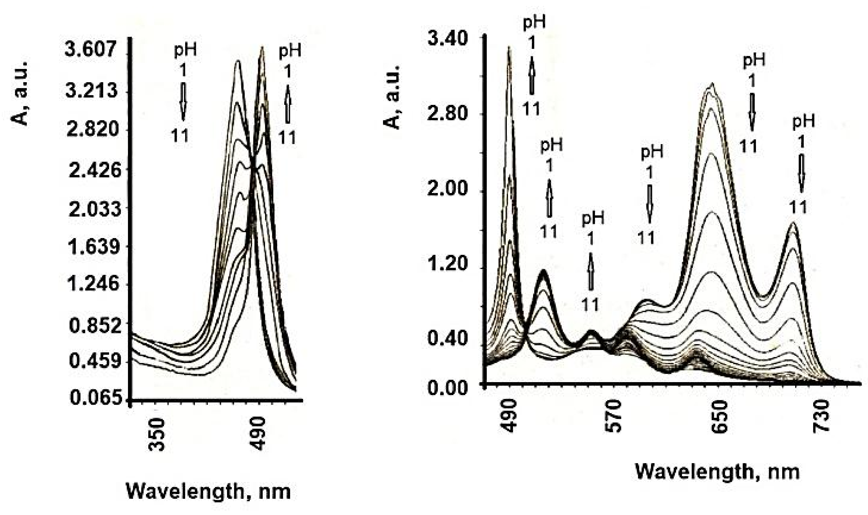
The formation of highly ordered TSPP aggregates at low pH values has been observed previously [72], these being formed by the intermolecular electrostatic attractions between the positively charged nucleus and the negatively charged periphery of the cycle. The protonation of the two pyrrole nitrogen atoms of the porphyrin ring introduces a change in the symmetry of the molecule from a 2-fold configuration to a 4-fold configuration. Both the B band (436 nm) and the maximum Q band are red shifted at protonation, and the color change of the solution from purple to green for free basic and deprotonated shapes, respectively, was recorded [73]. The structure of TSPP absorption spectra in an aqueous solution strongly depends on pH. At neutral pH, the absorption spectrum of TSPP consists of an intense Soret Band at 413 nm and four weak Q bands at 515, 550, 578, and 631 nm (Qy (1,0), Qy (0,0), Qx (1,0), Qx (0,0), respectively). In acidic solution (pH below 5.0) of low concentration, the TSPP absorption spectrum in the visible spectral region changes to a three-band spectrum composed of an intense absorption band at 645 nm, and weaker bands at 597 and 550 nm. The Soret band is red shifted to 435 nm with respect to that at neutral pH. Monoprotonated species of TSPP and dication forms (H2+P(SO3−)4) in acidic solutions are expected [74,75,76,77,78,79,80], and these spectral changes might be attributed to them. Further decrease of solution pH results in an appearance of the new absorption bands at 490 and 706 nm. The Soret band undergoes a slight shift to the blue and a decrease in intensity like the rest of the absorption bands belonging to the dication of TSPP (Figure 4). At pH 1.1, the TSPP absorption spectrum consists of the Soret Band at 430 nm; two intense bands at 490 and 709 nm; and three weak bands at 560, 640, and 670 nm. The band at 490 nm has a weak shoulder at 520 nm. The ratio between the intensities of both absorption bands at 490 and 709 nm and the absorption bands of dication vary depending on the total concentration of TSPP in acid solutions. Similar changes in absorption spectra can be induced by varying the ionic strength of the acidic solution of TSPP and have been assigned to the formation of J-aggregates [81].
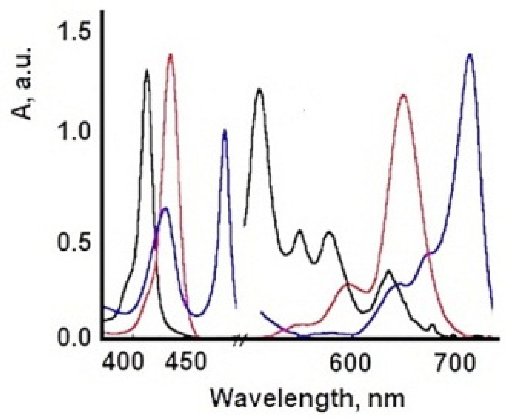
Due to its versatility, TSPP are now under reinvestigation, because this PS can adopt different cationic/anionic forms at different pH, temperature, concentrations, and ionic strength [82], Figure 5. In acidic environments, new absorption bands (from 490, 706 nm) become dominant when the TSPP concentration exceeds 10−5 M and these are attributed to the dicationic forms of TSPP and subsequently to the aggregate forms of this porphyrin. Aggregates J are formed with monomeric dicationic molecules arranged in a dimension so that the transition moments of the monomers are parallel and the angle between the transition moment and the line joining the molecular centers is zero [83]. The aggregation process causes further changes in the optical spectra of TSPP. The presence of intermolecular excitonic interactions determines the division of each of the monomeric bands into a blue and a red displacement band, associated with J and H type interactions [84]. The band at 490 nm comes from the J (head-tail) aggregates of the porphyrin molecules. In the dicationic form, due to Coulombic static repulsion, the two central N-H + fragments in the porphyrin macrocycle are probably distorted outside the aromatic plane, as reported elsewhere [85].
In contrast, the 401 nm and 422 nm bands occur in the H aggregate of porphyrin molecules [86] (face-to-face interaction) and occur at c > 2.5 × 10⁻³ M. H-aggregates, so named because of their band spectral, are characterized by the blue (hypochromatic) displacement in relation to the absorption band of the monomer and are found spatially by a face-to-face stacking of monomer species. In contrast, J aggregates (named after their discoverer, Scheibe Jelly) are edge or edge spatial assemblies that produce bathochromic (red) displacements, Table 2 and Figure 6.
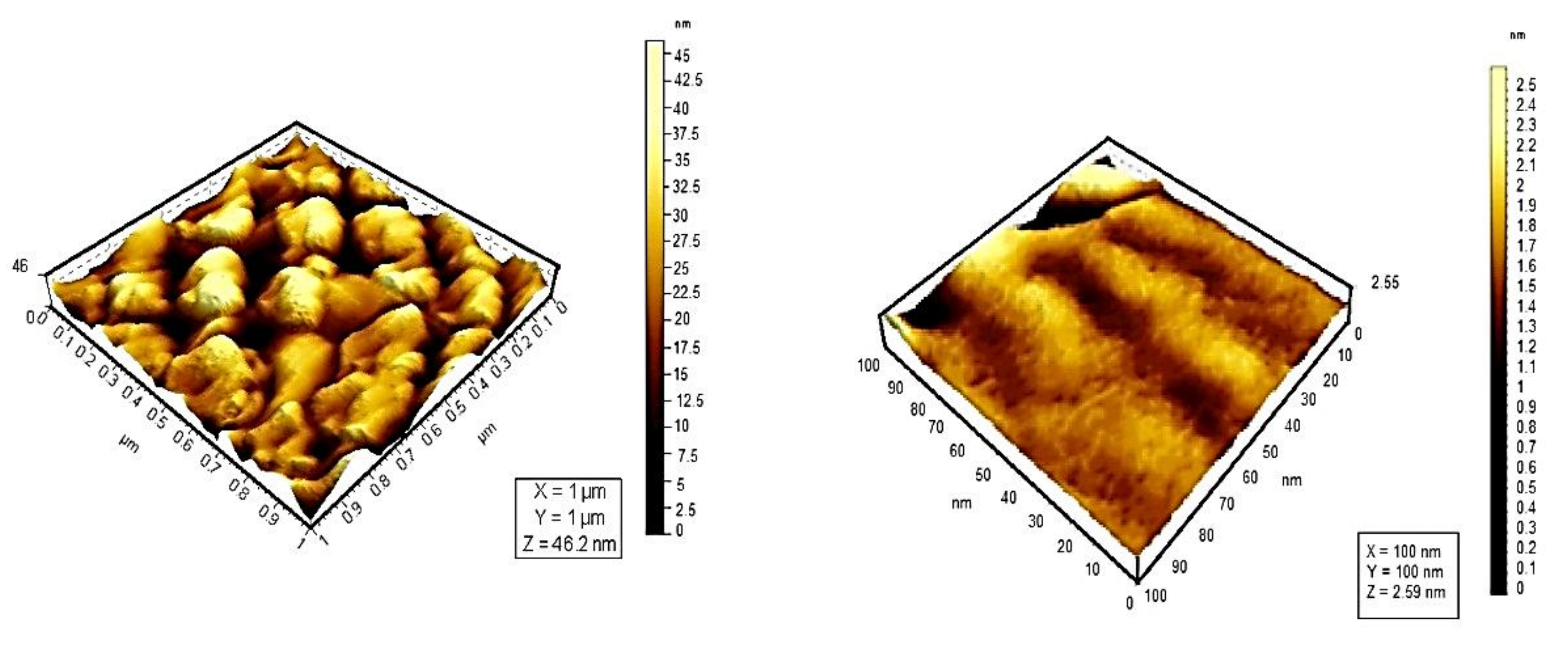
Whereas UV–vis and fluorescence techniques enabled us to determine the type of aggregates formed and the size of the assemblies in solution, AFM provided direct visualization of the aggregates. AFM experiments in air at room temperature with 100 μm acquired in tapping mode for additional image processing, Figure 7.
Topographic studies conducted by means of atomic force microscopy at the scale of 10 μm reveal that the distribution of porphyrins varies. TSPP shows a high density of particles on the same surface. From the analysis of the 3D images of porphyrins studied, could be observed an uniform distribution of particles on the analyzed surface; their average size was 23 nm for monomer form of TSPP, which tends to form aggregates of larger sizes (73.1 nm) than the other porphyrins studied [87].
7. Influence of Dicationic (J-Aggregates) TSPP form on aPDT
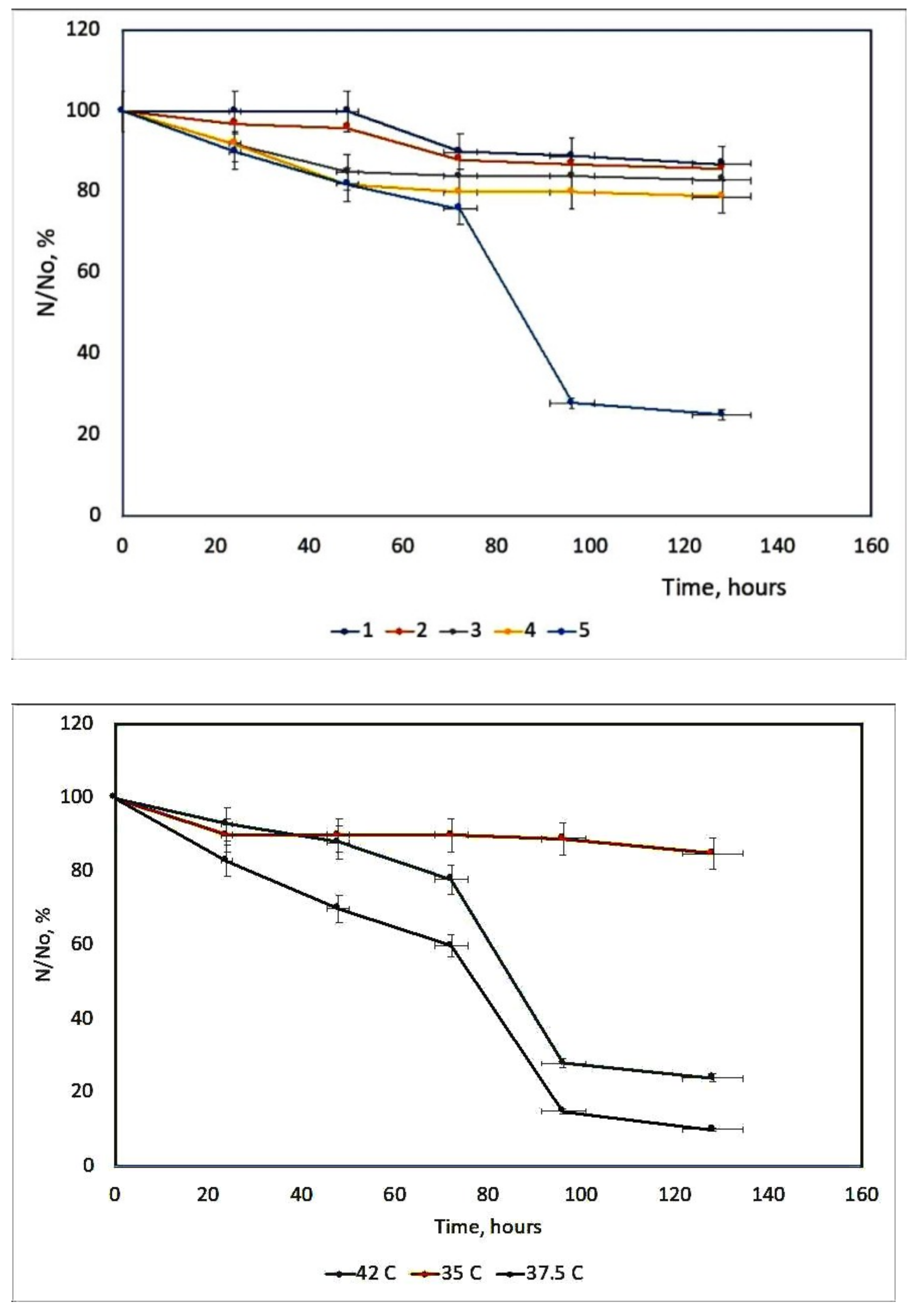
Herpes Simplex Virus (HSV) can be irreversibly and permanently made photosensitive by heterocyclic dyes so that brief exposure to visible light renders the virus non-infectious [88]. Photodynamic inactivation is dependent upon the dye concentration, temperature, and pH [89]. Membrane-photosensitizing dyes have the advantage of inactivating the virus at a site other than the genetic material [90]. Many systems as porphyrin derivatives have been tested in different culture cells [91,92]. Working with two viral suspensions: A type with a cell concentration 2.6–2.8 × 10⁵ per cell standard, and B type with a cell concentration of 2.7–2.9 × 10⁵ per cell standard. It is concluded that the survival curves of dermal HSV from rats during photosensitization with TSPP 1.377 × 10⁻⁵ M are the most efficient during the inactivation process of HSV. The other concentrations are not proper for this inactivation, Figure 8. This fact could be interpreted knowing different aggregated and ionized forms of TSPP [93] and geometrical configurations of these forms with their reduced photochemical activity, as H-aggregates [94].
The action of this PS is highly dependent on the temperature, Figure 9. The hyperthermia (37.5 and 42 °C) can potentiate the effect of PDT, due to the temperature effect on the basic photochemical processes [95].
Previous results show an increase in the reaction rate over the temperature range 15–45 °C. It can be seen that the optimum value of temperature is 37.5 °C because at this temperature TSPP could exist in the solution as a monomer and dication (J-aggregates) mixture in good agreement with other literature reports [96]. This porphyrin derivative demonstrates a remarkable virucidal activity upon light activation after 48 h, especially for HSV hen derma at 37.5 °C. The concentration and temperature effects were evaluated and also the time interval between dye treatment of cells and virus inoculation.
The viral membrane or protein coat might serve as a barrier to the penetration of the photosensitizing dye, and the sensitivity of the virus perhaps is determined primarily by the permeability of its exterior layer or interface with the suspending medium. The HSV envelope was found to be the major target for the photodynamic damage following dye inactivation. DNA damage is one crucial mechanism driving aPDI. aPDT leads to breaks in single-stranded and double-stranded DNA and the disappearance of the super-coiled fraction of plasmid DNA in both G+ and G− species [97]. An exemplification of the spectral interaction between TSPP with DNA, by UV-Vis spectrum (where from a new Soret band with a bathochromic shift, and a decrease of the same band), could be shown in Figure 10.
8. Extension of Studies to SARS-CoV-2 (COVID-19)
In the context of the above evidences, the difference between COVID-19 and HSV-1 should be mentioned. The severe acute respiratory syndrome coronavirus 2 (SARS-CoV-2) is a single-stranded RNA virus that causes a severe acute respiratory syndrome. The virus was originally called SARS-CoV-2 named officially by World Health Organization as COVID-19 and a global health emergency [98].
COVID-19 is the positive sense ssRNA whereas the Herpes viruses are the dsDNA with negative sense, but they both possess the T-cell-mediated immune responses. The structure of herpes viruses consists of a relatively large, double-stranded, linear DNA genome encased within an icosahedral protein cage called the capsid, which is wrapped in a lipid bilayer called the envelope. The envelope is joined to the capsid by means of a tegument. This complete particle is known as the virion [99].
In photodynamic treatment (PDT), using photosensitizers, such as porphyrin derivatives, can help attenuate COVID-19, namely to disinfect surfaces and very rarely water and air. Current drugs used against COVID-19 include steroids, interleukin-6 inhibitors, non-invasive oxygen therapy (high-flow nasal cannula), or invasive oxygen therapy (mechanical ventilation). Despite the many treatment options, none have proven overly successful. This is the reason for the exploration of the use of photodynamic therapy (PDT) to treat COVID-19 [100].
aPDT is an antimicrobial method, with an effect against emerging/unknown pathogens, being an alternative approach to inactivate the coronaviruses in wastewater. There are promising results of aPDT on inactivation of viruses, namely against those with an envelope, like the coronaviruses. Even though it is not yet clear whether the SARS-CoV-2 is viable in wastewater for a long period, it is of the utmost importance to develop wastewater treatment processes that effectively inactivate this virus, through the development of new technologies for wastewater treatment to inactivate microorganisms, including SARS-CoV-2. Different PSs are effective to photoinactivate microorganisms, including viruses, in samples of secondary-treated wastewater, including aPDT [101] at low PS concentrations (5–10 µM) under low artificial white light irradiance (380–700 nm, 4 mW cm−2) or under natural sunlight conditions.
The SARS-CoV-2-mediated attack on hemoglobin leads to desaturation of the lung tissue, which can lead to several organ failure. However, to reduce viral load, illumination of the target lung tissue using a fibre-optic catheter to deliver low-power light of a characteristic absorption wavelength for the photosensitizer (typical range 450–800 nm) causes photoactivation, yielding a highly reactive oxygen species capable of destroying the bonded SARS-CoV-2 virions to the photosensitizer molecules through peroxidation [102].
The sensitization with any of the photodynamically active dyes would be explained by assuming that the protein coat by this virus is virtually impermeable to dyes, or alternatively, that all of the important sites for combination of dye molecules with the RNA core of the virus are occupied or blocked by the peripheral protein. Some authors suggested that nucleic acids may be an important target for PDT inactivation in their model viruses [103].
Additionally, it has been demonstrated that PDT can be used to destroy pathogenic microorganisms, such as bacteria, viruses, protozoa, and fungi [104].
The predominant mechanism in PDT involves the generation of singlet oxygen (1O2). The diffusion distance of 1O2 is around 0.01–0.02 μm before being quenched, so the photosensitizers must be associated intimately with the target substrate for maximal impact. An antiviral potential of photodynamic therapy (PDT) using Methylene Blue and Radachlorin to inactivate and inhibit SARS-CoV-2 in vitro at very low concentrations (100–1000 times lower than usual), when activated by 662 nm light, caused inhibition of SARS-CoV-2 [105].
9. Photophysical and Photochemical Properties of PS
The effect of wavelength of excitation on aPDT efficacy
The absorption/excitation wavelength(s) of a given PS is a key selection criterion for its application in PDT or aPDT. Current light sources used are either ultraviolet (UV) or short-wavelength visible light, as most of the photosensitizers absorb in relatively short wavelengths, which possess limited tissue penetration ability that restricts the light to be delivered to the target sites. The UV activation of photoactive drugs does not exclude damages to the cells’ genetic information. Also, small wavelengths absorbed by the major tissue or cell chromophores should be avoided for excitation of the PS. The absorption spectra of these molecules define the optical window for PDT in the tissue in the wavelength range of 600–850 nm [106]. The upper limit of this window is required for the efficient production of singlet oxygen, considering the thermal loss of energy. However, due to a different composition of chromophores in microorganisms, the lower wavelength limit of the optical window defined for PDT applications does not necessarily apply for photosensitizers employed in aPDT.
As the photophysical processes in a fluorescing molecule are dependent on the solvent, excitation and emission spectra of a PS should be read in aqueous solutions (buffers).
The excitation wavelength dependence of the singlet oxygen quantum yield values indicates that different photochemical processes arise upon excitation of the porphyrin moieties [107].
The shorter the excitation wavelength, i.e., the more towards the ultraviolet region the fluorescence excitation occurs, the more of the visible spectrum that resulted from fluorescence signal is observed. The porphyrins as fluorophore molecules have a molecular structure that gives rise to fluorescence. Such molecules are frequently characterized by the presence of a series of conjugated double bonds, i.e., alternating single and double bonds. In addition to the emission wavelength, fluorescence is also characterized by its lifetime. The lifetime is dependent on the surroundings of the fluorophore. The lifetime for endogenous and exogenous fluorophores is in the order of nanoseconds (ns).
Laser-induced fluorescence provides non-invasive, real time monitoring of biological tissue. The technique is based on measurements of signals from fluorescent molecules within tissue. These signals are related to molecular concentration and micro-environment. Laser light in the near-ultraviolet or ultraviolet region is most often used as an excitation light source, and the fluorescence signal achieved from the tissue is subject to spectroscopic analyses.
Due to light exposure of a photosensitizer, the absorption and fluorescence properties of the photosensitizer itself can change, which indicates that photodegradation and/or photoproduct formation appeared, which results in a decreased photostability [108]. Such changes can be evidenced by the appearance of new absorption bands within the specific absorption spectra of the photosensitizer. Changes of the characteristic absorption spectra of a given photosensitizer depend on the wavelength of the illumination light: shorter wavelengths are more effective than longer wavelengths. If the photosensitizer is bleached too rapidly, either successful inactivation of microorganisms or tumor destruction will not be completed once the minimal inhibitory concentration of the nondegraded photosensitizer in the infected/tumor tissue is deseeded upon illumination [109]. Furthermore, it is unalterable to assure the nontoxicity of photodegraded products of the photosensitizer.
For the disinfection of objects, including fabrics and surfaces, besides artificial white light, natural sunlight can be used as a light source to inactivate the coronavirus. The use of sunlight as the light source turns out to be an inexpensive aPDT procedure since it is based on the use of a low cost and worldwide-available visible light source. In addition, photosensitizers like MB or porphyrin-based derivatives do not accumulate in the environment since they are degraded by exposure to sunlight, although they are able, in the meantime, to inactivate the microorganisms.
The main steps of photosensitization involved could be: dye partition into virus membrane; fluorescence quenching of bound dye; energy transfer experiments; and viral adhesion to the host cells [110]. These processes could be explained by different photophysical parameter measurements.
Transient difference spectra have been measured at saturation with respect to pump beam energy in which case at the end of the excitation pulse all molecules are in the excited state S1. Pumping in the Soret band populates the S2 state of porphyrin. This state relaxes back on femtosecond time scale to the first excited singlet state S1 [111,112], Scheme 1.
Therefore, the difference spectrum measured at 1 ns from excitation belongs to the S1 state. It decays in about 5 ns to a slightly different spectrum, which appears to remain stable in the nanosecond time range (Figure 11). The observed absorbance decay could be attributed to the depopulation of the singlet state S1. This attribution is in good agreement with the value of 5.2 ns previously reported for the S1 state lifetime of TSPP [113].
The intermediate detected at the end of the decay of the S1 state should be the triplet state T1 because it is known that porphyrins have a high quantum yields for triplet state formation (ΦT) 0.63.
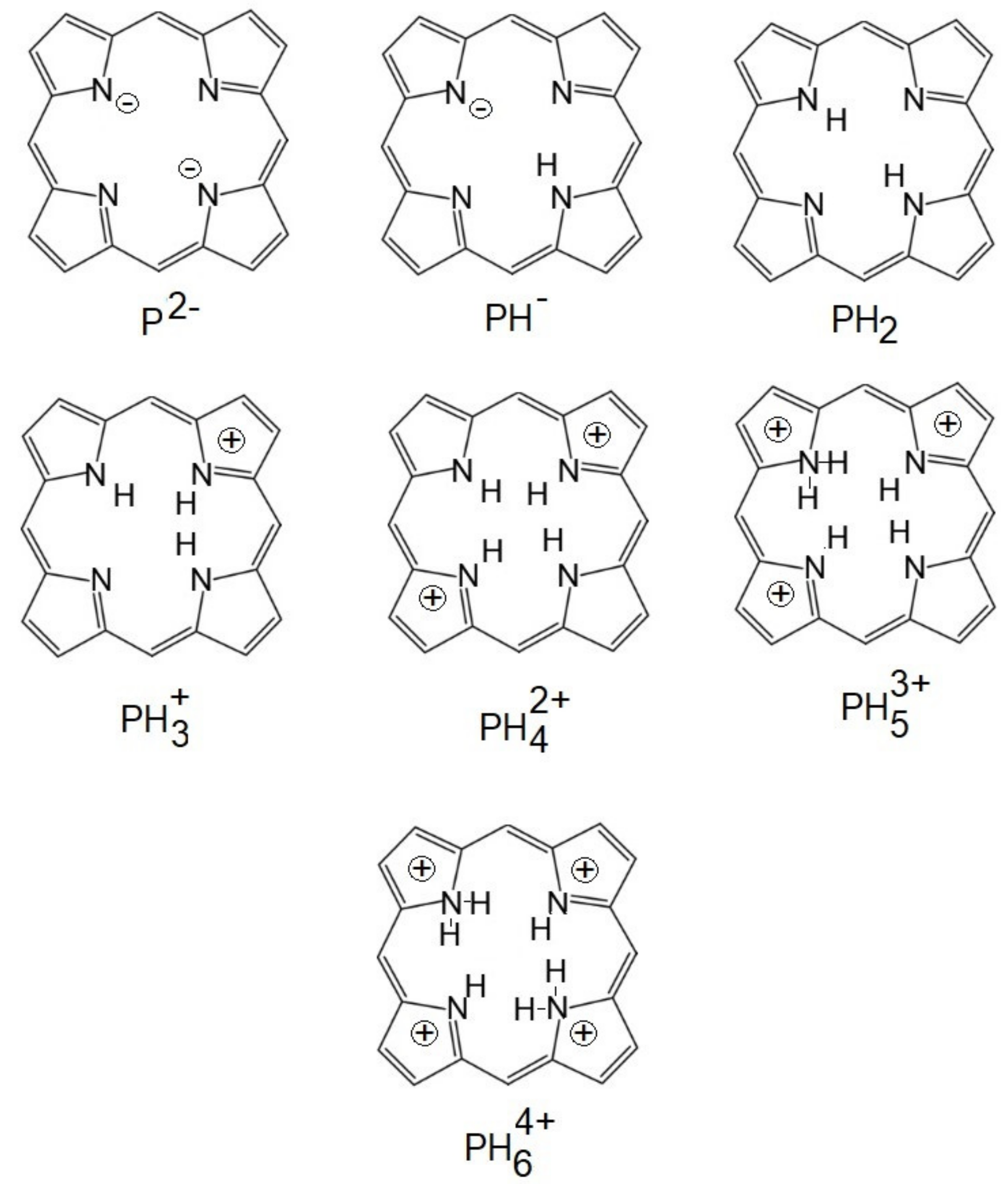
Also, the laser-induced optoacoustic spectroscopy (LIOAS) method was applied. The ionization of the peripheral charged/uncharged groups attached to the porphyrin ring and of the pyrrole nitrogen allows the dye molecule to exist as different ionic species depending on the pH of the surroundings. The pyrrole nitrogens can bind one or two protons leading to the monocation and to the dication as shown by Chow et al. [114].
In solutions of low pH (2–4), TSPP occurs in two forms: dicationic and J-aggregated form. Energy transfer between these forms could not be excluded. The energy transfer between the monomeric and dicationic dye forms in TSPP samples is possible, but with rather weak ET between the dicationic and aggregated dicationic species in TSPP [115]. The fluorescence quantum yield (ΦF), as the ratio of photons absorbed to photons emitted through fluorescence, is a measure of the probability of the excited state deactivated by fluorescence rather than by another, non-radiative mechanism. This value is 0.015 for TSPP aggregate and 0.179 for TSPP monomer. So, the aggregated forms have a longer lifetime to exist as an excited state. Such slow deactivated energy is generated predominantly by thermal deactivation of triplet states populated by intersystem crossing from the excited singlet state. By LIOAS, it was possible to measure this energy transfer, as follows: TSPP monomer has in an atmosphere without Ar α = 0.37677 and with Ar α = 0.37204. The aggregated TSPP in an atmosphere without Ar has a value for α = 0.5861 but in an atmosphere with Ar, this value becomes α = 0.1497, which means that for TSPP in the dicationic aggregated form, the deactivation of triplet states populated by intersystem crossing from the excited singlet state is low, being able to generate more singlet oxygen.
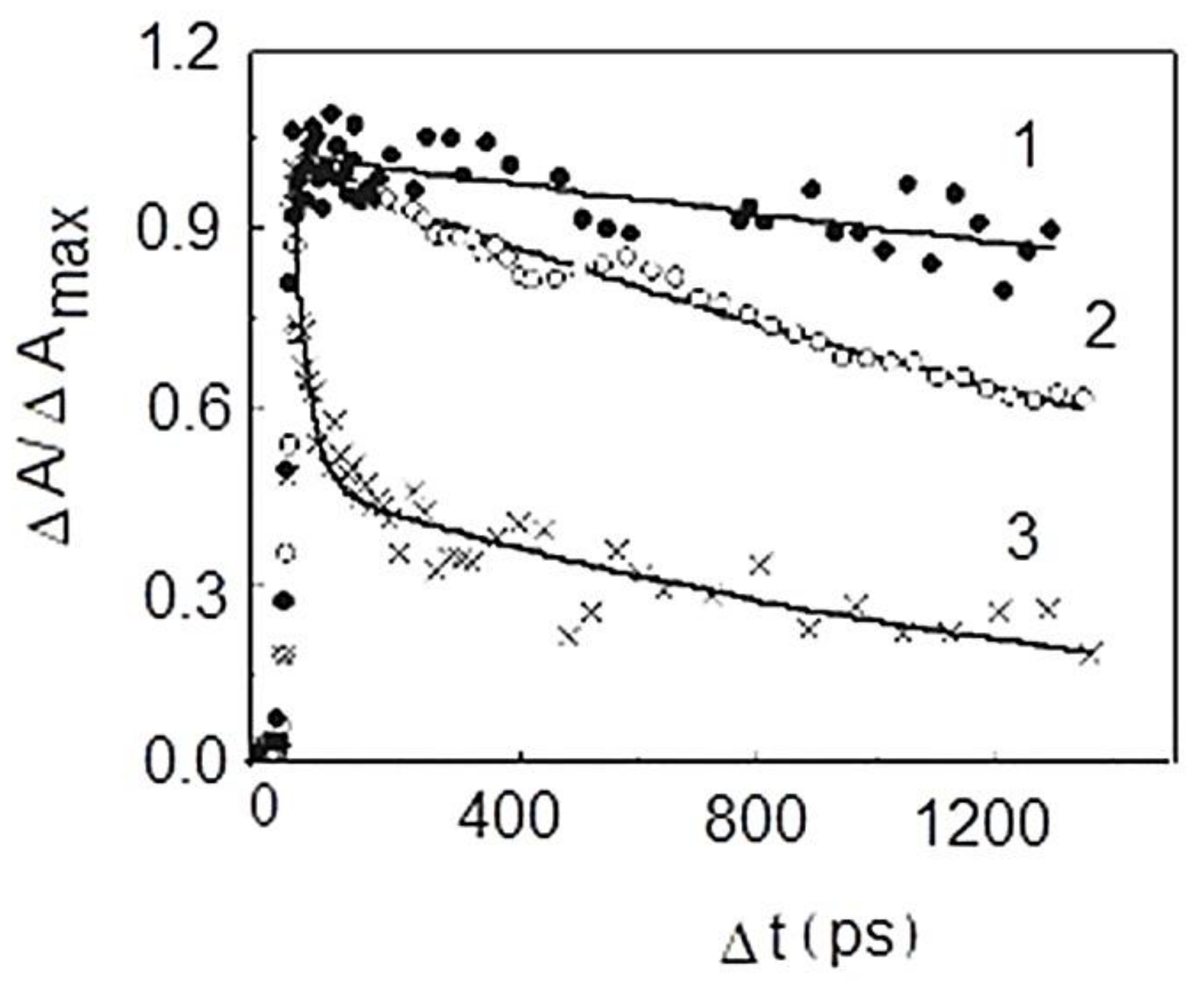
The absorption decay of TSPP monomer induced by excitation can be fitted biexponentially with the time constants about t1 = 27 ps and t2 = 1.44 ns, while for TSPP J-aggregates (Figure 11), is described a three exponential decay pattern with the time constants t1 = 24 ps, t2 = 290 ps, and t3 = 2–3 ns. In the case of TSPP J-aggregates, the shortest time constant seems to be related to exciton annihilation, whereas a nanosecond time constant reflects the relaxation of the excitation energy when a single excitation is present in the J aggregate [116,117,118,119].
The promising results of aPDT on inactivation of viruses, namely against those with an envelope, like the coronaviruses [120], suggest that this principle can be applied to surfaces treatment in order to inactivate the microorganisms, including SARS-CoV-2. The use of photodynamic therapy (PDT) can be an alternative approach against SARS-CoV-2 that deserves to be explored. In conclusion, photodynamic treatment (PDT), using photosensitizers such as porphyrin derivatives, can help to mitigate COVID-19, namely, to develop functional photoactive textiles and disinfect surfaces, water, and air. The active species is the dicationic forms, generated at low pH values, as precursors for J-aggregates, useful for such applications.
10. Conclusions
The use of photodynamic therapy (PDT) can be an alternative approach against SARS-CoV-2 that deserves to be explored. Some studies about Herpes Simplex virus type 1 (HSV-1) could develop new conclusions about COVID-19, knowing that without the help of HSV-1, the COVID-19 virus may not be able to cause serious illness or death in humans. As sensitizers, TSPP could be a very efficient agent in PDT, due to its capacity to generate dication and J-aggregate subsequently, which are decisive for PDT on HSV (ussualy responsive mostly at cationic agents). The mechanism of dicationic and J-aggregates has been presented in this paper, and the photophysical parameters have been collected and harmonized to support this aspect. This method could be a new direction for COVID treatment and immunization, either to prevent infections or to develop photoactive fabrics (e.g., masks, suits, gloves) to disinfect surfaces, under artificial light and/or natural sunlight. The use of photodynamic therapy (PDT) can be an alternative approach against SARS-CoV-2 that deserves to be explored.
Funding
This research received no external funding.
Institutional Review Board Statement
Not Applicable.
Informed Consent Statement
Not Applicable.
Conflicts of Interest
The author declares no conflict of interest.
References
- Nicholas, J.L. Photo(chemo)therapy: General principles. Clin. Dermatol. 1997, 15, 745–752. [Google Scholar]
- Ion, R.M. The photodynamic therapy of cancer-a photosensitisation or a photocatalytic process? Progr. Catal. 1997, 1, 55–62. [Google Scholar]
- Evensen, J.F.; Moan, J.; Winkelman, J.W. Toxic and phototoxic effects of tetraphenylporphine sulphonate and haematoporphyrin derivative in vitro. Int. J. Radiat. Biol. Relat. Stud. Phys. Chem. Med. 1987, 51, 477–491. [Google Scholar] [CrossRef]
- Qian, P.; Evensen, J.F.; Rimington, C.; Moan, J. A comparison of different photosensitizing dyes with respect to uptake C3H-tumors and tissues of mice. Cancer Lett. 1987, 36, 1–10. [Google Scholar] [CrossRef]
- Boda, D.; Neagu, M.; Constantin, C.; Diaconeasa, A.; Ianosi, S.; Ion, R.M.; Amalinei, C.; Stanoiu, B.; Crauciuc, E.; Toma, O. New photosensitizers versus aminolevulinic acid (ala) in experimental photodynamic therapy of actinic keratosis—A case report. Ann. Alexandru Ioan Cuza Univ. Sect. Genet. Mol. Biol. 2009, 3, 62–69. [Google Scholar]
- Winkelman, J.W.; Collins, G.H. Neurotoxicity of tetraphenylporphinesulfonate TPPS4 and its relation to photodynamic therapy. Photochem. Photobiol. 1987, 46, 801–807. [Google Scholar] [CrossRef] [PubMed]
- Streleckova, E.; Kodetova, D.; Pouckova, P.; Zadinova, M.; Lukas, E.; Rokyta, R.; Jirsa, M. Meso-tetra-(4-sulfonatophenyl)-porphine of low neurotoxicity. SB Lek. 1995, 96, 7–13. [Google Scholar] [PubMed]
- Huang, L.; El-Hussein, A.; Xuan, W.; Hamblin, M.R. Potentiation by potassium iodide reveals that the anionic porphyrin TPPS4 is a surprisingly effective photosensitizer for antimicrobial photodynamic inactivation. J. Photochem. Photobiol. B Biol. 2018, 178, 277–286. [Google Scholar] [CrossRef]
- Nunn, J.F. Ancient Egyptian Medicine; University of Oklahoma Press: Norman, OK, USA, 1996. [Google Scholar]
- Dougherty, T.; Henderson, J.; Schwartz, B.W.; Winkelman, J.W.; Lipson, R.L. Historical Perspective, in Photodynamic Therapy; Henderson, B.W., Dougherty, T.J., Eds.; Marcel Dekker: New York, NY, USA, 1992; pp. 1–15. [Google Scholar]
- Daniell, M.D.; Hill, J.S. A history of photodynamic therapy. Aust. N. Z. J. Surg. 1991, 61, 340–348. [Google Scholar] [CrossRef]
- Raab, O. Ueber die Wirkung fluorescirender Stoffe auf Iufusorien. Z. Biol. 1900, 39, 524. [Google Scholar]
- Yamamoto, N. Photodynamic inactivation of bacteriophage and its inhibition. J. Bacteriol. 1958, 75, 443–448. [Google Scholar] [CrossRef] [Green Version]
- Felber, T.D.; Smith, E.B.; Knox, J.M. Photodynamic inactivation of herpes simplex. JAMA 1973, 22, 289–295. [Google Scholar] [CrossRef]
- Wallis, C.; Melnick, J.L. Photodynamic inactivation of animal viruses: A review. Photochem. Photobiol. 1965, 4, 159–162. [Google Scholar] [CrossRef] [PubMed]
- Sperandio, F.F.; Huang, Y.-Y.; Hamblin, M.R. Antimicrobial photodynamic therapy to kill gram-negative bacteria. Recent Pat. Anti-Infect. Drug Discov. 2013, 8, 108–120. [Google Scholar] [CrossRef] [Green Version]
- Simon, M.I.; Vunakis, V. The photodynamic reaction of methylene blue with DNA. J. Mol. Biol. 1962, 4, 488–493. [Google Scholar] [CrossRef]
- Embleton, M.L.; Nair, S.P.; Heywood, W.; Menon, D.C.; Cookson, B.C.; Wilson, M. Development of a novel targeting system for lathal photosensitization of antibiotic-resistant strains of Staphylococcus aureus. Antimicrob. Agents Chemother. 2005, 49, 3690–3696. [Google Scholar] [CrossRef] [Green Version]
- Sahu, K.; Sharma, M.; Bansal, H.; Dube, A.; Gupta, P.K. Topical photodynamic treatment with poly-l-lysine–chlorin p6 conjugate improves wound healing by reducing hyperinflammatory response in Pseudomonas aeruginosa-infected wounds of mice. Lasers Med. Sci. 2012, 28, 465–471. [Google Scholar] [CrossRef]
- Cieplik, F.; Deng, D.; Crielaard, W.; Buchalla, W.; Hellwig, E.; Al-Ahmad, A.; Maisch, T. Antimicrobial photodynamic therapy—What we know and what we don’t. Crit. Rev. Microbiol. 2018, 44, 571–589. [Google Scholar] [CrossRef] [PubMed] [Green Version]
- Sobotta, L.; Skupin-Mrugalska, P.; Mielcarek, J.; Goslinski, T.; Balzarini, J. Photosensitizers mediated photodynamic inactivation against virus particles. Mini-Rev. Med. Chem. 2015, 15, 503–521. [Google Scholar] [CrossRef]
- Janouskova, O.; Rakusan, J.; Karaskova, M.; Holada, K. Photodynamic inactivation of prions by disulfonated hydroxyaluminium phthalocyanine. J. Gen. Virol. 2012, 93, 2512–2517. [Google Scholar] [CrossRef] [Green Version]
- Strasfeld, L.; Chou, S. Antiviral drug resistance: Mechanisms and clinical implications. Infect. Dis. Clin. N. Am. 2010, 24, 413–437. [Google Scholar] [CrossRef] [Green Version]
- Ion, R.M.; Corobea, M.C. Porphyrin models as sensitizers in photodynamic inactivation of Herpes Simplex Virus. In Proceedings of the IV International Conference on Antimicrobial Research—ICAR 2016, Torremolinos-Malaga, Spain, 29 June–1 July 2016. [Google Scholar]
- Wiehe, A.; O’Brien, J.M.; Senge, M.O. Trends and targets in antiviral phototherapy. Photochem. Photobiol. Sci. 2019, 18, 2565–2612. [Google Scholar] [CrossRef]
- Li, W.-Y.; Xu, J.-G.; He, X.-W. Characterization of the binding of methylene blue to DNA by spectroscopic methods. Anal. Lett. 2000, 33, 2453–2464. [Google Scholar] [CrossRef]
- Baptista, M.S.; Cadet, J.; Di Mascio, P.; Ghogare, A.A.; Greer, A.; Hamblin, M.R.; Lorente, C.; Nunez, S.C.; Ribeiro, M.S.; Thomas, A.H.; et al. Type I and type II photosensitized oxidation reactions: Guidelines and mechanistic pathways. Photochem. Photobiol. 2017, 93, 912–919. [Google Scholar] [CrossRef] [Green Version]
- Zhang, B.; Zheng, L.; Huang, Y.; Mo, Q.; Wang, X.; Qian, K. Detection of nucleic acid lesions during photochemical inac-tivation of RNA viruses by treatment with methylene blue and light using real-time PCR. Photochem. Photobiol. 2011, 87, 365–369. [Google Scholar] [CrossRef]
- Banerjee, I.; Douaisi, M.P.; Mondal, D.; Kane, R.S. Light-activated nanotube–porphyrin conjugates as effective antiviral agents. Nanotechnology 2012, 23, 105101. [Google Scholar] [CrossRef]
- Morikawa, K.; Suda, G.; Sakamoto, N. Viral life cycle of hepatitis B virus: Host factors and druggable targets. Hepatol. Res. 2016, 46, 871–877. [Google Scholar] [CrossRef]
- De Clercq, E.; Li, G. Approved antiviral drugs over the past 50 years. Clin. Microbiol. Rev. 2016, 29, 695–747. [Google Scholar] [CrossRef] [Green Version]
- Lenard, J.; Vanderoef, R. Photoinactivation of influenza virus fusion and infectivity by rose Bengal. Photochem. Photobiol. 1993, 58, 527–531. [Google Scholar] [CrossRef]
- Monjo, A.L.-A.; Pringle, E.S.; Thornbury, M.; Duguay, B.A.; Monro, S.M.A.; Hetu, M.; Knight, D.; Cameron, C.G.; McFarland, S.A.; McCormick, C. Photodynamic activation of herpes simplex viruses. Viruses 2018, 10, 532. [Google Scholar] [CrossRef] [Green Version]
- Belanger, J.M.; Raviv, Y.; Viard, M.; de la Cruz, J.M.; Nagashima, K.; Blumenthal, R. Characterization of the effects of ar-yl-azido compounds and UVA irradiation on the viral proteins and infectivity of human immunodeficiency virus type 1. Photochem. Photobiol. 2010, 86, 1099–1108. [Google Scholar] [CrossRef] [Green Version]
- Warfield, K.L.; Swenson, D.L.; Olinger, G.G.; Kalina, W.V.; Viard, M.; Aitichou, M.; Chi, X.; Ibrahim, S.; Blumenthal, R.; Raviv, Y.; et al. Ebola virus inactivation with preservation of antigenic and structural integrity by a photoinducible alkylating agent. J. Infect. Dis. 2007, 196, S276–S283. [Google Scholar] [CrossRef] [Green Version]
- Raviv, Y.; Blumenthal, R.; Tompkins, S.M.; Humberd, J.; Hogan, R.J.; Viard, M. Hydrophobic inactivation of influenza viruses confers preservation of viral structure with enhanced immunogenicity. J. Virol. 2008, 82, 4612–4619. [Google Scholar] [CrossRef] [Green Version]
- Dairou, J.; Vever-Bizet, C.; Brault, D. Interaction of sulfonated anionic porphyrins with HIV glycoprotein gp120: Photo-damages revealed by inhibition of antibody binding to V3 and C5 domains. Antiviral Res. 2004, 61, 37–47. [Google Scholar] [CrossRef]
- Tavares, A.; Carvalho, C.M.B.; Faustino, M.A.; Neves, M.G.P.M.S.; Tomé, J.P.C.; Tomé, A.C.; Cavaleiro, J.A.S.; Cunha, Â.; Gomes, N.C.M.; Alves, E.; et al. Antimicrobial photodynamic therapy: Study of bacterial recovery viability and potential development of resistance after treatment. Mar. Drugs 2010, 8, 91–105. [Google Scholar] [CrossRef] [PubMed] [Green Version]
- Almeida, A.; Cunha, A.; Faustino, M.A.F.; Tomé, A.C.; Neves, M.G.P.M.S. Porphyrins as antimibrobial photosensitizing agents. In Photodynamic Inactivation of Microbial Pathogens: Medical and Environmental Applications; Hamblin, M.R., Jori, G., Eds.; Royal Society of Chemistry: Cambridge, UK, 2011; pp. 83–160. [Google Scholar]
- Vardevanyan, P.O.; Antonyan, A.P.; Parsadanyan, M.A.; Shahinyan, M.A.; Ham-bardzumyan, L.A. Mechanisms for binding between methylene blue and DNA. J. Appl. Spectrosc. 2013, 80, 595–599. [Google Scholar] [CrossRef]
- Fekrazad, R. Photobiomodulation and antiviral photodynamic therapy as a possible novel approach in COVID-19 management. PhotobiomodulationPhotomed. Laser Surg. 2020, 38, 255–257. [Google Scholar] [CrossRef]
- Almeida, A.; Faustino, M.A.F.; Neves, M.G.P.M.S. Antimicrobial photodynamic therapy in the control of COVID-19. Antibiotics 2020, 9, 320. [Google Scholar] [CrossRef]
- Bond, P. Ethnicity and the relationship between covid-19 and the herpes simplex viruses. Med. Hypotheses 2021, 146, 110447. [Google Scholar] [CrossRef]
- Van Doremalen, N.; Bushmaker, T.; Morris, D.H.; Holbrook, M.G.; Gamble, A.; Williamson, B.N.; Tamin, A.; Harcourt, J.L.; Thornburg, N.J.; Gerber, S.I.; et al. Aerosol and surface stability of SARS-CoV-2 as compared with SARS-CoV-1. N. Engl. J. Med. 2020, 382, 1564–1567. [Google Scholar] [CrossRef]
- Mesquita, M.Q.; Dias, C.; Neves, M.G.P.M.S.; Almeida, A.; Faustino, M.A.F. Revisiting current photoactive materials for an-timicrobial photodynamic therapy. Molecules 2018, 23, 2424. [Google Scholar] [CrossRef] [Green Version]
- Foote, C.S. Mechanisms of photosensitized oxidation. Science 1968, 162, 963–970. [Google Scholar] [CrossRef]
- Ion, R.M. Near-Infrared Dyes for High Technology Applications; Daehne, S., Resch-Genger, U., Wolfbeis, O., Eds.; NATO ASI SERIES; Kluwer Academic Publishers: Dordrecht, The Netherlands; Boston, MA, USA; London, UK, 1998; Volume 3/52, pp. 87–114. ISBN 0-7923-5101-0. [Google Scholar]
- Kato, H.; Komagoe, K.; Inoue, T.; Masuda, K.; Katsu, T. Structure–activity relationship of porphyrin-induced photoinactivation with membrane function in bacteria and erythrocytes. Photochem. Photobiol. Sci. 2018, 17, 954–963. [Google Scholar] [CrossRef]
- Wainwright, M. Photodynamic antimicrobial chemotherapy (PACT). J. Antimicrob. Chemother. 1998, 42, 13–28. [Google Scholar] [CrossRef] [PubMed]
- Bertoloni, G.; Lauro, F.M.; Cortella, G.; Merchat, M. Photosensitizing activity of hematoporphyrin on Staphylococcus aureus cells. Biochim. Biophys. Acta Gen. Subj. 2000, 1475, 169–174. [Google Scholar] [CrossRef]
- Ion, R.M.; Planner, A.; Wiktorowicz, K.; Frackowiak, D. Incorporation of various porphyrins into human blood cells measured using the flow-cytometry, the absorption and emission spectroscopy. Acta Biochim. Pol. 1998, 45, 833–842. [Google Scholar] [CrossRef] [Green Version]
- Salva, K.A. Photodynamic therapy: Unapproved uses, dosages, or indications. Clin. Derm. 2002, 20, 571–581. [Google Scholar] [CrossRef]
- Juzeniene, A.; Juzenas, P.; Ma, L.-W.; Iani, V.; Moan, J. Effectiveness of different light sources for 5-aminolevulinic acid photodynamic therapy. Lasers Med. Sci. 2004, 19, 139–149. [Google Scholar] [CrossRef]
- Koshi, E.; Mohan, A.; Rajesh, S.; Philip, K. Antimicrobial photodynamic therapy: An overview. J. Indian Soc. Periodontol. 2011, 15, 323–327. [Google Scholar] [CrossRef]
- Kashef, N.; Huang, Y.-Y.; Hamblin, M.R. Advances in antimicrobial photodynamic inactivation at the nanoscale. Nanophotonics 2017, 6, 853–879. [Google Scholar] [CrossRef] [Green Version]
- Hu, X.; Huang, Y.-Y.; Wang, Y.; Wang, X.; Hamblin, M.R. Antimicrobial photodynamic therapy to control clinically relevant biofilm infections. Front. Microbiol. 2018, 9, 1299. [Google Scholar] [CrossRef] [Green Version]
- Kuo, W.-S.; Chang, C.-Y.; Chen, H.-H.; Hsu, C.-L.L.; Wang, J.-Y.; Kao, H.-F.; Chou, L.C.-S.; Chen, Y.-C.; Chen, S.-J.; Chang, W.-T.; et al. Two-photon photoexcited photodynamic therapy and contrast agent with antimicrobial graphene quantum dots. ACS Appl. Mater. Interfaces 2016, 8, 30467–30474. [Google Scholar] [CrossRef]
- Wozniak, A.; Grinholc, M. Combined antimicrobial activity of photodynamic inactivation and antimicrobials–state of the art. Front. Microbiol. 2018, 9, 930. [Google Scholar] [CrossRef] [PubMed]
- Bartolomeu, M.; Coimbra, S.; Cunha, A.; Neves, M.G.P.M.S.; Cavaleiro, J.A.S.; Faustino, M.A.F.; Almeida, A. Indirect and direct damage to genomic DNA induced by 5,10,15-tris(1-methylpyridinium-4-yl)-20-(pentafluorophenyl)porphyrin upon photody-namic action. J. Porph. Phthal. 2016, 20, 331–336. [Google Scholar] [CrossRef]
- Wainwright, M.; McLean, A. Rational design of phenothiazinium derivatives and photoantimicrobial drug discovery. Dyes Pigments 2017, 136, 590–600. [Google Scholar] [CrossRef]
- Yao, T.-T.; Wang, J.; Xue, Y.-F.; Yu, W.-J.; Gao, Q.; Ferreira, L.; Ren, K.-F.; Ji, J. A photodynamic antibacterial spray-coating based on the host–guest immobilization of the photosensitizer methylene blue. J. Mater. Chem. B 2019, 7, 5089–5095. [Google Scholar] [CrossRef]
- Friedman, L.I.; Skripchenko, A.; Wagner, S.J. Photodynamic Inactivation of Pathogens in Blood by Phenothiazines and Oxygen. Patent WO/2001/049328, 28 December 2000. [Google Scholar]
- Ion, R.-M.; Boda, D. Porphyrin—Based supramolecular nanotubes generated by aggregation processes. Rev. Chim. 2008, 59, 205–207. [Google Scholar] [CrossRef]
- Chatterjee, N.; Walker, G.C. Mechanisms of DNA damage, repair, and mutagenesis. Environ. Mol. Mutagen. 2017, 58, 235–263. [Google Scholar] [CrossRef] [Green Version]
- Snipes, W.; Keller, G.; Woog, J.; Vickroy, T.; Deering, R.; Keith, A. Inactivation of lipid-containing viruses by hydrophobic photosensitizers and near-UV radiation. Photochem. Photobiol. 1979, 29, 780–785. [Google Scholar] [CrossRef]
- Le Gall, T.; Lemercier, G.; Chevreux, S.; Tucking, K.-S.; Ravel, J.; Thetiot, F.; Jonas, U.; Schönherr, H.; Montier, T. Ruthenium (II) polypyridyl complexes as photosensitizers for antibacterial photodynamic therapy: A structure-activity study on clinical bac-terial strains. ChemMedChem 2018, 13, 2229–2239. [Google Scholar] [CrossRef]
- Minnock, A.; Vernon, D.I.; Schofield, J.; Griffiths, J.; Parish, J.H.; Brown, S.B. Mechanism of uptake of a cationic water-soluble pyridinium zinc phthalocyanine across the outer membrane of Escherichia coli. Antimicrob. Agents Chemo-ther. 2000, 44, 522–527. [Google Scholar] [CrossRef] [Green Version]
- Hancock, R.E.W. Alterations in outer membrane permeability. Annu. Rev. Microbiol. 1984, 38, 237–264. [Google Scholar] [CrossRef]
- Hancock, R.E. The bacterial outer membrane as a drug barrier. Trends Microbiol. 1997, 5, 37–42. [Google Scholar] [CrossRef]
- Hancock, R.E.; Farmer, S.W. Mechanism of uptake of deglucoteicoplanin amide derivatives across outer membranes of Escherichia coli and Pseudomonas aeruginosa. Antimicrob. Agents Chemother. 1993, 37, 453–456. [Google Scholar] [CrossRef] [Green Version]
- Vieira, C.; Gomes, A.T.; Mesquita, M.Q.; Moura, N.M.M.; Neves, M.G.P.M.S.; Faustino, M.A.F.; Almeida, A. An insight into the potentiation effect of potassium iodide on aPDT e_cacy. Front. Microbiol. 2018, 9, 2665–2670. [Google Scholar] [CrossRef] [Green Version]
- Vieira, C.; Santos, A.; Mesquita, M.Q.; Gomes, A.T.P.C.; Neves, M.G.P.M.S.; Faustino, M.A.F.; Almeida, A. Advances in aPDT based on the combination of a porphyrinic formulation with potassium iodide: Effectiveness on bacteria and fungi plankton-ic/biofilm forms and viruses. J. Porph. Phthal. 2019, 23, 534–545. [Google Scholar] [CrossRef]
- Costa, L.; Tomé, J.P.C.; Neves, M.D.G.P.M.S.; Tomé, A.C.; Cavaleiro, J.A.S.; Cunha, A.; Faustino, M.A.F.; Almeida, A. Sus-ceptibility of non-enveloped DNA- and RNA-type viruses to photodynamic inactivation. Photochem. Photobiol. Sci. 2012, 11, 1520–1530. [Google Scholar] [CrossRef]
- Wu, J.J.; Li, N.; Li, K.A.; Liu, F. J-aggregates of diprotonated tetrakis(4- sulfonatophenyl)porphyrin induced by ionic liquid 1-butyl-3-methylimidazolium tetrafluoroborate. J. Phys. Chem. B 2008, 112, 8134–8138. [Google Scholar] [CrossRef]
- Kemnitz, K.; Sakaguchi, T. Water-soluble porphyrin monomer-dimer systems: Fluorescence dynamics and thermodynamic properties. Chem. Phys. Lett. 1992, 196, 497–502. [Google Scholar] [CrossRef]
- Ribo, J.M.; Crusats, J.; Farrera, J.-A.; Valero, M.L. Aggregation in water solutions of tetrasodium diprotonated meso-tetrakis (4-sulfonatophenyl) porphyrin. J. Chem. Soc. Chem. Comm. 1974, 6, 681–690. [Google Scholar] [CrossRef]
- Hattori, S.; Ishii, K. Magneto-chiral dichroism of aromatic conjugated systems. Opt. Mater. Express 2014, 4, 2423–2432. [Google Scholar] [CrossRef]
- Farjtabar, A.; Gharib, F.; Farajtabar, A. Solvent effect on protonation constants of 5, 10, 15, 20-tetrakis(4-sulfonatophenyl)porphyrin in different aqueous solutions of methanol and ethanol. J. Solut. Chem. 2010, 39, 231–244. [Google Scholar] [CrossRef]
- Kobayashi, T. (Ed.) J-Aggregates; World Scientific Publishing: Singapore, 1996. [Google Scholar]
- Corsini, A.; Herrmann, O. Aggregation of meso-tetra-(p-sulphonatophenyl) porphine and its Cu(II) and Zn (II) complexes in aqeous solution. Talanta 1986, 33, 335–339. [Google Scholar] [CrossRef]
- Cunderlikova, B.; Bjørklund, E.G.; Pettersen, E.O.; Moan, J. pH-dependent spectral properties of HpIX, TPPS2a, mTHPP and mTHPC. Photochem. Photobiol. 2001, 74, 246–252. [Google Scholar] [CrossRef]
- Kadish, K.M.; Maiya, G.B.; Araullo, C.; Guillard, R. Micellar effects on the aggregation of tetraanionic porphyrins. Spectroscopic characterization of free-base meso-tetrakis(4-sulfonatophenyl)porphyrin, (TPPS)H2, and (TPPS)M (M = Zn(II), Cu(II), V02+) in aqueous micellar media. Inorg. Chem. 1989, 28, 2125–2131. [Google Scholar] [CrossRef]
- Kano, K.; Takei, M.; Hashimoto, S. Cationic porphyrins in water. 1H NMR and fluorescence studies on dimer and molecular complex formation. J. Phys. Chem. 1990, 94, 181–187. [Google Scholar] [CrossRef]
- Kano, K.; Tanaka, N.; Minamizono, H.; Kawakita, Y. Tetraarylporphyrins as probes for studying mechanism of inclusion-complex formation of cyclodextrins. Effect of microscopic environment on inclusion of ionic guests. Chem. Lett. 1996, 25, 925–926. [Google Scholar] [CrossRef]
- Valanciunaite, J.; Poderys, V.; Bagdonas, S.; Rotomskis, R.; Selskis, A. Protein induced formation of porphyrin (TPPS4) nanostructures. J. Phys. Conf. Ser. 2007, 61, 1207–1211. [Google Scholar] [CrossRef] [Green Version]
- Aggarwal, L.P.F.; Borissevitch, I.E. On the dynamics of the TPPS4 aggregation in aqueous solutions: Successive formation of H and J aggregates. Spectrochim. Acta Part A Mol. Biomol. Spectr. 2006, 63, 227–233. [Google Scholar] [CrossRef]
- Faraon, V.; Ion, R.-M.; Pop, S.-F.; Van-Staden, R.; Van-Staden, J.-F. Porphyrins as molecular nanomaterials. In Proceedings of the SPIE 7821, Advanced Topics in Optoelectronics, Microelectronics, and Nanotechnologies, V, 78212H, Constanta, Romania, 4 December 2010. [Google Scholar] [CrossRef]
- Jori, G.; Coppellotti, O. Inactivation of pathogenic microorganisms by photodynamic techniques: Mechanistic aspects and perspective applications. Anti-Infect. Agents Med. Chem. 2007, 6, 913–931. [Google Scholar] [CrossRef]
- Gottfried, V.; Kimmel, S. Temperature effects on photosensitized processes. J. Photochem. Photobiol. B Biol. 1991, 8, 419–430. [Google Scholar] [CrossRef]
- Kochevar, I.E.; Bouvier, J.; Lynch, M.; Lin, C.W.; Chi-Wei, L. Influence of dye and protein location on photosensitization of the plasma membrane. Biochim. Biophys. Acta Biomembr. 1994, 1196, 172–180. [Google Scholar] [CrossRef]
- Lytle, C.; Carney, P.; Felten, R.; Bushar, H.; Straight, R. Inactivation and mutagenesis of herpes virus by photodynamic treatment with therapeutic dyes. Photochem. Photobiol. 1989, 50, 367–371. [Google Scholar] [CrossRef]
- Ion, R.M.; Safta, I.; Natile, G. Photodynamic Inactivation of Herpes Simplex Viruses with Porphyrin Derivatives. Available online: http://www.photobiology.com/photobiology2000/rodica1/index.htm (accessed on 24 February 2021).
- Smetana, Z.; Ben-Hur, E.; Mendelson, E.; Salzberg, S.; Wagner, P.; Malik, Z. Herpes simplex virus proteins are damaged following photodynamic inactivation with phthalocyanines. J Photochem Photobiol B. 1998, 15, 77–83. [Google Scholar] [CrossRef] [PubMed]
- Gradova, M.A.; Kuryakov, V.N.; Lobanov, A.V. The role of the counterions in self-assembly of j-aggregates from meso-aryl substituted porphyrin diacids in aqueous solutions. Macroheterocycles 2015, 8, 244–251. [Google Scholar] [CrossRef]
- Conrado, P.C.V.; Sakita, K.M.; Arita, G.S.; Galinari, C.B.; Gonçalves, R.S.; Lopes, L.D.G.; Lonardoni, M.V.C.; Teixeira, J.J.V.; Bonfim-Mendonça, P.S.; Kioshima, E.S. A systematic review of photodynamic therapy as an antiviral treatment: Potential guidance for dealing with SARS-CoV-2. Photodiagnosis Photodyn Ther. 2021, 34, 102221. [Google Scholar] [CrossRef] [PubMed]
- Frackowiak, D.; Planner, A.; Ion, R.M.; Wiktorowicz, K. Incorporation of dyes in resting and stimulated leukocytes. In Synthesis, Properties and Applications of Near-Infrared Dyes in High Technology Fields; NATO ASI Series; Daehne, S., Ed.; Springer: Dordrecht, The Netherlands, 1998. [Google Scholar]
- Afrasiabi, S.; Pourhajibagher, M.; Raoofian, R.; Tabarzad, M.; Bahador, A. Therapeutic applications of nucleic acid aptamers in microbial infections. J. Biomed. Sci. 2020, 27, 6–13. [Google Scholar] [CrossRef]
- Heidary, F.; Gharebaghi, R. Ivermectin: A systematic review from antiviral effects to COVID-19 complementary regimen. J. Antibiot. 2020, 73, 593–602. [Google Scholar] [CrossRef]
- Mettenleiter, T.C.; Klupp, B.G.; Granzow, H. Herpes Virus assembly: A tale of two membranes. Curr. Opin. Microbiol. 2006, 9, 423–429. [Google Scholar] [CrossRef]
- Kipshidze, N.; Yeo, N.; Kipshidze, N. Photodynamic therapy for COVID-19. Nat. Photonics 2020, 14, 651–652. [Google Scholar] [CrossRef]
- Kharkwal, G.B.; Sharma, S.K.; Huang, Y.Y.; Dai, T.; Hamblin, M.R. Lasers in surgery and medicine. Lasers Surg. Med. 2011, 43, 755–767. [Google Scholar] [CrossRef] [PubMed] [Green Version]
- Majiya, H.; Adeyemi, O.O.; Stonehouse, N.J.; Millner, P. Photodynamic inactivation of bacteriophage MS2: The A-protein is the target of virus inactivation. J. Photochem. Photobiol. B Biol. 2018, 178, 404–411. [Google Scholar] [CrossRef] [Green Version]
- Bojadzic, D.; Alcazar, O.; Buchwald, P. Methylene Blue Inhibits the SARS-CoV-2 Spike–ACE2 Protein-Protein Interaction—A Mechanism that can Contribute to its Antiviral Activity Against COVID-19. Frontiers in Pharmacology. 2021, 11, 600372. [Google Scholar] [CrossRef]
- Baltazar, L.M.; Eray, A.; Santos, D.A.; Cisalpino, P.S.; Friedman, A.J.; Nosanchuk, J.D. Antimicrobial photodynamic therapy: An effective alternative approach to control fungal infections. Front. Microbiol. 2015, 6, 202. [Google Scholar] [CrossRef] [Green Version]
- Svyatchenko, V.A.; Nikonov, S.D.; Mayorov, A.P.; Gelfond, M.L.; Loktev, V.B. Antiviral photodynamic therapy: Inactivation and inhibition of SARS-CoV-2 in vitro using methylene blue and radahlorin. Photodiagnosis Photodyn. Ther. 2020, 33, 102112. [Google Scholar] [CrossRef]
- Engelhardt, V.; Krammer, B.; Plaetzer, K. Antibacterial photodynamic therapy using water-soluble formulations of hy-pericin or mTHPC is effective in inactivation of Staphylococcus aureus. Photochem. Photobiol. Sci. 2010, 9, 365–369. [Google Scholar] [CrossRef]
- Ogilby, R. Singlet oxygen: There is indeed something new under the sun. Chem. Soc. Rev. 2020, 39, 3181–3209. [Google Scholar] [CrossRef]
- Ericson, M.B.; Grapengiesser, S.; Gudmundson, F.; Wennberg, A.-M.; Larkö, O.; Moan, J.; Rosén, A. A spectroscopic study of the photobleaching of protoporphyrin IX in solution. Lasers Med. Sci. 2003, 18, 56–62. [Google Scholar] [CrossRef]
- Demidova, T.N.; Hamblin, M.R. Photodynamic therapy targeted to pathogens. Int. J. Immunopathol. Pharmacol. 2004, 17, 245–254. [Google Scholar] [CrossRef] [PubMed] [Green Version]
- O’Brien, J.M.; Gaffney, D.K.; Wang, T.P.; Sieber, F. Merocyanine 540-sensitized photoinactivation of enveloped viruses in blood products: Site and mechanism of phototoxicity. Blood 1992, 80, 277–285. [Google Scholar] [CrossRef] [Green Version]
- Narayana Rao, D.; Venugopal Rao, S.; Aranda, F.J.; Rao, D.V.G.L.H.; Nakashima, M.; Akkara, J.A. Ultrafast relaxation times of matalloporphyrins by time-resolved degenerate four-wave mixing with incoherent light. J. Opt. Soc. Am. B 1997, 14, 2710–2715. [Google Scholar] [CrossRef] [Green Version]
- Rodriguez, J.; Kirmaier, C.; Holten, D. Optical properties of metalloporphyrin excited states. J. Am. Chem. Soc. 1989, 111, 6500–6506. [Google Scholar] [CrossRef]
- Olejarz, B.; Bursa, B.; Szyperska, I.; Ion, R.-M.; Dudkowiak, A. Spectral properties and deactivation processes of anionic porphyrin coupled with TiO2 nanostructure. Int. J. Thermophys. 2009, 31, 163–171. [Google Scholar] [CrossRef]
- Chow, Y.F.A.; Dolphin, D.; Paine, J.P.; McGarvey, D.; Pottier, R.; Truscott, T.G. The excited states of covalently linked dimeric porphyrins: I: The excited singlet states. J. Photochem. Photobiol. B Biol. 1988, 2, 253–263. [Google Scholar] [CrossRef]
- Akins, D.L.; Özelik Zhu, V.S.H.-R.; Guo, C. Absorption and Raman scattering by aggregated meso-Tetrakis(p-sulfonatophenyl)porphine. J. Phys. Chem. 1996, 100, 14390–14396. [Google Scholar] [CrossRef]
- Kalyanasundaram, K.; Neumann-Spallart, M. Photophysical and redox properties of water-soluble porphyrins in aqueous media. J. Phys. Chem. 1982, 86, 5163–5169. [Google Scholar] [CrossRef]
- Karns, G.A.; Gallagher, W.A.; Elliott, W.B. Dimerization constants of water-soluble porphyrins in aqueouse alkali. Bioorg. Chem. 1979, 8, 69–81. [Google Scholar] [CrossRef]
- Luciano, M.; Brückner, C. Modifications of Porphyrins and Hydroporphyrins for Their Solubilization in Aqueous Media. Molecules 2017, 22, 980. [Google Scholar] [CrossRef] [Green Version]
- Tran-Thi, T.H.; Lipskier, J.F.; Maillard, P.; Momenteau, M.; Lopez-Castillo, J.L.; Jay-Gerin, J.-P. Effect of the exciton Coupling on the optical and photophysical properties of face-to-face porphyrin dimer and trimer. A treatment including the solvent stabilization effect. J. Phys. Chem. 1992, 96, 1073–1082. [Google Scholar] [CrossRef]
- Almeida, M.A.; Cavaleiro, J.A.S.; Rocha, J.; Carvalho, C.M.B.; Costa, L.A.S.; Alves, E.S.C.F.; Cunha, M.A.S.D.A.; Tomé, J.P.C.; Faustino, M.A.F.; Neves, M.G.P.M.S.; et al. Nanomagnet-Porphyrin Hybrid Materials: Synthesis and Water Disinfection Application. Portuguese Patent No. PT 103828, 21 September 2009. [Google Scholar]
Figure 1.
The structure of tetra-p-sulphonated-phenyl porphyrin (TSPP).

Figure 2.
Simplified Jablonski diagram that demonstrates the reactive states exploited during photodynamic therapy.
Figure 2.
Simplified Jablonski diagram that demonstrates the reactive states exploited during photodynamic therapy.

Figure 3.
The structure of 5,10,15,20-tetra-p-N-methyl pyrydil porphyrin (TMPyP); M = Cu, Ni, Mn, Fe, etc.
Figure 3.
The structure of 5,10,15,20-tetra-p-N-methyl pyrydil porphyrin (TMPyP); M = Cu, Ni, Mn, Fe, etc.

Figure 4.
Absorption spectra of TSPP in aqueous solution at different pH values. (concentration 5 × 10⁻⁶ M).
Figure 4.
Absorption spectra of TSPP in aqueous solution at different pH values. (concentration 5 × 10⁻⁶ M).

Figure 5.
The ionized forms of porphyrins.

Figure 6.
The ionized forms of TSPP (red: dicationic form; blue: J-aggregate; black: monomer.)

Figure 7.
3D topographic images for TSPP monomer (left) and TSPP J-aggregate (right).

Figure 8.
The survival curves of HSV derma from rats at different TSPP concentrations. 1 = 0.274 × 10⁻⁴ M; 2 = 0.6885 × 10⁻⁴ M; 3 = 2.754 × 10⁻⁶ M; 4 = 5.508 × 10⁻⁶ M; 5 = 1.377 × 10⁻⁵ M up) and the HSV-derma from rats’ inactivation kinetics at different temperatures (down).
Figure 8.
The survival curves of HSV derma from rats at different TSPP concentrations. 1 = 0.274 × 10⁻⁴ M; 2 = 0.6885 × 10⁻⁴ M; 3 = 2.754 × 10⁻⁶ M; 4 = 5.508 × 10⁻⁶ M; 5 = 1.377 × 10⁻⁵ M up) and the HSV-derma from rats’ inactivation kinetics at different temperatures (down).

Figure 9.
Temperature dependence of TSPP.

Figure 10.
The spectral interaction TSPP-DNA.

Scheme 1.
The photophysical processes responsible for absorption/emission decays.

Figure 11.
Relaxation kinetics (excitation at λex = 532 nm, measured at λm = 590 nm) of TSPP excited state decay (1 = reference; 2 = J-aggregates; 3 = monomer).
Figure 11.
Relaxation kinetics (excitation at λex = 532 nm, measured at λm = 590 nm) of TSPP excited state decay (1 = reference; 2 = J-aggregates; 3 = monomer).


Table 1.
Absorption maxima range of photosensitizer used in photodynamic inactivation (in water solution).
Table 1.
Absorption maxima range of photosensitizer used in photodynamic inactivation (in water solution).
| Photosensitizer | λabs (nm) |
|---|---|
| Psoralen | 300–380 |
| Acridine | 400–500 |
| Cyanine | 500–600 |
| Porphyrin | 600–650 |
| Perylenequinonoid | 600–650 |
| Phenothiazinium (methylene blue, toluidine blue O) | 620–660 |
| Phthalocyanine | 660–700 |
| Crystal violet | 550–610 |
| Rose Bengal | 450–650 |
| Neutral Red | 460–550 |
| Congo Red | 400–560 |
| Riboflavin | 300–600 |
| Eosin B | 514–544 |
Table 2.
Absorption bands of different species of TSPP.
| TSPP Form | Absorption Bands (nm) |
|---|---|
| neutral | 412; 515; 551; 579; 633 |
| dication | 433; 550; 594; 644 |
| J-aggregate | 422; 490; 707 |
| H-aggregate | 401; 517; 552; 593; 650 |
Publisher’s Note: MDPI stays neutral with regard to jurisdictional claims in published maps and institutional affiliations. |
© 2021 by the author. Licensee MDPI, Basel, Switzerland. This article is an open access article distributed under the terms and conditions of the Creative Commons Attribution (CC BY) license (http://creativecommons.org/licenses/by/4.0/).
Share and Cite
MDPI and ACS Style
Ion, R.-M. Revisiting Tetra-p-Sulphonated Porphyrin as Antimicrobial Photodynamic Therapy Agent. Coatings 2021, 11, 393. https://doi.org/10.3390/coatings11040393
AMA Style
Ion R-M. Revisiting Tetra-p-Sulphonated Porphyrin as Antimicrobial Photodynamic Therapy Agent. Coatings. 2021; 11(4):393. https://doi.org/10.3390/coatings11040393
Chicago/Turabian StyleIon, Rodica-Mariana. 2021. "Revisiting Tetra-p-Sulphonated Porphyrin as Antimicrobial Photodynamic Therapy Agent" Coatings 11, no. 4: 393. https://doi.org/10.3390/coatings11040393
Note that from the first issue of 2016, this journal uses article numbers instead of page numbers. See further details here.