
Era of Molecular Diagnostics Techniques before and after the COVID-19 Pandemic

1
Department of Clinical Laboratory Sciences, College of Applied Medical Sciences, King Khalid University, Abha 61413, Saudi Arabia
2
Cancer Research Unit, King Khalid University, Abha 61413, Saudi Arabia
3
Department of Natural Products and Alternative Medicine, Faculty of Pharmacy, King Abdulaziz University, Jeddah 21589, Saudi Arabia
4
Regenerative Medicine Unit, King Fahd Medical Research Center, King Abdulaziz University, Jeddah 21589, Saudi Arabia
5
Institute of Biotechnology and Genetic Engineering (Health Division), The University of Agriculture, Peshawar 25130, Pakistan
*
Authors to whom correspondence should be addressed.
Curr. Issues Mol. Biol. 2022, 44(10), 4769-4789; https://doi.org/10.3390/cimb44100325
Submission received: 1 September 2022
/
Revised: 28 September 2022
/
Accepted: 5 October 2022
/
Published: 11 October 2022
(This article belongs to the Special Issue Tailored Molecular and Pathophysiological Approach to COVID-19: Ambition and Need)
Abstract
:Despite the growth of molecular diagnosis from the era of Hippocrates, the emergence of COVID-19 is still remarkable. The previously used molecular techniques were not rapid enough to screen a vast population at home, in offices, and in hospitals. Additionally, these techniques were only available in advanced clinical laboratories.The pandemic outbreak enhanced the urgency of researchers and research and development companies to invent more rapid, robust, and portable devices and instruments to screen a vast community in a cost-effective and short time. There has been noteworthy progress in molecular diagnosing tools before and after the pandemic. This review focuses on the advancements in molecular diagnostic techniques before and after the emergence of COVID-19 and how the pandemic accelerated the implantation of molecular diagnostic techniques in most clinical laboratories towardbecoming routine tests.
1. Molecular Diagnostics before Emergence of COVID-19
1.1. Timeline and History of Molecular Diagnosis—From Hippocrates to NGS
Molecular diagnosis deals with identifying the patterns and alterations in DNA and RNA. These genomic and proteomic patterns are used for comprehending and classifying the broad knowledge of prognosis, therapeutic monitoring, and diagnosis for human healthcare. Molecular diagnostic techniques are an amalgam of molecular techniques with human genetics and medical knowledge. It is a broad term comprising variable fields such as medicine, clinical pathology, forensic testing, epigenetics, immunotherapy, molecular oncology, metagenomics, molecular biology, biotechnology, immunosuppression, toxicology, precision medicine, etc. [1].
In the early 1980s, before the era of molecular diagnosis, clinical laboratories used human disease histories to cure health problems.The timelines of molecular diagnosis techniques are an incredible journey starting from the speculation of any transferrable material to next-generation sequencing technologies. The journey of molecular diagnosis has been reported back to the millennia of Hippocrates, who was credited as being the first person to speculate about the presence of any genetic material to be involved in the transfer. Fast-forward to 1866, the laws of Gregor Mendel demonstrated the inheritance patterns in pea plants [2]. The discovery of nuclei by Friedrich Miescher in pus cells and the isolation of the nucleus by Albrecht Kossel in 1878 started building the foundation for an upcoming era of research [3]. Meanwhile, the clinical approaches were also revolutionized in 1902 by the study of recessive patterns of disease inheritance discovered in alkaptonuria patients [4].
1.1.1. The Invention of PCR
In the early 1990s, Oswald Avery’s transforming principle and Hershey–Chase experiments laid the foundation for comprehending the genetic transformation mechanism. The year 1953 is marked by a breakthrough in demonstrating the structure of DNA by Watson and Crick’s DNA model. With the isolation of DNA polymerase in 1958, the possibility of copying the genetic material was revealed. Karyotyping was first used to identify Down syndrome and trisomy 21, in 1959, and laid the foundation for analyzing the chromosomal aberrations[5]. The initiative of a significant wave of PCR invention in the history of science started with Khorana’s idea to synthesize a new gene using oligonucleotides.With the extraction of Taq Polymerase from Thermus Acquaticus, in 1976, molecular biology moved closer to achieving DNA replication [6].
The extraction of Taq Polymerase enabled Frederick Sanger to invent the Sanger sequencing method, in 1980, considered the first generation sequencing approach. Sanger sequencing works by incorporating random chain-terminating dideoxynucleotides during the process of DNA replication [7]. Finally, in 1985, Kary Mullis utilized the Sanger sequencing concepts in a productive way,which led to a significant wave of invention of PCR technology, which is a method of making billions of copies of desired DNA by using thermostable Taq Polymerase, extending the complimentary short sequences of primer under cyclic conditions [8,9].
1.1.2. The Era of Next-Generation Sequencing
Following the advent of PCR, Applied Biosystems invented the first ground-breaking automatic sequencing machine AB370 with the capillary electrophoresis method to increase the proficiency of sequencing [10]. The next target was to crack the human genome for diagnosis and prognosis using the previously developed techniques. Myriad, the first molecular diagnostic company, formed in 1991, announced the development of BRCA analysis for detecting tumor suppressing genes, i.e., BRCA1 and BRCA2 in breast cancer.In the late 1990s, the development of kits to test hepatitis C, cytomegalovirus, BK virus, herpes simplex virus (HSV), and Epstein–Barr virus were innovated. The late 1990s have been marked by many advances in molecular diagnosis by clinical aspects such as amplificationand detection of nucleic acid simultaneously, the development of whole genome shotgun sequencing for targeting the bacterial influenza genome, and fluorescence in situ hybridization (FISH), which works on the principle of detecting the required sequence by hybridizing them with specifically designed fluorescent probes and, in 1999,detected the signal under thefluorescent microscope for detection of lungs cancer [11].
In the early 2000s, most works were already performed by providing the catalyst for an incoming explosive era of modern technology.The turn of the millennium saw massive advancements in information technology in the form of bioinformatics. Then came the era of ground-breaking and remarkable innovation of next-generation sequencing (NGS)with the arrival of NGS, the Genome Sequencer 20 (GS20), which was introduced in 2005–2007 by 454 Life Sciences corporation and became the first sequencer worldwide, later onworking on the process named 454 pyrosequencing [12]. Similarly, many sequencing technologies, such as ion semiconductor sequencing, were innovated in this era. This technique works on the principle of sequencing by synthesis. In this technique, the hydrogen ions releaseddue tothe polymerization of DNA are detected. When dNTPs are added in the microwell containing DNA template, they bind to complementary bases, and the hydrogen ions are released, which is detected by the ion sensor as an indication of reaction completion [13]. Single-molecule real-time sequencing (SMRT)was invented by Pacific Biosciences, in 2005. In this approach, each SMRT cell consists of thousands of zero-mode waveguides (ZMWs), providing the smallest detection volume in the world. When the ZMW is illuminated from below, attenuated light from the excitation beam enters the lower 20 nm of ZMW as the light wave is too large to pass through. This makes a powerful microscopic resolution having a detection volume of 20 zeptolitres. The DNA is immobilized in the SMRT chambers, and four differently labeled fluorescent nucleotides are introduced. The incorporation signal of nucleotides is detected by the signal detector [14]. Illumina/Solexa developed the sequencing by synthesis approach, in 2007 [15]. Similarly, another sequencing method known as combinatorial probe-anchor synthesis (cPAS) was developed and is a combinatorial approach to the sequence by hybridization and sequence by ligation. Sequence by hybridization, also known as ChiP-sequencing, consists of billions of oligonucleotides embedded on a surface to bind with their target complementary genomic sequences. In contrast, the sequence of ligation known as SOLiD sequencing uses the mismatch activity ofDNA ligases to detect the underlying nucleotide sequence of DNA [16]. E.coli genome was sequenced, in 2005, with 99.9% accuracy using Polony sequencing, which is a multiplex sequencing technique performed on paired-end tags library as a template for emulsion PCR on microbeads to produce the polonies or polymerase colonies [17]. Similarly, DNA nanoball sequencing was developed, which works on the principle of rolling circular replication (RCR) for producing amplicons of required genetic material into nanoballs. Helicos single-molecule sequencing, developed by Helicos Biosciences, works by adding poly-A tail adapters to the fragments of DNA molecules. The molecules are subjected to extension-based sequencing, andthe cycles wash away the fluorescent labels for the detection of signals [18].The year wise timeline of events and molecular techniques in diagnostics is represented in Table 1.
1.2. Some Molecular Diagnostic Tools Used in Clinical Laboratory before COVID-19
After the advent of NGS and Bioinformatics, molecular diagnostics in clinical labs started to grow. The simple and most common molecular diagnostic techniques used in the clinical laboratory were FISH, PCR, microarrays, MALDI-TOF, ELISA, and nucleotide sequencing [41].
FISH works on the principle of detecting the required sequence by hybridizing them with specifically designed fluorescent probes and detecting the signal under a fluorescent microscope. FISH is used to diagnose specific features in nucleic acids found in tumors, cancers, amniotic fluids, etc. PCR is used to rapidly amplify any target DNA or RNA of humans, virus, or bacteria, in determining pathogenic and non-pathogenic bacteria and in SNPs analysis. It is the basics of diagnostics in the clinical laboratory. Microarrays detect a large mass of genetic material with a high-throughput screening approach which involves multiplex assays mounted in silicon or glass substrates. The microarrays are used for a wide range of medical applications such as detecting chromosomal abnormalities, SNP detection, determination of post-translational modifications, copy number analysis, gene expression, mutation analysis, and finding causative agents of diseases. Many gene ChiP companies are developing microarrays, such as Illumina, Array IT, Agilent, Affymetrix,Applied Microarrays, etc [42]. Matrix-assisted laser desorption/ionization–time of flight, fully abbreviated as MALDI-TOF, is used to measure the amount of genetic material, and it works by crystallization of the PCR amplicon followed by ionization and detection of ions by the detector. It is widely used in genotyping, molecular typing, somatic mutation profiling, antibiotic susceptibility testing, differentiating Gram-positive and Gram-negative bacterial species, quantitative gene expression, methylation analysis, etc [43]. Another tool, enzyme-linked immunosorbent assay (ELISA) is used to detect hormones, peptides, proteins, and antibodies in blood serum by using antibody specificity and enzyme sensitivity. ELISA can also be used to detect the early stages of ovarian and breast cancer, HIV, New Castle Disease Virus (NDV), West Nile Virus, and hormone gonadotropin in pregnant women [41].
2. Advances in Molecular Diagnostic Tool after Emergence of COVID-19
With the emergence of COVID-19, the era of molecular diagnosis has undergone many improvements. Conventionally, CT scan, hematological assays, and RT-PCR were used. However, due to rapidly increasing cases of COVID-19 and the urgent requirement for rapid and precise testing, there was a dire need to come up with some inventions. For instance, CT scans cannot differentiate the type of virus and disease detection in asymptomatic patients. Additionally, this was costly and unavailable at all hospitals [44]. Similarly, RT-PCR was a widespread test that was used, but it was timeconsuming, expensive, and not sensitive enough to detect the low viral load of the virus in the early stages of infection [45]. Therefore, the researchers came up with novel approaches to detect coronavirus which were less timeconsuming and more costeffective.
2.1. Advances in Molecular Diagnostic Techniques in COVID-19
2.1.1. Reverse Transcription Loop-Mediated Isothermal Amplification (RT-LAMP)
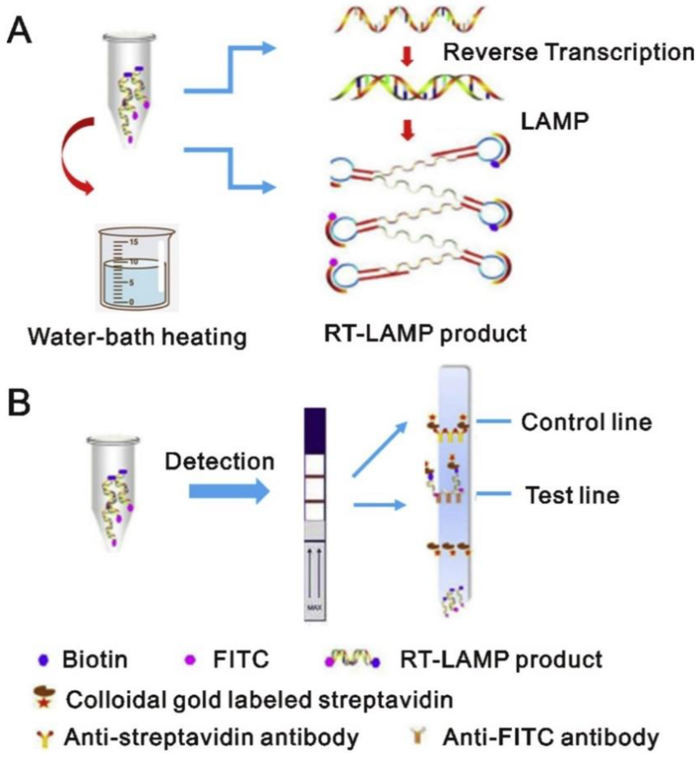
Loop-mediated isothermal amplification (LAMP) is an alternative to the cumbersome RT-PCR technique. It uses a single temperature, i.e., 60–65 °C, for amplification, which makes the technique much easier compared to RT-PCR and eliminates the need for a thermocycler, thus making it cheaper than RT-PCR. It can amplify the DNA in 25–35 min using polymerase with high DNA-strand replacement activity using 4–8 specific primers [46]. The high amount of DNA is amplified through this technique compared to conventional PCR. The LAMP reaction end products can be measured by either a fluorescent dye or by the turbidity of the sample, which relates directly to the viral content.Reverse transcription loop-mediated isothermal amplification, or RT-LAMP, uses reverse transcriptase to directly amplify RNA in a sample. Compared to RT-PCR and RT-LAMP, 76 nasopharyngeal samples demonstrated 97.6% sensitivity and 100% sensitivity, respectively [47]. According to recent reports, a novel single-tube real-time RT-LAMP assay has been developed with variable calorimetric versions with a limit of detection (LOD) of 119 copies per reaction [48]. Another robust modification of RT-LAMP has been developed, where samples from swabs can be directly used but with less sensitivity. Similarly, multiplex RT-LAMP combined with lateral flow biosensors has also been developed with high sensitivity and specificity [49]. LAMP can also be used for detection of SNPs for other diseases. Figure 1 illustrates the principle of amplification of SARS-CoV-2 and its detection [50].
2.1.2. Biosensors
Biosensors are the advanced and robust, cost-effective, portable, and simple technology designed for the detection of various biomolecules such as pathogens, proteins, glucose, etc [51]. Several studies have reported the manipulation of this technology detection of SARS-CoV-2 [52,53]. Anti-microbial peptides are produced in response to foreign evading bacterial or viral peptides in organisms. For analysis, instead of using a whole protein, their small representative peptides can be used and coated with biosensors to increasing their stability and avoid the degradation of proteins.
Field-Effect Transistors (FET)
Many research studies have developed novel biosensors for detecting SARS-CoV-2, such as field-effect transistors (FET). FET is a biosensor developed for SARS-CoV-2, the surface of which is graphene coated. It is then conjugated with the SARS-CoV-2 anti-spike antibody through a probe linker. FET can detect approximately 1–100 fg/mL of the spike protein of SARS-CoV-2 with 2.42 × 10² copies and LOD of 1.6 × 101 pfu/mL [54]. Another spike protein (S1) of SARS-CoV-2 is detected with a novel biosensor developed on a bioelectric recognition assay. The LOD of this biosensor is 1 fg/mL, and the detection time is only 3 min. Additionally, this portable biosensor can be controlled via smartphone or tablet. Figure 1 illustrates the working principle of FET biosensors [55].
Localized Surface Plasmon Resonance (LSPR) Sensor
LSPR is a technique for producing optical phenomena when light waves are confined in gold nanoparticles. A coherent localized plasmon oscillation is produced when incident light and the surface electrons in the conduction band interact with each other [56]. Various viral sequences such as E genes, ORF1ab COVID, and RdRp-COVID were detected via application ofLSPR and a plasmonic biosensor using photothermal effect. In this process, the converted plasmonic photothermal energy is used to provide stable heat energy for increasing hybridization of RdRp of COVID-19 with the target complementary DNA sequence. The slope graph obtained from the LSPR with photothermal effect was observed to be higher than the LSPR without the photothermal effect, and sensors without PPT showed a false-positive outcome. This sensor has a LOD of 0.2 pM and can differentiate between S-CoV-2 and S-CoV with a high precision rate of 96% [57].
Cell-Based Potentiometric Biosensor
This technique detects the SARS-CoV-2 S1 antigen through the SARS-CoV-2 Spike S1 antibody. By using electro-insertion technique, a membrane-engineered kidney cell was modified with the S1 antigen of SARS-CoV-2. A change in potential when the antibody interacts with the required antigen is detected. This biosensor is formulated on eight gold screen-printed electrodes. These printed electrodes are shielded by a polydimethylsiloxane (PDMS) layer with approximately eight wells. A potentiometric device is used to measure the signal after the suspension of membrane and protein solution is added to the wells. The detection limit of this device is 1 fg/mL [52].
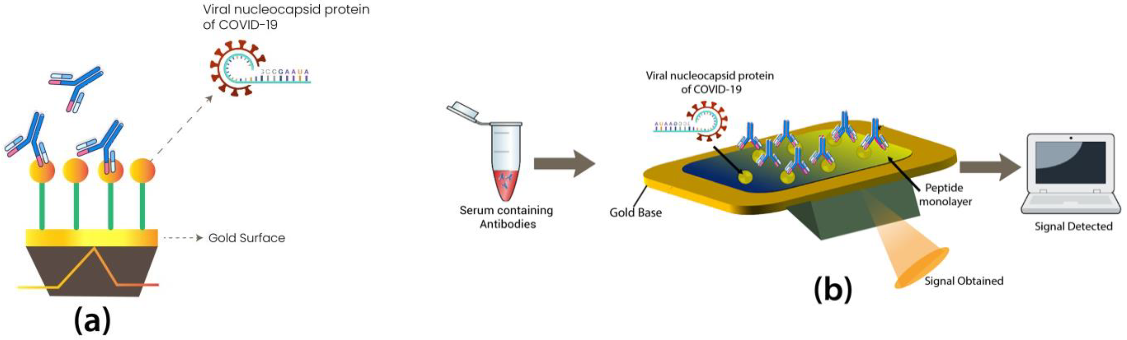
Another single-step detection field-deployable biosensor using saliva samples was developed based on plasmonic fiberoptic absorbance with LOD of 10−18 M [58]. Similarly, another biosensor that works on the principle of immunoassay and aptamer-based technology (Figure 2) has also been established, named fiber optic surface plasmon resonance (FO-SPR) biosensor. An SPR sensor was made by coating the sensor with a monolayer of recombinant N Ag to detect anti-SARS-CoV-2 Ab, which gave results in 15 min [55].
2.1.3. CRISPR-Based Diagnostics—SHERLOCK and DETECTR
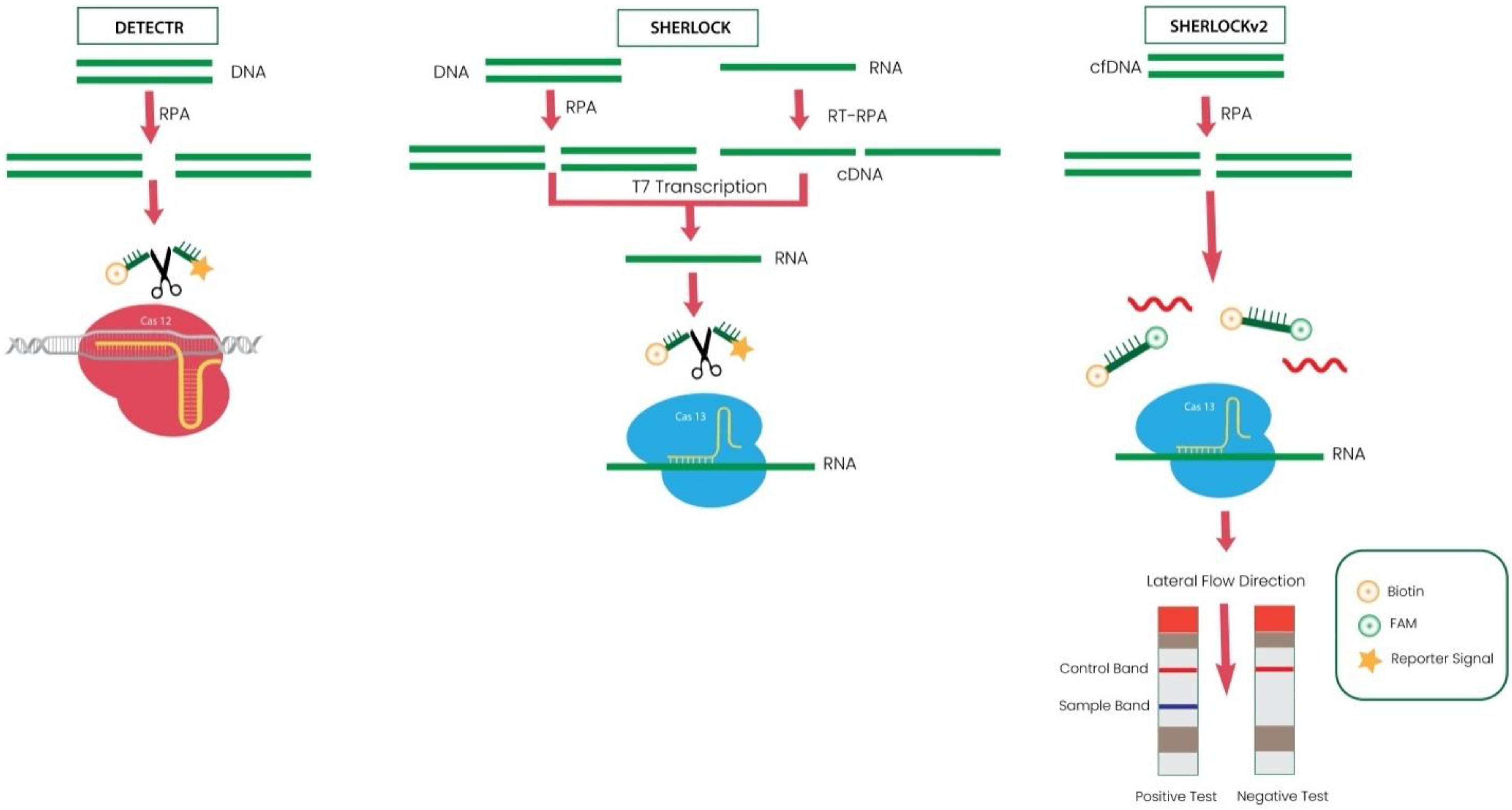
Clustered regularly interspaced short palindromic repeats (CRISPR) is the most significant innovation of the era. It has induced many advances in molecular diagnostics. Previously famous for its use in gene editing, CRISPR has revolutionized the field of diagnostics after the COVID-19 outbreak. CRISPR-Cas is a part of the natural immune system of microbes for protection against foreign material by recognizing them and eliminating them via CRISPR-associated endonuclease Cas enzymes [59]. The two CRISPR-based innovative kits launched recently for SARS-CoV-2 detection are SHERLOCK and DETECTR.
SARS-CoV-2 DETECTR
In the SARS-CoV-2 DETECTR experiment, the extracted RNA from the test samples is subjected to RT-PCR to increasethe copy number of RNase P, E, and N genes. CRISPR-Cas12 detects the copies of genome sequences and illuminates with a fluorescence signal after cleavage of the reporter dye. This novel detection kit combines RT-LAMP, CRISPR-Cas, and Lateral Flow Assay (LFA) in one process. This CRISPR-based biosensor is developed by Mammoth Biosciences Inc. and Abbott Viral Diagnostics [60]. This technique showed 90% sensitivity and 100% specificity. The specificity of this technique is so high that it can detect the difference among SARS-CoV, SARS-CoV-2, and MERS-CoV differing with a single nucleotide sequence only due to highly specific primers and probes used. Similarly, AIOD-CRISPR and FELUDA are other modified versions of techniques using CRISPR in diagnosis [61,62].
Specific High Sensitivity Enzymatic Reporter UnLOCKing (SHERLOCK)
The SHERLOCK system is based on the principle of the CRISPR-Cas VI system. SHERLOCK uses the Cas13 endonuclease activity from Leptotrichiawadei [63]. Recombinase polymerase amplification (RPA) is used to amplify target molecules Cas13 crRNA isothermally, and fluorescent RNA probes are mixed with amplified products [64]. If the amplified RNA products contain desired RNA, then Cas13 recognizes the desired RNA with the help of crRNA and cleaves the interaction between fluorophore and quencher. The cleavage of fluorescent signal results in illumination, and intensity of light directly depends on the quantity of the amplified sample. This technique is used to detect SNPs, Zika virus, pathogenic bacteria, and dengue virus [64]. Since the first SHERLOCK system was qualitative, the researchers developed SHERLOCKv2, a modified version. SHERLOCKv2 is 3.5-fold higher in sensitivity than the previous version, due to the combination of Cas13a with Csm6. The Csm6 endonuclease that supports the CRISPR type III can join Cas13a with the reporter signal, which enhances the signal quantity with diluted isothermal primers. Another advancement in SHERLOCKv2 is the development of commercial lateral flow strips to visualize colorimetric read-out. This version is fast, robust, sensitiveand superior to the previous one because it is a single-step assay in which unpurified samples can be directly applied to the strip without the need for purification and isolation [65]. Figure 3 illustrates the difference among DETECTR, SHERLOCK, and SHERLOCKv2.
2.1.4. Aptamer-Based Diagnostics
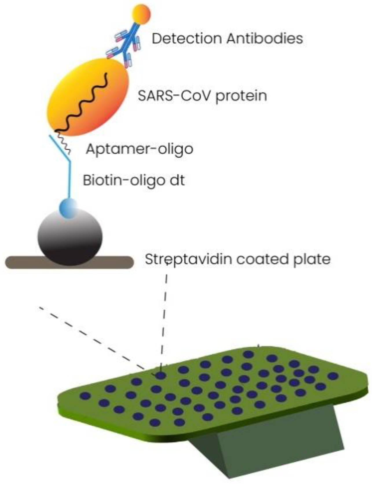
Aptamers are artificially made small oligonucleotide or peptide sequences that target specific DNA or RNA of interest. Aptamer-based detection is an emerging technique to target viral infections [66,67]. Synthetic aptamers have been made to bind the SARS-CoV-2 receptor binding domain (RBD) specifically with high affinity using the human angiotensin-converting enzyme 2 (ACE2) competition-based approach. The small size of this biomolecule makes it a suitable candidate for a stable target in diagnostic techniques. The inhibitory potential of this small anti-RBD aptamer can be used in therapeutical treatments and diagnosis. Sensitive splint-based one-pot isothermal RNA detection (SENSR) is an RNA aptamer-based rapid detection approach. This technique works on the principle of ligation via SplintR ligase and T7 RNA Pol. The target RNA is amplified, and a fluorescent signal is detected. This single-step technique can detect a variety of pathogens with a detection limit of 0.1 aM [68,69]. Figure 4 illustrates the working of aptamer-based technology for detection of SARS-CoV-2. Figure 4 illustrates the working of aptamer based technology for detection of SARS CoV-2 [70].
2.1.5. Molecular Imprinting Technology (MIT)-Based Diagnosis
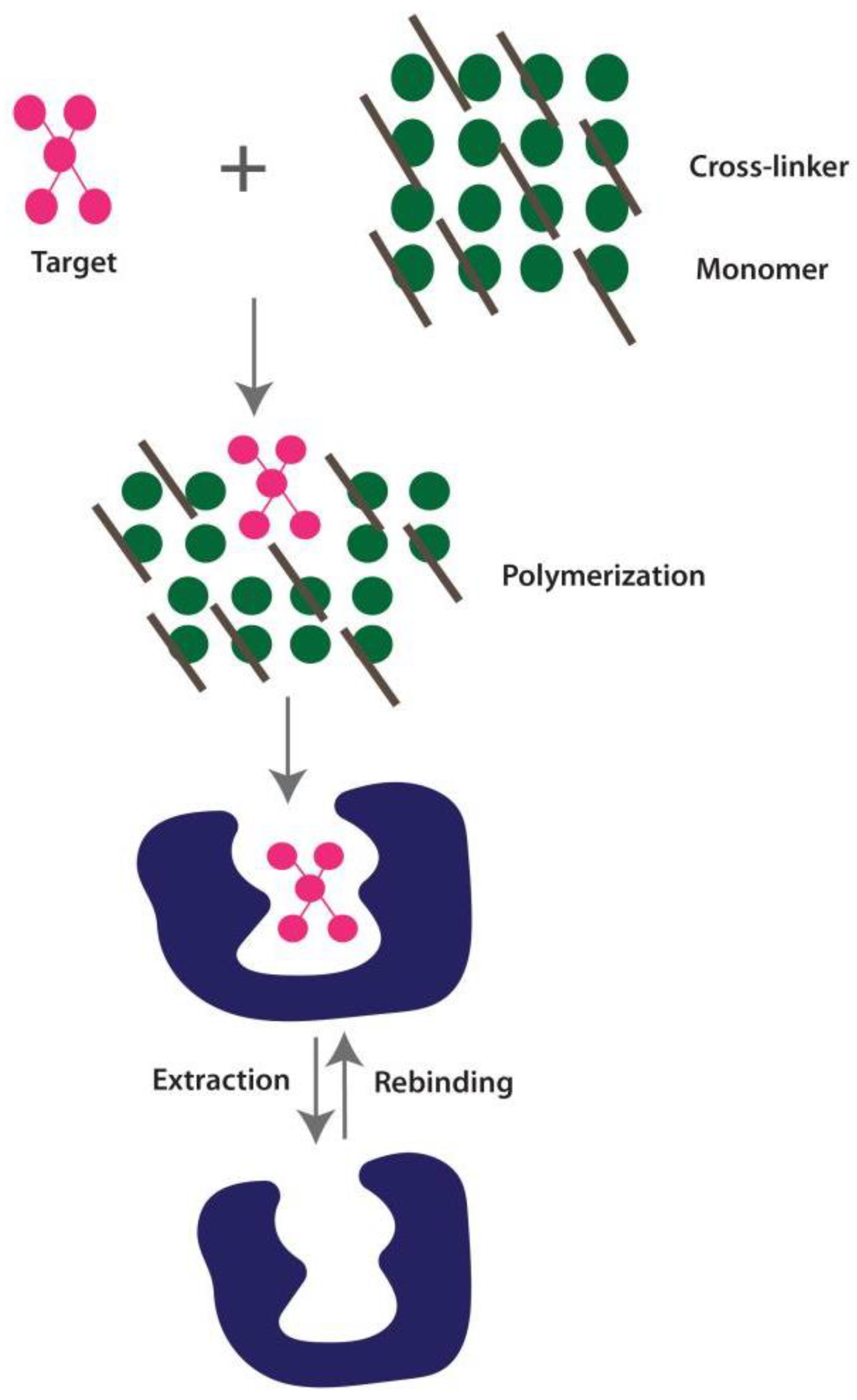
MIT is a diagnostic technique that works in the same way as the “Lock and Key Model” of enzyme and substrate reaction and binds to the most predictable structure with high specificity and affinity. MIPs are synthetically designed receptors for binding the complimentary target molecular of specific shape and orientation on a polymer [71]. The creation of MIPs with molecular recognition cavities that have specific selectivity for the template molecules is the basic principle. The formation of MIP involves the process of polumerization of a monomer and a cross-linker, both of which surround the target molecule. Covalent and non-covalent interactions promote this assembly of a monomer around a target molecule, as illustrated in Figure 5. A non-covalently produced MIP–target molecule complex is simpler to extract the target molecule from than a covalently formed MIP–target molecule complex [72].
MIPs have become widely employed in recent years for detection reasons, particularly for viral contamination. Ref. [73], recently reported the first work relating to the MIP-based detection of COVID-19. Utilizing a molecular imprinting approach, they created an electrochemical sensor for the detection of SARS-CoV-2 nucleoprotein. They showed that the nucleoprotein contained in nasopharyngeal swab samples from COVID-19-positive individuals could be detected by the MIP-based sensor. Their encouraging results support the need to create an effective, quick, and affordable MIP-based diagnostic tool for the identification of COVID-19 [73]. A rapid POC detection kit combined with SARS-CoV-2 specific aptamer and MIP sensor was proposed to detect the required target with greater affinity. Similarly, a MIP-based monoclonal antibody has been developed to bind SARS-CoV-2 selectively [74,75].
2.1.6. Microarray-Based Diagnosis
Microarrays are multifunctional tools that are used in retrospective research on SARS-CoV-2. This tool can study antigen–antibody interactions, pathogenic behavior, cross reactivity between specific species and target proteins, and immunogenic responses to diseases [76,77,78]. In this high-throughput tool, the RNA of SARS-Cov-2 is converted into cDNA using RT enzymes, and the probes are conjugated with it. A microarray plate is then used to detect the hybridization of labeled cDNA with fixed oligonucleotides [79]. Researchers proposed that a SARS-CoV Ab response can be calculated using commercial antisera against SARS-CoV-2 proteins. A comparative study was performed among different respiratory viruses, and microarray tools can be used in determining antigen selection for diagnosis, vaccine development, and differences in variable pathogen-specific Abs (Figure 6) [80]. The advanced diagnostics of SARS-CoV-19 in development are represented in Table 2.
2.2. Development of New Kits for SARS-CoV-19 Detection
With the emergence of COVID-19 many advances in new techniques have been made as explained above. Similarly, many new kits have been approved by FDA for detection of SARS-CoV-19. The purpose of these kits is to provide rapid and robust testing with minimal time. In August 2021, a list of FDA approved many categories of kits varying on the principle such as RT-PCR kit, antigen detection kit, and antibody detection kit for detection of COVID-19 virus as shown in Table 3 [82].
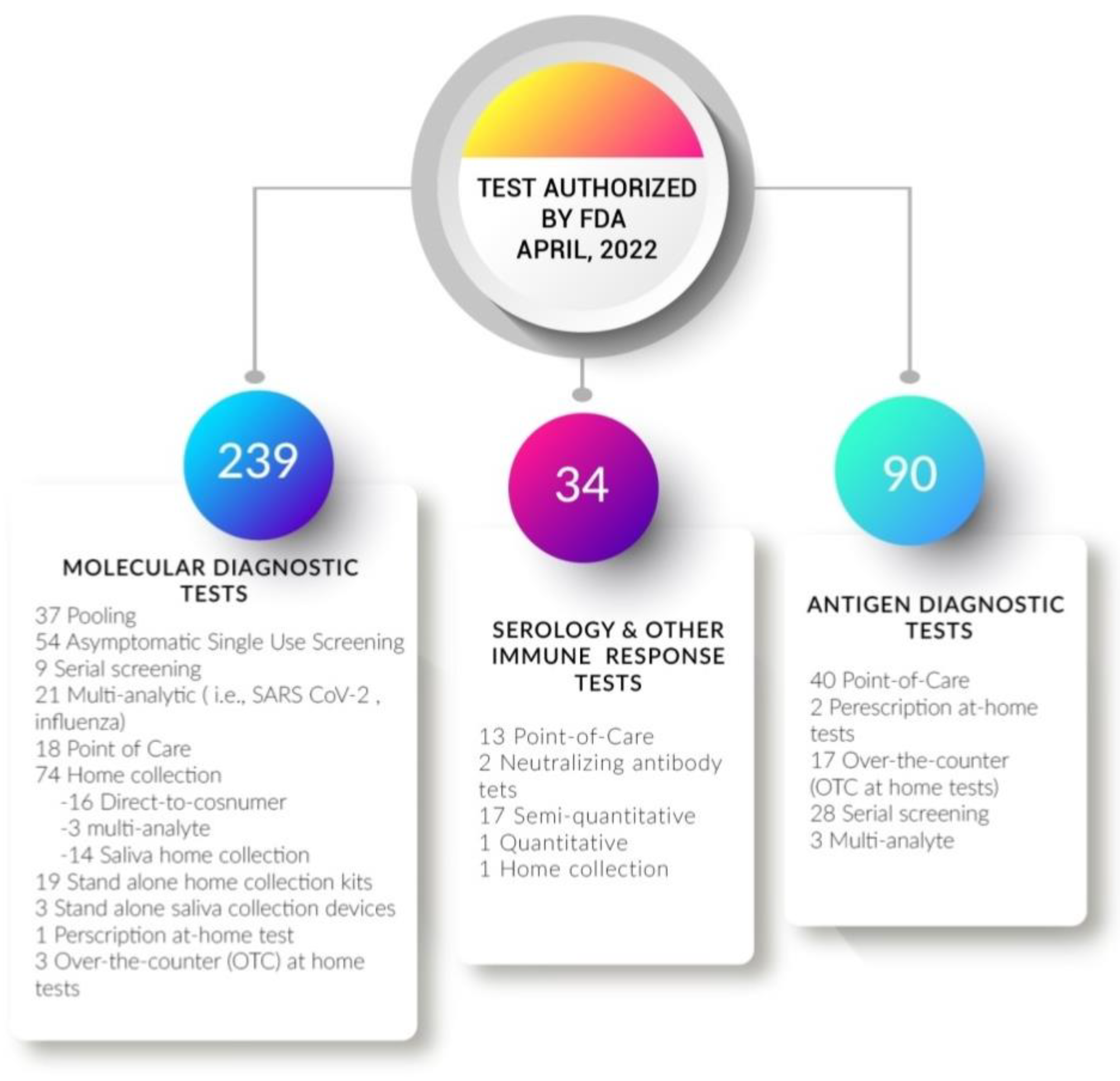
In the history of molecular diagnosis, the SARS-CoV-2 outbreak significantly impacted the transformation and development of molecular diagnostic techniques. This pandemic enhanced the speed of research for survival. Many new technologies have been made, and more inventions are in the development pipeline. According to FDA reports of April 2022, 294 advancements in molecular tests, 90 in antigen depending tests, and 34 serological tests have been approved [83].
2.3. Point-of-Care Diagnostics
Point-of-care (POC) tests are the medical testing performed near the point of care where the point of care is the patient. POC are portable, cost-effective devices, with reduced sample processing eliminating the need to transport samples to the laboratory. POC does not require any trained professional to collect samples and can measure symptomatic and asymptomatic patients. Protecting the community from spreading any viral infection can be controlled only when detected early. POC offers the rapid detection of viral presence or host antibody response without laboratory settings needed.It can perform thousands of public tests in a single day. POC analysis can be performed at home, in offices, in mobile vans, in healthcare centers, and in emergency rooms [84]. For SARS-CoV-19, the RNA, antigen of the virus, and antibody produced in response to the virus by humans are detected using POC devices.
2.3.1. Molecular Detection-Based Point-Of-Care Devices
The Isothermal amplification process is gaining much attention because it does not require the cycle at different temperatures and viral purification, thus making kits more rapid and easy to use. Many RT-PCR- and RT-LAMP-based POC devices have been developed since the emergence of COVID-19. Abbott Diagnostics Scarborough,Inc., developed the first RT-LAMP-based kit approved by EUA authorization, named the ID NOW COVID-19 test [83]. Similarly, another device, the Cue COVID-19 Test (EUA approved), works on the principle of isothermal amplification [85,86]. This device uses an electrochemical detection method and amplifies the RNA from direct nasal swabs in 20 min in a single step. Its LOD is 20 genomic copies per sample. Cue Health modified the kit for use at home on 5 March 2021. It became the first approved kit for use at home without prescription [87]. Cue COVID-19 Test (EUA approved) is another single-step rapid and robust battery-powered device that works on the RT-LAMP principle in 30 min using nasal swabs [88]. The list of some FDA-approved POC devices working on RT-PCR and RT-LAMP principles is shown in Table 4.
2.3.2. Antigen Detection-Based Point-of-Care Devices
In antigen detection tests, specific antibodies against SARS-CoV-19 structural proteins are used. The antigen approach is faster and more rapid than PCR tests but has the drawback of being less sensitive because there is no amplification [89]. Thus, antigen-based tests are qualitative rather than quantitative because they cannot measure the viral load. It mainly targets the N-proteinsand S-proteins ofthe SARS-CoV-19 virus [90]. Ellume COVID-19 Home Test is the first antigen-based POC device authorized by EUA, in Dec 2020, for home use without the need for a prescription. Similarly, BinaxNOW COVID-19 Ag Card Home Test is another POC device approved for home use [91]. The list of some EUA-approved POC devices working on the antigen detection principle is shown in Table 5.
2.3.3. Antibody Detection-Based Point-of Care Diagnostics
Antibodies produced bythe immune system in response to SARS-CoV-19 proteins can be detectedby using developed devices. They detect IgM and IgG antibodies from blood, serum, and plasma within 10–20 min. The list of some EUA-approved POC devices working on the antibody detection principle is shown in Table 6 [90].
3. Future of Molecular Techniques
Starting from the history to present, molecular diagnosis has revolutionized to a great extent with high-throughput technological advancements. It has become very convenient to study the entire genome with high accuracy and decreasing cost. Presently, many kits and tests are being developed which provide rapid detection as compared to the past, making molecular diagnosis indispensable in the clinical laboratory. As technology is overgrowing rapidly with time, the future of molecular diagnostics seems to be very promising. The wave of the corona accelerated and modernized the field of molecular diagnosis miraculously and analyzing the rapid growth in the pandemic assures us of a bright future ahead. The POC devices and biosensors can be potent candidates for molecular diagnosis because of their robustness. As the pandemics are likely to reoccur, investing efforts in molecular diagnosis seems highly imperative. As a result, the molecular diagnosis will be a central part of medical practice in the upcoming era. Whole genome sequencing will be the gold standard for identifying DNA variations associated with genetic diseases. Nevertheless, there is still a lot to discover before applying these techniques, such as cost-effectiveness, availability of resources, management of equipment, personnel training, and reproducibility. Along with proteomic-based testing, these developments will make further improvements in molecular diagnostics for implementation in hospitals, clinics, and healthcare departments, in public or private sectors.
4. Conclusions
The growth in molecular diagnostic labs before and after the emergence of coronavirus is notable. After the pandemic outbreak, the urge to develop fast and robust techniques exponentially increased. Many techniques have been modified to detect a massive community of coronavirus at home and offices. COVID-19 has boosted R&D struggles to launch innovative, more robust, rapid, precise and economical testing approaches than previously used tools. Currently, RT-LAMP, microarray-based detection, aptamer-based diagnosis, SHERLOCK, SHERLOCKv2, FET Biosensors, cell-based potentiometric diagnosis, molecular imprinting technology, etc. are used for diagnosis. Point-of-care (POC) diagnosis has rapidly increased to screen populations outside the hospitals. These POC devices are robust, portable, and can detect thousands of samples simultaneously. In previous times, the viral epidemic of SARS-CoV and MERS-CoV have led to the advancement in rapid diagnostics. For instance, the genome sequence identification of SARS-CoV-2 lead us toward developing nucleic acid-based diagnostic approaches, which assisted in controlling the outburst of COVID-19. The antibody-based and antigen-based diagnostic approaches helped in further comprehension. There is a lack of detailed research in the development of peptide- and aptamer-based biosensors, which could provide us more sensitivity and specificity in COVID-19 diagnosis, but the era of improvement has started, and yet, there is much to come. According to FDA reports, advancements in molecular tests, antigen-depending tests, and serological tests have been approved since the pandemic outbreak. Thus, the emergence has transformed the scientific community much more than any other life event. The scientific community’s collaboration to overcome the pandemic and break the chain of losing lives has increased the significance of molecular tools and molecular diagnosis.
Author Contributions
Conceptualization, A.M.A. and N.U.K.;writing—original draft preparation, A.M.A. and N.U.K.; writing—review and editing A.M.A., N.U.K., and F.A.A.; supervision, A.M.A.; funding acquisition, A.M.A. All authors have read and agreed to the published version of the manuscript.
Funding
Supported by Deanship of Scientific Research, King Khalid University, project number (G.R.P. 43).
Institutional Review Board Statement
Not applicable.
Informed Consent Statement
Not applicable.
Data Availability Statement
Not applicable.
Acknowledgments
The author appreciates the support of the Deanship of Scientific Research at King Khalid University Abha, Saudi Arabia through a project number (G.R.P. 43).
Conflicts of Interest
The authors declare no conflict of interest.
Abbreviations
| NGS | Next Generation Sequencing |
| PCR | Polymerase Chain Reaction |
| HSV | Herpes simplex virus |
| FISH | Fluorescence In Situ Hybridization |
| ZMWs | Zero-mode wave guides |
| SMRT | Single-molecule real-time sequencing |
| cPAS | Combinatorial Probe-Anchor Synthesis |
| RCR | Rolling Circular Replication |
| ELISA | Enzyme-linked Immunosorbent Assay |
| NDV | New Castle Disease Virus |
| RT-LAMP | Reverse Transcription Loop-Mediated Isothermal Amplification |
| LAMP | Loop-Mediated Isothermal Amplification |
| LOD | limit of detection |
| FET | Field-Effect Transistors |
| LSPR | Localized Surface Plasmon resonance |
| PDMS | polydimethylsiloxane |
| FO-SPR | Fiber Optic Surface Plasmon Resonance |
| CRISPR | Clustered Regularly Interspaced Short Palindromic Repeats |
| RPA | Recombinase Polymerase Amplification |
| RBD | Receptor binding domain |
| SENSOR | sensitive splint-based one-pot isothermal RNA detection |
| ACE2 | Angiotensin-converting enzyme 2 |
| MIT | Molecular imprinting technology |
| POC | Point-of-care |
References
- Coleman, W.B.; Tsongalis, G.J. Molecular Diagnostics: For the Clinical Laboratorian; Springer: Berlin/Heidelberg, Germany, 2006. [Google Scholar]
- Durmaz, A.A.; Karaca, E.; Demkow, U.; Toruner, G.; Schoumans, J.; Cogulu, O. Evolution of genetic techniques: Past, present, and beyond. BioMed Res. Int. 2015, 2015, 461524. [Google Scholar] [CrossRef] [PubMed]
- Jack, J. A pedagogy of sight: Microscopic vision in Robert Hooke’s Micrographia. Q. J. Speech 2009, 95, 192–209. [Google Scholar] [CrossRef]
- Vinkšel, M.; Writzl, K.; Maver, A.; Peterlin, B. Improving diagnostics of rare genetic diseases with NGS approaches. J. Community Genet. 2021, 12, 247–256. [Google Scholar] [CrossRef] [PubMed]
- Pacenza, N.; Pasqualini, T.; Gottlieb, S.; Knoblovits, P.; Costanzo, P.R.; Stewart Usher, J.; Rey, R.A.; Martínez, M.P.; Aszpis, S. Clinical presentation of Klinefelter’s syndrome: Differences according to age. Int. J. Endocrinol. 2012, 2012, 324835. [Google Scholar] [CrossRef]
- Bartlett, J.; Stirling, D. A short history of the polymerase chain reaction. In PCR Protocols; Humana Press: Totowa, NJ, USA, 2003; pp. 3–6. [Google Scholar]
- Fakruddin, M.; Chowdhury, A. Pyrosequencing an alternative to traditional Sanger sequencing. Am. J. Biochem. Biotechnol. 2012, 8, 14–20. [Google Scholar] [CrossRef]
- Singh, J.; Birbian, N.; Sinha, S.; Goswami, A. A critical review on PCR, its types and applications. Int. J. Adv. Res. Biol. Sci. 2014, 1, 65–80. [Google Scholar]
- Rolfs, A.; Schuller, I.; Finckh, U.; Weber-Rolfs, I. PCR: Clinical Diagnostics and Research; Springer: Berlin/Heidelberg, Germany, 1992. [Google Scholar]
- Liu, L.; Hu, N.; Wang, B.; Chen, M.; Wang, J.; Tian, Z.; He, Y.; Lin, D. A brief utilization report on the Illumina HiSeq 2000 sequencer. Mycology 2011, 2, 169–191. [Google Scholar]
- Lee, N.; Nielsen, P.H.; Andreasen, K.H.; Juretschko, S.; Nielsen, J.L.; Schleifer, K.-H.; Wagner, M. Combination of fluorescent in situ hybridization and microautoradiography—A new tool for structure-function analyses in microbial ecology. Appl. Environ. Microbiol. 1999, 65, 1289–1297. [Google Scholar] [CrossRef] [PubMed] [Green Version]
- Kulski, J.K. Next-generation sequencing—An overview of the history, tools, and “Omic” applications. In Next Generation Sequencing-Advances, Applications and Challenges; BoD—Books on Demand: Norderstedt, Germany, 2016; Volume 10, p. 61964. [Google Scholar]
- Park, J.; Shin, S.Y.; Kim, K.; Park, K.; Shin, S.; Ihm, C. Determining genotypic drug resistance by Ion semiconductor sequencing with the Ion AmpliSeq™ TB panel in multidrug-resistant Mycobacterium tuberculosis isolates. Ann. Lab. Med. 2018, 38, 316–323. [Google Scholar] [CrossRef] [PubMed] [Green Version]
- Ardui, S.; Ameur, A.; Vermeesch, J.R.; Hestand, M.S. Single molecule real-time (SMRT) sequencing comes of age: Applications and utilities for medical diagnostics. Nucleic Acids Res. 2018, 46, 2159–2168. [Google Scholar] [CrossRef] [Green Version]
- Jeck, W.R.; Reinhardt, J.A.; Baltrus, D.A.; Hickenbotham, M.T.; Magrini, V.; Mardis, E.R.; Dangl, J.L.; Jones, C.D. Extending assembly of short DNA sequences to handle error. Bioinformatics 2007, 23, 2942–2944. [Google Scholar] [CrossRef] [PubMed] [Green Version]
- Lin, B.; Wang, J.; Cheng, Y. Recent patents and advances in the next-generation sequencing technologies. Recent Pat. Biomed. Eng. 2008, 1, 60–67. [Google Scholar] [CrossRef] [PubMed]
- El-Metwally, S.; Ouda, O.M.; Helmy, M. First-and next-generations sequencing methods. In Next Generation Sequencing Technologies and Challenges in Sequence Assembly; Springer: Berlin/Heidelberg, Germany, 2014; pp. 29–36. [Google Scholar]
- Van den Oever, J.M.; Balkassmi, S.; Verweij, E.J.; van Iterson, M.; van Scheltema, P.N.A.; Oepkes, D.; van Lith, J.M.; Hoffer, M.J.; den Dunnen, J.T.; Bakker, E. Single molecule sequencing of free DNA from maternal plasma for noninvasive trisomy 21 detection. Clin. Chem. 2012, 58, 699–706. [Google Scholar] [CrossRef] [PubMed] [Green Version]
- Chehab, F.F. Molecular diagnostics: Past, present, and future. Hum. Mutat. 1993, 2, 331–337. [Google Scholar] [CrossRef] [PubMed]
- Demidov, V.V. DNA Diagnostics in the Fifty-Year Retrospect; Taylor & Francis: Abingdon, UK, 2003; pp. 121–124. [Google Scholar]
- Kresge, N.; Simoni, R.D.; Hill, R.L. Arthur Kornberg’s discovery of DNA polymerase I. J. Biol. Chem. 2005, 280, e46–e48. [Google Scholar] [CrossRef]
- Jensen, E. Technical review: In situ hybridization. Anat. Rec. 2014, 297, 1349–1353. [Google Scholar] [CrossRef]
- Di Felice, F.; Micheli, G.; Camilloni, G. Restriction enzymes and their use in molecular biology: An overview. J. Biosci. 2019, 44, 1–8. [Google Scholar] [CrossRef]
- Edberg, S. Principles of nucleic acid hybridization and comparison with monoclonal antibody technology for the diagnosis of infectious diseases. Yale J. Biol. Med. 1985, 58, 425. [Google Scholar]
- Sanger, F.; Nicklen, S.; Coulson, A.R. DNA sequencing with chain-terminating inhibitors. Proc. Natl. Acad. Sci. USA 1977, 74, 5463–5467. [Google Scholar] [CrossRef] [Green Version]
- Maxam, A.M.; Gilbert, W. [57] Sequencing end-labeled DNA with base-specific chemical cleavages. In Methods in Enzymology; Elsevier: Amsterdam, The Netherlands, 1980; pp. 499–560. [Google Scholar]
- Patrinos, G.P.; Ansorge, W.J. Molecular diagnostics: Past, present, and future. In Molecular Diagnostics; Elsevier: Amsterdam, The Netherlands, 2010; pp. 1–11. [Google Scholar]
- Saiki, R.K.; Scharf, S.; Faloona, F.; Mullis, K.B.; Horn, G.T.; Erlich, H.A.; Arnheim, N. Enzymatic amplification of β-globin genomic sequences and restriction site analysis for diagnosis of sickle cell anemia. Science 1985, 230, 1350–1354. [Google Scholar] [CrossRef]
- Mackay, I.M.; Arden, K.E.; Nitsche, A. Real-time PCR in virology. Nucleic Acids Res. 2002, 30, 1292–1305. [Google Scholar] [CrossRef] [PubMed] [Green Version]
- Elahi, E.; Ronaghi, M. Pyrosequencing. Bacterial Artificial Chromosomes; Springer: Berlin/Heidelberg, Germany, 2004; pp. 211–219. [Google Scholar]
- Psychesystems The History and Evolution of Molecular Diagnostics. 2020. Available online: https://psychesystems.com (accessed on 20 June 2022).
- Stranneheim, H.; Lundeberg, J. Stepping stones in DNA sequencing. Biotechnol. J. 2012, 7, 1063–1073. [Google Scholar] [CrossRef] [PubMed] [Green Version]
- Balzer, S.; Malde, K.; Lanzén, A.; Sharma, A.; Jonassen, I. Characteristics of 454 pyrosequencing data—Enabling realistic simulation with flowsim. Bioinformatics 2010, 26, i420–i425. [Google Scholar] [CrossRef] [PubMed]
- Sage, L. Faster, Cheaper DNA Sequencing; ACS Publications: Washington, DC, USA, 2005. [Google Scholar]
- Rasmussen, A.L. Probing the viromic frontiers. mBio 2015, 6, e01767-15. [Google Scholar] [CrossRef] [PubMed] [Green Version]
- Wong, K.-C.; Zhang, J.; Yan, S.; Li, X.; Lin, Q.; Kwong, S.; Liang, C. DNA sequencing technologies: Sequencing data protocols and bioinformatics tools. ACM Comput. Surv. (CSUR) 2019, 52, 1–30. [Google Scholar] [CrossRef]
- Chi, K.R. The year of sequencing: In 2007, the next-generation sequencing technologies have come into their own with an impressive array of successful applications. Nat. Methods 2008, 5, 11–15. [Google Scholar] [CrossRef]
- Liu, L.; Li, Y.; Li, S.; Hu, N.; He, Y.; Pong, R.; Lin, D.; Lu, L.; Law, M. Comparison of next-generation sequencing systems. J Biomed. Biotechnol. 2012, 2012, 251364. [Google Scholar] [CrossRef]
- Wang, S.; Zhang, S.; Wang, W.; Xiong, X.; Meng, F.; Cui, X. Efficient targeted mutagenesis in potato by the CRISPR/Cas9 system. Plant Cell Rep. 2015, 34, 1473–1476. [Google Scholar] [CrossRef] [Green Version]
- Jain, M.; Olsen, H.E.; Paten, B.; Akeson, M. The Oxford Nanopore MinION: Delivery of nanopore sequencing to the genomics community. Genome Biol. 2016, 17, 1–11. [Google Scholar]
- Choe, J.; Kim, J.; Han, H. Simple Molecular Diagnostic Tools in Clinical Medicine. J. Clin. Diagn. Res. 2017, 6, 141. [Google Scholar]
- Witt, M.; Walter, J.-G.; Stahl, F. Aptamer microarrays—Current status and future prospects. Microarrays 2015, 4, 115–132. [Google Scholar] [CrossRef] [PubMed] [Green Version]
- Spengler, B. Mass spectrometry imaging of biomolecular information. Anal. Chem. 2015, 87, 64–82. [Google Scholar] [CrossRef] [PubMed]
- Aminian, A.; Safari, S.; Razeghian-Jahromi, A.; Ghorbani, M.; Delaney, C.P. COVID-19 outbreak and surgical practice: Unexpected fatality in perioperative period. Ann. Surg. 2020, 272, e27–e29. [Google Scholar] [CrossRef] [PubMed]
- Nolan, T.; Hands, R.E.; Bustin, S.A. Quantification of mRNA using real-time RT-PCR. Nat. Protoc. 2006, 1, 1559–1582. [Google Scholar] [CrossRef]
- Carpenter, M.A.; Geletkanycz, M.A.; Sanders, W.G. Upper echelons research revisited: Antecedents, elements, and consequences of top management team composition. J. Manag. 2004, 30, 749–778. [Google Scholar] [CrossRef]
- Kitagawa, Y.; Orihara, Y.; Kawamura, R.; Imai, K.; Sakai, J.; Tarumoto, N.; Matsuoka, M.; Takeuchi, S.; Maesaki, S.; Maeda, T. Evaluation of rapid diagnosis of novel coronavirus disease (COVID-19) using loop-mediated isothermal amplification. J. Clin. Virol. 2020, 129, 104446. [Google Scholar] [CrossRef]
- Lu, R.; Wu, X.; Wan, Z.; Li, Y.; Jin, X.; Zhang, C. A novel reverse transcription loop-mediated isothermal amplification method for rapid detection of SARS-CoV-2. Int. J. Mol. Sci. 2020, 21, 2826. [Google Scholar] [CrossRef] [Green Version]
- Zhu, X.; Wang, X.; Han, L.; Chen, T.; Wang, L.; Li, H.; Li, S.; He, L.; Fu, X.; Chen, S. Multiplex reverse transcription loop-mediated isothermal amplification combined with nanoparticle-based lateral flow biosensor for the diagnosis of COVID-19. Biosens. Bioelectron. 2020, 166, 112437. [Google Scholar] [CrossRef]
- Shen, M.; Zhou, Y.; Ye, J.; Al-Maskri, A.A.A.; Kang, Y.; Zeng, S.; Cai, S. Recent advances and perspectives of nucleic acid detection for coronavirus. J. Pharm. Anal. 2020, 10, 97–101. [Google Scholar] [CrossRef]
- Hoyos-Nogués, M.; Gil, F.; Mas-Moruno, C. Antimicrobial peptides: Powerful biorecognition elements to detect bacteria in biosensing technologies. Molecules 2018, 23, 1683. [Google Scholar] [CrossRef] [Green Version]
- Mavrikou, S.; Moschopoulou, G.; Tsekouras, V.; Kintzios, S. Development of a portable, ultra-rapid and ultra-sensitive cell-based biosensor for the direct detection of the SARS-CoV-2 S1 spike protein antigen. Sensors 2020, 20, 3121. [Google Scholar] [CrossRef] [PubMed]
- Oishee, M.J.; Ali, T.; Jahan, N.; Khandker, S.S.; Haq, M.A.; Khondoker, M.U.; Sil, B.K.; Lugova, H.; Krishnapillai, A.; Abubakar, A.R. COVID-19 pandemic: Review of contemporary and forthcoming detection tools. Infect. Drug Resist. 2021, 14, 1049. [Google Scholar] [CrossRef] [PubMed]
- Seo, G.; Lee, G.; Kim, M.J.; Baek, S.-H.; Choi, M.; Ku, K.B.; Lee, C.-S.; Jun, S.; Park, D.; Kim, H.G. Rapid detection of COVID-19 causative virus (SARS-CoV-2) in human nasopharyngeal swab specimens using field-effect transistor-based biosensor. ACS Nano 2020, 14, 5135–5142. [Google Scholar] [CrossRef] [PubMed] [Green Version]
- Roberts, A.; Chouhan, R.S.; Shahdeo, D.; Shrikrishna, N.S.; Kesarwani, V.; Horvat, M.; Gandhi, S. A Recent Update on Advanced Molecular Diagnostic Techniques for COVID-19 Pandemic: An Overview. Front. Immunol. 2021, 12, 732756. [Google Scholar] [CrossRef]
- Petryayeva, E.; Krull, U.J. Localized surface plasmon resonance: Nanostructures, bioassays and biosensing—A review. Anal. Chim. Acta 2011, 706, 8–24. [Google Scholar] [CrossRef]
- Sheikhzadeh, E.; Eissa, S.; Ismail, A.; Zourob, M. Diagnostic techniques for COVID-19 and new developments. Talanta 2020, 220, 121392. [Google Scholar] [CrossRef]
- Murugan, D.; Bhatia, H.; Sai, V.; Satija, J. P-FAB: A fiber-optic biosensor device for rapid detection of COVID-19. Trans. Indian Natl. Acad. Eng. 2020, 5, 211–215. [Google Scholar] [CrossRef]
- Uygun, Z.O.; Yeniay, L.; Sağın, F.G. CRISPR-dCas9 powered impedimetric biosensor for label-free detection of circulating tumor DNAs. Anal. Chim. Acta 2020, 1121, 35–41. [Google Scholar] [CrossRef]
- Broughton, J.; Deng, X.; Yu, G.; Fasching, C.; Servellita, V.; Singh, J.; Zorn, K. CRISPR–Cas12-based detection of SARS-CoV-2. Nat. Biotechnol. 2020, 38, 870–874. [Google Scholar] [CrossRef] [Green Version]
- Ding, X.; Yin, K.; Li, Z.; Liu, C. All-in-One dual CRISPR-cas12a (AIOD-CRISPR) assay: A case for rapid, ultrasensitive and visual detection of novel coronavirus SARS-CoV-2 and HIV virus. bioRxiv 2020. [Google Scholar] [CrossRef] [Green Version]
- Azhar, M.; Phutela, R.; Ansari, A.H.; Sinha, D.; Sharma, N.; Kumar, M.; Aich, M.; Sharma, S.; Rauthan, R.; Singhal, K.; et al. Rapid, field-deployable nucleobase detection and identification using FnCas9. bioRxiv 2020. [Google Scholar] [CrossRef]
- Gootenberg, J.S.; Abudayyeh, O.O.; Lee, J.W.; Essletzbichler, P.; Dy, A.J.; Joung, J.; Verdine, V.; Donghia, N.; Daringer, N.M.; Freije, C.A. Nucleic acid detection with CRISPR-Cas13a/C2c2. Science 2017, 356, 438–442. [Google Scholar] [CrossRef] [PubMed] [Green Version]
- Kellner, M.J.; Koob, J.G.; Gootenberg, J.S.; Abudayyeh, O.O.; Zhang, F. SHERLOCK: Nucleic acid detection with CRISPR nucleases. Nat. Protoc. 2019, 14, 2986–3012. [Google Scholar] [CrossRef] [PubMed]
- Mustafa, M.I.; Makhawi, A.M. SHERLOCK and DETECTR: CRISPR-Cas systems as potential rapid diagnostic tools for emerging infectious diseases. J. Clin. Microbiol. 2021, 59, e00745-20. [Google Scholar] [CrossRef] [PubMed]
- Zou, X.; Wu, J.; Gu, J.; Shen, L.; Mao, L. Application of aptamers in virus detection and antiviral therapy. Front. Microbiol. 2019, 10, 1462. [Google Scholar] [CrossRef] [PubMed]
- Vickers, N.J. Animal communication: When I’m calling you, will you answer too? Curr. Biol. 2017, 27, R713–R715. [Google Scholar] [CrossRef] [PubMed]
- Chen, Z.; Wu, Q.; Chen, J.; Ni, X.; Dai, J. A DNA aptamer based method for detection of SARS-CoV-2 nucleocapsid protein. Virol. Sin. 2020, 35, 351–354. [Google Scholar] [CrossRef] [PubMed]
- Woo, C.H.; Jang, S.; Shin, G.; Jung, G.Y.; Lee, J.W. Sensitive fluorescence detection of SARS-CoV-2 RNA in clinical samples via one-pot isothermal ligation and transcription. Nat. Biomed. Eng. 2020, 4, 1168–1179. [Google Scholar] [CrossRef] [PubMed]
- Ahn, D.-G.; Jeon, I.-J.; Kim, J.D.; Song, M.-S.; Han, S.-R.; Lee, S.-W.; Jung, H.; Oh, J.-W. RNA aptamer-based sensitive detection of SARS coronavirus nucleocapsid protein. Analyst 2009, 134, 1896–1901. [Google Scholar] [CrossRef]
- Islam, K.U.; Iqbal, J. An update on molecular diagnostics for COVID-19. Front. Cell. Infect. Microbiol. 2020, 10, 560616. [Google Scholar] [CrossRef] [PubMed]
- Uygun, Z.O.; Uygun, H.D.E.; Ermiş, N.; Canbay, E. Molecularly imprinted sensors—New sensing technologies. In Biosensors–Micro and Nanoscale Applications; BoD—Books on Demand: Norderstedt, Germany, 2015; pp. 85–108. [Google Scholar]
- Raziq, A.; Kidakova, A.; Boroznjak, R.; Reut, J.; Öpik, A.; Syritski, V. Development of a portable MIP-based electrochemical sensor for detection of SARS-CoV-2 antigen. Biosens. Bioelectron. 2021, 178, 113029. [Google Scholar] [CrossRef] [PubMed]
- Nandy Chatterjee, T.; Bandyopadhyay, R. A molecularly imprinted polymer-based technology for rapid testing of COVID-19. Trans. Indian Natl. Acad. Eng. 2020, 5, 225–228. [Google Scholar] [CrossRef]
- Parisi, O.I.; Dattilo, M.; Patitucci, F.; Malivindi, R.; Pezzi, V.; Perrotta, I.; Ruffo, M.; Amone, F.; Puoci, F. “Monoclonal-type” plastic antibodies for SARS-CoV-2 based on Molecularly Imprinted Polymers. BioRxiv 2020. [Google Scholar] [CrossRef]
- Wang, H.; Wu, X.; Zhang, X.; Hou, X.; Liang, T.; Wang, D.; Teng, F.; Dai, J.; Duan, H.; Guo, S. SARS-CoV-2 proteome microarray for mapping COVID-19 antibody interactions at amino acid resolution. ACS Cent. Sci. 2020, 6, 2238–2249. [Google Scholar] [CrossRef] [PubMed]
- Makatsa, M.S.; Tincho, M.B.; Wendoh, J.M.; Ismail, S.D.; Nesamari, R.; Pera, F.; De Beer, S.; David, A.; Jugwanth, S.; Gededzha, M.P. SARS-CoV-2 antigens expressed in plants detect antibody responses in COVID-19 patients. Front. Plant Sci. 2021, 12, 550. [Google Scholar] [CrossRef]
- De Assis, R.R.; Jain, A.; Nakajima, R.; Jasinskas, A.; Felgner, J.; Obiero, J.M.; Norris, P.J.; Stone, M.; Simmons, G.; Bagri, A. Analysis of SARS-CoV-2 antibodies in COVID-19 convalescent blood using a coronavirus antigen microarray. Nat. Commun. 2021, 12, 1–9. [Google Scholar] [CrossRef]
- Chen, Q.; Li, J.; Deng, Z.; Xiong, W.; Wang, Q.; Hu, Y.-Q. Comprehensive detection and identification of seven animal coronaviruses and human respiratory coronavirus 229E with a microarray hybridization assay. Intervirology 2010, 53, 95–104. [Google Scholar] [CrossRef] [PubMed]
- Sheridan, C. COVID-19 spurs wave of innovative diagnostics. Nat. Biotechnol. 2020, 38, 769–773. [Google Scholar] [CrossRef] [PubMed]
- Nguyen, N.N.; McCarthy, C.; Lantigua, D.; Camci-Unal, G. Development of diagnostic tests for detection of SARS-CoV-2. Diagnostics 2020, 10, 905. [Google Scholar] [CrossRef] [PubMed]
- FDA List of COVID-19 Test Kits with FDA Special Certification and Performance Validation Conducted and/or Recommended by the Research Institute for Tropical Medicine (RITM). 2021 FDA Advisory No.2021–2094. Available online: https://www.fda.gov.ph/fda-advisory-no-2021-2094-list-of-covid-19-test-kits-with-fda-special-certification-and-performance-validation-conducted-and-or-recommended-by-the-research-institute-for-tropical-medicine-ritm/ (accessed on 20 June 2022).
- FDA. FDA Updates. 2021. Available online: https://www.cdc.gov/cliac/docs/april-2022/3_fda-update.pdf (accessed on 20 June 2022).
- Valera, E.; Jankelow, A.; Lim, J.; Kindratenko, V.; Ganguli, A.; White, K.; Kumar, J.; Bashir, R. COVID-19 point-of-care diagnostics: Present and future. ACS Nano 2021, 15, 7899–7906. [Google Scholar] [CrossRef]
- Ganguli, A.; Mostafa, A.; Berger, J.; Aydin, M.Y.; Sun, F.; de Ramirez, S.A.S.; Valera, E.; Cunningham, B.T.; King, W.P.; Bashir, R. Rapid isothermal amplification and portable detection system for SARS-CoV-2. Proc. Natl. Acad. Sci. USA 2020, 117, 22727–22735. [Google Scholar] [CrossRef] [PubMed]
- Ganguli, A.; Mostafa, A.; Berger, J.; de Ramirez, S.S.; Baltaji, A.; Roth, K.; Aamir, M.; Aedma, S.; Mady, M.; Mahajan, P. RT-LAMP assay for ultra-sensitive detection of SARS-CoV-2 in saliva and VTM clinical samples. medRxiv 2020. [Google Scholar] [CrossRef]
- Health, L. Lucira Check It. 2021. Available online: https://www.lucirahealth.com/ (accessed on 20 June 2022).
- Health, C. Cue Health Leading the fight against COVID-19. 2022. Available online: https://www.cuehealth.com/products/how-cue-detects-covid-19/ (accessed on 20 June 2022).
- US Food and Drug Administration. Visby Medical COVID-19 Point of Care Test. 2022. Available online: https://www.fda.gov/media/145914/download (accessed on 20 June 2022).
- Prince-Guerra, J.L.; Almendares, O.; Nolen, L.D.; Gunn, J.K.; Dale, A.P.; Buono, S.A.; Deutsch-Feldman, M.; Suppiah, S.; Hao, L.; Zeng, Y. Evaluation of Abbott BinaxNOW rapid antigen test for SARS-CoV-2 infection at two community-based testing sites—Pima County, Arizona, November 3–17, 2020. Morb. Mortal. Wkly. Rep. 2021, 70, 100. [Google Scholar] [CrossRef] [PubMed]
- Mojsoska, B.; Larsen, S.; Olsen, D.A.; Madsen, J.S.; Brandslund, I.; Alatraktchi, F.A. Rapid SARS-CoV-2 detection using electrochemical immunosensor. Sensors 2021, 21, 390. [Google Scholar] [CrossRef] [PubMed]
Figure 1.
(A) Figure illustrates the working principle of RT-LAMP. RNA from the sample is extracted and converted to cDNA and then amplified using multiple cross-linked primers. Two loop primers, i.e., LF and LB, are labeled with fluorescein isothiocyanate and biotin, respectively. The amplicons, after replication, are labeled with biotin and FITC. A complex is made consisting of biotin-labeled amplicons with gold particles conjugated with streptavidin. While the anti-FITC antibody coated on the strip captures the complex conjugated with FITC. (B) The results can be observed visually on the test strip.

Figure 2.
(a) Figure illustrates the working principle of SRP Biosensor. In this biosensor, the gold surface is coated with a single layer of viral nucleocapsid protein of SARS-CoV antigen to detect the anti-SARS-CoV-2 antibody present in the sample. (b) Figure illustrates the working principle of FET Biosensors, in which the isolated antibodies from the sample of patient is detected by SARS-CoV-2 antigens coated on the surface of sensor, and the signal obtained is detected by sensor.

Figure 3.
Figure illustrates the differences among DETECTR, SHERLOCK, and SHERLOCKv2. DETECTR (left) uses Cas12a for cleaving target DNA after RPA amplification. While SHERLOCK (middle) uses Cas13 programmed with crRNA to target ssRNA. DNA and RNA are amplified using RPA and RT-RPA, respectively. T7 transcription converts the DNA to RNA for processing by Cas13. SHERLOCKv2 (Right) uses cell-free DNA (cfDNA) and Cas13, Cas12a, Csm6. Direct visualization can be observed by lateral flow assay on strips.

Figure 4.
Figure illustrates the detection principle of SARS-CoV protein by aptamer-based technology. The streptavidin-coated plates,further coated with biotin labeled oligo (dT)16, are bound to aptamer oligo(A)16, which is attached to SARS-CoV N protein from patient sample. The detection antibodies such as polyclonal anti SARS-CoV N protein antibody bind to the N protein of SARS-CoV-2 for making complex and Alkaline phosphatase-conjugated goat anti-rabbit IgG antibody for measuring chemiluminescence.

Figure 5.
Figure illustrates the principle of molecular imprinting technology (MIT)- based diagnosis. A monomer and a cross-linker that surround the target molecule go through a process called polymerization to create MIP. This monomer–target molecule assembly is facilitated by both covalent and non-covalent interactions. The target molecule is complementary in shape to the MIP.

Figure 6.
Figure illustrates the list of tests approved by FDA, in 2022. Approximately 239 new molecular diagnostic tests, 34 novel serology and immune response tests, and 90 antigen diagnostic tests have been approved by FDA.
Figure 6.
Figure illustrates the list of tests approved by FDA, in 2022. Approximately 239 new molecular diagnostic tests, 34 novel serology and immune response tests, and 90 antigen diagnostic tests have been approved by FDA.


Table 1.
Timeline of events and techniques in molecular diagnostics.
| Year | Event/Invention | Reference |
|---|---|---|
| 1949 | Categorization of sickle cell anemia as a molecular disease | [19] |
| 1957 | Phosphonate synthesis assay for small oligodeoxynucleotides | [20] |
| 1958 | Isolation of DNA Polymerases by Arthur Kornberg | [21] |
| 1960 | Initial hybridization methods and electrochemical DNA Detection by Roy Britten | [20] |
| 1965 | Solid-phase oligodeoxynucleotide synthesis and Enzymatic synthesis of short RNAs | [20] |
| 1969 | Development of In situ hybridization technique by Gall and Pardue | [22] |
| 1970 | Isolation the first restriction enzyme and reverse transcriptase by Hamilton Smith | [23] |
| 1970 | Development of Nucleic acid hybridization methods | [24] |
| 1975 | Development of Southern blotting Technique | [20] |
| 1977 | Development of First Generation Sequencing technique-Sanger sequencing | [25] |
| 1980 | Maxim Gilbert Sequencing method | [26] |
| 1985 | Establishment of Restriction fragment length polymorphism analysis (RFLP) | [27] |
| 1985 | Invention of the polymerase chain reaction (PCR) | [9] |
| 1985 | Development of technique for detecting patient’s beta-globin gene for the diagnosis of sickle cell anemia | [28] |
| 1986 | Development of Fluorescence in situ hybridization (FISH) | [27] |
| 1988 | Discovery of the first thermostable DNA polymerase | [27] |
| 1988–1991 | Invention of first DNA Chip conceptions | [20] |
| 1991 | Designing of DNA/RNA mimics: peptide nucleic acid probes/PNA openers Ligase chain reaction; thermophilic DNA ligases | [20] |
| 1992 | Conception of real time PCR | [29] |
| 1992 | Assays for whole genome amplification and Strand-displacement amplification | [20] |
| 1992 | Development of Comparative genomic hybridization (CGH) | [27] |
| 1993 | Discovery of endonucleases for invasive cleavage assays | [27] |
| 1994 | Invention of DNA topological labeling | [20] |
| 1995 | Innovation of rolling amplification of circular probes | [20] |
| 1996 | First application of DNA microarrays | [27] |
| 1996 | Pyrosequencing technique-The next generation sequencing | [30] |
| 1998 | Lab-on-a-ChiP(microfluidics) for DNA analysis | [20] |
| 1985–1999 | Development of Immunoassays (Elisa, Western Blot, Immunostaining) | [20] |
| 2000 | Development of Massively parallel sequencing (MPS) by Lynx Therapeutics | [2] |
| 2001 | Application of protein profiling assays in diagnosis of human diseases | [27] |
| 2002 | HapMap project | [27] |
| 2002 | Development of Ion semiconductor sequencing | [31] |
| 2005 | Invention of Single molecule real time sequencing by Pacific Biosciences (SMRT) | [32] |
| 2005 | Invention of 454 Pyrosequencer system | [33] |
| 2005 | Invention of Polony sequencing by George M. Church | [34] |
| 2005 | Development of qRT-PCR, Virus microarrays | [35] |
| 2006 | Invention of Illumina/Solexa | [36] |
| 2007 | Invention of ABI/SoLID sequencing | [37,38] |
| 2013 | Invention of the CRISPR system | [39] |
| 2014 | Development of Portable oxford nanopore sequencing device | [40] |
| 2015 | Development of VirCapSeq-VERT | [35] |
Table 2.
SARS-CoV-19 advanced diagnostics in development [81].
Table 2.
SARS-CoV-19 advanced diagnostics in development [81].
| Device Name | Platform | Sample Used | Developer | Status |
|---|---|---|---|---|
| CARMEN-Cas13a (Combinatorial Arrayed Reactions For Multiplexed Evaluation of Nucleic Acids) |
|
| Broad Institute, Harvard University | Considered for 169 human viruses |
| CRISPR-ChiP | gFET connected with a portable digital reader |
| Cardea Bio |
|
| CRISPR-Cas based Electrochemical microfluidic sensors |
|
| University of Freiburg, Germany |
|
| Convat optical biosensor | A portable 25 × 15 × 25 cm box device controlled via tablet |
| Catalan Institute of Nanoscience and Nanotechnology (Spain) |
|
| COVID-19 biosensor | Change in electrical resistance |
| University of Utah |
|
| Dual functional plasmonic photothermal biosensor (PPT) | Glass surface associated with gold nanoislands Functionalized with cDNA sequences |
| Swiss Federal Institute of Technology in Zurich |
|
| FET Biosensors | gFET linked to a semiconductor analyzer |
| Korea Basic Science Institute |
|
| Femto Spot COVID-19 Rapid Diagnostic Test | Change in conductivity |
| Nano DiagnosiX |
|
| One-step COVID-19 test |
|
| Northwestern University, Stemloop |
|
| VIRRION (virus capture with rapid Raman spectroscopy detection and identification) | ChiP consisting of N-doped C nanotube arrays with gold nanoparticles for increasing Raman spectroscopic signals |
| Pennsylvania State University |
|
Table 3.
List of kits for SARS-CoV-19 approved by FDA 2021.
| Kit Name | Developer | Sensitivity | Specificity | |
|---|---|---|---|---|
| List of Permitted PCR Based Test Kits for Commercial Use | ||||
| 1 | SARS-CoV-2 Fluorescent PCR Kit | Maccura Biotechnology Co., Ltd. | 96.23% | 100% |
| 2 | BioFire Respiratory Panel 2.1 (RP2.1) | BioFire Diagnostic, LCC. | 100% | 100% |
| 3 | DENSY PACK UNIVERSAL REAGENT (i-DENSY PACK UNIVERSAL SARS-CoV-2 DETECTION SYSTEM) | ARKRAY INDUSTRY INC. | 100% | 100% |
| 4 | SARS-CoV-2 DETECTION PRIMER PROBE SET REAGENT (i-DENSY PACK UNIVERSAL SARS-CoV-2 DETECTION SYSTEM) | ARKRAY INDUSTRY INC. | 100% | 100% |
| 5 | SARS-COV-2 Nucleic Acid Detection Kit (PCR-Fluorescent Probe Method) | Zybio Inc. | 100% | 100% |
| 6 | Xpert Xpress SARS-CoV-2 | Macare Medicals, Inc | 100% | 99% |
| List of Permitted Antigen Test Kits for Commercial Use | ||||
| 1 | PanbioTM COVID-19 Ag Rapid Test Device | Abbot Rapid Diagnostics Jena GmbH | CT < 30 (97.83%) | 100% |
| 2 | PanbioTM COVID-19 Ag Rapid Test Device | Abbot Diagnostics Korea Inc | CT < 30 (97.83%) | 100% |
| 3 | SOFIA 2 SARS Antigen FIA | Quidel Corporation | CT < 30 (92.86%) | 100% |
| 4 | PanbioTM COVID-19 Ag Rapid Test Device | Abbott Rapid Diagnostics | CT < 30 (97.83%) | 100% |
| 5 | STANDARD™ Q COVID-19 Ag TEST KIT | SD Biosensor, Inc | CT < 30 93.1% | 100% |
| 6 | PanbioTM COVID-19 Ag Rapid Test Device | Abbott Rapid Diagnostics | CT < 30 (90.5%) | 100% |
| 7 | NowCheck COVID-19 Antigen Test | BioNote Inc-22 | CT < 30 (91.4%) | 100% |
| 8 | Novel Coronavirus (2019-nCoV) Antigen Detection Kit (Colloidal Gold Method)//Wondfo2019-nCoV Antigen Test (Lateral flow) | Guangzhou Wondfo Biotech Co., | CT < 30 (92.2%) | 100% |
| List of Permitted Antibody Rapid Test Kits for Commercial Use | ||||
| 1 | NADAL COVID-19 IgG/IgM Test | Nal Von Minden GmbH- Carl-Zeiss-Str. 12, 47,445 Moers, Germany | 92.67% | 100% |
| 2 | VivaDiagTM COVID-19 IgM/IgG Rapid Test | Vivachek Biotech Hangzhou Co. Ltd. | 92.00% | 99.33% |
Table 4.
List of EUA-approved POC devices working on RT-PCR/RT-LAMP for SARS-CoV-19 detection.
| Kit Name | Principle | Approved |
|---|---|---|
| Xpert Xpress SARS-CoV-2 | RT-PCR | EUA-approved |
| Xpert Xpress SARS-CoV-2/Flu/RSV | RT-PCR | EUA-approved |
| Xpert Xpress SARS-CoV-2 DoD | RT-PCR | EUA-approved |
| Accula SARS-CoV-2 Test (Mesa Biotech Inc.) | RT-PCR | EUA-approved |
| Cobas SARS-CoV-2 and Influenza A/B Nucleic Acid Test (Roche Molecular Systems, Inc.) | RT-PCR | EUA-approved |
| BioFire Respiratory Panel 2.1-EZ (BioFire Diagnostics, LLC) | RT-PCR | EUA-approved |
| Visby Medical COVID-19 Point-of-Care Test (Visby Medical, Inc.). | RT-PCR | EUA-approved |
| Visby Medical test | RT-PCR | EUA-approved |
| ID NOW COVID-19 test | RT-LAMP | EUA-approved |
| Cue COVID-19 Test | RT-LAMP | EUA-approved |
Table 5.
List of EUA-approved POC devices working on antigen detection principle for SARS-CoV-19 detection.
Table 5.
List of EUA-approved POC devices working on antigen detection principle for SARS-CoV-19 detection.
| Kit Name | Principle | Approved |
|---|---|---|
| LumiraDx SARS-CoV-2 Ag Test(LumiraDx UK Ltd.) | Antigen detection | EUA-approved |
| CareStart COVID-19 Antigen test (Access Bio, Inc.) | Antigen detection | EUA-approved |
| BinaxNOW COVID-19 Ag Card (Abbott Diagnostics Scarborough,Inc.), | Antigen detection | EUA-approved |
| BD Veritor System for Rapid Detection of SARS-CoV-2 (Becton, Dickinson and Company, LLC) | Antigen detection | EUA-approved |
| BD Veritor System for Rapid Detection of SARS-CoV-2 (Becton, Dickinson and Company, LLC), | Antigen detection | EUA-approved |
| QuickVue SARS Antigen Test | Antigen detection | EUA-approved |
| Sofia 2 SARS Antigen FIA | Antigen detection | EUA-approved |
| Sofia 2 Flu + SARS Antigen FIA (all three from Quidel Corporation) | Antigen detection | EUA-approved |
| Status COVID-19/Flu (Princeton BioMeditech Corp.) | Antigen detection | EUA-approved |
| Ellume COVID-19 Home Test | Antigen detection | EUA-approved |
| BinaxNOW COVID-19 Ag Card Home Test | Antigen detection | EUA-approved |
| QuickVue At-Home OTC COVID-19 Test | Antigen detection | EUA-approved |
Table 6.
List of EUA-approved POC devices working on antibody detection principle for SARS-CoV-19 detection.
Table 6.
List of EUA-approved POC devices working on antibody detection principle for SARS-CoV-19 detection.
| Kit Name | Principle | Approved |
|---|---|---|
| Assure COVID-19 IgG/IgM Rapid Test Device (Assure Tech.) | Antibody Detection | EUA-approved |
| RightSign COVID-19 IgG/IgM Rapid Test Cassette (Hangzhou Biotest Biotech) | Antibody Detection | EUA-approved |
| RapCov Rapid COVID-19 Test (Advaite, Inc.) | Antibody Detection | EUA-approved |
| MidaSpot COVID-19 Antibody Combo Detection Kit (Nirmidas Biotech, Inc.) | Antibody Detection | EUA-approved |
| Sienna-Clarity COVIBLOCK COVID-19 IgG/IgM Rapid Test Cassette (SalofaOy) | Antibody Detection | EUA-approved |
Publisher’s Note: MDPI stays neutral with regard to jurisdictional claims in published maps and institutional affiliations. |
© 2022 by the authors. Licensee MDPI, Basel, Switzerland. This article is an open access article distributed under the terms and conditions of the Creative Commons Attribution (CC BY) license (https://creativecommons.org/licenses/by/4.0/).
Share and Cite
MDPI and ACS Style
Alamri, A.M.; Alkhilaiwi, F.A.; Ullah Khan, N. Era of Molecular Diagnostics Techniques before and after the COVID-19 Pandemic. Curr. Issues Mol. Biol. 2022, 44, 4769-4789. https://doi.org/10.3390/cimb44100325
AMA Style
Alamri AM, Alkhilaiwi FA, Ullah Khan N. Era of Molecular Diagnostics Techniques before and after the COVID-19 Pandemic. Current Issues in Molecular Biology. 2022; 44(10):4769-4789. https://doi.org/10.3390/cimb44100325
Chicago/Turabian StyleAlamri, Ahmad M., Faris A. Alkhilaiwi, and Najeeb Ullah Khan. 2022. "Era of Molecular Diagnostics Techniques before and after the COVID-19 Pandemic" Current Issues in Molecular Biology 44, no. 10: 4769-4789. https://doi.org/10.3390/cimb44100325