Next Article in Journal
Why Bile Acids Are So Important in Non-Alcoholic Fatty Liver Disease (NAFLD) Progression
Next Article in Special Issue
Targeting mTOR and Metabolism in Cancer: Lessons and Innovations
Previous Article in Journal
Natural Sulfur-Containing Compounds: An Alternative Therapeutic Strategy against Liver Fibrosis
Previous Article in Special Issue
Disruption of the NF-κB/IL-8 Signaling Axis by Sulconazole Inhibits Human Breast Cancer Stem Cell Formation
 
 
Erratum published on 21 December 2020, see Cells 2020, 9(12), 2724.
Article

The Oncogene AF1Q is Associated with WNT and STAT Signaling and Offers a Novel Independent Prognostic Marker in Patients with Resectable Esophageal Cancer

1
Division of General Surgery, Department of Surgery, Medical University of Vienna, 1090 Vienna, Austria
2
Comprehensive Cancer Center, Medical University of Vienna, 1090 Vienna, Austria
3
Department of Experimental and Translational Pathology, Institute of Pathology, Medical University of Vienna, 1090 Vienna, Austria
4
PIZ—Patho im Zentrum GmbH, 3100 St. Poelten, Austria
5
Section of Biosimulation and Bioinformatics, Center for Medical Statistics, Informatics and Intelligent Systems (CeMSIIS), Medical University of Vienna, 1090 Vienna, Austria
6
James Graham Brown Cancer Center, University of Louisville School of Medicine, Louisville, KY 40202, USA
7
Division of Blood and Bone Marrow Transplantation, Department of Medicine, University of Louisville School of Medicine, Louisville, KY 40202, USA
8
Christian Doppler Laboratory for Applied Metabolomics (CDL-AM), Medical University of Vienna, 1090 Vienna, Austria
9
Institute of Laboratory Animal Pathology, University of Veterinary Medicine Vienna, 1210 Vienna, Austria
10
CBmed Core Lab 2, Medical University of Vienna, 1090 Vienna, Austria
*
Authors to whom correspondence should be addressed.
Cells 2019, 8(11), 1357; https://doi.org/10.3390/cells8111357
Submission received: 4 October 2019 / Revised: 24 October 2019 / Accepted: 29 October 2019 / Published: 30 October 2019
(This article belongs to the Special Issue Killing Cancer: Discovery and Selection of New Target Molecules)

Abstract

:
AF1q impairs survival in hematologic and solid malignancies. AF1q expression is associated with tumor progression, migration and chemoresistance and acts as a transcriptional co-activator in WNT and STAT signaling. This study evaluates the role of AF1q in patients with resectable esophageal cancer (EC). A total of 278 patients operated on for EC were retrospectively included and the expression of AF1q, CD44 and pYSTAT3 was analyzed following immunostaining. Quantified data were processed to correlational and survival analysis. In EC tissue samples, an elevated expression of AF1q was associated with the expression of CD44 (p = 0.004) and pYSTAT3 (p = 0.0002). High AF1q expression in primary tumors showed high AF1q expression in the corresponding lymph nodes (p = 0.016). AF1q expression was higher after neoadjuvant therapy (p = 0.0002). Patients with AF1q-positive EC relapsed and died earlier compared to patients with AF1q-negative EC (disease-free survival (DFS), p = 0.0005; disease-specific survival (DSS), p = 0.003); in the multivariable Cox regression model, AF1q proved to be an independent prognostic marker (DFS, p = 0.01; DSS, p = 0.03). AF1q is associated with WNT and STAT signaling; it impairs and independently predicts DFS and DSS in patients with resectable EC. Testing AF1q could facilitate prognosis estimation and provide a possibility of identifying the patients responsive to the therapeutic blockade of its oncogenic downstream targets.

1. Introduction

AF1Q was originally identified as an MLL fusion partner in acute myeloid leukemia patients with a t(1; 11)(q21; q23) translocation (MLLT11); here, multiple chromosomal translocations of the AF1q locus have been described [1]. Enhanced AF1q expression is associated with poor clinical outcome in hematologic and several solid malignancies, such as breast, thyroid as well as testicular cancer and neuroblastoma [2,3,4,5,6,7,8,9]. Further on, AF1q plays a role in cell differentiation and maintenance during neuronal development [10], and is involved in stem cell differentiation. Here, it promotes T cell development, and at the same time impairs B cell differentiation; in addition, CD34-enriched stem cells show high AF1q levels, which are diminished during cell differentiation [11]. We delineated the precise oncogenic function of AF1q in breast cancer models, where AF1q functions as transcriptional co-factor and interacts with the T-cell factor/lymphoid enhancer factor (TCF/LEF) transcriptional complex in the wingless-type MMTV integration site family (WNT) [3] and signal transducer and activator of transcription 3 (STAT3) [12] signaling pathway. These two core cancer pathways play a pivotal role in tumors of the gastrointestinal tract and are involved in tumor initiation, progression, metastases, and chemoresistance [13,14]. In colorectal cancer, WNT signaling is a major driver of oncogenicity, and AF1q has been reported to drive proliferation, migration, and invasion in vitro and in vivo [5,15]. Recently, the suppression of breast cancer dissemination has been subscribed to GATA3-driven miR29b induction, which reportedly inhibits AF1q [16]. Controversly, some studies reported γ-irradiation or doxorubicin-induced pro-apoptotic effects through Blc-2-antagonist of cell death (BAD) in liver and ovarian cancer cells that were mediated by AF1q [7,8]. However, the exact mechanisms that AF1q involves to promote cancer are largely unidentified.
Esophageal cancer (EC) is one of the 10 most common types of cancer and is responsible for more than half a million deaths worldwide [17]. Surgery is the treatment of choice in locally limited resectable disease, and surgical techniques have been extended recently to meet the resection challenges. For locally advanced stage cancers, preoperative treatment is recommended [18]. At the time of diagnosis, half of the patients are metastasized and might benefit from targeted therapies; still, randomized data in EC are scarce [18]. Since EC’s genetic landscape is very distinct and driver mutations differ largely among subgroups, the definition of therapeutic targets on the basis of a genetic analysis is challenging. However, several targets for blocking the proposed AF1q downstream pathways WNT and STAT have demonstrated satisfying tumor response in cancer patients [12,13,19,20,21,22].
The aim of this study was to elucidate the role of AF1q in resectable EC, which to date remains unclear. By correlating tumoral AF1q expression with the expression of downstream targets CD44 and STAT3 as well as clinciopathological parameters, we wanted to evaluate the oncogenic potential of AF1q and its prognostic value for postoperative survival. Our data provide evidence that AF1q is associated with both WNT and STAT signaling and serves as a prognostic factor in EC patients.

2. Materials and Methods

2.1. Study Population

Patients diagnosed with cancer of the esophagus and the esophagogastric junction who had undergone surgery between 1992 and 2002 at the Division of General Surgery, Medical University of Vienna were included into the study under ethical approval from the local review board (‘Ethikkommission’) of the Medical University of Vienna, #1197/2019). In case of locally advanced stage cancer, patients received neoadjuvant treatment according to the latest clinical practice guidelines at the time of diagnosis followed by surgery. Histopathological staging was conducted according to the AJCC/UICC staging system [23]. Surgical specimens were processed to tissue microarrays (TMAs), in which each tumor was represented by triplicate core biopsies. The location of the tumors was assessed according to the rules published in the fourth edition of the World Health Organization (WHO) classification of gastrointestinal tumors [24]. In brief, all squamous carcinomas from the tubular esophagus and from the area of the esophagogastric junction were considered to be carcinomas of the esophagus. The proximal extent of the gastric folds was used as the landmark for the esophagogastric junction. Due to similar initiating factors and prognosis of adenocarcinomas of the distal esophagus and adenocarcinomas of the esophagogastric junction (AEG) as well as challenges in clear clinical and histopathological distinction, these two groups were merged before analysis [18]. Histological response to neoadjuvant chemotherapy was graded according to Mandard [25].

2.2. Immunohistochemistry

The expression of AF1q as well as CD44 (as bona fide WNT target gene) as a possible AF1q downstream target was evaluated in resected human EC specimens; pySTAT3 expression was available from previous studies [26]. Immunostainings were performed using a standard protocol. Paraffin sections were de-waxed, and for the antigen retrieval, a citrate buffer pH 6 (CD44) or a Tris/Ethylenediaminetetraacetic acid (EDTA) buffer pH 9 (AF1q) was used. After endogenous peroxidase blocking, avidin and biotin blocking steps were performed. The antibodies were incubated overnight at 4 °C in PBS+1% bovine serum albumin (BSA). The following antibodies were used: CD44 (Santa Cruz, sc-9960) in a 1:200 dilution and AF1q (Abcam, ab109016) in a 1:200 dilution. Slides were washed with phosphate buffered saline (PBS) the following day and incubated with polyvalent-secondary antibody (IDetect Super Stain System HRP, ID laboratories) and horseradish peroxidase (HRP; IDetect Super Stain System HRP, ID laboratories). Signals were visualized with 3-amino-9-ethylcarbazole (ID laboratories). After counterstaining with hemalaun, the slides were mounted. The specimen were analyzed by a board-certified pathologist. A specimen was considered as positive when at least 50% of tumor cells showed moderate or strong cytoplasmic marker expression. Antibody specificity has been confirmed in previous studies [3,12,26,27,28,29].

2.3. Statistical Analysis

In terms of experimental characteristics, data were described by mean (± standard deviation). Statistical differences were analyzed by a Student’s t-test. A two-sided p-value less than 0.05 was considered statistically significant.
In terms of patient and tumor characteristics, numerical data were described by median (range) and categorical variables were described by frequencies. In order to compare AF1q expression with patient and tumor characteristics, the chi-square test was applied as appropriate. In order to describe the correlation between AF1q and ordinal or continuous variables, the Spearman rank correlation coefficient (rs) was calculated. The inverse Kaplan–Meier method was used to estimate the median follow-up time [30]. Survival estimates were calculated by the Kaplan–Meier method with the log-rank test for group comparisons. DSS was defined as time from esophageal surgery until death from EC. DFS was defined as time from esophageal surgery until EC recurrence. In order to identify independent predictive factors for survival, established prognostic factors such as neoadjuvant therapy and histological tumor subtype, as well as TNM staging, tumor grading, and resection margin [18,31], were entered into a multivariable Cox regression model in addition to AF1q. MATLAB Version 9.6/R2019a (MathWorks) was used for all statistical calculations. For all analyses, a two-sided p-value less than 0.05 was considered statistically significant.

3. Results

3.1. AF1q Expression Analysis in Human Esophageal Cancer Samples

In total, 278 patients with cancer of the esophagus and the esophagogastric junction as well as corresponding fully annotated tumor samples were retrospectively defined for analysis. The cohort consisted of 118 patients with esophageal squamous cell carcinoma (ESCC, 42.2%), 67 patients with esophageal adenocarcinoma (EAC, 24.1%) and 93 patients with adenocarcinoma of the esophagogastric junction (AEG, 33.5%); out of those, 138 tumor samples (49.6%) showed significant AF1q expression (ESCC, n = 54, 45.8%; EAC, n = 42, 62.7%; AEG, n = 42, 45.2%). An example of positive tumoral AF1q expression in EC is shown in Figure 1. Patient and tumor characteristics compared between AF1q-positive and AF1q-negative tumors are compiled in Table 1. In short, patients who received neoadjuvant therapy showed higher tumoral AF1q expression compared to patients who were resected upfront (p = 0.0002); histological response to neoadjuvant therapy did not correlate with AF1q expression (rs = 0.22, p = 0.09). Patients with AF1q-positive tumors suffered from a higher rate of positive resection margins (R1) compared to patients with AF1q-negative tumors (p = 0.004).
In order to evaluate an association of AF1q with WNT and STAT signaling, we correlated immunohistochemical (IHC) expression levels of CD44 (as bona fide WNT target gene) and tyrosine phosphorylated STAT3 (pYSTAT3) as possible AF1q downstream targets; we found a strong association of AF1q expression with both CD44 (n = 268, p = 0.004) as well as pYSTAT3 (n = 227, p = 0.0002) levels in EC (Table 2). Examples of positive tumoral CD44 as well as pYSTAT3 expression in EC are shown in Figure 1.
In order to explore AF1q-mediated dissemination features, we compared AF1q expression in lymph node metastases (n = 32) to AF1q expression in corresponding primary tumors and found a significant correlation (p = 0.016; Table 3).

3.2. Survival Analysis

During a median postoperative follow-up period of 71 months (range 52–90 months), 156 out of 278 (56.1%) patients relapsed, and 185 out of 278 (66.5%) patients had died of EC. The median DFS time was 17 months (range 13–20 months), and the median DSS time was 21 months (range 15–25 months). In patients with a high tumoral AF1q expression, the median DFS was 13 months (range 10–16 months) and the median DSS was 22 months (range 18–27 months) compared to 23 months (range 16–31 months) median DFS and 26 months (range 18–24 months) median DSS in patients with a low tumoral AF1q expression. Enhanced AF1q expression in the tumor proper resulted in significantly decreased DFS (Kaplan Meier/log rank; p = 0.0005; Figure 2) and DSS (Kaplan Meier/log rank, p = 0.003; Figure 3).
Cox regression analysis showed a 1.5 times higher risk for both disease recurrence and disease-specific death in patients with a high tumoral AF1q expression. Further prognostic factors were neoadjuvant therapy in regard to DFS (p = 0.0002), local tumor stage (DFS, p = 0.001 and DSS, p = 0.0004, respectively), regional lymph node metastases (DFS, p < 0.0001 and DSS, p < 0.0001, respectively), and resection margin (DFS, p = 0.02 and DSS, p = 0.01, respectively). In a multivariable Cox regression model, AF1q proved to be an independent factor for survival (DFS p = 0.01; DSS p = 0.03) next to the local tumor stage in regard to DSS (p = 0.007, respectively), regional lymph node metastases (DFS, p < 0.0001 and DSS, p < 0.0001, respectively), and histological tumor subtype (DFS p = 0.0004 and DSS, p = 0.003) as well as neoadjuvant therapy in regard to DFS (p = 0.005). The data of univariate and multivariable Cox regression analysis are compiled in Table 4.

4. Discussion

We show here for the first time the oncogenic potential as well as the utility of AF1q as a prognostic marker in esophageal cancer (EC). We demonstrate a significant association of AF1q expression with WNT as well as STAT3 signaling pathways, and show that tumoral AF1q expression impairs survival and serves as an independent prognostic factor in patients with resectable EC.
Well known for exerting multiple oncogenic functions, WNT and STAT3 have been shown to be of prognostic value in EC [13,14,26,32]. Both signaling pathways promote intestinal tumor growth and regeneration; interestingly, this can be reversed through gp130-JAK-STAT3 blockade [33]. These data imply that there might be a link between these two core cancer pathways. In fact, what WNT and STAT3 seem to share is AF1q as an activator of their target gene transcription. We previously reported that AF1q physically binds TCF/LEF and STAT3 and boosts their target gene transcription [3,12]. In breast cancer patients, the cooperation of AF1q with TCF7 led to the transcription of CD44 [3], which is a WNT target gene that is essentially involved in tumor progression and epithelial-to-mesenchymal transition [34,35]. In invasive breast cancer cells, AF1q further induced pYSTAT3 levels through the Src kinase-driven PDGFB/PDGFRB cascade [12]. Similarily to the findings in breast cancer, we here demonstrate an association of AF1q with both WNT and STAT signaling in the sense that the patients with AF1q-positive EC show enhanced tumoral levels of both proposed downstream targets CD44 and pYSTAT3.
AF1q expression is reportedly associated with metastatic spread in colorectal, lung, and breast cancer [5,12,36,37]. Since HER-2 is commonly expressed in EC, lung, and breast cancer [38,39,40,41], HER-2 might be another signaling pathway AF1q interacts with. However, further studies are needed to prove this concept.
We here demonstrate a correlation of AF1q expression in the local tumor with AF1q expression in the lymph node metastases of EC patients. In addition, AF1q-positive ECs show higher rates of positive resection margins compared to AF1q-negative ECs. We believe that the AF1q-induced co-activation of WNT and STAT signaling plays a key role in EC initiation, proliferation, and dissemination. Recently, Src kinase and JAK phosphorylation have been shown to facilitate STAT3 signaling [42], which further results in excessive proliferation and the malignant transformation of intestinal epithelial cells [43]. In invasive breast cancer cells, enhanced AF1q expression induced pYSTAT3 levels through the Src kinase-driven platelet-derived growth factor beta (PDGFB)/platelet-derived growth factor receptor beta (PDGFRB) cascade, which was reversible upon Src kinase blockade with protein phosphate 1 (PP1) or PDGFRB blockade using imatinib [12]. Consequently, patients with AF1q-positive EC are at exceptional risk to develop rapid disease progression that might respond to CD44 and STAT3 inhibition [13,19,20,21,22].
Conversely, AF1q has been reported to mediate the pro-apoptotic effects of cytotoxic agents such as doxorubicin, retinoic acid, or γ-irradiation through activation of the Blc-2-antagonist of cell death (BAD) [7,8,9]. In advanced stage EC, neoadjuvant (radio-)chemotherapy is applied to facilitate tumor shrinkage and consecutive R0 resection rates, and prevent recurrence [18,44]. Consistent with other findings, we showed an increased AF1q expression in patients treated with neoadjuvant therapy compared to treatment-naïve patients [7,8,12]. These findings support the fact that AF1q induction can be at least partly triggered by external factors such as cytotoxic agents and/or γ-irradiation [18]. Paradoxically, these data also indiciate that patients with AF1q-positive EC might suffer a disadvantage from currently applied oncologic treatment protocols due to fact that an enhanced induction of AF1q expression not only promotes EC dissemination, but might rather enforce EC’s metastic potential.
For a prognosis estimation of esophageal cancer, all staging efforts are recommended to be based on neoadjuvant therapy, histological tumor subtype, and TNM staging, as well as tumor grading and resection margin as important prognostic factors [18,31]. In this cohort of patients, AF1q proved to have a highly significant prognostic impact on both DFS and DSS in the univariate analysis. In fact, neoadjuvant therapy, histological tumor subtype, and regional lymph node status remained as independent prognostic factors for DFS and histological tumor subtype, local tumor stage, as well as regional lymph node status remained as independent prognostic factors for DSS in the model next to AF1q, respectively. Although none of these established prognostic factors correlated with tumoral AF1q expression, they demonstrate predominant importance for survival estimation in this selected cohort of patients with resectable esophageal cancer.

5. Conclusions

Our data underline the potency of AF1q to enforce and link the two major oncogenic pathways WNT and STAT3 involved in tumor initiation, progression, and dissemination. We demonstrate here for the first time a positive correlation between the expression of AF1q and the WNT and the STAT3 target genes CD44 and pYSTAT3 in EC, suggesting that AF1q may act as a cofactor for boosting the transcription of CD44 and pYSTAT3 in EC. Consequently, we demonstrate that patients with AF1q-positive EC relapse and die earlier. The association of AF1q with WNT and STAT3 signaling implicates various (combinatorial) targeting options such as CD44 and STAT3, JAK, and Src, as well as tyrosine kinases. Particularly, blockade of the AF1q/TCF7/CD44 regulatory axis as well as PDGF-B might hold therapeutic promise for patients with EC. Importantly, AF1q serves as an independent prognostic marker, and might add value to the estimation of resectability and disease progression. This study should prompt future clinical studies to validate these findings.

Author Contributions

Conception and design: E.S.G., G.O., M.S., P.B., L.K.; development of methodology: E.S.G., P.B., M.K., W.S.; acquisition of data: P.B., S.F.S., G.J.; analysis and interpretation of data: E.S.G., G.O., M.S., P.B., L.K.; Writing, review and/or revision of the manuscript: E.S.G., G.O., S.F.S., M.G., W.T., L.K.; administrative, technical or material support: none; study supervision: W.T., L.K.

Funding

This study was granted by the “Dr. Anton Oelzelt-Newin Stiftung”, Austrian Academy of Sciences. LK is supported by the Austrian Science Fund (FWF, grant no. P26011 and P 29251), the MCEU-ITN ALKATRAS. Network (grant no. 675712) as well as the BM Funds (grant no. 15142) and the Margaretha Hehberger Stiftung (grant no. 15142).

Acknowledgments

We thank Gertrude Krainz for her support in spelling and phraseology.

Conflicts of Interest

M.G. has received institutional research support from AstraZeneca, Roche, Novartis, and Pfizer, and has received lecture fees, honoraria for participation on advisory boards, and travel support from Amgen, AstraZeneca, Celgene, EliLilly, Invectys, Pfizer, Nanostring, Novartis, Roche, and Medison. He has served as a consultant for AstraZeneca and EliLilly, and an immediate family member is employed by Sandoz. None of the other authors have financial and personal relationships with individuals or organizations that could inappropriately influence their work.

References

  1. Tse, W.; Zhu, W.; Chen, H.S.; Cohen, A. A novel gene, AF1q, fused to MLL in t(1;11) (q21;q23), is specifically expressed in leukemic and immature hematopoietic cells. Blood 1995, 85, 650–656. [Google Scholar] [CrossRef] [PubMed] [Green Version]
  2. Strunk, C.J.; Platzbecker, U.; Thiede, C.; Schaich, M.; Illmer, T.; Kang, Z.; Leahy, P.; Li, C.; Xie, X.; Laughlin, M.J.; et al. Elevated AF1q expression is a poor prognostic marker for adult acute myeloid leukemia patients with normal cytogenetics. Am. J. Hematol. 2009, 84, 308–309. [Google Scholar] [CrossRef] [PubMed]
  3. Park, J.; Schlederer, M.; Schreiber, M.; Ice, R.; Merkel, O.; Bilban, M.; Hofbauer, S.; Kim, S.; Addison, J.; Zou, J.; et al. AF1q is a novel TCF7 co-factor which activates CD44 and promotes breast cancer metastasis. Oncotarget 2015, 6, 20697–20710. [Google Scholar] [CrossRef] [PubMed] [Green Version]
  4. Tse, W.; Meshinchi, S.; Alonzo, T.A.; Stirewalt, D.L.; Gerbing, R.B.; Woods, W.G.; Appelbaum, F.R.; Radich, J.P. Elevated expression of the AF1q gene, an MLL fusion partner, is an independent adverse prognostic factor in pediatric acute myeloid leukemia. Blood 2004, 104, 3058–3063. [Google Scholar] [CrossRef] [PubMed] [Green Version]
  5. Hu, J.; Li, G.; Liu, L.; Wang, Y.; Li, X.; Gong, J. AF1q Mediates Tumor Progression in Colorectal Cancer by Regulating AKT Signaling. Int. J. Mol. Sci. 2017, 18, 987. [Google Scholar] [CrossRef]
  6. Liu, T.; Bohlken, A.; Kuljaca, S.; Lee, M.; Nguyen, T.; Smith, S.; Cheung, B.; Norris, M.D.; Haber, M.; Holloway, A.J.; et al. The retinoid anticancer signal: Mechanisms of target gene regulation. Br. J. Cancer 2005, 93, 310–318. [Google Scholar] [CrossRef]
  7. Co, N.; Tsang, W.; Wong, T.; Cheung, H.; Tsang, T.; Kong, S.; Kwok, T. Oncogene AF1q enhances doxorubicin-induced apoptosis through BAD-mediated mitochondrial apoptotic pathway. Mol. Cancer Ther. 2008, 7, 3160–3168. [Google Scholar] [CrossRef] [Green Version]
  8. Co, N.; Tsang, W.; Tsang, T.; Yeung, H.; Yau, P.; Kong, S.K.; Kwok, T.T. AF1q enhancement of γ irradiation-induced apoptosis by up-regulation of BAD expression via NF-κB in human squamous carcinoma A431 cells. Oncol. Rep. 2010, 24, 547–554. [Google Scholar] [CrossRef]
  9. Tiberio, P.; Cavadini, E.; Callari, M.; Daidone, M.G.; Appierto, V. AF1q: A novel mediator of basal and 4-HPR-induced apoptosis in ovarian cancer cells. PLoS ONE 2012, 7, e39968. [Google Scholar] [CrossRef]
  10. Lin, H.; Shaffer, K.; Sun, Z.; Jay, G.; He, W.; Ma, W. AF1q, a differentially expressed gene during neuronal differentiation, transforms HEK cells into neuron-like cells. Mol. Brain Res. 2004, 131, 126–130. [Google Scholar] [CrossRef]
  11. Tse, W.; Deeg, H.; Stirewalt, D.; Appelbaum, F.; Radich, J.; Gooley, T. Increased AF1q gene expression in high-risk myelodysplastic syndrome. Br. J. Haematol. 2005, 128, 218–220. [Google Scholar] [CrossRef] [PubMed]
  12. Park, J.; Kim, S.; Joh, J.; Remick, S.C.; Miller, D.M.; Yan, J.; Kanaan, Z.; Chao, J.-H.; Krem, M.M.; Basu, S.K.; et al. MLLT11/AF1q boosts oncogenic STAT3 activity through Src-PDGFR tyrosine kinase signaling. Oncotarget 2016, 7, 43960–43973. [Google Scholar] [CrossRef] [PubMed] [Green Version]
  13. Anastas, J.N.; Moon, R.T. WNT signalling pathways as therapeutic targets in cancer. Nat. Rev. Cancer 2013, 13, 11–26. [Google Scholar] [CrossRef] [PubMed]
  14. Yu, H.; Lee, H.; Herrmann, A.; Buettner, R.; Jove, R. Revisiting STAT3 signalling in cancer: New and unexpected biological functions. Nat. Rev. Cancer 2014, 14, 736–746. [Google Scholar] [CrossRef] [PubMed]
  15. Vacante, M.; Borzì, A.M.; Basile, F.; Biondi, A. Biomarkers in colorectal cancer: Current clinical utility and future perspectives. World J. Clin. Cases 2018, 6, 869–881. [Google Scholar] [CrossRef] [PubMed]
  16. Xiong, Y.; Li, Z.; Ji, M.; Tan, A.; Bemis, J.; Tse, J.; Huang, G.; Park, J.; Ji, C.; Chen, J.; et al. MIR29B regulates expression of MLLT11 (AF1Q), an MLL fusion partner, and low MIR29B expression associates with adverse cytogenetics and poor overall survival in AML. Br. J. Haematol. 2011, 153, 753–757. [Google Scholar] [CrossRef]
  17. Bray, F.; Ferlay, J.; Soerjomataram, I.; Siegel, R.L.; Torre, L.A.; Jemal, A. Global cancer statistics 2018: GLOBOCAN estimates of incidence and mortality worldwide for 36 cancers in 185 countries. CA Cancer J. Clin. 2018, 68, 394–424. [Google Scholar] [CrossRef] [Green Version]
  18. Lordick, F.; Mariette, C.; Haustermans, K.; Obermannova, R.; Arnold, D.; Committee, E.G. Oesophageal cancer: ESMO Clinical Practice Guidelines for diagnosis, treatment and follow-up. Ann. Oncol. 2016, 27, v50–v57. [Google Scholar] [CrossRef]
  19. Buchert, M.; Burns, C.J.; Ernst, M. Targeting JAK kinase in solid tumors: Emerging opportunities and challenges. Oncogene 2016, 35, 939–951. [Google Scholar] [CrossRef]
  20. Hubbard, J.M.; Grothey, A. Napabucasin: An Update on the First-in-Class Cancer Stemness Inhibitor. Drugs 2017, 77, 1091–1103. [Google Scholar] [CrossRef]
  21. Dembowsky, K. A Safety and Pharmacokinetic Phase I/Ib Study of AMC303 in Patients With Solid Tumours. 2016. Available online: https://clinicaltrials.gov/ct2/show/NCT03009214 (accessed on 1 August 2016).
  22. Menke-van der Houven, C.W.; van Oordt, C.G.R.; van Herpen, C.; Coveler, A.L.; Mahalingam, D.; Verheul, H.M.; van der Graaf, W.T.; Christen, R.; Rüttinger, D.; Weigand, S.; et al. First-in-human phase I clinical trial of RG7356, an anti-CD44 humanized antibody, in patients with advanced, CD44-expressing solid tumors. Oncotarget 2016, 7, 80046–80058. [Google Scholar] [Green Version]
  23. Rice, T.W.; Patil, D.T.; Blackstone, E.H. 8th edition AJCC/UICC staging of cancers of the esophagus and esophagogastric junction: Application to clinical practice. Ann. Cardiothorac. Surg. 2017, 6, 119–130. [Google Scholar] [CrossRef] [PubMed]
  24. Odze, R.; Flejou, J.; Boffetta, P.; Hofler, H.; Montgomery, E.; Spechler, S. Adenocarcinoma of the oesophgogastric junction. In WHO Classification of Tumours of the Digestive System, World Health Organization Classification of Tumours; Bosman, F., Carneiro, F., Hruban, R., Theise, N., Eds.; IARC Press: Lyon, Paris, 2010; pp. 39–44. [Google Scholar]
  25. Mandard, A.M.; Dalibard, F.; Mandard, J.C.; Marnay, J.; Henry-Amar, M.; Petiot, J.F.; Roussel, A.; Jacob, J.H.; Segol, P.; Samama, G.; et al. Pathologic assessment of tumor regression after preoperative chemoradiotherapy of esophageal carcinoma. Clinicopathologic correlations. Cancer 1994, 73, 2680–2686. [Google Scholar] [CrossRef]
  26. Schoppmann, S.F.; Jesch, B.; Friedrich, J.; Jomrich, G.; Maroske, F.; Birner, P. Phosphorylation of signal transducer and activator of transcription 3 (STAT3) correlates with Her-2 status, carbonic anhydrase 9 expression and prognosis in esophageal cancer. Clin. Exp. Metastasis 2012, 29, 615–624. [Google Scholar] [CrossRef] [PubMed]
  27. Addison, J.B.; Koontz, C.; Fugett, J.H.; Creighton, C.J.; Chen, D.; Farrugia, M.K.; Padon, R.R.; Voronkova, M.A.; McLaughlin, S.L.; Livengood, R.H.; et al. KAP1 Promotes Proliferation and Metastatic Progression of Breast Cancer Cells. Cancer Res. 2015, 75, 344–355. [Google Scholar] [CrossRef] [PubMed]
  28. Chu, T.-H.; Chan, H.-H.; Hu, T.-H.; Wang, E.M.; Ma, Y.-L.; Huang, S.-C.; Wu, J.-C.; Chang, Y.-C.; Weng, W.-T.; Wen, Z.-H.; et al. Celecoxib enhances the therapeutic efficacy of epirubicin for Novikoff hepatoma in rats. Cancer Med. 2018, 7, 2567–2580. [Google Scholar] [CrossRef]
  29. Jin, H.; Sun, W.; Zhang, Y.; Yan, H.; Liufu, H.; Wang, S.; Chen, C.; Gu, J.; Hua, X.; Zhou, L.; et al. MicroRNA-411 Downregulation Enhances Tumor Growth by Upregulating MLLT11 Expression in Human Bladder Cancer. Mol. Ther. Nucleic Acids 2018, 11, 312–322. [Google Scholar] [CrossRef] [Green Version]
  30. Schemper, M.; Smith, T.L. A Note on Quantifying Follow-up in Studies of Failure Time. Control. Clin. Trials 1996, 17, 343–346. [Google Scholar] [CrossRef]
  31. Rice, T.; Kelsen, D.; Blackstone, E.; Ishwaran, H.; Patil, D.; Bass, A.; Erasmus, J.; Gerdes, H.; Hofstetter, W. Esophagus and Esophagogastric Junction. In AJCC Cancer Staging Manual, 8th ed.; Springer International Publishing: New York, NY, USA, 2017. [Google Scholar] [CrossRef]
  32. Yu, H.; Kortylewski, M.; Pardoll, D. Crosstalk between cancer and immune cells: Role of STAT3 in the tumour microenvironment. Nat. Rev. Immunol. 2007, 7, 41–51. [Google Scholar] [CrossRef]
  33. Phesse, T.J.; Buchert, M.; Stuart, E.; Flanagan, D.J.; Faux, M.; Afshar-Sterle, S.; Walker, F.; Zhang, H.H.; Nowell, C.J.; Jorissen, R. Partial inhibition of gp130-Jak-Stat3 signaling prevents Wnt–b-catenin–mediated intestinal tumor growth and regeneration. Cancer Biol. 2014, 7, 1–11. [Google Scholar] [CrossRef]
  34. Naor, D.; Wallach-Dayan, S.B.; Zahalka, M.A.; Sionov, R.V. Involvement of CD44, a molecule with a thousand faces, in cancer dissemination. Semin. Cancer Biol. 2008, 18, 260–267. [Google Scholar] [CrossRef] [PubMed]
  35. Zoller, M. CD44: Can a cancer-initiating cell profit from an abundantly expressed molecule? Nat. Rev. Cancer 2011, 11, 254–267. [Google Scholar] [CrossRef] [PubMed]
  36. Chang, X.Z.; Li, D.Q.; Hou, Y.F.; Wu, J.; Lu, J.S.; Di, G.H.; Jin, W.; Ou, Z.L.; Shen, Z.Z.; Shao, Z.M. Identification of the functional role of AF1Q in the progression of breast cancer. Breast Cancer Res. Treat. 2008, 111, 65–78. [Google Scholar] [CrossRef] [PubMed]
  37. Yang, S.; Dong, Q.; Yao, M.; Shi, M.; Ye, J.; Zhao, L.; Su, J.; Gu, W.; Xie, W.; Wang, K.; et al. Establishment of an experimental human lung adenocarcinoma cell line SPC-A-1BM with high bone metastases potency by (99m)Tc-MDP bone scintigraphy. Nucl. Med. Biol. 2009, 36, 313–321. [Google Scholar] [CrossRef] [PubMed]
  38. Bartley, A.N.; Washington, M.K.; Ventura, C.B.; Ismaila, N.; Colasacco, C.; Benson, A.B., III; Carrato, A.; Gulley, M.L.; Jain, D.; Kakar, S.; et al. HER2 Testing and Clinical Decision Making in Gastroesophageal Adenocarcinoma: Guideline From the College of American Pathologists, American Society for Clinical Pathology, and American Society of Clinical Oncology. Am. J. Clin. Pathol. 2016, 146, 647–669. [Google Scholar] [CrossRef]
  39. Schoppmann, S.F.; Jesch, B.; Friedrich, J.; Wrba, F.; Schultheis, A.; Pluschnig, U.; Maresch, J.; Zacherl, J.; Hejna, M.; Birner, P. Expression of Her-2 in carcinomas of the esophagus. Am. J. Surg. Pathol. 2010, 34, 1868–1873. [Google Scholar] [CrossRef]
  40. Cardoso, F.; Kyriakides, S.; Ohno, S.; Penault-Llorca, F.; Poortmans, P.; Rubio, I.T.; Zackrisson, S.; Senkus, E. Early Breast Cancer: ESMO Clinical Practice Guidelines. Ann. Oncol. 2019, 30, 1194–1220. [Google Scholar] [CrossRef]
  41. Kerr, K.; Bubendorf, L.; Edelman, M.; Marchetti, A.; Mok, T.; Novello, S.; O’Byrne, K.; Stahel, R.; Peters, S.; Felip, E. ESMO Consensus Guidelines: Pathology and molecular biomarkers for non-small-cell lung cancer. Ann. Oncol. 2014, 25, 1681–1690. [Google Scholar] [CrossRef]
  42. Qing, Y.; Stark, G.R. Alternative activation of STAT1 and STAT3 in response to interferon-gamma. J. Biol. Chem. 2004, 279, 41679–41685. [Google Scholar] [CrossRef]
  43. Peterson, L.W.; Artis, D. Intestinal epithelial cells: Regulators of barrier function and immune homeostasis. Nat. Rev. Immunol. 2014, 14, 141–153. [Google Scholar] [CrossRef]
  44. Smyth, E.C.; Verheij, M.; Allum, W.; Cunningham, D.; Cervantes, A.; Arnold, D.; Committee, E.G. Gastric cancer: ESMO Clinical Practice Guidelines for diagnosis, treatment and follow-up. Ann. Oncol. 2016, 27, v38–v49. [Google Scholar] [CrossRef] [PubMed]
Figure 1. Examples of immunohistochemical (IHC) stained esophageal adenocarcinoma. Examples of AF1q expression in high-risk esophageal adenocarcinoma: Right example showing enhanced AF1q expression in high-risk adenocarcinoma vs. left example showing no AF1q expression in low-risk esophageal adenocarcinoma, size bar 100 µm.
Cells 08 01357 g001
Figure 2. Kaplan–Meier analysis for disease-free survival in esophageal cancer (EC) patients. EC patients with high tumoral AF1q levels relapse earlier compared to patients with low or no tumoral AF1q expression (disease-free survival (DFS), log rank: p = 0.0005).
Cells 08 01357 g002
Figure 3. Kaplan–Meier analysis for disease-specific survival in esophageal cancer (EC) patients. EC patients with high tumoral AF1q die earlier compared to patients with low or no tumoral AF1q expression (disease-specific survival (DSS), log rank; p = 0.003).
Cells 08 01357 g003
Table 1. Patient and tumor characteristics compared between patients with AF1q-positive and AF1q-negative EC.
FactorPatients with EC, n = 278 (100.0)Patients with AF1q-positive EC, n = 138 (49.6)Patients with AF1q-negative EC, n = 140 (50.4)p-value
Female sex65 (23.4)28 (43.1)37 (56.9)0.23 *
Male sex213 (76.6)110 (51.6)103 (48.4)
Age63.3 (34–90)63.8 (38–90)63.9 (34–90)rs = 0.03 ** (0.65)
Adiposity16 (5.8)10 (62.5)6 (37.5)0.11 *
Reflux5 (1.8)2 (40.0)3 (60)0.22 *
Neoadjuvant therapy68 (24.5)47 (69.1)21 (30.9)0.0002 *
FactorPatients with EC, n = 278 (100.0)Patients with AF1q-positive EC, n = 138 (49.6)Patients with AF1q-negative EC, n = 140 (50.4)p-value
Histological tumor subtype
ESCC118 (42.4)54 (45.8)64 (54.2)rs = 0.02 ** (0.88)
AC160 (57.6)84 (52.5)76 (47.5)
EAC67 (24.1)42 (62.7)25 (37.3)n.a.
AEG93 (33.5)42 (45.2)51 (54.8)
AJCC/UICC tumor staging ESCC n = 118 (42.4)
IB3 (2.5)0 (0.0)3 (100.0)rs = 0.06 ** (0.55)
IIA32 (27.1)15 (46.9)17 (53.1)
IIIA13 (11.0)6 (46.2)7 (53.8)
IIIB45 (38.1)20 (44.4)25 (55.6)
IVA19 (15.3)10 (55.6)8 (44.4)
IVB7 (5.9)3 (42.9)4 (57.1)
AJCC/UICC tumor staging EAC n = 67 (24.1)/AEG n = 93 (33.5)
IC8 (5.0)4 (50.0)4 (50.0)rs = 0.12 ** (0.14)
IIA3 (1.9)0 (0.0)3 (100.0)
IIB21 (13.1.)10 (47.6)11 (32.4)
IIIA8 (5.0)2 (25.0)6 (75.0)
IIIB59 (36.9)33 (55.9)26 (44.1)
IVA61 (38.1)35 (57.4)26 (42.6)
Regional lymph nodes pN
N1119 (44.1)65 (54.6)54 (45.4)0.18 *
Tumor grading G
G111 (4.0)4 (36.4)7 (63.6)rs = 0.02 ** (0.69)
G2132 (47.5)66 (50.0)66 (50.0)
G3135 (48.6)68 (50.4)67 (49.6)
Resection margin R
R0228 (82.0)104 (45.6)124 (54.5)0.004 *
R150 (18.0)34 (68.0)16 (32.0)
Histological response to neoadjuvant therapy
none7 (10.3)7 (10.3)0 (0.0)rs = 0.22 ** (0.09)
poor30 (44.1)21 (70.0)9 (30.0)
moderate17 (25.0)11 (64.7)6 (35.3)
good4 (5.9)2 (50.0)2 (50.0)
Note. Continuous variables are shown as median and range, categorical variables are expressed as absolute and relative numbers, n (%); adiposity is defined as BMI >30 kg/m2; EC—esophageal cancer; ESCC—esophageal squamous cell carcinoma; AC—adenocarcinomas; EAC—esophageal adenocarcinoma; AEG—adenocarcinomas of the esophagogastric junction; AJCC/UICC—American Joint Committee on Cancer/Union for International Cancer Control (https://cancerstaging.org/Pages/default.aspx; https://www.uicc.org); n.a.—not applicable; * chi-square test, ** Spearman correlation coefficient.
Table 2. Correlation of AF1q expression with proposed downstream target expression (CD44, pYSTAT3) in tumors of EC patients.
FactorPatients with AF1q-positive EC, n = 133 (49.6)Patients with AF1q-negative EC, n = 135 (50.4)p-value
CD44, n = 268 (100)
positive, n = 94 (35.1)58 (21.6)36 (13.5)0.004
negative, n = 174 (74.9)75 (28.0)99 (36.9)
FactorPatients with AF1q-positive EC, n = 117 (51.5)Patients with AF1q-negative EC, n = 110 (48.5)p-value
pYSTAT3, n = 227 (100)
positive, n = 101 (44.5)66 (29.1)35 (15.4)0.0002
negative, n = 126 (55.5)51 (22.4)75 (33.1)
Note. EC—esophageal cancer; variables are expressed as absolute and relative numbers, n (%).
Table 3. Correlation of enhanced AF1q expression in primary tumors and lymph node metastases of EC patients.
FactorAF1q-positive local EC, n = 18 (56.3)AF1q-negative local EC, n = 14 (43.7)p-value
Lymph node metastases, n = 32 (100)
AF1q positive, n = 19 (59.4)14 (43.8)5 (15.6)0.016
AF1q negative, n = 13 (40.6)4 (12.5)9 (28.1)
Note. EC—esophageal cancer; variables are expressed as absolute and relative numbers, n (%).
Table 4. Univariable and multivariable Cox regression analysis in EC patients.
FactorUnivariate p-ValueMultivariable p-ValueHR95% CI Low95% CI High
Disease-free survival
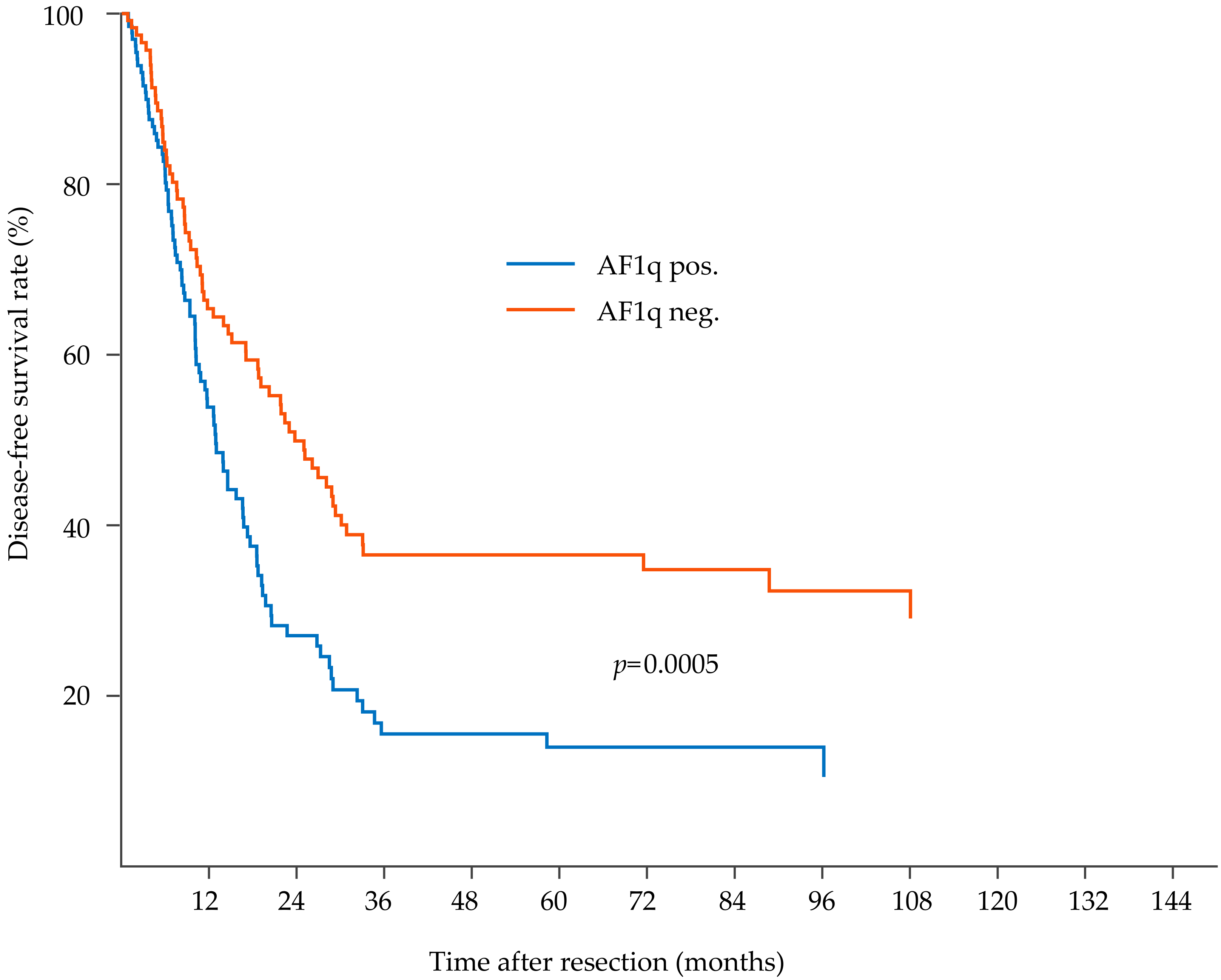
AF1q0.00050.011.51.12.2
Neoadjuvant therapy0.00020.0051.71.22.6
Histological tumor subtype *0.160.00041.91.32.7
Local tumor stage pT0.0010.07n.a.
Regional lymph nodes pN<0.0001<0.00012.31.63.2
Tumor grading G0.130.30n.a.
Resection margin R0.020.50n.a.
Disease-specific survival
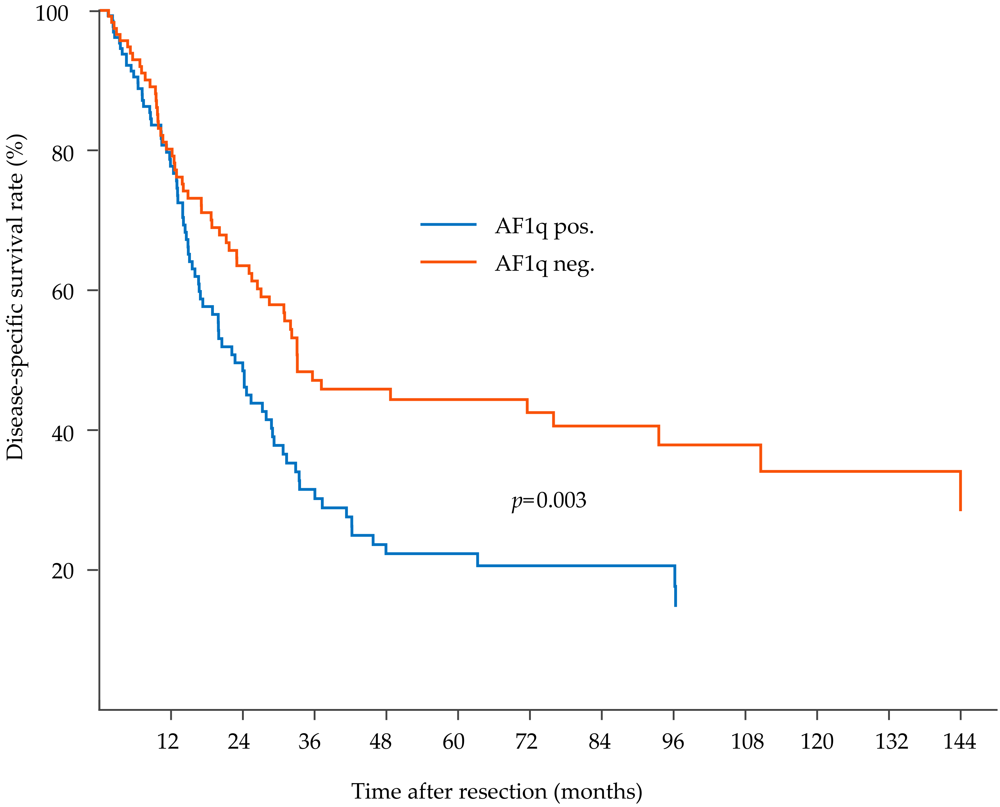
AF1q0.0030.031.51.02.1
Neoadjuvant therapy0.100.83n.a.
Histological tumor subtype *0.300.0031.81.22.7
Local tumor stage pT0.00040.0071.91.23.0
Regional lymph nodes pN<0.0001<0.00012.21.53.2
Tumor grading G0.220.48n.a.
Resection margin R0.010.31n.a.
Note. EC—esophageal cancer; HR—hazard ratio; CI—confidence interval; HR and CR refer to multivariable model; * adenocarcinoma and squamous cell carcinoma.

