Article

The Molecular Pathogenesis of Tumor-Suppressive miR-486-5p and miR-486-3p Target Genes: GINS4 Facilitates Aggressiveness in Lung Adenocarcinoma

1
Department of Pulmonary Medicine, Graduate School of Medical and Dental Sciences, Kagoshima University, Kagoshima 890-8544, Japan
2
Department of Functional Genomics, Chiba University Graduate School of Medicine, Chuo-ku, Chiba 260-8670, Japan
3
Head and Neck Surgery, Chiba Cancer Center, Nitona, Chiba 260-8717, Japan
*
Author to whom correspondence should be addressed.
These authors contributed equally to this work.
Cells 2023, 12(14), 1885; https://doi.org/10.3390/cells12141885
Submission received: 20 June 2023 / Revised: 14 July 2023 / Accepted: 16 July 2023 / Published: 18 July 2023

Abstract

:

Simple Summary

Two microRNAs (miRNAs) (miR-486-5p and miR-486-3p) derived from pre-miR-486 acted as tumor-suppressive miRNAs in lung adenocarcinoma (LUAD). We identified seven genes (MKI67, GINS4, RRM2, HELLS, MELK, TIMELESS, and SAPCD2) involved in the malignant phenotype of LUAD cells coordinately regulated by these miRNAs. It is possible to suppress the malignant transformation of LUAD by controlling these genes.

Abstract

The involvement of passenger strands of miRNAs in the molecular pathogenesis of human cancers is a recent concept in miRNA research, and it will broaden our understanding of the molecular mechanisms of miRNA-mediated cancer. The analysis of our miRNA signature of LUAD revealed that both strands of pre-miR-486 (miR-486-5p and miR-486-3p) were downregulated in LUAD tissues. Ectopic expression of both miRNAs induced cell cycle arrest in LUAD cells, suggesting both strands of miRNAs derived from pre-miR-486 were tumor suppressive. Our in silico analysis showed a total of 99 genes may be under the control of both miRNAs in LUAD cells. Importantly, among these targets, the high expression of seven genes (MKI67, GINS4, RRM2, HELLS, MELK, TIMELESS, and SAPCD2) predicted a poorer prognosis of LUAD patients (p < 0.05). We focused on GINS4, a DNA replication complex GINS protein that plays an essential role in the initiation of DNA replication. Our functional assays showed that GINS4 was directly controlled by both strands of pre-miR-486, and its aberrant expression facilitated the aggressive behavior of LUAD cells. GINS4 is attractive as a therapeutic target for this disease. MiRNA analysis, including passenger strands, will further improve our understanding of the molecular pathogenesis of LUAD.

1. Introduction

Lung cancer is the leading cause of cancer-related deaths in men and women worldwide, with approximately 2.3 million people diagnosed with lung cancer and approximately 1.8 million deaths from lung cancer each year [1]. Lung cancer is divided histologically into two groups: small cell lung cancer (SCLC), which accounts for 15% of patients, and non-SCLC (NSCLC), which accounts for 85% [2]. NSCLC are subdivided into squamous cell carcinoma (LUSQ), large cell carcinoma, and lung adenocarcinoma (LUAD); the latter accounts for approximately 60% of NSCLC [2].
The curative treatment for lung cancer patients at an early stage of the disease (stage I or II) is surgical resection; however, even those who undergo radical surgery have a 5-year survival rate of only 65% [3]. However, fewer than 30% of lung cancer patients are diagnosed early in the course of disease and proceed to surgical treatment. Patients diagnosed with advanced-stage lung cancer have a very poor prognosis, with only 20% of patients surviving 5 years [2,4].
Therapeutic strategies for advanced stage LUAD are developing in a remarkable way. Molecular-targeted drugs that counteract driver gene alteration (e.g., EGFR mutation, ALK rearrangement, ROS1 rearrangement, BRAF mutation, MET mutation, and KRAS mutation), and immune checkpoint inhibitors are showing therapeutic effects [5,6]. However, the prognosis for lung cancer patients remains extremely poor, with only 20% of those diagnosed at an advanced stage surviving for five years [2,4]. Therefore, the search for new diagnostic biomarkers and therapeutic target molecules is an important research theme for improving the prognosis of patients with LUAD.
Numerous non-coding RNAs (ncRNAs) are involved in a wide variety of biologic functions, e.g., basic metabolism and cell differentiation. At present, ncRNAs are thought to play important roles for maintaining cellular homeostasis [7,8,9]. A large number of studies have shown that various ncRNAs are aberrantly expressed in diverse tumors, indicating that such RNAs play important roles in tumorigenesis and development [10,11,12]. Among ncRNAs, miRNAs are small, single-stranded ncRNAs (19–22 nucleotides in length). They act as fine tuners of gene expression and modulate almost all biological processes [13,14]. Aberrant expressions of miRNAs are frequently detected in a wide range of cancers, including LUAD. Aberrant-expressed miRNAs are closely involved in the malignant transformation of human cancers, e.g., proliferation, metastasis, and resistance [15,16].
In the previous concept of miRNA biogenesis, only the guide strand of miRNAs derived from pre-miRNAs were actually functional miRNAs in cells. On the other hand, the passenger strand was thought to be broken down inside the cell and to have no function. In contrast to the miRNA theory, some passenger strands of miRNAs have been shown to regulate their target molecules [17,18]. These studies indicate that an miRNA analysis of gene regulation requires the inclusion of both the guide and passenger strands. For example, our recent studies on NSCLC cells demonstrated that both strands of miR-99a, miR-144, miR-145, and miR-150, had tumor-suppressive activity through their control of several oncogenic genes [19,20,21,22]. Based on these studies, we hypothesized that genes that are commonly regulated by both strands derived from pre-miRNA are highly involved in the oncogenesis of LUAD.
To identify dysregulated miRNAs in LUAD clinical tissues, we generated miRNA expression signatures by using small RNA sequencing technology. The analysis of the signatures revealed that both strands of pre-miR-486 (miR-486-5p, the guide strand, and miR-486-3p, the passenger strand) were downregulated in LUAD tissues. Moreover, they acted as antitumor miRNAs in our functional assays. Importantly, seven genes (MKI67, GINS4, RRM2, HELLS, MELK, TIMELESS, and SAPCD2) commonly regulated by miR-486-5p and miR-486-3p were closely involved in the molecular pathogenesis of LUAD. Furthermore, the aberrant expression of GINS4, a DNA replication complex GINS protein, facilitated LUAD cell aggressiveness.
Our signature-based miRNA analysis accelerates the discovery of genes closely involved in LUAD tumorigenesis. These genes are potential therapeutic targets for this disease.

2. Materials and Methods

2.1. Clinical Course of Patients with LUAD Cells

We obtained primary lesions and normal lung tissues from lung adenocarcinoma patients. The background and clinical characteristics of the patients are described in Table S1.

2.2. Cell Lines and Cell Culture

Two LUAD cell lines, A549 and H1299, were used in this study (American-Type Culture Collection (ATCC), Manassas, VA, USA). We have previously described the method of cell maintenance [21,23].

2.3. Construction of the miRNA Expression Signature in LUAD Based on RNA Sequencing

LUAD and normal lung specimens were sequenced using a the NextSeq 500 instrument (Illumina, Inc., San Diego, CA, USA) to evaluate miRNA expression. The raw sequencing data were registered in Gene Expression Omnibus (GEO; GEO accession number: GSE230229).

2.4. Identification of Oncogenic Targets Regulated by miR-486-5p and miR-486-3p in LUAD Cells

We used the expression profiles of genes from A549 cells transfected with miR-486-5p or miR-486-3p (GEO accession number: GSE230056) and TargetScanHuman ver.8.0 (https://www.targetscan.org/vert_80/, accessed on 12 January 2023) to search for miRNAs regulated by miR-486-5p and miR-486-3p.

2.5. Expression Levels of Genes and Prognosis by In Silico Analysis

The clinical significance of genes in LUAD was evaluated with The Cancer Genome Atlas (TCGA) datasets (https://www.cancer.gov/tcga, accessed on 17 January 2023). The data describing gene expression levels were obtained from FIREBROWSE (http://firebrowse.org/, accessed on 17 January 2023) and Genomic Data Commons (GDC) Data Portal (https://portal.gdc.cancer.gov/, accessed on 17 January 2023). The overall survival data were obtained from cBioPortal (https://www.cbioportal.org/, accessed on 17 January 2023) and OncoLnc (http://www.oncolnc.org/) (data downloaded on 17 January 2023).

2.6. Transfection with siRNA and miRNA

siRNA and miRNA were transfected into cell lines using Opti-MEM (catalog no.: 31985070, Gibco, Carlsbad, CA, USA) and LipofectamineTM RNAiMax Transfection Reagent (catalog no.: 13778150, Invitrogen, Carlsbad, CA, USA). Transfection protocols for siRNA and miRNA were described in our previous studies [21,23,24]. siRNA and miRNA used in this study are listed in Table S2.

2.7. RNA Extraction and Reverse Transcription Quantitative Polymerase Chain Reaction (RT-qPCR)

Total RNA obtained from LUAD cell lines was isolated using Isogen II (catalog no.: 311-07361, NIPPON GENE Co., Ltd., Tokyo, Japan). cDNA was synthesized using PrimeScriptTM RT Master Mix (catalog no.: RR036A, Takara Bio Inc., Shiga, Japan). Gene expression was analyzed by real-time PCR using a SYBR green assay (ThermoFisher Scientific, Rockford, IL, USA) on a StepOnePlus Real-Time PCR System (Applied Biosystems, Foster City, CA, USA). The internal control used in the gene expression assays was glyceraldehyde 3-phosphate dehydrogenase (GAPDH). The reagents used in this study are listed in Table S2.

2.8. Western Blotting

LUAD cells were lysed with the RIPA Lysis Buffer System (catalog no.: sc-24948, Santa Cruz Biotechnology Inc., Dallas, TX, USA). Protein concentrations were measured using a PierceTM BCA Protein Assay Kit (catalog no.: 23227, Thermo Fisher Scientific, Rockford, IL, USA). We used SuperSepTM Ace (7.5%, 13 well) (catalog no.: 198-14941, FUJIFILM Wako Pure Chemical Corporation, Osaka, Japan) as the SDS-PAGE gel for electrophoresis and Precision Plus ProteinTM Dual Color Standards (catalog no.: #1610374, Bio-Rad Laboratories, Inc., Hercules, CA, USA). The proteins were transferred to polyvinylidene fluoride membranes (catalog no.: PPVH00010, Merck KGaA, Darmstadt, Germany). The membranes were blocked with 5% skimmed milk (catalog no.: 190-12865, FUJIFILM Wako Pure Chemical Corporation, Osaka, Japan) in TBST. The signal was detected using Amersham ECL Prime Western Blotting Detection Reagent (Cytiva, Marlborough, MA, USA). The reagents used in this study are listed in Table S2.

2.9. Cell Proliferation and Cell Cycle Assays

Cell proliferation was evaluated with XTT assays using Cell Proliferation Kits (catalog no.: 20-300-1000, Biological Industries, Beit-Haemek, Israel). The cell cycle was evaluated using a BD CycletestTM Plus DNA Reagent Kit (catalog no.: 340242, BD Biosciences, Franklin Lakes, NJ, USA) and flow cytometry (BD FACSCelestaTM Flow Cytometer, BD Biosciences). The procedures for assessing cell proliferation and cell cycle behaviors were described previously [21,23,24].

2.10. Plasmid Construction and Dual-Luciferase Reporter Assays

The following two sequences were cloned into the psiCHECK-2 vector (C8021; Promega, Madison, WI, USA): the wild-type sequence of the 3′-untranslated regions (UTRs) of GINS4 and the deletion-type sequence, which lacked the miR-486-5p and miR-486-3p target sites of GINS4. The procedures for the transfection and dual-luciferase reporter assays were provided previously [21,23,24].

2.11. Immunohistochemical Staining

GINS4 expression was evaluated by immunohistochemical staining using tissue microarray slides (catalog no.: LC811a, US Biomax, Inc. Derwood, MD, USA). The VECTASTAIN Universal Elite ABC Kit (catalog no.: PK-6200, Vector Laboratories, Burlingame, CA, USA) was used for blocking, the primary antibody reaction, the secondary antibody reaction, and the binding of avidin to the biotin complex. Primary antibodies were diluted with Dako Real antibody diluent (catalog no.: K5007, Agilent, Santa Clara, CA, USA). Dako REALTM EnVisionTM Detection System Peroxidase/DAB+, Rabbit/Mouse (Agilent) was used to develop the chromogenic reaction. The primary antibody used in this study is described in Table S2. Clinical tissue information is presented in Table S3.

2.12. Putative miRNA Binding to GINS4 and miRNA Expression

We obtained the data for putative miRNA binding to GINS4 from TargetScanHuman database (release 8.0) and the data for miRNA expression levels from FIREBROWSE (http://firebrowse.org/, accessed on 31 October 2022) and Genomic Data Commons (GDC) Data Portal (https://portal.gdc.cancer.gov/) (accessed on 31 October 2022).

2.13. Statistical Analysis

Statistical analyses were performed using GraphPad Prism 8 (GraphPad Software, La Jolla, CA, USA) and R ver. 4.2.1 (R Core Team, Vienna, Austria; https://www.R-project.org/, accessed on 3 September 2022). The differences between 2 groups were analyzed by Student’s t- or Mann–Whitney U tests. Multiple group comparison was achieved using a one-way analysis of variance (ANOVA) and Tukey’s tests for post hoc analysis. Survival rates were analyzed by Kaplan–Meier survival curves and the log-rank test.

3. Results

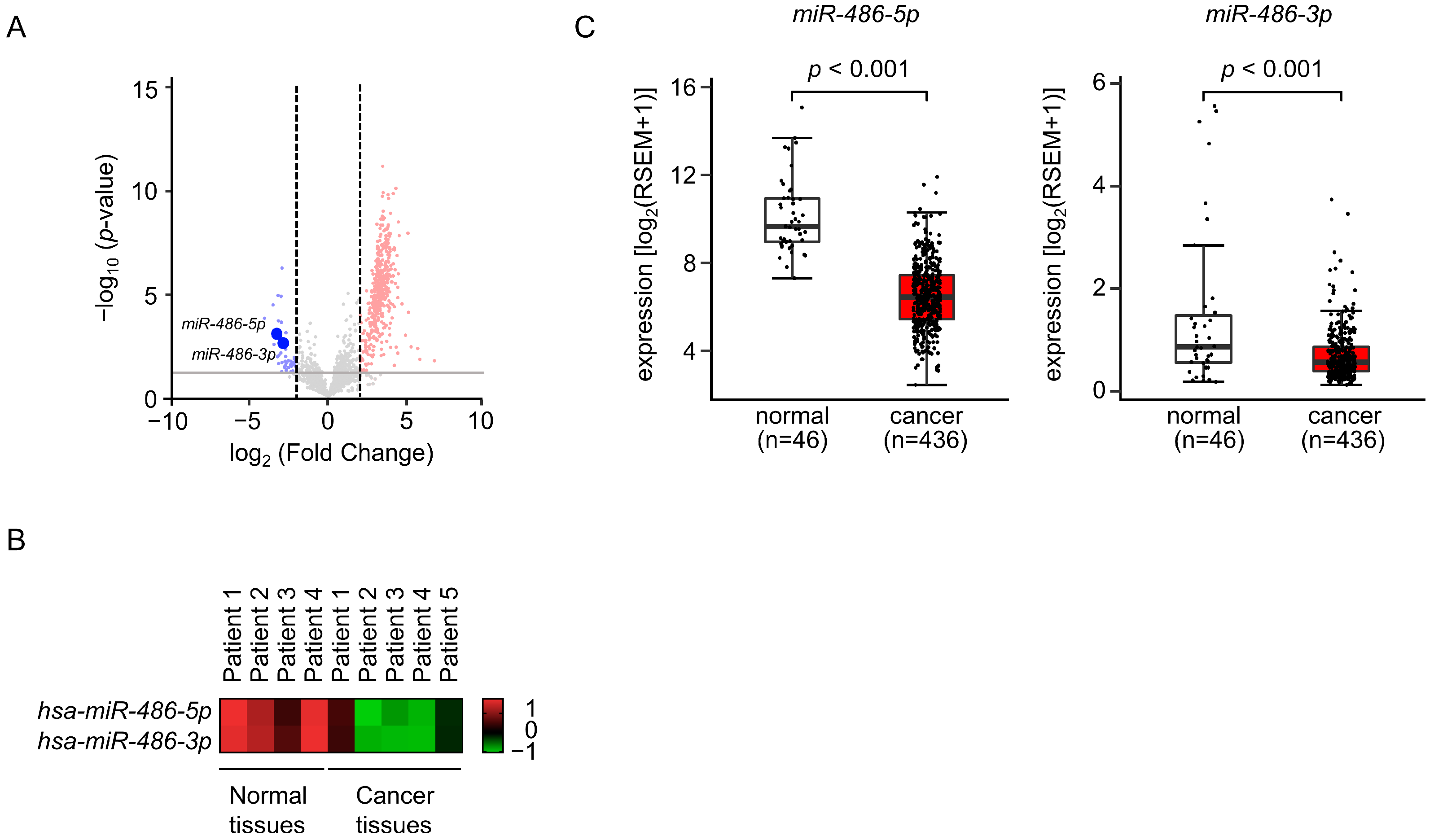
3.1. Selection of Downregulated miRNAs in LUAD Clinical Specimens by Small RNA Sequencing

To create the miRNA expression signatures of LUAD, we prepared nine cDNA libraries obtained from clinical specimens (five LUAD tissues and four normal lung tissues) and performed RNA sequencing. The clinical information for the LUAD tissues is summarized in Table S1. The processing of the data based on the RNA sequencing analysis and details of analyzed small RNA taxonomies are presented in Table S4. We successfully mapped a sufficient number of miRNA reads to the human genome (Table S4).
We analyzed RNA sequence data (a total of 41 miRNAs) in the LUAD tissues for comparison with normal lung tissues (Table 1, Figure 1A). We found they were significantly downregulated (log2 fold-change < −2.0 and p-value < 0.05). Interestingly, our signature revealed that both the guide and passenger strands of four miRNAs (miR-34c, miR-486, miR-34b, and miR-144) were downregulated in the LUAD tissues (Table 1). The involvement of passenger strands of miRNAs derived from pre-miRNAs in the molecular pathogenesis of human cancers is a recent concept in miRNA biology.

3.2. Expression Levels of miR-486-5p and miR-486-3p in LUAD Specimens and Cell Lines

To confirm our miRNA signature, we evaluated the expression levels of miR-486-5p and miR-486-3p in LUAD tissues and normal lung tissues.
Both miR-486-5p and miR-486-3p were significantly downregulated in LUAD tissues (Figure 1B). The TCGA dataset analysis confirmed that the expression levels of miR-486-5p (p < 0.001) and miR-486-3p (p < 0.001) were significantly lower in the LUAD tissues (n = 436) compared to normal tissues (n = 46) (Figure 1C).

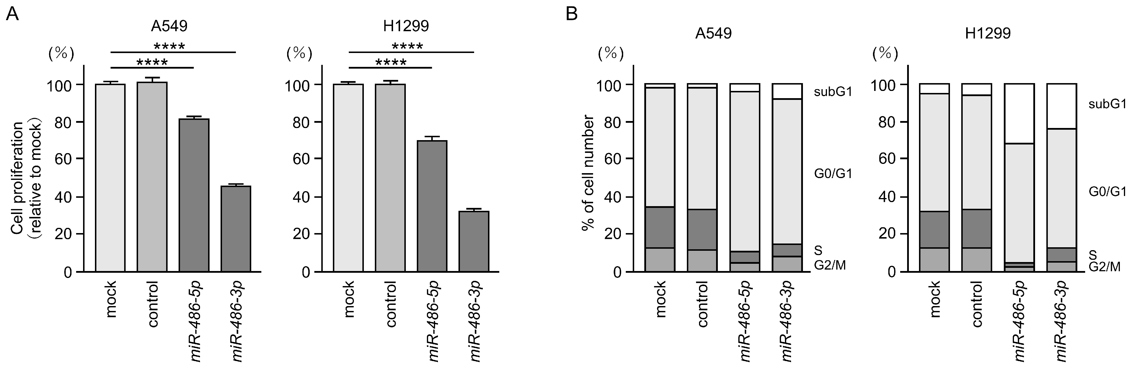
3.3. Antitumor Functions of miR-486-5p and miR-486-3p in LUAD Cells

In order to prove that both strands of pre-miR-486 had antitumor functions in LUAD cells, we performed an ectopic expression of these miRNAs in LUAD cells (A549 and H1299), and then investigated the behavior of the cancer cells.
Cancer cell proliferation was attenuated by the ectopic expression of miR-486-5p or miR-486-3p in LUAD cells (Figure 2A). The analysis showed typical cell cycle arrest (G0/G1 phase) after both miRNAs were transfected into LUAD cells (Figure 2B).
Based on these results, we conclude that the two types of miRNAs derived from pre-miR-486 are tumor suppressive miRNAs in LUAD cells.

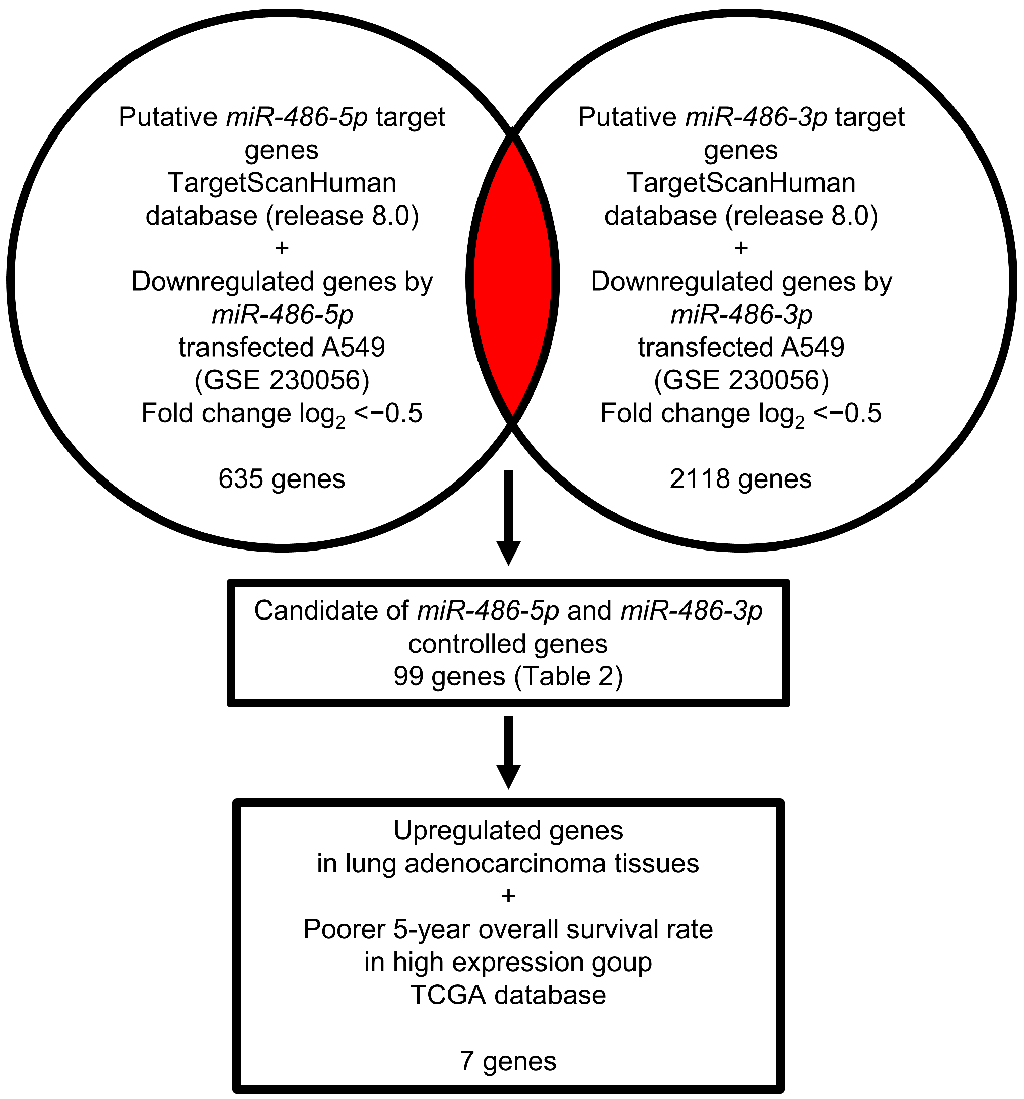
3.4. Identification of Genes Controlled by miR-486-5p and miR-486-3p in LUAD Cells

The fact that both miRNA strands derived from pre-miR-486 acted as antitumor miRNAs was quite interesting. The subsequent challenge is to identify the oncogenic targets controlled by these miRNAs in LUAD cells.
Our strategy for the search for miRNAs targets is shown in Figure 3. In this study, we obtained genome-wide gene expression data using miR-486-5p- or miR-486-3p-transfected A549 cells. Our gene expression data were deposited in the GEO database (accession number: GSE230056).
Using a combination of the TargetScan database and miRNA-transfected LUAD cells expression data, we searched for putative targets controlled by miR-486-5p (635 genes) and miR-486-3p (2118 genes). Notably, 99 genes were shown to be potential targets of both miR-486-5p and miR-486-3p in LUAD cells (Table 2).

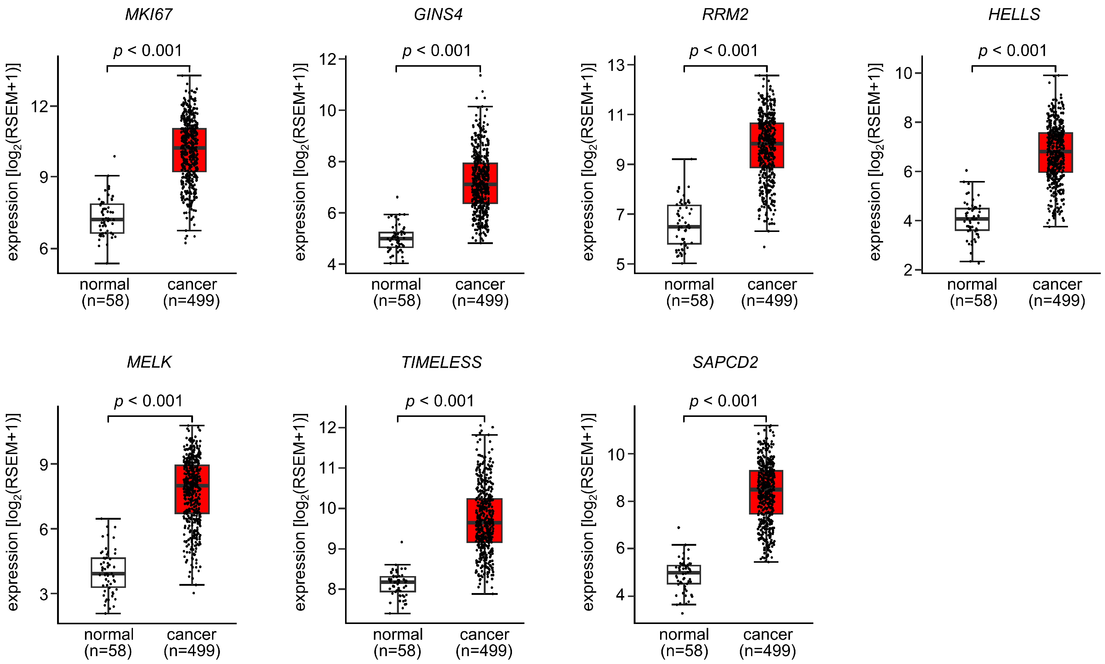
3.5. Expression and Clinical Significance of Both Strands of Pre-miR-486 Target Genes in LUAD

A further analysis of these 99 genes was performed to search for those that promoted cancer in LUAD cells.
We validated the expression levels of these 99 target genes using a large amount of clinical data (TCGA-LUAD). The expression of seven genes (MKI67, GINS4, RRM2, HELLS, MELK, TIMELESS, and SAPCD2) was upregulated in the LUAD tissues (n = 499) compared with normal lung tissues (n = 58) (Figure 4).
A clinicopathological analysis of the seven genes was performed using TCGA-LUAD datasets. Kaplan–Meier curve (5-year survival rates) analysis was performed according to the expression levels of the seven genes. The high expression of all genes significantly affected the poorer survival rates of the patients (Figure 5).

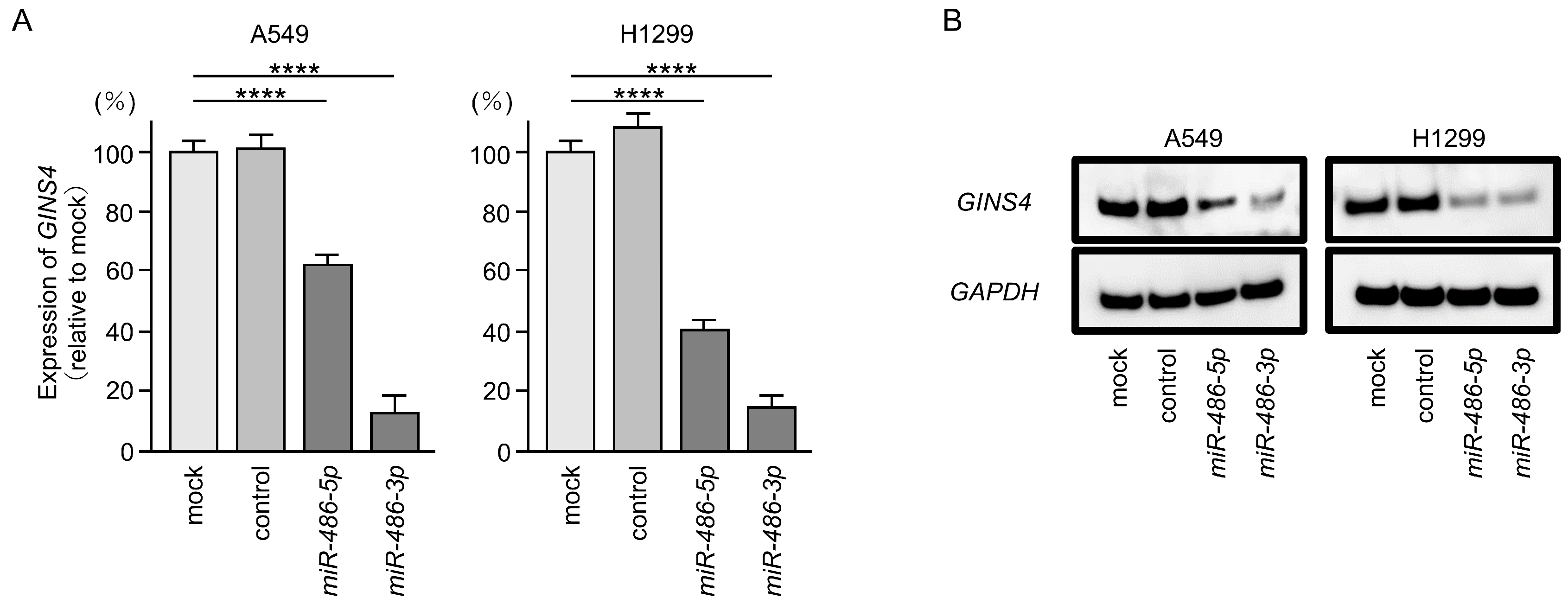
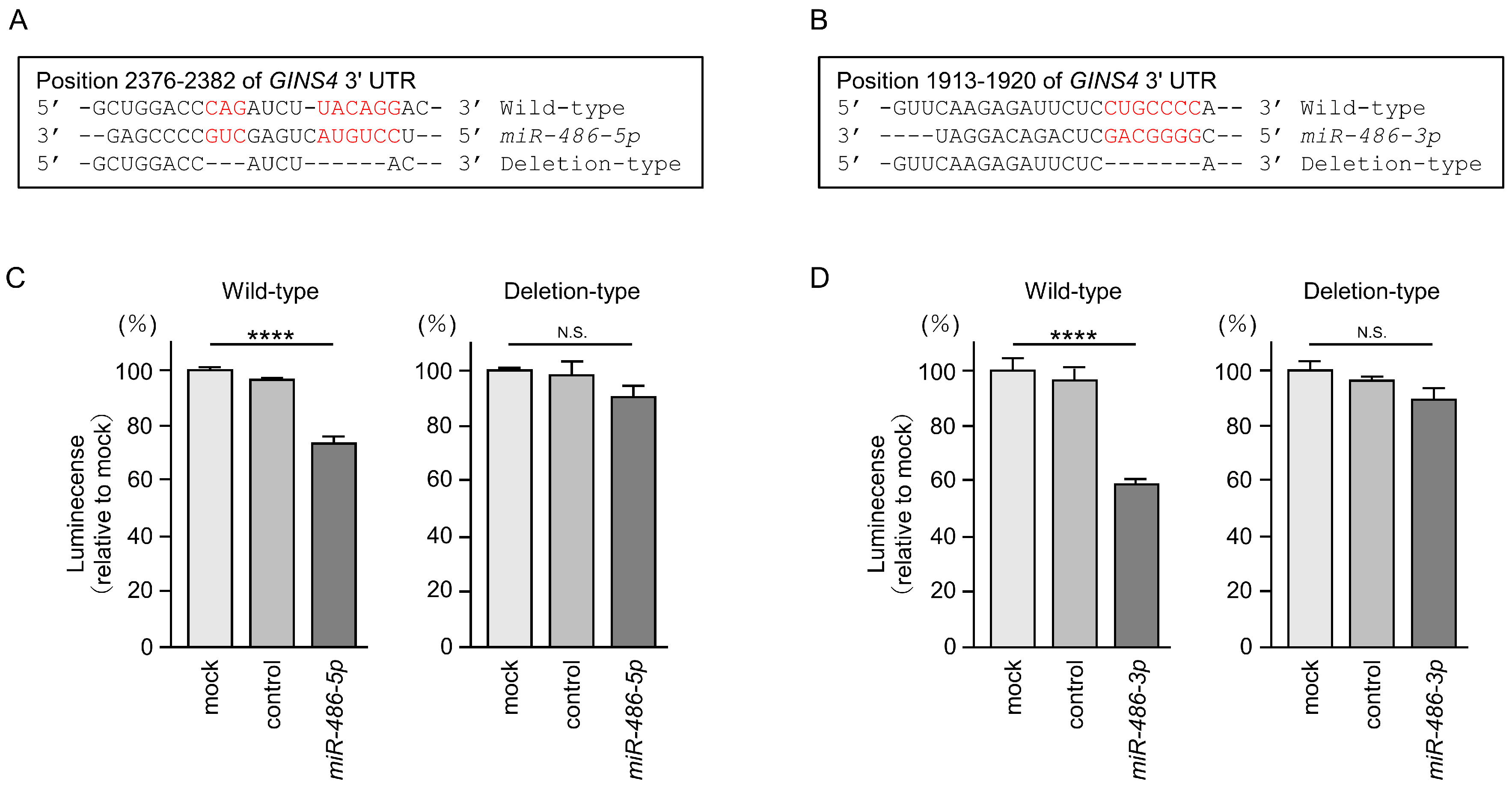
3.6. Direct Regulation of GINS4 by miR-486-5p and miR-486-3p in LUAD Cells

First, we investigated whether the expression of the seven selected genes was controlled by miR-486-5p and miR-486-3p in LUAD cells. The mRNA expression levels of all seven genes were remarkably suppressed in miR-486-3p-transfected A549 cells (Figure S1). In miR-486-5p-transfected cells, the expression levels of five genes (MKI67, GINS4, RRM2, HELLS and MELK) were significantly suppressed (Figure S1).
In our previous analysis, we focused on the genes involved in DNA replication [25,26]. In this study, we focused on GINS4 and investigated the oncogenic roles of its aberrant expression in LUAD cells.
We confirmed that GINS4 expression in LUAD cells was suppressed at the mRNA and protein levels by the ectopic expression of miR-486-5p or miR-496-3p (Figure 6A,B). Full-size images of Western blots are shown in Figure S2.
To demonstrate that both miRNAs directly bound to the 3′-UTR of the GINS4 gene in a sequence-dependent manner, we performed dual-luciferase reporter assays. The putative miR-486-5p binding site on the 3′-UTR of the GINS4 gene is shown in Figure 7A. Luciferase activity was significantly reduced when co-transfected with miR-486-5p and a vector containing binding sites for the 3′-UTR of GINS4 (Figure 7C). However, luciferase activity did not change when co-transfected with miR-486-5p and a vector lacking the miR-486-5p binding site (Figure 7C). Thus, miR-486-5p appeared to directly bind to the 3′-UTR of GINS4 in a sequence-dependent manner.
We also investigated the sequence-dependent direct binding of miR-486-3p and the 3′-UTR of the GINS4 gene. The putative miR-486-3p binding site on the 3′-UTR of the GINS4 gene is shown in Figure 7B. Luciferase activity was significantly reduced when co-transfected with miR-486-3p and a vector containing binding sites for the 3′-UTR of GINS4 (Figure 7D). However, there was no change after the co-transfection of miR-486-3p and a vector lacking the miR-486-3p binding site (Figure 7D). These results indicate that miR-486-3p directly binds to the 3′-UTR of GINS4 in a sequence-dependent manner.

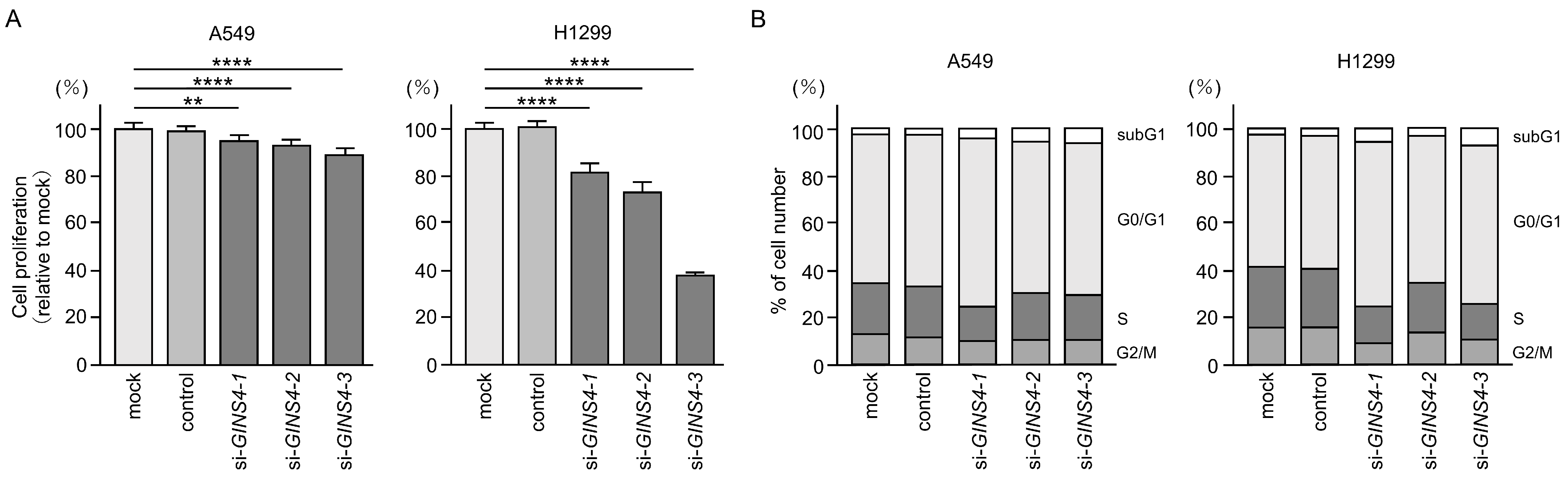
3.7. Functional Significance of GINS4 in LUAD Cells

To investigate the oncogenic function of GINS4 in LUAD cells, we made use of knockdown assays with siRNAs that were transfected into LUAD cells (A549 and H1299). We evaluated the knockdown efficiency of several siRNAs (siGINS4-1, siGINS4-2, and siGINS4-3) for GINS4. Transient transfection with three types of siRNAs significantly reduced GINS4 mRNA and protein expression in LUAD cells (Figures S3 and S4).
LUAD cell proliferation assays showed that cell growth was inhibited by suppressing the expression of GINS4 (Figure 8A). Moreover, cell cycle assays demonstrated that cell cycle arrest in the G0/G1 phase after the expression of GINS4 was suppressed in LUAD cells (Figure 8B). These results suggest that GINS4 is a cancer-promoting gene that modulates cell cycle progression.

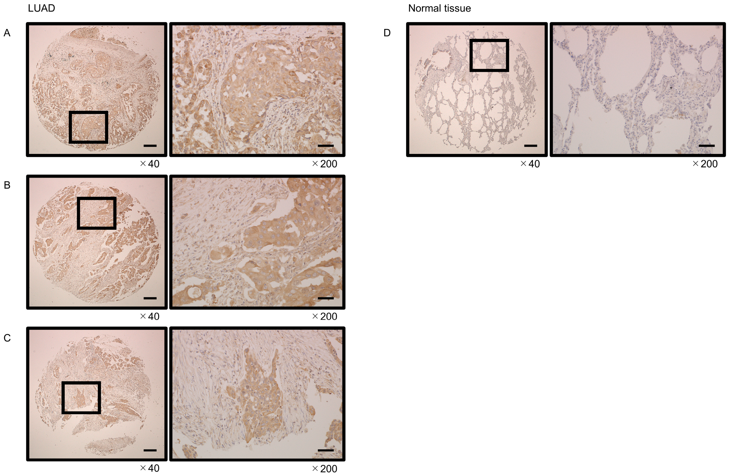
3.8. Immunostaining of GINS4 in LUAD Clinical Tissues

We examined the expression of GINS4 in tissue microarray studies. Compared with normal tissues, the GINS4 protein was overexpressed in LUAD tissues. In particular, cancer cells showed heavy cytoplasmic staining (Figure 9).

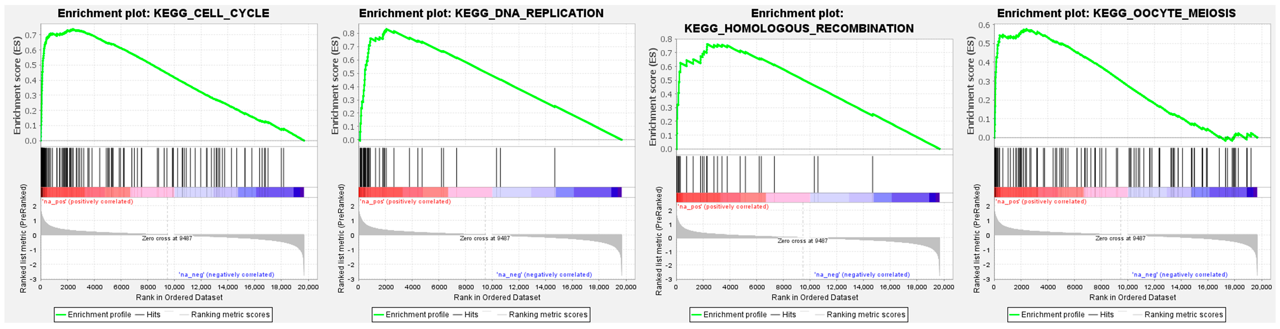
3.9. GINS4-Mediated Pathways Determined by Gene Set Enrichment Analysis (GSEA)

To investigate GINS4-modulated molecular pathways in LUAD cells, we used the Gene Set Enrichment Analysis (GSEA) based on TCGA–LUAD RNA sequencing data.
“Cell cycle”, “DNA replication”, “homologous recombination”, and “oocyte meiosis” pathways were enriched in patients with high expression of GINS4 compared to low-expression patients (Figure 10, Table 3).

4. Discussion

During classic miRNA maturation, pre-miRNA is transported to the cytoplasm where it is cleaved by Dicer to become a miRNA duplex. One strand derived from the miRNA duplex is incorporated into the RNA-Induced Silencing Complex (RISC), where it regulates specific target genes. That strand is defined as the guide strand. The non-loaded strand (the passenger strand) is degraded in the cytoplasm, as it has no known function [27]. However, in recent studies, both strands of the miRNA duplex were shown to be functional [27,28]. In this study, we confirmed that both strands of pre-miR-486 had tumor suppressive functions by regulating their respective target genes.
miR-486 is transcribed from the intron of the host gene ANK1 (Ankyrin 1) on human chromosome 8p11.21 [29]. There have been many reports describing miR-486-5p (the guide strand) in various cancer types [30,31,32,33]. Previous studies showed that the expression of miR-486-5p was reduced in cancer tissues, and that it functions as a tumor suppressor in breast cancer, colorectal cancer, gastric cancer, hepatocellular carcinoma, and renal cell carcinoma [34]. In contrast to the preceding examples, the overexpression of miR-486-5p was observed in prostate cancer, and its expression is associated with the malignant transformation of these cancers [35].
In previous reports of lung cancer, the function of miR-486-5p was that of tumor suppression, which is consistent with our results [31]. For example, the expression of miR-486-5p blocked mTOR pathways through its targeting of ribosomal proteins S6 kinase A1 and B1 [36]. Moreover, the overexpression of miR-486-5p attenuated tumor growth and inhibited metastasis according to in vivo assays [36,37]. A recent study showed that the anesthetic propofol induced the expression of miR-486-5p, resulting in the inhibition of the Ras-associated protein1-NF-κB pathway [38]. These events contributed to cisplatin-sensitivity in NSCLC cells [38].
Several papers have reported that miR-486-3p, which is the passenger strand of pre-miR-486, has anti-tumor functions in several cancers [34]. In recent years, it has become clear that overexpression of various types of circular RNAs adsorbs miRNAs and suppresses their functions in normal and pathological cells [39,40]. Various circular RNAs are overexpressed in lung cancer cells, e.g., circ_EPB41, circ_CSPP1, and circ_0011298, and the adsorption of miR-486-3p by these circular RNAs promotes a malignant transformation [41,42,43]. In addition, these studies revealed that eIF5A, BRD9, XRCC1, CYP1A1, and CRABP2 were miR-486-3p-regulated cancer-promoting genes in NSCLC cells [41,42,43,44]. The results of our functional analysis of miR-486-3p support those reports.
Since both strands of pre-miR-486 are tumor suppressive, our subsequent interest is to elucidate the molecular networks regulated by these miRNAs in LUAD cells. Numerous studies have characterized the target molecules of miRNAs, including miR-486-5p and miR-486-3p; however, none have searched for common targets of these miRNAs in LUAD cells. Our study revealed that 99 genes were putative targets of miR-486-5p and miR-486-3p regulations in LUAD cells. In fact, all 99 genes were regulated by both miR-486-5p and miR-486-3p in LUAD cells. It should be noted that high expression of seven genes (MKI67, GINS4, RRM2, HELLS, MELK, TIMELESS, and SAPCD2) had a negative impact on the prognosis of patients with LUAD. These genes are important for elucidating the molecular mechanisms of lung cancer malignancy.
For these genes, we referred to reports of genes under microRNA regulation in lung cancer cells. The RRM2 protein is one of the two subunits of the ribonucleotide reductase complex. This reductase is a key enzyme in DNA synthesis as it catalyzes the formation of deoxyribonucleotides from ribonucleotides [45]. Previous studies showed that the overexpression of RRM2 was detected in a wide range of cancers, including lung cancer [45,46]. A recent study showed that the expression of miR-203-3p was reduced in LUAD tissues, and its overexpression inhibited LUAD aggressive phenotypes through targeting RRM2 in LUAD cells [47]. Our previous study showed that miR-150-3p (the passenger strand) was significantly suppressed in LUSQ tissues, and performed a tumor-suppressive role in LUSQ cells via controlled several cell cycle-related genes, including HELLS [48]. HELLS belongs to the SNF2 family of chromatin-remodeling ATPases and is recruited to specific DNA sites to control the transcription of targeted genes [49,50]. Our data demonstrate that HELLS is directly regulated by miR-150-3p in LUSQ cells [48]. Previous reports revealed that SAPCD2 is highly expressed in various cancers and is highly involved in the malignant transformation of cancer cells [51]. Numerous studies have shown that SAPCD2 interacts with multiple proteins within the cell cycle interaction network and functions as a mitotic phase-promoting factor [51]. Notably, a recent study showed that miR-486-5p suppressed cell malignant progression in LUAD cells by targeting SAPCD2 [52]. This fact is consistent with our data and indicates that miR-486-5p-mediated molecular networks have pivotal effects on LUAD cell malignancy.
Among these targets, we focused on GINS4, and we showed that its expression facilitated the malignant transformation of LUAD cells. GINS4 is a member of the GINS complex, which consists of four different subunits, e.g., GINS1 to GINS4 [53]. Precisely maintained genomic DNA replication is critical for all forms of cellular life and requires a complex interplay of various protein factors. DNA helicases play a key role in unwinding double-stranded DNA during replication, recombination, and repair processes. The GINS complex forms the CMG (Cdc45-MCMs-GINS) complex with MCM (mini-chromosome maintenance) and CDC45. This complex functions as a replicative helicase that unwinds double-stranded DNA during chromosome replication [53,54].
The overexpression of GINS4 occurs in breast cancer, colorectal carcinoma, bladder cancer, pancreatic ductal adenocarcinoma, glioma, and gastric cancer [55]. In NSCLC cells, lymphoid-specific helicase binds to the 3′-UTR region of GINS4 and stabilizes GINS4 expression [56]. The overexpression of GINS4 facilitates lung cancer malignant transformation. The aberrant expression of other members of GINS (GINS1, GINS2, and GINS3) occurs in different types of human cancers [57,58]. The TCGA database analysis revealed that all members of GINS were upregulated in LUAD tissues and their high expression predicted the prognosis of the patients. Several studies demonstrated that the expression of a GINS member enhanced cancer cell aggressiveness, e.g., proliferation, drug resistance, and epithelial–mesenchymal transition [55,59,60]. Therefore, GINS members are closely involved with LUAD pathogenesis and may be potential therapeutic targets.

5. Conclusions

The analysis of the miRNA expression signature revealed that both strands of pre-miR-486 (miR-486-5p and miR-486-3p) were downregulated in LUAD tissues. From this study and previous reports, we confirmed that these miRNAs had tumor-suppressive functions in LUAD cells. A total of 99 genes were identified as cooperatively controlled by miR-486-5p and miR-486-3p in LUAD cells. Among these targets, seven genes (MKI67, GINS4, RRM2, HELLS, MELK, TIMELESS, and SAPCD2) were closely involved in the molecular pathogenesis of LUAD. Furthermore, GINS4 was directly regulated by these two miRNAs and the overexpression of GINS4 facilitated LUAD cell aggressiveness. Based on the tumor-suppressive miRNA analysis, it was possible to identify miRNA target genes closely involved in the molecular pathogenesis of LUAD.

Supplementary Materials

The following supporting information can be downloaded at: https://www.mdpi.com/article/10.3390/cells12141885/s1, Figure S1: suppression of mRNA expression levels of 7 target genes (MKI67, GINS4, RRM2, HELLS, MELK, TIMELESS, and SAPCD2) by ectopic expressions of miR-486-5p or miR-486-3p in A549 cells; Figure S2: full-sized images of Western blot analysis (GINS4 antibody) following ectopic expressions of miR-486-5p or miR-486-3p in A549 and H1299 cells; Figure S3: suppression of mRNA expression levels of GINS4 by the transfection of siRNAs (siGINS4-1, siGINS4-2, and siGINS4-3) in A549 and H1299 cells; Figure S4: full-sized images of Western blotting (GINS4 antibody) following the transfection of siRNAs (siGINS4-1, siGINS4-2, and siGINS4-3) in A549 and H1299 cells; Table S1: clinical features of LUAD patients created by the miRNA expression signature; Table S2: reagents used in this study; Table S3: information of tissues by immunostaining; Table S4: human genome-matched sequence reads.

Author Contributions

Conceptualization, T.S., N.S. and K.M.; methodology, N.S.; validation, H.I., N.S. and K.M.; formal analysis, Y.T., T.S., K.T., Y.H., M.S., S.A. and N.K.; investigation, Y.T., T.S., K.T., Y.H., M.S., S.A. and N.K.; resources, S.A.; data curation, Y.T., T.S. and K.T.; writing—original draft preparation, N.S.; writing—review and editing, Y.T., T.S. and N.S.; visualization, Y.T., T.S. and N.S.; supervision, N.S.; project administration, H.I., N.S. and K.M.; funding acquisition, N.S., N.K. and K.M. All authors have read and agreed to the published version of the manuscript.

Funding

This research was funded by KAKENHI; grant numbers 21K09577, 22K09679, and 22K08260.

Institutional Review Board Statement

The study was conducted in accordance with the Declaration of Helsinki and approved by the Ethics Committee on Epidemiological and its related Studies, Sakuragaoka Campus, Kagoshima University (approval no. 210101 eki-kai 1, 23 August 2022).

Informed Consent Statement

Informed consent was obtained from all subjects involved in the study.

Data Availability Statement

Publicly available datasets were analyzed in this study. These data can be accessed here: https://www.ncbi.nlm.nih.gov/geo/query/acc.cgi?acc=GSE230229 (accessed on 19 June 2023) and https://www.ncbi.nlm.nih.gov/geo/query/acc.cgi?acc=GSE230056 (accessed on 19 June 2023).

Conflicts of Interest

The authors declare no conflict of interest.

References

  1. Sung, H.; Ferlay, J.; Siegel, R.L.; Laversanne, M.; Soerjomataram, I.; Jemal, A.; Bray, F. Global Cancer Statistics 2020: GLOBOCAN Estimates of Incidence and Mortality Worldwide for 36 Cancers in 185 Countries. CA Cancer J. Clin. 2021, 71, 209–249. [Google Scholar] [CrossRef]
  2. Schabath, M.B.; Cote, M.L. Cancer Progress and Priorities: Lung Cancer. Cancer Epidemiol. Biomark. Prev. 2019, 28, 1563–1579. [Google Scholar] [CrossRef] [Green Version]
  3. Aokage, K.; Yoshida, J.; Hishida, T.; Tsuboi, M.; Saji, H.; Okada, M.; Suzuki, K.; Watanabe, S.; Asamura, H. Limited resection for early-stage non-small cell lung cancer as function-preserving radical surgery: A review. Jpn. J. Clin. Oncol. 2017, 47, 7–11. [Google Scholar] [CrossRef] [PubMed]
  4. Pirker, R. Conquering lung cancer: Current status and prospects for the future. Pulmonology 2020, 26, 283–290. [Google Scholar] [CrossRef] [PubMed]
  5. Tan, A.C.; Tan, D.S.W. Targeted Therapies for Lung Cancer Patients with Oncogenic Driver Molecular Alterations. J. Clin. Oncol. 2022, 40, 611–625. [Google Scholar] [CrossRef]
  6. Reck, M.; Remon, J.; Hellmann, M.D. First-Line Immunotherapy for Non-Small-Cell Lung Cancer. J. Clin. Oncol. 2022, 40, 586–597. [Google Scholar] [CrossRef]
  7. Fatica, A.; Bozzoni, I. Long non-coding RNAs: New players in cell differentiation and development. Nat. Rev. Genet. 2014, 15, 7–21. [Google Scholar] [CrossRef]
  8. Frías-Lasserre, D.; Villagra, C.A. The Importance of ncRNAs as Epigenetic Mechanisms in Phenotypic Variation and Organic Evolution. Front. Microbiol. 2017, 8, 2483. [Google Scholar] [CrossRef] [PubMed] [Green Version]
  9. Vienberg, S.; Geiger, J.; Madsen, S.; Dalgaard, L.T. MicroRNAs in metabolism. Acta Physiol. 2017, 219, 346–361. [Google Scholar] [CrossRef]
  10. Anastasiadou, E.; Jacob, L.S.; Slack, F.J. Non-coding RNA networks in cancer. Nat. Rev. Cancer 2018, 18, 5–18. [Google Scholar] [CrossRef]
  11. Chan, J.J.; Tay, Y. Noncoding RNA:RNA Regulatory Networks in Cancer. Int. J. Mol. Sci. 2018, 19, 1310. [Google Scholar] [CrossRef] [PubMed] [Green Version]
  12. Nadhan, R.; Isidoro, C.; Song, Y.S.; Dhanasekaran, D.N. Signaling by LncRNAs: Structure, Cellular Homeostasis, and Disease Pathology. Cells 2022, 11, 2517. [Google Scholar] [CrossRef]
  13. Bartel, D.P. MicroRNAs: Genomics, biogenesis, mechanism, and function. Cell 2004, 116, 281–297. [Google Scholar] [CrossRef] [Green Version]
  14. Bartel, D.P. MicroRNAs: Target recognition and regulatory functions. Cell 2009, 136, 215–233. [Google Scholar] [PubMed] [Green Version]
  15. Smolarz, B.; Durczyński, A.; Romanowicz, H.; Szyłło, K.; Hogendorf, P. miRNAs in Cancer (Review of Literature). Int. J. Mol. Sci. 2022, 23, 2805. [Google Scholar] [CrossRef] [PubMed]
  16. Hussen, B.M.; Hidayat, H.J.; Salihi, A.; Sabir, D.K.; Taheri, M.; Ghafouri-Fard, S. MicroRNA: A signature for cancer progression. Biomed. Pharmacother. 2021, 138, 111528. [Google Scholar] [CrossRef]
  17. Wu, K.L.; Tsai, Y.M.; Lien, C.T.; Kuo, P.L.; Hung, A.J. The Roles of MicroRNA in Lung Cancer. Int. J. Mol. Sci. 2019, 20, 1611. [Google Scholar] [CrossRef] [Green Version]
  18. Matranga, C.; Tomari, Y.; Shin, C.; Bartel, D.P.; Zamore, P.D. Passenger-strand cleavage facilitates assembly of siRNA into Ago2-containing RNAi enzyme complexes. Cell 2005, 123, 607–620. [Google Scholar] [CrossRef] [Green Version]
  19. Mizuno, K.; Tanigawa, K.; Nohata, N.; Misono, S.; Okada, R.; Asai, S.; Moriya, S.; Suetsugu, T.; Inoue, H.; Seki, N. FAM64A: A Novel Oncogenic Target of Lung Adenocarcinoma Regulated by Both Strands of miR-99a (miR-99a-5p and miR-99a-3p). Cells 2020, 9, 2083. [Google Scholar] [CrossRef]
  20. Uchida, A.; Seki, N.; Mizuno, K.; Misono, S.; Yamada, Y.; Kikkawa, N.; Sanada, H.; Kumamoto, T.; Suetsugu, T.; Inoue, H. Involvement of dual-strand of the miR-144 duplex and their targets in the pathogenesis of lung squamous cell carcinoma. Cancer Sci. 2019, 110, 420–432. [Google Scholar]
  21. Misono, S.; Seki, N.; Mizuno, K.; Yamada, Y.; Uchida, A.; Arai, T.; Kumamoto, T.; Sanada, H.; Suetsugu, T.; Inoue, H. Dual strands of the miR-145 duplex (miR-145–5p and miR-145–3p) regulate oncogenes in lung adenocarcinoma pathogenesis. J. Hum. Genet. 2018, 63, 1015–1028. [Google Scholar] [CrossRef] [PubMed]
  22. Misono, S.; Seki, N.; Mizuno, K.; Yamada, Y.; Uchida, A.; Sanada, H.; Moriya, S.; Kikkawa, N.; Kumamoto, T.; Suetsugu, T.; et al. Molecular Pathogenesis of Gene Regulation by the miR-150 Duplex: miR-150–3p Regulates TNS4 in Lung Adenocarcinoma. Cancers 2019, 11, 601. [Google Scholar] [CrossRef] [Green Version]
  23. Sanada, H.; Seki, N.; Mizuno, K.; Misono, S.; Uchida, A.; Yamada, Y.; Moriya, S.; Kikkawa, N.; Machida, K.; Kumamoto, T.; et al. Involvement of Dual Strands of miR-143 (miR-143–5p and miR-143–3p) and Their Target Oncogenes in the Molecular Pathogenesis of Lung Adenocarcinoma. Int. J. Mol. Sci. 2019, 20, 4482. [Google Scholar] [CrossRef] [Green Version]
  24. Tanigawa, K.; Misono, S.; Mizuno, K.; Asai, S.; Suetsugu, T.; Uchida, A.; Kawano, M.; Inoue, H.; Seki, N. MicroRNA signature of small-cell lung cancer after treatment failure: Impact on oncogenic targets by miR-30a-3p control. Mol. Oncol. 2023, 17, 328–343. [Google Scholar] [CrossRef]
  25. Misono, S.; Mizuno, K.; Suetsugu, T.; Tanigawa, K.; Nohata, N.; Uchida, A.; Sanada, H.; Okada, R.; Moriya, S.; Inoue, H.; et al. Molecular Signature of Small Cell Lung Cancer after Treatment Failure: The MCM Complex as Therapeutic Target. Cancers 2021, 13, 1187. [Google Scholar] [CrossRef]
  26. Toda, H.; Seki, N.; Kurozumi, S.; Shinden, Y.; Yamada, Y.; Nohata, N.; Moriya, S.; Idichi, T.; Maemura, K.; Fujii, T.; et al. RNA-sequence-based microRNA expression signature in breast cancer: Tumor-suppressive miR-101–5p regulates molecular pathogenesis. Mol. Oncol. 2020, 14, 426–446. [Google Scholar] [CrossRef] [Green Version]
  27. Mitra, R.; Adams, C.M.; Jiang, W.; Greenawalt, E.; Eischen, C.M. Pan-cancer analysis reveals cooperativity of both strands of microRNA that regulate tumorigenesis and patient survival. Nat. Commun. 2020, 11, 968. [Google Scholar] [CrossRef] [Green Version]
  28. Mitra, R.; Sun, J.; Zhao, Z. microRNA regulation in cancer: One arm or two arms? Int. J. Cancer 2015, 137, 1516–1518. [Google Scholar] [CrossRef] [Green Version]
  29. Hall, A.E.; Lu, W.T.; Godfrey, J.D.; Antonov, A.V.; Paicu, C.; Moxon, S.; Dalmay, T.; Wilczynska, A.; Muller, P.A.; Bushell, M. The cytoskeleton adaptor protein ankyrin-1 is upregulated by p53 following DNA damage and alters cell migration. Cell Death Dis. 2016, 7, e2184. [Google Scholar] [CrossRef] [PubMed] [Green Version]
  30. Kong, Y.; Li, Y.; Luo, Y.; Zhu, J.; Zheng, H.; Gao, B.; Guo, X.; Li, Z.; Chen, R.; Chen, C. circNFIB1 inhibits lymphangiogenesis and lymphatic metastasis via the miR-486–5p/PIK3R1/VEGF-C axis in pancreatic cancer. Mol. Cancer 2020, 19, 82. [Google Scholar] [CrossRef] [PubMed]
  31. Moro, M.; Fortunato, O.; Bertolini, G.; Mensah, M.; Borzi, C.; Centonze, G.; Andriani, F.; Di Paolo, D.; Perri, P.; Ponzoni, M.; et al. MiR-486–5p Targets CD133+ Lung Cancer Stem Cells through the p85/AKT Pathway. Pharmaceuticals 2022, 15, 297. [Google Scholar] [CrossRef]
  32. Wei, W.; Liu, C.; Yao, R.; Tan, Q.; Wang, Q.; Tian, H. miR-486–5p suppresses gastric cancer cell growth and migration through downregulation of fibroblast growth factor 9. Mol. Med. Rep. 2021, 24, 771. [Google Scholar] [CrossRef] [PubMed]
  33. Faur, C.I.; Roman, R.C.; Jurj, A.; Raduly, L.; Almășan, O.; Rotaru, H.; Chirilă, M.; Moldovan, M.A.; Hedeșiu, M.; Dinu, C. Salivary Exosomal MicroRNA-486–5p and MicroRNA-10b-5p in Oral and Oropharyngeal Squamous Cell Carcinoma. Medicina 2022, 58, 1478. [Google Scholar] [CrossRef]
  34. ElKhouly, A.M.; Youness, R.A.; Gad, M.Z. MicroRNA-486–5p and microRNA-486–3p: Multifaceted pleiotropic mediators in oncological and non-oncological conditions. Noncoding RNA Res. 2020, 5, 11–21. [Google Scholar] [CrossRef] [PubMed]
  35. Zhang, X.; Zhang, T.; Yang, K.; Zhang, M.; Wang, K. miR-486–5p suppresses prostate cancer metastasis by targeting Snail and regulating epithelial-mesenchymal transition. OncoTargets Ther. 2016, 9, 6909–6914. [Google Scholar] [CrossRef] [PubMed] [Green Version]
  36. Ding, L.; Tian, W.; Zhang, H.; Li, W.; Ji, C.; Wang, Y.; Li, Y. MicroRNA-486–5p Suppresses Lung Cancer via Downregulating mTOR Signaling In Vitro and In Vivo. Front. Oncol. 2021, 11, 655236. [Google Scholar] [CrossRef]
  37. Mohamed, M.A.; Mohamed, E.I.; El-Kaream, S.A.A.; Badawi, M.I.; Darwish, S.H. Underexpression of miR-486–5p but not Overexpression of miR-155 is Associated with Lung Cancer Stages. Microrna 2018, 7, 120–127. [Google Scholar] [CrossRef]
  38. Ling, Q.; Wu, S.; Liao, X.; Liu, C.; Chen, Y. Anesthetic propofol enhances cisplatin-sensitivity of non-small cell lung cancer cells through N6-methyladenosine-dependently regulating the miR-486–5p/RAP1-NF-κB axis. BMC Cancer 2022, 22, 765. [Google Scholar] [CrossRef]
  39. Wu, J.; Qi, X.; Liu, L.; Hu, X.; Liu, J.; Yang, J.; Yang, J.; Lu, L.; Zhang, Z.; Ma, S.; et al. Emerging Epigenetic Regulation of Circular RNAs in Human Cancer. Mol. Ther. Nucleic Acids 2019, 16, 589–596. [Google Scholar] [CrossRef] [Green Version]
  40. Zhu, L.P.; He, Y.J.; Hou, J.C.; Chen, X.; Zhou, S.Y.; Yang, S.J.; Li, J.; Zhang, H.D.; Hu, J.H.; Zhong, S.L.; et al. The role of circRNAs in cancers. Biosci. Rep. 2017, 37, BSR20170750. [Google Scholar] [CrossRef] [Green Version]
  41. Jin, M.; Liu, X.; Wu, Y.; Lou, Y.; Li, X.; Huang, G. Circular RNA EPB41 expression predicts unfavorable prognoses in NSCLC by regulating miR-486–3p/eIF5A axis-mediated stemness. Cancer Cell Int. 2022, 22, 219. [Google Scholar] [CrossRef]
  42. Xie, D.; Zhang, S.; Jiang, X.; Huang, W.; He, Y.; Li, Y.; Chen, S.; Xiong, H. Circ_CSPP1 Regulates the Development of Non-small Cell Lung Cancer via the miR-486–3p/BRD9 Axis. Biochem. Genet. 2023, 61, 1–20. [Google Scholar] [CrossRef]
  43. Wu, Y.; Xie, J.; Wang, H.; Hou, S.; Feng, J. Circular RNA hsa_circ_0011298 enhances Taxol resistance of non-small cell lung cancer by regulating miR-486–3p/CRABP2 axis. J. Clin. Lab. Anal. 2022, 36, e24408. [Google Scholar] [CrossRef] [PubMed]
  44. Pan, J.; Huang, G.; Yin, Z.; Cai, X.; Gong, E.; Li, Y.; Xu, C.; Ye, Z.; Cao, Z.; Cheng, W. Circular RNA FLNA acts as a sponge of miR-486–3p in promoting lung cancer progression via regulating XRCC1 and CYP1A1. Cancer Gene Ther. 2022, 29, 101–121. [Google Scholar] [CrossRef] [PubMed]
  45. Morikawa, T.; Maeda, D.; Kume, H.; Homma, Y.; Fukayama, M. Ribonucleotide reductase M2 subunit is a novel diagnostic marker and a potential therapeutic target in bladder cancer. Histopathology 2010, 57, 885–892. [Google Scholar] [CrossRef] [PubMed]
  46. Aye, Y.; Li, M.; Long, M.J.; Weiss, R.S. Ribonucleotide reductase and cancer: Biological mechanisms and targeted therapies. Oncogene 2015, 34, 2011–2021. [Google Scholar] [CrossRef] [PubMed]
  47. Cao, X.; Xue, F.; Chen, H.; Shen, L.; Yuan, X.; Yu, Y.; Zong, Y.; Zhong, L.; Huang, F. MiR-202–3p inhibits the proliferation and metastasis of lung adenocarcinoma cells by targeting RRM2. Ann. Transl. Med. 2022, 10, 1374. [Google Scholar] [CrossRef]
  48. Mizuno, K.; Tanigawa, K.; Misono, S.; Suetsugu, T.; Sanada, H.; Uchida, A.; Kawano, M.; Machida, K.; Asai, S.; Moriya, S.; et al. Regulation of Oncogenic Targets by Tumor-Suppressive miR-150–3p in Lung Squamous Cell Carcinoma. Biomedicines 2021, 9, 1883. [Google Scholar] [CrossRef]
  49. Law, C.T.; Wei, L.; Tsang, F.H.; Chan, C.Y.; Xu, I.M.; Lai, R.K.; Ho, D.W.; Lee, J.M.; Wong, C.C.; Ng, I.O.; et al. HELLS Regulates Chromatin Remodeling and Epigenetic Silencing of Multiple Tumor Suppressor Genes in Human Hepatocellular Carcinoma. Hepatology 2019, 69, 2013–2030. [Google Scholar] [CrossRef]
  50. Lungu, C.; Muegge, K.; Jeltsch, A.; Jurkowska, R.Z. An ATPase-deficient variant of the SNF2 family member HELLS shows altered dynamics at pericentromeric heterochromatin. J. Mol. Biol. 2015, 427, 1903–1915. [Google Scholar] [CrossRef]
  51. Baker, A.L.; Du, L. The Function and Regulation of SAPCD2 in Physiological and Oncogenic Processes. J. Cancer 2022, 13, 2374–2387. [Google Scholar] [CrossRef] [PubMed]
  52. Wei, D. MiR-486–5p specifically suppresses SAPCD2 expression, which attenuates the aggressive phenotypes of lung adenocarcinoma cells. Histol. Histopathol. 2022, 37, 909–917. [Google Scholar] [PubMed]
  53. Kamada, K. The GINS complex: Structure and function. Subcell. Biochem. 2012, 62, 135–156. [Google Scholar] [PubMed]
  54. Xu, Y.; Gristwood, T.; Hodgson, B.; Trinidad, J.C.; Albers, S.V.; Bell, S.D. Archaeal orthologs of Cdc45 and GINS form a stable complex that stimulates the helicase activity of MCM. Proc. Natl. Acad. Sci. USA 2016, 113, 13390–13395. [Google Scholar] [CrossRef] [PubMed]
  55. Usman, M.; Okla, M.K.; Asif, H.M.; AbdElgayed, G.; Muccee, F.; Ghazanfar, S.; Ahmad, M.; Iqbal, M.J.; Sahar, A.M.; Khaliq, G.; et al. A pan-cancer analysis of GINS complex subunit 4 to identify its potential role as a biomarker in multiple human cancers. Am. J. Cancer Res. 2022, 12, 986–1008. [Google Scholar] [PubMed]
  56. Yang, R.; Liu, N.; Chen, L.; Jiang, Y.; Shi, Y.; Mao, C.; Liu, Y.; Wang, M.; Lai, W.; Tang, H.; et al. LSH interacts with and stabilizes GINS4 transcript that promotes tumourigenesis in non-small cell lung cancer. J. Exp. Clin. Cancer Res. 2019, 38, 280. [Google Scholar] [CrossRef] [Green Version]
  57. Bu, F.; Zhu, X.; Yi, X.; Luo, C.; Lin, K.; Zhu, J.; Hu, C.; Liu, Z.; Zhao, J.; Huang, C.; et al. Expression Profile of GINS Complex Predicts the Prognosis of Pancreatic Cancer Patients. OncoTargets Ther. 2020, 13, 11433–11444. [Google Scholar] [CrossRef]
  58. Li, H.; Cao, Y.; Ma, J.; Luo, L.; Ma, B. Expression and prognosis analysis of GINS subunits in human breast cancer. Medicine 2021, 100, e24827. [Google Scholar] [CrossRef]
  59. Feng, H.; Zeng, J.; Gao, L.; Zhou, Z.; Wang, L. GINS Complex Subunit 2 Facilitates Gastric Adenocarcinoma Proliferation and Indicates Poor Prognosis. Tohoku J. Exp. Med. 2021, 255, 111–121. [Google Scholar] [CrossRef]
  60. Huang, L.; Chen, S.; Fan, H.; Ji, D.; Chen, C.; Sheng, W. GINS2 promotes EMT in pancreatic cancer via specifically stimulating ERK/MAPK signaling. Cancer Gene Ther. 2021, 28, 839–849. [Google Scholar] [CrossRef]
Figure 1. Expression levels of miR-486-5p and miR-486-3p in LUAD clinical specimens. (A) Volcano plot of the miRNA expression signature determined through RNA sequencing. The log2 fold-change (FC) is plotted on the x-axis and the log10 (p-value) is plotted on the y-axis. The blue points represent the downregulated miRNAs with an absolute log2 FC < 2.0 and p < 0.05. The red points represent the upregulated miRNAs with an absolute log2 FC > 2.0 and p < 0.05. Our miRNA expression data by RNA sequencing are deposited in the GEO database (accession number: GSE230229). (B) Heat map of the expression levels of miR-486-5p and miR-486-3p for normal lung and LUAD tissues based on the LUAD miRNA signature obtained by RNA sequencing. (C) The expression levels of miR-486-5p and miR-486-3p evaluated in an LUAD dataset from TCGA.
Cells 12 01885 g001
Figure 2. Antitumor roles of miR-486-5p and miR-486-3p in LUAD cells. (A) Cell proliferation was assessed using XTT assays 72 h after transfection with miR-486-5p or miR-486-3p in LUAD cells. (B) Cell cycle changes were analyzed by flow cytometry. Assays were performed 72 h after transfections with miR-486-5p or miR-486-3p in LUAD cells. ****, p < 0.0001.
Cells 12 01885 g002
Figure 3. Strategy for identifying oncogenic targets subject to miR-486-5p and miR-486-3p common regulations in LUAD cells. To identify miR-486-5p or miR-486-3p targets, we used the TargetScanHuman (release 8.0) database and gene expression profiles generated after miR-486-5p or miR-486-3p were transfected into A549 cells. Our original gene expression array data were deposited in the GEO database (accession number: GSE230056). In total, 99 genes were identified as possibly being controlled by both miR-486-5p and miR-486-3p in A549 cells.
Cells 12 01885 g003
Figure 4. Expression levels of putative target genes controlled by both miR-486-5p and miR-486-3p in LUAD clinical specimens. Among the 99 putative targets (Table 2), 7 target genes (MKI67, GINS4, RRM2, HELLS, MELK, TIMELESS, and SAPCD2) were upregulated in LUAD clinical specimens using TCGA-LUAD datasets.
Cells 12 01885 g004
Figure 5. Clinical significance of 7 target genes (MKI67, GINS4, RRM2, HELLS, MELK, TIMELESS, and SAPCD2) in LUAD clinical specimens. Kaplan–Meier curves of the five-year overall survival rates according to the expression levels of each gene. The low expression levels of all seven genes were significantly predictive of a poorer prognosis in patients with LUAD. The patients were divided into two groups—high- and low-expression groups—according to the median gene expression level. The red and blue lines represent high- and low-expression groups, respectively.
Cells 12 01885 g005
Figure 6. Ectopic expression levels of miR-486-5p and miR-486-3p reduced the expression level of GINS4 in LUAD cells. (A) Expression levels of GINS4 mRNA were significantly reduced in miR-486-5p- or miR-486-3p-transfected cells (A549 and H1299). Total RNAs were extracted 72 h after miRNA transfection and measured by real-time PCR methods. GAPDH was used as an internal control. The experiment was performed 3 times, with one-way ANOVA and Tukey’s tests for the post hoc analysis. ****, p < 0.001 (B) Expression levels of GINS4 proteins were significantly reduced in miR-486-5p- or miR-486-3p-transfected cells (A549 and H1299). Proteins were extracted 72 h after miRNAs transfection and measured by Western blotting methods. GAPDH was used as an internal control.
Cells 12 01885 g006
Figure 7. miR-486-5p and miR-486-3p were directly bound to the 3′-UTR of GINS4 in LUAD cells. (A,B) Putative miR-486-5p and miR-486-3p binding sites on the 3′-UTR of the GINS4 gene based on TargetScan database (release 8.0). (C,D) Dual-luciferase reporter assays showed reduced luminescence activity after co-transfection of the wild-type vector (containing the miR-486-5p binding site) with miR-486-5p in A549 cells. In contrast, no luminescence activity was seen after the co-transfection of the deletion-type vector (lacking the miR-486-5p binding site) with miR-486-5p in A549 cells. Similar analytic results were obtained for wild- or deletion-type vectors (with or without the miR-486-3p binding site) and miR-486-3p in A549 cells. ****, p < 0.001; N.S., not significant.
Cells 12 01885 g007
Figure 8. Effects of GINS4 knockdown in LUAD cells. Three types of siRNAs (siGINS4-1, siGINS4-2, and siGINS4-3) were used for functional assays for the knockdown of GINS4 expression. (A) Cell proliferation was assessed using an XTT assay 72 h after transfection with siRNAs (siGINS4-1 siGINS4-2, and siGINS4-3) in LUAD cells (A549 and H1299). ****, p < 0.0001; **, p < 0.05. (B) Cell cycle changes were analyzed by flow cytometry. Assays were performed 72 h after transfection with three types of siRNAs (siGINS4-1, siGINS4-2, and siGINS4-3) in LUAD cells (A549 and H1299).
Cells 12 01885 g008
Figure 9. Expression of GINS4 protein in LUAD clinical tissues assessed by immunostaining. (AC) High expression of GINS4 was detected in the cytoplasm of cancer lesions. (D) Weak expression of GINS4 was detected in normal lung tissues. Scale bar: 200 µm (low magnification); 50 µm (high magnification).
Cells 12 01885 g009
Figure 10. GINS4-mediated pathways identified by Gene Set Enrichment Analysis (GSEA). The top-4 enriched gene sets (enrichment plots) in patients in the high-GINS4 group compared with the low-expression group.
Cells 12 01885 g010
Table 1. Downregulated miRNAs in LUAD clinical tissues by RNA sequencing.
MicroRNAmiRBase Accession No.Guide or Passenger StrandLog2 FCp-ValueFDR
hsa-miR-517b-3pMIMAT0002857Guide strand−4.00<0.0010.002
hsa-miR-518a-3pMIMAT0002863Guide strand−3.46<0.001<0.001
hsa-miR-551b-5pMIMAT0004794Passenger strand−3.390.0040.022
hsa-miR-523-5pMIMAT0005449Passenger strand−3.180.0140.071
hsa-miR-4703-3pMIMAT0019802Guide strand−3.15<0.001<0.001
hsa-miR-6722-5pMIMAT0025853Passenger strand−3.12<0.0010.002
hsa-miR-34c-5pMIMAT0000686Guide strand−3.110.0300.129
hsa-miR-486-5pMIMAT0002177Guide strand−3.110.0020.009
hsa-miR-218-1-3pMIMAT0004565Passenger strand−3.070.0120.061
hsa-miR-518e-5pMIMAT0005450Passenger strand−3.010.0010.008
hsa-miR-34c-3pMIMAT0004677Passenger strand−2.960.0100.050
hsa-miR-1208MIMAT0005873Guide strand−2.95<0.0010.002
hsa-miR-4795-3pMIMAT0019969Passenger strand−2.95<0.001<0.001
hsa-miR-4455MIMAT0018977Guide strand−2.92<0.0010.002
hsa-miR-34b-3pMIMAT0004676Guide strand−2.900.0270.119
hsa-miR-603MIMAT0003271Guide strand−2.90<0.001<0.001
hsa-miR-519a-3pMIMAT0002869Guide strand−2.900.0020.012
hsa-miR-486-3pMIMAT0004762Passenger strand−2.860.0030.016
hsa-miR-34b-5pMIMAT0000685Passenger strand−2.730.0460.175
hsa-miR-4532MIMAT0019071Guide strand−2.700.0240.106
hsa-miR-4655-3pMIMAT0019722Passenger strand−2.700.0390.157
hsa-miR-4281MIMAT0016907Guide strand−2.660.0060.035
hsa-miR-518f-3pMIMAT0002842Guide strand−2.660.0140.068
hsa-miR-6813-3pMIMAT0027527Passenger strand−2.650.0010.007
hsa-miR-940MIMAT0004983Guide strand−2.630.0300.127
hsa-miR-371b-3pMIMAT0019893Passenger strand−2.500.0470.178
hsa-miR-516b-5pMIMAT0002859Guide strand−2.500.0220.100
hsa-miR-4483MIMAT0019017Guide strand−2.360.0340.141
hsa-miR-523-3pMIMAT0002840Guide strand−2.340.0230.106
hsa-miR-758-5pMIMAT0022929Passenger strand−2.310.0380.153
hsa-miR-1258MIMAT0005909Guide strand−2.310.0400.158
hsa-miR-4529-5pMIMAT0019236Passenger strand−2.220.0280.120
hsa-miR-518c-3pMIMAT0002848Guide strand−2.160.0270.117
hsa-miR-6768-5pMIMAT0027436Guide strand−2.130.0160.076
hsa-miR-3622a-5pMIMAT0018003Guide strand−2.120.0340.141
hsa-miR-144-5pMIMAT0004600Passenger strand−2.120.0180.086
hsa-miR-373-3pMIMAT0000726Guide strand−2.090.0380.152
hsa-miR-451aMIMAT0001631Guide strand−2.070.0380.153
hsa-miR-144-3pMIMAT0000436Guide strand−2.060.0240.107
hsa-miR-4723-5pMIMAT0019838Guide strand−2.050.0410.161
hsa-miR-5011-5pMIMAT0021045Passenger strand−2.020.0250.110
FDR, false discovery rate.
Table 2. Putative target genes regulated by miR-486-5p or miR-486-3p in A549 cells.
Gene IDGene SymbolGene NamemiR-486-5p Total SitesmiR-486-3p Total SitesmiR-486-5p Transfectant
Log2 FC
miR-486-3p Transfectant
Log2 FC
5141PDE4APhosphodiesterase 4A13−2.25−1.06
9783RIMS3Regulating synaptic membrane
Exocytosis 3
11−2.22−0.67
79856SNX22Sorting nexin 2221−2.15−2.67
4300MLLT3MLLT3 super-elongation
complex subunit
12−2.10−1.18
2012EMP1Epithelial membrane protein 121−2.06−0.90
79628SH3TC2SH3 domain and tetratricopeptide
repeats 2
31−1.96−1.97
195828ZNF367Zinc finger protein 36711−1.94−1.10
115650TNFRSF13CTNF-receptor superfamily
member 13C
15−1.85−0.61
84959UBASH3BUbiquitin-associated and SH3
domain containing B
12−1.62−2.16
246243RNASEH1Ribonuclease H111−1.60−1.21
339768ESPNLEspin-like12−1.55−1.46
220988HNRNPA3Heterogeneous nuclear
ribonucleoprotein A3
12−1.51−1.20
9411ARHGAP29Rho GTPase-activating
protein 29
21−1.48−3.37
81491GPR63G-protein-coupled receptor 6322−1.47−0.82
56906THAP10THAP domain containing 1011−1.47−1.63
54751FBLIM1Filamin-binding LIM protein 111−1.46−0.76
248ALPIAlkaline phosphatase, intestinal13−1.40−1.09
122953JDP2Jun dimerization protein 223−1.38−1.73
6689SPIBSpi-B transcription factor14−1.37−0.77
6722SRFSerum response factor14−1.32−0.72
64710NUCKS1Nuclear casein kinase and cyclin-dependent kinase substrate 111−1.30−1.45
29920PYCR2Pyrroline-5-carboxylate reductase 211−1.28−1.40
81621KAZALD1Kazal-type serine peptidase-inhibitor domain 111−1.27−1.76
7433VIPR1Vasoactive intestinal peptide
receptor 1
12−1.26−2.74
2304FOXE1Forkhead box E111−1.21−0.68
4288MKI67Marker of proliferation Ki-6721−1.21−3.01
84296GINS4GINS complex subunit 411−1.21−2.72
6241RRM2Ribonucleotide reductase
regulatory subunit M2
11−1.21−3.92
201292TRIM65Tripartite motif containing 6511−1.17−0.65
57153SLC44A2Solute carrier family 44 member 214−1.17−1.67
2649NR6A1Nuclear receptor subfamily 6
group A member 1
14−1.11−0.96
6720SREBF1Sterol regulatory element-binding
transcription factor 1
11−1.09−1.68
339834CCDC36Coiled-coil domain containing 3621−1.08−1.65
57506MAVSMitochondrial antiviral
signaling protein
44−1.07−0.51
85014TMEM141Transmembrane protein 14112−1.06−1.54
283349RASSF3Ras association domain family
member 3
11−1.04−1.81
160518DENND5BDENN domain containing 5B11−1.04−0.65
92126DSELDermatan sulfate epimerase-like11−1.03−0.72
3070HELLSHelicase, lymphoid specific12−1.02−1.90
11051NUDT21Nudix hydrolase 2112−1.01−1.06
345557PLCXD3Phosphatidylinositol-specific
phospholipase C X domain containing 3
11−1.00−2.20
11113CITCitron rho-interacting
serine/threonine kinase
11−1.00−2.89
145508CEP128Centrosomal protein 12811−0.99−2.54
3707ITPKBInositol-trisphosphate 3-kinase B12−0.97−1.07
59269HIVEP3HIVEP zinc finger 313−0.97−1.29
81563C1orf21Chromosome 1 open reading
frame 21
11−0.94−0.66
84515MCM8Minichromosome maintenance
8 homologous recombination
repair factor
11−0.94−1.80
93129ORAI3ORAI calcium release-activated
calcium modulator 3
11−0.94−0.62
119ADD2Adducin 221−0.92−4.18
5939RBMS2RNA binding motif single-stranded
interacting protein 2
12−0.90−0.51
55512SMPD3Sphingomyelin phosphodiesterase 313−0.89−2.43
9833MELKMaternal embryonic leucine
zipper kinase
11−0.86−2.20
30815ST6GALNAC6ST6 N-acetylgalactosaminide
alpha-2,6-sialyltransferase 6
12−0.85−0.73
64077LHPPPhospholysine phosphohistidine
inorganic pyrophosphate phosphatase
23−0.84−1.38
6526SLC5A3Solute carrier family 5 member 321−0.83−0.78
10272FSTL3Follistatin-like 312−0.80−0.71
10613ERLIN1ER lipid raft-associated 112−0.80−1.48
651746ANKRD33BAnkyrin repeat domain 33B13−0.79−1.28
26468LHX6LIM homeobox 611−0.78−1.80
196743PAOXPolyamine oxidase13−0.77−1.82
8624PSMG1Proteasome assembly chaperone 122−0.76−1.27
57546PDP2Pyruvate dehyrogenase phosphatase
catalytic subunit 2
13−0.75−1.31
10592SMC2Structural maintenance of chromosomes 221−0.74−1.92
54475NLE1Notchless homolog 112−0.73−1.95
8573CASKCalcium/calmodulin-dependent serine
protein kinase
21−0.72−0.51
153443SRFBP1Serum response factor-binding protein 111−0.72−0.65
10217CTDSPLCTD small-phosphatase-like11−0.72−0.76
81029WNT5BWnt family member 5B11−0.70−2.63
60312AFAP1Actin filament-associated protein 113−0.70−1.66
23216TBC1D1TBC1 domain family member 111−0.68−1.20
7301TYRO3TYRO3 protein tyrosine kinase11−0.67−1.52
2000ELF4E74-like ETS transcription factor 412−0.67−1.49
5064PALMParalemmin13−0.67−1.87
79622SNRNP25Small nuclear ribonucleoprotein
U11/U12 subunit 25
11−0.66−0.71
64399HHIPHedgehog interacting protein12−0.65−0.65
23075SWAP70Switching B-cell complex subunit
SWAP70
11−0.64−0.88
118980SFXN2Aideroflexin 211−0.64−2.07
4087SMAD2SMAD family member 211−0.64−0.86
317762CCDC85CCoiled-coil domain containing 85C11−0.63−2.02
84440RAB11FIP4RAB11 family interacting protein 418−0.60−0.73
131566DCBLD2Discoidin, CUB and LCCL domain
containing 2
11−0.60−2.78
4771NF2Neurofibromin 211−0.59−1.88
84083ZRANB3Zinc finger RANBP2-type containing 321−0.59−1.62
8914TIMELESSTimeless circadian regulator11−0.58−1.65
8125ANP32AAcidic nuclear phosphoprotein
32 family member A
12−0.57−1.57
25937WWTR1WW domain containing transcription
regulator 1
22−0.57−1.46
255104TMCO4Transmembrane and coiled-coil
domains 4
11−0.56−1.09
51308REEP2Receptor accessory protein 212−0.56−1.45
84908FAM136AFamily with sequence similarity 136
member A
11−0.56−0.82
25961NUDT13Nudix hydrolase 1311−0.55−1.49
81839VANGL1VANGL planar cell polarity protein 122−0.55−0.78
54820NDE1NudE neurodevelopment protein 111−0.55−1.40
117584RFFLRing finger and FYVE-like domain
containing E3 ubiquitin protein ligase
11−0.54−0.73
6839SUV39H1Suppressor of variegation 3–9 homolog 113−0.53−3.23
154810AMOTL1Angiomotin-like 112−0.51−1.09
79096C11orf49Chromosome 11 open reading frame 4912−0.51−1.84
89958SAPCD2Suppressor APC domain containing 214−0.51−1.98
9801MRPL19Mitochondrial ribosomal protein L1912−0.51−0.78
84948TIGD5Tigger transposable element-derived 511−0.50−0.91
Table 3. GINS4-mediated pathways by Gene Set Enrichment Analysis (GSEA).
PathwayEnrichment ScoreNormalized Enrichment Scorep-ValueFDR
KEGG_CELL_CYCLE0.742.91<0.001<0.001
KEGG_DNA_REPLICATION0.832.61<0.001<0.001
KEGG_HOMOLOGOUS_RECOMBINATION0.762.24<0.001<0.001
KEGG_MISMATCH_REPAIR0.772.22<0.001<0.001
KEGG_OOCYTE_MEIOSIS0.582.20<0.001<0.001
KEGG_SPLICEOSOME0.562.17<0.001<0.001
KEGG_PROTEASOME0.612.02<0.001<0.001
KEGG_P53_SIGNALING_PATHWAY0.531.92<0.0010.001
KEGG_NUCLEOTIDE_EXCISION_REPAIR0.581.88<0.0010.002
KEGG_BASAL_TRANSCRIPTION_FACTORS0.591.86<0.0010.002
FDR, false discovery rate.
Disclaimer/Publisher’s Note: The statements, opinions and data contained in all publications are solely those of the individual author(s) and contributor(s) and not of MDPI and/or the editor(s). MDPI and/or the editor(s) disclaim responsibility for any injury to people or property resulting from any ideas, methods, instructions or products referred to in the content.

