
The Role of Hypoxia-Associated Long Non-Coding RNAs in Breast Cancer


1
Cancer Functional Genomics Laboratory, Instituto Nacional de Medicina Genomica, Periférico Sur No. 4809, Tlalpan, Mexico City 14610, Mexico
2
Epigenetics Laboratory, Instituto Nacional de Medicina Genomica, Periférico Sur No. 4809, Tlalpan, Mexico City 14610, Mexico
*
Author to whom correspondence should be addressed.
Cells 2022, 11(10), 1679; https://doi.org/10.3390/cells11101679
Submission received: 12 April 2022
/
Revised: 28 April 2022
/
Accepted: 5 May 2022
/
Published: 18 May 2022
(This article belongs to the Special Issue Regulatory Roles of Non-coding RNAs in Cancer)
Abstract
:Breast cancer is the leading cause of cancer-related deaths in women worldwide. In the United States, even with earlier diagnosis and treatment improvements, the decline in mortality has stagnated in recent years. More research is needed to provide better diagnostic, prognostic, and therapeutic tools for these patients. Long non-coding RNAs are newly described molecules that have extensive roles in breast cancer. Emerging reports have shown that there is a strong link between these RNAs and the hypoxic response of breast cancer cells, which may be an important factor for enhanced tumoral progression. In this review, we summarize the role of hypoxia-associated lncRNAs in the classic cancer hallmarks, describing their effects on the upstream and downstream hypoxia signaling pathway and the use of them as diagnostic and prognostic tools.
1. Introduction
Breast cancer is the leading cause of cancer-related deaths in women worldwide. Its incidence has been increasing 0.5% per year since the last decade, mainly due to the decline in fertility rate and the increase in body weight among young women [1]. In the United States, even with earlier diagnosis and treatment improvements, the decline in mortality has stagnated in recent years, from a reported 3% to a 1% annually [2], so we need renovated research efforts to address this.
One of the most important mechanisms that drive breast cancer progression is hypoxia—a decrease in the microenvironment oxygen tension. Hypoxia in tumors results from the growth of aberrant new blood vessels developed during cancer progression, which cannot sustain an adequate blood supply. Tumor cells located more than 180 µm away from a vessel become anoxic and die by necrosis [3]. Cells in the immediate vicinity can survive in a chronic hypoxic condition by eliciting a strong cellular hypoxic response [4]. This response induces several local and distant physiological mechanisms, including a shift from aerobic to anaerobic cellular respiration, production of growth factors, pH regulation, proliferation, induction of distant production of erythropoietin from the kidney and local neo-angiogenesis.
2. Hypoxia Signaling Pathway
2.1. Canonical Hypoxia Signaling Pathway
Cells respond to hypoxia by a concerted physiological and sometimes pathological responses by activating the Hypoxia Signaling Pathway (HSP), which is centrally guided by a group of related proteins termed Hypoxia-Inducible-Factors (HIF). The HIF family is a conserved group of transcription factors that act as a heterodimer of alpha and beta subunits. In humans there are three alpha (HIF-1α, HIF-2α/EPAS and HIF-3α) and two beta paralogs (ARNT, ARNT2) [5]. In normoxic conditions, the canonical HIFα subunit, HIF-1α is bound to the von Hippel-Lindau (VHL) protein, allowing the activation of the ubiquitin ligase system, which renders it susceptible to its degradation by the proteasomal degradation complex (Figure 1). To bind VHL, HIF-1α proline residues need to be hydroxylated, a process that depends on several hydroxylases, including α-ketoglutarate-dependent dioxygenases and the prolyl hydroxylases (PHD). In addition, hydroxylation of an asparagine in the C-terminal transactivation domain by the asparaginyl hydroxylase (factor-inhibiting HIF (FIH)) prevents its interaction with the p300 coactivator and thus HIF transcriptional activity [6,7]. An oxygen level decrease inhibits the PHD and FIH, leading to a reduction in hydroxylation and thus, HIF-1α stabilization. Higher levels of this unit allow it to dimerize with the HIF-1β subunit, which induces their translocation to the nucleus. There, the dimer recruits additional co-activators such as CREB and p300 and acts as a transcription factor that binds to E-box-like hypoxia response elements (HREs) (5′-RCGTG-3′) in a diverse array of hypoxia-inducible promoters in at least a couple hundred genes, as described by integrative approaches [8]. The products of these genes not only regulate various biological processes, including cellular metabolism, growth, apoptosis, and migration, but also include several oncogenes and tumor suppressor genes. Since these same processes and genes are involved during carcinogenesis, it is not surprising that hypoxia is a key tumoral micro-ambient factor involved in cancer progression.
2.2. The Hypoxia Signaling Pathway and Breast Cancer
Most solid tumors have a hypoxic environment, which correlates with poor clinical outcomes. Early reports by Hockey et al. demonstrated that low oxygen in tumors was associated with increased metastasis and lower survival in patients with breast cancer [9]. Indeed, it has been estimated that 40% of all breast tumors and 50% of locally advanced breast cancers have hypoxic regions at the time of diagnosis [10], which add to the role of hypoxia during the early tumor progression. In common with other tumors, breast cancer tissues present higher levels of HIF-1α and hypoxia, which correlates with poor prognosis, including early relapse and metastatic disease [11]. As expected by the oxygen diffusion ability, breast precursor lesions such as ductal carcinoma in situ (DCIS) and early stage breast cancer already present HIF-1α overexpression [12].
The HSP is key to two main cancer progression processes: angiogenesis and metabolic reprogramming. As stated before, oxygen diffusion is limited to 180 µm, so tumoral growth is restricted to masses not larger than 1–2 mm before becoming hypoxic [13]. After reaching that volume, the HSP induces a complex stress response aimed mainly at supporting neo-angiogenesis and metabolic reprogramming [14]. This process is not straight-forward, since most of the new vessels are disorganized and leaky, which further increases hypoxic areas. Neo-angiogenesis relies heavily in the HSP, mediated by classical angiogenic inducers such as VEGF and Angiopoietin-like factors and angiogenesis receptors (e.g., VEGFR, ANGPTR) and microenvironment matrix elements that act not only in the tumor cells themselves, but also in the tumor endothelial cells [15]. In breast cancer, several reports [16,17,18] have shown that a complex interplay between tumor and stromal cells create a pro-angiogenic environment in which the HSP mediated by HIF members is the key regulator, as shown by loss-of-function experiments [19].
The second important oncogenic process regulated by the HSP is metabolic reprogramming, which includes carbohydrates, amino acids, and lipids. The main example is the modulation of the cellular energetic metabolism by hypoxia. In this case, HIF-1α induces a shift from mitochondrial respiration to glycolytic-dependent metabolism. This is achieved through up-regulation of glycolytic enzymes and pyruvate redistribution toward lactate production by several coupled mechanisms [20]. In parallel, hypoxia induces a concurrent increase in production and secretion of lactate, which acidifies the extracellular milieu [21]. Increases in glycogen synthesis and glucose uptake also accompanied this change, which add to a chemoresistance phenotype of breast cancer cells [22]. Accumulation of extracellular lactate and consequent acidification contribute to an important immunosuppressive microenvironment in breast tumors [23]. The principal actors in these changes are the HIF-responsive carbonic anhydrase 9 and monocarboxylate transporters (MCT) 1 and 4 [23,24]. The former catalyzes the conversion of CO2 and water to HCO3- and H+, whereas the latter mediates the lactate and H+ efflux from breast cancer cells [21,25]. Since most of the pyruvate in cancer cells is redirected away from the Krebs cycle, hypoxic cells require additional sources of Krebs cycle intermediates, such as cysteine and glutamine. This is achieved by an up regulation of several amino acid importers, such as SNAT2, SLC1A5, ASCT2, SLC7A11 and SLC7A5, all of which are HIF-responsive genes [26] and by an increase in the enzyme glutaminase, which converts glutamine to glutamate [27]. Cancer cells require fatty acids and lipids to support key ongoing oncogenic processes such as metabolism, signaling, intracellular oxidative adaptation and growth [28]. HIF proteins represses fatty acid oxidation and up regulates their synthesis by transactivating the genes of several enzymes involved in this process, such as the fatty acid synthase, lipin 1 and acetyl-CoA carboxylase (ACC). Simultaneously, the HSP is involved in an important increase in fatty acid uptake by up regulating fatty acid-binding proteins [29].
More recently, several authors have found that the HSP can modulate not only transient transcriptional responses, but also epigenetic programs. This is accomplished by changing the methylation status of both DNA and nuclear histones [30]. Induction of histone lysine demethylases (KDM) by the HSP is key to this process, as they stabilize HIF-1α complexes to initiate transcription of its key target genes. In addition, several KDM are members of the 2-oxoglutarate-dependent dioxygenase family (KDM3A, KDM2B, KDM4B, KDM5B, KDM6B and KDM4C), so they depend on oxygen and 2-oxoglutarate to present their enzymatic activity [30]. These enzymes act also as signal amplifiers and transcriptional facilitators for the expression of genes downstream of HIF signaling [31] and also mediate chromatin rearrangements to facilitate this [32]. Similarly, hypoxia reduces the activity of the TET demethylases, inducing DNA hyper-methylation [33]. Pediatric ependymomas underline the importance of hypoxia-mediated epigenetic reprogramming in cancer. As many of other pediatric tumors, these cancers have a very low number of recurrent mutations, so an epigenetic origin has been suggested. Ependymomas arise from putative stem cells embedded in a hypoxic compartment, in which the lack of oxygen establishes a gene regulatory program that depends on it [34,35]. These results show the HSP ability to regulate the epigenetic machinery to provide a stable response to hypoxia.
Several research groups have shown that a large part of what previously was considered “junk” DNA is actually transcribed [36] and at least part of it has important cellular functions [37,38]. Among the important transcribed DNA regions, non-coding RNAs genes have proven to be an enormous source of previously unknown regulatory elements that take part in physiological and pathological states. Among the latter, cancer stands out, perhaps due to the complex and profound genomic rearrangements that characterize it [39]. We can arbitrarily separate non-coding RNAs into two groups, small non-coding RNAs (sncRNAs) and long non-coding RNAs (lncRNAs), according to their length. Long non-coding RNAs (lncRNAs) are transcripts longer than 200 base pairs transcribed from intergenic or even genic regions. They are classified by its molecular function as decoy lncRNAs, when they sequester proteins, guide lncRNAs, which can recruit chromatin modifiers, scaffolding lncRNAs that act as protein adaptors and sponges, which act as competing endogenous molecules (ceRNAs) that prevent microRNAs to interact with mRNAs by being “sponges” and enhancer lncRNAs that stabilize chromosome loops [40]. These molecular activities allow them to take part in almost all cellular processes explored to date, as they converge into transcriptional and postranscriptional mechanisms, epigenetic modulation and even signal transduction participation. As expected by these facts, there are many reports showing deregulation of several lncRNAs in a long list of tumors [41]. Changes in lncRNAs expression have been also associated with clinical characteristics, prompting several authors to propose the use of them as diagnostic or prognostic tools [42]. Although the study of these RNAs has escalated in recent years, we are still in the initial steps of their understanding, due to the large number of lncRNAs, which may even exceed the number of coding RNAs [43], and their pleiotropic activity.
In breast cancer, lncRNAs have been extensively studied, but their role seems to be more complex that initially thought. These molecules participate in breast cancer cell proliferation [44,45], invasion [46], migration [45,46,47], apoptosis [48], epithelial-mesenchymal transition (EMT) [49], stem phenotype [50] and response to chemotherapeutic drugs [40,51] and most of the described cancer hallmarks [52]. Most of the studies have focused on the cell-autonomous effects of these RNAs, leaving the role of non-cell autonomous signaling provided by the Tumor Microenvironment (TME) unexplored. Since there is a remarkable intra- and inter-tumoral heterogeneity in breast cancer, more efforts are needed to study the role of the surrounding TME in gene expression regulation. In addition, recent reports have also shown that these non-coding RNAs can also take part in emerging cancer hallmarks such as phenotypic plasticity and non-mutational epigenetic reprogramming (NMER) [53]. TME is thus a key component required for full cancer progression, as it not only provides an adequate niche for tumor development, but also interacts with cancer cells in a bidirectional manner [54].
3. Hypoxia-Associated Long Non-Coding RNAs as Regulators of Breast Cancer
As stated previously, lncRNAs are critically involved in a bidirectional signaling circuit between TME and cancer cells. Two TME-related factors are key to drive cancer progression by providing important evolutionary forces involved in it: hypoxia and acidosis. In this review, we will focus only on the hypoxic response. In breast cancer, most of the attention has focused on the HIF-dependent or independent HSP, but new research has shown that non-coding RNAs may be key to understand the full response of tumors to hypoxia.
Recently, Lin et al. reported the discovery of hypoxia-regulated lncRNAs in breast cancer cells [55]. These authors used a RNASeq approach in MCF-7 cells exposed to normoxia, hypoxia and re-oxygenation conditions and found 472 lncRNAs that were differentially regulated during hypoxic conditions, validating three of them: lnc-CPN2-1, lnc-C11orf35-2 and lnc-NDRG1-1. Although not comprehensive since it was produced using only one cell line, this report showed that the number of hypoxia-responsive lncRNAs could be counted into the hundreds. A second article published in 2021 used two breast cancer cell lines to perform a similar RNASeq-based analysis [56]. Here, 104 and 282 regulated lncRNAs were found in each cell line, and only 43 were shared between them. Twenty-six RNAs were validated, but interestingly, 17 were not. Results from this study could help to provide more accurate network analyses and prognostic markers. For example, Gong, P. J. et al. recently used a panel of 13 hypoxia-related lncRNAs to generate a classification or “clusters” of breast cancer patients based on this signature [57]. The cluster with an unregulated hypoxic signature presented a differential immune infiltration profile, with lower CD8+ and CD4+ T but higher nTreg and iTreg cells number. A more comprehensive study using several cell lines and a more robust validation strategy, including in vivo experiments, is needed to establish the real number of hypoxia-related lncRNAs in breast cancer or at least those that are more important for the malignant phenotype. In addition, a study like this should help to add new diagnostic and prognostic factors and even possible therapeutic targets.
Most of the reports that have analyzed the role of lncRNA in the HSP have focused on RNAs that respond to hypoxia, although there are also several reports that have shown a direct role of lncRNAs that regulate the HSP and are even able to establish a positive signaling feedback to amplify the HSP [58,59]. Most of these reports analyzed lncRNAs that were previously associated with hypoxia in other tissues, so a more comprehensive discovery effort, as stated, should help to direct the efforts to the more relevant RNAs (Table 1).
The cellular processes modulated by hypoxic-associated lncRNAs are diverse, but most authors focused on proliferation, migration and invasion, epithelial-mesenchymal transition (EMT) and glycolysis, as these are probably the most studied hallmarks of cancer [52] (Table 1). Additional processes, such as apoptosis, stemness, and angiogenesis, were less studied, whereas recently described cancer hallmarks, such as non-mutational epigenetic reprogramming or senescence, have not been explored at all. We will next describe the role of specific hypoxia-related lncRNAs in each of these processes.
3.1. Growth
In 2015, Choudry et al. showed that hypoxia in breast cancer cells induced the formation of nuclear paraspeckles (a nuclear structure responsible for adenosine-to-inosine edition, among other functions). This led to the nuclear retention of the F11R transcript, due to the induction of the lncRNA NEAT1, which is part of this subcellular structure. Paraspeckle formation was associated with an increase in proliferation, enhanced clonogenic survival and reduced apoptosis [60]. Interestingly, the induction of NEAT1 depended on HIF-2α, not HIF-1α. More recently, three articles have shown that MALAT1, discovered initially in lung adenocarcinomas increase proliferation and invasion of breast cancer cells. Several mechanisms mediate this effect, including XBP1-HIF1α signaling [44], chromatin remodeling [65] and a ceRNA-mediated process that “sponge” the microRNA miR-3064-5p [66]. It is interesting to note that both HIF-1α and HIF-2α upregulate MALAT1 [66]. This lncRNA is regulated by hypoxia in several human cancers, but the main mechanism of action may vary between tumor types, as exemplified by the report showing that cytosolic functions of MALAT1 are more important in breast cancer [66], than in other tumors [84,85]. In 2020, the group of Cheng et al. demonstrated that the non-coding RNA LINC00662 increased the proliferation of breast cancer cells by acting as a sponge on miR-497-5p [68]. This action decreased the expression of the egl-9 family hypoxia-inducible factor 2 (EglN2), a prolyl hydroxylase that takes part in the hypoxia response. BRCT1 is a lncRNA that also regulates the proliferation of breast cancer cells by targeting miR-1303 [71]. The decrease of this miRNA prevents the degradation of PTBP3 mRNA, which is a breast tumor promoter. In addition to a role downstream of the HSP, there are several examples of long non-coding RNAs acting upstream of this signaling cascade (Table 1). VCAN-AS1 acts as a competitive endogenous molecule of miR-106a-5p [77]. The decrease in available miR-106a-5p stabilizes STAT3 mRNA and activates HIF-1α, inducing proliferation in breast cancer cells. Another example is HCG18, a lncRNA that induces proliferation of breast cancer cells by sponging miR-103a-3p, which is a regulator of the ubiquitin-conjugating enzyme W2O (UBE2O) [58]. This enzyme is part of the UBE2O/AMPKα2/mTORC1 signaling axis, which is also an inducer of the HCG18 by promoting the activation of HIF-1α and providing a positive feedback loop. Another example is LINC00649, which increased the stability and thus the levels of HIF-1α mRNA by interacting with the nuclear factor 90 (NF90)/NF45 complex, which is a signaling cascade responsive to double-stranded DNA, inducing proliferation of breast cancer cells [83]. GHET1 is a novel lncRNA that regulates the proliferation of breast cancer cells by decreasing HIF-1α expression by increasing the phosphorylation of LATS2 and YAP in order to inhibit the activation of the developmental Hippo pathway [79]. A similar example of lncRNA-mediated regulation of an important development-associated signaling pathway is mediated by RBM5-AS1, which is a lncRNA that is over expressed in breast cancer. This lncRNA increased proliferation after being induced by the RUNX2 transcription factor, which is also regulated by hypoxia [82]. This increase activated the Wnt pathway by preventing beta catenin degradation and stabilizing beta catenin-TCF4 transcriptional complexes. Interestingly, there are also examples of lncRNAs with a suppressor phenotype. For example, the novel hypoxia-induced mitochondrial-encoded RNA, mTORT1, reduced the proliferation of breast cancer cells by acting as a sponge for miR-26a-5p, which in turn regulated CREB1 and STK4 expression [74]. It is important to note that only two reports have shown that lncRNAs regulate tumor growth in vivo [56,59], so an additional effort is required to validate the role of these molecules on cell proliferation.
3.2. Migration, Invasion and Metastasis
Most of the reports that studied the role of hypoxia-associated lncRNAs in breast cancer proliferation also analyzed the effects of these molecules on migration, invasion, and metastasis [58,60,66,71,74,75,77,79,82,83,86,87,88]. Although this is an apparent bias caused by the researchers’ selection of analyzed phenotypes, there is indeed a biological basis for the shared cellular phenotype elicited by the lncRNAs, since most of the signaling cascades analyzed are oncogenic, which tend to have pleiotropic network effects. A clear example of this is the previously mentioned report the activation of the Wnt pathway by RBM5-AS1 [82], which induces not only proliferation but also migration, invasion, epithelial-mesenchymal transition (EMT) and stemness, all of which are cellular behaviors expected from the reported role of Wnt on development and carcinogenesis [89]. Nevertheless, there are three reports in the literature that analyzed only the role of hypoxia-related lncRNAs on migration, invasion, or metastasis.
The first of them is the analysis of the role of long non-coding RNAs expressed from the EFNA3 (Ephrin-A3) locus after hypoxia on the metastatic phenotype of breast cancer cells [61]. This effect was mediated by the increase of EFNA3 protein expression by an unknown mechanism. Since EFNA3 is a key cell surface protein required for efficient migration, invasion and metastasis of cancer cells, the induction of these lncRNAs could be one of the mechanisms associated with the effects of hypoxia on metastasis [90]. The second report involves the description of the effects of the well-known HOTAIR lncRNA. HOTAIR is highly expressed in breast cancer metastasis, inducing the overexpression of the HER2 oncogene through sequestration of miR-331-3p [91]. This lncRNA is induced by hypoxia in breast cancer cells where it acted as a competitive endogenous molecule toward miR-204, stimulating FAK1 mRNA degradation [72]. The third report showed that the intronic-derived SPRY4-IT1 increased breast cancer cell metastasis [80]. This lncRNA was shown to bind to the transcription elongation factor B subunit 1 (TCEB1) mRNA, increasing its stability, which in turn increased HIF-1α expression. Nevertheless, the molecular mechanism uncovered by these authors was described in colon cancer cells, so further corroboration of breast tumors is needed.
3.3. Glycolysis
Glycolysis is also one of the most studied metabolic pathways, so there are several reports of its modulation by hypoxia-related lncRNAs in breast cancer. The first analysis was reported by Lin et al., who found that LINK-A (long intergenic non-coding RNA for kinase activation) promoted the phosphorylation of HIF-1α by BRK and LRRK2 kinases [62]. This postraductional modification activated HIF in normoxic conditions, inducing glycolysis in breast cancer cells. Pyruvate dehydrogenase kinase 1 (PDK1) is a key enzyme in glucose metabolism. It has been shown that hypoxia induced this kinase by a mechanism that required the expression of the lncRNA H19 [64]. Furthermore, inhibition of this RNA decreased hypoxia-induced glycolysis. A similar postraductional mechanism was found for phosphoglycerate kinase, a key glycolytic enzyme. Enhanced ubiquitination by the E3 ligase STUB1 mediated by LINC00926 downregulated this enzyme [76]. MIR210HG is a hypoxia-induced lncRNA that promoted the Warburg effect in breast cancer cells [69]. This was achieved by increasing the translation of HIF-1α and the consequent transactivation of glycolytic enzymes. Finally, it is worth mentioning that GHET1 lncRNA not only increase proliferation of breast cancer cells, but also increases glycolysis in them [79].
3.4. Stemness
Tumor Initiating Cells (TIC) or Cancer Stem Cells (CSC) are a small subpopulation of tumoral cells that resemble progenitor tissue cells from the original organ from which the tumor arose. CSC are important drivers of initiation, progression, invasion, metastasis and drug resistance in all the tumors examined so far [92], including breast cancer [93]. Breast cancer stem cells (BCSC) were first isolated by Al-Hajj et al. using cell surface markers (EpCAM+/CD44+/CD24−) [94]. Abnormal expression of lncRNAs contribute to almost all aspects of cancer progression, as stated previously. In breast cancer, modulation of several lncRNAs contribute to the stemness phenotype, including well-known non-coding RNAs such as MALAT-1, HOTAIR and H19 [95]. Nevertheless, there is a paucity of reports exploring the role of these RNAs in the HSP. Previous reports have shown that inhibition of HIF-1α decreased the proportion of breast cancer stem cells in xenografts and that a hypoxic state induced more malignant traits in these cells [96,97]. As expected by these results, we [70] and other researchers [58,82] (Table 1) have shown that lncRNAs could be key unexplored regulators of the stem phenotype, by means of acting as ceRNAs for stem-related microRNAs, such as miR-103 [58,82,98]. Using a three-dimensional in vitro tumoral model we showed that the expression of lncMat2B, a lncRNA expressed in the hypoxic regions of multicellular tumor spheroids (MCTS), is increased in the CD44+/CD24- breast cancer cell subpopulation [70]. Inhibition of this lncRNA decreased the stem properties of this subpopulation, as assessed by clonogenic assays and in vivo xenografts. In a similar work, Liu et al. showed that a decrease in the expression of the lncRNA HCG18 reduced the expression of stem markers and the growth of tumors in a mice xenograft model [58]. Although not fully explored, preliminary reports support the view that microRNA sponging may activate stem signaling pathways or genes such as c-Myc [81] or AMPKα2 [58]. More research is clearly needed to address the full relevance of non-coding RNAs in breast cancer stem cells functions.
4. Hypoxia-Associated lncRNAs as Breast Cancer Clinical Molecular Markers
As expected by the role of hypoxia in cancer, several research groups have explored the potential use of hypoxia-related lncRNAs as molecular markers. In 2017, Liu et al., created a 3-lncRNA signature that could classify breast tumors as triple-negative or non-triple negative tumors based on plasma obtained from patients [99] (Table 2). Gong et al. used a 13-gene signature that included four lncRNAs for predicting poor prognosis of breast cancer patients using a network approach that also uncovered immunological differences [57]. A more recent work using a four-lncRNA signature had prognostic power for overall survival in patients with breast cancer when used to stratify patients in low- and high-risk groups [100]. Gu et al. recently proposed a more extensive signature with 12 lncRNAs [101]. This signature was used to predict the survival outcome, classifying breast cancer patients in high and low-risk groups. Patients in the high-risk group had shorter median overall and disease-free survival and lower chemosensitivity compared with those in the low-risk group. More importantly, the score provided by these authors was an independent prognostic factor. Finally, there are additional works that have explored the use of single lncRNAs as markers, such as the report from Wang et al. in 2019, which found that HIF1A-AS2 expression had prognostic value for lymph node and distant metastasis, unfavorable histological grade and shorter overall survival [102].
These recent reports are encouraging, but there is still the need to provide further validation in distinct populations using larger number of patients in studies that also incorporate breast cancer molecular subtypes in order to implement these as routine markers.
5. Discussion
Since the discovery that most of the human DNA is transcribed and have a potential function [36,37,38], extensive research has shown that non-coding RNAs genes provide a large number of regulatory elements in cancer. Due to the sheer number of these molecules and lower evolutionary conservation compared to coding DNA, we have only a fragmentary glimpse of their function in these diseases. Besides, the non-cell autonomous role of lncRNAs and the importance of TME in breast cancer progression make the task more difficult. Nevertheless, several advances have been made. In this review, we provided a general overview of the lncRNAs´ role in the hypoxic response in these tumors. Most of the studies provide information about single lncRNAs functions over the classic cancer hallmarks, and only a few of them aimed to discover all the possible hypoxic-responsive lncRNAs or the role of non-coding RNAs upstream of the HSP. High-throughput experiments in a larger number of cells lines stratified by breast cancer molecular subtypes with extensive in vivo validation are now required to gain a more comprehensive insight on the lncRNAs role in the hypoxic response. These results could not only be useful for this, but also to provide better diagnostic, prognostic, and perhaps therapeutic tools for breast cancer patients.
Author Contributions
Bibliography research was performed by V.M. and J.M.-Z.; writing—original draft preparation, V.M. and J.M.-Z.; writing—review and editing, V.M., J.M.-Z. All authors have read and agreed to the published version of the manuscript.
Funding
The research groups of the authors were funded by Consejo Nacional de Ciencia y Tecnologia (CONACyT) grants number A1-S-8462 and A1-S-33543 to J.M.-Z. and V.M., respectively. The APC was funded by Instituto Nacional de Medicina Genomica.
Institutional Review Board Statement
Not applicable.
Informed Consent Statement
Not applicable.
Conflicts of Interest
The authors declare no conflict of interest.
References
- Pfeiffer, R.M.; Webb-Vargas, Y.; Wheeler, W.; Gail, M.H. Proportion of U.S. Trends in Breast Cancer Incidence Attributable to Long-term Changes in Risk Factor Distributions. Cancer Epidemiol. Biomark. Prev. 2018, 27, 1214–1222. [Google Scholar] [CrossRef] [PubMed] [Green Version]
- Siegel, R.L.; Miller, K.D.; Fuchs, H.E.; Jemal, A. Cancer statistics, 2022. CA Cancer J. Clin. 2022, 72, 7–33. [Google Scholar] [CrossRef] [PubMed]
- Thomlinson, R.H.; Gray, L.H. The histological structure of some human lung cancers and the possible implications for radiotherapy. Br. J. Cancer 1955, 9, 539–549. [Google Scholar] [CrossRef] [Green Version]
- Ivan, M.; Fishel, M.L.; Tudoran, O.M.; Pollok, K.E.; Wu, X.; Smith, P.J. Hypoxia signaling: Challenges and opportunities for cancer therapy. Semin. Cancer Biol. 2021. [Google Scholar] [CrossRef] [PubMed]
- Graham, A.M.; Presnell, J.S. Hypoxia Inducible Factor (HIF) transcription factor family expansion, diversification, divergence and selection in eukaryotes. PLoS ONE 2017, 12, e0179545. [Google Scholar] [CrossRef] [PubMed] [Green Version]
- Myllyharju, J. Prolyl 4-hydroxylases, master regulators of the hypoxia response. Acta Physiol. 2013, 208, 148–165. [Google Scholar] [CrossRef] [PubMed] [Green Version]
- Yu, M.; Lun, J.; Zhang, H.; Zhu, L.; Zhang, G.; Fang, J. The non-canonical functions of HIF prolyl hydroxylases and their dual roles in cancer. Int. J. Biochem. Cell Biol. 2021, 135, 105982. [Google Scholar] [CrossRef]
- Ortiz-Barahona, A.; Villar, D.; Pescador, N.; Amigo, J.; del Peso, L. Genome-wide identification of hypoxia-inducible factor binding sites and target genes by a probabilistic model integrating transcription-profiling data and in silico binding site prediction. Nucleic Acids Res. 2010, 38, 2332–2345. [Google Scholar] [CrossRef]
- Hockel, M.; Vaupel, P. Tumor hypoxia: Definitions and current clinical, biologic, and molecular aspects. J. Natl. Cancer Inst. 2001, 93, 266–276. [Google Scholar] [CrossRef] [Green Version]
- Vaupel, P.; Briest, S.; Hockel, M. Hypoxia in breast cancer: Pathogenesis, characterization and biological/therapeutic implications. Wien. Med. Wochenschr. 2002, 152, 334–342. [Google Scholar] [CrossRef]
- Dales, J.P.; Garcia, S.; Meunier-Carpentier, S.; Andrac-Meyer, L.; Haddad, O.; Lavaut, M.N.; Allasia, C.; Bonnier, P.; Charpin, C. Overexpression of hypoxia-inducible factor HIF-1alpha predicts early relapse in breast cancer: Retrospective study in a series of 745 patients. Int. J. Cancer 2005, 116, 734–739. [Google Scholar] [CrossRef] [PubMed]
- Bos, R.; Zhong, H.; Hanrahan, C.F.; Mommers, E.C.; Semenza, G.L.; Pinedo, H.M.; Abeloff, M.D.; Simons, J.W.; van Diest, P.J.; van der Wall, E. Levels of hypoxia-inducible factor-1 alpha during breast carcinogenesis. J. Natl. Cancer Inst. 2001, 93, 309–314. [Google Scholar] [CrossRef] [PubMed]
- Munoz, L.; Espinosa, M.; Quintanar-Jurado, V.; Hidalgo, A.; Melendez-Zajgla, J.; Maldonado, V. Paradoxial changes in the expression of estrogen receptor alpha in breast cancer multicellular spheroids. Tissue Cell 2010, 42, 334–337. [Google Scholar] [CrossRef] [PubMed]
- Munoz-Galindo, L.; Melendez-Zajgla, J.; Pacheco-Fernandez, T.; Rodriguez-Sosa, M.; Mandujano-Tinoco, E.A.; Vazquez-Santillan, K.; Castro-Oropeza, R.; Lizarraga, F.; Sanchez-Lopez, J.M.; Maldonado, V. Changes in the transcriptome profile of breast cancer cells grown as spheroids. Biochem. Biophys. Res. Commun. 2019, 516, 1258–1264. [Google Scholar] [CrossRef]
- Hashimoto, T.; Shibasaki, F. Hypoxia-inducible factor as an angiogenic master switch. Front. Pediatr. 2015, 3, 33. [Google Scholar] [CrossRef] [Green Version]
- Palazon, A.; Tyrakis, P.A.; Macias, D.; Velica, P.; Rundqvist, H.; Fitzpatrick, S.; Vojnovic, N.; Phan, A.T.; Loman, N.; Hedenfalk, I.; et al. An HIF-1alpha/VEGF-A Axis in Cytotoxic T Cells Regulates Tumor Progression. Cancer Cell 2017, 32, 669–683.e5. [Google Scholar] [CrossRef] [Green Version]
- Incio, J.; Ligibel, J.A.; McManus, D.T.; Suboj, P.; Jung, K.; Kawaguchi, K.; Pinter, M.; Babykutty, S.; Chin, S.M.; Vardam, T.D.; et al. Obesity promotes resistance to anti-VEGF therapy in breast cancer by up-regulating IL-6 and potentially FGF-2. Sci. Transl. Med. 2018, 10, eaag0945. [Google Scholar] [CrossRef] [Green Version]
- Semenza, G.L. The hypoxic tumor microenvironment: A driving force for breast cancer progression. Biochim. Biophys. Acta 2016, 1863, 382–391. [Google Scholar] [CrossRef]
- Tang, N.; Wang, L.; Esko, J.; Giordano, F.J.; Huang, Y.; Gerber, H.P.; Ferrara, N.; Johnson, R.S. Loss of HIF-1alpha in endothelial cells disrupts a hypoxia-driven VEGF autocrine loop necessary for tumorigenesis. Cancer Cell 2004, 6, 485–495. [Google Scholar] [CrossRef] [Green Version]
- Semenza, G.L. HIF-1 mediates metabolic responses to intratumoral hypoxia and oncogenic mutations. J. Clin. Investig. 2013, 123, 3664–3671. [Google Scholar] [CrossRef] [Green Version]
- Jamali, S.; Klier, M.; Ames, S.; Barros, L.F.; McKenna, R.; Deitmer, J.W.; Becker, H.M. Hypoxia-induced carbonic anhydrase IX facilitates lactate flux in human breast cancer cells by non-catalytic function. Sci. Rep. 2015, 5, 13605. [Google Scholar] [CrossRef] [PubMed] [Green Version]
- Altemus, M.A.; Goo, L.E.; Little, A.C.; Yates, J.A.; Cheriyan, H.G.; Wu, Z.F.; Merajver, S.D. Breast cancers utilize hypoxic glycogen stores via PYGB, the brain isoform of glycogen phosphorylase, to promote metastatic phenotypes. PLoS ONE 2019, 14, e0220973. [Google Scholar] [CrossRef] [PubMed] [Green Version]
- Boedtkjer, E. Na+,HCO3- cotransporter NBCn1 accelerates breast carcinogenesis. Cancer Metastasis Rev. 2019, 38, 165–178. [Google Scholar] [CrossRef] [PubMed]
- Wykoff, C.C.; Beasley, N.J.; Watson, P.H.; Turner, K.J.; Pastorek, J.; Sibtain, A.; Wilson, G.D.; Turley, H.; Talks, K.L.; Maxwell, P.H.; et al. Hypoxia-inducible expression of tumor-associated carbonic anhydrases. Cancer Res. 2000, 60, 7075–7083. [Google Scholar] [PubMed]
- Ames, S.; Andring, J.T.; McKenna, R.; Becker, H.M. CAIX forms a transport metabolon with monocarboxylate transporters in human breast cancer cells. Oncogene 2020, 39, 1710–1723. [Google Scholar] [CrossRef] [PubMed]
- de Heer, E.C.; Jalving, M.; Harris, A.L. HIFs, angiogenesis, and metabolism: Elusive enemies in breast cancer. J. Clin. Investig. 2020, 130, 5074–5087. [Google Scholar] [CrossRef]
- Altman, B.J.; Stine, Z.E.; Dang, C.V. From Krebs to clinic: Glutamine metabolism to cancer therapy. Nat. Rev. Cancer 2016, 16, 619–634. [Google Scholar] [CrossRef] [Green Version]
- Butler, L.M.; Perone, Y.; Dehairs, J.; Lupien, L.E.; de Laat, V.; Talebi, A.; Loda, M.; Kinlaw, W.B.; Swinnen, J.V. Lipids and cancer: Emerging roles in pathogenesis, diagnosis and therapeutic intervention. Adv. Drug Deliv. Rev. 2020, 159, 245–293. [Google Scholar] [CrossRef]
- Rohrig, F.; Schulze, A. The multifaceted roles of fatty acid synthesis in cancer. Nat. Rev. Cancer 2016, 16, 732–749. [Google Scholar] [CrossRef]
- Choudhry, H.; Harris, A.L. Advances in Hypoxia-Inducible Factor Biology. Cell Metab. 2018, 27, 281–298. [Google Scholar] [CrossRef]
- Choudhry, H.; Schodel, J.; Oikonomopoulos, S.; Camps, C.; Grampp, S.; Harris, A.L.; Ratcliffe, P.J.; Ragoussis, J.; Mole, D.R. Extensive regulation of the non-coding transcriptome by hypoxia: Role of HIF in releasing paused RNApol2. EMBO Rep. 2014, 15, 70–76. [Google Scholar] [CrossRef] [PubMed]
- Platt, J.L.; Salama, R.; Smythies, J.; Choudhry, H.; Davies, J.O.; Hughes, J.R.; Ratcliffe, P.J.; Mole, D.R. Capture-C reveals preformed chromatin interactions between HIF-binding sites and distant promoters. EMBO Rep. 2016, 17, 1410–1421. [Google Scholar] [CrossRef] [PubMed] [Green Version]
- Thienpont, B.; Steinbacher, J.; Zhao, H.; D’Anna, F.; Kuchnio, A.; Ploumakis, A.; Ghesquiere, B.; Van Dyck, L.; Boeckx, B.; Schoonjans, L.; et al. Tumour hypoxia causes DNA hypermethylation by reducing TET activity. Nature 2016, 537, 63–68. [Google Scholar] [CrossRef] [PubMed]
- Lin, G.L.; Monje, M. Understanding the Deadly Silence of Posterior Fossa A Ependymoma. Mol. Cell 2020, 78, 999–1001. [Google Scholar] [CrossRef] [PubMed]
- Michealraj, K.A.; Kumar, S.A.; Kim, L.J.Y.; Cavalli, F.M.G.; Przelicki, D.; Wojcik, J.B.; Delaidelli, A.; Bajic, A.; Saulnier, O.; MacLeod, G.; et al. Metabolic Regulation of the Epigenome Drives Lethal Infantile Ependymoma. Cell 2020, 181, 1329–1345.e24. [Google Scholar] [CrossRef]
- Pennisi, E. Genomics. ENCODE project writes eulogy for junk DNA. Science 2012, 337, 1159–1161. [Google Scholar] [CrossRef]
- Ge, S.X. Exploratory bioinformatics investigation reveals importance of “junk” DNA in early embryo development. BMC Genom. 2017, 18, 200. [Google Scholar] [CrossRef] [Green Version]
- Bernardi, G. The “Genomic Code”: DNA Pervasively Moulds Chromatin Structures Leaving no Room for “Junk”. Life 2021, 11, 342. [Google Scholar] [CrossRef]
- Ling, H.; Vincent, K.; Pichler, M.; Fodde, R.; Berindan-Neagoe, I.; Slack, F.J.; Calin, G.A. Junk DNA and the long non-coding RNA twist in cancer genetics. Oncogene 2015, 34, 5003–5011. [Google Scholar] [CrossRef] [Green Version]
- Melendez-Zajgla, J.; Maldonado, V. The Role of lncRNAs in the Stem Phenotype of Pancreatic Ductal Adenocarcinoma. Int. J. Mol. Sci. 2021, 22, 6374. [Google Scholar] [CrossRef]
- Jiang, M.C.; Ni, J.J.; Cui, W.Y.; Wang, B.Y.; Zhuo, W. Emerging roles of lncRNA in cancer and therapeutic opportunities. Am. J. Cancer Res. 2019, 9, 1354–1366. [Google Scholar] [PubMed]
- Qian, Y.; Shi, L.; Luo, Z. Long Non-coding RNAs in Cancer: Implications for Diagnosis, Prognosis, and Therapy. Front. Med. 2020, 7, 612393. [Google Scholar] [CrossRef] [PubMed]
- Hon, C.C.; Ramilowski, J.A.; Harshbarger, J.; Bertin, N.; Rackham, O.J.; Gough, J.; Denisenko, E.; Schmeier, S.; Poulsen, T.M.; Severin, J.; et al. An atlas of human long non-coding RNAs with accurate 5’ ends. Nature 2017, 543, 199–204. [Google Scholar] [CrossRef] [PubMed] [Green Version]
- Xiping, Z.; Bo, C.; Shifeng, Y.; Feijiang, Y.; Hongjian, Y.; Qihui, C.; Binbin, T. Roles of MALAT1 in development and migration of triple negative and Her-2 positive breast cancer. Oncotarget 2018, 9, 2255–2267. [Google Scholar] [CrossRef] [PubMed] [Green Version]
- Fernandez-Rojas, M.A.; Melendez-Zajgla, J.; Lagunas, V.M. lincRNA-RP11400K9.4 Regulates Cell Survival and Migration of Breast Cancer Cells. Cancer Genom. Proteom. 2020, 17, 769–779. [Google Scholar] [CrossRef] [PubMed]
- Wang, J.; Chen, X.; Hu, H.; Yao, M.; Song, Y.; Yang, A.; Xu, X.; Zhang, N.; Gao, J.; Liu, B. PCAT-1 facilitates breast cancer progression via binding to RACK1 and enhancing oxygen-independent stability of HIF-1α. Mol. Ther. Nucleic Acids 2021, 24, 310–324. [Google Scholar] [CrossRef] [PubMed]
- Shi, G.; Cheng, Y.; Zhang, Y.; Guo, R.; Li, S.; Hong, X. Long non-coding RNA LINC00511/miR-150/MMP13 axis promotes breast cancer proliferation, migration and invasion. Biochim. Biophys. Acta Mol. Basis Dis. 2021, 1867, 165957. [Google Scholar] [CrossRef]
- Wang, X.; Chen, T.; Zhang, Y.; Zhang, N.; Li, C.; Li, Y.; Liu, Y.; Zhang, H.; Zhao, W.; Chen, B.; et al. Long noncoding RNA Linc00339 promotes triple-negative breast cancer progression through miR-377-3p/HOXC6 signaling pathway. J. Cell. Physiol. 2019, 234, 13303–13317. [Google Scholar] [CrossRef]
- Wang, Y.; Dong, T.; Wang, P.; Li, S.; Wu, G.; Zhou, J.; Wang, Z. LINC00922 regulates epithelial-mesenchymal transition, invasive and migratory capacities in breast cancer through promoting NKD2 methylation. Cell. Signal. 2021, 77, 109808. [Google Scholar] [CrossRef]
- Lu, G.; Li, Y.; Ma, Y.; Lu, J.; Chen, Y.; Jiang, Q.; Qin, Q.; Zhao, L.; Huang, Q.; Luo, Z.; et al. Long noncoding RNA LINC00511 contributes to breast cancer tumourigenesis and stemness by inducing the miR-185-3p/E2F1/Nanog axis. J. Exp. Clin. Cancer Res. 2018, 37, 289. [Google Scholar] [CrossRef]
- Du, T.; Shi, Y.; Xu, S.; Wan, X.; Sun, H.; Liu, B. Long Non-Coding RNAs in Drug Resistance of Breast Cancer. Onco Targets Ther. 2020, 13, 7075–7087. [Google Scholar] [CrossRef] [PubMed]
- Hanahan, D. Hallmarks of Cancer: New Dimensions. Cancer Discov. 2022, 12, 31–46. [Google Scholar] [CrossRef] [PubMed]
- Gagliano, T.; Brancolini, C. Epigenetic Mechanisms beyond Tumour-Stroma Crosstalk. Cancers 2021, 13, 914. [Google Scholar] [CrossRef]
- Li, J.J.; Tsang, J.Y.; Tse, G.M. Tumor Microenvironment in Breast Cancer-Updates on Therapeutic Implications and Pathologic Assessment. Cancers 2021, 13, 4233. [Google Scholar] [CrossRef] [PubMed]
- Lin, H.C.; Yeh, C.C.; Chao, L.Y.; Tsai, M.H.; Chen, H.H.; Chuang, E.Y.; Lai, L.C. The hypoxia-responsive lncRNA NDRG-OT1 promotes NDRG1 degradation via ubiquitin-mediated proteolysis in breast cancer cells. Oncotarget 2018, 9, 10470–10482. [Google Scholar] [CrossRef] [PubMed] [Green Version]
- Chen, L.; Bao, L.; Niu, Y.; Wang, J.E.; Kumar, A.; Xing, C.; Wang, Y.; Luo, W. LncIHAT Is Induced by Hypoxia-Inducible Factor 1 and Promotes Breast Cancer Progression. Mol. Cancer Res. 2021, 19, 678–687. [Google Scholar] [CrossRef]
- Gong, P.J.; Shao, Y.C.; Huang, S.R.; Zeng, Y.F.; Yuan, X.N.; Xu, J.J.; Yin, W.N.; Wei, L.; Zhang, J.W. Hypoxia-Associated Prognostic Markers and Competing Endogenous RNA Co-Expression Networks in Breast Cancer. Front. Oncol. 2020, 10, 579868. [Google Scholar] [CrossRef]
- Liu, X.; Qiao, K.; Zhu, K.; Li, X.; Zhao, C.; Li, J.; Feng, D.; Fang, Y.; Wang, P.; Qian, C.; et al. Long Noncoding RNA HCG18 Promotes Malignant Phenotypes of Breast Cancer Cells via the HCG18/miR-103a-3p/UBE2O/mTORC1/HIF-1α-Positive Feedback Loop. Front. Cell Dev. Biol. 2021, 9, 675082. [Google Scholar] [CrossRef]
- Zheng, F.; Chen, J.; Zhang, X.; Wang, Z.; Chen, J.; Lin, X.; Huang, H.; Fu, W.; Liang, J.; Wu, W.; et al. The HIF-1α antisense long non-coding RNA drives a positive feedback loop of HIF-1α mediated transactivation and glycolysis. Nat. Commun. 2021, 12, 1341. [Google Scholar] [CrossRef]
- Choudhry, H.; Albukhari, A.; Morotti, M.; Haider, S.; Moralli, D.; Smythies, J.; Schödel, J.; Green, C.M.; Camps, C.; Buffa, F.; et al. Tumor hypoxia induces nuclear paraspeckle formation through HIF-2α dependent transcriptional activation of NEAT1 leading to cancer cell survival. Oncogene 2015, 34, 4482–4490. [Google Scholar] [CrossRef] [Green Version]
- Gómez-Maldonado, L.; Tiana, M.; Roche, O.; Prado-Cabrero, A.; Jensen, L.; Fernandez-Barral, A.; Guijarro-Muñoz, I.; Favaro, E.; Moreno-Bueno, G.; Sanz, L.; et al. EFNA3 long noncoding RNAs induced by hypoxia promote metastatic dissemination. Oncogene 2015, 34, 2609–2620. [Google Scholar] [CrossRef] [PubMed] [Green Version]
- Lin, A.; Li, C.; Xing, Z.; Hu, Q.; Liang, K.; Han, L.; Wang, C.; Hawke, D.H.; Wang, S.; Zhang, Y.; et al. The LINK-A lncRNA activates normoxic HIF1alpha signalling in triple-negative breast cancer. Nat. Cell Biol. 2016, 18, 213–224. [Google Scholar] [CrossRef] [PubMed]
- Yeh, C.C.; Luo, J.L.; Phan, N.N.; Cheng, Y.C.; Chow, L.P.; Tsai, M.H.; Chuang, E.Y.; Lai, L.C. Different effects of long noncoding RNA NDRG1-OT1 fragments on NDRG1 transcription in breast cancer cells under hypoxia. RNA Biol. 2018, 15, 1487–1498. [Google Scholar] [CrossRef] [PubMed] [Green Version]
- Peng, F.; Wang, J.H.; Fan, W.J.; Meng, Y.T.; Li, M.M.; Li, T.T.; Cui, B.; Wang, H.F.; Zhao, Y.; An, F.; et al. Glycolysis gatekeeper PDK1 reprograms breast cancer stem cells under hypoxia. Oncogene 2018, 37, 1062–1074. [Google Scholar] [CrossRef] [Green Version]
- Stone, J.K.; Kim, J.H.; Vukadin, L.; Richard, A.; Giannini, H.K.; Lim, S.S.; Tan, M.; Ahn, E.E. Hypoxia induces cancer cell-specific chromatin interactions and increases MALAT1 expression in breast cancer cells. J. Biol. Chem. 2019, 294, 11213–11224. [Google Scholar] [CrossRef]
- Shih, C.H.; Chuang, L.L.; Tsai, M.H.; Chen, L.H.; Chuang, E.Y.; Lu, T.P.; Lai, L.C. Hypoxia-Induced MALAT1 Promotes the Proliferation and Migration of Breast Cancer Cells by Sponging MiR-3064-5p. Front. Oncol. 2021, 11, 658151. [Google Scholar] [CrossRef]
- Chen, F.; Chen, J.; Yang, L.; Liu, J.; Zhang, X.; Zhang, Y.; Tu, Q.; Yin, D.; Lin, D.; Wong, P.P.; et al. Extracellular vesicle-packaged HIF-1alpha-stabilizing lncRNA from tumour-associated macrophages regulates aerobic glycolysis of breast cancer cells. Nat. Cell Biol. 2019, 21, 498–510. [Google Scholar] [CrossRef]
- Cheng, L.; Xing, Z.; Zhang, P.; Xu, W. Long non-coding RNA LINC00662 promotes proliferation and migration of breast cancer cells via regulating the miR-497-5p/EglN2 axis. Acta Biochim. Pol. 2020, 67, 229–237. [Google Scholar] [CrossRef]
- Du, Y.; Wei, N.; Ma, R.; Jiang, S.H.; Song, D. Long Noncoding RNA MIR210HG Promotes the Warburg Effect and Tumor Growth by Enhancing HIF-1α Translation in Triple-Negative Breast Cancer. Front. Oncol. 2020, 10, 580176. [Google Scholar] [CrossRef]
- Garcia-Venzor, A.; Mandujano-Tinoco, E.A.; Ruiz-Silvestre, A.; Sanchez, J.M.; Lizarraga, F.; Zampedri, C.; Melendez-Zajgla, J.; Maldonado, V. lncMat2B regulated by severe hypoxia induces cisplatin resistance by increasing DNA damage repair and tumor-initiating population in breast cancer cells. Carcinogenesis 2020, 41, 1485–1497. [Google Scholar] [CrossRef]
- Liang, Y.; Song, X.; Li, Y.; Chen, B.; Zhao, W.; Wang, L.; Zhang, H.; Liu, Y.; Han, D.; Zhang, N.; et al. LncRNA BCRT1 promotes breast cancer progression by targeting miR-1303/PTBP3 axis. Mol. Cancer 2020, 19, 85. [Google Scholar] [CrossRef] [PubMed]
- Lozano-Romero, A.; Astudillo-de la Vega, H.; Terrones-Gurrola, M.; Marchat, L.A.; Hernández-Sotelo, D.; Salinas-Vera, Y.M.; Ramos-Payan, R.; Silva-Cázares, M.B.; Nuñez-Olvera, S.I.; Hernández-de la Cruz, O.N.; et al. HOX Transcript Antisense RNA HOTAIR Abrogates Vasculogenic Mimicry by Targeting the AngiomiR-204/FAK Axis in Triple Negative Breast Cancer Cells. Noncoding RNA 2020, 6, 19. [Google Scholar] [CrossRef] [PubMed]
- Niu, Y.; Bao, L.; Chen, Y.; Wang, C.; Luo, M.; Zhang, B.; Zhou, M.; Wang, J.E.; Fang, Y.V.; Kumar, A.; et al. HIF2-Induced Long Noncoding RNA RAB11B-AS1 Promotes Hypoxia-Mediated Angiogenesis and Breast Cancer Metastasis. Cancer Res. 2020, 80, 964–975. [Google Scholar] [CrossRef] [PubMed]
- Cheng, Y.C.; Su, L.Y.; Chen, L.H.; Lu, T.P.; Chuang, E.Y.; Tsai, M.H.; Chuang, L.L.; Lai, L.C. Regulatory Mechanisms and Functional Roles of Hypoxia-Induced Long Non-Coding RNA MTORT1 in Breast Cancer Cells. Front. Oncol. 2021, 11, 663114. [Google Scholar] [CrossRef] [PubMed]
- Choudhry, H. UCA1 Overexpression Promotes Hypoxic Breast Cancer Cell Proliferation and Inhibits Apoptosis via HIF-1α Activation. J. Oncol. 2021, 2021, 5512156. [Google Scholar] [CrossRef] [PubMed]
- Chu, Z.; Huo, N.; Zhu, X.; Liu, H.; Cong, R.; Ma, L.; Kang, X.; Xue, C.; Li, J.; Li, Q.; et al. FOXO3A-induced LINC00926 suppresses breast tumor growth and metastasis through inhibition of PGK1-mediated Warburg effect. Mol. Ther. 2021, 29, 2737–2753. [Google Scholar] [CrossRef]
- Du, P.; Luo, K.; Li, G.; Zhu, J.; Xiao, Q.; Li, Y.; Zhang, X. Long non-coding RNA VCAN-AS1 promotes the malignant behaviors of breast cancer by regulating the miR-106a-5p-mediated STAT3/HIF-1α pathway. Bioengineered 2021, 12, 5028–5044. [Google Scholar] [CrossRef]
- Stone, J.K.; Vukadin, L.; Ahn, E.E. eNEMAL, an enhancer RNA transcribed from a distal MALAT1 enhancer, promotes NEAT1 long isoform expression. PLoS ONE 2021, 16, e0251515. [Google Scholar] [CrossRef]
- Wang, Y.; Liu, S. LncRNA GHET1 Promotes Hypoxia-Induced Glycolysis, Proliferation, and Invasion in Triple-Negative Breast Cancer Through the Hippo/YAP Signaling Pathway. Front. Cell Dev. Biol. 2021, 9, 643515. [Google Scholar] [CrossRef]
- Zhao, L.; Jiang, L.; Zhang, M.; Zhang, Q.; Guan, Q.; Li, Y.; He, M.; Zhang, J.; Wei, M. NF-κB-activated SPRY4-IT1 promotes cancer cell metastasis by downregulating TCEB1 mRNA via Staufen1-mediated mRNA decay. Oncogene 2021, 40, 4919–4929. [Google Scholar] [CrossRef]
- Zhu, P.; He, F.; Hou, Y.; Tu, G.; Li, Q.; Jin, T.; Zeng, H.; Qin, Y.; Wan, X.; Qiao, Y.; et al. A novel hypoxic long noncoding RNA KB-1980E6.3 maintains breast cancer stem cell stemness via interacting with IGF2BP1 to facilitate c-Myc mRNA stability. Oncogene 2021, 40, 1609–1627. [Google Scholar] [CrossRef] [PubMed]
- Li, X.; Yang, J.; Ni, R.; Chen, J.; Zhou, Y.; Song, H.; Jin, L.; Pan, Y. Hypoxia-induced lncRNA RBM5-AS1 promotes tumorigenesis via activating Wnt/beta-catenin signaling in breast cancer. Cell Death Dis. 2022, 13, 95. [Google Scholar] [CrossRef] [PubMed]
- Zhang, J.; Du, C.; Zhang, L.; Wang, Y.; Zhang, Y.; Li, J. LncRNA LINC00649 promotes the growth and metastasis of triple-negative breast cancer by maintaining the stability of HIF-1alpha through the NF90/NF45 complex. Cell Cycle 2022, 21, 1034–1047. [Google Scholar] [CrossRef]
- Zhao, Y.; Zhou, L.; Li, H.; Sun, T.; Wen, X.; Li, X.; Meng, Y.; Li, Y.; Liu, M.; Liu, S.; et al. Nuclear-Encoded lncRNA MALAT1 Epigenetically Controls Metabolic Reprogramming in HCC Cells through the Mitophagy Pathway. Mol. Ther. Nucleic Acids 2021, 23, 264–276. [Google Scholar] [CrossRef] [PubMed]
- Onoguchi-Mizutani, R.; Kirikae, Y.; Ogura, Y.; Gutschner, T.; Diederichs, S.; Akimitsu, N. Identification of a heat-inducible novel nuclear body containing the long noncoding RNA MALAT1. J. Cell Sci. 2021, 134, jcs253559. [Google Scholar] [CrossRef] [PubMed]
- Mondal, P.; Meeran, S.M. Long non-coding RNAs in breast cancer metastasis. Noncoding RNA Res. 2020, 5, 208–218. [Google Scholar] [CrossRef]
- Liu, L.; Zhang, Y.; Lu, J. The roles of long noncoding RNAs in breast cancer metastasis. Cell Death Dis. 2020, 11, 749. [Google Scholar] [CrossRef]
- Zhang, T.; Hu, H.; Yan, G.; Wu, T.; Liu, S.; Chen, W.; Ning, Y.; Lu, Z. Long Non-Coding RNA and Breast Cancer. Technol. Cancer Res. Treat. 2019, 18, 1533033819843889. [Google Scholar] [CrossRef] [Green Version]
- Parsons, M.J.; Tammela, T.; Dow, L.E. WNT as a Driver and Dependency in Cancer. Cancer Discov. 2021, 11, 2413–2429. [Google Scholar] [CrossRef]
- Kandouz, M. The Eph/Ephrin family in cancer metastasis: Communication at the service of invasion. Cancer Metastasis Rev. 2012, 31, 353–373. [Google Scholar] [CrossRef]
- Xin, X.; Li, Q.; Fang, J.; Zhao, T. LncRNA HOTAIR: A Potential Prognostic Factor and Therapeutic Target in Human Cancers. Front. Oncol. 2021, 11, 679244. [Google Scholar] [CrossRef] [PubMed]
- Zhang, T.; Zhou, H.; Wang, K.; Wang, X.; Wang, M.; Zhao, W.; Xi, X.; Li, Y.; Cai, M.; Zhao, W.; et al. Role, molecular mechanism and the potential target of breast cancer stem cells in breast cancer development. Biomed. Pharmacother. 2022, 147, 112616. [Google Scholar] [CrossRef] [PubMed]
- Saha, T.; Lukong, K.E. Breast Cancer Stem-Like Cells in Drug Resistance: A Review of Mechanisms and Novel Therapeutic Strategies to Overcome Drug Resistance. Front. Oncol. 2022, 12, 856974. [Google Scholar] [CrossRef]
- Al-Hajj, M.; Wicha, M.S.; Benito-Hernandez, A.; Morrison, S.J.; Clarke, M.F. Prospective identification of tumorigenic breast cancer cells. Proc. Natl. Acad. Sci. USA 2003, 100, 3983–3988. [Google Scholar] [CrossRef] [Green Version]
- Flores-Huerta, N.; Silva-Cazares, M.B.; Arriaga-Pizano, L.A.; Prieto-Chavez, J.L.; Lopez-Camarillo, C. LncRNAs and microRNAs as Essential Regulators of Stemness in Breast Cancer Stem Cells. Biomolecules 2021, 11, 380. [Google Scholar] [CrossRef]
- Xiang, L.; Gilkes, D.M.; Chaturvedi, P.; Luo, W.; Hu, H.; Takano, N.; Liang, H.; Semenza, G.L. Ganetespib blocks HIF-1 activity and inhibits tumor growth, vascularization, stem cell maintenance, invasion, and metastasis in orthotopic mouse models of triple-negative breast cancer. J. Mol. Med. 2014, 92, 151–164. [Google Scholar] [CrossRef] [Green Version]
- Shiraishi, A.; Tachi, K.; Essid, N.; Tsuboi, I.; Nagano, M.; Kato, T.; Yamashita, T.; Bando, H.; Hara, H.; Ohneda, O. Hypoxia promotes the phenotypic change of aldehyde dehydrogenase activity of breast cancer stem cells. Cancer Sci. 2017, 108, 362–372. [Google Scholar] [CrossRef] [Green Version]
- Chen, H.Y.; Lang, Y.D.; Lin, H.N.; Liu, Y.R.; Liao, C.C.; Nana, A.W.; Yen, Y.; Chen, R.H. miR-103/107 prolong Wnt/beta-catenin signaling and colorectal cancer stemness by targeting Axin2. Sci. Rep. 2019, 9, 9687. [Google Scholar] [CrossRef] [PubMed] [Green Version]
- Liu, M.; Xing, L.Q.; Liu, Y.J. A three-long noncoding RNA signature as a diagnostic biomarker for differentiating between triple-negative and non-triple-negative breast cancers. Medicine 2017, 96, e6222. [Google Scholar] [CrossRef]
- Zhao, Y.; Liu, L.; Zhao, J.; Du, X.; Yu, Q.; Wu, J.; Wang, B.; Ou, R. Construction and Verification of a Hypoxia-Related 4-lncRNA Model for Prediction of Breast Cancer. Int. J. Gen. Med. 2021, 14, 4605–4617. [Google Scholar] [CrossRef]
- Gu, P.; Zhang, L.; Wang, R.; Ding, W.; Wang, W.; Liu, Y.; Wang, W.; Li, Z.; Yan, B.; Sun, X. Development and Validation of a Novel Hypoxia-Related Long Noncoding RNA Model With Regard to Prognosis and Immune Features in Breast Cancer. Front. Cell Dev. Biol. 2021, 9, 796729. [Google Scholar] [CrossRef] [PubMed]
- Wang, Y.; Zhang, G.; Han, J. HIF1A-AS2 predicts poor prognosis and regulates cell migration and invasion in triple-negative breast cancer. J. Cell. Biochem. 2019, 120, 10513–10518. [Google Scholar] [CrossRef] [PubMed]
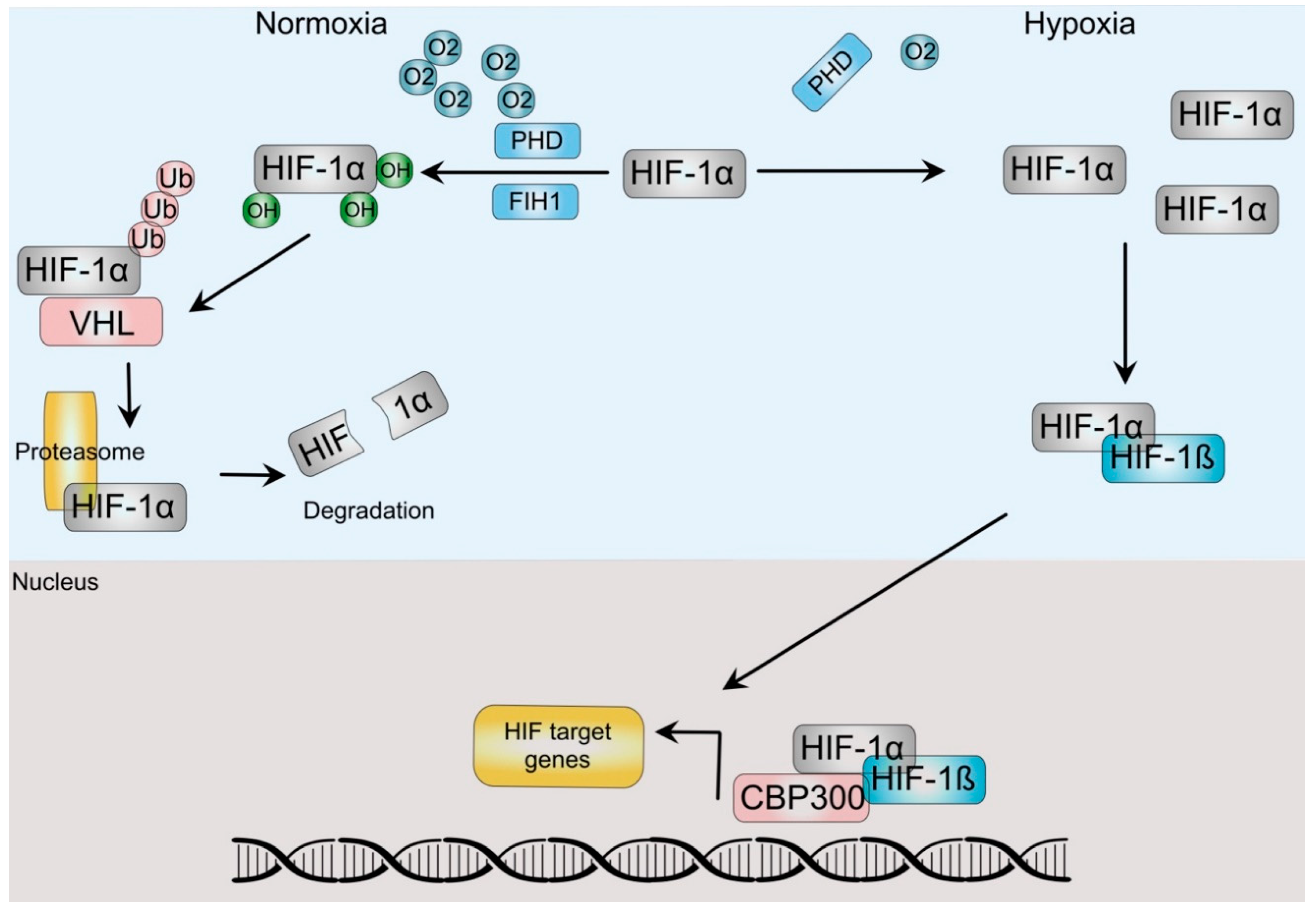
Figure 1.
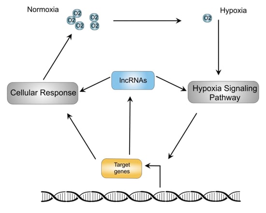
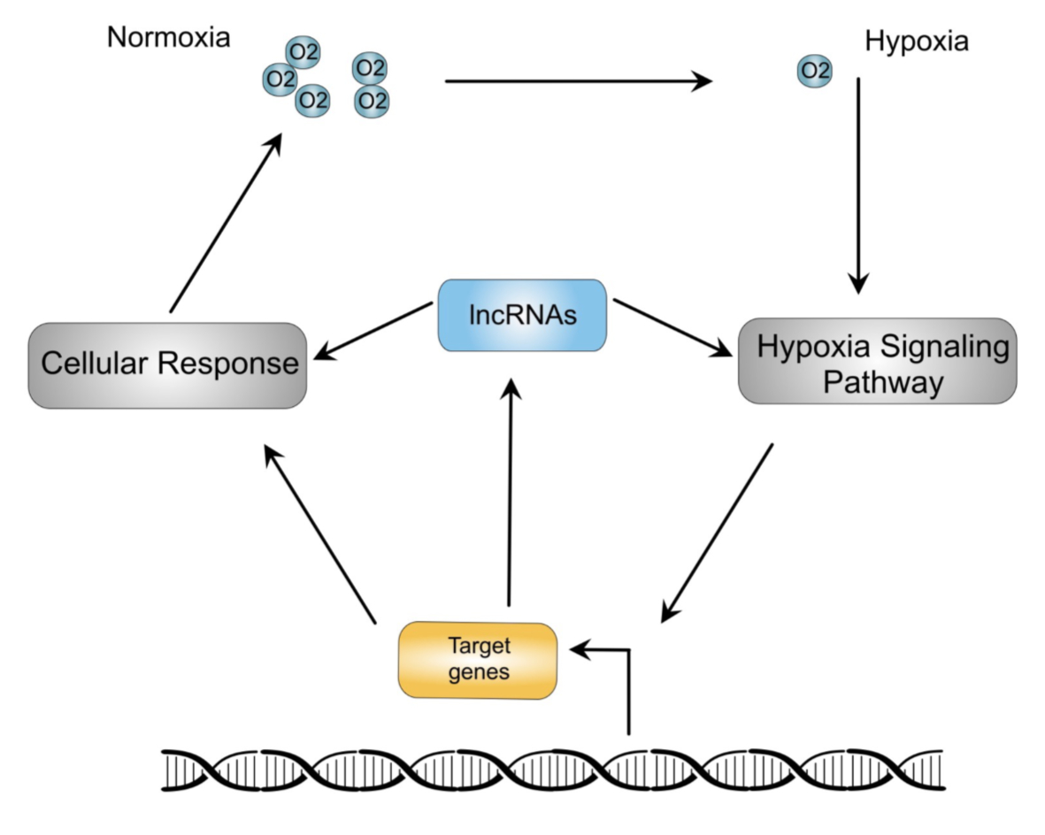
HIF-dependent hypoxia signaling pathway. In normoxia, prolyl-hydrolyses (PHD) and Factor-inhibiting hypoxia-inducible factor (FIH) hydroxylate HIF-α. Hydroxylation from the latter impairs HIF transactivation of target genes, whereas the former allows dimerization with the Von Hippel Lindau (VHL) protein, which directs HIF to ubiquitination and degradation by the proteasome. Hypoxic conditions stabilize HIF-α after PHD and FIH inactivation, allowing dimerization with HIF-ß subunits, translocation to the nucleus and association with coactivators, such as p300/CBP to regulate gene expression.
Figure 1.
HIF-dependent hypoxia signaling pathway. In normoxia, prolyl-hydrolyses (PHD) and Factor-inhibiting hypoxia-inducible factor (FIH) hydroxylate HIF-α. Hydroxylation from the latter impairs HIF transactivation of target genes, whereas the former allows dimerization with the Von Hippel Lindau (VHL) protein, which directs HIF to ubiquitination and degradation by the proteasome. Hypoxic conditions stabilize HIF-α after PHD and FIH inactivation, allowing dimerization with HIF-ß subunits, translocation to the nucleus and association with coactivators, such as p300/CBP to regulate gene expression.


Table 1.
Hypoxia-related lncRNA in breast cancer.
| Year | lncRNA | Expression in Breast Cancer | Regulated by Hypoxia | Regulates Hypoxia Signaling | Molecular | Phenotype | References |
|---|---|---|---|---|---|---|---|
| 2015 | NEAT1 | Paraspeckle formation | Proliferation, apoptosis inhibition | [60] | |||
| 2015 | EFNA3 | Induce Ephrin-A3 accumulation | Increased metastatic potential | [61] | |||
| 2016 | LINK-A | Increased in TNBC | Allows HIF-1α phosphorylation and stabilization by BRK and LRRK2 | Glycolysis | [62] | ||
| 2018 | NDRG-OT1 | Inhibition of NDRG1 by ubiquitination-mediated proteolysis. Inhibition of NDRG1 promoter activation | [55,63] | ||||
| 2018 | H19 | miRNA let-7 endogenous competitor. HIF-1α activator | PDK-1-mediated Increased glycolysis | [64] | |||
| 2018, 2019, 2021 | MALAT1, TALAM | Increased | XBP1-HIF-1α and HER2 pathway-mediated effects. miRNA-3064-5p endogenous competitor. Chromatin remodeling | Proliferation and invasion | [44,65,66] | ||
| 2019 | HISLA | Increased in breast cancer TAMs | HIF-1α stabilization by blocking PHD2 | Macrophage-mediated enhanced glycolysis | [67] | ||
| 2020 | LINC00662 | Increased | miR-497-5p endogenous competitor mediated EglN2 regulation | Proliferation and migration | [68] | ||
| 2020 | MIR210HG | Increased | HIF-1α translation enhancement | Glycolysis | [69] | ||
| 2020 | lncMat2B | Stemness | [70] | ||||
| 2020 | BRCT1 | Increased | miR-1303 endogenous competitor-mediated stabilization of PTBP3 | Proliferation, migration, increased metastatic potential | [71] | ||
| 2020 | HOTAIR | Increased | miR-204 endogenous competitor-mediated FAK induction | Migration | [72] | ||
| 2020 | RAB11B-AS1 | Increased | Induces the expression of VEGFA and ANGPTL4 by recruitment of RNA polymerase II | Migration, invasion, and angiogenesis | [73] | ||
| 2021 | LncIHAT | Promotes expression of PDK1 and ITGA6 | Tumor growth and metastasis | [56] | |||
| 2021 | MTORT1 | miR-26a-5p endogenous competitor-mediated CREB1 and STK4 regulation | Proliferation and migration | [74] | |||
| 2021 | UCA1 | Increased | Proliferation and apoptosis | [75] | |||
| 2021 | LINC00926 | Regulation of PGK1 via ubiquitination mediated by STUB1 | Glycolysis | [76] | |||
| 2021 | VCAN-AS1 | Increased | miR-106a-5p endogenous competitor-mediated regulation of STAT3 | Proliferation, migration, invasion, and EMT. | [77] | ||
| 2021 | HCG18 | Increased | miR-103a-3p endogenous competitor-mediated UBE2O/AMPKα2/mTORC1 activation | Proliferation, invasion and stemness | [58] | ||
| 2021 | eNEMAL | Alternative splicing of NEAT1 | [78] | ||||
| 2021 | PCAT-1 | Increased | Stabilizes HIF-1α by preventing RACK1 binding | [46] | |||
| 2021 | GHET1 | Increased in TNBC | Decreased phosphorylation of LATS1 allowing YAP nuclear translocation | Proliferation, viability, invasion, glycolysis | [79] | ||
| 2021 | SPRY4-IT1 | STAU1 recruitment to TCEB1, upregulating HIF-1α | Metastasis | [80] | |||
| 2021 | HIFAL | Increased | Recruitment of PHD3 to PKM2 to enhance HIF-1α activity | Tumor growth | [59] | ||
| 2021 | KB-1980E6.3 | Increased | Activation of IGF2BP1/c-Myc signaling axis to stabilize c-Myc | Stemness | [81] | ||
| 2022 | RBM5-AS1 | Increased | Activation of the Wnt pathway | Proliferation, migration, invasion, EMT, and stemness | [82] | ||
| 2022 | LINC00649 | Increased in TNBC | Stabilization of HIF-1α via NF90/NF45 interaction | Proliferation, migration, and invasion | [83] |
TNBC: Triple Negative Breast Cancer; TAMs: Tumor-Associated Macrophages. Gray bars show regulation (upstream or downstream)
Table 2.
Hypoxia-associated molecular lncRNA markers.
| Year | Marker Genes | Signature Derived From | Features | Reference |
|---|---|---|---|---|
| 2017 | 3-gene signature. ANRIL, HIF1A-AS2, UCA1 | Plasma | Differentiate TNBC vs. other | [99] |
| 2020 | 13-gene signature that includes SNHG16, LINC00899, PSMG3-AS1 and PAXIP-AS1 lncRNAs | Tumoral tissue | Prognosis | [57] |
| 2021 | 12-gene signature. LINC01614, LINCO2384, AL109955.1, AC044849.1, LINCO2084, LINC01615, YTHDF3-AS1, AL451085.3, AL512380.1, HSD11B1-AS1, AC110995.1, AC004847.1, TRG-AS1, AC011978.2, CDK6-AS1, TDRKH-AS1, OTUD6B-AS1, MIR4435-2HG, AP003774.2, SLC12A5-AS1 | Tumoral tissue | Prognosis and immune features | [101] |
| 2021 | 4-gene signature. AL031316.1, AC004585.1, LINC01235, and ACTA2-AS1 | Tumoral tissue | Prognosis | [100] |
| 2018 | MALAT1 | Tumoral tissue | Number of metastatic lymph nodes | [44] |
| 2019 | HIF1A-AS2 | Tumoral tissue | Lymph node metastasis, distant metastasis, and unfavorable histological grade. Shorter overall survival | [102] |
Publisher’s Note: MDPI stays neutral with regard to jurisdictional claims in published maps and institutional affiliations. |
© 2022 by the authors. Licensee MDPI, Basel, Switzerland. This article is an open access article distributed under the terms and conditions of the Creative Commons Attribution (CC BY) license (https://creativecommons.org/licenses/by/4.0/).
Share and Cite
MDPI and ACS Style
Maldonado, V.; Melendez-Zajgla, J. The Role of Hypoxia-Associated Long Non-Coding RNAs in Breast Cancer. Cells 2022, 11, 1679. https://doi.org/10.3390/cells11101679
AMA Style
Maldonado V, Melendez-Zajgla J. The Role of Hypoxia-Associated Long Non-Coding RNAs in Breast Cancer. Cells. 2022; 11(10):1679. https://doi.org/10.3390/cells11101679
Chicago/Turabian StyleMaldonado, Vilma, and Jorge Melendez-Zajgla. 2022. "The Role of Hypoxia-Associated Long Non-Coding RNAs in Breast Cancer" Cells 11, no. 10: 1679. https://doi.org/10.3390/cells11101679
Note that from the first issue of 2016, this journal uses article numbers instead of page numbers. See further details here.