
Pre-Clinical and Clinical Applications of Small Interfering RNAs (siRNA) and Co-Delivery Systems for Pancreatic Cancer Therapy


 ,
, 

Abstract
:1. Introduction
2. SiRNAs: From Basic to Application in Cancer Therapy
3. SiRNA and Pancreatic Cancer Therapy
3.1. Proliferation and Growth
3.2. Metastasis and Angiogenesis
3.3. Immune Regulation
3.4. Therapy Response and Synergistic Therapy
4. Chemical Modification of siRNAs
5. Co-Delivery Systems
5.1. Lipid-Based Nanoparticles
5.2. Polymeric Nanoparticles
5.3. Carbon-Based Nanoparticles
5.4. Dendrimers
5.5. Metal-Based Nanoparticles
5.6. Viral Vectors
6. SiRNA and Pancreatic Cancer: Clinical Applications
7. Conclusions and Remarks
Author Contributions
Funding
Conflicts of Interest
Abbreviations
| PC | Pancreatic cancer |
| siRNA | Small interfering RNA |
| BBB | Blood-brain barrier |
| BTB | Blood-tumor barrier |
| RNAi | RNA interference |
| mRNA | Messenger RNA |
| RISC | RNA-induced silencing |
| Ago | Argonaute |
| HSP27 | Heat shock protein 27 |
| PARP | Poly (ADP-ribose) polymerase |
| ESM-1 | Endothelial cell-specific molecule-1 |
| proNGF | Precursor of nerve growth factor |
| ATG5 | Autophagy-related gene 5 |
| RAP80 | Receptor-associated protein 80 |
| HIF-1α | Hypoxia inducible factor-1α |
| NF-κB | Nuclear factor-kappaB |
| COX-2 | Cyclooxygenase-2 |
| EGFR | Epidermal growth factor receptor |
| VEGF | Vascular endothelial growth factor |
| HDAC | Histone deacetylase |
| HRE | Hypoxia response element |
| HPA | Heparinase |
| FGF2 | Fibroblast growth factor 2 |
| SDC1 | Syndecan-1 |
| EMT | Epithelial-to-mesenchymal transition |
| NELFE | Negative elongation factor E |
| NDRG2 | N-Myc downstream-regulated gene 2 |
| lncRNA | long non-coding RNA |
| miRNA | microRNA |
| MMP | Matrix metalloproteinase |
| TGF-β | Transforming growth factor-beta |
| RIG-I | Retinoic acid-inducible gene I |
| CADM1 | Cell adhesion molecule 1 |
| RR | Ribonucleotide reductase |
| DOX | Doxorubicin |
| GEM | Gemcitabine |
| hTERT | Human telomerase reverse transcriptase |
| hnRNP | Heterogenous nuclear ribonucleotide protein A2/B1 |
| TS | Thymidylate synthase |
| TRAIL | TNF-related apoptosis-inducing ligand |
| Mcl-1 | Myeloid cell leukemia 1 |
| EPR | Enhanced permeability and retention |
| Fab | Fragment |
| PIC | Polyion complex |
| TF | Tissue factor |
| BCPVs | Biodegradable charged polyester-based vectors |
| iRGD | Internalizing RGD peptide |
| IV | Intravenous |
| EPAS1 | Endothelial PAS domain protein 1 |
| PLGA | Polyethylenimine-poly (lactic-coglycolide) |
| GPCR | G protein-coupled receptor |
| ABC | ATP-binding cassette |
| YAP | Yes-associated protein |
| LODER | Local drug EluteR |
| 2D | Two-dimensional |
| PEG | Polyethylene glycol |
| PTX | Paclitaxel |
| PTP | Plectin-1 targeted peptide |
| SPIONs | Superparamagnetic iron oxide nanoparticles |
| PLK1 | Polo-like kinase 1 |
| NGF | Nerve growth factor |
| PI3K | Phosphoinositide 3-kinase |
| Akt | Protein kinase-B |
| MSNPs | Mesoporous silica nanoparticles |
| PEI | Polyethyleneimine |
References
- Paternoster, S.; Falasca, M. The Intricate Relationship between Diabetes, Obesity and Pancreatic Cancer. Biochim. Biophys. Acta Rev. Cancer 2020, 1873, 188326. [Google Scholar] [CrossRef]
- Daoud, A.Z.; Mulholland, E.J.; Cole, G.; McCarthy, H.O. MicroRNAs in pancreatic cancer: Biomarkers, prognostic, and therapeutic modulators. BMC Cancer 2019, 19, 1130. [Google Scholar] [CrossRef] [Green Version]
- Siegel, R.L.; Miller, K.D.; Fuchs, H.E.; Jemal, A. Cancer Statistics, 2021. CA Cancer J. Clin. 2021, 71, 7–33. [Google Scholar] [CrossRef] [PubMed]
- Ashrafizadeh, M.; Bakhoda, M.R.; Bahmanpour, Z.; Ilkhani, K.; Zarrabi, A.; Makvandi, P.; Khan, H.; Mazaheri, S.; Darvish, M.; Mirzaei, H. Apigenin as Tumor Suppressor in Cancers: Biotherapeutic Activity, Nanodelivery, and Mechanisms with Emphasis on Pancreatic Cancer. Front. Chem. 2020, 8, 829. [Google Scholar] [CrossRef]
- Siegel, R.L.; Miller, K.D.; Jemal, A. Cancer Statistics, 2019. CA Cancer J. Clin. 2019, 69, 7–34. [Google Scholar] [CrossRef] [PubMed] [Green Version]
- Ilic, M.; Ilic, I. Epidemiology of Pancreatic Cancer. World J. Gastroenterol. 2016, 22, 9694–9705. [Google Scholar] [CrossRef] [PubMed]
- Yang, D.; Hu, Z.; Xu, J.; Tang, Y.; Wang, Y.; Cai, Q.; Zhu, Z. MiR-760 Enhances Sensitivity of Pancreatic Cancer Cells to Gemcitabine through Modulating INTEGRIN β1. Biosci. Rep. 2019, 39, BSR20192358. [Google Scholar] [CrossRef] [PubMed]
- Liu, Y.; Guo, C.; Li, F.; Wu, L. LncRNA LOXL1-AS1/miR-28-5p/SEMA7A Axis Facilitates Pancreatic Cancer Progression. Cell Biochem. Funct. 2020, 38, 58–65. [Google Scholar] [CrossRef] [PubMed]
- Gong, Y.; Dai, H.S.; Shu, J.J.; Liu, W.; Bie, P.; Zhang, L.D. LNC00673 Suppresses Proliferation and Metastasis of Pancreatic Cancer via Target miR-504/ HNF1A. J. Cancer 2020, 11, 940–948. [Google Scholar] [CrossRef] [PubMed] [Green Version]
- Ercan, G.; Karlitepe, A.; Ozpolat, B. Pancreatic Cancer Stem Cells and Therapeutic Approaches. Anticancer Res. 2017, 37, 2761–2775. [Google Scholar] [CrossRef] [Green Version]
- Chiorean, E.G.; Coveler, A.L. Pancreatic Cancer: Optimizing Treatment Options, New, and Emerging Targeted Therapies. Drug Des. Devel. Ther. 2015, 9, 3529–3545. [Google Scholar] [CrossRef] [PubMed] [Green Version]
- Jung, Y.Y.; Ko, J.H.; Um, J.Y.; Chinnathambi, A.; Alharbi, S.A.; Sethi, G.; Ahn, K.S. LDL Cholesterol Promotes the Proliferation of Prostate and Pancreatic Cancer Cells by Activating the STAT3 Pathway. J. Cell Physiol. 2021, 236, 5253–5264. [Google Scholar] [CrossRef] [PubMed]
- Pandya, G.; Kirtonia, A.; Sethi, G.; Pandey, A.K.; Garg, M. The Implication of Long Non-Coding RNAs in the Diagnosis, Pathogenesis and Drug Resistance of Pancreatic Ductal Adenocarcinoma and Their Possible Therapeutic Potential. Biochim. Biophys. Acta Rev. Cancer 2020, 1874, 188423. [Google Scholar] [CrossRef]
- Ashrafizadeh, M.; Ahmadi, Z.; Mohamadi, N.; Zarrabi, A.; Abasi, S.; Dehghannoudeh, G.; Tamaddondoust, R.N.; Khanbabaei, H.; Mohammadinejad, R.; Thakur, V.K. Chitosan-Based Advanced Materials for Docetaxel and Paclitaxel Delivery: Recent Advances and Future Directions in Cancer Theranostics. Int. J. Biol. Macromol. 2020, 145, 282–300. [Google Scholar] [CrossRef] [PubMed]
- Ashrafizadeh, M.; Zarrabi, A.; Hashemi, F.; Zabolian, A.; Saleki, H.; Bagherian, M.; Azami, N.; Bejandi, A.K.; Hushmandi, K.; Ang, H.L. Polychemotherapy with Curcumin and Doxorubicin via Biological Nanoplatforms: Enhancing Antitumor Activity. Pharmaceutics 2020, 12, 1084. [Google Scholar] [CrossRef]
- Yao, Y.; Wang, T.; Liu, Y.; Zhang, N. Co-Delivery of Sorafenib and VEGF-siRNA via pH-Sensitive Liposomes for the Synergistic Treatment of Hepatocellular Carcinoma. Artif. Cells Nanomed. Biotechnol. 2019, 47, 1374–1383. [Google Scholar] [CrossRef] [Green Version]
- He, Y.; Guo, S.; Wu, L.; Chen, P.; Wang, L.; Liu, Y.; Ju, H. Near-Infrared Boosted ROS Responsive siRNA Delivery and Cancer Therapy with Sequentially Peeled Upconversion Nano-Onions. Biomaterials 2019, 225, 119501. [Google Scholar] [CrossRef]
- Wang, D.; Zhang, F.; Gao, G. CRISPR-Based Therapeutic Genome Editing: STRATEGIES and in Vivo Delivery by aav Vectors. Cell 2020, 181, 136–150. [Google Scholar] [CrossRef] [PubMed]
- Zhen, S.; Li, X. Liposomal Delivery of CRISPR/Cas9. Cancer Gene Ther. 2020, 27, 515–527. [Google Scholar] [CrossRef]
- Mirzaei, S.; Abbaspour, A.; Fallahzadeh, P.; Noori, M.; Hashemi, F.; Hushmandi, K.; Daneshi, S.; Kumar, A.P. Small Interfering RNA (siRNA) to Target Genes and Molecular Pathways in Glioblastoma Therapy: Current Status with an Emphasis on Delivery Systems. Life Sci. 2021, 275, 119368. [Google Scholar] [CrossRef]
- Ashrafizade, M.; Delfi, M.; Hashemi, F.; Zabolian, A.; Saleki, H.; Bagherian, M.; Azami, N.; Farahani, M.V.; omid Sharifzadeh, S.; Hamzehlou, S. Biomedical Application of Chitosan-Based Nanoscale Delivery Systems: Potential Usefulness in siRNA Delivery for Cancer Therapy. Carbohydr. Polym. 2021, 260, 117809. [Google Scholar] [CrossRef]
- Mirzaei, S.; Gholami, M.H.; Hashemi, F.; Zabolian, A.; Hushmandi, K.; Rahmanian, V.; Entezari, M.; Girish, Y.R.; Kumar, K.S.S.; Aref, A.R. Employing siRNA Tool and Its Delivery Platforms in Suppressing Cisplatin Resistance: Approaching to a New Era of Cancer Chemotherapy. Life Sci. 2021, 277, 119430. [Google Scholar] [CrossRef]
- Ashrafizadeh, M.; Zarrabi, A.; Hushmandi, K.; Hashemi, F.; Rahmani Moghadam, E.; Raei, M.; Kalantari, M.; Tavakol, S.; Mohammadinejad, R.; Najafi, M. Progress in Natural Compounds/siRNA Co-Delivery Employing Nanovehicles for Cancer Therapy. ACS Comb. Sci. 2020, 22, 669–700. [Google Scholar] [CrossRef]
- Ashrafizadeh, M.; Hushmandi, K.; Rahmani Moghadam, E.; Zarrin, V.; Hosseinzadeh Kashani, S.; Bokaie, S.; Najafi, M.; Tavakol, S.; Mohammadinejad, R.; Nabavi, N. Progress in Delivery of siRNA-Based Therapeutics Employing Nano-Vehicles for Treatment of Prostate Cancer. Bioengineering 2020, 7, 91. [Google Scholar] [CrossRef]
- Peer, D.; Karp, J.M.; Hong, S.; Farokhzad, O.C.; Margalit, R.; Langer, R. Nanocarriers as an Emerging Platform for Cancer Therapy. Nat. Nanotechnol. 2007, 2, 751–760. [Google Scholar] [CrossRef]
- Rui, Y.; Wilson, D.R.; Choi, J.; Varanasi, M.; Sanders, K.; Karlsson, J.; Lim, M.; Green, J.J. Carboxylated Branched Poly (β-Amino ester) Nanoparticles Enable Robust Cytosolic Protein Delivery and CRISPR-Cas9 Gene Editing. Sci. Adv. 2019, 5, eaay3255. [Google Scholar] [CrossRef] [PubMed] [Green Version]
- Jubair, L.; Fallaha, S.; McMillan, N.A.J. Systemic Delivery of CRISPR/Cas9 Targeting HPV Oncogenes is Effective at Eliminating Established Tumors. Mol. Ther. 2019, 27, 2091–2099. [Google Scholar] [CrossRef] [PubMed]
- Wei, T.; Cheng, Q.; Farbiak, L.; Anderson, D.G.; Langer, R.; Siegwart, D.J. Delivery of Tissue-Targeted Scalpels: Opportunities and Challenges for in Vivo CRISPR/Cas-Based Genome Editing. ACS Nano 2020, 14, 9243–9262. [Google Scholar] [CrossRef]
- Yu, Q.; Zhang, B.; Zhou, Y.; Ge, Q.; Chang, J.; Chen, Y.; Zhang, K.; Peng, D.; Chen, W. Co-Delivery of Gambogenic Acid and VEGF-siRNA with Anionic Liposome and Polyethylenimine Complexes to HepG2 Cells. J. Liposome Res. 2019, 29, 322–331. [Google Scholar] [CrossRef]
- Vocelle, D.; Chan, C.; Walton, S.P. Endocytosis Controls siRNA Efficiency: Implications for siRNA Delivery Vehicle Design and Cell-Specific Targeting. Nucleic Acid Ther. 2020, 30, 22–32. [Google Scholar] [CrossRef]
- Li, S.; Liu, Z.; Ji, F.; Xiao, Z.; Wang, M.; Peng, Y.; Zhang, Y.; Liu, L.; Liang, Z.; Li, F. Delivery of Quantum Dot-siRNA Nanoplexes in SK-N-SH Cells for BACE1 Gene Silencing and Intracellular Imaging. Mol. Ther. Nucleic Acids 2012, 1, 22–32. [Google Scholar] [CrossRef] [PubMed]
- Kozielski, K.L.; Ruiz-Valls, A.; Tzeng, S.Y.; Guerrero-Cázares, H.; Rui, Y.; Li, Y.; Vaughan, H.J.; Gionet-Gonzales, M.; Vantucci, C.; Kim, J.; et al. Cancer-Selective Nanoparticles for Combinatorial siRNA Delivery to Primary Human GBM in Vitro and in Vivo. Biomaterials 2019, 209, 79–87. [Google Scholar] [CrossRef]
- Kulkarni, J.A.; Darjuan, M.M.; Mercer, J.E.; Chen, S.; van der Meel, R.; Thewalt, J.L.; Tam, Y.Y.C.; Cullis, P.R. On the Formation and morphology of lipid nanoparticles containing ionizable cationic lipids and siRNA. ACS Nano 2018, 12, 4787–4795. [Google Scholar] [CrossRef] [Green Version]
- Shaat, H.; Mostafa, A.; Moustafa, M.; Gamal-Eldeen, A.; Emam, A.; El-Hussieny, E.; Elhefnawi, M. Modified gold nanoparticles for Intracellular Delivery of Anti-Liver Cancer siRNA. Int. J. Pharm. 2016, 504, 125–133. [Google Scholar] [CrossRef]
- Yamakawa, K.; Nakano-Narusawa, Y.; Hashimoto, N.; Yokohira, M.; Matsuda, Y. Development and Clinical Trials of Nucleic Acid Medicines for Pancreatic Cancer Treatment. Int. J. Mol. Sci. 2019, 20, 4224. [Google Scholar] [CrossRef] [PubMed] [Green Version]
- Kurtanich, T.; Roos, N.; Wang, G.; Yang, J.; Wang, A.; Chung, E.J. Pancreatic Cancer Gene Therapy Delivered by Nanoparticles. SLAS Technol. 2019, 24, 151–160. [Google Scholar] [CrossRef] [PubMed]
- Takakura, K.; Kawamura, A.; Torisu, Y.; Koido, S.; Yahagi, N.; Saruta, M. The Clinical Potential of Oligonucleotide Therapeutics against Pancreatic Cancer. Int. J. Mol. Sci. 2019, 20, 3331. [Google Scholar] [CrossRef] [Green Version]
- Kokkinos, J.; Ignacio, R.M.C.; Sharbeen, G.; Boyer, C.; Gonzales-Aloy, E.; Goldstein, D.; Australian Pancreatic Cancer Genome Initiative, A.; McCarroll, J.A.; Phillips, P.A. Targeting the Undruggable in Pancreatic Cancer Using Nano-Based Gene Silencing Drugs. Biomaterials 2020, 240, 119742. [Google Scholar] [CrossRef] [PubMed]
- Sato-Dahlman, M.; Wirth, K.; Yamamoto, M. Role of Gene Therapy in Pancreatic Cancer—A Review. Cancers 2018, 10, 103. [Google Scholar] [CrossRef] [Green Version]
- Liu, Y.; Wu, W.; Wang, Y.; Han, S.; Yuan, Y.; Huang, J.; Shuai, X.; Peng, Z. Recent Development of Gene Therapy for Pancreatic Cancer Using Non-Viral Nanovectors. Biomater. Sci. 2021, 9, 6673–6690. [Google Scholar] [CrossRef] [PubMed]
- Galanopoulos, M.; Doukatas, A.; Gkeros, F.; Viazis, N.; Liatsos, C. Room for Improvement in the Treatment of Pancreatic Cancer: Novel Opportunities from Gene Targeted Therapy. World J. Gastroenterol. 2021, 27, 3568–3580. [Google Scholar] [CrossRef] [PubMed]
- Kim, H.J.; Kim, A.; Miyata, K.; Kataoka, K. Recent Progress in Development of siRNA Delivery Vehicles for Cancer Therapy. Adv. Drug Deliv. Rev. 2016, 104, 61–77. [Google Scholar] [CrossRef] [PubMed] [Green Version]
- Kim, D.H.; Rossi, J.J. Strategies for Silencing Human Disease Using RNA Interference. Nat. Rev. Genet. 2007, 8, 173–184. [Google Scholar] [CrossRef]
- Singh, A.; Trivedi, P.; Jain, N.K. Advances in siRNA Delivery in Cancer Therapy. Artif. Cells Nanomed. Biotechnol. 2018, 46, 274–283. [Google Scholar] [CrossRef]
- Houseley, J.; Tollervey, D. The Many Pathways of RNA Degradation. Cell 2009, 136, 763–776. [Google Scholar] [CrossRef] [Green Version]
- De La Sierra-Gallay, I.; Zig, L.; Jamalli, A.; Putzer, H. Structural Insights into the Dual Activity of RNase. J. Nat. Struct. Mol. Biol. 2008, 15, 206–212. [Google Scholar] [CrossRef]
- Mathy, N.; Bénard, L.; Pellegrini, O.; Daou, R.; Wen, T.; Condon, C. 5′-to-3′ Exoribonuclease Activity in Bacteria: Role of RNase J1 in rRNA Maturation and 5′ Stability of mRNA. Cell 2007, 129, 681–692. [Google Scholar] [CrossRef] [Green Version]
- Matranga, C.; Tomari, Y.; Shin, C.; Bartel, D.P.; Zamore, P.D. Passenger-Strand Cleavage Facilitates Assembly of siRNA into Ago2-Containing RNAi Enzyme Complexes. Cell 2005, 123, 607–620. [Google Scholar] [CrossRef] [Green Version]
- Tolia, N.H.; Joshua-Tor, L. Slicer and the Argonautes. Nat. Chem. Biol. 2007, 3, 36–43. [Google Scholar] [CrossRef]
- Ameres, S.L.; Martinez, J.; Schroeder, R. Molecular Basis for Target RNA Recognition and Cleavage by Human RISC. Cell 2007, 130, 101–112. [Google Scholar] [CrossRef] [Green Version]
- Rand, T.A.; Petersen, S.; Du, F.; Wang, X. Argonaute2 Cleaves the Anti-Guide Strand of siRNA during RISC Activation. Cell 2005, 123, 621–629. [Google Scholar] [CrossRef] [Green Version]
- Dykxhoorn, D.M.; Lieberman, J. Knocking down Disease with siRNAs. Cell 2006, 126, 231–235. [Google Scholar] [CrossRef] [Green Version]
- Novina, C.D.; Sharp, P.A. The RNAi Revolution. Nature 2004, 430, 161–164. [Google Scholar] [CrossRef] [PubMed]
- Lingel, A.; Sattler, M. Novel Modes of Protein–RNA Recognition in the RNAi Pathway. Curr. Opin. Struct. Biol. 2005, 15, 107–115. [Google Scholar] [CrossRef] [PubMed]
- George, J.; Banik, N.L.; Ray, S.K. Bcl-2 siRNA Augments Taxol Mediated Apoptotic Death in Human Glioblastoma U138MG and U251MG Cells. Neurochem. Res. 2009, 34, 66–78. [Google Scholar] [CrossRef] [PubMed]
- Salehi Khesht, A.M.; Karpisheh, V.; Sahami Gilan, P.; Melnikova, L.A.; Olegovna Zekiy, A.; Mohammadi, M.; Hojjat-Farsangi, M.; Majidi Zolbanin, N.; Mahmoodpoor, A.; Hassannia, H.; et al. Blockade of CD73 Using siRNA Loaded Chitosan Lactate Nanoparticles Functionalized with TAT-Hyaluronate Enhances Doxorubicin Mediated Cytotoxicity in Cancer Cells Both In Vitro and In Vivo. Int. J. Biol. Macromol. 2021, 186, 849–863. [Google Scholar] [CrossRef]
- Li, Y.; Tan, X.; Liu, X.; Liu, L.; Fang, Y.; Rao, R.; Ren, Y.; Yang, X.; Liu, W. Enhanced Anticancer Effect of Doxorubicin by TPGS-Coated Liposomes with Bcl-2 siRNA-Corona for Dual Suppression of Drug Resistance. Asian J. Pharm. Sci. 2020, 15, 646–660. [Google Scholar] [CrossRef]
- Zhang, Y.; Fu, X.; Jia, J.; Wikerholmen, T.; Xi, K.; Kong, Y.; Wang, J.; Chen, H.; Ma, Y.; Li, Z.; et al. Glioblastoma Therapy Using Codelivery of Cisplatin and Glutathione Peroxidase Targeting siRNA from Iron Oxide Nanoparticles. ACS Appl. Mater. Interfaces 2020, 12, 43408–43421. [Google Scholar] [CrossRef]
- Patel, V.; Lalani, R.; Vhora, I.; Bardoliwala, D.; Patel, A.; Ghosh, S.; Misra, A. Co-Delivery of Cisplatin and siRNA through Hybrid Nanocarrier Platform for Masking Resistance to Chemotherapy in Lung Cancer. Drug Deliv. Transl. Res. 2020, 11, 2052–2071. [Google Scholar] [CrossRef]
- Lai, Y.; Kong, Z.; Zeng, T.; Xu, S.; Duan, X.; Li, S.; Cai, C.; Zhao, Z.; Wu, W. PARP1-siRNA Suppresses Human Prostate Cancer Cell Growth and Progression. Oncol. Rep. 2018, 39, 1901–1909. [Google Scholar] [CrossRef] [Green Version]
- Rebollo, J.; Geliebter, J.; Reyes, N. ESM-1 siRNA Knockdown Decreased Migration and Expression of CXCL3 in Prostate Cancer Cells. Int. J. Biomed. Sci. 2017, 13, 35–42. [Google Scholar] [PubMed]
- Birmingham, A.; Anderson, E.M.; Reynolds, A.; Ilsley-Tyree, D.; Leake, D.; Fedorov, Y.; Baskerville, S.; Maksimova, E.; Robinson, K.; Karpilow, J. 3′ UTR Seed Matches, But Not Overall Identity, Are Associated with RNAi off-Targets. Nat. Methods. 2006, 3, 199–204. [Google Scholar] [CrossRef]
- Ozcan, G.; Ozpolat, B.; Coleman, R.L.; Sood, A.K.; Lopez-Berestein, G. Preclinical and Clinical Development of siRNA-Based Therapeutics. Adv. Drug Deliv. Rev. 2015, 87, 108–119. [Google Scholar] [CrossRef] [PubMed] [Green Version]
- Yang, Q.; Humphreys, S.C.; Lade, J.M.; Li, A.P. Prolonged Cultured Human Hepatocytes as an in Vitro Experimental System for the Evaluation of Potency and Duration of Activity of RNA Therapeutics: Demonstration of Prolonged Duration of Gene Silencing Effects of a GalNAc-Conjugated Human Hypoxanthine Phosphoribosyl Transferase (HPRT1) siRNA. Biochem. Pharmacol. 2021, 189, 114374. [Google Scholar] [CrossRef] [PubMed]
- Anselmo, A.C.; Mitragotri, S. Nanoparticles in the Clinic. Bioeng. Transl. Med. 2016, 1, 10–29. [Google Scholar] [CrossRef] [PubMed]
- Shi, M.; Zhang, J.; Huang, Z.; Chen, Y.; Pan, S.; Hu, H.; Qiao, M.; Chen, D.; Zhao, X. Stimuli-Responsive Release and Efficient siRNA Delivery in Non-Small Cell Lung Cancer by a Poly(l-Histidine)-Based Multifunctional Nanoplatform. J. Mater. Chem. B 2020, 8, 1616–1628. [Google Scholar] [CrossRef]
- Li, S.; Saw, P.E.; Lin, C.; Nie, Y.; Tao, W.; Farokhzad, O.C.; Zhang, L.; Xu, X. Redox-Responsive Polyprodrug Nanoparticles for Targeted siRNA Delivery and Synergistic Liver Cancer Therapy. Biomaterials 2020, 234, 119760. [Google Scholar] [CrossRef]
- Lan, B.; Wu, J.; Li, N.; Pan, C.; Yan, L.; Yang, C.; Zhang, L.; Yang, L.; Ren, M. Hyperbranched Cationic Polysaccharide Derivatives for Efficient siRNA Delivery and Diabetic Wound Healing Enhancement. Int. J Biol. Macromol. 2020, 154, 855–865. [Google Scholar] [CrossRef] [PubMed]
- Di Silvio, D.; Martínez-Moro, M.; Salvador, C.; de Los Angeles Ramirez, M.; Caceres-Velez, P.R.; Ortore, M.G.; Dupin, D.; Andreozzi, P.; Moya, S.E. Self-Assembly of Poly(Allylamine)/siRNA Nanoparticles, Their Intracellular Fate and siRNA Delivery. J. Colloid Interface Sci. 2019, 557, 757–766. [Google Scholar] [CrossRef]
- Karlsson, J.; Rui, Y.; Kozielski, K.L.; Placone, A.L.; Choi, O.; Tzeng, S.Y.; Kim, J.; Keyes, J.J.; Bogorad, M.I.; Gabrielson, K.; et al. Engineered Nanoparticles for Systemic siRNA Delivery to Malignant Brain Tumours. Nanoscale 2019, 11, 20045–20057. [Google Scholar] [CrossRef] [PubMed]
- Chen, M.; Chen, M.; He, J. Cancer Cell Membrane Cloaking Nanoparticles for Targeted Co-Delivery of Doxorubicin and PD-L1 siRNA. Artif. Cells Nanomed. Biotechnol. 2019, 47, 1635–1641. [Google Scholar] [CrossRef]
- Hanurry, E.Y.; Hsu, W.H.; Darge, H.F.; Birhan, Y.S.; Mekonnen, T.W.; Andrgie, A.T.; Chou, H.Y.; Cheng, C.C.; Lai, J.Y.; Tsai, H.C. In vitro siRNA delivery via diethylenetriamine- and tetraethylenepentamine-modified carboxyl group-terminated Poly(amido)amine generation 4.5 dendrimers. Mater. Sci. Eng. C Mater. Biol. Appl. 2020, 106, 110245. [Google Scholar] [CrossRef]
- Zou, Y.; Sun, X.; Wang, Y.; Yan, C.; Liu, Y.; Li, J.; Zhang, D.; Zheng, M.; Chung, R.S.; Shi, B. Single siRNA Nanocapsules for Effective siRNA Brain Delivery and Glioblastoma Treatment. Adv. Mater. 2020, 32, e2000416. [Google Scholar] [CrossRef]
- Ghaffari, M.; Dehghan, G.; Baradaran, B.; Zarebkohan, A.; Mansoori, B.; Soleymani, J.; Ezzati Nazhad Dolatabadi, J.; Hamblin, M.R. Co-Delivery of Curcumin and Bcl-2 siRNA by PAMAM Dendrimers for Enhancement of the Therapeutic Efficacy in HeLa Cancer Cells. Colloids Surf. B Biointerfaces 2020, 188, 110762. [Google Scholar] [CrossRef]
- Mainini, F.; Eccles, M.R. Lipid and Polymer-Based Nanoparticle siRNA Delivery Systems for Cancer Therapy. Molecules 2020, 25, 2692. [Google Scholar] [CrossRef] [PubMed]
- Erdene-Ochir, T.; Ganbold, T.; Zandan, J.; Han, S.; Borjihan, G.; Baigude, H. Alkylation Enhances Biocompatibility and siRNA Delivery Efficiency of Cationic Curdlan Nanoparticles. Int. J. Biol. Macromol. 2020, 143, 118–125. [Google Scholar] [CrossRef]
- Choi, M.; Jeong, H.; Kim, S.; Kim, M.; Lee, M.; Rhim, T. Targeted Delivery of Chil3/Chil4 siRNA to Alveolar Macrophages Using ternary Complexes Composed of HMG and Oligoarginine Micelles. Nanoscale 2020, 12, 933–943. [Google Scholar] [CrossRef] [PubMed]
- Bulbake, U.; Kommineni, N.; Ionov, M.; Bryszewska, M.; Khan, W. Comparison of Cationic Liposome and PAMAM Dendrimer for Delivery of Anti-Plk1 siRNA in Breast Cancer Treatment. Pharm. Dev. Technol. 2020, 25, 9–19. [Google Scholar] [CrossRef]
- Zhao, L.; Gu, C.; Gan, Y.; Shao, L.; Chen, H.; Zhu, H. Exosome-Mediated siRNA Delivery to Suppress Postoperative Breast Cancer Metastasis. J. Control. Release 2020, 318, 1–15. [Google Scholar] [CrossRef] [PubMed]
- Baran-Rachwalska, P.; Torabi-Pour, N.; Sutera, F.M.; Ahmed, M.; Thomas, K.; Nesbit, M.A.; Welsh, M.; Moore, C.B.T.; Saffie-Siebert, S.R. Topical siRNA Delivery to the Cornea and Anterior Eye by Hybrid Silicon-Lipid Nanoparticles. J. Control. Release 2020, 326, 192–202. [Google Scholar] [CrossRef]
- Azambuja, J.H.; Schuh, R.S.; Michels, L.R.; Gelsleichter, N.E.; Beckenkamp, L.R.; Iser, I.C.; Lenz, G.S.; de Oliveira, F.H.; Venturin, G.; Greggio, S.; et al. Nasal Administration of Cationic Nanoemulsions as CD73-siRNA Delivery System for Glioblastoma Treatment: A New Therapeutical Approach. Mol. Neurobiol. 2020, 57, 635–649. [Google Scholar] [CrossRef]
- Xia, Y.; Tang, G.; Wang, C.; Zhong, J.; Chen, Y.; Hua, L.; Li, Y.; Liu, H.; Zhu, B. Functionalized Selenium Nanoparticles for Targeted siRNA Delivery Silence Derlin1 and Promote Antitumor Efficacy against Cervical Cancer. Drug Deliv. 2020, 27, 15–25. [Google Scholar] [CrossRef] [Green Version]
- Kara, G.; Parlar, A.; Cakmak, M.C.; Cokol, M.; Denkbas, E.B.; Bakan, F. Silencing of Survivin and Cyclin B1 through siRNA-Loaded Arginine Modified Calcium Phosphate Nanoparticles for Non-Small-Cell Lung Cancer Therapy. Colloids Surf. B. Biointerfaces 2020, 196, 111340. [Google Scholar] [CrossRef]
- Zhang, C.; Yuan, W.; Wu, Y.; Wan, X.; Gong, Y. Co-Delivery of EGFR and BRD4 siRNA by Cell-Penetrating Peptides-Modified Redox-Responsive Complex in Triple Negative Breast Cancer Cells. Life Sci. 2021, 266, 118886. [Google Scholar] [CrossRef]
- Khodaei, M.; Rostamizadeh, K.; Taromchi, A.H.; Monirinasab, H.; Fathi, M. DDAB Cationic Lipid-mPEG, PCL Copolymer Hybrid Nano-Carrier Synthesis and Application for Delivery of siRNA Targeting IGF-1R into Breast Cancer Cells. Clin. Transl. Oncol. 2021, 23, 1167–1178. [Google Scholar] [CrossRef]
- Wang, F.; Zhang, Z. Nanoformulation of Apolipoprotein E3-Tagged Liposomal Nanoparticles for the Co-Delivery of KRAS-siRNA and Gemcitabine for Pancreatic Cancer Treatment. Pharm. Res. 2020, 37, 247. [Google Scholar] [CrossRef] [PubMed]
- Li, M.; Guo, H.; Wang, Q.; Chen, K.; Marko, K.; Tian, X.; Yang, Y. Pancreatic Stellate Cells Derived Exosomal miR-5703 Promotes Pancreatic Cancer by Downregulating CMTM4 and Activating PI3K/Akt Pathway. Cancer Lett. 2020, 490, 20–30. [Google Scholar] [CrossRef] [PubMed]
- Fu, X.; Tang, N.; Xie, W.; Mao, L.; Qiu, Y. Mind Bomb 1 Promotes Pancreatic Cancer Proliferation by Activating β-Catenin Signaling. J. Nanosci. Nanotechnol. 2020, 20, 7276–7282. [Google Scholar] [CrossRef] [PubMed]
- Wang, L.; Gao, P.; Yuan, P.; Zhou, P.; Fan, H.; Lin, X.; Yuan, X.; Zhu, M.; Fan, X.; Lu, Y.; et al. miR-573 Suppresses Pancreatic Cancer Cell Proliferation, Migration, and Invasion through Targeting TSPAN1. Strahlenther. Onkol. 2021, 197, 438–448. [Google Scholar] [CrossRef]
- Swami, P.; Thiyagarajan, S.; Vidger, A.; Indurthi, V.S.K.; Vetter, S.W.; Leclerc, E. RAGE up-Regulation Differently Affects Cell Proliferation and Migration in Pancreatic Cancer Cells. Int. J. Mol. Sci. 2020, 21, 7723. [Google Scholar] [CrossRef]
- Li, A.; Feng, L.; Niu, X.; Zeng, Q.; Li, B.; You, Z. Downregulation of OIP5-AS1 Affects proNGF-Induced Pancreatic Cancer Metastasis by Inhibiting p75NTR Levels. Aging (Albany NY) 2021, 13, 10688–10702. [Google Scholar] [CrossRef] [PubMed]
- Fofaria, N.M.; Srivastava, S.K. STAT3 Induces Anoikis Resistance, Promotes Cell Invasion and Metastatic Potential in Pancreatic Cancer Cells. Carcinogenesis 2015, 36, 142–150. [Google Scholar] [CrossRef] [Green Version]
- Lagadec, C.; Romon, R.; Tastet, C.; Meignan, S.; Com, E.; Page, A.; Bidaux, G.; Hondermarck, H.; Le Bourhis, X. Ku86 Is Important for TrkA Overexpression-Induced Breast Cancer Cell Invasion. Proteomics. Clin. Appl. 2010, 4, 580–590. [Google Scholar] [CrossRef]
- Bao, X.; Shi, J.; Xie, F.; Liu, Z.; Yu, J.; Chen, W.; Zhang, Z.; Xu, Q. Proteolytic Release of the p75NTR Intracellular Domain by ADAM10 Promotes Metastasis and Resistance to Anoikis. Cancer Res. 2018, 78, 2262–2276. [Google Scholar] [CrossRef] [Green Version]
- Xu, J.; Song, J.; Yang, X.; Guo, J.; Wang, T.; Zhuo, W. ProNGF siRNA inhibits cell proliferation and invasion of pancreatic cancer cells and promotes anoikis. Biomed Pharmacother 2019, 111, 1066–1073. [Google Scholar] [CrossRef]
- Ashrafizadeh, M.; Zarrabi, A.; Orouei, S.; Hushmandi, K.; Hakimi, A.; Zabolian, A.; Daneshi, S.; Samarghandian, S.; Baradaran, B.; Najafi, M. MicroRNA-Mediated Autophagy Regulation in Cancer Therapy: The Role in Chemoresistance/Chemosensitivity. Eur. J. Pharmacol. 2021, 892, 173660. [Google Scholar] [CrossRef] [PubMed]
- Ashrafizadeh, M.; Ahmadi, Z.; Farkhondeh, T.; Samarghandian, S. Autophagy Regulation Using Luteolin: New Insight into Its Anti-Tumor Activity. Cancer Cell Int. 2020, 20, 537. [Google Scholar] [CrossRef]
- Patra, S.; Mishra, S.R.; Behera, B.P.; Mahapatra, K.K.; Panigrahi, D.P.; Bhol, C.S.; Praharaj, P.P.; Sethi, G.; Patra, S.K.; Bhutia, S.K. Autophagy-Modulating Phytochemicals in Cancer Therapeutics: Current Evidences and Future Perspectives. Semin. Cancer Biol. 2020, S1044-1579X(1020)30104-30108. [Google Scholar] [CrossRef] [PubMed]
- Nabetani, A.; Koujin, T.; Tsutsumi, C.; Haraguchi, T.; Hiraoka, Y. A Conserved Protein, Nuf2, is Implicated in Connecting the Centromere to the Spindle during Chromosome Segregation: A Link between the Kinetochore Function and the Spindle Checkpoint. Chromosoma 2001, 110, 322–334. [Google Scholar] [CrossRef]
- DeLuca, J.G.; Moree, B.; Hickey, J.M.; Kilmartin, J.V.; Salmon, E.D. hNuf2 Inhibition Blocks Stable Kinetochore-Microtubule Attachment and Induces Mitotic Cell Death in HeLa Cells. J. Cell Biol. 2002, 159, 549–555. [Google Scholar] [CrossRef] [Green Version]
- Hu, P.; Chen, X.; Sun, J.; Bie, P.; Zhang, L.D. siRNA-Mediated Knockdown against NUF2 Suppresses Pancreatic Cancer Proliferation in Vitro and in Vivo. Biosci. Rep. 2015, 35, e00170. [Google Scholar] [CrossRef] [PubMed]
- Wang, L.; Liu, H.L.; Li, Y.; Yuan, P. Proteomic Analysis of Pancreatic Intraepithelial Neoplasia and Pancreatic Carcinoma in Rat Models. World J. Gastroenterol. 2011, 17, 1434. [Google Scholar] [CrossRef]
- Yan, Z.; Kim, Y.S.; Jetten, A.M. RAP80, a Novel Nuclear Protein That Interacts With the Retinoid-Related Testis-Associated Receptor. J. Biol. Chem. 2002, 277, 32379–32388. [Google Scholar] [CrossRef] [Green Version]
- Di Fiore, P.P.; Polo, S.; Hofmann, K. When Ubiquitin Meets Ubiquitin Receptors: A Signalling Connection. Nat. Rev. Mol. Cell Biol. 2003, 4, 491–497. [Google Scholar] [CrossRef]
- Hoeller, D.; Crosetto, N.; Blagoev, B.; Raiborg, C.; Tikkanen, R.; Wagner, S.; Kowanetz, K.; Breitling, R.; Mann, M.; Stenmark, H. Regulation of Ubiquitin-Binding Proteins by Monoubiquitination. Nat. Cell Biol. 2006, 8, 163–169. [Google Scholar] [CrossRef] [Green Version]
- Yan, J.; Kim, Y.S.; Yang, X.P.; Li, L.P.; Liao, G.; Xia, F.; Jetten, A.M. The Ubiquitin-Interacting Motif–Containing Protein RAP80 Interacts with BRCA1 and Functions in DNA Damage Repair Response. Cancer Res. 2007, 67, 6647–6656. [Google Scholar] [CrossRef] [Green Version]
- Wang, B.; Matsuoka, S.; Ballif, B.A.; Zhang, D.; Smogorzewska, A.; Gygi, S.P.; Elledge, S.J. Abraxas and RAP80 Form a BRCA1 Protein Complex Required for the DNA Damage Response. Science 2007, 316, 1194–1198. [Google Scholar] [CrossRef] [Green Version]
- Kim, H.; Chen, J.; Yu, X. Ubiquitin-Binding Protein RAP80 Mediates BRCA1-Dependent DNA Damage Response. Science 2007, 316, 1202–1205. [Google Scholar] [CrossRef] [PubMed]
- Li, Y.; Gu, W.J.; Liu, H.L. Induction of Pancreatic Cancer Cell Apoptosis and Enhancement of Gemcitabine Sensitivity by RAP80 siRNA. Dig. Dis. Sci. 2012, 57, 2072–2078. [Google Scholar] [CrossRef] [PubMed]
- Yang, X.; Lu, Y.; Hang, J.; Zhang, J.; Zhang, T.; Huo, Y.; Liu, J.; Lai, S.; Luo, D.; Wang, L.; et al. Lactate-Modulated Immunosuppression of Myeloid-Derived Suppressor Cells Contributes to the Radioresistance of Pancreatic Cancer. Cancer Immunol. Res. 2020, 8, 1440–1451. [Google Scholar] [CrossRef]
- Liu, Y.F.; Luo, D.; Li, X.; Li, Z.Q.; Yu, X.; Zhu, H.W. PVT1 Knockdown Inhibits Autophagy and Improves Gemcitabine Sensitivity by Regulating the MiR-143/HIF-1α/VMP1 Axis in Pancreatic Cancer. Pancreas 2021, 50, 227–234. [Google Scholar] [CrossRef] [PubMed]
- Liu, A.; Xu, J. Circ_03955 Promotes Pancreatic Cancer Tumorigenesis and Warburg Effect by Targeting the miR-3662/HIF-1α Axis. Clin. Transl. Oncol. 2021, 23, 1905–1914. [Google Scholar] [CrossRef] [PubMed]
- Chen, C.; Yu, Z. siRNA Targeting HIF-1alpha Induces Apoptosis of Pancreatic Cancer Cells through NF-kappaB-Independent and-Dependent Pathways under Hypoxic Conditions. Anticancer Res. 2009, 29, 1367–1372. [Google Scholar]
- Yan, C.Q.; Zhao, Y.P. The Significance of VEGF siRNA and bFGF siRNA in Invasion and Proliferation of Pancreatic Carcinoma cell. Zhonghua Wai Ke Za Zhi 2010, 48, 610–614. [Google Scholar] [PubMed]
- Xu, M.; Gao, J.; Du, Y.Q.; Gao, D.J.; Zhang, Y.Q.; Li, Z.S.; Zhang, Y.L.; Gong, Y.F.; Xu, P. Reduction of Pancreatic Cancer Cell Viability and Induction of Apoptosis Mediated by siRNA Targeting DNMT1 through Suppression of Total DNA Methyltransferase Activity. Mol. Med. Rep. 2010, 3, 699–704. [Google Scholar] [CrossRef]
- Boyer, P.L.; Colmenares, C.; Stavnezer, E.; Hughes, S.H. Sequence and Biological Activity of Chicken snoN cDNA Clones. Oncogene 1993, 8, 457–466. [Google Scholar]
- Pearson-White, S.; Crittenden, R. Proto-Oncogene Sno Expression, Alternative Isoforms and Immediate Early Serum Response. Nucleic Acids Res. 1997, 25, 2930–2937. [Google Scholar] [CrossRef] [Green Version]
- Imoto, I.; Pimkhaokham, A.; Fukuda, Y.; Yang, Z.Q.; Shimada, Y.; Nomura, N.; Hirai, H.; Imamura, M.; Inazawa, J. SNO is a Probable Target for Gene Amplification at 3q26 in Squamous-Cell Carcinomas of the Esophagus. Biochem. Biophys. Res. Commun. 2001, 286, 559–565. [Google Scholar] [CrossRef] [PubMed]
- Bonni, S.; Wang, H.R.; Causing, C.G.; Kavsak, P.; Stroschein, S.L.; Luo, K.; Wrana, J.L. TGF-Beta Induces Assembly of a Smad2-Smurf2 Ubiquitin Ligase Complex that Targets SnoN for Degradation. Nat. Cell Biol. 2001, 3, 587–595. [Google Scholar] [CrossRef]
- Stroschein, S.L.; Wang, W.; Zhou, S.; Zhou, Q.; Luo, K. Negative Feedback Regulation of TGF-Beta Signaling by the SnoN Oncoprotein. Science 1999, 286, 771–774. [Google Scholar] [CrossRef] [Green Version]
- Liu, C.; Zhang, H.; Zang, X.; Wang, C.; Kong, Y.; Zhang, H. The Influence of SnoN Gene Silencing by siRNA on the Cell Proliferation and Apoptosis of Human Pancreatic Cancer Cells. Diagn. Pathol. 2015, 10, 30. [Google Scholar] [CrossRef] [PubMed] [Green Version]
- Ocker, M.; Neureiter, D.; Lueders, M.; Zopf, S.; Ganslmayer, M.; Hahn, E.G.; Herold, C.; Schuppan, D. Variants of bcl-2 Specific siRNA for Silencing Antiapoptotic bcl-2 in Pancreatic Cancer. Gut 2005, 54, 1298–1308. [Google Scholar] [CrossRef] [PubMed]
- Fry, A.M.; Meraldi, P.; Nigg, E.A. A Centrosomal Function for the Human Nek2 Protein Kinase, a Member of the NIMA Family of Cell Cycle Regulators. EMBO J. 1998, 17, 470–481. [Google Scholar] [CrossRef] [PubMed] [Green Version]
- Hayward, D.G.; Fry, A.M. Nek2 Kinase in Chromosome Instability and Cancer. Cancer Lett. 2006, 237, 155–166. [Google Scholar] [CrossRef]
- Zhang, X.; Huang, X.; Xu, J.; Li, E.; Lao, M.; Tang, T.; Zhang, G.; Guo, C.; Zhang, X.; Chen, W.; et al. NEK2 Inhibition Triggers Anti-Pancreatic Cancer Immunity by Targeting PD-L1. Nat. Commun. 2021, 12, 4536. [Google Scholar] [CrossRef]
- Kokuryo, T.; Hibino, S.; Suzuki, K.; Watanabe, K.; Yokoyama, Y.; Nagino, M.; Senga, T.; Hamaguchi, M. Nek2 siRNA Therapy Using a Portal Venous Port-Catheter System for Liver Metastasis in Pancreatic Cancer. Cancer Sci. 2016, 107, 1315–1320. [Google Scholar] [CrossRef] [Green Version]
- Li, C.; Ge, M.; Chen, D.; Sun, T.; Jiang, H.; Xie, Y.; Lu, H.; Zhang, B.; Han, L.; Chen, J.; et al. RPL21 siRNA blocks proliferation in Pancreatic Cancer Cells by Inhibiting DNA Replication and Inducing G1 Arrest and Apoptosis. Front. Oncol. 2020, 10, 1730. [Google Scholar] [CrossRef]
- Yu, Q.; Qiu, Y.; Chen, X.; Wang, X.; Mei, L.; Wu, H.; Liu, K.; Liu, Y.; Li, M.; Zhang, Z.; et al. Chemotherapy Priming of the Pancreatic Tumor Microenvironment Promotes Delivery and Anti-Metastasis Efficacy of Intravenous Low-Molecular-Weight Heparin-Coated lipid-siRNA Complex. Theranostics 2019, 9, 355–368. [Google Scholar] [CrossRef]
- Merarchi, M.; Sethi, G.; Shanmugam, M.K.; Fan, L.; Arfuso, F.; Ahn, K.S. Role of Natural Products in Modulating Histone Deacetylases in Cancer. Molecules 2019, 24, 1047. [Google Scholar] [CrossRef] [Green Version]
- Wang, L.; Syn, N.L.; Subhash, V.V.; Any, Y.; Thuya, W.L.; Cheow, E.S.H.; Kong, L.; Yu, F.; Peethala, P.C.; Wong, A.L.; et al. Pan-HDAC Inhibition by Panobinostat Mediates Chemosensitization to Carboplatin in Non-Small Cell Lung Cancer via Attenuation of EGFR Signaling. Cancer Lett. 2018, 417, 152–160. [Google Scholar] [CrossRef]
- Zhang, Y.; LeRoy, G.; Seelig, H.P.; Lane, W.S.; Reinberg, D. The Dermatomyositis-Specific Autoantigen Mi2 is a Component of a Complex Containing Histone Deacetylase and Nucleosome Remodeling Activities. Cell 1998, 95, 279–289. [Google Scholar] [CrossRef] [Green Version]
- McKinsey, T.A.; Zhang, C.L.; Lu, J.; Olson, E.N. Signal-Dependent Nuclear Export of a Histone Deacetylase Regulates Muscle Differentiation. Nature 2000, 408, 106–111. [Google Scholar] [CrossRef]
- Shanmugam, M.K.; Arfuso, F.; Arumugam, S.; Chinnathambi, A.; Jinsong, B.; Warrier, S.; Wang, L.Z.; Kumar, A.P.; Ahn, K.S.; Sethi, G.; et al. Role of Novel Histone Modifications in Cancer. Oncotarget 2018, 9, 11414–11426. [Google Scholar] [CrossRef] [Green Version]
- Johnstone, R.W. Histone-Deacetylase Inhibitors: Novel Drugs for the Treatment of Cancer. Nat. Rev. Drug Discov. 2002, 1, 287–299. [Google Scholar] [CrossRef] [PubMed]
- Ng, H.H.; Bird, A. Histone Deacetylases: Silencers For hire. Trends Biochem. Sci. 2000, 25, 121–126. [Google Scholar] [CrossRef]
- Sixto-López, Y.; Gómez-Vidal, J.A.; de Pedro, N.; Bello, M.; Rosales-Hernández, M.C.; Correa-Basurto, J. Hydroxamic Acid Derivatives as HDAC1, HDAC6 and HDAC8 Inhibitors with Antiproliferative Activity in Cancer Cell Lines. Sci. Rep. 2020, 10, 10462. [Google Scholar] [CrossRef] [PubMed]
- Zhu, S.; He, C.; Deng, S.; Li, X.; Cui, S.; Zeng, Z.; Liu, M.; Zhao, S.; Chen, J.; Jin, Y.; et al. MiR-548an, Transcriptionally Downregulated by HIF1α/HDAC1, Suppresses Tumorigenesis of Pancreatic Cancer by Targeting Vimentin Expression. Mol. Cancer Ther. 2016, 15, 2209–2219. [Google Scholar] [CrossRef] [PubMed] [Green Version]
- Aghdassi, A.; Sendler, M.; Guenther, A.; Mayerle, J.; Behn, C.O.; Heidecke, C.D.; Friess, H.; Büchler, M.; Evert, M.; Lerch, M.M.; et al. Recruitment of Histone Deacetylases HDAC1 and HDAC2 by the Transcriptional Repressor ZEB1 Downregulates E-Cadherin Expression in Pancreatic Cancer. Gut 2012, 61, 439–448. [Google Scholar] [CrossRef]
- Gao, D.J.; Xu, M.; Zhang, Y.Q.; Du, Y.Q.; Gao, J.; Gong, Y.F.; Man, X.H.; Wu, H.Y.; Jin, J.; Xu, G.M.; et al. Upregulated Histone Deacetylase 1 Expression in Pancreatic Ductal Adenocarcinoma and Specific siRNA Inhibits the Growth of Cancer Cells. Pancreas 2010, 39, 994–1001. [Google Scholar] [CrossRef]
- Meng, L.D.; Shi, G.D.; Ge, W.L.; Huang, X.M.; Chen, Q.; Yuan, H.; Wu, P.F.; Lu, Y.C.; Shen, P.; Zhang, Y.H.; et al. Linc01232 Promotes the Metastasis of Pancreatic Cancer by Suppressing the Ubiquitin-Mediated Degradation of HNRNPA2B1 and Activating the A-Raf-induced MAPK/ERK Signaling Pathway. Cancer Lett. 2020, 494, 107–120. [Google Scholar] [CrossRef] [PubMed]
- Geng, Y.; Fan, J.; Chen, L.; Zhang, C.; Qu, C.; Qian, L.; Chen, K.; Meng, Z.; Chen, Z.; Wang, P. A Notch-Dependent Inflammatory Feedback Circuit between Macrophages and Cancer Cells Regulates Pancreatic Cancer Metastasis. Cancer Res. 2021, 81, 64–76. [Google Scholar] [CrossRef]
- Lee, J.H.; Chiang, S.Y.; Nam, D.; Chung, W.S.; Lee, J.; Na, Y.S.; Sethi, G.; Ahn, K.S. Capillarisin Inhibits Constitutive and Inducible STAT3 Activation through Induction of SHP-1 and SHP-2 Tyrosine Phosphatases. Cancer Lett. 2014, 345, 140–148. [Google Scholar] [CrossRef]
- Kim, S.M.; Lee, J.H.; Sethi, G.; Kim, C.; Baek, S.H.; Nam, D.; Chung, W.S.; Kim, S.H.; Shim, B.S.; Ahn, K.S. Bergamottin, a Natural Furanocoumarin Obtained from Grapefruit Juice Induces Chemosensitization and Apoptosis through the Inhibition of STAT3 Signaling Pathway IN Tumor Cells. Cancer Lett. 2014, 354, 153–163. [Google Scholar] [CrossRef]
- Zhang, J.; Ahn, K.S.; Kim, C.; Shanmugam, M.K.; Siveen, K.S.; Arfuso, F.; Samym, R.P.; Deivasigamanim, A.; Lim, L.H.; Wang, L.; et al. Nimbolide-Induced Oxidative Stress Abrogates STAT3 Signaling Cascade and Inhibits Tumor Growth in Transgenic Adenocarcinoma of Mouse Prostate Model. Antioxid. Redox. Signal 2016, 24, 575–589. [Google Scholar] [CrossRef]
- Fang, D.; Zhang, C.; Xu, P.; Liu, Y.; Mo, X.; Sun, Q.; Abdelatty, A.; Hu, C.; Xu, H.; Zhou, G.; et al. S100A16 Promotes Metastasis and Progression of Pancreatic Cancer through FGF19-Mediated AKT and ERK1/2 Pathways. Cell Biol. Toxicol. 2021, 37, 555–571. [Google Scholar] [CrossRef]
- Wang, C.; Zhang, T.; Liao, Q.; Dai, M.; Guo, J.; Yang, X.; Tan, W.; Lin, D.; Wu, C.; Zhao, Y. Metformin Inhibits Pancreatic Cancer Metastasis Caused by SMAD4 Deficiency and Consequent HNF4G Upregulation. Protein Cell 2021, 12, 128–144. [Google Scholar] [CrossRef] [PubMed]
- Xu, S.; Lei, S.L.; Liu, K.J.; Yi, S.G.; Yang, Z.L.; Yao, H.L. circSFMBT1 Promotes Pancreatic Cancer Growth and Metastasis via Targeting miR-330-5p/PAK1 Axis. Cancer Gene Ther. 2021, 28, 234–249. [Google Scholar] [CrossRef] [PubMed]
- Yarla, N.S.; Bishayee, A.; Sethi, G.; Reddanna, P.; Kalle, A.M.; Dhananjaya, B.L.; Dowluru, K.S.; Chintala, R.; Duddukuri, G.R. Targeting Arachidonic Acid Pathway by Natural Products for Cancer Prevention and Therapy. Semin. Cancer Biol. 2016, 40–41, 48–81. [Google Scholar] [CrossRef] [PubMed]
- Shanmugam, M.K.; Ong, T.H.; Kumar, A.P.; Lun, C.K.; Ho, P.C.; Wong, P.T.; Hui, K.M.; Sethi, G. Ursolic Acid Inhibits the Initiation, Progression of Prostate Cancer and Prolongs the Survival of TRAMP Mice by Modulating Pro-Inflammatory Pathways. PLoS ONE 2012, 7, e32476. [Google Scholar] [CrossRef] [PubMed]
- Li, F.; Shanmugam, M.K.; Chen, L.; Chatterjee, S.; Basha, J.; Kumar, A.P.; Kundu, T.K.; Sethi, G. Garcinol, a Polyisoprenylated Benzophenone Modulates Multiple Proinflammatory Signaling Cascades Leading to the Suppression of Growth and Survival of Head and Neck Carcinoma. Cancer Prev. Res. 2013, 6, 843–854. [Google Scholar] [CrossRef] [PubMed] [Green Version]
- Hu, H.; Han, T.; Zhuo, M.; Wu, L.L.; Yuan, C.; Wu, L.; Lei, W.; Jiao, F.; Wang, L.W. Elevated COX-2 Expression Promotes Angiogenesis through EGFR/p38-MAPK/Sp1-Dependent Signalling in Pancreatic Cancer. Sci. Rep. 2017, 7, 470. [Google Scholar] [CrossRef] [PubMed] [Green Version]
- Matsubayashi, H.; Infante, J.R.; Winter, J.; Klein, A.P.; Schulick, R.; Hruban, R.; Visvanathan, K.; Goggins, M. Tumor COX-2 Expression and Prognosis of Patients with Resectable Pancreatic Cancer. Cancer Biol. Ther. 2007, 6, 1569–1575. [Google Scholar] [CrossRef] [Green Version]
- Eibl, G.; Takata, Y.; Boros, L.G.; Liu, J.; Okada, Y.; Reber, H.A.; Hines, O.J. Growth Stimulation of COX-2-Negative Pancreatic Cancer by a Selective COX-2 Inhibitor. Cancer Res. 2005, 65, 982–990. [Google Scholar]
- Shanmugam, M.K.; Warrier, S.; Kumar, A.P.; Sethi, G.; Arfuso, F. Potential Role of Natural Compounds as Anti-Angiogenic Agents in Cancer. Curr. Vasc. Pharmacol. 2017, 15, 503–519. [Google Scholar] [CrossRef] [PubMed]
- Zhong, Y.; Xia, Z.; Liu, J.; Lin, Y.; Zan, H. The Effects of Cyclooxygenase-2 Gene Silencing by siRNA on Cell Proliferation, Cell Apoptosis, Cell Cycle and Tumorigenicity of Capan-2 Human Pancreatic Cancer Cells. Oncol. Rep. 2012, 27, 1003–1010. [Google Scholar] [CrossRef]
- Jung, Y.Y.; Lee, J.H.; Nam, D.; Narula, A.S.; Namjoshi, O.A.; Blough, B.E.; Um, J.Y.; Sethi, G.; Ahn, K.S. Anti-Myeloma Effects of Icariin are Mediated through the Attenuation of JAK/STAT3-Dependent Signaling Cascade. Front. Pharmacol. 2018, 9, 531. [Google Scholar] [CrossRef]
- Arora, L.; Kumar, A.P.; Arfuso, F.; Chng, W.J.; Sethi, G. The Role of Signal Transducer and Activator of Transcription 3 (Stat3) and Its Targeted Inhibition in Hematological Malignancies. Cancers (Basel) 2018, 10, 327. [Google Scholar] [CrossRef] [Green Version]
- Kim, C.; Lee, S.G.; Yang, W.M.; Arfuso, F.; Um, J.Y.; Kumar, A.P.; Bian, J.; Sethi, G.; Ahn, K.S. Formononetin-induced Oxidative Stress Abrogates the Activation of STAT3/5 Signaling Axis and Suppresses the Tumor Growth in Multiple Myeloma Preclinical Model. Cancer Lett. 2018, 431, 123–141. [Google Scholar] [CrossRef] [PubMed]
- Ma, H.; Zhang, J.; Zhou, L.; Wen, S.; Tang, H.Y.; Jiang, B.; Zhang, F.; Suleman, M.; Sun, D.; Chen, A.; et al. c-Src Promotes Tumorigenesis and Tumor Progression by Activating PFKFB3. Cell Rep. 2020, 30, 4235-4249.e6. [Google Scholar] [CrossRef] [PubMed]
- Tsuboi, Y.; Oyama, M.; Kozuka-Hata, H.; Ito, A.; Matsubara, D.; Murakami, Y. CADM1 Suppresses c-Src Activation by Binding with Cbp on Membrane Lipid Rafts and Intervenes Colon Carcinogenesis. Biochem. Biophys. Res. Commun. 2020, 529, 854–860. [Google Scholar] [CrossRef]
- Mayoral-Varo, V.; Calcabrini, A.; Sánchez-Bailón, M.P.; Martínez-Costa, Ó.H.; González-Páramos, C.; Ciordia, S.; Hardisson, D.; Aragón, J.J.; Fernández-Moreno, M.; Martín-Pérez, J. c-Src Functionality Controls self-Renewal and Glucose Metabolism in MCF7 Breast Cancer Stem Cells. PLoS ONE 2020, 15, e0235850. [Google Scholar] [CrossRef] [PubMed]
- Zhang, S.; Li, G.; Liu, C.; Lu, S.; Jing, Q.; Chen, X.; Zheng, H.; Ma, H.; Zhang, D.; Ren, S.; et al. miR-30e-5p Represses Angiogenesis and Metastasis by Directly Targeting AEG-1 in Squamous Cell Carcinoma of the Head and Neck. Cancer Sci. 2020, 111, 356–368. [Google Scholar] [CrossRef] [PubMed] [Green Version]
- Chen, R.Y.; Yen, C.J.; Liu, Y.W.; Guo, C.G.; Weng, C.Y.; Lai, C.H.; Wang, J.M.; Lin, Y.J.; Hung, L.Y. CPAP Promotes Angiogenesis and Metastasis by Enhancing STAT3 Activity. Cell Death Differ. 2020, 27, 1259–1273. [Google Scholar] [CrossRef] [PubMed]
- Shanmugam, M.K.; Ahn, K.S.; Hsu, A.; Woo, C.C.; Yuan, Y.; Tan, K.H.B.; Chinnathambi, A.; Alahmadi, T.A.; Alharbi, S.A.; Koh, A.P.F.; et al. Thymoquinone Inhibits Bone Metastasis of Breast Cancer Cells through Abrogation of the CXCR4 Signaling Axis. Front. Pharmacol. 2018, 9, 1294. [Google Scholar] [CrossRef]
- Zhao, X.; Li, D.C.; Zhao, H.; Li, Z.; Wang, J.X.; Zhu, D.M.; Zhou, J.; Cen, J.N. A Study of the Suppressive Effect on Human Pancreatic Adenocarcinoma Cell Proliferation and Angiogenesis by Stable Plasmid-Based siRNA Silencing of c-Src Gene Expression. Oncol. Rep. 2012, 27, 628–636. [Google Scholar] [CrossRef] [PubMed]
- Ashrafizadeh, M.; Najafi, M.; Orouei, S.; Zabolian, A.; Saleki, H.; Azami, N.; Sharifi, N.; Hushmandi, K.; Zarrabi, A.; Ahn, K.S. Resveratrol Modulates Transforming Growth Factor-Beta (tgf-β) Signaling Pathway for Disease Therapy: A New Insight into Its Pharmacological Activities. Biomedicines 2020, 8, 261. [Google Scholar] [CrossRef] [PubMed]
- Ashrafizadeh, M.; Zarrabi, A.; Hushmandi, K.; Zarrin, V.; Moghadam, E.R.; Hashemi, F.; Makvandi, P.; Samarghandian, S.; Khan, H.; Hashemi, F. Toward Regulatory Effects of Curcumin on Transforming Growth Factor-Beta across Different Diseases: A Review. Front. Pharmacol. 2020, 11, 1785. [Google Scholar] [CrossRef]
- Yoshimoto, S.; Tanaka, F.; Morita, H.; Hiraki, A.; Hashimoto, S. Hypoxia-Induced HIF-1α and ZEB1 Are Critical for the Malignant Transformation of Ameloblastoma via TGF-β-Dependent EMT. Cancer Med. 2019, 8, 7822–7832. [Google Scholar] [CrossRef] [Green Version]
- Cheng, J.T.; Wang, L.; Wang, H.; Tang, F.R.; Cai, W.Q.; Sethi, G.; Xin, H.W.; Ma, Z. Insights into Biological Role of LncRNAs in Epithelial-Mesenchymal Transition. Cells 2019, 8, 1178. [Google Scholar] [CrossRef] [PubMed] [Green Version]
- Lee, J.H.; Chinnathambi, A.; Alharbi, S.A.; Shair, O.H.M.; Sethi, G.; Ahn, K.S. Farnesol Abrogates Epithelial to Mesenchymal Transition Process through Regulating Akt/mTOR Pathway. Pharmacol. Res. 2019, 150, 104504. [Google Scholar] [CrossRef] [PubMed]
- Yoneyama, M.; Fujita, T. RNA Recognition and Signal Transduction by RIG-I-like Receptors. Immunol. Rev. 2009, 227, 54–65. [Google Scholar] [CrossRef]
- Besch, R.; Poeck, H.; Hohenauer, T.; Senft, D.; Häcker, G.; Berking, C.; Hornung, V.; Endres, S.; Ruzicka, T.; Rothenfusser, S.; et al. Proapoptotic Signaling Induced by RIG-I and MDA-5 Results in Type I Interferon-Independent Apoptosis in Human Melanoma Cells. J. Clin. Investig. 2009, 119, 2399–2411. [Google Scholar] [CrossRef] [PubMed] [Green Version]
- Poeck, H.; Besch, R.; Maihoefer, C.; Renn, M.; Tormo, D.; Morskaya, S.S.; Kirschnek, S.; Gaffal, E.; Landsberg, J.; Hellmuth, J. 5′-Triphosphate-siRNA: Turning Gene Silencing and Rig-I Activation against Melanoma. Nat. Med. 2008, 14, 1256–1263. [Google Scholar] [CrossRef] [PubMed]
- Ellermeier, J.; Wei, J.; Duewell, P.; Hoves, S.; Stieg, M.R.; Adunka, T.; Noerenberg, D.; Anders, H.J.; Mayr, D.; Poeck, H.; et al. Therapeutic Efficacy of Bifunctional siRNA Combining TGF-β1 Silencing with RIG-I Activation in Pancreatic Cancer. Cancer Res. 2013, 73, 1709–1720. [Google Scholar] [CrossRef] [PubMed] [Green Version]
- Schnurr, M.; Duewell, P. Breaking Tumor-Induced Immunosuppression with 5′-Triphosphate siRNA Silencing TGFβ and Activating RIG-I. Oncoimmunology 2013, 2, e24170. [Google Scholar] [CrossRef] [PubMed] [Green Version]
- Ruan, Z.; Liang, M.; Shang, L.; Lai, M.; Deng, X.; Su, X. Shikonin-Mediated PD-L1 Degradation Suppresses Immune Evasion in Pancreatic Cancer by Inhibiting NF-κB/STAT3 and NF-κB/CSN5 Signaling Pathways. Pancreatology 2021, 21, 630–664. [Google Scholar] [CrossRef] [PubMed]
- Ashrafizadeh, M.; Zarrabi, A.; Hushmandi, K.; Zarrin, V.; Moghadam, E.R.; Zabolian, A.; Tavakol, S.; Samarghandian, S.; Najafi, M. PD-1/PD-L1 axis regulation in cancer therapy: The role of long non-coding RNAs and microRNAs. Life Sci. 2020, 256, 117899. [Google Scholar] [CrossRef] [PubMed]
- Manu, K.A.; Shanmugam, M.K.; Ramachandran, L.; Li, F.; Siveen, K.S.; Chinnathambi, A.; Zayed, M.E.; Alharbi, S.A.; Arfuso, F.; Kumar, A.P.; et al. Isorhamnetin Augments the Anti-Tumor Effect of Capecitabine through the Negative Regulation of NF-κB Signaling Cascade in Gastric Cancer. Cancer Lett. 2015, 363, 28–36. [Google Scholar] [CrossRef] [PubMed]
- Manu, K.A.; Shanmugam, M.K.; Li, F.; Chen, L.; Siveen, K.S.; Ahn, K.S.; Kumar, A.P.; Sethi, G. Simvastatin Sensitizes Human Gastric Cancer Xenograft in Nude Mice to Capecitabine by Suppressing Nuclear Factor-Kappa B-Regulated Gene Products. J. Mol. Med. (Berl) 2014, 92, 267–276. [Google Scholar] [CrossRef] [PubMed] [Green Version]
- Chen, C.; Tang, P.; Yue, J.; Ren, P.; Liu, X.; Zhao, X.; Yu, Z. Effect of siRNA Targeting HIF-1alpha Combined L-Ascorbate on Biological Behavior of Hypoxic MiaPaCa2 Cells. Technol. Cancer Res. Treat 2009, 8, 235–240. [Google Scholar] [CrossRef]
- Xie, W.; Chu, M.; Song, G.; Zuo, Z.; Han, Z.; Chen, C.; Li, Y.; Wang, Z.W. Emerging Roles of Long Noncoding RNAs in Chemoresistance of Pancreatic Cancer. Semin. Cancer Biol. 2020, S1044-1579X(1020)30222-30224. [Google Scholar] [CrossRef] [PubMed]
- Gao, Z.; Wu, J.; Wu, X.; Zheng, J.; Ou, Y. SRPX2 Boosts Pancreatic Cancer Chemoresistance by Activating PI3K/AKT Axis. Open Med. (Wars) 2020, 15, 1072–1082. [Google Scholar] [CrossRef] [PubMed]
- Chen, H.; Zhu, X.; Sun, R.; Ma, P.; Zhang, E.; Wang, Z.; Fan, Y.; Zhou, G.; Mao, R. Ubiquitin-Specific Protease 7 Is a Druggable Target That Is Essential for Pancreatic Cancer Growth And Chemoresistance. Investig. New Drugs 2020, 38, 1707–1716. [Google Scholar] [CrossRef] [PubMed]
- Elledge, S.J.; Zhou, Z.; Allen, J.B. Ribonucleotide Reductase: Regulation, Regulation, Regulation. Trends Biochem. Sci. 1992, 17, 119–123. [Google Scholar] [CrossRef]
- Youns, M.; Askoura, M.; Abbas, H.A.; Attia, G.H.; Khayyat, A.N.; Goda, R.M.; Almalki, A.J.; Khafagy, E.S.; Hegazy, W.A.H. Celastrol Modulates Multiple Signaling Pathways to Inhibit Proliferation of Pancreatic Cancer via DDIT3 and ATF3 up-Regulation and RRM2 and MCM4 Down-Regulation. Onco Targets Ther. 2021, 14, 3849–3860. [Google Scholar] [CrossRef]
- Zheng, S.; Wang, X.; Weng, Y.H.; Jin, X.; Ji, J.L.; Guo, L.; Hu, B.; Liu, N.; Cheng, Q.; Zhang, J.; et al. siRNA Knockdown of RRM2 Effectively Suppressed Pancreatic Tumor Growth Alone or Synergistically with Doxorubicin. Mol. Ther. Nucleic Acids 2018, 12, 805–816. [Google Scholar] [CrossRef] [Green Version]
- Guo, H.; Liu, F.; Yang, S.; Xue, T. Emodin Alleviates Gemcitabine Resistance in Pancreatic Cancer by Inhibiting MDR1/P-Glycoprotein and MRPs Expression. Oncol Lett. 2020, 20, 167. [Google Scholar] [CrossRef]
- Tan, J.; Zhou, X.; Zhu, H. hTERT-siRNA Could Potentiate the Cytotoxic Effect of Gemcitabine to Pancreatic Cancer cells Bxpc-3. Exp. Clin. Transplant. 2012, 10, 386–393. [Google Scholar] [CrossRef] [PubMed] [Green Version]
- Zhong, Y.Q.; Xia, Z.S.; Fu, Y.R.; Zhu, Z.H. Knockdown of hTERT by SiRNA Suppresses Growth of Capan-2 Human Pancreatic Cancer Cell via the Inhibition of Expressions of Bcl-2 and COX-2. J. Dig. Dis. 2010, 11, 176–184. [Google Scholar] [CrossRef]
- Ashrafizadeh, M.; Zarrabi, A.; Hushmandi, K.; Kalantari, M.; Mohammadinejad, R.; Javaheri, T.; Sethi, G. Association of the Epithelial-Mesenchymal Transition (EMT) with Cisplatin Resistance. Int. J. Mol. Sci. 2020, 21, 4002. [Google Scholar] [CrossRef]
- Mirzaei, S.; Gholami, M.H.; Hashemi, F.; Zabolian, A.; Farahani, M.V.; Hushmandi, K.; Zarrabi, A.; Goldman, A.; Ashrafizadeh, M.; Orive, G. Advances in Understanding the Role of P-gp in Doxorubicin Resistance: Molecular Pathways, Therapeutic Strategies, and Prospects. Drug Discov. Today 2021, S1359-6446(21)00428-1. [Google Scholar] [CrossRef]
- Paskeh, M.D.A.; Mirzaei, S.; Orouei, S.; Zabolian, A.; Saleki, H.; Azami, N.; Hushmandi, K.; Baradaran, B.; Hashmi, M.; Aref, A.R. Revealing the Role of miRNA-489 as a New Onco-Suppressor Factor in Different Cancers Based on Pre-Clinical and Clinical Evidence. Int. J. Biol. Macromol. 2021, 191, 727–737. [Google Scholar] [CrossRef] [PubMed]
- Ashrafizadeh, M.; Mirzaei, S.; Gholami, M.H.; Hashemi, F.; Zabolian, A.; Raei, M.; Hushmandi, K.; Zarrabi, A.; Voelcker, N.H.; Aref, A.R. Hyaluronic acid-Based Nanoplatforms for Doxorubicin: A Review of Stimuli-Responsive Carriers, Co-Delivery And Resistance Suppression. Carbohydr. Polym. 2021, 272, 118491. [Google Scholar] [CrossRef] [PubMed]
- Mirzaei, S.; Gholami, M.H.; Zabolian, A.; Saleki, H.; Farahani, M.V.; Hamzehlou, S.; Far, F.B.; Sharifzadeh, S.O.; Samarghandian, S.; Khan, H. Caffeic Acid and Its Derivatives as Potential Modulators of Oncogenic Molecular Pathways: New Hope in the Fight against Cancer. Pharmacol. Res. 2021, 171, 105759. [Google Scholar] [CrossRef]
- Chopra, P.; Sethi, G.; Dastidar, S.G.; Ray, A. Polo-like Kinase Inhibitors: An Emerging Opportunity for Cancer Therapeutics. Expert Opin. Investig. Drugs 2010, 19, 27–43. [Google Scholar] [CrossRef]
- Mishra, S.; Verma, S.S.; Rai, V.; Awasthee, N.; Chava, S.; Hui, K.M.; Kumar, A.P.; Challagundla, K.B.; Sethi, G.; Gupta, S.C. Long Non-Coding RNAs Are Emerging Targets of Phytochemicals for Cancer and Other Chronic Diseases. Cell Mol. Life Sci. 2019, 76, 1947–1966. [Google Scholar] [CrossRef] [PubMed]
- Ashrafizaveh, S.; Ashrafizadeh, M.; Zarrabi, A.; Husmandi, K.; Zabolian, A.; Shahinozzaman, M.; Aref, A.R.; Hamblin, M.R.; Nabavi, N.; Crea, F. Long Non-Coding RNA in the Doxorubicin Resistance of Cancer Cells. Cancer Lett. 2021, 508, 104–114. [Google Scholar] [CrossRef] [PubMed]
- Martinez-Contreras, R.; Cloutier, P.; Shkreta, L.; Fisette, J.F.; Revil, T.; Chabot, B. hnRNP Proteins and Splicing Control. Adv. Exp. Med. Biol. 2007, 623, 123–147. [Google Scholar] [CrossRef]
- Gu, W.; Liu, W.; Shen, X.; Shi, Y.; Wang, L.; Liu, H. Emergence of Heterogeneous Nuclear Ribonucleoprotein A2/B1 vs. Loss of E-Cadherin: Their Reciprocal Immunoexpression Profiles in Human Pancreatic Cancer. Ann. Diagn. Pathol. 2013, 17, 14–17. [Google Scholar] [CrossRef]
- Begines, P.; Sevilla-Horrillo, L.; Puerta, A.; Puckett, R.; Bayort, S.; Lagunes, I.; Maya, I.; Padrón, J.M.; López, Ó.; Fernández-Bolaños, J.G. Masked Phenolic-Selenium Conjugates: Potent and Selective Antiproliferative Agents Overcoming P-gp Resistance. Pharmaceuticals 2020, 13, 358. [Google Scholar] [CrossRef]
- Gu, W.J.; Liu, H.L. Induction of Pancreatic Cancer Cell Apoptosis, Invasion, Migration, and Enhancement of Chemotherapy Sensitivity of Gemcitabine, 5-FU, and Oxaliplatin by hnRNP A2/B1 siRNA. Anticancer Drugs 2013, 24, 566–576. [Google Scholar] [CrossRef] [PubMed]
- Goan, Y.G.; Zhou, B.; Hu, E.; Mi, S.; Yen, Y. Overexpression of Ribonucleotide Reductase as a Mechanism of Resistance to 2,2-Difluorodeoxycytidine in the Human KB Cancer Cell Line. Cancer Res. 1999, 59, 4204–4207. [Google Scholar] [PubMed]
- Eriksson, S.; Martin, D.W.J. Ribonucleotide Reductase in Cultured Mouse Lymphoma Cells. Cell Cycle-Dependent Variation in the Activity of Subunit Protein M2. J. Biol. Chem. 1981, 256, 9436–9440. [Google Scholar] [CrossRef]
- Duxbury, M.S.; Ito, H.; Zinner, M.J.; Ashley, S.W.; Whang, E.E. RNA Interference Targeting the M2 Subunit of Ribonucleotide Reductase Enhances Pancreatic Adenocarcinoma Chemosensitivity to Gemcitabine. Oncogene 2004, 23, 1539–1548. [Google Scholar] [CrossRef] [PubMed] [Green Version]
- Réjiba, S.; Bigand, C.; Parmentier, C.; Hajri, A. Gemcitabine-Based Chemogene Therapy for Pancreatic Cancer Using Ad-dCK::UMK GDEPT and TS/RR siRNA Strategies. Neoplasia 2009, 11, 637–650. [Google Scholar] [CrossRef] [PubMed] [Green Version]
- Yang, L.; Kang, W.K. The Effect of HIF-1alpha siRNA on Growth and Chemosensitivity of MIA-Paca Cell Line. Yonsei Med. J. 2008, 49, 295–300. [Google Scholar] [CrossRef] [PubMed] [Green Version]
- Al Hanjori, A.S.; Alshaer, W.; Anati, B.; Wehaibi, S.; Zihlif, M. Studying Antitumor Effects of siRNA Gene Silencing of Some Metabolic Genes in Pancreatic Ductal Adenocarcinoma. Curr.Mol. Pharmacol. 2020, 14, 604–619. [Google Scholar] [CrossRef]
- Kami, K.; Doi, R.; Koizumi, M.; Toyoda, E.; Mori, T.; Ito, D.; Kawaguchi, Y.; Fujimoto, K.; Wada, M.; Miyatake, S.; et al. Downregulation of Survivin by siRNA Diminishes Radioresistance of Pancreatic Cancer Cells. Surgery 2005, 138, 299–305. [Google Scholar] [CrossRef]
- Liu, W.S.; Yan, H.J.; Qin, R.Y.; Tian, R.; Wang, M.; Jiang, J.X.; Shen, M.; Shi, C.J. siRNA Directed against Survivin Enhances Pancreatic Cancer Cell Gemcitabine Chemosensitivity. Dig. Dis. Sci. 2009, 54, 89–96. [Google Scholar] [CrossRef]
- Li, N.; Song, M.M.; Chen, X.H.; Liu, L.H.; Li, F.S. S100A4 siRNA Inhibits Human Pancreatic Cancer Cell Invasion in Vitro. Biomed. Environ. Sci. 2012, 25, 465–470. [Google Scholar] [CrossRef]
- Liu, D.; Zhang, Y.; Dang, C.; Ma, Q.; Lee, W.; Chen, W. siRNA Directed against TrkA Sensitizes Human Pancreatic Cancer Cells to Apoptosis Induced by Gemcitabine through an Inactivation of PI3K/Akt-Dependent Pathway. Oncol. Rep. 2007, 18, 673–677. [Google Scholar] [CrossRef]
- Wang, W.; Wang, C.Y.; Dong, J.H.; Chen, X.; Zhang, M.; Zhao, G. Identification of Effective siRNA against K-Ras in Human Pancreatic Cancer Cell Line MiaPaCa-2 by siRNA Expression Cassette. World J. Gastroenterol. 2005, 11, 2026–2031. [Google Scholar] [CrossRef]
- Chiu, Y.L.; Rana, T.M. RNAi in Human Cells: Basic Structural and Functional Features of Small Interfering RNA. Mol. Cell 2002, 10, 549–561. [Google Scholar] [CrossRef]
- Chiu, Y.L.; Rana, T.M. siRNA Function in RNAi: A Chemical Modification Analysis. RNA 2003, 9, 1034–1048. [Google Scholar] [CrossRef] [PubMed] [Green Version]
- Rana, T.M. Illuminating the Silence: Understanding the Structure and Function of Small RNAs. Nat. Rev. Mol. Cell Biol. 2007, 8, 23–36. [Google Scholar] [CrossRef]
- Wang, Y.; Juranek, S.; Li, H.; Sheng, G.; Wardle, G.S.; Tuschl, T.; Patel, D.J. Nucleation, Propagation and Cleavage of Target RNAs in Ago Silencing Complexes. Nature 2009, 461, 754–761. [Google Scholar] [CrossRef] [PubMed]
- Egli, M.; Manoharan, M. Re-Engineering RNA Molecules into Therapeutic Agents. Acc. Chem. Res. 2019, 52, 1036–1047. [Google Scholar] [CrossRef] [PubMed]
- Setten, R.L.; Rossi, J.J.; Han, S. The Current State and Future Directions of RNAi-Based Therapeutics. Nat. Rev. Drug Discov. 2019, 18, 421–446. [Google Scholar] [CrossRef] [PubMed]
- Khvorova, A.; Watts, J.K. The Chemical Evolution of Oligonucleotide Therapies of Clinical utility. Nat. Biotechnol. 2017, 35, 238–248. [Google Scholar] [CrossRef] [PubMed]
- Ge, Q.; Dallas, A.; Ilves, H.; Shorenstein, J.; Behlke, M.A.; Johnston, B.H. Effects of Chemical Modification on the Potency, Serum Stability, and Immunostimulatory Properties of Short shRNAs. RNA 2010, 16, 118–130. [Google Scholar] [CrossRef] [Green Version]
- Meade, B.R.; Gogoi, K.; Hamil, A.S.; Palm-Apergi, C.; Van Den Berg, A.; Hagopian, J.C.; Springer, A.D.; Eguchi, A.; Kacsinta, A.D.; Dowdy, C.F. Efficient Delivery of RNAi Prodrugs Containing Reversible Charge-Neutralizing Phosphotriester Backbone Modifications. Nat. Biotechnol. 2014, 32, 1256–1261. [Google Scholar] [CrossRef] [PubMed] [Green Version]
- Bollu, A.; Hassan, M.K.; Dixit, M.; Sharma, N.K. The 2’-Caged-Tethered-siRNA Shows Light-Dependent Temporal Controlled RNAi Activity for GFP Gene into HEK293T Cells. Bioorg. Med. Chem. 2021, 30, 115932. [Google Scholar] [CrossRef] [PubMed]
- Qiu, Y.; Lo, J.C.K.; Kwok, K.C.W.; Mason, A.J.; Lam, J.K.W. Modification of KL4 Peptide Revealed the Importance of Alpha-Helical Structure for Efficient siRNA Delivery. Nucleic Acid Ther. 2021, 31, 220–228. [Google Scholar] [CrossRef]
- Irie, A.; Sato, K.; Hara, R.I.; Wada, T.; Shibasaki, F. An Artificial Cationic Oligosaccharide Combined with Phosphorothioate Linkages Strongly Improves siRNA Stability. Sci. Rep. 2020, 10, 14845. [Google Scholar] [CrossRef]
- Davis, S.M.; Sousa, J.; Vangjeli, L.; Hassler, M.R.; Echeverria, D.; Knox, E.; Turanov, A.A.; Alterman, J.F.; Khvorova, A. 2’-O-Methyl at 20-Mer Guide Strand 3’ Termini May Negatively Affect Target Silencing Activity of Fully Chemically Modified siRNA. Mol. Ther. Nucleic Acids 2020, 21, 266–277. [Google Scholar] [CrossRef]
- Shinohara, F.; Oashi, T.; Harumoto, T.; Nishikawa, T.; Takayama, Y.; Miyagi, H.; Takahashi, Y.; Nakajima, T.; Sawada, T.; Koda, Y.; et al. siRNA Potency Enhancement via Chemical Modifications of Nucleotide Bases at the 5’-End of the siRNA Guide Strand. RNA 2021, 27, 163–173. [Google Scholar] [CrossRef] [PubMed]
- Brechin, V.; Shinohara, F.; Saito, J.I.; Seitz, H.; Tomari, Y. Mechanistic Analysis of the Enhanced RNAi Activity by 6-mCEPh-Purine at the 5’ End of the siRNA Guide Strand. RNA 2021, 27, 151–162. [Google Scholar] [CrossRef] [PubMed]
- Rydzik, A.M.; Gottschling, D.; Simon, E.; Skronska-Wasek, W.; Rippmann, J.F.; Riether, D. Epigenetic Modification 6-Methyladenosine Can Impact the Potency and Specificity of siRNA. Chembiochem 2021, 22, 491–495. [Google Scholar] [CrossRef]
- Strand, M.S.; Krasnick, B.A.; Pan, H.; Zhang, X.; Bi, Y.; Brooks, C.; Wetzel, C.; Sankpal, N.; Fleming, T.; Goedegebuure, S.P.; et al. Precision Delivery of RAS-Inhibiting siRNA to KRAS Driven Cancer via Peptide-Based Nanoparticles. Oncotarget 2019, 10, 4761–4775. [Google Scholar] [CrossRef] [PubMed] [Green Version]
- Wang, J.; Lu, Z.; Wang, J.; Cui, M.; Yeung, B.Z.; Cole, D.J.; Wientjes, M.G.; Au, J.L. Paclitaxel Tumor Priming Promotes Delivery and Transfection of Intravenous lipid-siRNA in Pancreatic Tumors. J. Control. Release 2015, 216, 103–110. [Google Scholar] [CrossRef] [Green Version]
- Wang, J.; Lu, Z.; Yeung, B.Z.; Wientjes, M.G.; Cole, D.J.; Au, J.L. Tumor Priming Enhances siRNA Delivery and Transfection in Intraperitoneal Tumors. J. Control. Release 2014, 178, 79–85. [Google Scholar] [CrossRef] [PubMed] [Green Version]
- McCarroll, J.A.; Sharbeen, G.; Kavallaris, M.; Phillips, P.A. The Use of Star Polymer Nanoparticles for the Delivery of siRNA to Mouse Orthotopic Pancreatic Tumor Models. Methods Mol. Biol. 2019, 1974, 329–353. [Google Scholar] [CrossRef] [PubMed]
- Norouzi, P.; Amini, M.; Dinarvand, R.; Arefian, E.; Seyedjafari, E.; Atyabi, F. Co-Delivery of Gemcitabine Prodrug along with Anti NF-κB siRNA by Tri-Layer Micelles Can Increase Cytotoxicity, Uptake and Accumulation of the System in the Cancers. Mater. Sci. Eng. C Mater. Biol. Appl. 2020, 116, 111161. [Google Scholar] [CrossRef] [PubMed]
- Wang, Z.; Wu, H.; Shi, H.; Wang, M.; Huang, C.; Jia, N. A Novel Multifunctional Biomimetic Au@ BSA Nanocarrier as a Potential siRNA Theranostic Nanoplatform. J. Mater. Chem. B 2016, 4, 2519–2526. [Google Scholar] [CrossRef] [PubMed]
- Wang, Y.; Yang, C.; Hu, R.; Toh, H.T.; Liu, X.; Lin, G.; Yin, F.; Yoon, H.S.; Yong, K.T. Assembling Mn: ZnSe Quantum Dots-siRNA Nanoplexes for Gene Silencing in Tumor Cells. Biomater. Sci. 2015, 3, 192–202. [Google Scholar] [CrossRef]
- Ramasamy, T.; Haidar, Z.S.; Tran, T.H.; Choi, J.Y.; Jeong, J.H.; Shin, B.S.; Choi, H.G.; Yong, C.S.; Kim, J.O. Layer-by-Layer Assembly of Liposomal Nanoparticles with PEGylated Polyelectrolytes Enhances Systemic Delivery of Multiple Anticancer Drugs. Acta Biomater. 2014, 10, 5116–5127. [Google Scholar] [CrossRef]
- Tran, T.H.; Ramasamy, T.; Choi, J.Y.; Nguyen, H.T.; Pham, T.T.; Jeong, J.H.; Ku, S.K.; Choi, H.G.; Yong, C.S.; Kim, J.O. Tumor-Targeting, pH-Sensitive Nanoparticles for Docetaxel Delivery to Drug-Resistant Cancer Cells. Int. J. Nanomed. 2015, 10, 5249. [Google Scholar] [CrossRef] [Green Version]
- Moballegh Nasery, M.; Abadi, B.; Poormoghadam, D.; Zarrabi, A.; Keyhanvar, P.; Khanbabaei, H.; Ashrafizadeh, M.; Mohammadinejad, R.; Tavakol, S.; Sethi, G. Curcumin Delivery Mediated by Bio-Based Nanoparticles: A Review. Molecules 2020, 25, 689. [Google Scholar] [CrossRef] [Green Version]
- Ashrafizadeh, M.; Ahmadi, Z.; Kotla, N.G.; Afshar, E.G.; Samarghandian, S.; Mandegary, A.; Pardakhty, A.; Mohammadinejad, R.; Sethi, G. Nanoparticles Targeting STATs in Cancer Therapy. Cells 2019, 8, 1158. [Google Scholar] [CrossRef] [Green Version]
- Brand, T.M.; Iida, M.; Li, C.; Wheeler, D.L. The Nuclear Epidermal Growth Factor Receptor Signaling Network and Its Role in Cancer. Discov. Med. 2011, 12, 419. [Google Scholar]
- Masuda, H.; Zhang, D.; Bartholomeusz, C.; Doihara, H.; Hortobagyi, G.N.; Ueno, N.T. Role of Epidermal Growth Factor Receptor in Breast Cancer. Breast Cancer Res. Treat 2012, 136, 331–345. [Google Scholar] [CrossRef] [Green Version]
- Li, Z.; Zhao, R.; Wu, X.; Sun, Y.; Yao, M.; Li, J.; Xu, Y.; Gu, J. Identification and Characterization of a Novel Peptide Ligand of Epidermal Growth Factor Receptor for Targeted Delivery of Therapeutics. FASEB J. 2005, 19, 1978–1985. [Google Scholar] [CrossRef] [PubMed]
- Schäfer, A.; Pahnke, A.; Schaffert, D.; Van Weerden, W.M.; De Ridder, C.M.A.; Rödl, W.; Vetter, A.; Spitzweg, C.; Kraaij, R.; Wagner, E. Disconnecting the Yin and Yang Relation of Epidermal Growth Factor Receptor (EGFR)-Mediated Delivery: A Fully Synthetic, EGFR-Targeted Gene Transfer System Avoiding Receptor Activation. Hum. Gene Ther. 2011, 22, 1463–1473. [Google Scholar] [CrossRef] [PubMed]
- Zhu, Y.; Wu, F.; Gui, W.; Zhang, N.; Matro, E.; Zhu, L.; Eserberg, D.T.; Lin, X. A Positive Feedback Regulatory Loop Involving the lncRNA PVT1 and HIF-1α in Pancreatic Cancer. J. Mol. Cell Biol. 2021, mjab042. [Google Scholar] [CrossRef] [PubMed]
- Kang, G.; Hu, M.; Ren, H.; Wang, J.; Cheng, X.; Li, R.; Yuan, B.; Balan, Y.; Bai, Z.; Huang, H. VHH212 Nanobody Targeting the Hypoxia-Inducible Factor 1α Suppresses Angiogenesis and Potentiates Gemcitabine Therapy in Pancreatic Cancer in Vivo. Cancer Biol. Med. 2021, 18, 772–787. [Google Scholar] [CrossRef] [PubMed]
- Lin, C.; Hu, Z.; Yuan, G.; Su, H.; Zeng, Y.; Guo, Z.; Zhong, F.; Jiang, K.; He, S. HIF1α-siRNA and Gemcitabine Combination-Based GE-11 Peptide Antibody-Targeted Nanomedicine for Enhanced Therapeutic Efficacy in Pancreatic Cancers. J. Drug Target 2019, 27, 797–805. [Google Scholar] [CrossRef] [PubMed]
- Wang, Y.; Gao, F.; Jiang, X.; Zhao, X.; Wang, Y.; Kuai, Q.; Nie, G.; He, M.; Pan, Y.; Shi, W.; et al. Co-Delivery of Gemcitabine and Mcl-1 SiRNA via Cationic Liposome-Based System Enhances the Efficacy of Chemotherapy in Pancreatic Cancer. J. Biomed. Nanotechnol. 2019, 15, 966–978. [Google Scholar] [CrossRef]
- Kanasty, R.; Dorkin, J.R.; Vegas, A.; Anderson, D. Delivery Materials for siRNA Therapeutics. Nat. Mater. 2013, 12, 967–977. [Google Scholar] [CrossRef]
- Oh, Y.K.; Park, T.G. siRNA Delivery Systems for Cancer Treatment. Adv. Drug Deliv. Rev. 2009, 61, 850–862. [Google Scholar] [CrossRef]
- Li, J.; Huang, L. Targeted Delivery of RNAi Therapeutics for Cancer Therapy. Nanomedicine 2010, 5, 1483–1486. [Google Scholar] [CrossRef]
- Wang, H.; Wu, Y.; Zhao, R.; Nie, G. Engineering the Assemblies of Biomaterial Nanocarriers for Delivery of Multiple Theranostic Agents with Enhanced Antitumor Efficacy. Adv. Mater. 2013, 25, 1616–1622. [Google Scholar] [CrossRef] [PubMed]
- Aigner, A. Nonviral in Vivo Delivery of Therapeutic Small Interfering RNAs. Curr. Opin. Mol. Ther. 2007, 9, 345–352. [Google Scholar]
- Mandal, B.; Bhattacharjee, H.; Mittal, N.; Sah, H.; Balabathula, P.; Thoma, L.A.; Wood, G.C. Core–Shell-Type Lipid–Polymer Hybrid Nanoparticles as a Drug Delivery Platform. Nanomedicine 2013, 9, 474–491. [Google Scholar] [CrossRef]
- Raemdonck, K.; Braeckmans, K.; Demeester, J.; De Smedt, S.C. Merging the Best of Both Worlds: Hybrid Lipid-Enveloped Matrix Nanocomposites in Drug Delivery. Chem. Soc. Rev. 2014, 43, 444–472. [Google Scholar] [CrossRef] [PubMed]
- Zhao, X.; Li, F.; Li, Y.; Wang, H.; Ren, H.; Chen, J.; Nie, G.; Hao, J. Co-Delivery of HIF1α siRNA and Gemcitabine via Biocompatible Lipid-Polymer Hybrid Nanoparticles for Effective Treatment of Pancreatic Cancer. Biomaterials 2015, 46, 13–25. [Google Scholar] [CrossRef]
- Matsumura, Y.; Maeda, H. A New Concept for Macromolecular Therapeutics in Cancer Chemotherapy: Mechanism of Tumoritropic Accumulation of Proteins and the Antitumor Agent Smancs. Cancer Res. 1986, 46, 6387–6392. [Google Scholar]
- Cabral, H.; Matsumoto, Y.; Mizuno, K.; Chen, Q.; Murakami, M.; Kimura, M.; Terada, Y.; Kano, M.R.; Miyazono, K.; Uesaka, M. Accumulation of Sub-100 nm Polymeric Micelles in Poorly Permeable Tumours Depends on Size. Nat. Nanotechnol. 2011, 6, 815–823. [Google Scholar] [CrossRef] [PubMed]
- Kataoka, K.; Harada, A.; Nagasaki, Y. Block Copolymer Micelles for Drug Delivery: Design, Characterization and Biological Significance. Adv. Drug Deliv. Rev. 2012, 64, 37–48. [Google Scholar] [CrossRef]
- Wagner, E. Polymers for siRNA Delivery: Inspired by Viruses to be Targeted, Dynamic, and Precise. Acc. Chem. Res. 2012, 45, 1005–1013. [Google Scholar] [CrossRef]
- Bertrand, N.; Wu, J.; Xu, X.; Kamaly, N.; Farokhzad, O.C. Cancer Nanotechnology: The Impact of Passive and Active Targeting in the Era of Modern Cancer Biology. Adv. Drug Deliv. Rev. 2014, 66, 2–25. [Google Scholar] [CrossRef] [Green Version]
- Min, H.S.; Kim, H.J.; Ahn, J.; Naito, M.; Hayashi, K.; Toh, K.; Kim, B.S.; Matsumura, Y.; Kwon, I.C.; Miyata, K.; et al. Tuned Density of Anti-Tissue Factor Antibody Fragment onto siRNA-Loaded Polyion Complex Micelles for Optimizing Targetability into Pancreatic Cancer Cells. Biomacromolecules 2018, 19, 2320–2329. [Google Scholar] [CrossRef]
- Kashyap, D.; Tuli, H.S.; Yerer, M.B.; Sharma, A.; Sak, K.; Srivastava, S.; Pandey, A.; Garg, V.K.; Sethi, G.; Bishayee, A. Natural Product-Based Nanoformulations for Cancer Therapy: Opportunities and Challenges. Semin. Cancer Biol. 2021, 69, 5–23. [Google Scholar] [CrossRef]
- Chen, C.K.; Law, W.C.; Aalinkeel, R.; Nair, B.; Kopwitthaya, A.; Mahajan, S.D.; Reynolds, J.L.; Zou, J.; Schwartz, S.A.; Prasad, P.N. Well-Defined Degradable Cationic Polylactide as Nanocarrier for the Delivery of siRNA to Silence Angiogenesis in Prostate Cancer. Adv. Healthc. Mater. 2012, 1, 751–761. [Google Scholar] [CrossRef]
- Repasky, G.A.; Chenette, E.J.; Der, C.J. Renewing the Conspiracy Theory Debate: Does Raf Function Alone to Mediate Ras Oncogenesis? Trends Cell Biol. 2004, 14, 639–647. [Google Scholar] [CrossRef] [PubMed]
- Campbell, S.L.; Khosravi-Far, R.; Rossman, K.L.; Clark, G.J.; Der, C.J. Increasing Complexity of Ras Signaling. Oncogene 1998, 17, 1395–1413. [Google Scholar] [CrossRef] [Green Version]
- Barbacid, M.M.M.; Barbacid, M. Ras Oncogenes: The First 30 Years. Nat. Rev. Cancer 2003, 3, 459–465. [Google Scholar] [CrossRef]
- Hanahan, D.; Weinberg, R.A. The Hallmarks of Cancer. Cell 2000, 100, 57–70. [Google Scholar] [CrossRef] [Green Version]
- Lin, G.; Chen, C.K.; Yin, F.; Yang, C.; Tian, J.; Chen, T.; Xu, G.; He, C.; Lin, M.C.; Wang, J.; et al. Biodegradable Nanoparticles as siRNA Carriers for in Vivo Gene Silencing and Pancreatic Cancer Therapy. J. Mater. Chem. B 2017, 5, 3327–3337. [Google Scholar] [CrossRef]
- Sugahara, K.N.; Teesalu, T.; Karmali, P.P.; Kotamraju, V.R.; Agemy, L.; Girard, O.M.; Hanahan, D.; Mattrey, R.F.; Ruoslahti, E. Tissue-Penetrating Delivery of Compounds and Nanoparticles into Tumors. Cancer Cell 2009, 16, 510–520. [Google Scholar] [CrossRef] [PubMed] [Green Version]
- Yang, D.; Wang, H.; Sun, C.; Zhao, H.; Hu, K.; Qin, W.; Ma, R.; Yin, F.; Qin, X.; Zhang, Q.; et al. Development of a High Quantum Yield Dye for Tumour Imaging. Chem. Sci. 2017, 8, 6322–6326. [Google Scholar] [CrossRef] [PubMed] [Green Version]
- Huang, Y.; Li, X.; Sha, H.; Zhang, L.; Bian, X.; Han, X.; Liu, B. sTRAIL-iRGD is a Promising Therapeutic Agent for Gastric Cancer Treatment. Sci. Rep. 2017, 7, 579. [Google Scholar] [CrossRef] [Green Version]
- Lo, J.H.; Hao, L.; Muzumdar, M.D.; Raghavan, S.; Kwon, E.J.; Pulver, E.M.; Hsu, F.; Aguirre, A.J.; Wolpin, B.M.; Fuchs, C.S.; et al. iRGD-Guided Tumor-Penetrating Nanocomplexes for Therapeutic siRNA Delivery to Pancreatic Cancer. Mol. Cancer Ther. 2018, 17, 2377–2388. [Google Scholar] [CrossRef] [Green Version]
- Kim, H.A.; Nam, K.; Kim, S.W. Tumor Targeting RGD Conjugated Bio-Reducible Polymer for VEGF siRNA Expressing Plasmid Delivery. Biomaterials 2014, 35, 7543–7552. [Google Scholar] [CrossRef] [PubMed] [Green Version]
- Xie, Y.; Hang, Y.; Wang, Y.; Sleightholm, R.; Prajapati, D.R.; Bader, J.; Yu, A.; Tang, W.; Jaramillo, L.; Li, J.; et al. Stromal Modulation and Treatment of Metastatic Pancreatic Cancer with Local Intraperitoneal Triple miRNA/siRNA Nanotherapy. ACS Nano 2020, 14, 255–271. [Google Scholar] [CrossRef] [PubMed]
- Ma, Z.; Wang, L.Z.; Cheng, J.T.; Lam, W.S.T.; Ma, X.; Xiang, X.; Wong, A.L.; Goh, B.C.; Gong, Q.; Sethi, G.; et al. Targeting Hypoxia-Inducible Factor-1-Mediated Metastasis for Cancer Therapy. Antioxid. Redox. Signal 2021, 34, 1484–1497. [Google Scholar] [CrossRef] [PubMed]
- Zhen, Q.; Zhang, Y.; Gao, L.; Wang, R.; Chu, W.; Zhao, X.; Li, Z.; Li, H.; Zhang, B.; Lv, B.; et al. EPAS1 Promotes Peritoneal Carcinomatosis of Non-Small-Cell Lung Cancer by Enhancing Mesothelial-Mesenchymal Transition. Strahlenther. Onkol. 2020, 197, 141–149. [Google Scholar] [CrossRef]
- Islam, F.; Gopalan, V.; Law, S.; Lam, A.K.; Pillai, S. Molecular Deregulation of EPAS1 in the Pathogenesis of Esophageal Squamous Cell Carcinoma. Front. Oncol. 2020, 10, 1534. [Google Scholar] [CrossRef] [PubMed]
- Pan, X.; Zhu, Q.; Sun, Y.; Li, L.; Zhu, Y.; Zhao, Z.; Zuo, J.; Fang, W.; Li, K. PLGA/Poloxamer Nanoparticles Loaded with EPAS1 siRNA for the Treatment of Pancreatic Cancer in Vitro and in Vivo. Int. J. Mol. Med. 2015, 35, 995–1002. [Google Scholar] [CrossRef] [PubMed]
- Wittenberger, T.; Schaller, H.C.; Hellebrand, S. An Expressed Sequence Tag (EST) Data Mining Strategy Succeeding in the Discovery of New G-Protein Coupled Receptors. J. Mol. Biol. 2001, 307, 799–813. [Google Scholar] [CrossRef]
- Wang, L.; Zhou, W.; Zhong, Y.; Huo, Y.; Fan, P.; Zhan, S.; Xiao, J.; Jin, X.; Gou, S.; Yin, T. Overexpression of G Protein-Coupled Receptor GPR87 Promotes Pancreatic Cancer Aggressiveness and Activates NF-κB Signaling Pathway. Mol. Cancer. 2017, 16, 61. [Google Scholar] [CrossRef] [PubMed] [Green Version]
- Zhang, Y.; Scoumanne, A.; Chen, X. G Protein-Coupled Receptor 87: A Promising Opportunity for Cancer Drug Discovery. Mol. Cell. Pharmacol. 2010, 2, 111. [Google Scholar]
- Glatt, S.; Halbauer, D.; Heindl, S.; Wernitznig, A.; Kozina, D.; Su, K.C.; Puri, C.; Garin-Chesa, P.; Sommergruber, W. hGPR87 Contributes to Viability of Human Tumor Cells. Int. J. Cancer 2008, 122, 2008–2016. [Google Scholar] [CrossRef] [PubMed]
- Dauer, P.; Sharma, N.S.; Gupta, V.K.; Nomura, A.; Dudeja, V.; Saluja, A.; Banerjee, S. GRP78-Mediated Antioxidant Response and ABC Transporter Activity Confers Chemoresistance to Pancreatic Cancer Cells. Mol. Oncol. 2018, 12, 1498–1512. [Google Scholar] [CrossRef] [PubMed] [Green Version]
- Gopal, U.; Mowery, Y.; Young, K.; Pizzo, S.V. Targeting Cell Surface GRP78 Enhances Pancreatic Cancer Radiosensitivity through YAP/TAZ Protein Signaling. J. Biol. Chem. 2019, 294, 13939–13952. [Google Scholar] [CrossRef]
- Ceylan, S.; Bahadori, F.; Akbas, F. Engineering of siRNA Loaded PLGA Nano-Particles for Highly Efficient Silencing of GPR87 Gene as a Target for Pancreatic Cancer Treatment. Pharm. Dev. Technol. 2020, 25, 855–864. [Google Scholar] [CrossRef] [PubMed]
- Zeng, L.; Li, J.; Wang, Y.; Qian, C.; Chen, Y.; Zhang, Q.; Wu, W.; Lin, Z.; Liang, J.; Shuai, X.; et al. Combination of siRNA-Directed Kras Oncogene Silencing and Arsenic-Induced Apoptosis Using a Nanomedicine Strategy for the Effective Treatment of Pancreatic Cancer. Nanomedicine 2014, 10, 463–472. [Google Scholar] [CrossRef] [PubMed]
- Ramot, Y.; Rotkopf, S.; Gabai, R.M.; Zorde Khvalevsky, E.; Muravnik, S.; Marzoli, G.A.; Domb, A.J.; Shemi, A.; Nyska, A. Preclinical Safety Evaluation in Rats of a Polymeric Matrix Containing an siRNA Drug Used as a Local and Prolonged Delivery System for Pancreatic Cancer Therapy. Toxicol. Pathol. 2016, 44, 856–865. [Google Scholar] [CrossRef] [Green Version]
- Pittella, F.; Zhang, M.; Lee, Y.; Kim, H.J.; Tockary, T.; Osada, K.; Ishii, T.; Miyata, K.; Nishiyama, N.; Kataoka, K. Enhanced Endosomal Escape of siRNA-Incorporating Hybrid Nanoparticles from Calcium Phosphate and PEG-Block Charge-Conversional Polymer for Efficient Gene Knockdown with Negligible Cytotoxicity. Biomaterials 2011, 32, 3106–3114. [Google Scholar] [CrossRef]
- Lin, G.; Hu, R.; Law, W.C.; Chen, C.K.; Wang, Y.; Li Chin, H.; Nguyen, Q.T.; Lai, C.K.; Yoon, H.S.; Wang, X.; et al. Biodegradable Nanocapsules as siRNA Carriers for Mutant K-Ras gene Silencing of Human Pancreatic Carcinoma Cells. Small 2013, 9, 2757–2763. [Google Scholar] [CrossRef]
- Kim, H.J.; Oba, M.; Pittella, F.; Nomoto, T.; Cabral, H.; Matsumoto, Y.; Miyata, K.; Nishiyama, N.; Kataoka, K. PEG-Detachable Cationic Polyaspartamide Derivatives Bearing Stearoyl Moieties for Systemic siRNA Delivery toward Subcutaneous BxPC3 Pancreatic Tumor. J. Drug Target 2012, 20, 33–42. [Google Scholar] [CrossRef]
- Yin, F.; Lan, R.; Zhang, X.; Zhu, L.; Chen, F.; Xu, Z.; Liu, Y.; Ye, T.; Sun, H.; Lu, F.; et al. LSD1 Regulates Pluripotency of Embryonic Stem/Carcinoma Cells through Histone Deacetylase 1-Mediated Deacetylation of Histone H4 at Lysine 16. Mol. Cell Biol. 2014, 34, 158–179. [Google Scholar] [CrossRef] [PubMed] [Green Version]
- Chen, Y.; Wang, L.; Shi, J. Two-Dimensional Non-Carbonaceous Materials-Enabled Efficient Photothermal Cancer Therapy. Nano Today 2016, 11, 292–308. [Google Scholar] [CrossRef]
- Izadi, S.; Moslehi, A.; Kheiry, H.; Karoon Kiani, F.; Ahmadi, A.; Masjedi, A.; Ghani, S.; Rafiee, B.; Karpisheh, V.; Hajizadeh, F.; et al. Codelivery of HIF-1α siRNA and Dinaciclib by Carboxylated Graphene Oxide-Trimethyl Chitosan-Hyaluronate Nanoparticles Significantly Suppresses Cancer Cell progression. Pharm. Res. 2020, 37, 196. [Google Scholar] [CrossRef] [PubMed]
- Liu, J.; Cui, L.; Losic, D. Graphene and Graphene Oxide as New Nanocarriers for Drug Delivery Applications. Acta Biomater. 2013, 9, 9243–9257. [Google Scholar] [CrossRef]
- Yin, F.; Hu, K.; Chen, Y.; Yu, M.; Wang, D.; Wang, Q.; Yong, K.T.; Lu, F.; Liang, Y.; Li, Z. SiRNA Delivery with PEGylated Graphene Oxide Nanosheets for Combined Photothermal and Genetherapy for Pancreatic Cancer. Theranostics 2017, 7, 1133–1148. [Google Scholar] [CrossRef] [Green Version]
- Zhang, X.; Zhang, Z.; Xu, X.; Li, Y.; Li, Y.; Jian, Y.; Gu, Z. Bioinspired Therapeutic Dendrimers as Efficient Peptide Drugs Based on Supramolecular Interactions for Tumor Inhibition. Angew. Chem. Int. Ed. Engl. 2015, 127, 4363–4368. [Google Scholar] [CrossRef]
- Zhang, Z.; Zhang, X.; Xu, X.; Li, Y.; Li, Y.; Zhong, D.; He, Y.; Gu, Z. Virus-Inspired Mimics Based on Dendritic Lipopeptides for Efficient Tumor-Specific Infection and Systemic Drug Delivery. Adv. Funct. Mater. 2015, 25, 5250–5260. [Google Scholar] [CrossRef]
- Dong, Y.; Chen, Y.; Zhu, D.; Shi, K.; Ma, C.; Zhang, W.; Rocchi, P.; Jiang, L.; Liu, X. Self-Assembly of Amphiphilic Phospholipid Peptide Dendrimer-Based Nanovectors for Effective Delivery of siRNA Therapeutics in Prostate Cancer Therapy. J. Control. Release 2020, 322, 416–425. [Google Scholar] [CrossRef]
- Amreddy, N.; Ahmed, R.A.; Munshi, A.; Ramesh, R. Tumor-Targeted Dendrimer Nanoparticles for Combinatorial Delivery of siRNA and Chemotherapy for Cancer Treatment. Methods Mol. Biol. 2020, 2059, 167–189. [Google Scholar] [CrossRef]
- Li, Y.; Wang, H.; Wang, K.; Hu, Q.; Yao, Q.; Shen, Y.; Yu, G.; Tang, G. Targeted Co-Delivery of PTX and TR3 siRNA by PTP Peptide Modified Dendrimer for the Treatment of Pancreatic Cancer. Small 2017, 13, 1602697. [Google Scholar] [CrossRef]
- Vyas, D.; Lopez-Hisijos, N.; Gandhi, S.; El-Dakdouki, M.; Basson, M.D.; Walsh, M.F.; Huang, X.; Vyas, A.K.; Chaturvedi, L.S. Doxorubicin-Hyaluronan Conjugated Super-Paramagnetic Iron Oxide Nanoparticles (DOX-HA-SPION) Enhanced Cytoplasmic Uptake of Doxorubicin and Modulated Apoptosis, IL-6 Release and NF-kappaB Activity in Human MDA-MB-231 Breast Cancer Cells. J Nanosci. Nanotechnol. 2015, 15, 6413–6422. [Google Scholar] [CrossRef] [PubMed] [Green Version]
- Hamzian, N.; Hashemi, M.; Ghorbani, M.; Aledavood, S.A.; Ramezani, M.; Bahreyni Toosi, M.H. In-Vitro Study of Multifunctional PLGA-SPION Nanoparticles Loaded with Gemcitabine as Radiosensitizer Used in Radiotherapy. Iran J. Pharm. Res. 2019, 18, 1694–1703. [Google Scholar] [CrossRef] [PubMed]
- Hajizadeh, F.; Moghadaszadeh Ardebili, S.; Baghi Moornani, M.; Masjedi, A.; Atyabi, F.; Kiani, M.; Namdar, A.; Karpisheh, V.; Izadi, S.; Baradaran, B.; et al. Silencing of HIF-1α/CD73 Axis by siRNA-Loaded TAT-Chitosan-Spion Nanoparticles Robustly Blocks Cancer Cell Progression. Eur. J. Pharmacol. 2020, 882, 173235. [Google Scholar] [CrossRef] [PubMed]
- Li, J.; Wang, R.; Schweickert, P.G.; Karki, A.; Yang, Y.; Kong, Y.; Ahmad, N.; Konieczny, S.F.; Liu, X. Plk1 Inhibition Enhances the Efficacy of Gemcitabine in Human Pancreatic Cancer. Cell Cycle 2016, 15, 711–719. [Google Scholar] [CrossRef] [Green Version]
- Song, B.; Liu, X.S.; Rice, S.J.; Kuang, S.; Elzey, B.D.; Konieczny, S.F.; Ratliff, T.L.; Hazbun, T.; Chiorean, E.G.; Liu, X. Plk1 Phosphorylation of orc2 and hbo1 Contributes to Gemcitabine Resistance in Pancreatic Cancer. Mol. Cancer Ther. 2013, 12, 58–68. [Google Scholar] [CrossRef] [Green Version]
- Chen, B.; Zhu, A.; Tian, L.; Xin, Y.; Liu, X.; Peng, Y.; Zhang, J.; Miao, Y.; Wei, J. miR-23a Suppresses Pancreatic Cancer Cell Progression by Inhibiting PLK-1 Expression. Mol. Med. Rep. 2018, 18, 105–112. [Google Scholar] [CrossRef]
- Mahajan, U.M.; Teller, S.; Sendler, M.; Palankar, R.; van den Brandt, C.; Schwaiger, T.; Kühn, J.P.; Ribback, S.; Glöckl, G.; Evert, M.; et al. Tumour-Specific Delivery of siRNA-Coupled Superparamagnetic Iron Oxide Nanoparticles, Targeted against PLK1, Stops Progression of Pancreatic Cancer. Gut 2016, 65, 1838–1849. [Google Scholar] [CrossRef] [Green Version]
- Ghosh, P.; Han, G.; De, M.; Kim, C.K.; Rotello, V.M. Gold Nanoparticles in Delivery Applications. Adv. Drug Deliv. Rev. 2008, 60, 1307–1315. [Google Scholar] [CrossRef]
- Ding, Y.; Jiang, Z.; Saha, K.; Kim, C.S.; Kim, S.T.; Landis, R.F.; Rotello, V.M. Gold Nanoparticles for Nucleic Acid Delivery. Mol. Ther. 2014, 22, 1075–1083. [Google Scholar] [CrossRef] [PubMed] [Green Version]
- Zhao, Y.; Chen, Z.; Chen, Y.; Xu, J.; Li, J.; Jiang, X. Synergy of Non-Antibiotic Drugs and Pyrimidinethiol on Gold Nanoparticles against Superbugs. J. Am. Chem. Soc. 2013, 135, 12940–12943. [Google Scholar] [CrossRef]
- Zhao, Y.; Ye, C.; Liu, W.; Chen, R.; Jiang, X. Tuning the Composition of AuPt Bimetallic Nanoparticles for Antibacterial Application. Angew. Chem. Int. Ed. Engl. 2014, 53, 8127–8131. [Google Scholar] [CrossRef] [PubMed]
- Xianyu, Y.; Wang, Z.; Jiang, X. A Plasmonic Nanosensor for Immunoassay via Enzyme-Triggered Click Chemistry. ACS Nano 2014, 8, 12741–12747. [Google Scholar] [CrossRef] [PubMed]
- Xianyu, Y.; Xie, Y.; Wang, N.; Wang, Z.; Jiang, X. A Dispersion-Dominated Chromogenic Strategy for Colorimetric Sensing of Glutathione at the Nanomolar Level Using Gold Nanoparticles. Small 2015, 11, 5510–5514. [Google Scholar] [CrossRef]
- Cutler, J.I.; Auyeung, E.; Mirkin, C.A. Spherical Nucleic Acids. J. Am. Chem. Soc. 2012, 134, 1376–1391. [Google Scholar] [CrossRef] [PubMed]
- Jensen, S.A.; Day, E.S.; Ko, C.H.; Hurley, L.A.; Luciano, J.P.; Kouri, F.M.; Merkel, T.J.; Luthi, A.J.; Patel, P.C.; Cutler, J.I.; et al. Spherical Nucleic Acid Nanoparticle Conjugates as an RNAi-Based Therapy for Glioblastoma. Sci. Transl. Med. 2013, 5, 209ra152. [Google Scholar] [CrossRef] [PubMed] [Green Version]
- Randeria, P.S.; Seeger, M.A.; Wang, X.Q.; Wilson, H.; Shipp, D.; Mirkin, C.A.; Paller, A.S. siRNA-Based Spherical Nucleic Acids Reverse Impaired Wound Healing in Diabetic Mice by Ganglioside GM3 Synthase Knockdown. Proc. Natl. Acad. Sci. USA 2015, 112, 5573–5578. [Google Scholar] [CrossRef] [PubMed] [Green Version]
- Pierucci, D.; Cicconi, S.; Bonini, P.; Ferrelli, F.; Pastore, D.; Matteucci, C.; Marselli, L.; Marchetti, P.; Ris, F.; Halban, P.; et al. NGF-Withdrawal Induces Apoptosis in Pancreatic Beta Cells in Vitro. Diabetologia 2001, 44, 1281–1295. [Google Scholar] [CrossRef] [Green Version]
- Jiang, J.; Bai, J.; Qin, T.; Wang, Z.; Han, L. NGF from Pancreatic Stellate Cells Induces Pancreatic Cancer Proliferation and Invasion by PI3K/AKT/GSK Signal Pathway. J. Cell Mol. Med. 2020, 24, 5901–5910. [Google Scholar] [CrossRef] [Green Version]
- Lei, Y.; Tang, L.; Xie, Y.; Xianyu, Y.; Zhang, L.; Wang, P.; Hamada, Y.; Jiang, K.; Zheng, W.; Jiang, X. Gold Nanoclusters-Assisted Delivery of NGF siRNA for Effective Treatment of Pancreatic Cancer. Nat. Commun. 2017, 8, 15130. [Google Scholar] [CrossRef] [PubMed]
- Vallet-Regí, M.; Balas, F.; Arcos, D. Mesoporous Materials for Drug Delivery. Angew. Chem. Int. Ed. Engl. 2007, 46, 7548–7558. [Google Scholar] [CrossRef]
- Slowing, I.I.; Vivero-Escoto, J.L.; Wu, C.W.; Lin, V.S. Mesoporous Silica Nanoparticles as Controlled Release Drug Delivery and Gene Transfection Carriers. Adv. Drug Deliv. Rev. 2008, 60, 1278–1288. [Google Scholar] [CrossRef]
- Borm, P.; Klaessig, F.C.; Landry, T.D.; Moudgil, B.; Pauluhn, J.; Thomas, K.; Trottier, R.; Wood, S. Research Strategies for Safety Evaluation of Nanomaterials, Part V: Role of Dissolution in Biological Fate and Effects of Nanoscale Particles. Toxicol. Sci. 2006, 90, 23–32. [Google Scholar] [CrossRef] [Green Version]
- Finnie, K.S.; Waller, D.J.; Perret, F.L.; Krause-Heuer, A.M.; Lin, H.Q.; Hanna, J.V.; Barbé, C.J. Biodegradability of Sol–Gel Silica Microparticles for Drug Delivery. J. Sol-Gel Sci. Technol. 2009, 49, 12–18. [Google Scholar] [CrossRef]
- Wang, Y.; Xie, Y.; Kilchrist, K.V.; Li, J.; Duvall, C.L.; Oupický, D. Endosomolytic and Tumor-Penetrating Mesoporous Silica Nanoparticles for siRNA/miRNA Combination Cancer Therapy. ACS Appl. Mater. Interfaces 2020, 12, 4308–4322. [Google Scholar] [CrossRef]
- Hanafi-Bojd, M.Y.; Ansari, L.; Malaekeh-Nikouei, B. Codelivery of Anticancer Drugs and siRNA by Mesoporous Silica Nanoparticles. Ther. Deliv. 2016, 7, 649–655. [Google Scholar] [CrossRef] [PubMed]
- Bhattarai, S.R.; Muthuswamy, E.; Wani, A.; Brichacek, M.; Castañeda, A.L.; Brock, S.L.; Oupicky, D. Enhanced Gene and siRNA Delivery by Polycation-Modified Mesoporous Silica Nanoparticles Loaded with Chloroquine. Pharm. Res. 2010, 27, 2556–2568. [Google Scholar] [CrossRef] [PubMed]
- Xia, T.; Kovochich, M.; Liong, M.; Meng, H.; Kabehie, S.; George, S.; Zink, J.I.; Nel, A.E. Polyethyleneimine Coating Enhances the Cellular Uptake of Mesoporous Silica Nanoparticles and Allows Safe Delivery of siRNA and DNA Constructs. ACS Nano 2009, 3, 3273–3286. [Google Scholar] [CrossRef]
- Devroe, E.; Silver, P.A. Retrovirus-Delivered siRNA. BMC Biotechnol. 2002, 2, 15. [Google Scholar] [CrossRef] [Green Version]
- Barton, G.M.; Medzhitov, R. Retroviral Delivery of Small Interfering RNA into Primary Cells. Proc. Natl. Acad. Sci. USA 2002, 99, 14943–14945. [Google Scholar] [CrossRef] [Green Version]
- Xia, H.; Mao, Q.; Paulson, H.L.; Davidson, B.L. siRNA-Mediated Gene Silencing in Vitro and in Vivo. Nat. Biotechnol. 2002, 20, 1006–1010. [Google Scholar] [CrossRef] [PubMed]
- Miller, A.D. Human Gene Therapy Comes of Age. Nature 1992, 357, 455–460. [Google Scholar] [CrossRef] [PubMed]
- Berkner, K.L. Development of Adenovirus Vectors for the Expression of Heterologous Genes. Biotechniques 1988, 6, 616–629. [Google Scholar] [PubMed]
- He, T.C.; Da Costa, L.T.; Yu, J.; Kinzler, K.W.; Vogelstein, B. A Simplified System for Generating Recombinant Adenoviruses. Proc. Natl. Acad. Sci. USA 1998, 95, 2509–2514. [Google Scholar] [CrossRef] [PubMed] [Green Version]
- Chen, L.M.; Le, H.Y.; Qin, R.Y.; Kumar, M.; Du, Z.Y.; Xia, R.J.; Deng, J. Reversal of the Phenotype by K-rasval12 Silencing Mediated by Adenovirus-Delivered siRNA in Human Pancreatic Cancer Cell Line Panc-1. World J. Gastroenterol. 2005, 11, 831. [Google Scholar] [CrossRef]
- Akgul, C.; Turner, P.; White, M.; Edwards, S. Functional Analysis of the Human MCL-1 Gene. Cell Mol. Life Sci. 2000, 57, 684–691. [Google Scholar] [CrossRef] [PubMed]
- Miyamoto, Y.; Hosotani, R.; Wada, M.; Lee, J.U.; Koshiba, T.; Fujimoto, K.; Tsuji, S.; Nakajima, S.; Doi, R.; Kato, M. Immunohistochemical Analysis of Bcl-2, Bax, Bcl-X, and Mcl-1 Expression in Pancreatic Cancers. Oncology 1999, 56, 73–82. [Google Scholar] [CrossRef] [PubMed]
- Boucher, M.J.; Morisset, J.; Vachon, P.H.; Reed, J.C.; Lainé, J.; Rivard, N. MEK/ERK Signaling Pathway Regulates the Expression of Bcl-2, Bcl-XL, and Mcl-1 and Promotes Survival of Human Pancreatic Cancer Cells. J. Cell Biochem. 2000, 79, 355–369. [Google Scholar] [CrossRef]
- Guoan, X.; Hanning, W.; Kaiyun, C.; Hao, L. Adenovirus-Mediated siRNA Targeting Mcl-1 Gene Increases Radiosensitivity of Pancreatic Carcinoma Cells in Vitro and in Vivo. Surgery 2010, 147, 553–561. [Google Scholar] [CrossRef]
- Guo, J.; Gao, J.; Li, Z.; Gong, Y.; Man, X.; Jin, J.; Wu, H. Adenovirus Vector-Mediated Gli1 siRNA Induces Growth Inhibition and Apoptosis in Human Pancreatic Cancer with Smo-Dependent or Smo-Independent Hh Pathway Activation in Vitro and in Vivo. Cancer Lett. 2013, 339, 185–194. [Google Scholar] [CrossRef] [PubMed]
- Anderson, T.; Hu, R.; Yang, C.; Yoon, H.S.; Yong, K.T. Pancreatic Cancer Gene Therapy Using an siRNA-Functionalized Single Walled Carbon Nanotubes (SWNTs) Nanoplex. Biomater. Sci. 2014, 2, 1244–1253. [Google Scholar] [CrossRef]


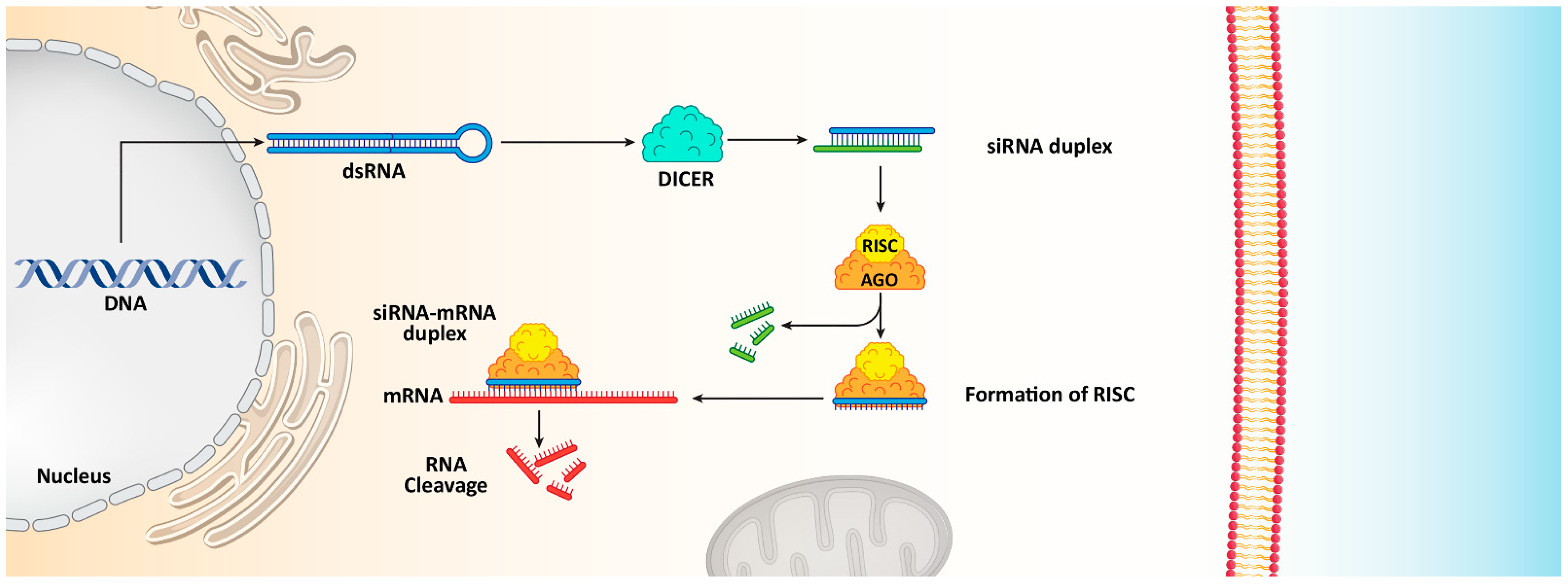
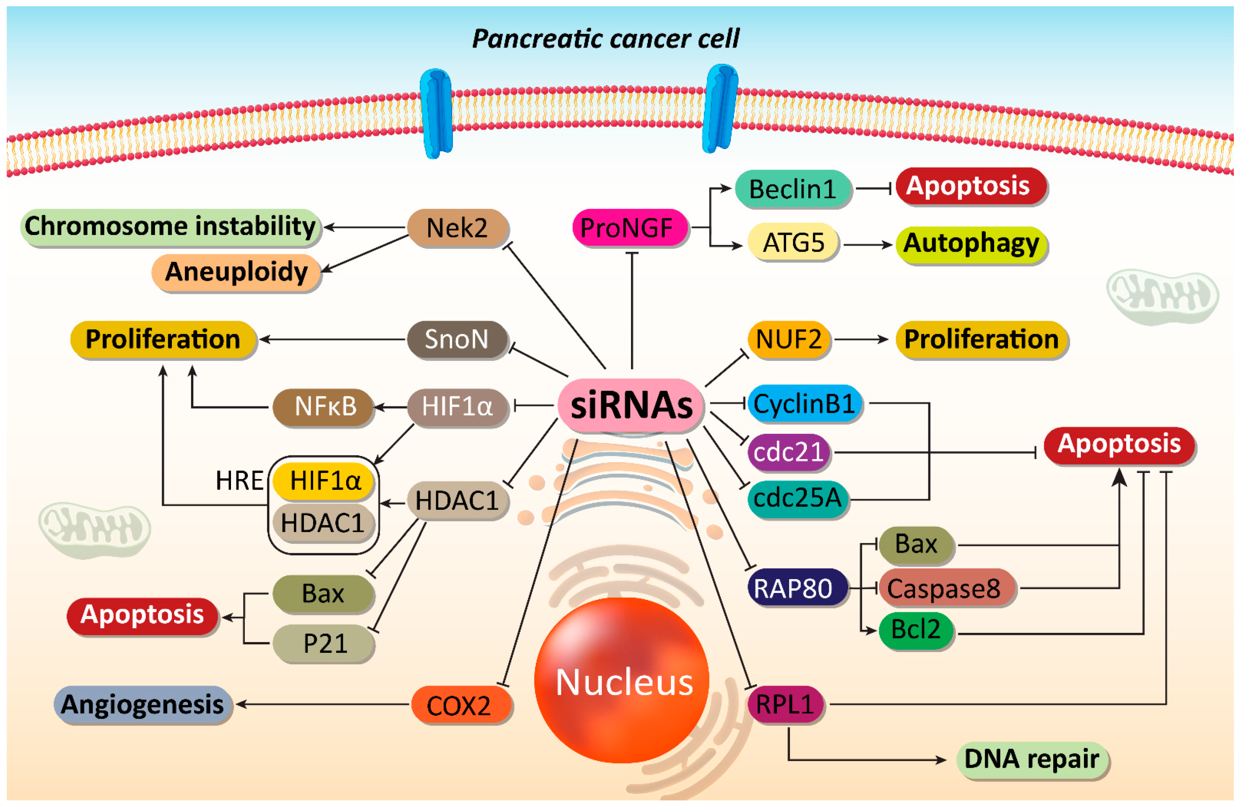
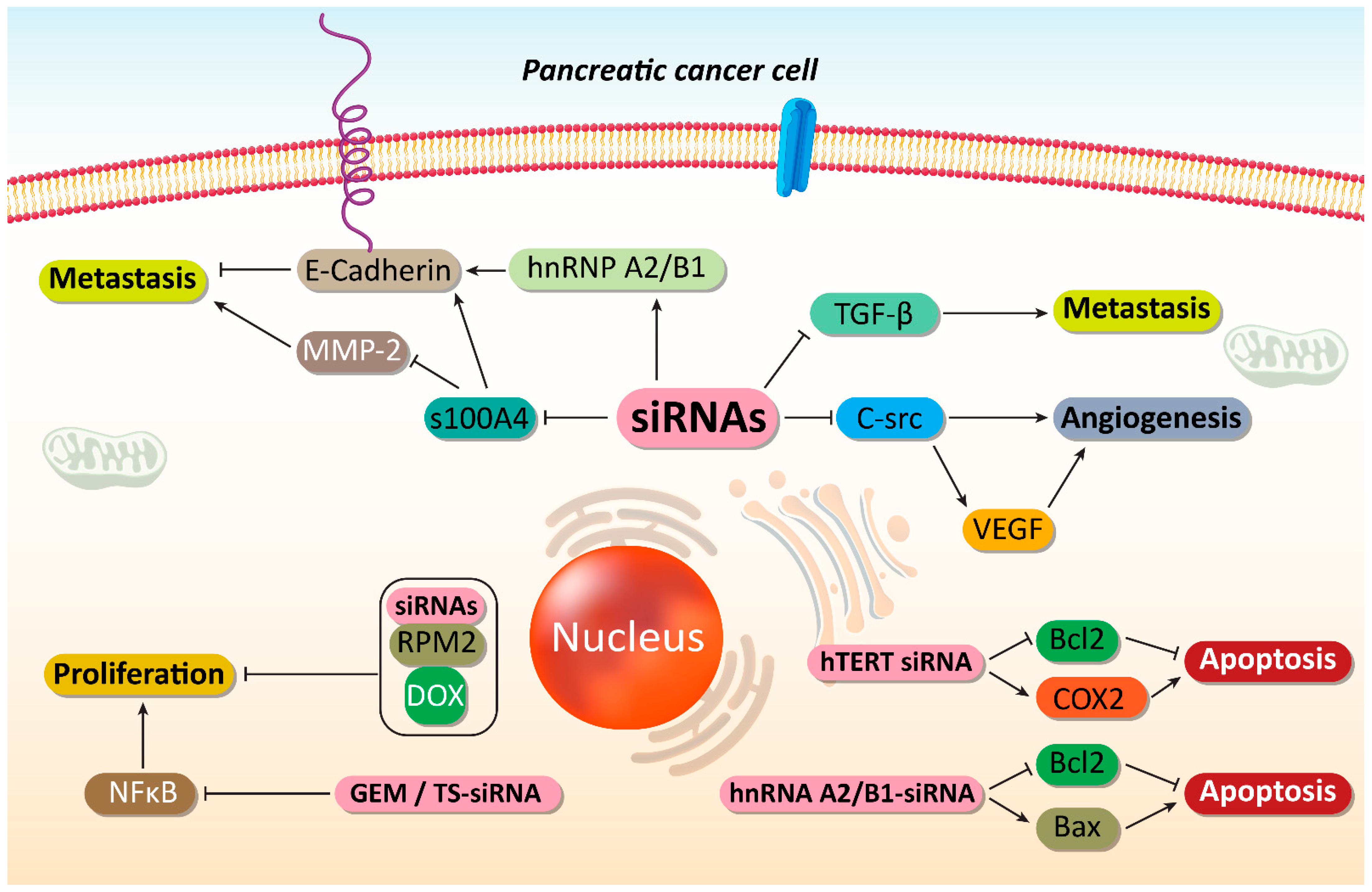
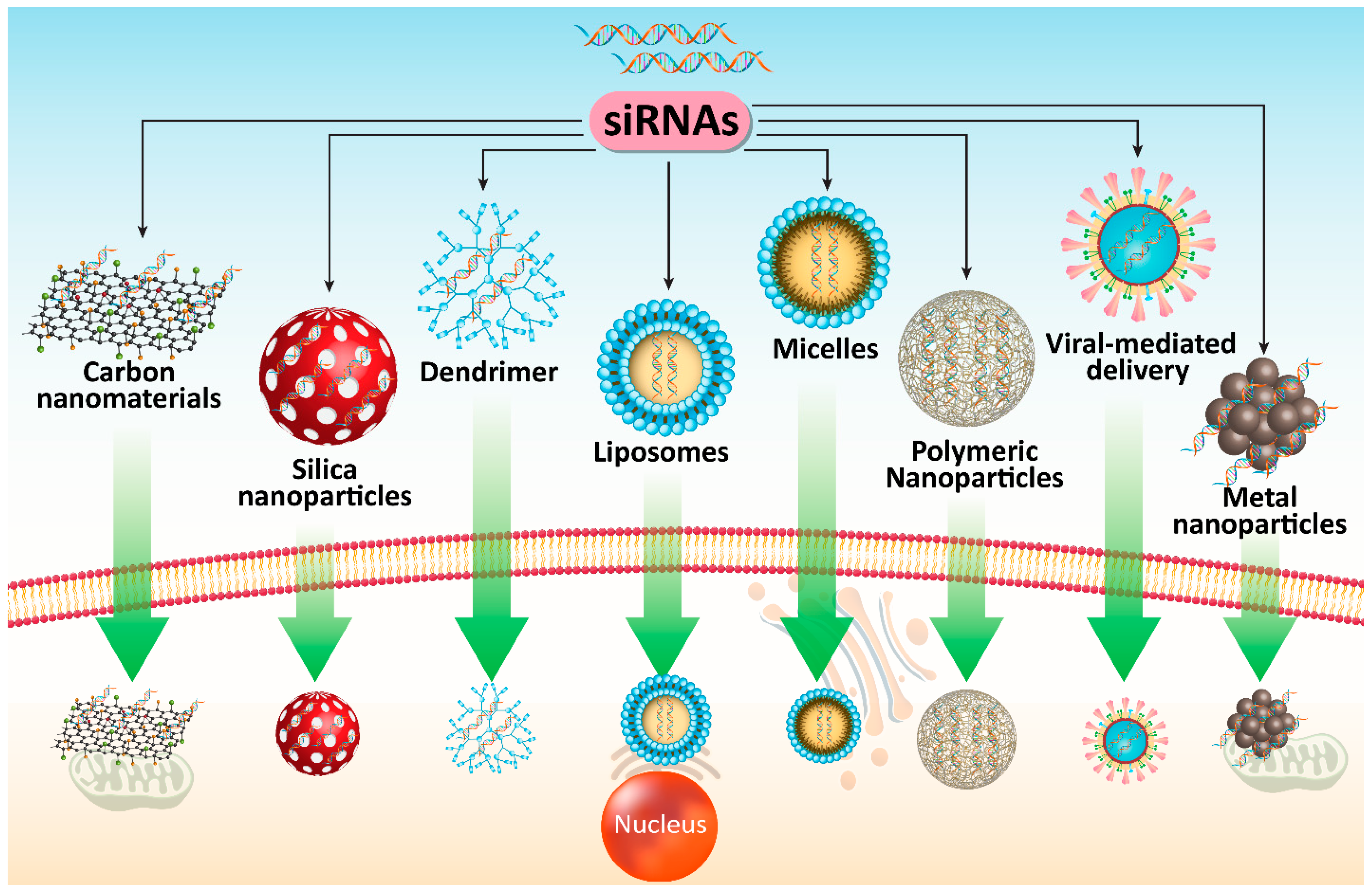
| siRNA | Cnacer Type | Phase | Aim | Trial Number |
|---|---|---|---|---|
| Atu027 | Advanced solid tumors | Phase I | 34 participants that receive eight treatments within 4 weeks The aim is to determine best and optimal dose in cancer treatment | NCT00938574 |
| EphA2 | Advanced or recurrent solid tumors | Phase I | The EphA2 leads to cancer growth and progression siRNA targeting EphA2 paves way in cancer treatment Determing dose and side effects | NCT01591356 |
| CALAA-01 | Solid tumors | Phase I | Determing pharmacokinetics and safety profile | NCT00689065 |
| PLK1 | Liver cancer | Phase I | Testing a new drug, known as TKM-080301 that is a liposomal nanoformulation containing siRNA-PLK1 in cancer treatment | NCT01437007 |
| siRNA taregting immunoproteasome | Melanoma | Phase I | The aim is to improve anti-tumor immunity and prevent immune evasion of cancer cells | NCT00672542 |
| MYC | Solid tumors Lymphoma Multiple myeloma | Phase I | Encapsulation of siRNA-MYC by lipid nanoparticlesin cancer therapy | NCT02110563 |
| PD-L1/PD-L2 | Heamtological malignancies | Phase I Phase II | Developing a new vaccine and mediating immunotherapy | NCT02528682 |
| In Vitro/In Vivo | Cell Line/Animal Model | SiRNA | Outcomes | Refs |
|---|---|---|---|---|
|
In vitro In vivo | PANC-1 and Sw1990 cell lines Xenograft nude mice | NUF2 | Cell cycle arrest at G0/G1 phase Down-regulation of Cdc2, Cyclin B1 and Cdc25A Suppressing carcinogenesis | [101] |
| In vitro | AsPC-1, SUIT-2, and Panc-1 cells | Survivin | Reducing promoter activity and mRNA expression of survivin Inducing caspase-3 expression and DNA fragmentation Enhancing radiosensitivity | [208] |
| In vitro | SW1990 and Capan-2 cells | RAP80 | Down-regulating Bcl-2 and up-regulating Bax Inducing apoptotic cell death Increasing TRAIL-mediated apoptosis Promoting GEM sensitivity | [109] |
| In vitro | Panc-1 and BxPC3 cells | Survivin | Decreasing mRNA and protein levels of survivin Suppressing cell proliferation Triggering cell cycle arrest at G0/G1 phase | [209] |
| In vitro | BxPC3 cells | S100A4 | Reducing gene expression by 17% Down-regulating MMP-2 Enhancing levels of E-cadherin and TSP-1 Suppressing cancer invasion and metastasis | [210] |
| In vitro | PANC-1, MIA-PaCa-2 and ASPC-1 cells | TrKA | TrKA down-regulation is associated with GEM sensitivity Inducing apoptotic cell death Inhibiting PI3K/Akt signaling pathway | [211] |
| In vitro | BxPC3 cells | hTERT | Apoptosis stimulation Cell cycle arrest at G0/G1 phase Enhancing GEM sensitivity | [188] |
| In vitro | MiaPaCa2 cells | HIF-1α | Interfering with cancer proliferation Apoptosis induction Disrupting cancer growth under hypoxic conditions | [113] |
| In vitro | PaTu8988 cells | DNMT1 | Apoptosis induction and inhibiting tumor growth by cell cycle arrest (S phase) DNMT1 down-regulation and subsequent activation of hMLH1 as a tumor-suppressor factor | [115] |
| In vitro | PANC-1 cells | RRM2 | Exerting synergistic effect with doxorubicin and enhancing cytotoxicity against cancer cells by 4-fold | [186] |
| In vitro | MiaPaCa-2 cells | K-Ras | Down-regulating K-Ras expression Triggering apoptosis | [212] |
| In vitro | SW1990 cells | SnoN | Down-regulating SnoN expression and reducing cancer cell proliferation Apoptosis induction | [121] |
| In vitro | SW1990 and BxPC-3 cells | hnRNP A2/B1 | Stimulating apoptosis via Bcl-2 down-regulation and Bax up-regulation Mediating TRAIL-induced apoptosis P-glycoprotein down-regulation Suppressing cancer metastasis via enhancing E-cadherin levels | [201] |
|
In vitro In vivo | Capan-2 cells Nude mice | COX-2 | Cell cycle arrestApoptosis induction Decreasing cancer cell proliferation | [155] |
| In vitro | PaTu8988 cells | HDAC-1 | Disrupting cancer growth and survival Inducing cell cycle arrest (S phase) and apoptosis Enhancing Bax and p21 expressions | [139] |
|
In vitro In vivo | PANC-1 and BxPC-3 cells | RPL21 | Cell cycle arrest at G1 phase Apoptosis induction via mitochondrial pathway Caspase-8 activation | [127] |
| Vehicle | SiRNA | In Vitro/In Vivo | Cell Line | Surface Modification | Particle Size (nm) Zeta Potential (mV) Encapsulation Efficiency (%) | Remarks | Refs |
|---|---|---|---|---|---|---|---|
| Polymeric nanoparticles | K-Ras | In vitro In vivo | KPC-derived cell lines and MIA PaCa-2 cells | RGD | Not reported | Gene down-regulation efficiency more than 95% High cellular uptake Great internalization Suppressing PC progression | [272] |
| Polymer hybrid nanoparticles | VEGF | In vitro In vivo | BxPC3 cells | N/A | 120–140 nm 35 mV | 100 nm in size, spherical shape and narrow dispersion High gene silencing efficiency Reducing tumor growth | [290] |
| Lipid-polymer hybrid nanoparticles | HIF-1α | In vitro In vivo | PANC-1 cells | N/A | 120–140 nm −34 mV | Co-delivery of GEM and siRNA in exerting synergistic effect Prolonged lifetime in bloodstream and improved drug release via the enhanced tumor vasculature effect in tumor tissues Suppressing tumor growth and metastasis Down-regulating HIF-1α Enhancing GEM sensitivity | [255] |
| Polymeric nanoparticles | KRAS | In vivo | KPC8060 cells | N/A | Not reported | Intraperitoneal injection enhances intracellular accumulation of nanoparticles to intravenous administration (15-fold higher) Enhancing infiltration of T cytotoxic cells Inducing delay in tumor growth Suppressing metastasis Increasing survival | [274] |
| Polymeric nanoparticles | EPAS1 | In vitro In vivo | BxPC3 cells | N/A | 160–220 nm −0.41 mV 40% | Prolonged-release behavior Suppressing cancer growthTriggering apoptotic cell death Down-regulating EPAS1 Reducing tumor vessels and VEGF inhibition | [278] |
| Polymeric nanoparticles | GPR87 | In vitro | HEK293T cells | N/A | Average size of 100–200 nm Up to −15 mV Up to 31.14% | Reducing gene expression up to 87% High efficiency and cytotoxicity against cancer growth | [285] |
| Polymeric nanoparticles | K-Ras | In vivo | MiaPaCa-2 cells | N/A | 97.99 nm 39.71 mV | High biocompatibility Potentiality in siRNA delivery and gene silencing in suppressing cancer progression | [268] |
| Polymeric nanoparticles | K-Ras | In vitro In vivo | PANC-1 and BxPC3 cells | N/A | Not reported | Apoptosis stimulationCell cycle arrest at G0/G1 phase Enhanced efficiency in gene silencing | [286] |
| Gold nanocluster | NGF | In vitro In vivo | Panc-1 cells Tumor models | N/A | Not reported | High cellular uptake and intracellular accumulation NGF down-regulation Inhibiting PC proliferation and viability | [319] |
| Liposome | HIF-1α | In vitro In vivo | Panc-1 cells | GE11 | 166.4 nm 22.5 mV | Enhancing GEM sensitivity of cancer cells via HIF-1αdown-regulation | [246] |
| Liposome | Mcl-1 | In vitro | PANC-1 and BxPC3 cells | N/A | N/A | Increased efficiency in down-regulating Mcl-1 Suppressing GEM resistance | [247] |
| Peptide nanoparticles | KRAS | In vitro In vivo | KPC-1 murine PDAC cells | N/A | Not reported | Precision delivery to tumor site High cellular uptake Potentiality in gene silencing | [229] |
| Single wall carbon nanotubes | K-Ras | In vitro | PANC-1 cells | N/A | 110–150 nm +40 mV | High transfection efficiency and cellular internalization Down-regulation of target gene | [340] |
| Graphene oxide nanosheet | HDAC1 K-Ras | In vitro In vivo | MIA PaCa-2 cells | N/A | 550–637 nm +32 to +29 mV | Synergistic effect by combining two siRNAs Simultaneous phototherapy and gene therapy Apoptosis induction Cell cycle arrest and inhibiting cancer growth Reducing tumor growth by more than 80% | [295] |
Publisher’s Note: MDPI stays neutral with regard to jurisdictional claims in published maps and institutional affiliations. |
© 2021 by the authors. Licensee MDPI, Basel, Switzerland. This article is an open access article distributed under the terms and conditions of the Creative Commons Attribution (CC BY) license (https://creativecommons.org/licenses/by/4.0/).
Share and Cite
Mirzaei, S.; Gholami, M.H.; Ang, H.L.; Hashemi, F.; Zarrabi, A.; Zabolian, A.; Hushmandi, K.; Delfi, M.; Khan, H.; Ashrafizadeh, M.; et al. Pre-Clinical and Clinical Applications of Small Interfering RNAs (siRNA) and Co-Delivery Systems for Pancreatic Cancer Therapy. Cells 2021, 10, 3348. https://doi.org/10.3390/cells10123348
Mirzaei S, Gholami MH, Ang HL, Hashemi F, Zarrabi A, Zabolian A, Hushmandi K, Delfi M, Khan H, Ashrafizadeh M, et al. Pre-Clinical and Clinical Applications of Small Interfering RNAs (siRNA) and Co-Delivery Systems for Pancreatic Cancer Therapy. Cells. 2021; 10(12):3348. https://doi.org/10.3390/cells10123348
Chicago/Turabian StyleMirzaei, Sepideh, Mohammad Hossein Gholami, Hui Li Ang, Farid Hashemi, Ali Zarrabi, Amirhossein Zabolian, Kiavash Hushmandi, Masoud Delfi, Haroon Khan, Milad Ashrafizadeh, and et al. 2021. "Pre-Clinical and Clinical Applications of Small Interfering RNAs (siRNA) and Co-Delivery Systems for Pancreatic Cancer Therapy" Cells 10, no. 12: 3348. https://doi.org/10.3390/cells10123348