Article

Transcriptomic Biomarkers for Tuberculosis: Validation of NPC2 as a Single mRNA Biomarker to Diagnose TB, Predict Disease Progression, and Monitor Treatment Response

by
Leonardo S. de Araujo
1,2,†,
Marcelo Ribeiro-Alves
3,
Matthew F. Wipperman
4,5,
Charles Kyriakos Vorkas
4,6,
Frank Pessler
2,7,8,*,‡ and
Maria Helena Féres Saad
1,*,‡
1
Cellular Microbiology Laboratory, Oswaldo Cruz Institute (IOC), Oswaldo Cruz Foundation (FIOCRUZ), Rio de Janeiro 20045-360, Brazil
2
Research Group Biomarkers for Infectious Diseases, TWINCORE Centre for Experimental and Clinical Infection Research, 30519 Hannover, Germany
3
National Institute of Infectology Evandro Chagas, Oswaldo Cruz Foundation (FIOCRUZ), Rio de Janeiro 21040-360, Brazil
4
Immunology Program, Memorial Sloan Kettering Cancer Center, New York, NY 10021, USA
5
Clinical and Translational Science Center, Weill Cornell Medicine, New York, NY 10021, USA
6
Division of Infectious Diseases, Renaissance School of Medicine, Stony Brook University, Stony Brook, NY 11794, USA
7
Centre for Individualised Infection Medicine, 30625 Hannover, Germany
8
Helmholtz Center for Infection Research, 38124 Braunschweig, Germany
*
Authors to whom correspondence should be addressed.
Leonardo Silva de Araujo’s current affiliation: Research Center Borstel, 23845 Borstel, Germany.
These authors contributed to this work equally.
Cells 2021, 10(10), 2704; https://doi.org/10.3390/cells10102704
Submission received: 20 August 2021 / Revised: 27 September 2021 / Accepted: 28 September 2021 / Published: 9 October 2021
(This article belongs to the Special Issue Biomarkers for Human and Veterinary Infectious Diseases)

Abstract

:
External validation in different cohorts is a key step in the translational development of new biomarkers. We previously described three host mRNA whose expression in peripheral blood is significantly higher (NPC2) or lower (DOCK9 and EPHA4) in individuals with TB compared to latent TB infection (LTBI) and controls. We have now conducted an independent validation of these genes by re-analyzing publicly available transcriptomic datasets from Brazil, China, Haiti, India, South Africa, and the United Kingdom. Comparisons between TB and control/LTBI showed significant differential expression of all three genes (NPC2high p < 0.01, DOCK9low p < 0.01, and EPHA4low p < 0.05). NPC2high had the highest mean area under the ROC curve (AUROC) for the differentiation of TB vs. controls (0.95) and LTBI (0.94). In addition, NPC2 accurately distinguished TB from the clinically similar conditions pneumonia (AUROC, 0.88), non-active sarcoidosis (0.87), and lung cancer (0.86), but not from active sarcoidosis (0.66). Interestingly, individuals progressing from LTBI to TB showed a constant increase in NPC2 expression with time when compared to non-progressors (p < 0.05), with a significant change closer to manifestation of active disease (≤3 months, p = 0.003). Moreover, NPC2 expression normalized with completion of anti-TB treatment. Taken together, these results validate NPC2 mRNA as a diagnostic host biomarker for active TB independent of host genetic background. Moreover, they reveal its potential to predict progression from latent to active infection and to indicate a response to anti-TB treatment.

1. Introduction

Tuberculosis (TB) is a curable infectious disease that remains a serious health problem worldwide, mainly due to inadequate diagnosis and treatment of infected individuals. Although concentrated in the poorest population groups, Mycobacterium tuberculosis (Mtb), the etiological agent of TB, may infect anyone. The complexity of Mtb control is clearly reflected by TB disease statistics: there are an estimated 10 million new cases yearly, one-third of the global population are latently infected with Mtb (LTBI), and there are 1.2 million deaths per year. In addition, drug-resistant TB strains remain a public health threat with around half a million cases worldwide [1]. Thus, a reduction in the global impact of TB requires improved diagnostic methods in order to identify both latently and actively infected individuals as early as possible, as well as tools to monitor treatment outcome. Mtb infection spans the spectrum between LTBI and active TB, and a given patient may be anywhere along this spectrum depending on the interplay between immune response and bacillary activity, which makes diagnosis even more complex. Tests based on the host’s immune response, such as tuberculin skin test (TST) and interferon gamma (INF-γ) release assays (IGRAs), although offering some help in clinical practice in the detection of LTBI, have important limitations. They do not distinguish LTBI from active TB among latently infected individuals, they do not indicate who is at greater risk of progressing to active disease (especially in populations with a high bacillary load), and they are not accurate in immunosuppressed individuals [2,3,4,5]. Moreover, aside from the complicated and time-consuming microbiological culture, there are no other tools available to monitor treatment.
Considering these shortcomings, there have been intense efforts to identify biomarkers that could serve as more accurate diagnostics and help to stratify individuals along the above-mentioned spectrum of TB. RNA biomarkers have received great attention in this regard because they often accurately reflect the regulation of processes underlying disease development and progression [6,7,8,9].
In our previous study of whole blood transcriptomes from Brazilian patients with latent and active TB, we identified three mRNA, NPC2, EPHA4, and DOCK9, with the highest AUROC (≥0.94) for TB in comparison to LTBI/controls. In the same previous study, we expanded the cohort and carried out a confirmatory analysis of these genes using quantitative polymerase chain reaction ((RT)qPCR). This confirmatory analysis showed that (at 92% specificity) sensitivity was highest for NPC2 (85%) and less promising for EPHA4 (53%) and DOCK9 (19%) [10]. These three mRNA candidates achieved the optimal specificity (>80%) recommended for a community-based triage or referral test to identify people suspected of having TB according to the World Health Organization’s (WHO) TB target product profile (TPP). However, only NPC2 approached the minimum TPP threshold for sensitivity (>90%) [9]. Moreover, confounders such as the broad clinical spectrum of TB infection and suboptimal detection of LTBI [5] might have limited the accuracy of those results. Interestingly, in a longitudinal analysis of a small number of samples, expression of EPHA4 (p = 0.0003) and NPC2 (p = 0.004) correlated significantly with a clinical response to anti-TB treatment [10], suggesting that the full biomarker potential of these genes was not completely investigated in our previous studies [10,11], and that NPC2, EPHA4, and DOCK9 should be further evaluated in order to validate our initial findings.
The biological roles of EPHA4 and DOCK9 mRNA have not been completely elucidated. Previous studies suggest that both DOCK9 [12] and EPHA4 [13] proteins can interact with the Rho GTPase CDC42. Among other functions, Rho GTPases are described to regulate cytoskeletal kinetics and signal transduction pathways, playing a key role in the coordination of immune responses, including in the activation of T cells [14].
NPC1/NPC2 is a crucial pathway of intracellular cholesterol trafficking. While the protein NPC2, the product of the NPC2 gene, is a soluble cholesterol-binding luminal protein, NPC1 is an anchored transmembrane glycoprotein [15,16,17]. In the lumen of late endosomes/lysosomes NPC2 transfers cholesterol to NPC1, then NPC1 transfers cholesterol to other vesicular pathways, e.g., endoplasmic reticulum [15,16,17]. Using an in vitro M. tuberculosis infection model, Wheelwright et al. (2014) [18] showed that supplementation with vitamin A, or all-trans retinoic acid (ATRA), contributed to the acidification of the lysosome, decreased intracellular cholesterol, altogether with a negative effect on mycobacterial viability. This mechanism was described as NPC2-dependent, up-regulated after the infection of phagocytes by M. tuberculosis, but the same was not the case with the NPC1 protein [18]. Indeed, NPC1 mRNA was later shown not to be differentially expressed in the blood of TB patients [10].
In the present work, we performed an extensive evaluation of NPC2, EPHA4, and DOCK9 mRNA levels (i) as diagnostic biomarkers according to WHO TPP criteria for a community-based triage or referral test to identify people suspected of having TB, (ii) as potential biomarkers for predicting progression from latent TB infection to active disease [19], and (iii) as correlates of a clinical response to anti-TB treatment. For this purpose, we analyzed previously published and unpublished datasets from cross-sectional tuberculosis cohorts from Brazil, Haiti, India, South Africa, and the United Kingdom, as well as from two prospective studies from China and South Africa.

2. Materials and Methods

2.1. Ethics Statements

The Brazilian study was approved by the Ethics Committee of the Oswaldo Cruz Foundation under registration code 560-10 [10]. Details about sample collection and ethical procedures of the Haitian cohort were previously published [20,21]. The data from the other cohorts were publicly available at GEO [22].

2.2. Terminology

We used the following case definitions and abbreviations. Control = healthy uninfected individuals recently exposed to a TB index case or not (the control subjects recruited by de Araujo et al. [10,11] and Wipperman et al. [20,21] were known to have been exposed to a TB index case but TB infection was subsequently ruled out). Symptomatic non-TB (S-NTB) = symptomatic adults self-presenting for investigation of pulmonary tuberculosis and showing no laboratorial evidence of active TB disease, regardless of the history of known exposure to a TB index case (for more details please check [6]). LTBI = defined by a positive Mantoux TST and/or IGRA and absence of active TB diagnostic [5]. TB = active tuberculosis diagnosed by sputum smear and/or culture and/or GeneXpert MTB/RIF [6,7,10,11,23,24,25]. TBtt = drug treatment for TB [7,24]. OD = other non-TB pulmonary diseases, such as active (aSARC) and non-active sarcoidosis (naSARC), lung cancer (LC), and pneumonia (PN) [23].

2.3. Inclusion Criteria for Eligible Published Datasets

The following search keywords were used to identify eligible datasets on GEO and ArrayXpress: human, transcriptomic, tuberculosis, and blood. Datasets that were deposited until March 2020 and were not present in our previous study [10] were included in this re-analysis. Inclusion criteria were studies containing the following characteristics. Biological specimens: whole blood or peripheral blood mononuclear cells (PBMC); subjects: adults (≥18 years old) with active pulmonary TB, LTBI, and controls with or without other diseases; transcriptomic profiling by RNAseq or microarray analysis. Exclusion criteria were samples from subjects <18 years of age or positive HIV status and non-human samples.
Table 1 summarizes the included cohorts. The Brazilian cohort is composed of a joint analysis of sub-cohorts from our two previous studies. In the first, we had originally discovered NPC2, DOCK9, and EPHA4 mRNA as potential biomarkers for TB [10]. The second was focused on small noncoding RNA (sncRNA) expression in a larger group of samples [11], but we also used some of them to extract the normalized mRNA expression values for the present study. One unpublished dataset containing a cohort of Haitians and publicly available datasets from India were also included, following the criteria detailed above. Using all five cross-sectional cohorts, we evaluated expression of the three mRNA in TB patients from different geographic areas, also for the differentiation from non-TB pulmonary infections. In the longitudinal cohorts (two public datasets from China and South Africa), we evaluated their expression (i) in the progression from LTBI to active TB and (ii) during follow-up of anti-TB treatment.
For the Brazilian and Haitian cohorts, peripheral whole blood was collected in Paxgene RNA tubes (PreAnalytiX, SWZ) and processed and analyzed as described previously [10].

2.4. Acquisition and Normalization of Datasets

Transcriptomic data (microarray or RNAseq) of whole blood or PBMC were obtained as follows. GEO2R web tool [27] was used to gather normalized expression values of microarray studies. For RNAseq data, the FASTQ files were exported to the GREIN tool [28] and submitted to standard normalization. Processed expression data of E-MTAB-8290 [6] were downloaded from the ArrayExpress platform (https://www.ebi.ac.uk/arrayexpress/experiments/E-MTAB-8290/?page=1&pagesize=250, accessed on 31 March 2020) and included in our analysis. The normalized microarray or RNAseq expression values were exported to Prism 6 (GraphPad Software, 6.07, San Diego, CA, USA) for statistical analysis.

2.5. Statistical Analysis

Significance of differences between two groups was assessed with the Mann–Whitney (cross-sectional) or Wilcoxon (longitudinal) test. For comparisons of >2 groups, the Kruskal–Wallis (cross-sectional) or Friedman test (longitudinal) was used. Means, medians, standard deviations (SD), dispersion plots, area under the receiver operating characteristics curve (AUROC) values, 95% confidence intervals (CI), and coefficient of variation (CV) were computed using Prism 6 (GraphPad Software).

3. Results

3.1. Cross-Sectional Studies: Group Comparisons and ROC Analysis
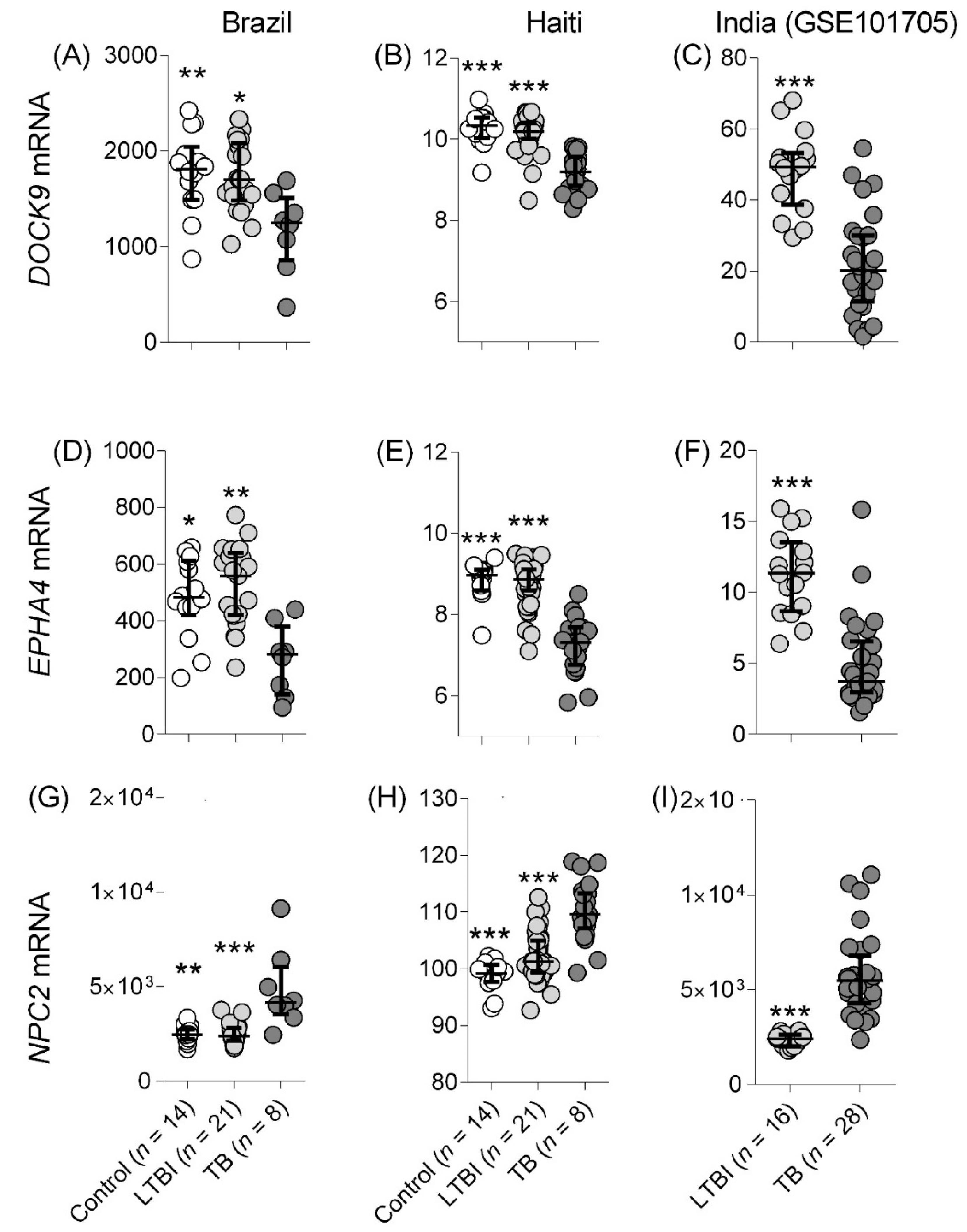
3.1.1. TB Detection

Studies Comparing with Control and LTBI

Cross-sectional published datasets were included in the re-analysis, which comprise subjects enrolled between 2009 and 2013 in India and the United Kingdom (London). Together with the Brazilian cohort enrolled from 2010 to 2013 and the Haitian cohort collected from 2016 to 2020, four cohorts from four different countries were, thus, available for analysis. In a first step, we assessed the diagnostic biomarker potential of DOCK9, EPHA4, and NPC2 mRNA to distinguish among active TB, LTBI, and control groups.
In accordance with our previous findings [10], active TB induced significantly higher NPC2 mRNA levels and lower expression of DOCK9 and EPHA4 mRNA in the cohorts from Haiti and India (Figure 1). An NPC2high expression pattern (similar to the one observed among TB cases) was more frequent among LTBI than controls in the cohorts from Haiti (Figure 1H) and Brazil (Figure 1G), whilst LTBI from the Indian cohort showed less dispersed expression (Figure 1I). The control group showed a more heterogeneous expression profile of DOCK9 and EPHA4, i.e., greater dispersion along the y-axis, which is more noticeable among the Brazilian (compare Figure 1A,D with Figure 1G) and Indian (compare Figure 1C,F with Figure 1I) cohorts. Overall, this analysis showed (i) that expression of all three mRNA changed in a similar fashion in blood of TB cases from the different geographic areas and (ii) that LTBI cases were more likely than non-infected samples to exhibit the “TB-like” pattern NPC2high.
ROC analysis of the distinction control vs. TB revealed high AUROC values (indicating accurate classification) with low variation for all three mRNAs among the different cohorts (DOCK9: 0.8–0.96; EPHA4: 0.89–0.97; NPC2: 0.91–0.99). Comparisons between latent and active infection also showed a similarly low variation of AUROC values (DOCK9: 0.86–0.95; EPHA4: 0.91–0.96; NPC2: 0.8 –0.98). This suggests that the high accuracy of these markers is reproducible among these ethnically different populations from diverse geographical locations.
In line with our previous findings [10], in this new reanalysis, NPC2 showed the highest AUROC values for TB vs. non-TB discrimination among Brazilians (AUROC, TB vs. control/LTBI: 0.94), which was validated in the Indian (TB vs. LTBI: 0.98) as well as the British (TB vs. control: 0.99) cohort. Intriguingly, a slightly lower performance of NPC2 (AUROC, TB vs. LTBI = 0.89) to differentiate TB vs. LTBI compared to DOCK9 (TB vs. LTBI = 0.95) and EPHA4 (TB vs. LTBI = 0.96) was observed in the Haitian cohort. As in high TB-burden settings, the chances of infection and progression to disease are higher, this might indicate that some subjects could actually be in the initial stages of progression, which could interfere with the performance of NPC2high as a biomarker for the binary distinction TB vs. LTBI (Figure 1).
Overall, the biomarker potential of NPC2high was successfully validated in these new analyses, which showed the highest mean AUROC values in the comparisons between TB and control (mean AUROC = 0.95) or LTBI (mean AUROC 0.94, Table 2). Further analysis will be performed in the next sections to obtain the sensitivity and specificity values of NPC2high for TB detection across the different cohorts included here. DOCK9 and EPHA4 also showed interesting biomarker value in this context.

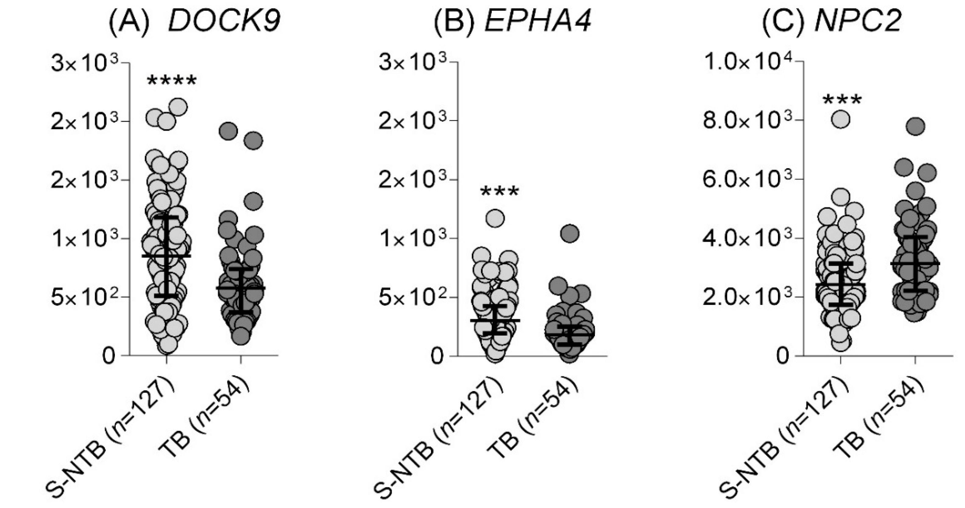
Identification of TB in Individuals Presenting with Respiratory Symptoms

Data from whole blood of individuals with respiratory symptoms, self-presenting for investigation of pulmonary TB, were available from the South African cohort [6]. Even though all subjects had respiratory symptoms, those diagnosed with TB by positive sputum smear and/or sputum liquid culture and/or GeneXpert TB/RIF test showed significant differences in DOCK9 (p-value = 0.0002), EPHA4 (p < 0.0001), and NPC2 (p < 0.0001) mRNA expression when compared with non-TB subjects, as defined by negative results by the aforementioned diagnostic tests (Figure 2). However, all three mRNA were only moderately accurate in classifying the subjects into TB and non-TB cases: EPHA4 showed a slightly higher AUROC (0.71, 95% CI: 0.63 to 0.79) than NPC2 (0.68, 95% CI: 0.60 to 0.77) or DOCK9 (0.675, 95% CI: 0.59 to 0.76).

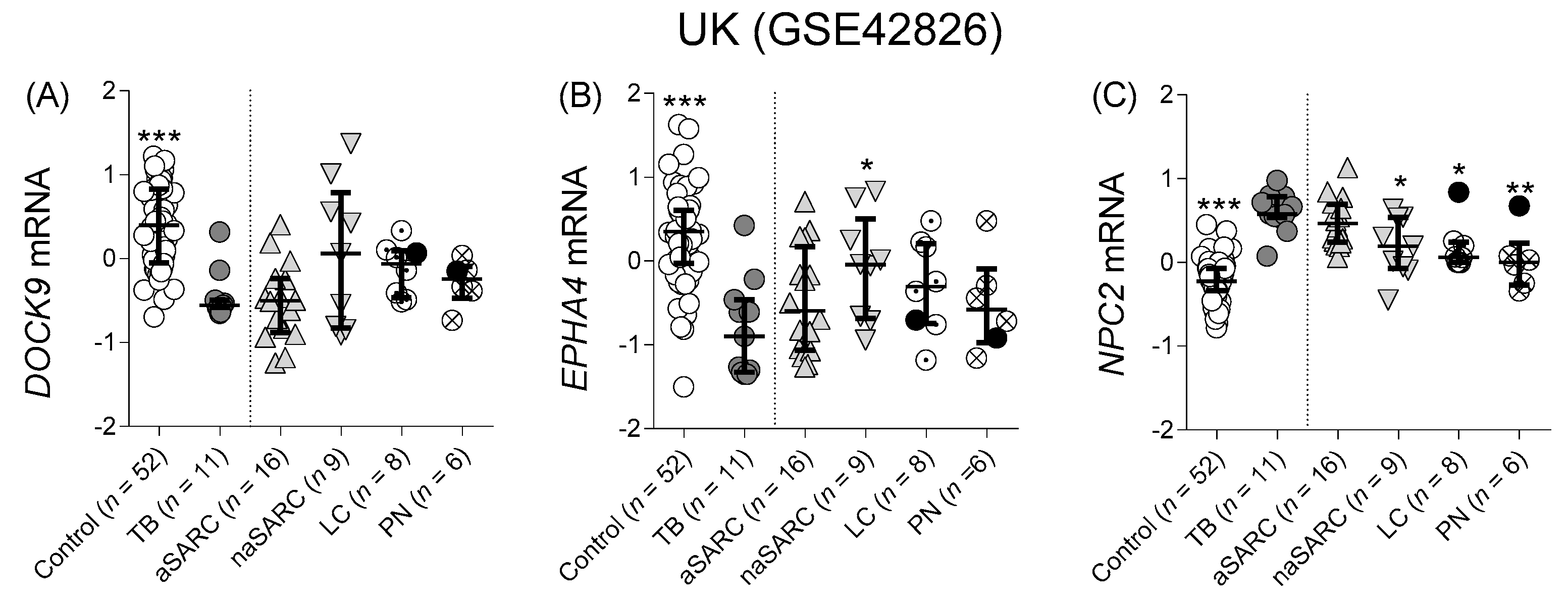
Differentiation from Other Pulmonary Diseases

Using the available datasets from the United Kingdom (GSE42826), we evaluated the potential of the three mRNAs to discriminate between TB and other lung diseases that are likely to constitute clinically important confounders. This study generated whole blood microarray transcriptional data from patients with TB or OD and from controls (Figure 3).
Similarly to the observation in Section Studies Comparing with Control and LTBI, in the British cohort, DOCK9 and EPHA4 expression was also significantly lower (p ≤ 0.0001) in TB patients compared to control (Figure 3A,B). However, only NPC2 expression levels were significantly higher in TB than in the majority of the other lung diseases, such as non-active sarcoidosis (p = 0.021), lung cancer (p = 0.018), and pneumonia (p = 0.006) (Figure 3C).
Similarly to TB, but on a smaller magnitude, patients with active sarcoidosis had significantly higher blood levels of NPC2 in comparison with control (p < 0.0001), which was not observed for any of the other disease groups (Figure 3C, control vs. non-active sarcoidosis: 0.07, vs. lung cancer: 0.052, vs. pneumonia > 0.99). In fact, TB induced the highest median NPC2 blood levels (0.58, 95% CI 0.37–0.80), which was followed by active sarcoidosis (0.46, 95% CI 0.23–0.71). Lower median values were observed in non-active sarcoidosis (0.19, 95% CI 0.091–0.54), lung cancer (0.063, 95% CI 0.0018–0.84), and pneumonia (0.00109, 95% CI 0.34–0.675) and the lowest in the control group (−0.22, 95% CI 0.296–0.15). Thus, higher NPC2 levels in peripheral blood might underlie immunopathological processes that are similar in active sarcoidosis and in TB, but are less common during the cessation of sarcoidosis symptoms, and even lower in lung cancer and pneumonia.
In general, DOCK9 and EPHA4 (Figure 3; Table 2, mean AUROC ≤ 0.65) showed lower potential to differentiate between TB and these clinical confounders when compared to NPC2 (Table 2, mean AUROC = 0.73). All mRNAs had low AUROC values for the distinction TB vs. active sarcoidosis (Table 2, AUROC between 0.51 and 0.69). In contrast, a moderate to high potential to discriminate TB from non-active sarcoidosis (AUROC = 0.87), lung cancer (0.86), and pneumonia (0.88) was observed. In fact, only one patient diagnosed with lung cancer and one with pneumonia showed an NPC2high profile (black dots, Figure 3C).

3.2. Prospective Studies: Group Comparisons and ROC Analysis

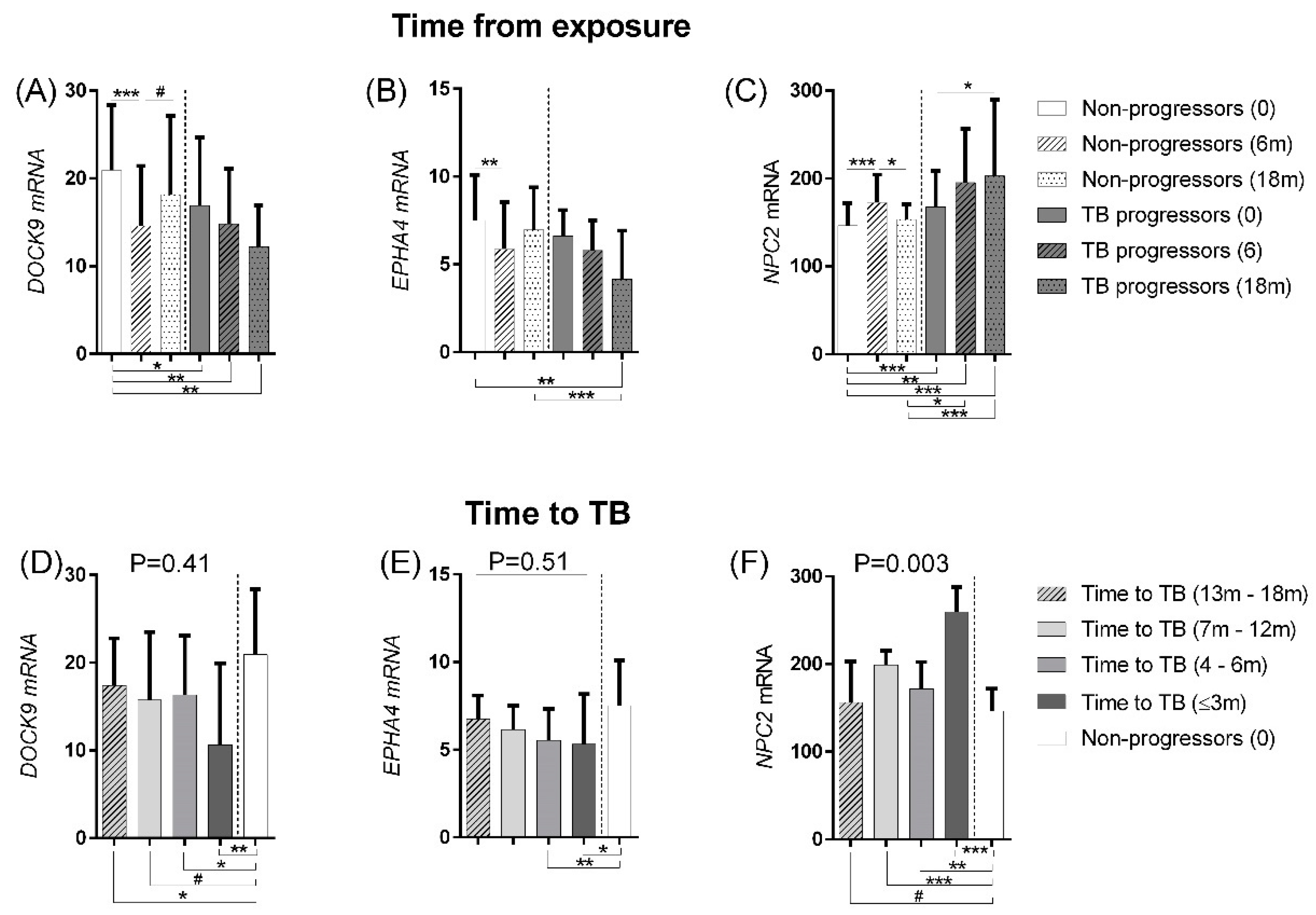
3.2.1. Disease Progression

Our previous observation of a subgroup of LTBI subjects with NPC2 TB-like expression profile [10] raised the question whether this would indicate progression of sub-clinical cases.
The Pan African cohort comprises a series of samples collected from household contacts after a person who was recently diagnosed with TB returned to the household (GSE94438). Based on samples collected three times at intervals of 6 months, subjects who developed TB are here classified as “TB progressors” (n = 64), and those who did not develop TB in the 18-month window are classified as “non-progressors” (n = 208) [24]. At the time of Mtb exposure (time 0), the TB progressors already show differential expression of DOCK9 and NPC2 in comparison to the non-progressors (Figure 4A–C, small clades under the graph; p < 0.026). During follow up, non-progressors and TB progressors initially showed similar expression changes until month 6, i.e., down-regulation of DOCK9 (compare white and grey bars, Figure 4A) and EPHA4 (Figure 4B) and up-regulation of NPC2 (Figure 4C). Interestingly, after month 6 of exposure, expression of all three mRNA showed a trend toward normalization in non-progressors, although a significant difference was only observed for NPC2 (as indicated by the significant down-regulation between the dashed and dotted white bars in Figure 4C). In contrast, NPC2 mRNA levels in TB progressors continued to increase throughout the follow-up period (Figure 4C, compare white and grey bars).
In order to understand the dynamics of transcriptional changes from the time of infection to disease onset, we performed additional analyses on the Pan African GSE94438 dataset. They were grouped into TB progressors, according to the time elapsed between the blood collection and the diagnosis of TB (T<3 m = <3 m, T4–6m = 4–6 m, T7–12m = 7–12 m, or T13–18 m = 13–18 m) and non-progressors at enrollment (time 0) for comparison. This analysis showed that TB progressors already had a significantly lower expression of DOCK9 at T13–18m before disease development (Figure 4D). Significant up-regulation of NPC2 among TB progressors was observed from T7 to 12m (Figure 4F), while EPHA4 showed later expression changes at T4–6m (Figure 4E).
We expected that these expression changes would be more pronounced with decreasing time to disease onset. Indeed, a median decrease in DOCK9 (Figure 4D) and EPHA4 (Figure 4E) was observed in individuals with time-to-disease of 3 and 6 months, but only NPC2 up-regulation was significantly increased up to one year of time-to-disease, with the highest expression observed in individuals with the shortest time-to-disease (3 months) (p = 0.003; Figure 4F, dark grey bars). Altogether, these findings suggest that monitoring NPC2 expression in blood might serve as biomarker for progression to TB among individuals recently exposed to Mtb or among household contacts of recently diagnosed TB cases.

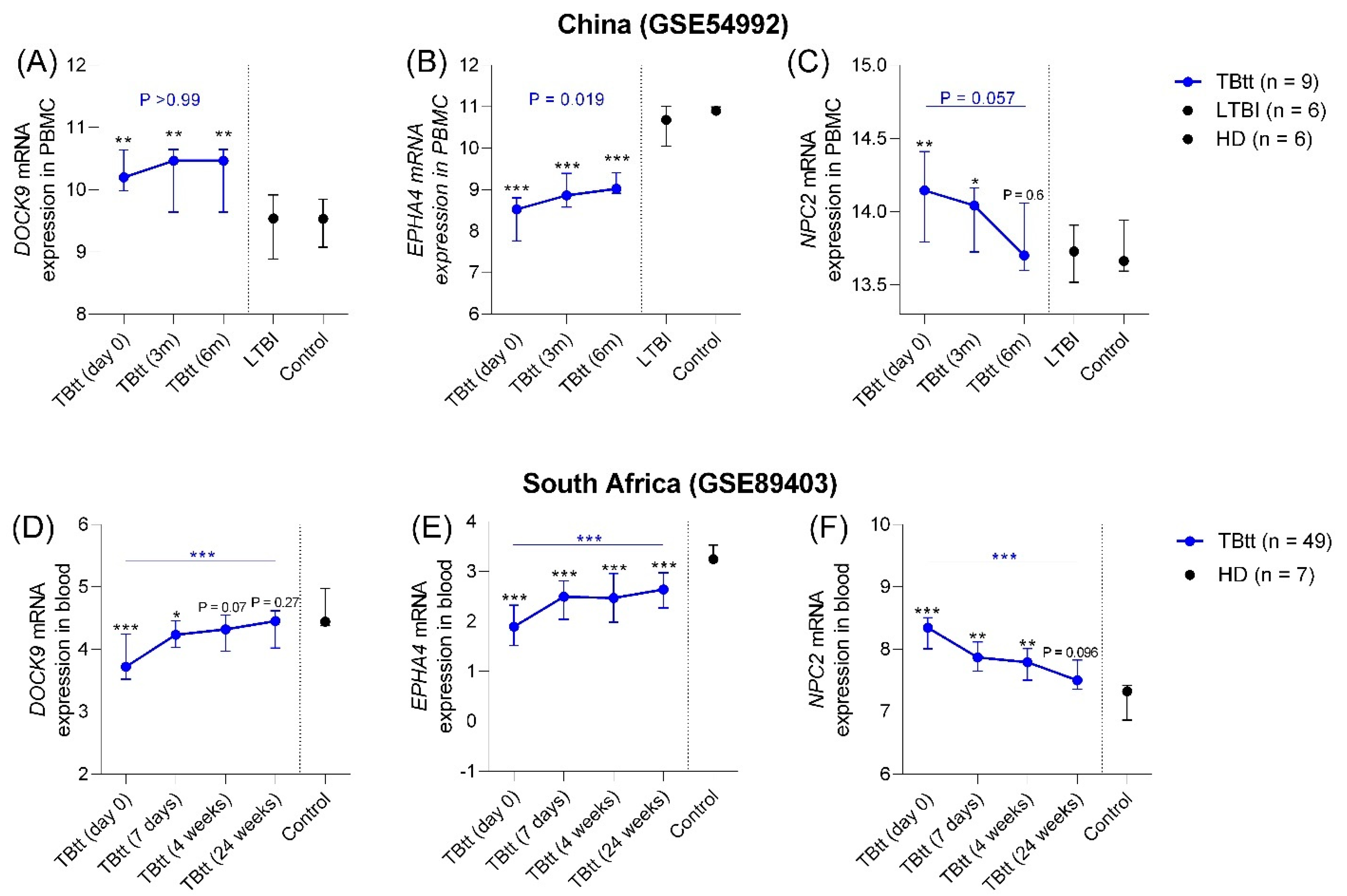
3.2.2. Correlation with Completion of Anti-TB Treatment

Besides their potential to detect disease cases, expression of optimal biomarkers for TB should reflect successful completion of anti-TB treatment. To evaluate this aspect, we used two datasets, China GSE54992 [25] and South Africa GSE89403 [7], which featured prospective sampling during anti-TB treatment. The dataset GSE54992 comprises microarray expression data from PBMC, and GSE89403 is an RNAseq-based study of whole blood.
In accordance with the cross-sectional evaluation shown in Figure 1 and Figure 2, in this section, significant differences in DOCK9, EPHA4, and NPC2 expression were also evident in all pairwise comparisons between untreated active TB patients (TB) and LTBI and controls (see Figure 5, p-values in black font). However, although DOCK9 was down-regulated in the previous analyses, it was up-regulated in PBMC from TB cases in the Chinese dataset (Figure 5A). Apart from this discrepancy, significant DOCK9low (GSE89403 only; p < 0.0001), EPHA4low (GSE54992 and GSE89403; p < 0.001) and NPC2high (GSE54992 and GSE89403; p ≤ 0.03) expression differences in the comparison TB vs. control/LTBI were also observed among these datasets from China and South Africa.
During anti-TB chemotherapy, expression of these three genes changed significantly over time. Even though expression of all three tended to normalize, it is notable that only NPC2 expression levels did not differ between control/LTBI groups and TBtt by the end of therapy in both cohorts (Figure 5C [control/LTBI vs. TBtt (6 m), p = 0.6] and 5F [control vs. TBtt (24 weeks), p = 0.096]). In the South African cohort, DOCK9 also showed a gradual normalization and no statistically different expression levels between control and TBtt at the end of therapy.
Moreover, expression data available from the Haitian cohort after two weeks of anti-TB treatment (Figure S1) corroborated the significant reduction in NPC2 mRNA blood levels even in this early stage of anti-TB treatment.

3.3. NPC2 Accuracy: Sensitivity and Specificity Analysis

Overall, in the previous sections, NPC2 showed better discriminatory potential for TB across the different group comparison analyses. Therefore, we proceeded with a more detailed ROC curve analysis exclusively for NPC2.
As the selection of cut-off values can be adjusted in order to improve either the sensitivity or the specificity of a given test, we decided to assess NPC2 accuracy in various possible diagnostic scenarios. For this purpose, we calculated its sensitivity and specificity for the detection of TB according to: (i) maximum Youden index; (ii) the TPP for a community-based triage or referral test to identify people suspected of having TB (9); (iii) the TPP for a test for predicting progression from TB infection to active disease (12).
As shown in Table 3, analyses performed at the maximum Youden index (sensitivity + specificity/2) showed high mean sensitivities for TB detection varying between 87.5% and 100% vs. controls and moderate values when compared to LTBI (72.7–75%), while exhibiting high mean specificity (90.2–100%). The analysis comparing TB against OD showed the lowest mean specificity in the case of TB vs. active sarcoidosis (56.3%), but for the other TB clinical confounders, these values were ≥79%, maintaining a sensitivity of ≥72.7%. However, discriminatory power decreased when comparing TB vs. S-NTB, as sensitivity and specificity were ≤67.8%.
By adjusting the accuracy analysis to meet the TPP for a community-based triage or referral test to identify people suspected of having TB, we observed that most of the analyses meet the minimum sensitivity (>90%) and specificity (>70%) requirements (Table 3, bold font). Only the cohorts from Brazil and South Africa (in which the control and S-NTB groups were composed of recent close contacts or symptomatic respiratory patients, respectively) and comparisons between TB and naSARC and aSARC did not meet this TPP minimum criteria.
In contrast, it is noteworthy that in the longitudinal analysis, NPC2high fulfilled the minimum TPP sensitivity and specificity (>75%) criteria for a monitoring test for prediction of progression from latent to active TB [19]. Here, NPC2high could detect subjects that will progress to active TB in a time interval <3 m before disease onset (Pan African cohort GSE94438, see bold numbers in Table 3), but not later than that. If we aim for a maximum detection of TB cases, i.e., higher sensitivity, NPC2high demonstrated 92.3% mean sensitivity and 75% mean specificity to detect TB < 3 m before disease onset.

4. Discussion

We have performed an external validation/re-evaluation of the mRNA triplet DOCK9, EPHA4, and NPC2, which we had previously identified as potential biomarkers in whole blood of Brazilian TB patients [10]. We observed similar changes in expression among subjects from different countries, regardless of differences in genetic background and local TB incidence ( Figure 1 and Figure 2, Table 2). Our results suggest that the gene set was differentially expressed among patients with active TB and that NPC2high should perform better even in high burden areas such as South Africa (520/100.000), India (199/100.000), and Haiti (176/100.000). Notably, the single microarray dataset included in this study (GSE42826) confirmed the differential expression of the three genes originally uncovered by RNAseq, corroborating a previous study showing the equivalence of microarray and RNAseq to assess differential gene expression [29].
Our study, which was done in different cohorts, shows mean AUROC values > 0.90 of NPC2 for the detection of active TB, which was consistent in all cohorts for the discrimination between TB cases and control, with the highest mean AUROC values compared to EPHA4 and DOCK9. In addition, NPC2 expression was significantly lower in other lung diseases, except for active sarcoidosis (Figure 3). Even though mycobacteria and propionibacteria are the most commonly implicated etiologic agents of sarcoidosis, based on studies using PCR amplification of microbial DNA, so far, Propionibacterium acnes is the only microorganism successfully isolated from sarcoid lesions by bacterial culture, which may help in the diagnostic differentiation from TB [30,31,32,33].
The biomarker reproducibility of a signature with a small number of genes may be a concern, as some studies have reported that increasing the number of genes may improve sensitivity and specificity [26]. Yet, other studies have reported that a signature comprising 16 genes, which initially predicted progression from LTBI to TB, decreased in accuracy during external validation, failing in the validation in cohorts from other countries [24]. More recently, a systematic review evaluated the accuracy of several proposed transcriptional signatures in a setting with a high burden of TB and HIV in South Africa [6]. Note that none of 27 selected signatures met the WHO optimum or minimum criteria for triage (95% sensitivity and 80% specificity) or confirmatory test (65% sensitivity and 98% specificity), including NPC2 [10]. In contrast, in the present study, especially NPC2 demonstrated high mean sensitivity (>87.5%) to distinguish between TB and LTBI, even though a “TB-like” expression profile was also observed among some subjects classified as LTBI and, more frequently, among S-NTB. It is also important to mention that the study by Turner et al. [6] did not feature a confirmatory analysis of these genes, whereas differential expression of NPC2 in TB was already confirmed by (RT) qPCR in a different Brazilian cohort not included in the present study [10]. Additionally, a diagnostic algorithm tree combining NPC2 expression cut-off values combined with the results provided by the low-cost chest X-ray examination enabled accurate discrimination between TB and LTBI individuals [10]. When applied in a population to be prospectively evaluated for TB, this type of holistic approach combining imaging findings with transcriptional signatures should be considered in future studies.
Furthermore, we also found that NPC2high met the minimum sensitivity and specificity TPP criteria for predicting progression from latent tuberculosis to active TB in most of the cohorts. The lower sensitivity/specificity observed for the comparisons with the South African respiratory symptomatic cohort (E-MTAB-8290, Table 3) might seem to be a drawback at first. However, we have to consider the limitations of the current diagnostic tests for TB detection [2,3,4,5] and that an NPC2high pattern may already be observed at earlier stages of the TB progression spectrum (as seem on Figure 4F). Thus, S-NTB individuals showing an NPC2high profile in blood could be harboring a sub-clinical/paucibacillary TB infection.
We identified NPC2 as an accurate marker for identifying individuals at high risk of progressing from LTBI to TB. If the sensitivity of 92.3% and specificity of 75% (Table 3) for predicting progression from latent to active TB is corroborated in additional studies, monitoring NPC2 expression in blood can contribute to the detection and early treatment of those LTBI cases at risk of progression to active TB by using a simpler method, (RT) qPCR, which was already validated for this marker [10]. In addition, chest X-ray would be an easy tool to perform in order to screen high-risk LTBI vs. active TB cases among subjects with an NPC2high profile. On the other hand, considering that all-trans retinoic acid (ATRA) triggers an NPC2-dependent antimicrobial response against Mtb [18], it is important to investigate whether vitamin A deficiency could contribute to false-negative results in individuals otherwise expected to have an NPC2high pattern. Clearly, there is a need for additional prospective studies to validate our current findings.
Our data of non-progressor household TB contacts showed significant changes in NPC2 gene expression only 6 months after exposure to the initial index case, with a return of expression to the initial level after 18 months (Figure 4). However, a different dynamic was observed for the TB progressors, which showed a continual up-regulation of this gene expression toward the TB profile with increasing proximity to disease onset (Figure 4F). Nowadays, TST and IGRA are the eligible tests to identify M. tuberculosis exposure, although they cannot distinguish TB vs. LTBI or identify individuals who will progress from latent to active TB within the next two years [5]. These immune response-based tests do not reflect the presence of live bacilli in the host and are still positive after completion of treatment of the infection. As exemplified by NPC2 mRNA, this drawback can be overcome by measuring the expression of genes that play a functional role in host defenses against the pathogen. Measuring its activity at the mRNA level may be a particularly attractive option due to the highly dynamic nature of transcriptional responses in the host’s biological processes [34]. The sensitivity and high dynamic range of many transcriptomic responses likely also explains the ability of NPC2 to predict progression to TB and its correlation with treatment completion.
TB control critically relies on the identification of individuals with active disease and the administration of complete drug treatment. However, to follow the response to treatment, the tools available, such as in vitro culture of clinical specimens, are slow and laborious. Rapid molecular tests, such as GeneXpert®, as well as the less expensive sputum stain for acid-fast bacilli, may produce false positive results due to the detection of residual nucleic acids and structure of dead bacilli, respectively [35]. For the South African cohort, we observed a significant decrease (p = 0.0037, Figure 5F) in NPC2 levels during treatment, with a borderline significance (p = 0.057, Figure 5C) for the Chinese cohort. The presence of drug-resistant strains can affect the outcome of anti-TB treatments and could explain the borderline significance observed on Figure 5C (blue line). Unfortunately, we cannot do any further analysis in this regard, since information on the presence of drug resistance is not available for these cohorts.

5. Conclusions

In summary, this analysis of publicly available datasets from different geographic areas validates our previous findings that NPC2 is a promising host biomarker for diagnosing TB. Potential use as a differential diagnostic between TB and other lung diseases was also observed, although the diagnostic performance was slightly lower among subjects from South Africa with respiratory symptoms. Notably, we obtained additional evidence indicating that up-regulation of this gene in blood might also be used for predicting progression from latent to active infection (also fitting to the minimum TPP criteria from WHO) and for monitoring response to anti-TB treatment. The relatively low number of subjects in the independent validation cohorts is an important limitation of this study. Further studies are required to corroborate our findings, including heterogeneous cohorts with larger sample sizes, different TB clinical confounders, and doing prospective evaluations during disease progression and anti-TB treatments.

Supplementary Materials

The following are available online at https://www.mdpi.com/article/10.3390/cells10102704/s1, Figure S1: Effects of anti-tuberculosis chemotherapy on expression of DOCK9, EPHA4 and NPC2 mRNAs in blood specimens from tuberculosis (TB) patients.

Author Contributions

L.S.d.A., M.H.F.S., F.P.: Study conception and design, collection and assembly of data, data analysis and interpretation, and manuscript writing. C.K.V., M.R.-A., M.F.W.: Data collection and analysis. L.S.d.A., M.H.F.S., F.P.: Study conception and design, data analysis and interpretation, M.H.F.S., F.P.: funding acquisition: M.H.F.S., F.P., final manuscript approval: L.S.d.A., C.K.V., M.R.-A., M.F.W., M.H.F.S., F.P. All authors have read and agreed to the published version of the manuscript.

Funding

This work was supported by CNPq (Science and Technology Ministry, Brazil, Proc n. 478344/2012-20); Research Program for the Unified Health System (PPSUS-FAPERJ, Brazil, Proc n. E-26/110.299/2014-9); Program for Excellence Research Support of the Institute Oswaldo Cruz (PAEF, Health Ministry, FIOCRUZ, and Brazil, Proc n. VPPLR-002-Fio-14-9); and by the Helmholtz Association’s Initiative on Individualised Medicine (iMed).

Institutional Review Board Statement

The study was conducted according to the guidelines of the Declaration of Helsinki, and approved by the Ethics Committee of the Ethics Committee of the Oswaldo Cruz Foundation under registration code 560-10.

Informed Consent Statement

Informed consent was obtained from all subjects involved in the study.

Data Availability Statement

The data supporting the reported results can be found as follows:

Acknowledgments

We thank Michael Glickman and his team for kindly providing transcriptional data of the Haitian cohort, which was made possible by the Tri-I TBRU grant.

Conflicts of Interest

The authors declare no conflict of interest.

References

  1. WHO. Global Tuberculosis Report; WHO: Geneva, Switzerland, 2020. [Google Scholar]
  2. Andersen, P.; Munk, M.; Pollock, J.; Doherty, T. Specific immune-based diagnosis of tuberculosis. Lancet 2000, 356, 1099–1104. [Google Scholar] [CrossRef]
  3. Denholm, J. Immigration screening for latent tuberculosis infection. Med. J. Aust. 2013, 199, 654. [Google Scholar] [CrossRef]
  4. Gocmen, O.; Babayigit, C.; Ozer, B.; Inandi, T.; Ozer, C.; Duran, N. Performance of QuantiFERON-TB Gold In-Tube test and Tuberculin Skin Test for diagnosis of latent tuberculosis infection in BCG vaccinated health care workers. Med. Sci. Monit. 2014, 20, 521–529. [Google Scholar] [CrossRef] [PubMed] [Green Version]
  5. Behr, M.A.; Kaufmann, E.; Duffin, J.; Edelstein, P.H.; Ramakrishnan, L. Latent Tuberculosis: Two Centuries of Confusion. Am. J. Respir. Crit. Care Med. 2021. [Google Scholar] [CrossRef] [PubMed]
  6. Turner, C.T.; Gupta, R.; Tsaliki, E.; Roe, J.K.; Mondal, P.; Nyawo, G.R.; Palmer, Z.; Miller, R.F.; Reeve, B.W.; Theron, G.; et al. Blood transcriptional biomarkers for active pulmonary tuberculosis in a high-burden setting: A prospective, observational, diagnostic accuracy study. Lancet Respir. Med. 2020, 8, 407–419. [Google Scholar] [CrossRef]
  7. Thompson, E.G.; Du, Y.; Malherbe, S.T.; Shankar, S.; Braun, J.; Valvo, J.; Ronacher, K.; Tromp, G.; Tabb, D.; Alland, D.; et al. Host blood RNA signatures predict the outcome of tuberculosis treatment. Tuberculosis 2017, 107, 48–58. [Google Scholar] [CrossRef] [PubMed]
  8. Gupta, R.K.; Turner, C.T.; Venturini, C.; Esmail, H.; Rangaka, M.X.; Copas, A.; Lipman, M.; Abubakar, I.; Noursadeghi, M. Concise whole blood transcriptional signatures for incipient tuberculosis: A systematic review and patient-level pooled meta-analysis. Lancet Respir. Med. 2020, 8, 395–406. [Google Scholar] [CrossRef]
  9. World Health Organization. High-Priority Target Product Profiles for New Tuberculosis Diagnostics Report of a Consensus Meeting; World Health Organization: Geneva, Switzerland, 2014. [Google Scholar]
  10. De Araujo, L.S.; Vaas, L.A.I.; Ribeiro-Alves, M.; Geffers, R.; Mello, F.C.Q.; De Almeida, A.S.; Moreira, A.D.S.R.; Kritski, A.L.; Silva, J.R.L.; Moraes, M.O.; et al. Transcriptomic Biomarkers for Tuberculosis: Evaluation of DOCK9, EPHA4, and NPC2 mRNA Expression in Peripheral Blood. Front. Microbiol. 2016, 7, 1586. [Google Scholar] [CrossRef]
  11. De Araujo, L.S.; Ribeiro-Alves, M.; Leal-Calvo, T.; Leung, J.; Durán, V.; Samir, M.; Talbot, S.; Tallam, A.; Mello, F.C.d.; Geffers, R.; et al. Reprogramming of small non-coding RNA populations in peripheral blood reveals host biomarkers for latent and active Mycobacterium tuberculosis Infection. mBio 2019, 10, 6. [Google Scholar] [CrossRef] [Green Version]
  12. Yang, J.; Zhang, Z.; Roe, S.M.; Marshall, C.J.; Barford, D. Activation of Rho GTPases by DOCK Exchange Factors Is Mediated by a Nucleotide Sensor. Science 2009, 325, 1398–1402. [Google Scholar] [CrossRef]
  13. Bisson, N.; Poitras, L.; Mikryukov, A.; Tremblay, M.; Moss, T. EphA4 signaling regulates blastomere adhesion in the Xenopus embryo by recruiting Pak1 to suppress Cdc42 function. Mol. Biol. Cell 2007, 18, 1030–1043. [Google Scholar] [CrossRef] [Green Version]
  14. Pernis, A.B. Rho GTPase-mediated pathways in mature CD4+ T cells. Autoimmun. Rev. 2009, 8, 199–203. [Google Scholar] [CrossRef] [PubMed]
  15. Subramanian, K.; Balch, W.E. NPC1/NPC2 function as a tag team duo to mobilize cholesterol: Fig. 1. Proc. Natl. Acad. Sci. USA 2008, 105, 15223–15224. [Google Scholar] [CrossRef] [PubMed] [Green Version]
  16. Yu, X.; Li, X.; Zhao, G.; Xiao, J.; Mo, Z.; Yin, K.; Jiang, Z.; Fu, Y.; Zha, X.; Tang, C. OxLDL up-regulates Niemann-Pick type C1 expression through ERK1/2/COX-2/PPAR -signaling pathway in macrophages. Acta Biochim. et Biophys. Sin. 2012, 44, 119–128. [Google Scholar] [CrossRef] [Green Version]
  17. Tseng, W.-C.; Loeb, H.E.; Pei, W.; Morris, C.H.T.; Xu, L.; Cluzeau, C.V.; Wassif, C.A.; Feldman, B.; Burgess, S.M.; Pavan, W.J.; et al. Modeling Niemann-Pick disease type C1 in zebrafish: A robust platform for in vivo screening of candidate therapeutic compounds. Dis. Model. Mech. 2018, 11. [Google Scholar] [CrossRef] [PubMed] [Green Version]
  18. Wheelwright, M.; Kim, E.W.; Inkeles, M.S.; De Leon, A.; Pellegrini, M.; Krutzik, S.R.; Liu, P.T. All-TransRetinoic Acid–Triggered Antimicrobial Activity against Mycobacterium tuberculosis Is Dependent on NPC2. J. Immunol. 2014, 192, 2280–2290. [Google Scholar] [CrossRef] [Green Version]
  19. World Health Organization. Development of a Target Product Profile (TPP) and a Framework for Evaluation for a Test for Predicting Progression from Tuberculosis Infection to Active Disease; World Health Organization: Geneva, Switzerland, 2017. [Google Scholar]
  20. Walsh, K.F.; McAulay, K.; Lee, M.H.; Vilbrun, S.C.; Mathurin, L.; Francois, D.J.; Zimmerman, M.; Kaya, F.; Zhang, N.; Saito, K.; et al. Early Bactericidal Activity Trial of Nitazoxanide for Pulmonary Tuberculosis. Antimicrob. Agents Chemother. 2020, 64. [Google Scholar] [CrossRef]
  21. Wipperman, M.F.; Bhattarai, S.K.; Vorkas, C.K.; Maringati, V.S.; Taur, Y.; Mathurin, L.; McAulay, K.; Vilbrun, S.C.; Francois, D.; Bean, J.; et al. Gastrointestinal microbiota composition predicts peripheral inflammatory state during treatment of human tuberculosis. Nat. Commun. 2021, 12, 1–17. [Google Scholar] [CrossRef]
  22. Barrett, T. NCBI GEO: Mining millions of expression profiles--database and tools. Nucleic Acids Res. 2004, 33, D562–D566. [Google Scholar] [CrossRef] [Green Version]
  23. Bloom, C.I.; Berry, M.P.; Rozakeas, F.; Redford, P.S.; Wang, Y.; Xu, Z.; Wilkinson, K.A.; Wilkinson, R.J.; Kendrick, Y.; Devouassoux, G.; et al. Transcriptional blood signatures distinguish pulmonary tuberculosis, pulmonary sar-coidosis, pneumonias and lung cancers. PLoS ONE 2013, 8, e70630. [Google Scholar] [CrossRef]
  24. Suliman, S.; Thompson, E.G.; Sutherland, J.; Rd, J.W.; Ota, M.O.C.; Shankar, S.; Penn-Nicholson, A.; Thiel, B.; Erasmus, M.; Maertzdorf, J.; et al. Four-Gene Pan-African Blood Signature Predicts Progression to Tuberculosis. Am. J. Respir. Crit. Care Med. 2018, 197, 1198–1208. [Google Scholar] [CrossRef] [PubMed]
  25. Cai, Y.; Yang, Q.; Tang, Y.; Zhang, M.; Liu, H.; Zhang, G.; Deng, Q.; Huang, J.; Gao, Z.; Zhou, B.; et al. Increased Complement C1q Level Marks Active Disease in Human Tuberculosis. PLoS ONE 2014, 9, e92340. [Google Scholar] [CrossRef] [PubMed] [Green Version]
  26. Leong, S.; Zhao, Y.; Joseph, N.M.; Hochberg, N.; Sarkar, S.; Pleskunas, J.; Hom, D.; Lakshminarayanan, S.; Horsburgh, C.; Roy, G.; et al. Existing blood transcriptional classifiers accurately discriminate active tuberculosis from latent infection in individuals from south India. Tuberculosis 2018, 109, 41–51. [Google Scholar] [CrossRef]
  27. Barrett, T.; Wilhite, S.E.; Ledoux, P.; Evangelista, C.; Kim, I.F.; Tomashevsky, M.; Marshall, K.A.; Phillippy, K.H.; Sherman, P.M.; Holko, M.; et al. NCBI GEO: Archive for functional genomics data sets–update. Nucleic Acids Res. 2013, 41, D991–D995. [Google Scholar] [CrossRef] [Green Version]
  28. Al Mahi, N.; Najafabadi, M.F.; Pilarczyk, M.; Kouril, M.; Medvedovic, M. GREIN: An Interactive Web Platform for Re-analyzing GEO RNA-seq Data. Sci. Rep. 2019, 9, 1–9. [Google Scholar] [CrossRef] [Green Version]
  29. Wang, C.; Gong, B.; Bushel, P.R.; Thierry-Mieg, J.; Thierry-Mieg, D.; Xu, J.; Fang, H.; Hong, H.; Shen, J.; Su, Z.; et al. The concordance between RNA-seq and microarray data depends on chemical treatment and transcript abundance. Nat. Biotechnol. 2014, 32, 926–932. [Google Scholar] [CrossRef]
  30. Ishige, I.; Usui, Y.; Takemura, T.; Eishi, Y. Quantitative PCR of mycobacterial and propionibacterial DNA in lymph nodes of Japanese patients with sarcoidosis. Lancet 1999, 354, 120–123. [Google Scholar] [CrossRef]
  31. Brownell, I.; Ramírez-Valle, F.; Sanchez, M.; Prystowsky, S. Evidence for Mycobacteria in Sarcoidosis. Am. J. Respir. Cell Mol. Biol. 2011, 45, 899–905. [Google Scholar] [CrossRef] [Green Version]
  32. Abe, C.; Iwai, K.; Mikami, R.; Hosoda, Y. Frequent isolation of propionibacterium acnes from sarcoidosis lymph nodes. Zent. Bakteriol. Mikrobiol. Hygiene. 1. Abt. Originale. A Med. Mikrobiol. Infekt. Parasitol. 1984, 256, 541–547. [Google Scholar] [CrossRef]
  33. Homma, J.Y.; Abe, C.; Chosa, H.; Ueda, K.; Saegusa, J.; Nakayama, M.; Homma, H.; Washizaki, M.; Okano, H. Bacteriological investigation on biopsy specimens from patients with sarcoidosis. Jpn. J. Exp. Med. 1978, 48, 251–255. [Google Scholar]
  34. Riedmaier, I.; Pfaffl, M. Transcriptional biomarkers—High throughput screening, quantitative verification, and bioinformatical validation methods. Methods 2013, 59, 3–9. [Google Scholar] [CrossRef]
  35. Theron, G.; Venter, R.; Smith, L.; Esmail, A.; Randall, P.; Sood, V.; Oelfese, S.; Calligaro, G.; Warren, R.; Dheda, K. False-Positive Xpert MTB/RIF Results in Retested Patients with Previous Tuberculosis: Frequency, Profile, and Prospective Clinical Outcomes. J. Clin. Microbiol. 2018, 56. [Google Scholar] [CrossRef] [Green Version]
Figure 1. Individual normalized mRNA expression values of DOCK9, EPHA4, and NPC2 mRNA in blood. Diagnostic groups comprise controls, latent tuberculosis infection (LTBI), and active tuberculosis (TB). (AC) DOCK9 mRNA expression in cohorts from Brazil (A), Haiti (B), and India (C). (DF) EPHA4 mRNA expression in cohorts from Brazil (D), Haiti (E), and India (F). (GI) NPC2 mRNA expression in cohorts from Brazil (G), Haiti (H), and India (I).The Mann–Whitney test was used to assess significance between 2 groups. The Kruskal–Wallis test was used to assess significance of differences across more than 2 groups, followed by Dunn’s multiple comparison tests correction. * p-value < 0.05; ** p-value < 0.01; *** p-value < 0.005 with respect to TB.
Cells 10 02704 g001
Figure 2. Individual normalized expression values of (A) DOCK9, (B) EPHA4, and (C) NPC2 mRNA in blood in a South African cohort (E-MTAB-8290). The diagnostic groups comprise adults with respiratory symptoms self-presenting for investigation of pulmonary TB who were ultimately diagnosed as having TB or not. S-NTB: symptomatic adults showing no laboratorial evidence of active TB disease, regardless of the history of known exposure to a TB index case. TB = active tuberculosis. The Mann–Whitney test was used to assess significance between two groups: *** p-value < 0.005, **** p-value < 0.001.
Cells 10 02704 g002
Figure 3. Individual normalized mRNA expression values of (A)DOCK9, (B) EPHA4, and (C) NPC2 mRNA in blood. Diagnostic groups comprise: controls; TB = active tuberculosis; aSARC = active sarcoidosis; naSARC = non-active sarcoidosis; LC = lung cancer; PN = pneumonia. The Kruskal–Wallis test was used to assess significance of differences across more than 2 groups, followed by Dunn’s multiple comparison tests correction. * p-value < 0.05; ** p-value < 0.01; *** p-value < 0.005 with respect to TB. ● single LC and PN patients presenting an NPC2high profile. UK = United Kingdom.
Cells 10 02704 g003
Figure 4. Changes in DOCK9, EPHA4, and NPC2 expression during progression to active TB. Median values (interquartile ranges). The same samples were arranged according to the time from exposure to index case (AC) or time to development of active TB (DF). Expression values were obtained from the dataset GSE94438. The Mann–Whitney test was used to assess significance between two groups. The Kruskal–Wallis test was used to assess significance of global differences across more than two groups, followed by Dunn’s multiple comparison tests correction. Small bar: p-value comparing groups. # p-value < 0.1; * p-value < 0.05; ** p-value < 0.01; *** p-value < 0.005.
Cells 10 02704 g004
Figure 5. Effects of anti-tuberculosis chemotherapy on the expression of DOCK9, EPHA4, and NPC2 mRNAs in blood samples from TB patients. Transcriptomic data were obtained from two publicly available datasets, China (GSE54992, AC) and South Africa (GSE89403, DF). Follow-up samples from TB-treated (TBtt) patients were collected before treatment initiation (day 0), at different time intervals during therapy (GSE54992: 3 months [m]; GSE89403: 7 days, and 1 month), and at the end of therapy (GSE54992: and GSE89403: 6 months). The Friedman test was used to assess significance of the longitudinal analysis among TB cases during treatment. The Mann–Whitney test was used to assess significance between two groups vs. LTBI or vs. control. The Kruskal–Wallis test was used to assess significance of global differences across more than two groups, followed by Dunn’s multiple comparison tests correction. p-values obtained comparing TBtt time intervals against non-TB cases (LTBI and control) are shown in black (●). p-values obtained comparing expression changes in TBtt patients during treatment are shown in blue (). * p-value < 0.05; ** p-value < 0.01; *** p-value < 0.005.
Cells 10 02704 g005
Table 1. Overview of the included cohorts.
StudyPublication YearStudy CountryMethodDiagnostic GroupsPublic IDReference
ControlS-NTBLTBITBTBttOD
Cross-sectional2016
2019
BrazilRNAseq14 d-21 e8 f--GSE84076
GSE131174
[10]
[11]
2019HaitiRNAseq14-4122--Not deposited[20,21]
2017IndiaRNAseq--2816--GSE101705[26]
2012UKMicroarray52--11-39 cGSE42826[23]
2020SARNAseq-127-54--E-MTAB-8290[6]
2017Pan ARNAseq208 a--64 b--GSE94438[24]
Prospective2014ChinaMicroarray6--69-GSE54992[25]
2016SARNAseq7---49-GSE89403[7]
All studies used whole blood as clinical specimen except the Chinese cohort, which worked with PBMC. a Non-progressors to active TB during the period of the respective study. b Progressors to active TB during the period of the respective study. c Other respiratory diseases (OD) comprising active (n = 16) or non-active sarcoidosis (n = 9), lung cancer (n = 8), and pneumonia (n = 6). d,e,f contain 12, 14, and 6 samples previously analyzed in de Araujo et al. 2016, respectively. SA = South Africa, UK = United Kingdom, Pan A = Pan-African. Specimen collection and RNA isolation.
Table 2. ROC statistics.
StudyPlatformAge GroupStudy SiteTB Incidence ¥Study Period (Month/Year)SpecimenReference
Negative (n)
Reference Positive (n)AUROC (95% CI)
DOCK9EPHA4NPC2
de Araujo et al RNAseqAdultsBrazil4503/2010–08/2013Whole bloodControl (14)TB (6)0.860.890.94
(0.70 to 1.0)(0.76 to 1.0)(0.81 to 1.0)
LTBI (21)0.860.910.94
(0.71 to 1.0)(0.80 to 1.0)(0.83 to 1.0)
HaitiRNAseqAdultsHaiti17602/2016–08/2020Whole bloodControl (14)TB (22)0.960.970.91
(0.89 to 1.0)(0.92 to 1.0)(0.79 to 1.0)
IGRApos (41)0.950.960.89
(0.89 to 1.0)(0.92 to 1.0)(0.80 to 0.98)
GSE101705RNAseqAdultsIndia199NAWhole bloodLTBI (28)TB (16)0.920.930.98
(0.84 to 1.0)(0.85 to 1.0)(0.93 to 1.0)
GSE42826MicroarrayAdultsUnited Kingdom809/2009–03/2012Whole bloodControl (52)TB (11)0.930.900.99
(0.84 to 1.0)(0.79 to 1.0)(0.97 to 1.0)
aSARC (16)0.510.690.66
(0.29 to 0.74)(0.47 to 0.90)(0.45 to 0.87)
naSARC (9)0.610.800.87
(0.31 to 0.91)(0.60 to 1.0)(0.71 to 1.0)
LC (8)0.880.750.86
(0.71 to 1.0)(0.53 to 0.97)(0.65 to 1.0)
PN (6)0.710.680.88
(0.41 to 1.0)(0.42 to 0.94)(0.68 to 1.0)
Mean
(all of the above)
Control (80)TB (55)0.920.920.95
(0.79 to 1.0)(0.81 to 1.0)(0.84 to 1.0)
LTBI (90)0.910.930.94
(0.87 to 0.99)(0.87 to 1.0)(0.83 to 1.0)
OD (37)0.650.630.73
(0.50 to 0.80)(0.62to 0.79)(0.56 to 0.90)
¥ Cases per 100,000 inhabitants/year, source: https://www.who.int/tb/country/data/profiles/en/; diagnostic groups are composed of controls, latent tuberculosis infection (LTBI), and active tuberculosis (TB). aSARC = active sarcoidosis; naSARC = non-active sarcoidosis; LC = lung cancer; PN = pneumonia; NA = not available.
Table 3. Accuracy of NPC2 transcription s following WHO´s target product profile (TPP) for blood biomarkers for TB.
Study or GEO Accession NumberCountryComparisonSensitivity and Specificity Analysis Adjusted to:
Maximum
Youden Index
TPP for a Community-Based Triage or Referral Test to Identify People Suspected of Having TB aTPP for a Test for Predicting Progression from TB Infection to Active Disease a
Minimum
SENSITIVITY: ≥90%
Minimum
SPECIFICITY: ≥70%
Minimum
SENSITIVITY: ≥75
Minimum
SPECIFICITY: ≥75
Sensitivity (95% CI)Specificity (95% CI)Sensitivity (95% CI)Specificity (95% CI)Sensitivity (95% CI)Specificity (95% CI)Sensitivity (95% CI)Specificity (95% CI)Sensitivity (95% CI)Specificity (95% CI)
Cross-sectional analyses
Control or LTBI vs TB
de Araujo et al 2016BRControl
(n = 14) b
vs TB
(n = 6)
87.5
(47.4–99.7)
100
(76.8–100)
100
(63.1–100)
50
(23–77)
87.5
(47.4–99.7)
100
(76.8–100)
87.5
(47.4–99.7)
100
(76.8–100)
87.5
(47.4–99.7)
100
(76.8–100)
LTBI
(n = 21)
75
(34.9–96.8)
100 (83.9–100)100
(63.1–100)
57.1
(34–78.2)
87.5
(47.4–99.7)
90.5
(69.6–98.9)
75
(34.9–96.8)
100
(83.9–100)
87.5
(47.4–99.7)
90.5
(69.6–98.8)
HaitiHControl
(n = 14) c
vs TB
(n = 22)
90.9
(70.8–98.9)
100
(75.3–100)
90.9
(70.8–98.9)
100
(75.3–100)
95.5
(77.2–99.9)
76.9
(46.2–95)
90.9
(70.8–98.9)
100
(75.3–100)
95.5
(77.2–99.9)
76.9
(46.2–95)
LTBI
(n = 41)
72.7
(49.8–89.3)
90.2
(76.8–97.3)
90.9
(70.8–98.9)
78.1
(62.4–89.4)
90.9
(70.8–98.9)
70.7
(54.5–83.9)
77.3
(54.6–92.2)
87.8
(73.8–95.9)
90.9
(70.8–98.9)
78.1
(62.4–89.4)
GSE101705ILTBI
(n = 28)
vs TB
(n = 16)
96.4
(81.7–99.9)
100
(79.4–100)
96.4
(81.7–99.9)
100
(79.4–100)
96.4
(81.7–99.9)
100
(79.4–100)
96.4
(81.7–99.9)
100
(79.4–100)
96.4
(81.7–99.9)
75
(47.6–92.7)
OD vs TB
GSE42826UKControl
(n = 52) c
vs TB
(n = 11)
100
(71.5–100)
90.4
(79–96.8)
90.9
(58.7–99.8)
96.2
(86.8–99.5)
100
(71.5–100)
71.2
(56.9–82.9)
81.8
(48.2–97.7)
100
(93.2–100)
100
(71.5–100)
75
(61.1–86)
aSARC
(n = 16)
81.8
(48.2–97.8)
56.3
(29.9–80.3)
90.9
(58.7–99.8)
43.8
(19.8–70.1)
45.5
(16.8–76.6)
75
(47.6–92.8)
81.8
(48.2–97.7)
56.3
(29.9–80.3)
45.5
(16.8–76.6)
75
(47.6–92.7)
naSARC
(n = 9)
72.7
(39–94)
88.9
(51.8–99.7)
90.9
(58.7–99.8)
66.7
(29.9–92.5)
81.8
(48.2–97.7)
77.8
(40–97.2)
81.8
(48.2–97.7)
77.8
(40–97.2)
81.8
(48.2–97.7)
77.8
(40–97)
LC
(n = 8)
81.8
(48.2–97.7)
87.5
(47.4–99.7)
90.9
(58.7–99.8)
87.5
(47.4–99.7)
90.9
(58.7–99.8)
75
(34.9–96.8)
81.8
(48.2–97.7)
87.5
(47.4–99.7)
90.9
(58.7–99.8)
87.5
(47.4–99.7)
PN
(n = 6)
90.9
(58.7–99.8)
83.3
(35.9–99.6)
90.9
(58.7–99.8)
83.3
(35.9–99.6)
90.9
(58.7–99.8)
83.3
(35.9–99.6)
81.8
(48.2–97.7)
87.5
(47.4–99.7)
90.9
(58.7–99.8)
83.3
(35.9–99.6)
Mean
All of the aboveControl
(n = 80) c
TB
(n = 55)
92.8
(76.8–100)
96.8
(83–100)
93.9
(80.9–100)
82.1
(12.9–100)
94.3
(78.6–100)
82.7
(44.8–100)
86.7
(75.3–98.2)
100
(100–100)
94.3
(78.6–100)
84
(49.4–100)
LTBI
(n = 90)
81.4
(48.9–100)
96.7
(82.7–100)
95.8
(84.4–100)
78.4
(25.1–100)
91.6
(80.4–100)
87.1
(49.9–100)
82.9
(53.7–100)
95.9
(78.4–100)
91.6
(80.4–100)
81.2
(60.8–100)
OD
(n = 39)
81.8
(70–93.6)
79
(54.6–100)
90.9
(90.9–90.9)
70.3
(38.8–100)
77.3
(42.9–100)
77.8
(71.6–84)
NANANANA
Symptomatic respiratory
E-MTAB-8290SAS-NTB
(n = 127)
TB
(n = 54)
61.1
(46.9–74.1)
67.8
(58.9–75.7)
90.7
(79.7–96.9)
26
(18.6–34.5)
57.4
(43.2–70.8)
70
(61.3–77.9)
75.9
(62.4–86.5)
49.6
(40.6–58.6)
64.81
(50.6–77.3)
91.3
(85.0–95.6)
Prospective analyses
GSE94438PAControl
(n = 208)
vs TB(≤3 m)
(n = 13)
92.3
(64–99.8)
83.2
(77.4–88)
92.3
(64–99.8)
83.2
(77.4–88)
100
(75.3–100)
74.5
(68–80.39)
76.9
(46.2–95)
87.5
(82.2–91.7)
92.3
(64–99.8)
75
(68.5–80.7)
Control
(n = 208)
vs TB(4–6m)
(n = 34)
76.5
(58.8–89.3)
53.9
(46.8–60.8)
91.2
(76.3–98.1)
23.1
(17.5–29.4)
52.9
(35.1–70.2)
70.2
(63.5–76.3)
76.5
(58.8–89.3)
53.9
(46.8–60.8)
50
(32.4–67.6)
75
(68.5–80.7)
Control
(n = 208)
vs TB(7–12m)
(n = 19)
57.9
(33.5–79.8)
90.4
(85.5–94)
94.7
(74–99.9)
31.3
(25–38)
63.2
(38.4–83.7)
76
(69.6–81.6)
79
(54.4–94)
61.1
(54.7–67.7)
63.2
(38.4–83.7)
75
(68.5–80.7)
Control
(n = 208)
vs TB(13–18m)
(n = 32)
84.4
(67.2–94.7)
44.7
(37.8–51.7)
90.6
(75–98)
30.3
(24.1–379)
40.6
(23.7–59.4)
70.2
(63.5–76.3)
75
(56.6–88.5)
50
(43–57)
34.4
(18.6–53.2)
75
(68.5–80.7)
Diagnostic groups are composed of exposed controls, latent tuberculosis infection (LTBI) and active tuberculosis (TB). S-NTB = symptomatic non-TB; aSARC = active sarcoidosis; naSARC = non-active sarcoidosis; LC = lung cancer; PN = Pneumonia. BR = Brazil, H = Haiti; I = India; UK = United Kingdom; PA = Pan African (SA, the Gambia, Ethiopia, and Uganda). a Results that fulfilled the TPP minimum sensitivity and specificity recommendations are printed in bold. b Healthy individuals recently exposed to a TB index case. c Healthy individuals with no known recent contact with a TB index case. NA = non-applicable.
Publisher’s Note: MDPI stays neutral with regard to jurisdictional claims in published maps and institutional affiliations.

