
Herb Sanqi-Derived Compound K Alleviates Oxidative Stress in Cultured Human Melanocytes and Improves Oxidative-Stress-Related Leukoderma in Guinea Pigs

 , ,
, , 
Abstract
:1. Introduction
2. Materials and Methods
2.1. Materials
2.2. Cell Culture
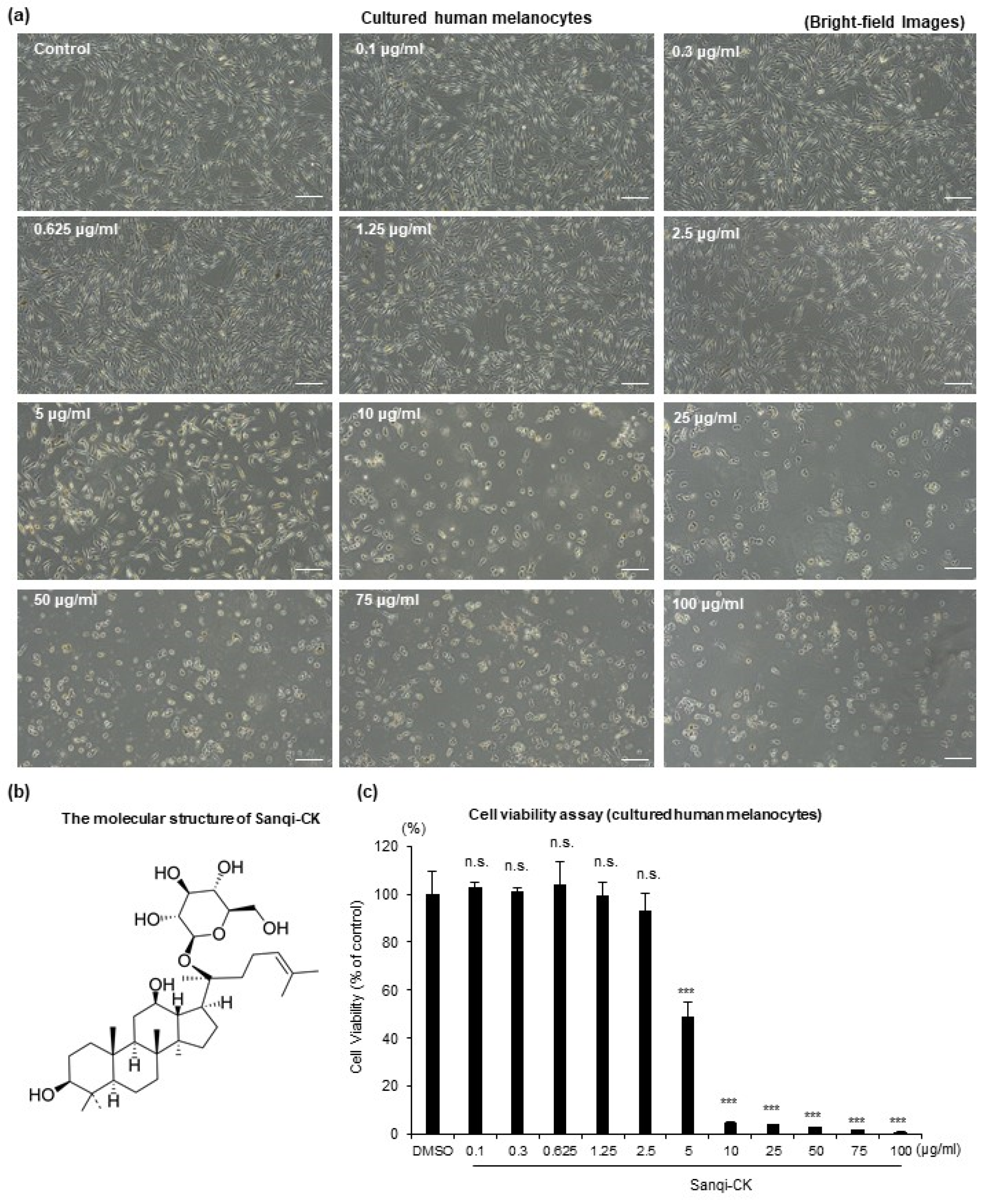
2.3. MTT Assay
2.4. Melanin Content Assay
2.5. Cell Counting Assay
2.6. L-Dopa Reaction
2.7. Trosinase Activity Assay
2.8. RNA Isolation and Real-Time RT-PCR Analysis
2.9. Oxidative Stress Assessment
2.10. Measurement of Reduced and Oxidized Glutathione
2.11. Measurement of Glutathione Reductase Activity
2.12. Animals and In Vivo Experimental Procedures
2.13. Measurement of Skin Color
2.14. Fontana-Masson Staining
2.15. Fluorescent Immunohistochemical Staining of Melanocytes
2.16. Statistical Analyses
3. Results
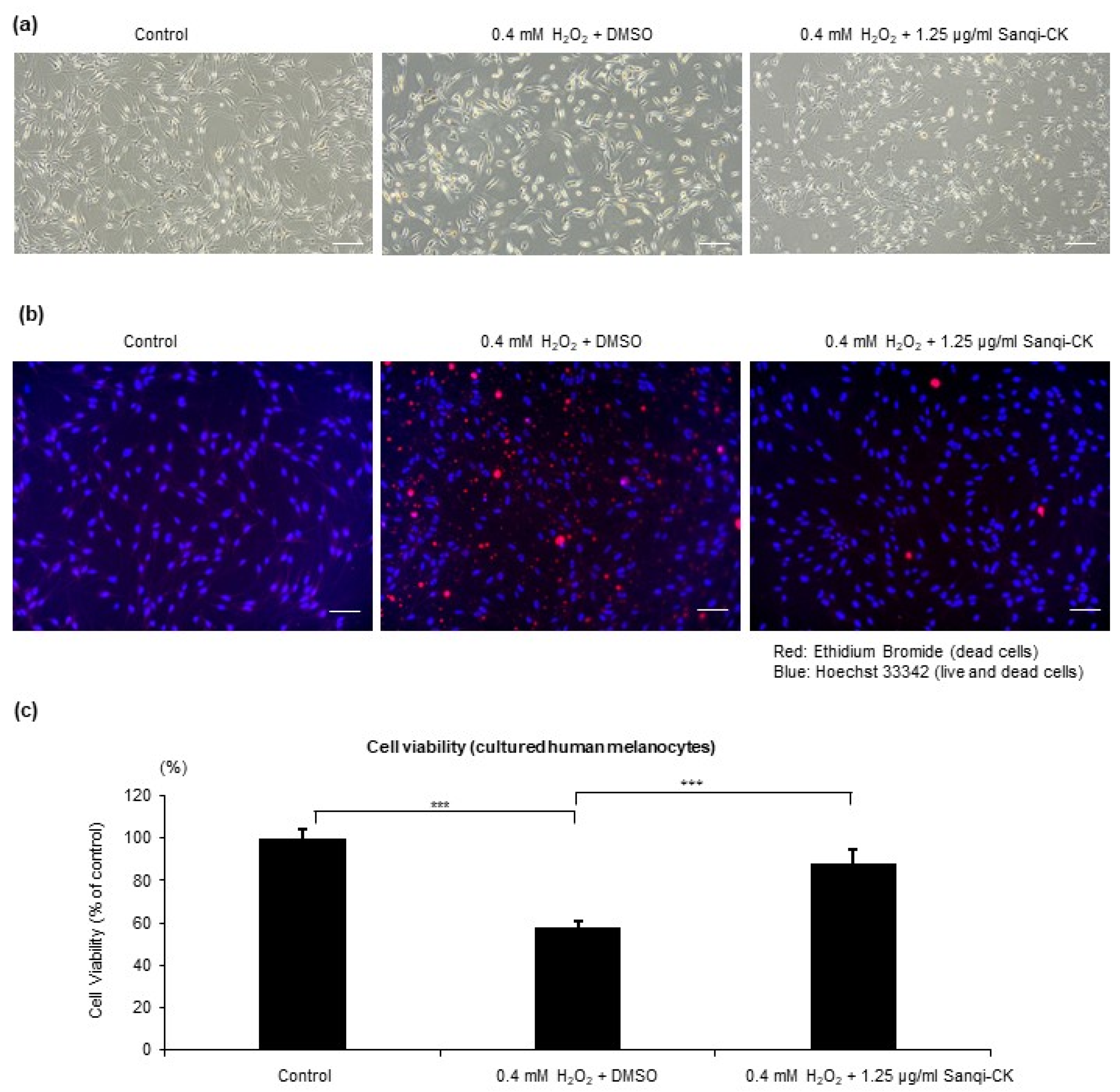
3.1. Sanqi-CK Attenuates H2O2-Induced Cytotoxicity in Human Primary Epidermal Melanocytes
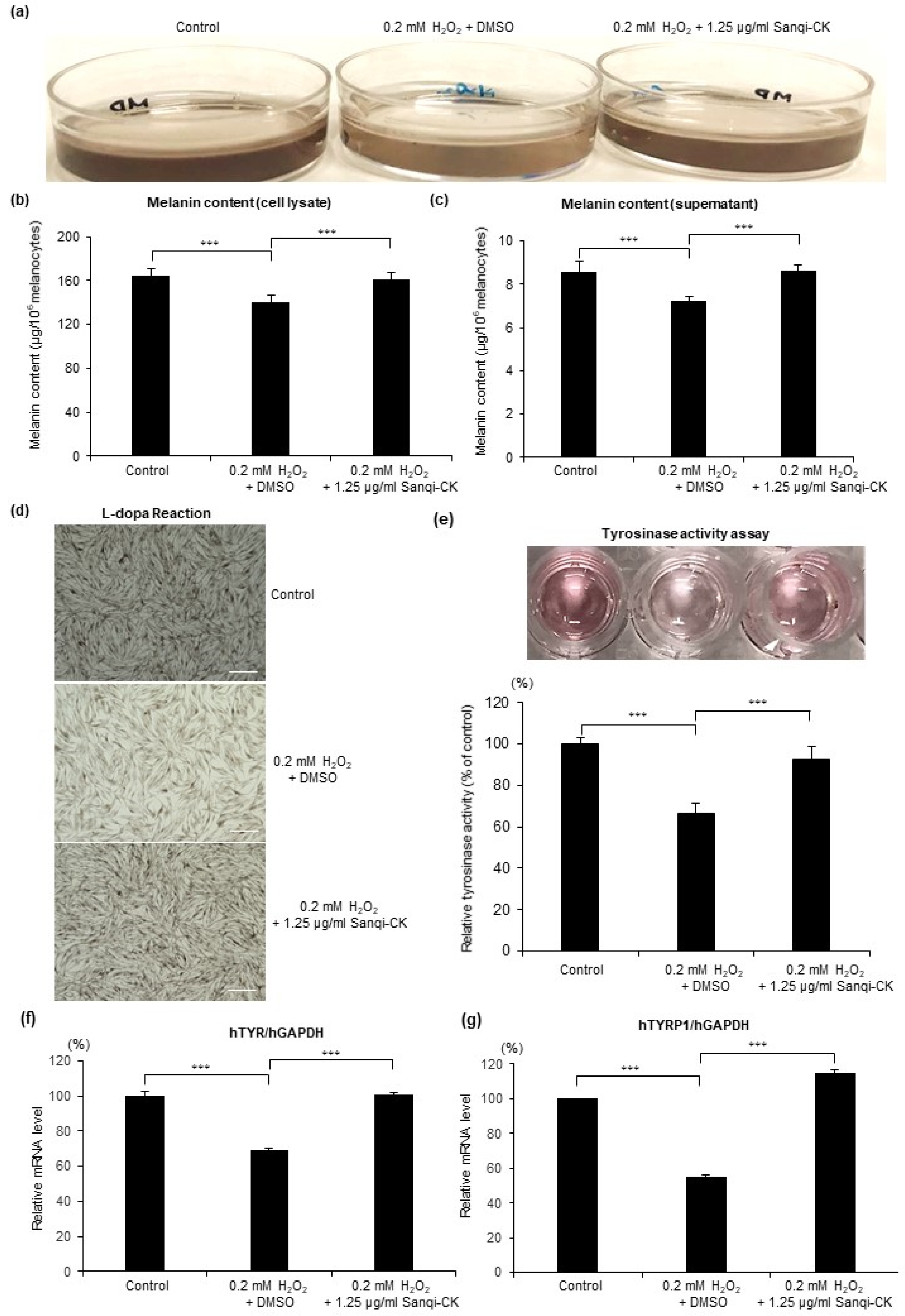
3.2. Sanqi-CK Attenuates H2O2-Induced Suppression of Melanogenesis in Human Primary Epidermal Melanocytes
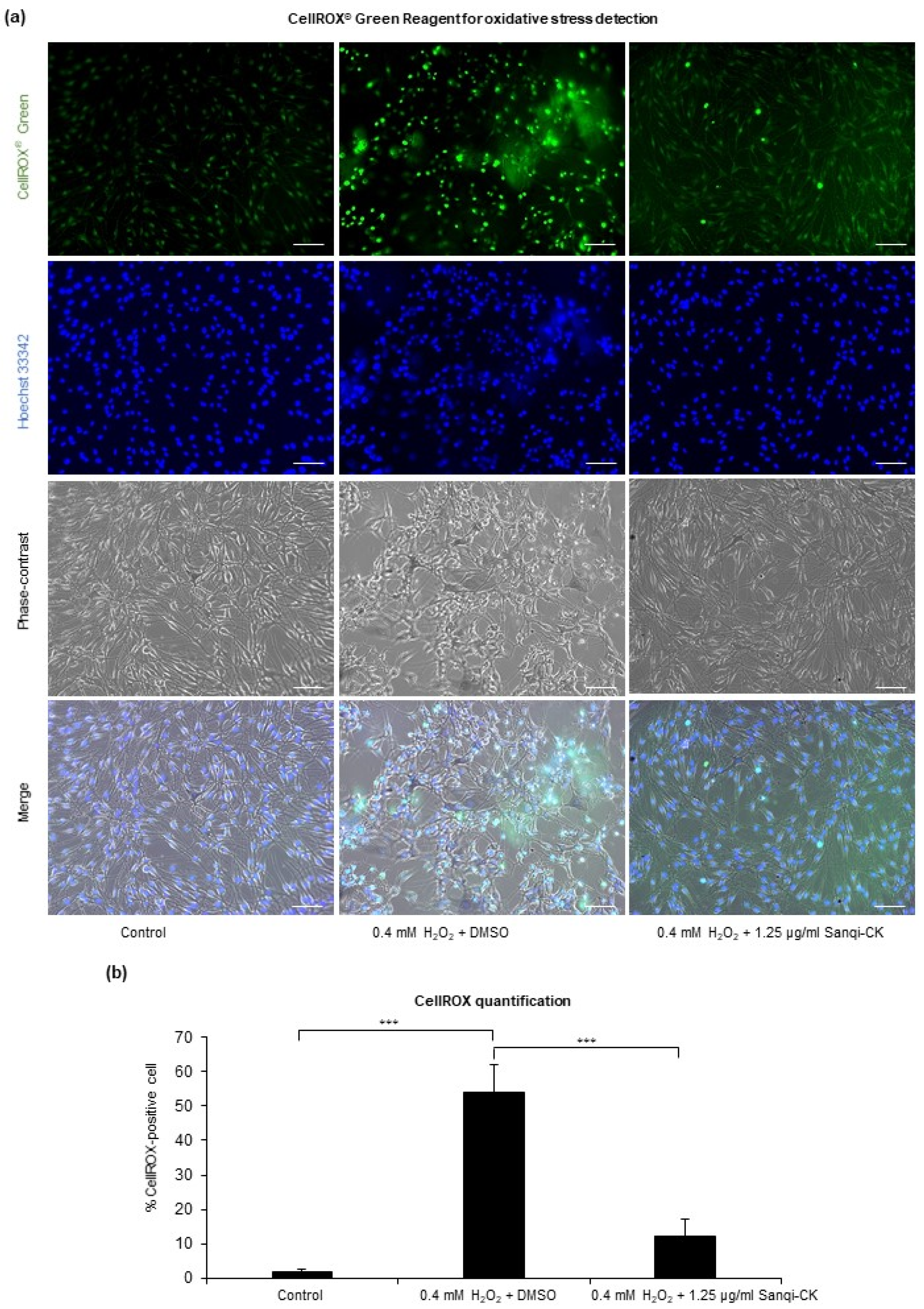
3.3. Sanqi-CK Reduces H2O2-Induced Oxidative Stress in Human Primary Epidermal Melanocytes
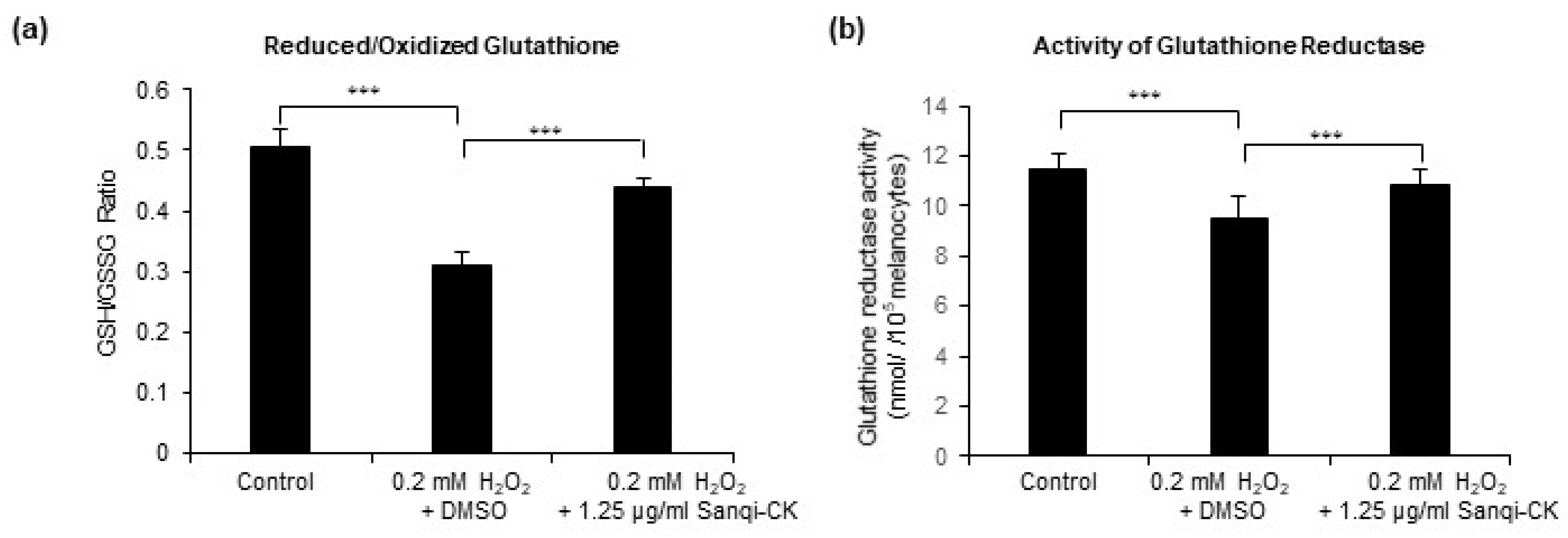
3.4. Sanqi-CK Modulates Redox Balance and Activates Glutathione Reductase in H2O2-Treated Human Primary Epidermal Melanocyte
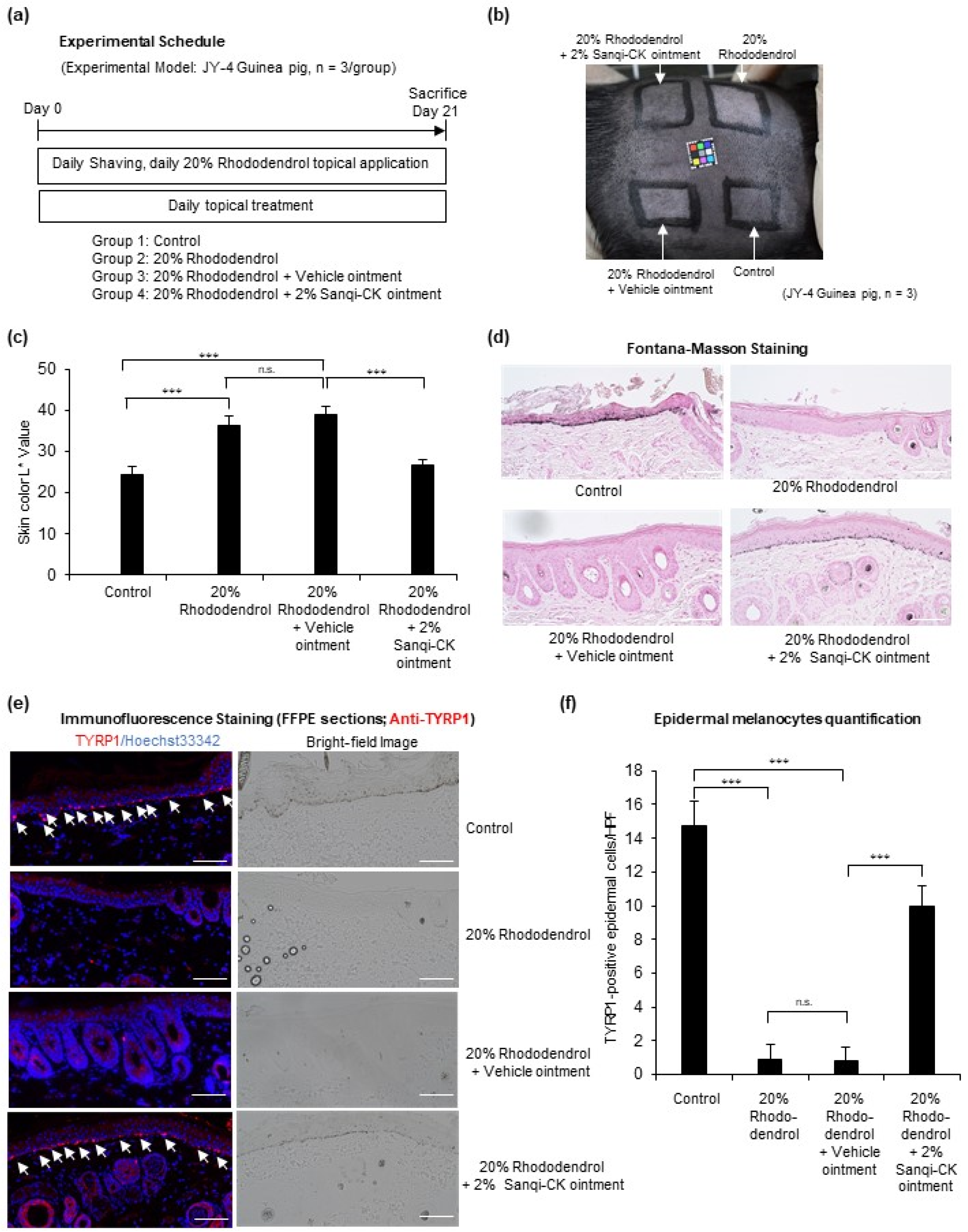
3.5. Topical Sanqi-CK Ointment Treatment Suppresses Rhododendrol-Induced Depigmentation in Guinea Pigs
4. Discussion
Author Contributions
Funding
Institutional Review Board Statement
Informed Consent Statement
Data Availability Statement
Acknowledgments
Conflicts of Interest
References
- Fitzpatrick, T.B.; Szabo, G. The melanocyte: Cytology and cytochemistry. J. Investig. Dermatol. 1959, 32, 197–209. [Google Scholar] [CrossRef] [Green Version]
- Denat, L.; Kadekaro, A.L.; Marrot, L.; Leachman, S.A.; Abdel-Malek, Z.A. Melanocytes as instigators and victims of oxidative stress. J. Investig. Dermatol. 2014, 134, 1512–1518. [Google Scholar] [CrossRef] [PubMed] [Green Version]
- Ezzedine, K.; Eleftheriadou, V.; Whitton, M.; van Geel, N. Vitiligo. Lancet 2015, 386, 74–84. [Google Scholar] [CrossRef]
- Picardo, M.; Dell’Anna, M.L.; Ezzedine, K.; Hamzavi, I.; Harris, J.E.; Parsad, D.; Taieb, A. Vitiligo. Nat. Rev. Dis. Primers 2015, 1, 15011. [Google Scholar] [CrossRef] [PubMed]
- Taieb, A. Intrinsic and extrinsic pathomechanisms in vitiligo. Pigment. Cell Res. 2000, 13 (Suppl. S8), 41–47. [Google Scholar] [CrossRef]
- Maresca, V.; Roccella, M.; Roccella, F.; Camera, E.; Del Porto, G.; Passi, S.; Grammatico, P.; Picardo, M. Increased sensitivity to peroxidative agents as a possible pathogenic factor of melanocyte damage in vitiligo. J. Investig. Dermatol. 1997, 109, 310–313. [Google Scholar] [CrossRef] [Green Version]
- Schallreuter, K.U.; Moore, J.; Wood, J.M.; Beazley, W.D.; Gaze, D.C.; Tobin, D.J.; Marshall, H.S.; Panske, A.; Panzig, E.; Hibberts, N.A. In vivo and in vitro evidence for hydrogen peroxide (H2O2) accumulation in the epidermis of patients with vitiligo and its successful removal by a UVB-activated pseudocatalase. J. Investig. Dermatol. Symp. Proc. 1999, 4, 91–96. [Google Scholar] [CrossRef] [PubMed] [Green Version]
- Jimbow, K.; Chen, H.; Park, J.S.; Thomas, P.D. Increased sensitivity of melanocytes to oxidative stress and abnormal expression of tyrosinase-related protein in vitiligo. Br. J. Dermatol. 2001, 144, 55–65. [Google Scholar] [CrossRef]
- Abdel-Malek, Z.A.; Jordan, C.; Ho, T.; Upadhyay, P.R.; Fleischer, A.; Hamzavi, I. The enigma and challenges of vitiligo pathophysiology and treatment. Pigment. Cell Melanoma Res. 2020, 33, 778–787. [Google Scholar] [CrossRef] [Green Version]
- Passi, S.; Grandinetti, M.; Maggio, F.; Stancato, A.; De Luca, C. Epidermal oxidative stress in vitiligo. Pigment. Cell Res. 1998, 11, 81–85. [Google Scholar] [CrossRef]
- Peng, M.; Yi, Y.X.; Zhang, T.; Ding, Y.; Le, J. Stereoisomers of Saponins in Panax notoginseng (Sanqi): A Review. Front. Pharmacol. 2018, 9, 188. [Google Scholar] [CrossRef] [Green Version]
- Duan, L.; Xiong, X.; Hu, J.; Liu, Y.; Li, J.; Wang, J. Panax notoginseng Saponins for Treating Coronary Artery Disease: A Functional and Mechanistic Overview. Front. Pharmacol. 2017, 8, 702. [Google Scholar] [CrossRef] [PubMed] [Green Version]
- Du, Y.; Fu, M.; Wang, Y.T.; Dong, Z. Neuroprotective Effects of Ginsenoside Rf on Amyloid-beta-Induced Neurotoxicity in vitro and in vivo. J. Alzheimers Dis. 2018, 64, 309–322. [Google Scholar] [CrossRef]
- Xie, W.; Meng, X.; Zhai, Y.; Zhou, P.; Ye, T.; Wang, Z.; Sun, G.; Sun, X. Panax Notoginseng Saponins: A Review of Its Mechanisms of Antidepressant or Anxiolytic Effects and Network Analysis on Phytochemistry and Pharmacology. Molecules 2018, 23, 940. [Google Scholar] [CrossRef] [Green Version]
- Zhao, H.; Han, Z.; Li, G.; Zhang, S.; Luo, Y. Therapeutic Potential and Cellular Mechanisms of Panax Notoginseng on Prevention of Aging and Cell Senescence-Associated Diseases. Aging Dis. 2017, 8, 721–739. [Google Scholar] [CrossRef] [PubMed] [Green Version]
- Bellei, B.; Pitisci, A.; Ottaviani, M.; Ludovici, M.; Cota, C.; Luzi, F.; Dell’Anna, M.L.; Picardo, M. Vitiligo: A possible model of degenerative diseases. PLoS ONE 2013, 8, e59782. [Google Scholar] [CrossRef] [PubMed] [Green Version]
- Xiao, J.; Chen, H.; Kang, D.; Shao, Y.; Shen, B.; Li, X.; Yin, X.; Zhu, Z.; Li, H.; Rao, T.; et al. Qualitatively and quantitatively investigating the regulation of intestinal microbiota on the metabolism of panax notoginseng saponins. J. Ethnopharmacol. 2016, 194, 324–336. [Google Scholar] [CrossRef]
- Xia, W.; Sun, C.; Zhao, Y.; Wu, L. Hypolipidemic and antioxidant activities of sanchi (radix notoginseng) in rats fed with a high fat diet. Phytomedicine 2011, 18, 516–520. [Google Scholar] [CrossRef]
- Song, W.; Wei, L.; Du, Y.; Wang, Y.; Jiang, S. Protective effect of ginsenoside metabolite compound K against diabetic nephropathy by inhibiting NLRP3 inflammasome activation and NF-kappaB/p38 signaling pathway in high-fat diet/streptozotocin-induced diabetic mice. Int. Immunopharmacol. 2018, 63, 227–238. [Google Scholar] [CrossRef]
- Yang, Q.; Lin, J.; Zhang, H.; Liu, Y.; Kan, M.; Xiu, Z.; Chen, X.; Lan, X.; Li, X.; Shi, X.; et al. Ginsenoside Compound K Regulates Amyloid beta via the Nrf2/Keap1 Signaling Pathway in Mice with Scopolamine Hydrobromide-Induced Memory Impairments. J. Mol. Neurosci. 2019, 67, 62–71. [Google Scholar] [CrossRef]
- Yang, F.; Yang, L.; Wataya-Kaneda, M.; Yoshimura, T.; Tanemura, A.; Katayama, I. Uncoupling of ER/Mitochondrial Oxidative Stress in mTORC1 Hyperactivation-Associated Skin Hypopigmentation. J. Investig. Dermatol. 2018, 138, 669–678. [Google Scholar] [CrossRef] [Green Version]
- Kim, Y.J.; Kim, M.J.; Kweon, D.K.; Lim, S.T.; Lee, S.J. Quantification of Hypopigmentation Activity In Vitro. J. Vis. Exp. 2019, e58185. [Google Scholar] [CrossRef] [Green Version]
- Meister, A. Glutathione metabolism and its selective modification. J. Biol. Chem. 1988, 263, 17205–17208. [Google Scholar] [CrossRef]
- Ito, S.; Wakamatsu, K. Biochemical Mechanism of Rhododendrol-Induced Leukoderma. Int. J. Mol. Sci. 2018, 19, 552. [Google Scholar] [CrossRef] [Green Version]
- Abe, Y.; Okamura, K.; Kawaguchi, M.; Hozumi, Y.; Aoki, H.; Kunisada, T.; Ito, S.; Wakamatsu, K.; Matsunaga, K.; Suzuki, T. Rhododenol-induced leukoderma in a mouse model mimicking Japanese skin. J. Dermatol. Sci. 2016, 81, 35–43. [Google Scholar] [CrossRef] [PubMed]
- Abe, Y.; Hozumi, Y.; Okamura, K.; Kawaguchi, M.; Kunisada, T.; Aoki, H.; Suzuki, T. A Mouse Model of Leukoderma Induced by Rhododendrol. J. Dermatol. Sci. 2016, 84, e86. [Google Scholar] [CrossRef]
- Kuroda, Y.; Takahashi, Y.; Sakaguchi, H.; Matsunaga, K.; Suzuki, T. Depigmentation of the skin induced by 4-(4-hydroxyphenyl)-2-butanol is spontaneously re-pigmented in brown and black guinea pigs. J. Toxicol. Sci. 2014, 39, 615–623. [Google Scholar] [CrossRef] [Green Version]
- Yang, L.; Yang, F.; Teng, L.; Katayama, I. 6-Shogaol Protects Human Melanocytes against Oxidative Stress through Activation of the Nrf2-Antioxidant Response Element Signaling Pathway. Int. J. Mol. Sci. 2020, 21, 3537. [Google Scholar] [CrossRef] [PubMed]
- Wang, C.-Z.; McEntee, E.; Wicks, S.; Wu, J.-A.; Yuan, C.-S. Phytochemical and Analytical Studies of Panax Notoginseng (Burk.) F.H. Chen. J. Nat. Med. 2006, 60, 97–106. [Google Scholar] [CrossRef]
- Xu, C.; Wang, W.; Wang, B.; Zhang, T.; Cui, X.; Pu, Y.; Li, N. Analytical methods and biological activities of Panax notoginseng saponins: Recent trends. J. Ethnopharmacol. 2019, 236, 443–465. [Google Scholar] [CrossRef] [PubMed]
- Dai, C.; Liang, Y.; Hao, H.; Zheng, X.; Xie, L.; Guan, T.; Zhou, Y.; Wang, G. Global detection and identification of components from Yunnan Baiyao based on liquid chromatography hybrid ion trap time-of-flight mass spectrometry. J. Sep. Sci. 2013, 36, 1935–1944. [Google Scholar] [CrossRef] [PubMed]
- Couto, N.; Wood, J.; Barber, J. The role of glutathione reductase and related enzymes on cellular redox homoeostasis network. Free Radic. Biol. Med. 2016, 95, 27–42. [Google Scholar] [CrossRef] [PubMed]
- Zedan, H.; Abdel-Motaleb, A.A.; Kassem, N.M.; Hafeez, H.A.; Hussein, M.R. Low glutathione peroxidase activity levels in patients with vitiligo. J. Cutan. Med. Surg. 2015, 19, 144–148. [Google Scholar] [CrossRef] [PubMed]
- Kondo, M.; Kawabata, K.; Sato, K.; Yamaguchi, S.; Hachiya, A.; Takahashi, Y.; Inoue, S. Glutathione Maintenance Is Crucial for Survival of Melanocytes after Exposure to Rhododendrol. Pigment. Cell Melanoma Res. 2016, 29, 541–549. [Google Scholar] [CrossRef] [PubMed]


Publisher’s Note: MDPI stays neutral with regard to jurisdictional claims in published maps and institutional affiliations. |
© 2021 by the authors. Licensee MDPI, Basel, Switzerland. This article is an open access article distributed under the terms and conditions of the Creative Commons Attribution (CC BY) license (https://creativecommons.org/licenses/by/4.0/).
Share and Cite
Tang, S.; Yang, L.; Kuroda, Y.; Lai, S.; Xie, S.; Zhang, H.; Katayama, I. Herb Sanqi-Derived Compound K Alleviates Oxidative Stress in Cultured Human Melanocytes and Improves Oxidative-Stress-Related Leukoderma in Guinea Pigs. Cells 2021, 10, 2057. https://doi.org/10.3390/cells10082057
Tang S, Yang L, Kuroda Y, Lai S, Xie S, Zhang H, Katayama I. Herb Sanqi-Derived Compound K Alleviates Oxidative Stress in Cultured Human Melanocytes and Improves Oxidative-Stress-Related Leukoderma in Guinea Pigs. Cells. 2021; 10(8):2057. https://doi.org/10.3390/cells10082057
Chicago/Turabian StyleTang, Suwei, Lingli Yang, Yasutaka Kuroda, Sylvia Lai, Shaoqiong Xie, Huimin Zhang, and Ichiro Katayama. 2021. "Herb Sanqi-Derived Compound K Alleviates Oxidative Stress in Cultured Human Melanocytes and Improves Oxidative-Stress-Related Leukoderma in Guinea Pigs" Cells 10, no. 8: 2057. https://doi.org/10.3390/cells10082057