Review

Cyclic AMP Signaling in Biliary Proliferation: A Possible Target for Cholangiocarcinoma Treatment?

1
Hepatology Unit, University of Tor Vergata, 00133 Rome, Italy
2
Hepatology and Gastroenterology, Medicine, Indiana University, Indianapolis, IN 46202, USA
3
Richard L. Roudebush VA Medical Center, Indianapolis, IN 46202, USA
4
Division of Transplant Surgery, Department of Surgery, Indiana University, Indianapolis, IN 46202, USA
5
Department of Medical Physiology, Texas A&M University College of Medicine, Bryan, TX 77807, USA
*
Authors to whom correspondence should be addressed.
These authors contributed equally to this work.
Cells 2021, 10(7), 1692; https://doi.org/10.3390/cells10071692
Submission received: 25 May 2021 / Revised: 30 June 2021 / Accepted: 30 June 2021 / Published: 4 July 2021
(This article belongs to the Special Issue New Aspects and Mechanisms in Liver Diseases)

Abstract

:
Cholangiocarcinoma is a lethal disease with scarce response to current systemic therapy. The rare occurrence and large heterogeneity of this cancer, together with poor knowledge of its molecular mechanisms, are elements contributing to the difficulties in finding an appropriate cure. Cholangiocytes (and their cellular precursors) are considered the liver component giving rise to cholangiocarcinoma. These cells respond to several hormones, neuropeptides and molecular stimuli employing the cAMP/PKA system for the translation of messages in the intracellular space. For instance, in physiological conditions, stimulation of the secretin receptor determines an increase of intracellular levels of cAMP, thus activating a series of molecular events, finally determining in bicarbonate-enriched choleresis. However, activation of the same receptor during cholangiocytes’ injury promotes cellular growth again, using cAMP as the second messenger. Since several scientific pieces of evidence link cAMP signaling system to cholangiocytes’ proliferation, the possible changes of this pathway during cancer growth also seem relevant. In this review, we summarize the current findings regarding the cAMP pathway and its role in biliary normal and neoplastic cell proliferation. Perspectives for targeting the cAMP machinery in cholangiocarcinoma therapy are also discussed.

1. Introduction

The biliary tree is lined by epithelial cells (i.e., cholangiocytes), which appear to originate from a common stem cell compartment, the hepatic progenitor cells, similar to hepatocytes [1]. These pluripotent cells are located at the interface between the hepatocyte canaliculi and bile ductules in the canals of Hering (Figure 1), the latter being the smaller branches of the biliary tree [2].
The canals of Hering extend to the interlobular ducts (i.e., bile ductules, 15–100 μm in diameter), then continue in the septal area, segmental and finally, in the right and left hepatic ducts, joining together at the hepatic hilum. This portion of the biliary epithelium is regarded as the intrahepatic one, whereas the part extending outside the liver and comprising the common bile duct, the gallbladder (with cystic duct) and choledochus is defined as extrahepatic [1]. Via electron microscopy, two different cholangiocyte subpopulations in rodent livers are well recognized (small and large), lining small ducts (≤15 μm in diameter) or large ducts (≥15 μm in diameter) according to their size [1,3]. Large cyclic adenosine monophosphate (cAMP)-dependent cholangiocytes are considered the functional part of the biliary tree since they respond to gastrointestinal hormones (e.g., secretin), neuro-peptides and other modulators [4,5,6]. On the other hand, small Ca2+-dependent cholangiocytes [7] do not respond to secretin and proliferate to replenish the biliary epithelium (during damage to the large, cAMP-dependent cholangiocytes) by amplification of Ca2+-dependent signaling and de novo acquisition of large cholangiocyte phenotypes [6,8,9]. While, at the beginning, just bile duct secretory activities were studied with regard to biliary tract diseases, lately also, biliary proliferative phenotypes have gained interest [10,11,12]. For instance, atypical cholangiocyte proliferation, characterized by truncated tortuous bile ducts, has been observed in adult cholestatic liver diseases such as primary biliary cholangitis (PBC) and primary sclerosing cholangitis (PSC) [13,14]. Both diseases are defined as ductopenia, since changes and evolution of proliferation determine a reduction of total bile ducts in the end [15].
In cholangiocarcinoma (CCA), cellular growth and expansion also deserve interest as possible targets for therapy. Several molecular cascades such as the Janus kinase/signal transducer and activator of transcription, p38 MAP kinase (MAPK), Akt (AKA, protein kinase B, PKB) or fibroblast growth factor/fibroblast growth factor receptor may stimulate cell growth and impact biliary proliferation [16,17]. Both hyperplastic and neoplastic cholangiocyte growth are frequently associated with changes in intracellular cAMP levels [18]. In this review, after a general description of cAMP signaling and its role in parenchymal and biliary cell functions, we will discuss the most recent findings regarding the relationship between the cAMP-dependent pathway and cancer of the biliary tract (e.g., cholangiocarcinoma).

2. cAMP Signaling

cAMP was first discovered in 1958 and introduced the concept of a “second messenger” system [19]. In fact, this molecule, together with cyclic guanosine monophosphate (cGMP), has been identified as an important intracellular translator of membrane signaling originating from hormones, growth factors, cytokines and other molecules [20]. In the general transduction mechanism, the stimulated receptor activates the corresponding G-coupled protein, leading to increased adenylyl cyclase-mediated cAMP synthesis. The following steps include cAMP binding to protein kinase A (PKA) or an exchange protein activated by cAMP (EPAC) and also cyclic nucleotide-gated ion channels [21]. Canonical G-protein-coupled receptor/cAMP/PKA signaling is depicted in Figure 2.
cAMP in eukaryotic cells serves as a secondary messenger for several biological activities such as the growth, differentiation and expression of selected mRNAs [22]. As described before, cAMP is primarily synthesized by G-protein-dependent stimulation of adenyl cyclase; however, G-protein-independent synthesis of cAMP may also occur upon stimulation of a soluble adenylyl cyclase induced by bicarbonate [23]; its activity is interrupted by phosphodiesterase-mediated degradation, when the biological target is achieved. The activity of PKA is prompted by cAMP binding to the R subunit that allows detachment of the C catalytic portion [24]; PKA is tetrameric so it is composed of two R and two C portions. Two different PKA isoforms (I and II) may be distinguished on the basis of different R subunits (I and II), while the C portion remains unchanged. At the same time, both the RI and RII subunits may present two forms called α and β, respectively [25]. Heterogeneity in the composition of subunits, and its different distribution in cellular districts is the basis for the several effects that PKA may exert upon stimulation [26]. Several cytoplasmic and nuclear proteins are targeted by PKA [20]. On the other hand, significant gene transcription activity is associated with PKA-dependent stimulation of the cAMP response element-binding protein (CREB) [27]. While PKA was considered the unique cAMP target for several years, recently, EPAC 1 and 2 were identified as sharing an identical PKA-subunit R-binding domain [28]. EPACs also regulate several physiological mechanisms including cellular secretion, adhesion and differentiation [22]. Finally, the cAMP binding domain has been identified at cyclic nucleotide-gated ion channels; cAMP-mediated channels’ stimulation remains of paramount importance for neuronal impulse transmission and cardiomyocyte contractility [29].

3. cAMP Signaling in Hepatocytes

3.1. General Signaling Effects

Similar to what is observed in other organs, in the liver, cAMP signaling represents an important point of intersection for signaling activities both in normal and diseased conditions. In this section, we discuss the main activities supported by the cAMP transduction pathway machinery in hepatocytes. The cAMP/PKA axis is involved in both liver gluconeogenesis and glycogenolysis [30]. In fact, during starvation, glucagon stimulates cAMP/PKA/CREB signaling. The maximal stimulation for transcription of target glucogenic genes is acquired by binding CREB, together with its CREB-regulated transcription coactivator 2 (CRTC2) [31]; conversely, glycogenolysis may be stimulated by PKA, leading to enhanced activity of glycogen phosphorylase. Nonetheless, fat metabolism may be regulated in the liver by cAMP levels. In fact, cAMP-stimulated PKA intracellular levels may phosphorylate and inhibit key enzymes of lipogenesis [32]. Evidence supporting the role of cAMP signaling in glucose and fat homeostasis has stimulated studies on liver diseases characterized by metabolic derangement, such as alcoholic liver disease (ALD) or non-alcoholic fatty liver disease (NAFLD).

3.2. cAMP in Liver Diseases with Metabolic Impairment

Reduced levels of cAMP have been shown in peripheral mononuclear cells from ALD patients in the early eighties [33]. Recently, decreased activity of cytochrome P450 2E1 (CYP2E1, the main enzyme producing toxic acetaldehyde from ethanol) by cAMP, was demonstrated in an experimental rodent model of ethanol feeding [34]. Similarly, in an analog animal model, a study demonstrated that ethanol-related phosphodiesterase-4 stimulation induced a decrease in intracellular cAMP levels and subsequent fat accumulation within the liver [35]. Beside the effects on lipid homeostasis, cAMP may also exhibit anti-inflammatory properties in ALD. In fact, a study in isolated monocytes and rat Kupffer cells demonstrated decreased cAMP levels associated with increased release of tumor necrosis factor-α (TNF-α) after alcohol exposure [36]. Furthermore, cAMP has been shown to attenuate alcohol-induced oxidative damage stimulating nitric oxide synthase expression [37]. With regards to NAFLD, a study has shown that cAMP acts as a second messenger after glucagon-like peptide (GLP) 1 receptor stimulation, improving fatty liver accumulation, glucose homeostasis and liver serum chemistry in leptin-deficient (ob/ob) mice [38]. Recent findings suggest that, together with peroxisome proliferator-activated receptor alpha (PPAR-α) and forkhead box O1 (FOXO1), the cAMP/responsive element-binding protein H (CREBH) signaling pathways remain of paramount importance in metabolic liver diseases [39]. Although a conclusive model describing the complex relationship between the cAMP/CREBH pathway and NAFLD (also for the involvement of this pathway in glucose homeostasis, the stress response and the inflammatory process) is lacking, this signaling system may be a good therapeutic target for the human metabolic liver diseases [40]. Finally, the role of cAMP in hepatocyte proliferation is well-defined [41]. In the model of 70% partial hepatectomy, which represents an ideal system to evaluate cellular regeneration, a two-phase increase in cAMP and activation of different PKA subtypes is observed. Therefore, important CREB phosphorylation is reported, thus stimulating the downstream growth genes; this process requires a functional G-protein, Gαs [42].

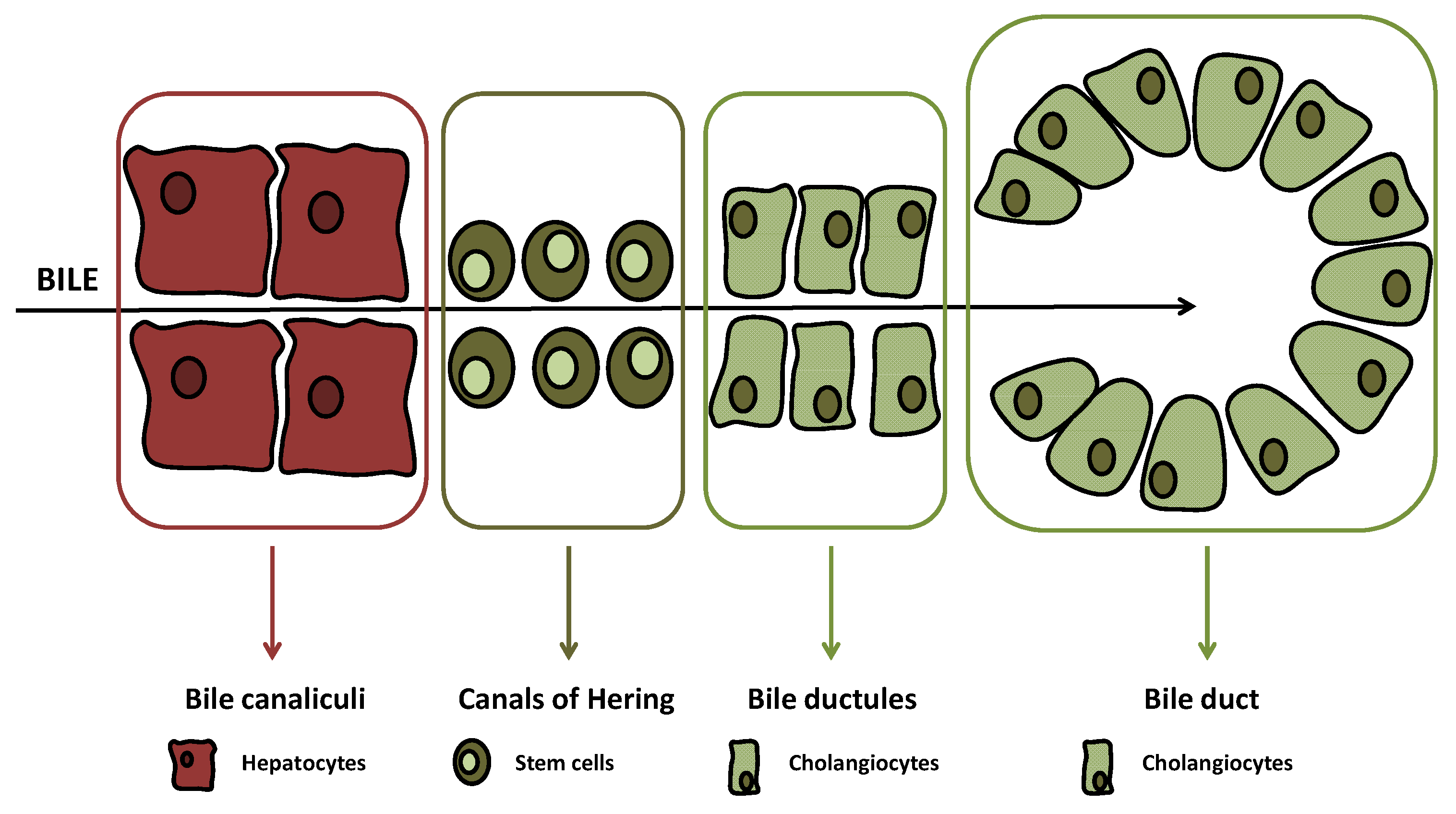
4. cAMP Signaling in Cholangiocytes

4.1. cAMP and Biliary Secretion

Since the beginning of research on biliary epithelia, cAMP attracted attention for its involvement in secretin-stimulated biliary secretion [4]. In fact, several studies established the role of secretin in the stimulation of biliary bicarbonate secretion by specifically increasing cAMP intracellular levels in cholangiocytes, but not hepatocytes [4,43,44]. In a seminal study, secretin-stimulated exocytosis was examined in detail in isolated cholangiocytes [44]; the study demonstrated that exocytosis was closely associated with enhanced intracellular cAMP levels (more than 200% increase compared to unstimulated cholangiocytes), independent of cGMP intracellular levels, as well as stimulated by forskolin (a drug enhancing cAMP synthesis). Following interaction with its basolateral receptors [4,5,45], secretin-stimulated choleresis is mediated by sequential activation of PKA, the cystic fibrosis transmembrane conductance regulator (CFTR) and anion exchanger 2 (AE2), leading to enhanced bicarbonate secretion [4,5,46,47]. In the bile duct-ligated (BDL) hyperplastic rodent model, this event is amplified [47] but is inhibited by insulin, decreasing cAMP intracellular levels through a Ca2+/PKC-dependent mechanism [48].

4.2. cAMP and Biliary Proliferation

A number of studies also demonstrated the effects of cAMP signaling on biliary proliferation in response to liver injury [10,49,50,51]. Recently, research specifically focused on the net effect of cAMP on proliferation of biliary epithelia was performed [49]. Administration of forskolin (an activator of adenylyl cyclase) to Fisher male rats significantly increased intrahepatic biliary mass, as well as biliary secretion. Also, the signaling pathway was characterized in this study, demonstrating the involvement of cAMP in the activation of a signaling cascade composed by the PKA/Src/MEK/ERK1/2 axis [49]. This study clarified some relevant concepts, such as how: (i) cAMP plays an important messenger role for both secretive and proliferative biliary functions; (ii) a functional cAMP/ERK signaling axis is present in cholangiocytes; and (iii) in the biliary ductal system, the cAMP/ERK signaling axis promotes cell growth [10,49]. Another study demonstrated that prolonged secretin administration stimulated cAMP-dependent biliary growth in normal rats [52]. Minipump administration of the hormone (2.5 nmoles/kg BW/day) for one week increased biliary proliferation and hyperplasia by enhancing intracellular cAMP levels. Furthermore, in BDL rats lacking a secretin receptor (knockout SR−/−) [53], there was reduced biliary growth through a significant decrease in intracellular cAMP and ERK1/2 phosphorylation. Other studies underscored the important function of cAMP as a common hormonal and neuroendocrine signal mediating cholangiocyte growth [10,49]. With regard to gastrointestinal hormones, while secretin and GLP-1 stimulate biliary proliferation by enhancing cAMP machinery [10,53,54], others such as gastrin and somatostatin reduced biliary hyperplasia. However, while the inhibitory effects of somatostatin on cAMP-dependent biliary proliferation were directly linked to cAMP, gastrin inhibition of cAMP-dependent biliary growth was mediated by changes in the Ca2+-dependent protein kinase C (PKC) alpha [55,56,57]. The neuropeptides acetylcholine, serotonin, histamine, melatonin and others also modulate cholangiocyte growth, reacting with specific receptors and altering cAMP levels [58,59,60,61].
Finally, bile acids (BAs) are probably the most important non-endocrine regulators of biliary growth. In molecular studies, some BAs G-protein-coupled receptors have been identified in cholangiocytes such as the Takeda G-protein-coupled receptor 5 (TGR5), the subtype 3 muscarinic (M3) receptor and the sphingosine-1-phosphate receptor 2 (S1PR2) [62]. Among these, TGR5 has been found to regulate cell growth, modulate the cAMP pathway and have an opposite effect when comparing non-ciliated cholangiocytes (increases growth) with ciliated ones (decreases growth) [63]. In fact, activation of TGR5 located in the cilia determines receptor coupling with Gαi instead of Gαs, thus decreasing cAMP intracellular levels and cell proliferation, a mechanism impaired in polycystic liver disease. With regard to this latter disease, characterized by an exuberant growth of cysts, possible downregulation of cAMP signaling may be hypothesized as a possible therapeutic approach.
Data from in vivo studies have shown that feeding taurocholic or taurolithocholic acid to rats increases biliary proliferation and secretin-stimulated ductal secretion (a functional index of cholangiocyte growth) [10,49,64], phenotypes that were mediated by increased biliary cAMP levels [65]. On the other hand, ursodeoxycholic acid (UDCA), used for therapy in PBC [66], reduced cholangiocyte proliferation in the cholestatic rodent model of bile duct ligation (BDL) through a decrease in cAMP signaling mediated by activation of PKC alpha [67]. Taken together, the findings suggest that modulation of cholangiocyte growth based on the activity of the cAMP axis normally occurs in physiologic conditions; moreover, this signaling pathway may represent an important target for pharmacological modulation of biliary secretive and proliferative activity in cholangiopathies. However, is to be underscored that the final effect of the increase (or decrease) of cAMP in cholangiocytes is variable according to different molecular factors. In fact, this second messenger lies at the intersection between several signaling cascades with possible divergent downstream biological effects.

5. cAMP and Cancer

The role of cAMP and its main effector PKA in cancer has been recognized [68], and targeting of this signaling axis has been identified as a possible strategy for cancer therapy [69]. Changes to or modulation of cAMP/PKA signaling exert effects in a number of cancers involving the brain [70,71], lungs [72], prostate [73] or blood [74]. However, it has to be emphasized that stimulation of cAMP signaling may have opposing results depending on the tissue and cancer type. For instance, in tumor spheres obtained from a primary cell culture of medulloblastoma, cancer growth was inhibited by forskolin and enhanced by PKA inhibition [75]. On the other hand, cAMP/PKA signaling was involved in enhanced expression of the androgen receptor and tumor growth in prostate cancer [76]. Since PKA composed of an RI subunit was linked to increased proliferation, and enhanced expression of the RIα-type was observed in tumors, in the early 1990s, a study with an antisense oligonucleotide-repressing RIα was conducted on cancer cells [77]. Neoplastic cell lines from the stomach, colon and breast (TMK-1, LS-174T and MCF-7 lines) all exhibited impaired growth upon antisense treatment [77]. A following study demonstrated that the antisense oligonucleotide GEM231 also exhibited anticancer activity in vivo [77]. Further, a separate study demonstrated that the antisense oligonucleotide GEM231 also exhibited anticancer activity in vivo [78]. In fact, in a nude mouse model of human prostate, colon, lung and pancreas cancer cells’ xenotransplantation, simultaneous GEM231 administration determined enhanced irinotecan antitumor activity (68). In a Phase I study on 20 patients with different solid tumors, GEM231 (220 mg/m2, twice a week) was administered and coupled with a variable dose of Docetaxel [79]. Liver toxicity was frequently observed (increase of transaminases levels in 75% of patients) as well as neutropenia (45%) and fatigue (40%). Neurologic side effects occurred in five patients (20%), with one exhibiting toxic neuropathy; three serious adverse events occurred during the study: a cardiac arrest, a pulmonary embolism and fever as a consequence of neutropenia. Results of further attempts of this study have not been published thus far. Other preclinical studies evaluated an approach with cAMP analogs. In this perspective, 8-Cl-cAMP (i.e., tacladesine) with a preferential inhibition of PKAI evidenced a significant effect on the growth of the following cancer cellular lines: HL-60 leukemia cells [80], MCF-7 (breast), LS-174T (colon) and A549 (lung) [81]. Two trials were registered with 8-Cl-cAMP, one (Phase II, NCT00004902) on multiple myeloma and one (Phase I, NCT00021268) on colon cancer [82]. The results of these studies (despite beginning 20 years ago) have not been disclosed so far. Use of phosphodiesterase for the modulation of cAMP has been suggested by some authors for cancer therapy, but preclinical or clinical results employing this approach have not been published [28].

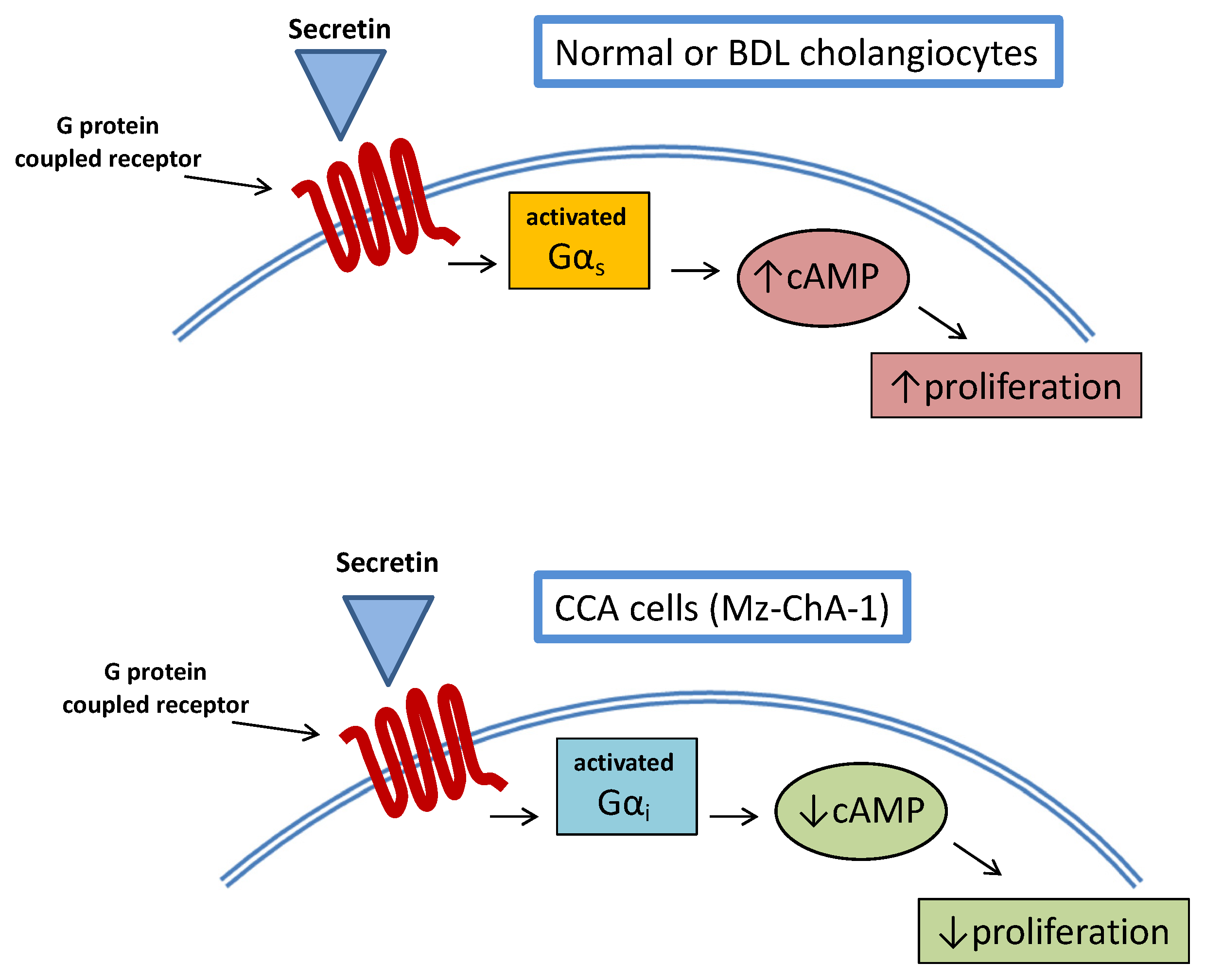
6. CCA and cAMP Signaling

6.1. CCA

CCA is a rare and lethal disease representing the most frequent primary liver cancer after hepatocellular carcinoma (HCC) [83]. Incidences of CCA change widely among different countries, ranging from 85 to less than 1 out of 100,000, thus reflecting the prevalence of some risk factors such as parasitic infections (Clonorchis sinensis and Opisthorchis viverrini), biliary tract disorders (PSC, hepatolithiasis, biliary cystic diseases) and also inflammatory bowel diseases [84]. CCA’s clinical classification relies on its anatomical location along the biliary tree. Intrahepatic (iCCA), perihilar (pCCA) and distal (dCCA) forms are recognized with different prevalence and prognoses [85]. Histological classification is complex since heterogeneity is found in genetic alterations, pathogenesis and cellular origin, which are all factors contributing to a variable morphological picture [86]. Of concern, while some forms of CCA (iCCA) increased their global prevalence and associated mortality in the last decades, treatment of this cancer did not show, in parallel, any major improvements [87,88]. Improved understanding of the processes at the origin of CCA proliferation within the biliary tract may probably stimulate targeted therapy for this cancer in the future. In this perspective, since cAMP signaling plays an important role in the normal proliferative activity of cholangiocytes, it is likely that changes to this pathway may occur in biliary tract cancer.

6.2. Hormones/Neuropeptides Modulation of cAMP in CCA

Secretin-stimulated intracellular cAMP levels represent one of the most studied mechanisms at the origin of cholangiocyte proliferation [89]. A study examined the relationship between cAMP and secretin stimulation in CCA cell lines [90]. Interestingly, the study demonstrated decreased expression of the secretin receptor in the majority of CCA lines (Mz-ChA-1, HuH-28, SG231, CCLP1) used. The same was observed in CCA human liver specimens. More interestingly, stimulation of CCA cell lines with secretin did not increase intracellular cAMP levels so that proliferative activities were not enhanced. This was also confirmed in an in vivo model of Mz-ChA-1 xenotransplantation in nude mice, where tumor growth was delayed by secretin treatment. The opposite effect of secretin on CCA cell growth was related to an aberrant (cancer-related) coupling of the secretin receptor with Gαi rather than Gαs (Figure 3).
This study also had the merit of demonstrating once again the complex interplay between CCA and cAMP signaling, evidencing a mechanism similar to what was already observed, upon stimulation of the TGR5 receptor in cholangiocytes’ cilia [66]. A number of studies suggest a multifaceted interaction between hormones/neuropeptides and cAMP signaling with regard to CCA growth. Somatostatin has been shown to reduce secretin-induced biliary secretion and proliferation, downregulating cAMP intracellular levels in cholangiocytes from BDL rats [56]. There is also evidence that somatostatin inhibits CCA growth [91]. In fact, human CCA specimens and the human CCA line, SK-ChA-1, express a functional somatostatin receptor subtype 2 (SSTR2) [91]. Treatment with a somatostatin analog (SS-14, octreotide) significantly decreased CCA cell growth in CCA lines, whereas lanreotide (a long-acting somatostatin analog) determined a 33% reduced growth of SK-ChA-1 cells implanted in athymic mice. However, this study did not observe any major changes in intracellular cAMP levels, thus excluding a possible role of this signaling system in somatostatin-induced decreased CCA proliferation. On the other hand, a relationship was found between the cAMP system and CCA growth upon stimulation of the α2-adrenergic receptor (α2-AR) [92]. Mz-ChA-1 and TFK-1 human CCA cell lines expressed α2-AR. Exposure of these CCA cells to the α2-AR agonist (UK14,304) decreased their growth as demonstrated by reduced [3H] thymidine incorporation and PCNA mRNA expression. This effect was related to increased cAMP/PKA signaling activity, inhibiting Raf-1 and B-Raf with a consequent downregulation of MAPK. Another study evaluated the effect of γ-aminobutyric acid (GABA) on CCA growth [93]. Mz-ChA-1, HuH-28 and TFK-1 human CCA cell lines all expressed functional GABA receptors and exposure to GABA decreased the proliferation index and PCNA mRNA expression in all three cell lines. In addition, Mz-ChA-1 cells’ transplantation in nude mice demonstrated reduced tumor growth after 82 days of GABA injection. These effects were related to increased cAMP/PKA signaling activity since: (i) this pathway was enhanced by GABA and; (ii) CCA growth was restored, adding into the cell culture a PKA inhibitor (Rp-cAMP116816). Also, the migration of CCA cells seemed to be impaired by GABA. In fact, wound closure (obtained by linear abrasion to culture plate) required a longer time in GABA-treated cells (>96 h).

6.3. BAs Modulation of cAMP in CCA

Other studies examined the possible role of the BA receptor TGR5 (known as a significant stimulator of cAMP/PKA signaling in cholangiocytes) [63] in CCA growth. This receptor has been found to be overexpressed in human CCA specimens when compared to surrounding normal tissue [94,95,96]. Moreover, the TGR5 expression rate was associated with less-differentiated CCA in one study [95]. Stimulation of this receptor in CCA cell cultures (EGI-1 and TFK-1) sustained the proliferation and invasiveness of tumoral cells [94]. TGR5’s stimulating effect on CCA growth seems to occur by a dual mechanism; (i) increased proliferation for stimulation of the EGFR/ERK1/2 pathway; and (ii) inhibition of apoptosis by a cAMP/PKA/CD95 cascade [62]. These observations underscore the differential and complex effect of TGR5/cAMP signaling in cholangiocytes, according to different species and conditions. In fact, while in H69 human biliary cells (both ciliated or non-ciliated), TGR5–induced proliferation is strictly related to fluctuations of cAMP levels [63], in murine cholangiocytes and human CCA cell lines (EGI-1 and TFK-1), cAMP signaling mainly contributes to apoptosis suppression, and growth is maintained by stimulation of the EGFR/ERK1/2 pathway [94]. Further studies, possibly on human tissue, would be needed to improve our knowledge of the TGR5/cAMP relationship in cholangiocytes and possible targeting in CCA.

6.4. PKA Subunits Changes in CCA

Changes in PKA subunits have been associated with CCA. Increased expression of RIα was found in human CCA in comparison with the surrounding non-tumoral tissue [97]. In the same study, also in CCA cell lines (M156, OCA17, KKU100 and M214), an enrichment of PKA subunit RIα was observed. Lentiviral transfection-induced knockdown of RIα in M156 and OCA17 cell cultures led to a significant decrease of PKA activity and cellular growth. Reduction of cellular proliferation was in part also related to increased apoptosis. Similar effects were also reported when testing H-89 (a PKA inhibitor) or 8-Cl cAMP on M156, OCA17, KKU100 and M214 cell culture growth. In fact, reduced CCA cellular proliferation, accounting for 20–40% growth inhibition, was observed, according to different doses of the inhibitor or cellular line. Resulting from these findings, an attempt was made to develop T-cell expansion against CCA, employing self-differentiated monocyte-derived dendritic cells presenting an RIα subunit [98]. Effector T-cells, activated with this strategy, were able to eliminate nearly 60% of CCA cells in vitro. PRKACA and PRKACB, the genes involved in transcription of the α and β PKA catalytic subunits, have also been found altered in some cases of CCA [99]. Their gene fusion with ATP1B may determine increased transcription with downstream activation of proliferative signals [100]. Similar fusion (DNAJB1-PRKACA) has been observed in fibrolamellar HCC; however, this genetic aberration should not be considered specific for this latter tumor since it was also identified in pancreatic and biliary tract cancer [101]. Recently, a study focused on intraductal oncocytic papillary neoplasm (IOPN) and PRKACA or PRKACB fusion [102]. Among 23 patients, three were harboring an IOPN of the biliary tract. All of them had ATP1B1–PRKACB gene fusion. The same alterations were not found in other malignancies of the pancreas or biliary tract, suggesting that PRKACA or PRKACB fusion may be characteristic of the IOPN type. The main preclinical studies evaluating the cAMP pathway in CCA are reported in Table 1.

7. Conclusions/Future Perspectives

CCA is a severe disease and we are still in search of appropriate treatment. While a limited number of patients may undergo surgical resection with improved survival, the majority of subjects harboring CCA are expected to die within one year of diagnosis [103]. Several efforts have been made in preclinical studies to identify possible strategies for CCA systemic therapy [99]. Targeting genetic aberrations or the use of immune checkpoint inhibitors represent possible opportunities in the future [88]; however, exploring new pathways for therapy seems wise given the suboptimal results of the current medical practice. cAMP/PKA signaling is a highly conserved pathway in eukaryotic cells and its important contribution in parenchymal and non-parenchymal epithelial liver cell growth has been largely demonstrated. Since important changes to this pathway occur in several human cancers, its targeting has been attempted with antisense oligonucleotide or cAMP analogs in preclinical models. Data on CCA when employing this approach are scanty, despite the fact that cAMP/PKA signaling has been demonstrated to be of paramount importance in the regulation of cholangiocyte proliferative activities in physiologic conditions and during damage. Moreover, as cholangiocytes are the main liver collectors of several hormones, neuropeptides and angiogenic factor signals employing cAMP as a second messenger, the possible modulation of this pathway employing these molecules may be hypothesized. In this context, we demonstrated that secretin receptor stimulation in CCA cells is not followed by the physiologic increase of cAMP that supports proliferation as observed in normal cholangiocytes. Also, the stimulation of α2-AR and GABA receptors (using cAMP as a second messenger) seems to inhibit CCA growth, suggesting possible anticancer activity. Finally, TGR5, another G-protein-coupled receptor, is constitutively overexpressed in CCA, where it sustains tumor expansion and diffusion with a mechanism partially involving cAMP and different from what is observed in normal cholangiocytes. These and other findings suggest that the study of cAMP signaling in CCA may be useful to understand the differences between normal and neoplastic biliary cells and help to design strategies to abolish or mitigate cancer growth with systemic therapy.

Author Contributions

L.B. conceptualization, original draft preparation, I.L., M.M., L.K., K.S., W.Z., B.E., L.C., V.M., S.G. review and editing, G.A. and H.F. proposal, supervision, funding acquisition, review and editing. All authors have read and agreed to the published version of the manuscript.

Funding

This work was supported by the Hickam Endowed Chair, Gastroenterology, Medicine, Indiana University; the Indiana University Health–Indiana University School of Medicine Strategic Research Initiative; the VA Merit awards to G.A. (5I01BX000574) and H.F. (1I01BX003031) from the United States Department of Veterans Affairs; the Biomedical Laboratory Research and Development Service and NIH grants DK108959 and DK119421 (H.F.) and DK054811, DK115184, DK076898, DK107310, DK110035, DK062975 and AA028711 (G.A. and S.G.); the PSC Partners Seeking a Cure (G.A.). Some of these studies were supported by resources at the Central Texas Veterans’ Health Care System (Temple, TX), the Richard L. Roudebush VA Medical Center (Indianapolis, IN) and Medical Physiology (Medical Research Building, Temple, TX). The views expressed in this article are those of the authors and do not necessarily represent the views of the Department of Veterans Affairs.

Conflicts of Interest

The authors declare no conflict of interest.

References

  1. Han, Y.; Glaser, S.; Meng, F.; Francis, H.; Marzioni, M.; McDaniel, K.; Alvaro, D.; Venter, J.; Carpino, G.; Onori, P.; et al. Recent advances in the morphological and functional heterogeneity of the biliary epithelium. Exp. Biol. Med. 2013, 238, 549–565. [Google Scholar] [CrossRef] [PubMed] [Green Version]
  2. Paliwal, J.K.; Ramesh, D.; Gupta, R.C. Synthesis and disposition of 14C-labelled 81/470, a new anthelminthic agent in rats. Int. J. Clin. Pharmacol. Res. 1997, 17, 23–30. [Google Scholar]
  3. Kanno, N.; LeSage, G.; Glaser, S.; Alvaro, D.; Alpini, G. Functional heterogeneity of the intrahepatic biliary epithelium. Hepatology 2000, 31, 555–561. [Google Scholar] [CrossRef] [PubMed]
  4. Kanno, N.; LeSage, G.; Glaser, S.; Alpini, G. Regulation of cholangiocyte bicarbonate secretion. Am. J. Physiol. Gastrointest. Liver Physiol. 2001, 281, G612–G625. [Google Scholar] [CrossRef] [PubMed]
  5. Alpini, G.; Glaser, S.; Robertson, W.; Rodgers, R.E.; Phinizy, J.L.; Lasater, J.; LeSage, G.D. Large but not small intrahepatic bile ducts are involved in secretin-regulated ductal bile secretion. Am. J. Physiol. 1997, 272, G1064–G1074. [Google Scholar] [CrossRef] [PubMed]
  6. Wu, N.; Baiocchi, L.; Zhou, T.; Kennedy, L.; Ceci, L.; Meng, F.; Sato, K.; Wu, C.; Ekser, B.; Kyritsi, K.; et al. Functional role of the secretin/secretin receptor signaling during cholestatic liver injury. Hepatology 2020, 72, 2219–2227. [Google Scholar] [CrossRef] [PubMed]
  7. Francis, H.; Glaser, S.; DeMorrow, S.; Gaudio, E.; Ueno, Y.; Venter, J.; Dostal, D.; Onori, P.; Franchitto, A.; Marzioni, M.; et al. Small mouse cholangiocytes proliferate in response to H1 histamine receptor stimulation by activation of the IP3/CaMK I/CREB pathway. Am. J. Physiol. Cell Physiol. 2008, 295, C499–C513. [Google Scholar] [CrossRef] [Green Version]
  8. Mancinelli, R.; Franchitto, A.; Gaudio, E.; Onori, P.; Glaser, S.; Francis, H.; Venter, J.; Demorrow, S.; Carpino, G.; Kopriva, S.; et al. After damage of large bile ducts by gamma-aminobutyric acid, small ducts replenish the biliary tree by amplification of calcium-dependent signaling and de novo acquisition of large cholangiocyte phenotypes. Am. J. Pathol. 2010, 176, 1790–1800. [Google Scholar] [CrossRef]
  9. LeSage, G.; Benedetti, A.; Glaser, S.; Marucci, L.; Tretjak, Z.; Caligiuri, A.; Rodgers, R.; Phinizy, J.L.; Baiocchi, L.; Francis, H.; et al. Acute carbon tetrachloride feeding selectively damages large, but not small, cholangiocytes from normal rat liver. Hepatology 1999, 29, 307–319. [Google Scholar] [CrossRef]
  10. Sato, K.; Marzioni, M.; Meng, F.; Francis, H.; Glaser, S.; Alpini, G. Ductular reaction in liver diseases: Pathological mechanisms and translational significances. Hepatology 2019, 69, 420–430. [Google Scholar] [CrossRef] [Green Version]
  11. Guicciardi, M.E.; Trussoni, C.E.; LaRusso, N.F.; Gores, G.J. The spectrum of reactive cholangiocytes in primary sclerosing cholangitis. Hepatology 2020, 71, 741–748. [Google Scholar] [CrossRef]
  12. Banales, J.M.; Huebert, R.C.; Karlsen, T.; Strazzabosco, M.; LaRusso, N.F.; Gores, G.J. Cholangiocyte pathobiology. Nat. Rev. Gastroenterol. Hepatol. 2019, 16, 269–281. [Google Scholar] [CrossRef]
  13. Lee, S.J.; Park, J.B.; Kim, K.H.; Lee, W.R.; Kim, J.Y.; An, H.J.; Park, K.K. Immunohistochemical study for the origin of ductular reaction in chronic liver disease. Int. J. Clin. Exp. Pathol. 2014, 7, 4076–4085. [Google Scholar] [PubMed]
  14. LeSage, G.; Glaser, S.; Alpini, G. Regulation of cholangiocyte proliferation. Liver 2001, 21, 73–80. [Google Scholar] [CrossRef] [PubMed]
  15. Alvaro, D.; Gigliozzi, A.; Attili, A.F. Regulation and deregulation of cholangiocyte proliferation. J. Hepatol. 2000, 33, 333–340. [Google Scholar] [CrossRef]
  16. Sato, K.; Francis, H.; Zhou, T.; Meng, F.; Kennedy, L.; Ekser, B.; Baiocchi, L.; Onori, P.; Mancinelli, R.; Gaudio, E.; et al. Neuroendocrine changes in cholangiocarcinoma growth. Cells 2020, 9, 436. [Google Scholar] [CrossRef] [PubMed] [Green Version]
  17. Labib, P.L.; Goodchild, G.; Pereira, S.P. Molecular pathogenesis of cholangiocarcinoma. BMC Cancer 2019, 19, 185. [Google Scholar] [CrossRef]
  18. Hall, C.; Sato, K.; Wu, N.; Zhou, T.; Kyritsi, K.; Meng, F.; Glaser, S.; Alpini, G. Regulators of cholangiocyte proliferation. Gene Expr. 2017, 17, 155–171. [Google Scholar] [CrossRef] [Green Version]
  19. Sutherland, E.W.; Rall, T.W. Fractionation and characterization of a cyclic adenine ribonucleotide formed by tissue particles. J. Biol. Chem. 1958, 232, 1077–1091. [Google Scholar] [CrossRef]
  20. Sassone-Corsi, P. The cyclic AMP pathway. Cold Spring Harb. Perspect. Biol. 2012, 4. [Google Scholar] [CrossRef] [PubMed]
  21. Cheng, X.; Ji, Z.; Tsalkova, T.; Mei, F. Epac and PKA: A tale of two intracellular cAMP receptors. Acta Biochim. Biophys. Sin. 2008, 40, 651–662. [Google Scholar] [CrossRef] [PubMed] [Green Version]
  22. Chin, K.V.; Yang, W.L.; Ravatn, R.; Kita, T.; Reitman, E.; Vettori, D.; Cvijic, M.E.; Shin, M.; Iacono, L. Reinventing the wheel of cyclic AMP: Novel mechanisms of cAMP signaling. Ann. N. Y. Acad. Sci. 2002, 968, 49–64. [Google Scholar] [CrossRef] [PubMed]
  23. Kleinboelting, S.; Diaz, A.; Moniot, S.; van den Heuvel, J.; Weyand, M.; Levin, L.R.; Buck, J.; Steegborn, C. Crystal structures of human soluble adenylyl cyclase reveal mechanisms of catalysis and of its activation through bicarbonate. Proc. Natl. Acad. Sci. USA 2014, 111, 3727–3732. [Google Scholar] [CrossRef] [PubMed] [Green Version]
  24. Taylor, S.S.; Knighton, D.R.; Zheng, J.; Ten Eyck, L.F.; Sowadski, J.M. Structural framework for the protein kinase family. Annu. Rev. Cell Biol. 1992, 8, 429–462. [Google Scholar] [CrossRef] [PubMed]
  25. Tasken, K.; Skalhegg, B.S.; Solberg, R.; Andersson, K.B.; Taylor, S.S.; Lea, T.; Blomhoff, H.K.; Jahnsen, T.; Hansson, V. Novel isozymes of cAMP-dependent protein kinase exist in human cells due to formation of RI alpha-RI beta heterodimeric complexes. J. Biol. Chem. 1993, 268, 21276–21283. [Google Scholar] [CrossRef]
  26. Rich, T.C.; Fagan, K.A.; Tse, T.E.; Schaack, J.; Cooper, D.M.; Karpen, J.W. A uniform extracellular stimulus triggers distinct cAMP signals in different compartments of a simple cell. Proc. Natl. Acad. Sci. USA 2001, 98, 13049–13054. [Google Scholar] [CrossRef] [PubMed] [Green Version]
  27. Shaywitz, A.J.; Greenberg, M.E. CREB: A stimulus-induced transcription factor activated by a diverse array of extracellular signals. Annu. Rev. Biochem. 1999, 68, 821–861. [Google Scholar] [CrossRef] [PubMed]
  28. Zhang, H.; Kong, Q.; Wang, J.; Jiang, Y.; Hua, H. Complex roles of cAMP-PKA-CREB signaling in cancer. Exp. Hematol. Oncol. 2020, 9, 32. [Google Scholar] [CrossRef]
  29. Akimoto, M.; VanSchouwen, B.; Melacini, G. The structure of the apo cAMP-binding domain of HCN4—A stepping stone toward understanding the cAMP-dependent modulation of the hyperpolarization-activated cyclic-nucleotide-gated ion channels. FEBS J. 2018, 285, 2182–2192. [Google Scholar] [CrossRef] [Green Version]
  30. Yang, H.; Yang, L. Targeting cAMP/PKA pathway for glycemic control and type 2 diabetes therapy. J. Mol. Endocrinol. 2016, 57, R93–R108. [Google Scholar] [CrossRef] [Green Version]
  31. Jitrapakdee, S. Transcription factors and coactivators controlling nutrient and hormonal regulation of hepatic gluconeogenesis. Int. J. Biochem. Cell Biol. 2012, 44, 33–45. [Google Scholar] [CrossRef] [PubMed]
  32. Lent, B.A.; Kim, K.H. Phosphorylation and activation of acetyl-coenzyme A Carboxylase kinase by the catalytic subunit of cyclic AMP-dependent protein kinase. Arch. Biochem. Biophys. 1983, 225, 972–978. [Google Scholar] [CrossRef]
  33. Barlas, N.; Mutchnick, M.G.; Grant, G.J.; Trainin, N. The effect of thymic humoral factor on intracellular lymphocyte cyclic AMP in alcoholic liver disease. Thymus 1983, 5, 433–437. [Google Scholar] [PubMed]
  34. Gouillon, Z.Q.; Miyamoto, K.; Donohue, T.M.; Wan, Y.J.; French, B.A.; Nagao, Y.; Fu, P.; Reitz, R.C.; Hagbjork, A.; Yap, C.; et al. Role of CYP2E1 in the pathogenesis of alcoholic liver disease: Modifications by cAMP and ubiquitin-proteasome pathway. Front. Biosci. 1999, 4, A16–A25. [Google Scholar] [CrossRef] [PubMed] [Green Version]
  35. Avila, D.V.; Barker, D.F.; Zhang, J.; McClain, C.J.; Barve, S.; Gobejishvili, L. Dysregulation of hepatic cAMP levels via altered Pde4b expression plays a critical role in alcohol-induced steatosis. J. Pathol. 2016, 240, 96–107. [Google Scholar] [CrossRef] [Green Version]
  36. Gobejishvili, L.; Barve, S.; Joshi-Barve, S.; Uriarte, S.; Song, Z.; McClain, C. Chronic ethanol-mediated decrease in cAMP primes macrophages to enhanced LPS-inducible NF-kappaB activity and TNF expression: Relevance to alcoholic liver disease. Am. J. Physiol. Gastrointest. Liver Physiol. 2006, 291, G681–G688. [Google Scholar] [CrossRef] [Green Version]
  37. Mandal, S.; Nelson, V.K.; Mukhopadhyay, S.; Bandhopadhyay, S.; Maganti, L.; Ghoshal, N.; Sen, G.; Biswas, T. 14-Deoxyandrographolide targets adenylate cyclase and prevents ethanol-induced liver injury through constitutive NOS dependent reduced redox signaling in rats. Food Chem. Toxicol. 2013, 59, 236–248. [Google Scholar] [CrossRef] [PubMed]
  38. Ding, X.; Saxena, N.K.; Lin, S.; Gupta, N.A.; Anania, F.A. Exendin-4, a glucagon-like protein-1 (GLP-1) receptor agonist, reverses hepatic steatosis in ob/ob mice. Hepatology 2006, 43, 173–181. [Google Scholar] [CrossRef] [PubMed]
  39. Yang, Z.; Roth, K.; Agarwal, M.; Liu, W.; Petriello, M.C. The transcription factors CREBH, PPARa, and FOXO1 as critical hepatic mediators of diet-induced metabolic dysregulation. J. Nutr. Biochem. 2021, 95, 108633. [Google Scholar] [CrossRef] [PubMed]
  40. Wang, M.; Zhao, S.; Tan, M. bZIP transmembrane transcription factor CREBH: Potential role in non-alcoholic fatty liver disease (Review). Mol. Med. Rep. 2016, 13, 1455–1462. [Google Scholar] [CrossRef] [PubMed] [Green Version]
  41. Della Fazia, M.A.; Servillo, G.; Sassone-Corsi, P. Cyclic AMP signalling and cellular proliferation: Regulation of CREB and CREM. FEBS Lett. 1997, 410, 22–24. [Google Scholar] [CrossRef] [Green Version]
  42. Lu, C.; Xia, J.; Zhou, Y.; Lu, X.; Zhang, L.; Gou, M.; Li, L.; Zhang, X.; Ji, H.; Zhu, K.; et al. Loss of Gsalpha impairs liver regeneration through a defect in the crosstalk between cAMP and growth factor signaling. J. Hepatol. 2016, 64, 342–351. [Google Scholar] [CrossRef]
  43. Lenzen, R.; Alpini, G.; Tavoloni, N. Secretin stimulates bile ductular secretory activity through the cAMP system. Am. J. Physiol. 1992, 263, G527–G532. [Google Scholar] [CrossRef]
  44. Kato, A.; Gores, G.J.; LaRusso, N.F. Secretin stimulates exocytosis in isolated bile duct epithelial cells by a cyclic AMP-mediated mechanism. J. Biol. Chem. 1992, 267, 15523–15529. [Google Scholar] [CrossRef]
  45. Farouk, M.; Vigna, S.R.; McVey, D.C.; Meyers, W.C. Localization and characterization of secretin binding sites expressed by rat bile duct epithelium. Gastroenterology 1992, 102, 963–968. [Google Scholar] [CrossRef]
  46. Medina, J.F. Role of the anion exchanger 2 in the pathogenesis and treatment of primary biliary cirrhosis. Dig. Dis. 2011, 29, 103–112. [Google Scholar] [CrossRef] [PubMed]
  47. Alpini, G.; Ulrich, C.; Roberts, S.; Phillips, J.O.; Ueno, Y.; Podila, P.V.; Colegio, O.; LeSage, G.D.; Miller, L.J.; LaRusso, N.F. Molecular and functional heterogeneity of cholangiocytes from rat liver after bile duct ligation. Am. J. Physiol. 1997, 272, G289–G297. [Google Scholar] [CrossRef]
  48. LeSage, G.; Marucci, L.; Alvaro, D.; Glaser, S.S.; Benedetti, A.; Marzioni, M.; Patel, T.; Francis, H.; Phinizy, J.L.; Alpini, G. Insulin inhibits secretin-induced ductal secretion by activation of PKC alpha and inhibition of PKA activity. Hepatology 2002, 36, 641–651. [Google Scholar] [CrossRef]
  49. Francis, H.; Glaser, S.; Ueno, Y.; Lesage, G.; Marucci, L.; Benedetti, A.; Taffetani, S.; Marzioni, M.; Alvaro, D.; Venter, J.; et al. cAMP stimulates the secretory and proliferative capacity of the rat intrahepatic biliary epithelium through changes in the PKA/Src/MEK/ERK1/2 pathway. J. Hepatol. 2004, 41, 528–537. [Google Scholar] [CrossRef]
  50. Francis, H.L.; Demorrow, S.; Franchitto, A.; Venter, J.K.; Mancinelli, R.A.; White, M.A.; Meng, F.; Ueno, Y.; Carpino, G.; Renzi, A.; et al. Histamine stimulates the proliferation of small and large cholangiocytes by activation of both IP3/Ca2+ and cAMP-dependent signaling mechanisms. Lab. Investig. 2012, 92, 282–294. [Google Scholar] [CrossRef] [Green Version]
  51. Alpini, G.; Ulrich, C.D., 2nd; Phillips, J.O.; Pham, L.D.; Miller, L.J.; LaRusso, N.F. Upregulation of secretin receptor gene expression in rat cholangiocytes after bile duct ligation. Am. J. Physiol. 1994, 266, G922–G928. [Google Scholar] [CrossRef]
  52. Guerrier, M.; Attili, F.; Alpini, G.; Glaser, S. Prolonged administration of secretin to normal rats increases biliary proliferation and secretin-induced ductal secretory activity. Hepatobiliary Surg. Nutr. 2014, 3, 118–125. [Google Scholar] [CrossRef] [PubMed]
  53. Glaser, S.; Lam, I.P.; Franchitto, A.; Gaudio, E.; Onori, P.; Chow, B.K.; Wise, C.; Kopriva, S.; Venter, J.; White, M.; et al. Knockout of secretin receptor reduces large cholangiocyte hyperplasia in mice with extrahepatic cholestasis induced by bile duct ligation. Hepatology 2010, 52, 204–214. [Google Scholar] [CrossRef] [Green Version]
  54. Marzioni, M.; Alpini, G.; Saccomanno, S.; Candelaresi, C.; Venter, J.; Rychlicki, C.; Fava, G.; Francis, H.; Trozzi, L.; Glaser, S.; et al. Glucagon-like peptide-1 and its receptor agonist exendin-4 modulate cholangiocyte adaptive response to cholestasis. Gastroenterology 2007, 133, 244–255. [Google Scholar] [CrossRef]
  55. Glaser, S.; Benedetti, A.; Marucci, L.; Alvaro, D.; Baiocchi, L.; Kanno, N.; Caligiuri, A.; Phinizy, J.L.; Chowdury, U.; Papa, E.; et al. Gastrin inhibits cholangiocyte growth in bile duct-ligated rats by interaction with cholecystokinin-B/Gastrin receptors via D-myo-inositol 1,4,5-triphosphate-, Ca(2+)-, and protein kinase C alpha-dependent mechanisms. Hepatology 2000, 32, 17–25. [Google Scholar] [CrossRef]
  56. Tietz, P.S.; Alpini, G.; Pham, L.D.; Larusso, N.F. Somatostatin inhibits secretin-induced ductal hypercholeresis and exocytosis by cholangiocytes. Am. J. Physiol. 1995, 269, G110–G118. [Google Scholar] [CrossRef]
  57. Alpini, G.; Glaser, S.S.; Ueno, Y.; Pham, L.; Podila, P.V.; Caligiuri, A.; LeSage, G.; LaRusso, N.F. Heterogeneity of the proliferative capacity of rat cholangiocytes after bile duct ligation. Am. J. Physiol 1998, 274, G767–G775. [Google Scholar] [CrossRef] [PubMed]
  58. Alvaro, D.; Mancino, M.G.; Glaser, S.; Gaudio, E.; Marzioni, M.; Francis, H.; Alpini, G. Proliferating cholangiocytes: A neuroendocrine compartment in the diseased liver. Gastroenterology 2007, 132, 415–431. [Google Scholar] [CrossRef] [PubMed]
  59. LeSage, G.; Alvaro, D.; Benedetti, A.; Glaser, S.; Marucci, L.; Baiocchi, L.; Eisel, W.; Caligiuri, A.; Phinizy, J.L.; Rodgers, R.; et al. Cholinergic system modulates growth, apoptosis, and secretion of cholangiocytes from bile duct-ligated rats. Gastroenterology 1999, 117, 191–199. [Google Scholar] [CrossRef]
  60. Francis, H.; Franchitto, A.; Ueno, Y.; Glaser, S.; DeMorrow, S.; Venter, J.; Gaudio, E.; Alvaro, D.; Fava, G.; Marzioni, M.; et al. H3 histamine receptor agonist inhibits biliary growth of BDL rats by downregulation of the cAMP-dependent PKA/ERK1/2/ELK-1 pathway. Lab. Investig. 2007, 87, 473–487. [Google Scholar] [CrossRef] [Green Version]
  61. Baiocchi, L.; Zhou, T.; Liangpunsakul, S.; Ilaria, L.; Milana, M.; Meng, F.; Kennedy, L.; Kusumanchi, P.; Yang, Z.; Ceci, L.; et al. Possible application of melatonin treatment in human diseases of the biliary tract. Am. J. Physiol. Gastrointest. Liver Physiol. 2019, 317, G651–G660. [Google Scholar] [CrossRef]
  62. Deutschmann, K.; Reich, M.; Klindt, C.; Droge, C.; Spomer, L.; Haussinger, D.; Keitel, V. Bile acid receptors in the biliary tree: TGR5 in physiology and disease. Biochim. Biophys. Acta Mol. Basis Dis. 2018, 1864, 1319–1325. [Google Scholar] [CrossRef]
  63. Masyuk, A.I.; Huang, B.Q.; Radtke, B.N.; Gajdos, G.B.; Splinter, P.L.; Masyuk, T.V.; Gradilone, S.A.; LaRusso, N.F. Ciliary subcellular localization of TGR5 determines the cholangiocyte functional response to bile acid signaling. Am. J. Physiol. Gastrointest. Liver Physiol. 2013, 304, G1013–G1024. [Google Scholar] [CrossRef] [PubMed]
  64. Alpini, G.; Glaser, S.; Robertson, W.; Phinizy, J.L.; Rodgers, R.E.; Caligiuri, A.; LeSage, G. Bile acids stimulate proliferative and secretory events in large but not small cholangiocytes. Am. J. Physiol. 1997, 273, G518–G529. [Google Scholar] [CrossRef] [PubMed]
  65. Alpini, G.; Glaser, S.S.; Ueno, Y.; Rodgers, R.; Phinizy, J.L.; Francis, H.; Baiocchi, L.; Holcomb, L.A.; Caligiuri, A.; LeSage, G.D. Bile acid feeding induces cholangiocyte proliferation and secretion: Evidence for bile acid-regulated ductal secretion. Gastroenterology 1999, 116, 179–186. [Google Scholar] [CrossRef] [Green Version]
  66. Suraweera, D.; Rahal, H.; Jimenez, M.; Viramontes, M.; Choi, G.; Saab, S. Treatment of primary biliary cholangitis ursodeoxycholic acid non-responders: A systematic review. Liver Int. 2017, 37, 1877–1886. [Google Scholar] [CrossRef] [PubMed]
  67. Alpini, G.; Baiocchi, L.; Glaser, S.; Ueno, Y.; Marzioni, M.; Francis, H.; Phinizy, J.L.; Angelico, M.; Lesage, G. Ursodeoxycholate and tauroursodeoxycholate inhibit cholangiocyte growth and secretion of BDL rats through activation of PKC alpha. Hepatology 2002, 35, 1041–1052. [Google Scholar] [CrossRef]
  68. Caretta, A.; Mucignat-Caretta, C. Protein kinase a in cancer. Cancers 2011, 3, 913–926. [Google Scholar] [CrossRef] [Green Version]
  69. Cho-Chung, Y.S.; Nesterova, M.; Becker, K.G.; Srivastava, R.; Park, Y.G.; Lee, Y.N.; Cho, Y.S.; Kim, M.K.; Neary, C.; Cheadle, C. Dissecting the circuitry of protein kinase A and cAMP signaling in cancer genesis: Antisense, microarray, gene overexpression, and transcription factor decoy. Ann. N. Y. Acad. Sci. 2002, 968, 22–36. [Google Scholar] [CrossRef] [PubMed]
  70. Chen, T.C.; Hinton, D.R.; Zidovetzki, R.; Hofman, F.M. Up-regulation of the cAMP/PKA pathway inhibits proliferation, induces differentiation, and leads to apoptosis in malignant gliomas. Lab. Investig. 1998, 78, 165–174. [Google Scholar]
  71. Moon, E.Y.; Lee, G.H.; Lee, M.S.; Kim, H.M.; Lee, J.W. Phosphodiesterase inhibitors control A172 human glioblastoma cell death through cAMP-mediated activation of protein kinase A and Epac1/Rap1 pathways. Life Sci. 2012, 90, 373–380. [Google Scholar] [CrossRef] [PubMed]
  72. Shaikh, D.; Zhou, Q.; Chen, T.; Ibe, J.C.; Raj, J.U.; Zhou, G. cAMP-dependent protein kinase is essential for hypoxia-mediated epithelial-mesenchymal transition, migration, and invasion in lung cancer cells. Cell Signal. 2012, 24, 2396–2406. [Google Scholar] [CrossRef]
  73. Pollack, A.; Bae, K.; Khor, L.Y.; Al-Saleem, T.; Hammond, M.E.; Venkatesan, V.; Byhardt, R.W.; Asbell, S.O.; Shipley, W.U.; Sandler, H.M. The importance of protein kinase A in prostate cancer: Relationship to patient outcome in Radiation Therapy Oncology Group trial 92-02. Clin. Cancer Res. 2009, 15, 5478–5484. [Google Scholar] [CrossRef] [Green Version]
  74. Tan, Y.; Watkins, A.A.; Freeman, B.B.; Meyers, J.A.; Rifkin, I.R.; Lerner, A. Inhibition of type 4 cyclic nucleotide phosphodiesterase blocks intracellular TLR signaling in chronic lymphocytic leukemia and normal hematopoietic cells. J. Immunol. 2015, 194, 101–112. [Google Scholar] [CrossRef] [PubMed]
  75. Cohen, J.R.; Resnick, D.Z.; Niewiadomski, P.; Dong, H.; Liau, L.M.; Waschek, J.A. Pituitary adenylyl cyclase activating polypeptide inhibits gli1 gene expression and proliferation in primary medulloblastoma derived tumorsphere cultures. BMC Cancer 2010, 10, 676. [Google Scholar] [CrossRef] [PubMed] [Green Version]
  76. Merkle, D.; Hoffmann, R. Roles of cAMP and cAMP-dependent protein kinase in the progression of prostate cancer: Cross-talk with the androgen receptor. Cell Signal. 2011, 23, 507–515. [Google Scholar] [CrossRef] [PubMed]
  77. Yokozaki, H.; Budillon, A.; Tortora, G.; Meissner, S.; Beaucage, S.L.; Miki, K.; Cho-Chung, Y.S. An antisense oligodeoxynucleotide that depletes RI alpha subunit of cyclic AMP-dependent protein kinase induces growth inhibition in human cancer cells. Cancer Res. 1993, 53, 868–872. [Google Scholar]
  78. Agrawal, S.; Kandimalla, E.R.; Yu, D.; Ball, R.; Lombardi, G.; Lucas, T.; Dexter, D.L.; Hollister, B.A.; Chen, S.F. GEM 231, a second-generation antisense agent complementary to protein kinase A RIalpha subunit, potentiates antitumor activity of irinotecan in human colon, pancreas, prostate and lung cancer xenografts. Int. J. Oncol. 2002, 21, 65–72. [Google Scholar]
  79. Goel, S.; Desai, K.; Macapinlac, M.; Wadler, S.; Goldberg, G.; Fields, A.; Einstein, M.; Volterra, F.; Wong, B.; Martin, R.; et al. A phase I safety and dose escalation trial of docetaxel combined with GEM231, a second generation antisense oligonucleotide targeting protein kinase A R1alpha in patients with advanced solid cancers. Investig. New Drugs 2006, 24, 125–134. [Google Scholar] [CrossRef]
  80. Rohlff, C.; Clair, T.; Cho-Chung, Y.S. 8-Cl-cAMP induces truncation and down-regulation of the RI alpha subunit and up-regulation of the RII beta subunit of cAMP-dependent protein kinase leading to type II holoenzyme-dependent growth inhibition and differentiation of HL-60 leukemia cells. J. Biol. Chem. 1993, 268, 5774–5782. [Google Scholar] [CrossRef]
  81. Katsaros, D.; Tortora, G.; Tagliaferri, P.; Clair, T.; Ally, S.; Neckers, L.; Robins, R.K.; Cho-Chung, Y.S. Site-selective cyclic AMP analogs provide a new approach in the control of cancer cell growth. FEBS Lett. 1987, 223, 97–103. [Google Scholar] [CrossRef] [Green Version]
  82. Man, S.; Lu, Y.; Yin, L.; Cheng, X.; Ma, L. Potential and promising anticancer drugs from adenosine and its analogs. Drug Discov. Today 2021. [Google Scholar] [CrossRef]
  83. Tyson, G.L.; El-Serag, H.B. Risk factors for cholangiocarcinoma. Hepatology 2011, 54, 173–184. [Google Scholar] [CrossRef]
  84. Khan, S.A.; Tavolari, S.; Brandi, G. Cholangiocarcinoma: Epidemiology and risk factors. Liver Int. 2019, 39 (Suppl. S1), 19–31. [Google Scholar] [CrossRef] [PubMed] [Green Version]
  85. Banales, J.M.; Cardinale, V.; Carpino, G.; Marzioni, M.; Andersen, J.B.; Invernizzi, P.; Lind, G.E.; Folseraas, T.; Forbes, S.J.; Fouassier, L.; et al. Expert consensus document: Cholangiocarcinoma: Current knowledge and future perspectives consensus statement from the European Network for the Study of Cholangiocarcinoma (ENS-CCA). Nat. Rev. Gastroenterol. Hepatol. 2016, 13, 261–280. [Google Scholar] [CrossRef] [PubMed]
  86. Kendall, T.; Verheij, J.; Gaudio, E.; Evert, M.; Guido, M.; Goeppert, B.; Carpino, G. Anatomical, histomorphological and molecular classification of cholangiocarcinoma. Liver Int. 2019, 39 (Suppl. S1), 7–18. [Google Scholar] [CrossRef] [Green Version]
  87. Bertuccio, P.; Malvezzi, M.; Carioli, G.; Hashim, D.; Boffetta, P.; El-Serag, H.B.; La Vecchia, C.; Negri, E. Global trends in mortality from intrahepatic and extrahepatic cholangiocarcinoma. J. Hepatol. 2019, 71, 104–114. [Google Scholar] [CrossRef]
  88. Baiocchi, L.; Sato, K.; Ekser, B.; Kennedy, L.; Francis, H.; Ceci, L.; Lenci, I.; Alvaro, D.; Franchitto, A.; Onori, P.; et al. Cholangiocarcinoma: Bridging the translational gap from preclinical to clinical development and implications for future therapy. Expert Opin. Investig. Drugs 2021, 30, 365–375. [Google Scholar] [CrossRef]
  89. Munshi, M.K.; Priester, S.; Gaudio, E.; Yang, F.; Alpini, G.; Mancinelli, R.; Wise, C.; Meng, F.; Franchitto, A.; Onori, P.; et al. Regulation of biliary proliferation by neuroendocrine factors: Implications for the pathogenesis of cholestatic liver diseases. Am. J. Pathol. 2011, 178, 472–484. [Google Scholar] [CrossRef] [PubMed] [Green Version]
  90. Onori, P.; Wise, C.; Gaudio, E.; Franchitto, A.; Francis, H.; Carpino, G.; Lee, V.; Lam, I.; Miller, T.; Dostal, D.E.; et al. Secretin inhibits cholangiocarcinoma growth via dysregulation of the cAMP-dependent signaling mechanisms of secretin receptor. Int. J. Cancer 2010, 127, 43–54. [Google Scholar] [CrossRef] [PubMed]
  91. Tan, C.K.; Podila, P.V.; Taylor, J.E.; Nagorney, D.M.; Wiseman, G.A.; Gores, G.J.; LaRusso, N.F. Human cholangiocarcinomas express somatostatin receptors and respond to somatostatin with growth inhibition. Gastroenterology 1995, 108, 1908–1916. [Google Scholar] [CrossRef]
  92. Kanno, N.; Lesage, G.; Phinizy, J.L.; Glaser, S.; Francis, H.; Alpini, G. Stimulation of alpha2-adrenergic receptor inhibits cholangiocarcinoma growth through modulation of Raf-1 and B-Raf activities. Hepatology 2002, 35, 1329–1340. [Google Scholar] [CrossRef] [PubMed]
  93. Fava, G.; Marucci, L.; Glaser, S.; Francis, H.; De Morrow, S.; Benedetti, A.; Alvaro, D.; Venter, J.; Meininger, C.; Patel, T.; et al. gamma-Aminobutyric acid inhibits cholangiocarcinoma growth by cyclic AMP-dependent regulation of the protein kinase A/extracellular signal-regulated kinase 1/2 pathway. Cancer Res. 2005, 65, 11437–11446. [Google Scholar] [CrossRef] [Green Version]
  94. Reich, M.; Deutschmann, K.; Sommerfeld, A.; Klindt, C.; Kluge, S.; Kubitz, R.; Ullmer, C.; Knoefel, W.T.; Herebian, D.; Mayatepek, E.; et al. TGR5 is essential for bile acid-dependent cholangiocyte proliferation in vivo and in vitro. Gut 2016, 65, 487–501. [Google Scholar] [CrossRef]
  95. Li, A.D.; Xie, X.L.; Qi, W.; Wang, W.B.; Ma, J.J.; Zhao, D.Q.; Jiang, X.Y.; Chen, L.; Bai, Y.; Jiang, H.Q. TGR5 promotes cholangiocarcinoma by interacting with mortalin. Exp. Cell Res. 2020, 389, 111855. [Google Scholar] [CrossRef] [PubMed]
  96. Erice, O.; Labiano, I.; Arbelaiz, A.; Santos-Laso, A.; Munoz-Garrido, P.; Jimenez-Aguero, R.; Olaizola, P.; Caro-Maldonado, A.; Martin-Martin, N.; Carracedo, A.; et al. Differential effects of FXR or TGR5 activation in cholangiocarcinoma progression. Biochim. Biophys. Acta Mol. Basis Dis. 2018, 1864, 1335–1344. [Google Scholar] [CrossRef]
  97. Loilome, W.; Juntana, S.; Namwat, N.; Bhudhisawasdi, V.; Puapairoj, A.; Sripa, B.; Miwa, M.; Saya, H.; Riggins, G.J.; Yongvanit, P. PRKAR1A is overexpressed and represents a possible therapeutic target in human cholangiocarcinoma. Int. J. Cancer 2011, 129, 34–44. [Google Scholar] [CrossRef] [PubMed]
  98. Panya, A.; Thepmalee, C.; Sawasdee, N.; Sujjitjoon, J.; Phanthaphol, N.; Junking, M.; Wongkham, S.; Yenchitsomanus, P.T. Cytotoxic activity of effector T cells against cholangiocarcinoma is enhanced by self-differentiated monocyte-derived dendritic cells. Cancer Immunol. Immunother. 2018, 67, 1579–1588. [Google Scholar] [CrossRef]
  99. Rizvi, S.; Gores, G.J. Emerging molecular therapeutic targets for cholangiocarcinoma. J. Hepatol. 2017, 67, 632–644. [Google Scholar] [CrossRef]
  100. Nakamura, H.; Arai, Y.; Totoki, Y.; Shirota, T.; Elzawahry, A.; Kato, M.; Hama, N.; Hosoda, F.; Urushidate, T.; Ohashi, S.; et al. Genomic spectra of biliary tract cancer. Nat. Genet. 2015, 47, 1003–1010. [Google Scholar] [CrossRef]
  101. Vyas, M.; Hechtman, J.F.; Zhang, Y.; Benayed, R.; Yavas, A.; Askan, G.; Shia, J.; Klimstra, D.S.; Basturk, O. DNAJB1-PRKACA fusions occur in oncocytic pancreatic and biliary neoplasms and are not specific for fibrolamellar hepatocellular carcinoma. Mod. Pathol. 2020, 33, 648–656. [Google Scholar] [CrossRef] [PubMed]
  102. Singhi, A.D.; Wood, L.D.; Parks, E.; Torbenson, M.S.; Felsenstein, M.; Hruban, R.H.; Nikiforova, M.N.; Wald, A.I.; Kaya, C.; Nikiforov, Y.E.; et al. Recurrent rearrangements in PRKACA and PRKACB in intraductal oncocytic papillary neoplasms of the pancreas and bile duct. Gastroenterology 2020, 158, 573–582 e572. [Google Scholar] [CrossRef] [PubMed] [Green Version]
  103. Valle, J.W.; Borbath, I.; Khan, S.A.; Huguet, F.; Gruenberger, T.; Arnold, D.; Committee, E.G. Biliary cancer: ESMO Clinical Practice Guidelines for diagnosis, treatment and follow-up. Ann. Oncol. 2016, 27, v28–v37. [Google Scholar] [CrossRef] [PubMed]
Figure 1. The main cells and ultrastructural districts encountered by the bile (black arrow) while moving from the hepatocytes to the bile ducts.
Cells 10 01692 g001
Figure 2. The typical mechanism of PKA activation upon stimulation of G-protein-coupled receptor. Ligand-receptor binding determines an increase of intracellular cAMP. This, in turn, attaches to the reactive portions of PKA (R), allowing the release of the catalytic subunits (C) responsible for the following biological effects.
Cells 10 01692 g002
Figure 3. The mechanism and opposing effect of secretin-stimulated G-protein-coupled receptor on normal (or BDL) cholangiocytes and CCA cells (Mz-ChA-1 line).
Cells 10 01692 g003
Table 1. Main preclinical studies evaluating cAMP/PKA signaling in cholangiocarcinoma (CCA).
Author ReferenceTargetExperimental SettingcAMP LevelsResults
Tan, C.K. [91]Stimulation of somatostatin receptor (SSTR2)CCA cell culture
Human CCA specimens
CCA xenotransplantation in nude mice
UnchangedReduced cancer growth
Kanno, N. [92]Stimulation of α2-adrenergic receptorCCA cell cultureIncreasedReduced cancer growth
Fava, G. [93]Stimulation of γ-aminobutyric acid (GABA) receptorCCA cell culture
CCA xenotransplantation in nude mice
Increased Reduced cancer growth and spread
Onori, P. [90]Stimulation of secretin receptorCCA cell culture
Human CCA specimens
CCA xenotransplantation in nude mice
Unchanged Reduced cancer growth
Reich, M. [94]Stimulation of Takeda G-protein-coupled receptor 5 (TGR5), receptorCCA cell culture
CCA xenotransplantation in nude mice
IncreasedEnhanced cancer growth, reduced apoptosis
Loilome, W. [97]PKA subunit RIα suppressionRIα knockdown in CCA cell cultureNot assessedReduced cancer growth
Panya, A. [98]PKA subunit RIα suppressionT-cells against RIα subunit in CCA cell cultureNot assessedReduced cancer growth
Publisher’s Note: MDPI stays neutral with regard to jurisdictional claims in published maps and institutional affiliations.

