
Enhanced Photocatalytic and Photoluminescence Properties Resulting from Type-I Band Alignment in the Zn2GeO4/g-C3N4 Nanocomposites

 ,
,
Abstract
:1. Introduction
2. Results and Discussion
3. Materials and Methods
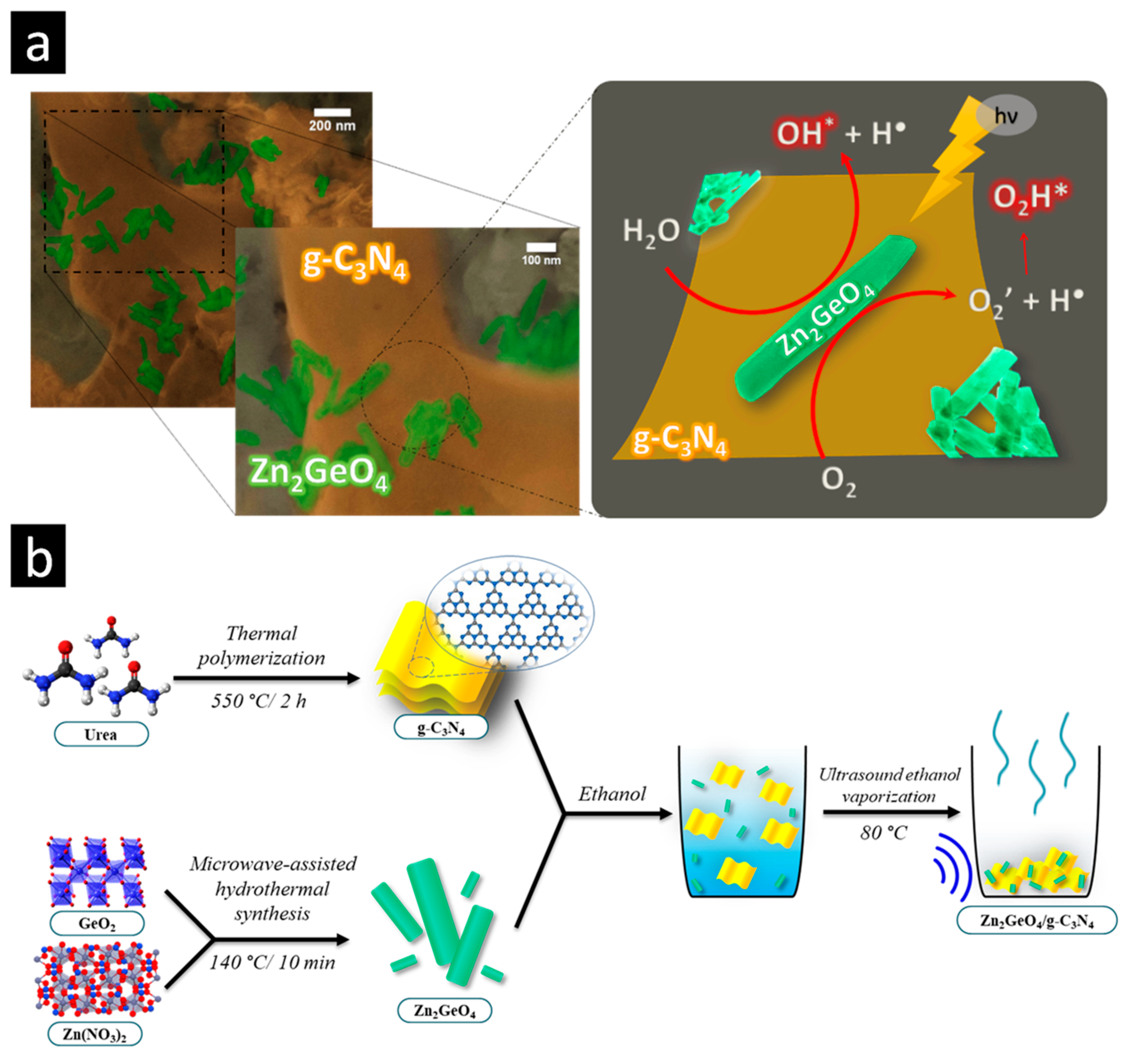
3.1. Synthesis of Zn2GeO4 Nanorods
3.2. Synthesis of g-C3N4
3.3. Preparation of Zn2GeO4/g-C3N4 Nanocomposite
3.4. Materials Characterization
3.5. Measurement of Photocatalytic Activity
4. Conclusions
Author Contributions
Funding
Acknowledgments
Conflicts of Interest
References
- Borges, W.M.S.; Guerreiro, M.C.; Anconi, C.P.A.; Magalhães, K.T.; Castro, G.M.M.; Neto, J.L.; Rossi, M.A.L.S. Coordination of iron (III) to modified silica surface containing pyrazine acid groups and its application in advanced oxidative processes. Surf. Interfaces 2022, 29, 10770. [Google Scholar] [CrossRef]
- Bento, A.C.; Emídio, E.S.; Hammer, P.; Nogueira, R.F.P. Degradation of Acid Red 8 Dye Using Photo-Fenton Reaction Mediated by Titanium Modified Catalysts. J. Braz. Chem. Soc. 2019, 30, 2170–2181. [Google Scholar] [CrossRef]
- Camargo, L.P.; Lucilha, A.C.; Gomes, G.A.B.; Liberatti, V.R.; Andrello, A.C.; da Silva, P.R.C.; Dall’Antonia, L.H. Copper pyrovanadate electrodes prepared by combustion synthesis: Evaluation of photoelectroactivity. J. Solid State Electrochem. 2020, 24, 1935–1950. [Google Scholar] [CrossRef]
- Robinson, T.; McMullan, G.; Marchant, R.; Nigam, P. Remediation of dyes in textile effluent: A critical review on current treatment technologies with a proposed alternative. Bioresour. Technol. 2001, 77, 247–255. [Google Scholar] [CrossRef]
- Alkanad, K.; Hezam, A.; Sujay Shekar, G.C.; Drmosh, Q.A.; Amrutha Kala, A.L.; AL-Gunaid, M.Q.A.; Lokanath, N.K. Magnetic recyclable α-Fe2O3–Fe3O4/Co3O4–CoO nanocomposite with a dual Z-scheme charge transfer pathway for quick photo-Fenton degradation of organic pollutants. Catal. Sci. Technol. 2021, 11, 3084–3097. [Google Scholar] [CrossRef]
- Sharma, G.; Kumar, A.; Naushad, M.; Thakur, B.; Vo, D.V.; Gao, B.; Al-Kahtani, A.A.; Stadler, F.J. Adsorptional-photocatalytic removal of fast sulphon black dye by using chitin-cl-poly(itaconic acid-co-acrylamide)/zirconium tungstate nanocomposite hydrogel. J. Hazard. Mater. 2021, 416, 125714. [Google Scholar] [CrossRef]
- Ivetić, T.B.; Finčur, N.L.; Šojić Merkulov, D.V.; Despotović, V.N.; Četojević-Simin, D.D.; Armaković, S.J.; Uzelac, M.M.; Bognár, S.I.; Zec, N.J.; Lukić-Petrović, S.R.; et al. Water-Active Titanium/Molybdenum/Mixed-Oxides: Removal Efficiency of Organic Water Pollutants by Adsorption and Photocatalysis and Toxicity Assessment. Catalysts 2021, 11, 1054. [Google Scholar] [CrossRef]
- Costa, G.S.; Costa, M.J.; Oliveira, H.G.; Lima, L.C.; Luz, G.E.; Cavalcante, L.S.; Santos, R.S. Effect of the applied potential condition on the photocatalytic properties of Fe2O3|WO3 heterojunction films. J. Inorg. Organomet. Polym. 2020, 30, 2851–2862. [Google Scholar] [CrossRef]
- Ayodhya, D.; Veerabhadram, G. A review on recent advances in photodegradation of dyes using doped and heterojunction based semiconductor metal sulfide nanostructures for environmental protection. Mater. Today Energy 2018, 9, 83–113. [Google Scholar] [CrossRef]
- Bai, H.; Li, X.; Zhao, Y.; Fan, W.; Liu, Y.; Gao, Y.; Xu, D.; Ding, J.; Shi, W. Fabrication of BiVO4-Ni/Co3O4 photoanode for enhanced photoelectrochemical water splitting. Appl. Surf. Sci. 2021, 538, 148150. [Google Scholar] [CrossRef]
- Marschall, R. Semiconductor Composites: Strategies for Enhancing Charge Carrier Separation to Improve Photocatalytic Activity. Adv. Funct. Mater. 2014, 24, 2421–2440. [Google Scholar] [CrossRef]
- Kumar, A.; Sharma, S.K.; Sharma, G.; Guo, C.; Vo, D.V.; Iqbal, J.; Naushad, M.; Stadler, F.J. Silicate glass matrix@Cu2O/Cu2V2O7 p-n heterojunction for enhanced visible light photo-degradation of sulfamethoxazole: High charge separation and interfacial transfer. J. Hazard. Mater. 2021, 402, 123790. [Google Scholar] [CrossRef] [PubMed]
- Li, X.; Feng, Y.; Li, M.; Li, W.; Wei, H.; Song, D. Smart hybrids of Zn2GeO4 nanoparticles and ultrathin gC3N4 layers: Synergistic lithium storage and excellent electrochemical performance. Adv. Funct. Mater. 2015, 25, 6858–6866. [Google Scholar] [CrossRef]
- Pei, L.; Xu, Y.; Liu, J.; Wu, J.; Han, Y.; Zhang, X. Effects of solvent-induced morphology evolution of Zn2GeO4 on photocatalytic activities of g-C3N4/Zn2GeO4 composites. J. Am. Ceram. Soc. 2019, 102, 6517–6528. [Google Scholar] [CrossRef]
- Zhang, L.; Zhang, Q.; Jiu, H.; He, G. Preparation of g-C3N4/Zn2GeO4 heterojunctions with enhanced photocatalytic activity under visible light. J. Mater. Sci. Mater. Electron. 2016, 27, 7311–7317. [Google Scholar] [CrossRef]
- Wang, S.; Li, C.; Wang, T.; Zhang, P.; Li, A.; Gong, J. Controllable synthesis of nanotube-type graphitic C3N4 and their visible-light photocatalytic and fluorescent properties. J. Mater. Chem. A 2014, 2, 2885–2890. [Google Scholar] [CrossRef]
- Qian, X.; Meng, X.; Sun, J.; Jiang, L.; Wang, Y.; Zhang, J.; Hu, X.; Shalom, M.; Zhu, J. Salt-Assisted Synthesis of 3D Porous g-C3N4 as a Bifunctional Photo- and Electrocatalyst. ACS Appl. Mater. Interfaces 2019, 11, 27226–27232. [Google Scholar] [CrossRef]
- Zhou, C.; Shi, R.; Shang, L.; Wu, L.Z.; Tung, C.H.; Zhang, T. Template-free large-scale synthesis of g-C3N4 microtubes for enhanced visible light-driven photocatalytic H2 production. Nano Res. 2018, 11, 3462–3468. [Google Scholar] [CrossRef] [Green Version]
- de Jesus, J.; Santos, A.C.; Pinto, F.M.; Taft, C.A.; La Porta, F.A. Review: Theoretical and experimental investigation of the intrinsic properties of Zn2GeO4 nanocrystals. J. Mater. Sci. 2021, 56, 4552–4568. [Google Scholar] [CrossRef]
- Li, J.; Diao, X.; Xiao, Y.; Qin, L.; Lin, H.; Li, Q.; Liao, B. Effect of surfactant SDS on the morphology and photocatalytic performance of Zn2GeO4 nanorods. Mater. Res. Express 2020, 7, 085005. [Google Scholar] [CrossRef]
- La Porta, F.A.; Taft, C.A. Emerging Research in Science and Engineering Based on Advanced Experimental and Computational Strategies; Springer: Cham, Switzerland, 2020; pp. 1–530. [Google Scholar]
- Longo, E.; La Porta, F.A. Recent Advances in Complex Functional Materials: From Design to Application; Springer: Cham, Switzerland, 2017; pp. 1–454. [Google Scholar]
- La Porta, F.A.; Taft, C.A. (Eds.) Functional Properties of Advanced Engineering Materials and Biomolecules, 1st ed.; Springer International Publishing: Cham, Switzerland, 2021; ISBN 978-3-030-62225-1. [Google Scholar]
- Oliveira, L.H.; Ramírez, M.A.; Ponce, M.A.; Ramajo, L.A.; Albuquerque, A.R.; Sambrano, J.R.; Longo, E.; Castro, M.S.; La Porta, F.A. Optical and gas-sensing properties, and electronic structure of the mixed-phase CaCu3Ti4O12/CaTiO3 composites. Mater. Res. Bull. 2017, 93, 47–55. [Google Scholar] [CrossRef] [Green Version]
- Diez-Cabanes, V.; Morales-Garcia, A.; Illas, F.; Pastore, M. Tuning the Interfacial Energetics in WO3/WO3 and WO3/TiO2 Heterojunctions by Nanostructure Morphological Engineering. J. Phys. Chem. Lett. 2021, 12, 11528–11533. [Google Scholar] [CrossRef] [PubMed]
- Hossain, M.N.; Prslja, P.; Flox, C.; Muthuswamy, N.; Sainio, J.; Kannan, A.M.; Suominen, M.; Lopez, N.; Kallio, T. Temperature dependent product distribution of electrochemical CO2 reduction on CoTPP/MWCNT composite. Appl. Catal. B 2022, 304, 120863. [Google Scholar] [CrossRef]
- Liu, J. Origin of High Photocatalytic Efficiency in Monolayer g-C3N4/CdS Heterostructure: A Hybrid DFT Study. J. Phys. Chem. C 2015, 119, 28417–28423. [Google Scholar] [CrossRef]
- Le Bahers, T.; Rérat, M.; Sautet, P. Semiconductors Used in Photovoltaic and Photocatalytic Devices: Assessing Fundamental Properties from DFT. J. Phys. Chem. C 2014, 118, 5997–6008. [Google Scholar] [CrossRef]
- Liu, W.; Li, Y.; Liu, F.; Jiang, W.; Zhang, D.; Liang, J. Visible-light-driven photocatalytic degradation of diclofenac by carbon quantum dots modified porous g-C3N4: Mechanisms, degradation pathway and DFT calculation. Water Res. 2019, 151, 9–19. [Google Scholar] [CrossRef] [PubMed]
- Chan, M.K.Y.; Ceder, G. Efficient Band Gap Prediction for Solids. Phys. Rev. Lett. 2010, 105, 196403. [Google Scholar] [CrossRef]
- Amorin, L.H.; Suzuki, V.Y.; de Paula, N.H.; Duarte, J.L.; da Silva, M.A.T.; Taft, C.A.; La Porta, F.A. Electronic, structural, optical, and photocatalytic properties of graphitic carbon nitride. New J. Chem. 2019, 43, 13647–13653. [Google Scholar] [CrossRef]
- Yuan, Y.; Zhang, L.; Xing, J.; Utama, M.I.; Lu, X.; Du, K.; Li, Y.; Hu, X.; Wang, S.; Genç, A.; et al. High-yield synthesis and optical properties of g-C3N4. Nanoscale 2015, 7, 12343–12350. [Google Scholar] [CrossRef] [Green Version]
- Yuan, X.; Zhou, C.; Jin, Y.; Jing, Q.; Yang, Y.; Shen, X.; Tang, Q.; Mu, Y.; Du, A.K. Facile synthesis of 3D porous thermally exfoliated g-C3N4 nanosheet with enhanced photocatalytic degradation of organic dye. J. Colloid Interface Sci. 2016, 468, 211–219. [Google Scholar] [CrossRef] [Green Version]
- Li, X.; Zhang, J.; Shen, L.; Ma, Y.; Lei, W.; Cui, Q.; Zou, G. Preparation and characterization of graphitic carbon nitride through pyrolysis of melamine. Appl. Phys. A Mater. Sci. Process. 2009, 94, 387–392. [Google Scholar] [CrossRef]
- Su, L.; Qi, Y.; Jia, C.-J.; Jin, Z.; Fan, W. Enhanced visible-light photocatalytic activity of g-C3N4/Zn2GeO4 heterojunctions with effective interfaces based on band match. Nanoscale 2014, 6, 2649–2659. [Google Scholar]
- Thurston, J.H.; Hunter, N.M.; Cornell, K.A. Preparation and characterization of photoactive antimicrobial graphitic carbon nitride (g-C3N4) films. RSC Adv. 2016, 6, 42240–42248. [Google Scholar] [CrossRef] [PubMed] [Green Version]
- Wagner, C.D.; Muilenberg, G.E. Handbook of X-ray Photoelectron Spectroscopy: A Reference Book of Standard Data for Use in X-ray Photoelectron Spectroscopy; Physical Electronics Division, Perkin-Elmer Corp.: Eden Prairie, MN, USA, 1979. [Google Scholar]
- Boonprakob, N.; Wetchakun, N.; Phanichphant, S.; Waxler, D.; Sherrell, P.; Nattestad, A.; Chen, J.; Inceesungvorn, B. Enhanced visible-light photocatalytic activity of g-C3N4/TiO2 films. J. Colloid Interface Sci. 2014, 417, 402–409. [Google Scholar] [CrossRef]
- Chidhambaram, N.; Ravichandran, K. Single step transformation of urea into metal-free g-C3N4 nanoflakes for visible light photocatalytic applications. Mater. Lett. 2017, 207, 44–48. [Google Scholar] [CrossRef]
- Zhang, R.; Zhang, X.; Liu, S.; Tong, J.; Kong, F.; Sun, N.; Han, X.; Zhang, Y. Enhanced photocatalytic activity and optical response mechanism of porous graphitic carbon nitride (g-C3N4) nanosheets. Mater. Res. Bull. 2021, 140, 111263. [Google Scholar] [CrossRef]
- Yamaguchi, O.; Hidaka, J.I.; Hirota, K. Formation and characterization of alkoxy-derived Zn2GeO4. J. Mater. Sci. Lett. 1991, 10, 1471–1474. [Google Scholar] [CrossRef]
- Viegas, J.I.; Moreira, R.L.; Dias, A. Optical-vibration and intrinsic dielectric properties of low-k high-Q Zn2GeO4 ceramics. J. Phys. Chem. Solids 2021, 148, 109693. [Google Scholar] [CrossRef]
- Duan, X.; Yuan, M.; Ou, K.; Zhao, W.; Tian, T.; Duan, W.; Zhang, X.; Yi, L. Controlled preparation of undoped Zn2GeO4 microcrystal and the luminescent properties resulted from the inner defects. Mater. Today Commun. 2021, 27, 102359. [Google Scholar] [CrossRef]
- Alaghmandfard, A.; Ghandi, K. A Comprehensive Review of Graphitic Carbon Nitride (g-C3N4)–Metal Oxide-Based Nanocomposites: Potential for Photocatalysis and Sensing. Nanomaterials 2022, 12, 294. [Google Scholar] [CrossRef]
- Xie, Z.; Lu, H.; Zhang, Y.; Sun, Q.; Zhou, P.; Ding, S.; Zhang, D.W. The electronic structures and optical properties of Zn2GeO4 with native defects. J. Alloys Compd. 2015, 619, 368–371. [Google Scholar] [CrossRef]
- Suzuki, V.Y.; de Paula, N.H.; Gonçalves, R.; Li, M.S.; Pereira, E.C.; Longo, E.; La Porta, F.A. Exploring effects of microwave-assisted thermal annealing on optical properties of Zn2GeO4 nanostructured films. Mater. Sci. Eng. B 2019, 246, 7–12. [Google Scholar] [CrossRef]
- Suzuki, V.Y.; Amorin, L.H.; Lima, N.M.; Machado, E.G.; Carvalho, P.E.; Castro, S.B.; Alves, C.S.; Carli, A.P.; Li, M.S.; Longo, E.; et al. Characterization of the structural, optical, photocatalytic and: In vitro and in vivo anti-inflammatory properties of Mn2+ doped Zn2GeO4 nanorods. J. Mater. Chem. C 2019, 7, 8216–8225. [Google Scholar] [CrossRef]
- Rietveld, H.M. Line profiles of neutron powder-diffraction peaks for structure refinement. Acta Crystallogr. 1967, 22, 151–152. [Google Scholar] [CrossRef]
- Sharma, A.; Varshney, M.; Chae, K.H.; Won, S.O. Mechanistic investigations on emission characteristics from g-C3N4, g-C3N4@Pt and g-C3N4@Ag nanostructures using X-ray absorption spectroscopy. Curr. Appl. Phys. 2018, 18, 1458–1464. [Google Scholar] [CrossRef]
- Shi, Q.; Zhang, X.; Liu, X.; Xu, L.; Liu, B.; Zhang, J.; Xu, H.; Han, Z.; Li, G. In-situ exfoliation and assembly of 2D/2D g-C3N4/TiO2(B) hierarchical microflower: Enhanced photo-oxidation of benzyl alcohol under visible light. Carbon 2022, 196, 401–409. [Google Scholar] [CrossRef]
- Waheed, A.; Shi, Q.; Maeda, N.; Meier, D.M.; Qin, Z.; Li, G.; Baiker, A. Strong Activity Enhancement of the Photocatalytic Degradation of an Azo Dye on Au/TiO2 Doped with FeOx. Catalysts 2020, 10, 933. [Google Scholar] [CrossRef]
- Sharma, G.; Kumar, A.; Sharma, S.; Naushad, M.; Dhiman, P.; Vo, D.V.; Stadler, F.J. Fe3O4/ZnO/Si3N4 nanocomposite based photocatalyst for the degradation of dyes from aqueous solution. Mater. Lett. 2020, 278, 128359. [Google Scholar] [CrossRef]
- Suzuki, V.Y.; Amorin, L.H.; de Paula, N.H.; Albuquerque, A.R.; Li, M.S.; Sambrano, J.R.; Longo, E.; La Porta, F.A. New insights into the nature of the bandgap of CuGeO3 nanofibers: Synthesis, electronic structure, and optical and photocatalytic properties. Mater. Today Commun. 2021, 26, 101701. [Google Scholar] [CrossRef]
- Karimi, M.A.; Atashkadi, M.; Ranjbar, M.; Habibi-Yangjeh, A. Novel visible-light-driven photocatalyst of NiO/Cd/g-C3N4 for enhanced degradation of methylene blue. Arab. J. Chem. 2020, 13, 5810–5820. [Google Scholar] [CrossRef]
- Karimi, M.A.; Iliyat, M.; Atashkadi, M.; Ranjbar, M.; Habibi-Yangjeh, A. Microwave-assisted synthesis of the Fe2O3/g-C3N4 nanocomposites with enhanced photocatalytic activity for degradation of methylene blue. J. Chin. Chem. Soc. 2020, 67, 2032–2041. [Google Scholar] [CrossRef]
- Hakimi-Tehrani, M.J.; Hassanzadeh-Tabrizi, S.A.; Koupaei, N.; Saffar-Teluri, A.; Rafiei, M. Facile thermal synthesis of g–C3N4/ZnO nanocomposite with antibacterial properties for photodegradation of Methylene blue. Mater. Res. Express 2021, 8, 125002. [Google Scholar] [CrossRef]
- Pham, T.-T.; Shim, E.W. Inhibition of charge recombination of NiTiO3 photocatalyst by the combination of Mo-doped impurity state and Z-scheme charge transfer. Appl. Surf. Sci. 2020, 501, 143992. [Google Scholar] [CrossRef]
- Toby, B.H.; Von Dreele, R.B. GSAS-II: The genesis of a modern open-source all purpose crystallography software package. J. Appl. Crystallogr. 2013, 46, 544–549. [Google Scholar] [CrossRef]
- Dovesi, R.; Saunders, V.R.; Roetti, C.; Orlando, R.; Zicovich-Wilson, C.M.; Pascale, F.; Civalleri, B.; Doll, K.; Harrison, N.M.; Bush, I.J.; et al. CRYSTAL17—User’s Manual; University of Torino: Torino, Italy, 2018. [Google Scholar]
- Heyd, J.; Scuseria, G.E.; Ernzerhof, M. Hybrid functionals based on a screened Coulomb potential. J. Chem. Phys. 2003, 118, 8207. [Google Scholar] [CrossRef] [Green Version]
- CRYSTAL—Basis Sets Library, (n.d.). Available online: http://www.crystal.unito.it/basis-sets.php (accessed on 5 April 2022).
- La Porta, F.A.; Gracia, L.; Andrés, J.; Sambrano, J.R.; Varela, J.A.; Longo, E. A DFT Study of Structural and Electronic Properties of ZnS Polymorphs and its Pressure-Induced Phase Transitions. J. Am. Ceram. Soc. 2014, 97, 4011–4018. [Google Scholar] [CrossRef] [Green Version]
- de Jesus, J.P.A.; Jimenez, M.Z.; La Porta, F.A. Theoretical investigation on the effects of electric field on the electronic structure and spectroscopic properties of Zn6−xCdxS6 clusters as model systems of semiconductor quantum dots. Comput. Mater. Sci. 2021, 188, 110147. [Google Scholar] [CrossRef]
- Marana, N.L.; Albuquerque, A.R.; La Porta, F.A.; Longo, E.; Sambrano, J.R. Periodic density functional theory study of structural and electronic properties of single-walled zinc oxide and carbon nanotubes. J. Solid State Chem. 2016, 237, 36–47. [Google Scholar] [CrossRef]
- Kokalj, A. XCrySDen—A new program for displaying crystalline structures and electron densities. J. Mol. Graph. Model. 1999, 17, 176–179. [Google Scholar] [CrossRef]


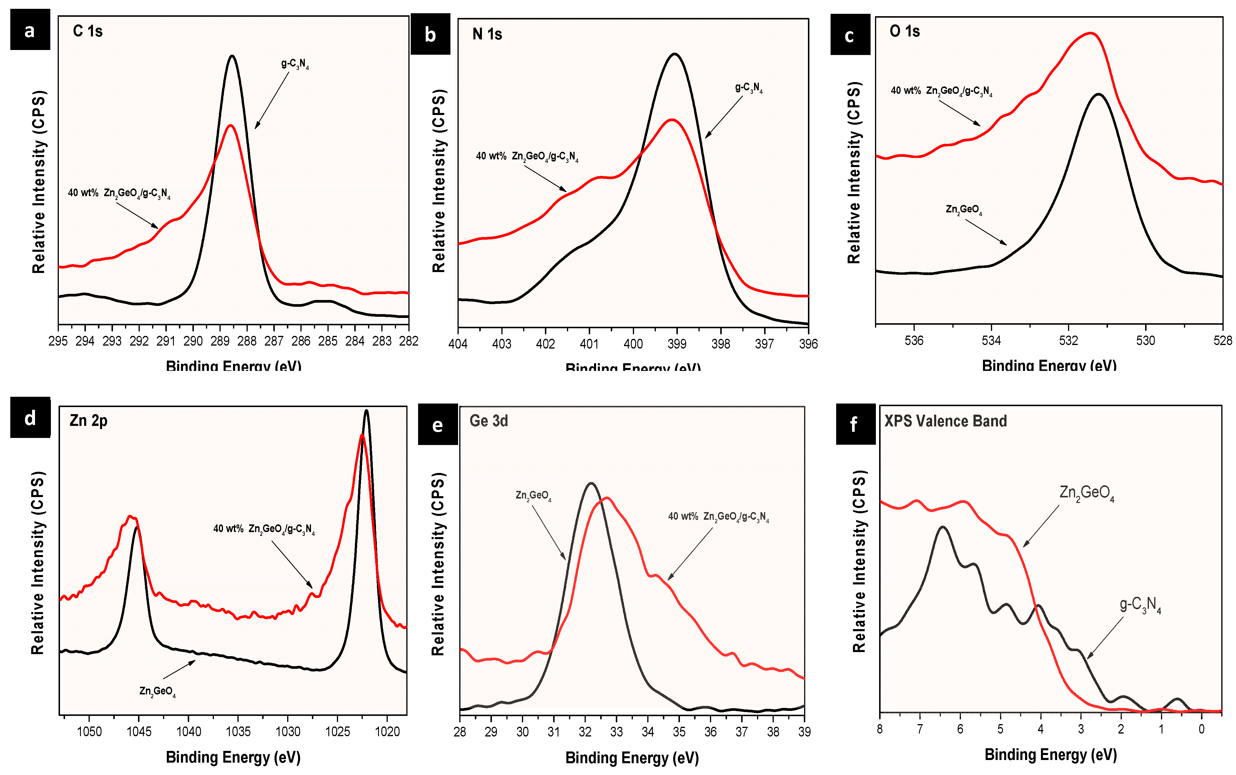
| g-C3N4 | Zn2GeO4 | 40% Zn2GeO4 | |
|---|---|---|---|
| C (main) | 283.3 | - | 288.35 |
| N (main) | 398.6 | - | 398.92 |
| Zn 2p 3/2 | - | 1022.15 | 10.22 |
| Zn 3d | - | 10.83 | 10.95 |
| Ge 3d | - | 32.25 | 32.5 |
| O | - | 531.25 | 531.3 |
| XPS VB (top) | 2.53 | 3.83 (3.15) | 2.9–3.03 |
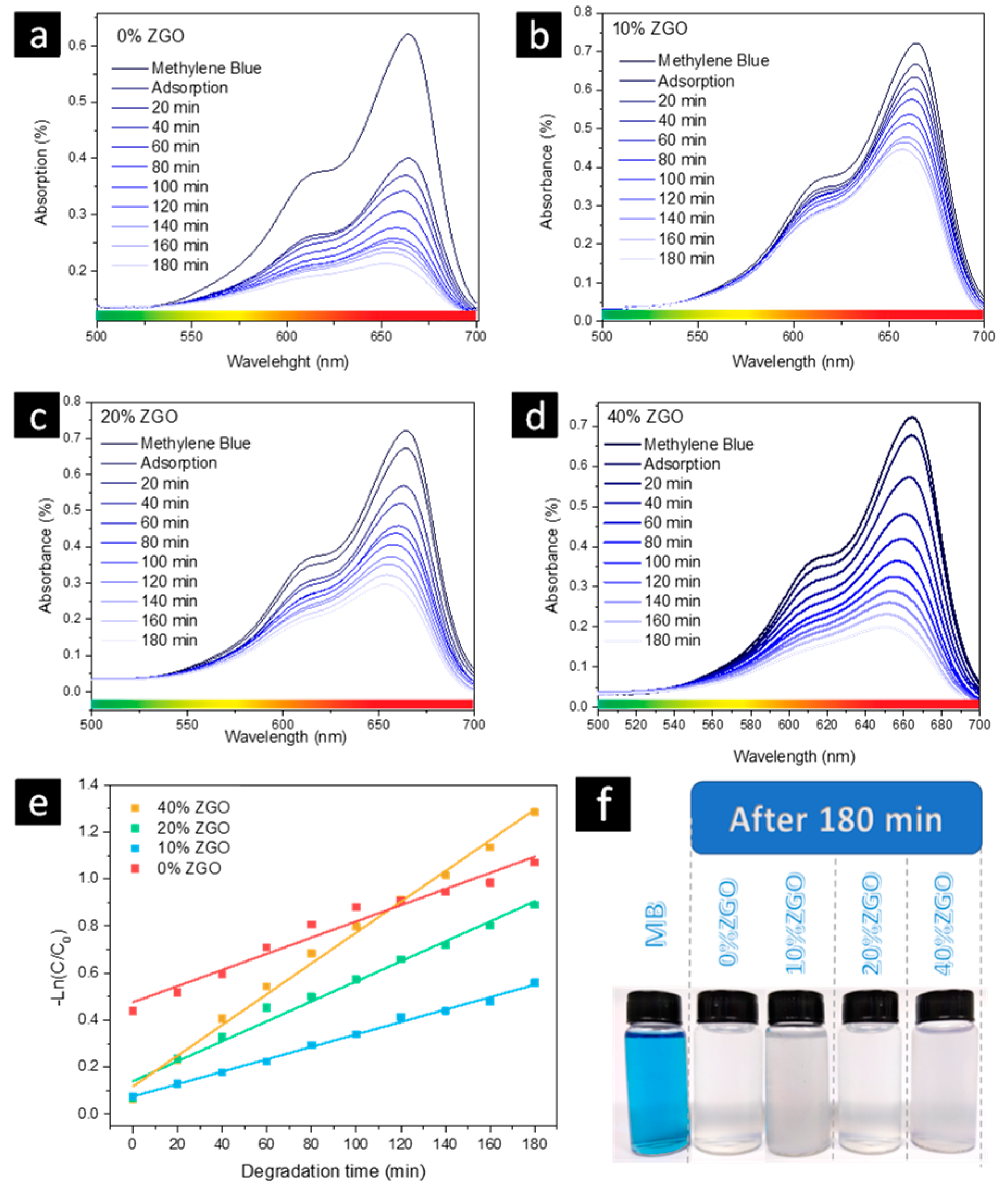
| Sample | k [10−3 min−1] | R2 [%] | Adsorption [%] | Degradation [%] |
|---|---|---|---|---|
| 0% ZGO | 3.44 | 96.77 | 35.53 | 46.73 |
| 10% ZGO | 2.64 | 99.57 | 7.42 | 38.26 |
| 20% ZGO | 4.25 | 98.28 | 6.51 | 56.12 |
| 40% ZGO | 6.54 | 99.35 | 6.05 | 70.52 |
Publisher’s Note: MDPI stays neutral with regard to jurisdictional claims in published maps and institutional affiliations. |
© 2022 by the authors. Licensee MDPI, Basel, Switzerland. This article is an open access article distributed under the terms and conditions of the Creative Commons Attribution (CC BY) license (https://creativecommons.org/licenses/by/4.0/).
Share and Cite
Suzuki, V.Y.; Amorin, L.H.C.; Fabris, G.S.L.; Dey, S.; Sambrano, J.R.; Cohen, H.; Oron, D.; La Porta, F.A. Enhanced Photocatalytic and Photoluminescence Properties Resulting from Type-I Band Alignment in the Zn2GeO4/g-C3N4 Nanocomposites. Catalysts 2022, 12, 692. https://doi.org/10.3390/catal12070692
Suzuki VY, Amorin LHC, Fabris GSL, Dey S, Sambrano JR, Cohen H, Oron D, La Porta FA. Enhanced Photocatalytic and Photoluminescence Properties Resulting from Type-I Band Alignment in the Zn2GeO4/g-C3N4 Nanocomposites. Catalysts. 2022; 12(7):692. https://doi.org/10.3390/catal12070692
Chicago/Turabian StyleSuzuki, Victor Y., Luis H. C. Amorin, Guilherme S. L. Fabris, Swayandipta Dey, Julio R. Sambrano, Hagai Cohen, Dan Oron, and Felipe A. La Porta. 2022. "Enhanced Photocatalytic and Photoluminescence Properties Resulting from Type-I Band Alignment in the Zn2GeO4/g-C3N4 Nanocomposites" Catalysts 12, no. 7: 692. https://doi.org/10.3390/catal12070692