Review

Pathogenesis, Diagnosis and Risk Stratification in Arrhythmogenic Cardiomyopathy

1
Division of Internal Medicine, University of Campania “Luigi Vanvitelli”, 80131 Naples, Italy
2
Laboratory of Cardiomyopathies and Inherited Cardiac Diseases, Aristotle University of Thessaloniki 1st Cardiology Department, AHEPA University Hospital, 546 21 Thessaloniki, Greece
3
Inherited and Rare Cardiovascular Disease Unit, Department of Translational Medical Sciences, University of Campania “Luigi Vanvitelli”, 80131 Naples, Italy
*
Author to whom correspondence should be addressed.
Cardiogenetics 2021, 11(4), 263-289; https://doi.org/10.3390/cardiogenetics11040025
Submission received: 6 May 2021 / Revised: 31 October 2021 / Accepted: 23 November 2021 / Published: 8 December 2021
(This article belongs to the Special Issue Cardiogenetics: Feature Papers 2021)

Abstract

:
Arrhythmogenic cardiomyopathy (ACM) is a genetically determined myocardial disease associated with sudden cardiac death (SCD). It is most frequently caused by mutations in genes encoding desmosomal proteins. However, there is growing evidence that ACM is not exclusively a desmosome disease but rather appears to be a disease of the connexoma. Fibroadipose replacement of the right ventricle (RV) had long been the hallmark of ACM, although biventricular involvement or predominant involvement of the left ventricle (LD-ACM) is increasingly found, raising the challenge of differential diagnosis with arrhythmogenic dilated cardiomyopathy (a-DCM). A-DCM, ACM, and LD-ACM are increasingly acknowledged as a single nosological entity, the hallmark of which is electrical instability. Our aim was to analyze the complex molecular mechanisms underlying arrhythmogenic cardiomyopathies, outlining the role of inflammation and autoimmunity in disease pathophysiology. Secondly, we present the clinical tools used in the clinical diagnosis of ACM. Focusing on the challenge of defining the risk of sudden death in this clinical setting, we present available risk stratification strategies. Lastly, we summarize the role of genetics and imaging in risk stratification, guiding through the appropriate patient selection for ICD implantation.

1. Introduction

Initially assumed as developmental anomaly, the term arrhythmogenic right-ventricular dysplasia (ARVD) was used to describe a condition characterized by replacement of the right-ventricular (RV) myocardium with fibrofatty tissue [1]. Arrhythmogenic right-ventricular cardiomyopathy (ARVC) has been included in the European Cardiomyopathies Working Group classification since 1994. On that iteration, morphological abnormalities were predominantly confined to the RV with little or no left-ventricle (LV) impairment [2]. Over the recent past, the term arrhythmogenic cardiomyopathy (ACM) has been increasingly used to describe a broader spectrum of primary myocardial diseases. This emerging term encompasses morphological involvement of the RV, the LV, or both. Importantly, the hallmark feature in ACM is prominent nonischemic fibrosis/scarring and ventricular arrhythmias of genetic or nongenetic etiology [3]; the adjective arrhythmogenic is disease-specific and denotes the distinctive propensity of the ACM to develop ventricular arrhythmias due to the underlying fibroadipose myocardial replacement [4].
ACM is typically inherited as an autosomal dominant trait with incomplete penetrance and variable expressivity. Naxos disease and Carvajal syndrome are cardio-cutaneous syndromes in which ACM is inherited as a recessive trait and associated with palmoplantar keratosis and woolly hair [5]. About half of ACM cases are caused by mutations in five genes encoding desmosomal proteins, with plakophilin-2 (PKP2) being the most involved, accounting for approximately 43% of the cases [6]; desmoplakin (DSP), desmoglein-2 (DSG2), democollin-2 (DSC2), and plakoglobin (JUP) are the others [7]. More recently, mutations in fascia adherens genes cadherin-2 (CDH2) and catenin-α3 (CTNNA3) have been identified in ACM patients [8,9].
A consistent proportion (2–6%) of mutation-positive ACM patients harbors a second pathogenic variant [10], mostly among ACM patients with DSG2 and DSC2 variants [11]. Furthermore, there is strong evidence that exercise contributes to the pathogenesis of ACM, promoting disease phenotypic expression, worsening structural damage, and increasing risk of arrhythmias and sudden cardiac death [12].
The intent of this review is to shed light on the newer aspects of the etiopathogenesis, diagnosis, and risk stratification of ACM, underling the necessity to reappraise the currently available rigid classification system in the clinical setting.

2. Pathophysiology

Cardiomyocyte loss, fibrosis, adipogenesis, inflammation, and arrhythmogenesis are classical features of ACM pathophysiology. The molecular genetics of ACM indicate that it is a desmosomal disease. Desmosomes and fascia adherens junctions (together forming the area composita, located at intercalated discs (ID) in the epithelium and muscle tissue) maintain mechanical adhesion, while gap junctions and ion channel complexes provide electrical continuity between cardiomyocytes [13]. Growing evidence seems to confirm that these subcomplexes form a single functional entity, the connexome [14]: a common protein interaction network controlling excitability, electrical coupling, and intercellular adhesion. Therefore, ACM seems to be not just a desmosomal disease, but rather a disease of the connexome, whose pathogenetic mechanism may include loss of mechanical integrity, altered signaling pathways, and disruption of ion channel complexes and gap junctions [15].

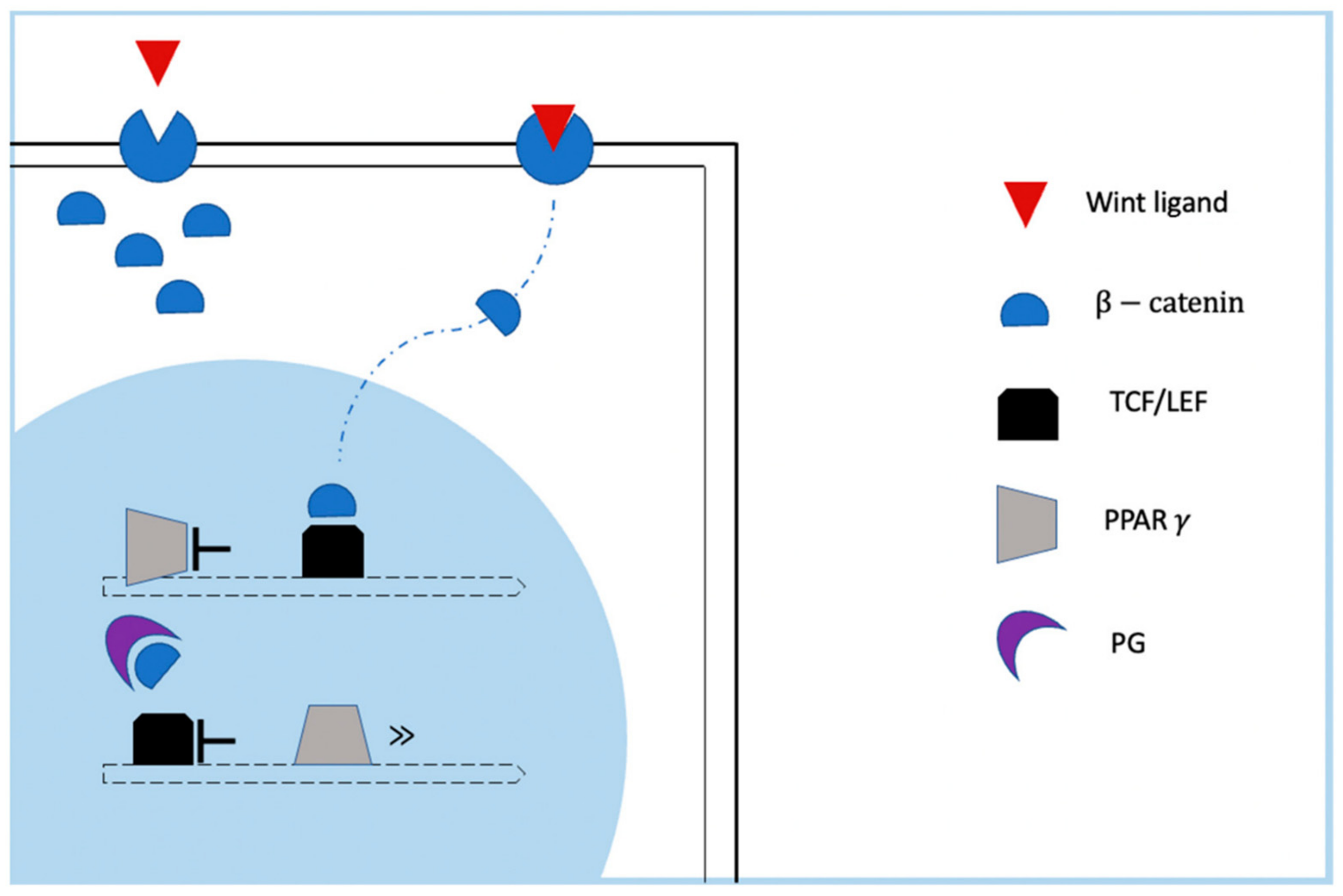
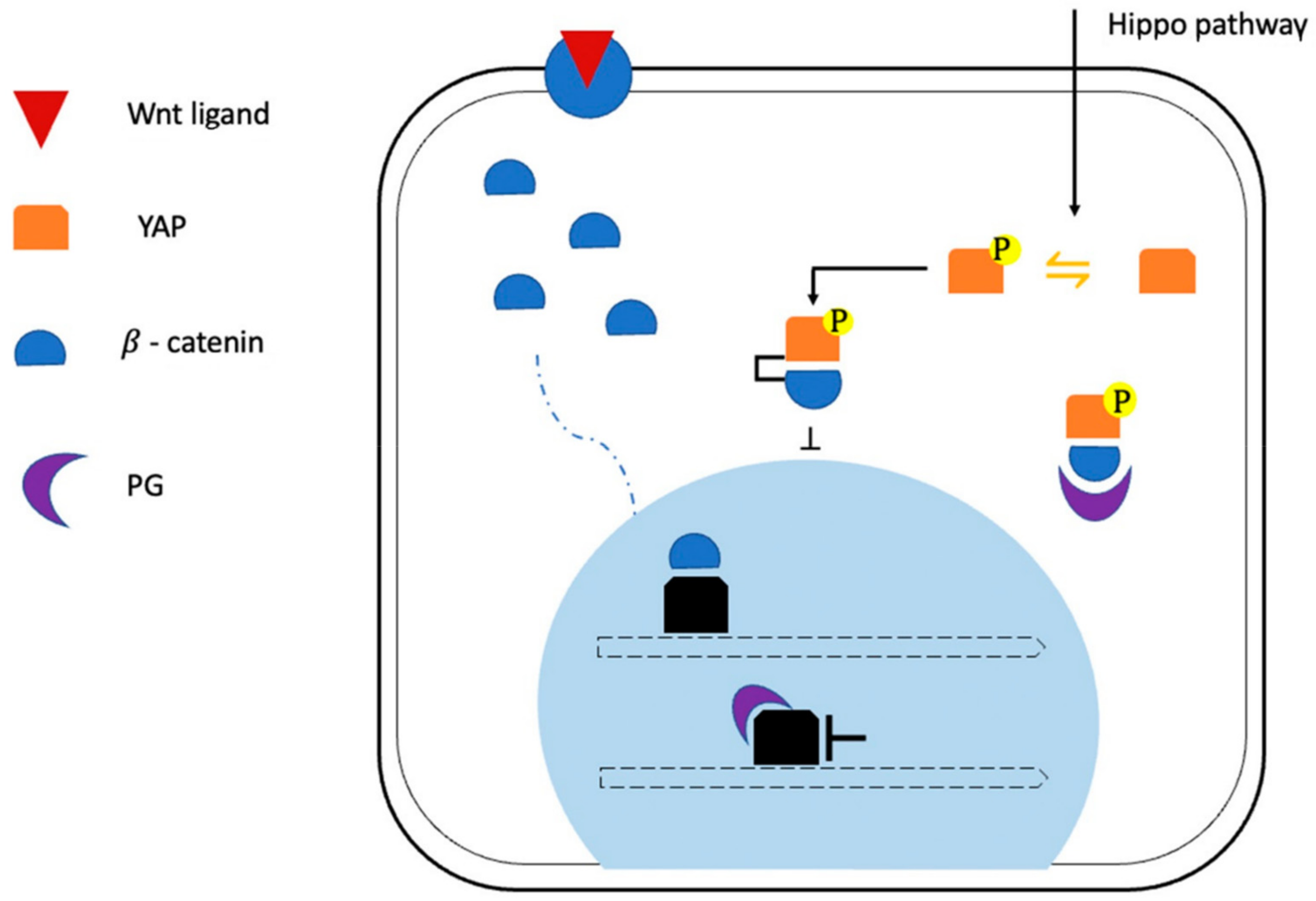
2.1. Role of Altered Signaling Pathways in ACM Pathogenesis: Desmosomal and Junctional Gene Mutations

Desmosomal and junctional gene mutations cause the disruption of the normal interactions in the area composita, altering the signaling function of the intercalated disc and, in particular, the following pathways: Wnt/β-catenin, Hippo/Yes-associated protein (YAP), and transforming growth factor-β (TGF-β), which have been implied in cardiomyocyte loss, adipogenesis, and fibrosis in ACM.
Wnt ligand signaling through β1-catenin is known as canonical Wnt/β-catenin signaling [16]. β-catenin participates in intercellular adhesion and is also a transcriptional coactivator of the Wnt/β-catenin signaling pathway. This pathway is a key regulator of myogenesis versus adipogenesis and plays an essential role in heart development, as well in cardiac tissue homeostasis. Its abnormal regulation has been linked to a variety of cardiac disease conditions, including fibrosis and arrhythmias. Plakoglobin (PG), also known as γ-catenin, has some structural similarities to β-catenin, and it is an important component of the cardiac desmosome. Binding of Wnt ligands to their receptors allows the translocation of β1-catenin to the nucleus, where it coactivates target genes of the Tcf/lef transcription factor family [17]. On the other hand, in the absence of Wnt ligand–receptor, the intracellular β1-catenin is rapidly degraded by a cytoplasmic destruction complex. Nuclear PG localization, as a consequence of PG mutation, has been proposed to contribute to ACM pathogenesis by suppressing canonical Wnt signaling interfering with β1-catenin transcriptional activities, thereby enhancing adipogenesis driven by PPARγ and C/ERBα [16] (Figure 1).
Mutation in catenin-α3 (CTNNA3) seems to affect the canonical Wnt/β-catenin signaling pathway in a similar manner [9]. The abnormal Wnt/β-catenin signaling pathway causes redirection of cells destined to become cardiomyocytes toward an alternative mesoderm fate [18], malformation of the outflow tract [19], and abnormal development of the cardiac conduction system [20].
Moreover, Wnt ligands can activate signaling pathways that are independent of β1-catenin. This noncanonical Wnt/β-catenin signaling pathway, through its effector pathway ROCK (Rho GTPase/Rho-associated protein kinase), has been implicated in ACM-associated adipogenesis. Indeed, Rho has a key role in desmosomal architecture, and its disruption has been linked to PG nuclear localization and subsequent β1-catenin inhibition, as described in classical desmosomal mutations [21].
Another intracellular signaling pathway implicated in ACM pathogenesis is the Hippo/YAP [22]. This pathway controls the activity of transcriptional coactivators that stimulate the expression of genes that promote proliferation; it is a key regulator of organ growth that has been linked to cardiomyocyte proliferation and heart size [23]. Intercalated disc abnormalities, such as impaired ID assembly, reduced ID stability, and abnormal regulation of ID gene expression by nuclear PG, seem a probable cause of the Hippo/YAP pathway deregulation [22]. Interestingly, a tight crosstalk between Wnt and Hippo/YAP pathways in controlling cardiomyocyte proliferation has been proven. Indeed, YAP interacts with β1-catenin and suppresses its nuclear translocation in presence of inhibitory Hippo kinase activity [24]. However, the Wnt-Hippo/YAP crosstalk seems more complex, happens at multiple other levels, and remains to be explored (Figure 2).
In ACM, the Wnt/β-catenin signaling pathway could be suppressed, activated, or unchanged. Wnt/β-catenin signaling suppression in concomitance with Hippo pathway activation causes the inability of β-catenin to translocate into the nucleus. As a consequence, the expression of the effector of both Hippo and Wnt pathways is suppressed, and adipogenesis in enhanced [22]. Moreover, in the presence of Wnt/β-catenin signaling activation, the loss of PG leads to activation of Akt and subsequent inhibition of glycogen synthase kinase 3β (GSK3β), resulting in the stabilization of β-catenin and its translocation in the nucleus. Here, β-catenin interacts with Tcf/Lef, causing the enhanced expression of effectors such c-myc, c-fos, and cyclin D1 and promoting cardiac hypertrophy [25]. Lastly, the disruption of ID integrity can result in the increased presence of β-catenin without the involvement of Wnt/β-catenin signaling. However, the increased expression of transforming growth factor β-1 (TGFβ1), phospho-SMAD2 (pSMAD2), and Pai1 is consistent with activation of the TGFβ pathway responsible for progressive fibrosis in ACM hearts [26]. These findings suggest a central role of Wnt/β-catenin signaling in the disease pathogenesis. However, the direct causal relationship between mutant desmosomal proteins and perturbed Wnt signaling pathway remains poorly understood.

2.2. Role of Altered Signaling Pathways in ACM Pathogenesis: Non-Desmosomal Gene Mutations

Some variants of non-desmosomal genes have been linked to ACM: CDH2, desmin (DES), CTNNA3, phospholamban (PLN), sodium voltage-gated channel alpha subunit 5 (SC5A), cardiac ryanodine receptor (RYR2), filamin C (FLNC), lamin a/c (LMNA), titin (TTN), transmembrane protein 43 (TMEM43), transforming growth factor β-3 (TGFβ3), and p63 (TP63) [2]. However, albeit indirectly, some of these also play a role in cellular adhesion mechanisms [27], but their pathogenic role is controversial in ACM.
SCN5A variants have been described in some ACM families [28]. SCN5A encodes NaV1.5, a pore-forming subunit of the voltage-gated cardiac sodium channel (VGSC). To date, over 300 pathogenetic variants of SCN5A have been associated with Brugada syndrome (BrS). Similarly, most of the other genes implicated in BrS pathogenesis encode cardiac ion channel proteins, suggesting that BrS is a pure channelopathy [29]. Leo-Macias et al. demonstrated that NaV1.5 and N-cadherin are implicated in adhesion and excitability molecular complexes in the intercalated disc, suggesting that NaV1.5 can contribute to intercellular adhesion strength [30]. Indeed, NaV1.5 appears to be sensitive to local mechanical forces with its proximity to mechanical junctions, conferring mechanical stability and limiting membrane deformation during the cardiac cycle [31]. This function of NaV1.5 as a modulator of cell adhesion and mechanical integrity is a likely explanation of how SCN5A can lead not only to rhythm disorders but also to structural abnormalities. Inversely, the close interconnection between mechanical and electrical subcomplexes would explain the early occurrence of major arrhythmic events in desmosomal mutation carriers, even before structural damage becomes overtly detectable.
Intriguingly, the first non-ion channel-encoding gene to be implicated in BrS was PKP2, the most frequently causative gene for ACM. Cerrone et al. identified PKP2 variants in five of the 200 patients with BrS diagnosis, no mutations in BrS-related genes (SCN5A, CACNa1c, GPD1L, and MOG1), and no macroscopic signs of ACM [32]. This study experimentally demonstrated that siRNA-mediated loss of PKP2 expression in isolated cells affected the amplitude and kinetics of the sodium current (INa), exhibiting that PKP2 not only participates in intercellular coupling, but also interacts, directly or indirectly, with the VGSC complex [33].
Similarly, the intensity of immunoreactive Nav1.5 has been shown to be reduced in heart sections obtained from ACM patients with conventional desmosomal mutations [34]. Therefore, a reduction in Nav1.5 abundance may be a component of the phenotype in subjects with ACM, and impaired INa could play a role in arrhythmia susceptibility in the “concealed” phase of ACM. As such, BrS and ACM could represent two clinical entities on the opposite ends of the same disease spectrum. Clinical studies have shown that BrS patients may show minor RV structural abnormalities [35], whereas desmosomal mutation carriers can develop arrhythmic events in the absence of overt structural disease [36] (Figure 3). Likewise, both PKP2 and SCN5A mutations have been associated with phenotypes on both ends of the disease spectrum, ranging from long QT syndrome and BrS to ACM [37].
Mutations in Ca2+ cycling genes and impaired calcium homeostasis are described in arrhythmogenic diseases such as BrS, catecholaminergic polymorphic ventricular tachycardia (CPVT), and long and short QT syndromes, as well as in cardiomyopathies at elevated arrhythmic risk such as a-DCM and ACM [38]. Forms of ACM associated with RYR2 gene mutations were first described [39] and then reconsidered [40] due to a lack of sufficient abnormalities to maintain the initial ACM diagnosis. The RYR2 channel is responsible for the release of Ca2+ from the sarcoplasmic reticulum (SR) into the cytoplasm during the excitation–contraction process in cardiomyocytes. The gene mutations destabilize the tetrameric structure of the channel, leading to failure of Ca2+ retention in the SR. This results in enhanced spontaneous release of Ca2+, determining delayed after-depolarizations (DADs) and ventricular arrhythmia, as described in some ACM patients [41].

2.3. Role of Large Genomic Rearrangements of Desmosomal Genes and Whole-Exome Sequencing in ACM

Conventional mutation screening in desmosomal genes is performed as a gold standard to identify point mutations in ACM patients. However, as stated before, it only allows detecting mutations in about half of ACM probands. Therefore, it has been proposed to include copy number variations (CNVs) (large genomic rearrangements such as deletions, insertions, and duplications), which have been recently described in ACM but can also be omitted by conventional screening mutation, into routine genetic testing in ACM. In a study by Pilichou et al., conducted on an Italian cohort of ACM patients, genomic rearrangements were detected in about 7% of ACM probands negative for desmosomal gene point mutations, highlighting the potential role of CNVs in increasing the diagnostic yield of clinical genetic testing [42].
Moreover, the contribution of whole-exome sequencing in ACM patients was highlighted in a recent study by Fedida et al. in which putative pathogenic variants were screened in 96 candidate genes associated with other cardiomyopathies and channelopathies in 22 ACM patients with negative routine genetic testing for desmosomal gene mutation. All suspected deletions were verified by multiplex ligation-dependent probe amplification (MLPA) and performed in 50 additional gene-negative probands. About 6% of large PKP2 deletions, undetectable by routine sequencing, were detected as the cause of ACM [43]. However, due to the difficulties in interpreting the many variants of unknown significance (VUS), the classification of genetic variants remains challenging, and the WES remains confined to research rather than diagnostic purposes.
Nevertheless, these two studies highlight the possible role, in the future, of CNVs and next-generation sequencing analysis in expanding the diagnostic yield of routine genetic testing in ACM.

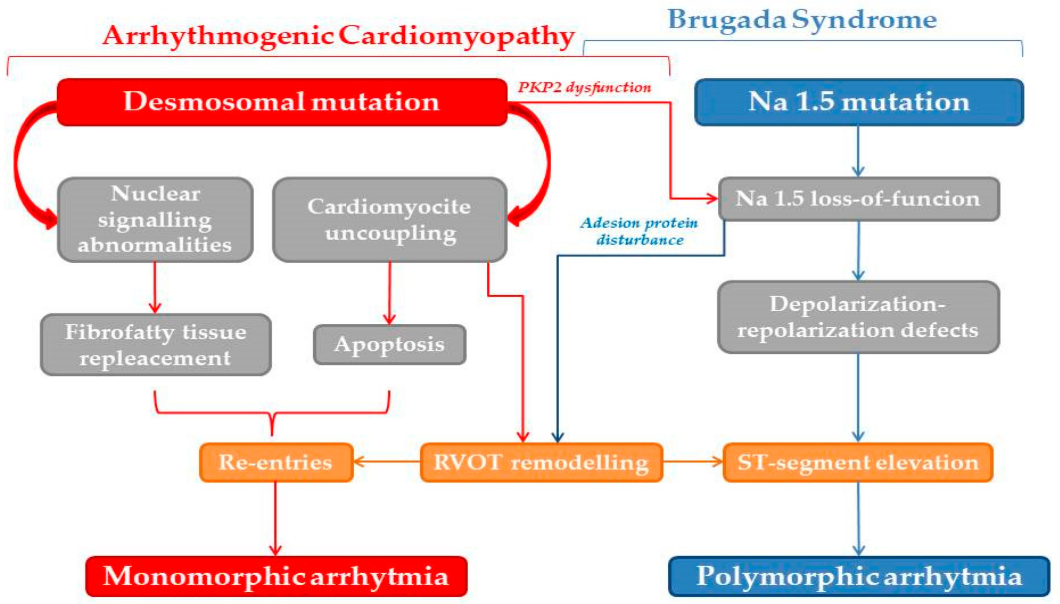
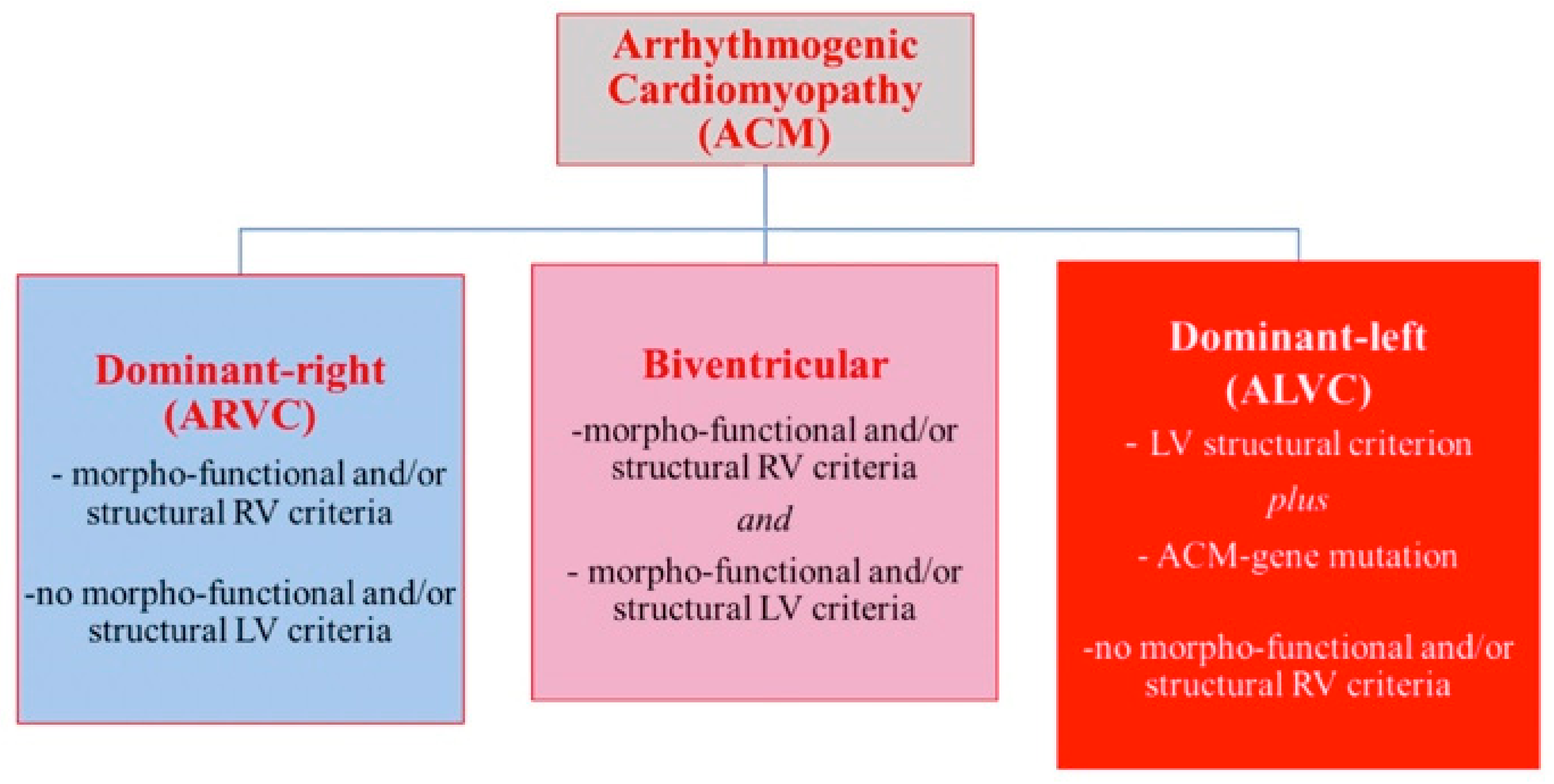
2.4. Arrhythmogenic Cardiomyopathy Is Not a Right-Ventricular Disease

2.4.1. Arrhythmogenic Phenotypes

ACM includes different clinical phenotypes, and Sen-Chowdry et al. for the first time described three patterns of disease expression: the classic ARVC, which primarily affects the RV, left-dominant arrhythmogenic cardiomyopathy (LD-ACM), primarily involving the LV, and the biventricular forms [44]. They defined the clinical and genetic profile of LD-ACM and suggested that idiopathic myocardial fibrosis (IMF) might be another clinical manifestation of arrhythmogenic cardiomyopathy [45]. Distinctive ECG features of LV involvement in ACM include (1) T-wave inversion in the inferolateral leads with low QRS voltages (<0.5 mV) in limb leads, and (2) monomorphic ventricular tachycardia with a right branch block (RBBB) morphology, denoting its origin from the LV. The typical LV imaging phenotype is characterized by a ventricular remodeling pattern consisting of mild LV dysfunction with no or mild LV dilatation, in association with subepicardial/mid-myocardial (nonischemic) late gadolinium enhancement (LGE) affecting the LV (predominantly the inferolateral wall regions) at tissue characterization in cardiac magnetic resonance (CMR). The degree of systolic LV dysfunction appears related to the global extent of LGE which, in the advanced disease stage, affects multiple septal and LV free-wall segments [46].
In order to better define ACM, a recently published International Expert Consensus Document proposed an upgrade of the 2010 Task Force diagnostic criteria for the diagnosis of ACM phenotypic variants. The novelty of the new proposed diagnostic criteria (so-called Padua criteria) consists of the introduction of tissue characterization by CMR for detection of fibrofatty myocardial replacement of both ventricles and the addition of new ECG criteria specific to the LV involvement. The accuracy of diagnostic criteria for left-sided ACM varies according to the disease variant, whether biventricular or left-ventricular dominant. In the context of biventricular ACM, the disease specificity of the left-sided abnormalities is ensured by the concomitant fulfilment of International Task Force (ITF) criteria for the RV phenotype. On the other hand, in patients with no (or minor) clinical RV abnormalities (not fulfilling the updated 2010 ITF criteria), the diagnosis of LD-ACM cannot be achieved only considering the LV phenotypic criteria. In fact, morpho-functional and structural LV abnormalities of ACM do not provide a sufficient disease specificity because of the overlap with the phenotypic features of other heart muscle diseases such as DCM, myocarditis, and cardiac sarcoidosis. Hence, diagnosis of LD-ACM requires, in addition to consistent LV phenotypic features, the demonstration of a positive genotyping for ACM-causing gene mutation [4] (Figure 4).

2.4.2. The Arrhythmogenic Profile in Left-Side Dilated Hearts: Role of Genetics

A subset of patients with familial DCM present with disproportionate arrhythmogenic risk considering the degree of the morphological anomalies and systolic dysfunction, with consequently increased risk of sudden cardiac death [47,48]. Current guidelines recommend ICD implantation for primary prevention in DCM patients whose ejection fraction remains <35% despite optimal medical therapy [49]. In a general nonischemic DCM population, the DANISH trial demonstrated a reduction in sudden death mortality; however, this did not translate into a substantial total mortality benefit [50]. This suggested that the majority of deaths in this population were a consequence of heart failure rather than arrhythmic in origin. Nonetheless, considering the high risk of SCD and phenotypic overlap with DCM, accurate recognition of LD-ACM which would derive maximum benefit from ICD implantation could be a crucial step in risk stratification. Applying genetic knowledge to clinical practice could impact clinical care; however, genetic screening is not routinely performed for DCM, except in individuals with concomitant conduction system disorders where genetic testing for mutations in the LMNA and SCN5A genes is recommended [51].
Furthermore, while ACM is more often caused by desmosomal variants, interestingly, genetic defects in non-desmosomal genes are thought to be more frequently involved in LD-ACM. Mutations in LMNA and SCN5A have been described in ACM, as already mentioned, but are mainly responsible for a-DCM and LD-ACM. Mutations in LMNA, which encodes the nuclear proteins lamin A and C, have been identified in over 5% of familial DCM cases. Mutations in the LMNA lead to a more aggressive course as they are associated with conduction disturbances and ventricular arrhythmias [52] and the burden of ventricular arrhythmias is disproportionate to the underlying structural disease, overlapping LD-ACM. Frameshift mutations of SCN5A, classically causative of BrS, also cause a-DCM with atrial or ventricular arrhythmias that exceed the degree of LV dysfunction [28]. SCN5A is the exemplary gene of overlap across channelopathies, DCM, and ACM.
Moreover, filamins anchor membrane proteins to the cytoskeleton in cardiac and skeletal muscles; filamin C binds to several proteins of the Z-disc of the sarcomere [53]. Truncating mutations have been reported in families with an overlapping a-DCM and LD-ACM phenotype; an early onset, an aggressive ventricular dysfunction with a high burden of atrial and ventricular arrhythmias, and an increased incidence of SCD were common. CMR demonstrated extensive fibrosis, mainly of the LV, and, in a percentage of patients the LGE was subepicardial and circumferential [54]. Moreover, a recent study showed fibrofatty infiltration of the LV in addition to interstitial fibrosis of the RV among patients with truncating FLNC variants [55]. Interestingly, immunostaining assays of the LV showed decreased desmoplakin staining of cellular junctions, supporting the concept of the LD-ACM and a-DCM overlap.
Bermúdez-Jiménez et al. for the first time described the largest LD-ACM family with p.Glu401Asp mutation in the DES gene [56]. Desmin, encoded by the gene DES, is a structural intermediate filament present in the cytoskeleton of the leiomyocytes, rhabdomyocytes, and cardiomyocytes. More than 70% of the described pathogenic DES mutations are associated with cardiac involvement and can be related to DCM most commonly, but also to restrictive, hypertrophic, and ACM. This usually implies specific conduction system disturbance and skeletal myopathy [57]. Instead, they investigated the pathogenicity of this novel DES mutation as a cause of biventricular inherited ACM with dominant primary LV affection, without conduction system abnormality or signs of skeletal muscular involvement. A nearly exclusive LV affection was seemingly determined, with hypokinesia localized on the mid-apical inferolateral wall of the LV, mildly depressed LV ejection fraction, and no ventricular dilation; CMR revealed extensive late gadolinium enhancement with a typical circumferential subepicardial pattern in most of the cases. The p.Glu401Asp mutation is located in segment 2B of the central rod domain and could eventually produce a critical break in the intra- and interhelical ionic bridges between desmin dimers, leading to a loss of structural integrity and, consequently, of cellular adhesion [56].
Moreover, a PLN mutation (R14del) has been associated with DCM, as well as with a-DCM/LD-ACM [58]. Lastly, a high-penetrance mutation in TMEM43 (S358L), encoding a nuclear envelope protein (LUMA), was identified among Canadian population with a reported association with LDAC and high risk for SCD [59].

2.4.3. Desmoplakin Cardiomyopathy

Among desmosomal genes, DSP mutations are mainly responsible for LD-ACM, and they are identified in about 3% of patients diagnosed with DCM [60]. Multiple case series identified DSP mutations in LD-ACM [44,61,62]. Smith et al. [63] described desmoplakin cardiomyopathy as a distinct nosological entity marked by a high proclivity for LV fibrosis and arrhythmias and associated with intermittent myocardial inflammatory episodes that appear clinically similar to myocarditis or sarcoidosis. They found that DSP cardiomyopathy involves the LV in almost all cases and often without any apparent RV involvement in contrast to PKP2 cardiomyopathy, which always involved the RV predominantly and most often in isolation. Interestingly, in contrast to the DSP group in their study, none of the PKP2 patients had documented episodes of acute myocardial injury. Desmoplakin interacts with intermediate filaments, binds them to desmosomal plaques [64], and seems to be capable of sensing exposure to external mechanical stresses and reacting to them [65]. Truncating DSP mutations are evenly distributed throughout the DSP coding sequence without any clear correlation between specific truncating mutations and clinical presentation; DSP loss of function leads to significant LV dysfunction. Conversely, specific missense mutations may contribute to the specific disease phenotype (e.g., mutations in the desmin versus plakophilin/plakoglobin-binding domains), less frequently leading to LV dysfunction. Furthermore, risk stratification variables that perform well for PKP2-associated ACM and DCM appear to exhibit poor accuracy for diagnosis and risk assessment for DSP cardiomyopathy. In particular, Castelletti et al. found that the standard DCM LVEF threshold of <35% was an insensitive marker for future severe ventricular arrhythmias in DSP cardiomyopathy, with many events occurring in an LVEF range of 35–55% and occasionally at an EF >55%. Even more, criteria predictive of events in “classic” ARVC, including RV systolic dysfunction, anterior T-wave inversion, and male gender would play no role in either clinical recognition or risk stratification in DSP cardiomyopathy [66]. Heliö et al. identified the DSP c.6310delA, p.(Thr2104Glnfs*12) in 10 Finnish index patients with established diagnosis of DCM; the major findings were ventricular arrhythmias and dilatation of the LV, with three of the patients dying because of arrhythmogenic events rather than ventricular dysfunction, confirming the significance of DSP gene as a cause of arrhythmogenic cardiomyopathy [67]. A genotype-specific management approach might be useful for DSP cardiomyopathy.
Therefore, the distinction between LD-ACM and a-DCM in practical terms may be challenging, particularly in cases which have mild phenotypic expression or are detected early in disease course due to better cascade family screening [68].
In conclusion, genetic and clinical overlaps between a-DCM and LD-ACM seem obvious, and, on these bases, an expert panel of the Heart Rhythm Society (HRS) has proposed to include a-DCM, ACM, and LD-ACM in a common nosological entity whose hallmark is electrical instability [3].
Advances in our understanding of the complex pathophysiological pathways implicated in the development of cardiomyopathic phenotype obviate the need to reappraise classification systems and defined diagnostic–therapeutic pathways. Novel, flexible approaches incorporating genetic, functional, and ultrastructural characteristics beyond simple morphological features should be explored, reflecting the complexity often met in clinical practice.

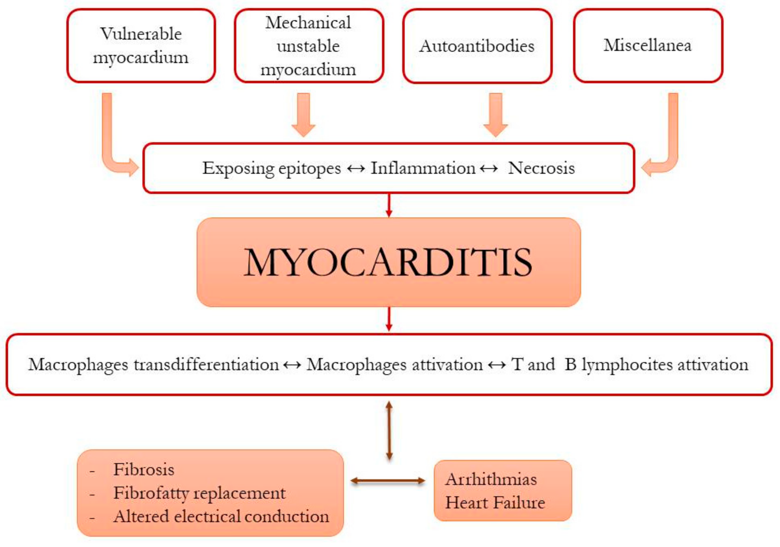
2.5. Role of Immunity in ACM Pathophysiology

Pathological findings in ACM patients and laboratory models show that inflammatory infiltration can be present in two-thirds of cases [69,70]. On this basis, focus has recently shifted toward the potential role of inflammatory insults in myocardial necrosis and in the subsequent fibrotic replacement [6,68,71], raising the key question of whether inflammation is the primary cause of myocardial damage or a consequence of it.
Myocardial necrosis can be acute, accompanied by substantial troponin elevation in the absence of coronary artery obstructions. In such cases, it can often be preceded by episodes of ventricular arrhythmia (clinically perceived as chest pain and palpitations) in the absence of typical pathological and ECG findings of ACM [72,73], suggesting an initial “hot phase” [74] of the disease predating phenotypical expression in its typical form. A viral origin of myocardial inflammation has been sought and, in some cases, found [75,76], but data are insufficient to establish an unequivocal cause–effect connection. On the other hand, a possible genetic predisposition to myocarditis has been demonstrated. Introducing the concept of vulnerable myocardium, Campuzano et al. analyzed sera of three patients suddenly dying because of myocarditis and found various types of mutations typically associated with ACM, even if signs of the disease were absent at autopsies [77]. Furthermore, a large clinical and genetical study of patients’ relatives unveiled further carriers of desmosomal mutations. Interesting elements were also underlined in a study conducted on cardiac sarcoidosis and giant cell myocarditis [78]; histopathological analyses of these patients showed an altered distribution of plakoglobin, desmoplakin, and plakophilin-2 not only in the damaged area but also in apparently unaffected regions. The local expression of cytokines was analyzed, and a high level of IL-17 and TNFα was found in both tissues and blood. Similar alterations were found in the serum and myocardium of ACM patients, raising the hypothesis that a local production of inflammation mediators could play a role in the pathogenesis and evolution of both inflammatory cardiac disease and ACM.
A study on a murine model with mutated or knockout DSG2 demonstrated that, when myocardial necrosis is established, the immune system reacts to eliminate dead cells and repair the damage. In particular, in the early, acute stage when macroscopical phenotypic features are not yet detectable, but the risk of sudden cardiac death may already be increased, local and blood-derived macrophages are activated by neutrophils to a much higher degree compared to the chronic stage. Normally, resident macrophages (CD11b+ and CD206+) can be found in healthy hearts and participate in electrical impulse conduction, but those implicated in scar development express different markers (MMP12 an SPP1) which correspond to proteins involved in proinflammatory processes. On this basis, it is reasonable to assume that they are not equally able to participate in electrical propagation, enhancing the proarrhythmogenic feature of this phase [79]. On the other hand, B lymphocytes (CD45+) and T cells (CD3 and CD4 mainly) are more involved in the acute phase; whether their stable persistence in the chronic phase suggests a modulating effect remains to be proven.
In a murine model study with DSG2 mutations, similar inflammatory cells and cytokines were involved [80]. In addition, this study showed that inhibition of GSK3β (a kinase that promotes inflammation via the NF-κB pathway) was able to reduce inflammatory cells and molecules leading to clinical improvement, reflected by less ventricular necrosis and fibrosis, better systolic function, and reduced arrhythmic episodes. The authors underlined that the presence of mutations typically associated with ACM (PKP2 in particular) could alone activate the NF-κB pathway, recalling in a certain way the concept of genetically vulnerable myocardium.
Recently, an autoimmune hypothesis emerged, and three types of autoantibodies were identified, as outlined below.
Anti-heart (AHA, with alpha and beta myosin heavy chain being the principal antigens identified) and anti-intercalated disc (AIDA) antibodies were found in 45% of patients with sporadic ACM and 85% of familial cases [81]. Clinically, patients AHA-positive presented with more severe LV involvement and with higher incidence of chest pain, arrythmias, ICD implantation, thicker septum and LV posterior wall, and lower LVEF; AIDA presence was associated with biventricular manifestations. Follow-up of antibody-positive relatives with no disease features was suggested as a means to elucidate whether detection of AHA or AIDA constitutes an early sign of disease.
Anti-desmoglein 2 autoantibodies (anti DSG2) were studied by Chatterjee et al. [82] in a small group of genetically diagnosed patients plus sera of boxer dogs affected by ACM. AntiDSG2 antibodies were present in all subjects with definite disease, exhibiting high sensitivity and specificity for its detection. Moreover, the intensity of the immune reaction correlated closely with disease severity, as expressed by ventricular ectopic burden.
Neither of the abovementioned studies could reach a conclusion on whether autoantibodies were the triggers inducing myocardial damage in genetically predisposed patients or if the presence of altered junctional proteins induced the production of autoantibodies perpetuating myocardial damage.
We would like to report another potential category: antimitochondrial autoantibodies (AMA) are notoriously linked to primary biliary cirrhosis and other autoimmune diseases, in which the cardiac involvement mostly progresses in dilated cardiomyopathy [83]. A Japanese case report described an AMA-positive myocarditis with arrhythmias, severe HF, and a fibrofatty replacement of myocardium at autopsy (a genetic study was not performed), revealing a hypothetical unexplored scenario [84].
In another case series [85], two brothers with a truncated variant of desmoplakin were diagnosed with ACM after multiple episodes of myocarditis following sessions of intense physical exercise. In both, autoantibodies against troponin and myosin were detectable, suggesting that exercise induced a sort of “mechanical unstable” myocardium necrosis by exposing epitopes which triggered autoimmune reaction. As for the interaction between immune system and exercise, recent evidence is intriguing; a more general analysis conducted by Campbell and Turner showed that single episodes of intense exercise recruit T cells, particularly CD8+, and redistribute them in areas with ongoing damage (necrotic or neoplastic tissues for example), thereby enhancing the immune response. On the other hand, regular intensity physical activity seems to also play a role in B-cell and antibody production [86]. Moreover, there are also data [87] highlighting how, in a patient with known ACM, adrenergic stimulation due to physical activity can indirectly activate intracellular Ca2+-mediated pathways, enhancing myocardial necrosis and a subsequent inflammatory response, in addition to impaired mechano-transduction due to an altered desmosomal structure (Figure 5). The sum of these elements could create a molecular microenvironment able to induce cardiomyocyte trans-differentiation, resulting in fibrotic and fibrofatty replacement. In Figure 3, we summarize the interplay among the many possible starters and perpetuators of the damage in ACM patients.
Novel cardiac imaging techniques such as SPECT [88], CMR [89,90], or PET CT scanning [91] could be used to effectively diagnose early stages of ACM, allowing early detection of myocardial inflammation, especially when the LV is involved, triggering further genetic screening of patients presenting with acute inflammation.
From a therapeutic perspective [92] immune modulation, could be a promising treatment option aimed at ameliorating disease progression and improving patient outcomes.

3. Diagnosis

Clinical diagnosis of ACM is often difficult because of the nonspecific nature of the disease features and the broad spectrum of phenotypic expressions. Indeed, in the early phases, the so-called “concealed” forms prevail. They are characterized by propensity toward ventricular arrhythmias in the presence of well-preserved ventricular morphology and function. As the disease progresses, the morpho-functional abnormalities, caused by myocyte loss, inflammation, and fibrofatty scar replacement (starting from the epi- or midmyocardium and extending to become transmural), become more evident [93].
Considering that most ACM patients are asymptomatic and sudden death often represents the first manifestation of the disease, diagnosis can ultimately be challenging. A scoring system to establish the diagnosis was developed on the basis of the fulfillment of major and minor criteria encompassing morphologic, electrocardiographic, clinical, and genetic components according to the statement initially proposed by the international Task Force in 1994 and revised in 2010 in an attempt to achieve better diagnostic specificity. ACM diagnosis can be established as “definite”, “borderline”, or “possible” on the basis of a qualitative score including both minor (one point each) and major criteria (two points each) [94,95]. Definite ACM is diagnosed when one of the following combinations is satisfied: (i) two major criteria, (ii) one major and two minor criteria, or (iii) four minor criteria (score ≥ 4). “Borderline” is considered in the presence of at least (i) one major and one minor criteria, or (ii) three minor criteria. Lastly, the diagnosis is considered “possible” when either one major or two minor criteria are satisfied (see Table 1).

3.1. Electrocardiographic Signs

Electrocardiogram (EKG) represents the first instrumental exam, often crucial to address diagnosis in ACM patients. Fibroadipose replacement interferes with the physiological conduction of the electric impulse through the ventricular myocardium, triggering electrocardiographic abnormalities in up to 90% of patients [2]. As such, a correct setting of the electrocardiograph is crucial for a correct interpretation. In particular, sensitivity can be improved by changing the 40 Hz low-pass filters (which may not detect either “fragmented” or “epsilon” waves) to 100 or 250 Hz [96]. In addition, double amplification and an increased scrolling speed (50 mm/s) might be useful [97]. EKG changes, particularly in precordial leads, depend on both the extent and the location of the disease. EKG criteria from the 2010 Task Force include T wave inversion in the precordial leads, presence of “epsilon” waves (late potentials between the end of QRS complex and the beginning of T wave), and an extension of the terminal part of QRS (nadir-end interval of the S wave >55 ms). These changes may not be manifest in at least 30% of patients. However, other nonspecific abnormalities may occur, such as ST segment changes or QRS “fragmentation” [98,99]. Late potentials by signal-averaged EKG (SAECG) represented an electrical diagnostic marker of ACM, classified in the 2010 International Task Force guidelines as a minor diagnostic criterion [46]. However, due to the technological advances by contrast enhancement CMR and the availability of molecular genetic testing for preclinical diagnosis based on demonstration of causative gene defects, use of the SAECG technique has been questioned because of its low diagnostic accuracy compared to these modern diagnostic tests [100]. In addition, data on the efficacy of late potentials in predicting clinical arrhythmic outcome are conflicting and insufficient to recommend their use for risk stratification [101]. Actually, the majority of cardiomyopathy centers no longer routinely employ late potentials in the evaluation of patients with ACM.
T-wave inversion in right precordial leads (up to V3) is present in most adult ACM patients; it correlates with the extent of RV dilation and, over the years, it may extend to left leads [102]. However, other conditions associated with T-wave inversion, either physiological or pathological, must be excluded. This finding in the right leads, indeed, may manifest in young Afro-Caribbean athletes, in ischemic heart disease, in acute pulmonary embolism, and in RBBB. T-wave inversion in the inferior leads is instead associated with the “left dominant” form. Alterations in the QRS (epsilon waves, enlarged S wave, QRS fragmentation, atypical RBBB) usually reflect the extent of the scar and slower conduction in the RV. In particular, the epsilon waves correspond to a slowed perivalvular epicardial activation, whilst S wave widening is the expression of a slowed perivalvular endocardial activation and of the right infundibulum.

3.2. First-Line Echocardiography

Echocardiography is the first-line imaging modality in ACM, as well as the most commonly used tool in the follow-up of patients with ACM. Evaluation of the RV by echocardiography can be challenging due to its retrosternal position and complex geometry. Moreover, the assessment of wall motion abnormalities is highly subjective even for experienced operators. Echocardiography allows for quantitative evaluation of RV dilation and dysfunction, as well as changes in segmental kinetics (segmental akinesia, dyskinesia, or dyssynchrony). Both minor and major ITFC criteria require the presence of wall motion abnormalities, differing with regard to the extent of RV dilatation and severity of dysfunction. However, these criteria, although specific, are hampered by poor sensitivity, particularly in the early stages of the disease. Some structural alterations, such as accentuated RV apical trabeculation and thickening of the moderator band, are also poorly specific. Echocardiographic techniques, such as 3D ultrasound and speckle-tracking have proven useful to increase the sensitivity of disease detection, mostly in the early stages [103]. A recent study recognized echocardiographic strain as a useful tool to predict structural disease progression in ACM, suggesting that baseline RV free-wall strain and rate of deformation could both be useful in identifying patients at risk of disease progression, who may require closer follow-up and treatment [104]. Previous studies using serial echocardiograms [105,106] demonstrated disease progression expressed as RVOT dilatation and RV-FAC reduction in two-thirds of patients during a mean follow-up of 6.4 years. Worsening of the peak RV free-wall longitudinal systolic strain and strain rate has also been associated with an increase in the size of the RVOT, as obtained from PSAX and PLAX views. Nonetheless, echocardiography is considered less sensitive than CMR in determining structural disease progression, given the improved capacity of the latter to identify segmental RV dilatation or focal wall motion abnormalities [107].

3.3. The Power of Cardiac Magnetic Resonance

CMR represents the gold standard as compared to echocardiography, as it allows a more precise estimate of morphology, volumes, thicknesses, mass, and segmental and global kinetics of the ventricles. The presence of regional akinesia or dyskinesia and accurate RV volume assessment may lead to the diagnosis of ARVD or biventricular ACM [95]. Extensive, sometimes circumferential late enhancement has been recognized as a phenotypical feature of LD-ACM patients, and it may be the only imaging finding in carriers of FLNC truncating mutations. Thus, LGE can help differentiate patients with arrhythmic DCM and suggest potential high-risk genotypes [45,55]. CMR is able to identify the presence of both fibrous and adipose tissue [108], which, in addition to corroborating the ACM diagnosis, represents a useful tool for the prognostic stratification [3]. The usefulness of a combined evaluation of the movement of the regional wall and the characterization of tissues by CMR in ACM diagnosis has been recently reported. Highest precision (98%) is obtained when wall motion abnormalities are accompanied by pre/post-contrast alterations on tissue characterization [109]. Identification of LV involvement on CMR is associated with a negative prognostic significance. In such cases, the subepicardial/intramyocardial distribution of fibroadipose replacement may explain why early disease stages are visualized as normal in size, function, and segmentary kinetics on echocardiography [46,110]. LV systolic dysfunction is accompanied by increased LGE extent, affecting more LV segments, with a more transmural involvement [111]. Compared to the LV, assessment of late enhancement on the thin walls of the RV can be limited by spatial resolution restrictions [112,113].
CMR findings such as RV dilatation, severe RV dysfunction, and segmental wall motion akinesia or aneurysms are highly specific for gene carriers. On the other hand, specificity has been reported as low as 56% for abnormal trabeculae and 44% for mild localized RV dilation and/or regional wall movement abnormalities. Interobserver variability in detecting features of ACM has been described, being more substantial among inexperienced operators [114,115,116]. Due to this reason, CMR should ideally be performed in a center with high expertise in evaluating abnormalities suggestive of ACM.
In recent years, myocardial wall motion abnormalities in ACM have been assessed by CMR strain analysis using feature tracking to measure regional and global ventricular dysfunction in order to identify the patients affected in the early stages of the disease [117,118], identify those at risk of progression [105], and stratify the prognosis [119].

3.4. The Role of Computed Tomography

Multidetector computed tomography (MDCT) can identify morphological features of ACM, such as increased RV chamber size, RV trabeculation, and intramyocardial fat. Modern MDCT scanners enable fast acquisition and high isotropic spatial resolution (0.5 mm), enabling precise ventricular volume measurements [120]. An advantage of 4D CT imaging is its ability to reveal the complex anatomy of RV, alleviating the risk of image overlap and limited views of standard 2D angiography. In addition to these functional parameters, a high spatial resolution, combined with the high native contrast of adipose tissue, allows a precise representation of fat infiltration within the thin wall of the RV, either with or without contrast injection [121]. CT-detected fibrosis correlated closely with epicardial and endocardial low-voltage areas on electro-anatomical mapping [122]. Fusion imaging using CT could assist ablative therapy of the culprit lesions. MDCT is widely available, fast, and affordable and can be considered a viable option in patients for whom MRI is challenging or contraindicated (e.g., those with severe arrhythmia, claustrophobia, implantable cardioverter defibrillators, or with suspicion of focal ARVC/D) [2].

4. Risk Stratification

Once the diagnosis is established, the most challenging aspect in ACM patient management is to stratify arrhythmic risk [102]. Implantable cardioverter defibrillator (ICD) implantation is a lifelong preventive measure for SCD with an established efficacy and safety in high-risk patients [123]; however, it is also associated with both short- and long-term complications (malfunction, device infection, inappropriate shocks, and others) [124].
Many single-center reports and several small multi-center registries have reported a number of clinical predictors of adverse events and death. Most of these studies focused on the outcomes of patients who already had ICDs implanted [125,126,127,128,129]. These studies have used ICD therapy for fast VT or VF as a surrogate for aborted SCD, according to the assumption that these ventricular arrhythmias would have led to death if not terminated by the device, although ICD shocks have been found to be an imperfect substitute for SCD [130]. The main clinical variables identified as independent predictors of poor outcome were history of sustained VT or VF, non-sustained VT syncope, male gender, young age at the time of diagnosis, proband status, T-wave inversion extent at 12 lead ECG, a premature ventricular complex (PVC) burden >1000/24 h, VT inducibility at EPS study, and RV and/or LV dysfunction. Only a few studies have examined risk stratification in patients with no ICD implanted. Among these, a recent study by Brun et al. concluded that sustained or non-sustained VT and LV dysfunction are risk factors for arrhythmic events [131].
The International Task Force of experts from Europe and North America have produced a consensus document (ITFC) on the treatment of ACM [132], suggesting the use of a simple algorithm to stratify the risk of SCD in patient with definite ACM. According to the presence of past major arrhythmic events and defined “major” and “minor” risk factors, three categories of risk for SCD were defined: a high-risk category, with an estimated annual event rate >10%, a low-risk category, characterized by an estimated rate of arrhythmic event <1%/year, and an intermediate risk category between those two.
Patients with a previous cardiac arrest due to VT or VF (secondary prevention) are considered in the high-risk category, as are those with severe RV and/or LV dysfunction. This group has a class I indication for ICD implantation. The intermediate risk group includes patients with ≥1 “major” risk factor (syncope, NSVT, and moderate RV and/or LV dysfunction), in whom it is reasonable to implant an ICD (class IIa). In the same risk group, if only minor risk factors are present (young age at the time of diagnosis, male gender, proband status, compound and digenetic heterozygosity of desmosomal gene mutations, extent of T-wave inversion, and VT inducibility at EPS), ICD implantation can be considered (class IIb) on an individual basis, especially if there are multiple minor risk factors [128]. This decision will need to take into account the overall patient clinical profile, age, device complication rates, and patient preferences. Lastly, the low-risk category includes healthy gene carriers and patients who meet diagnostic criteria for ACM but without risk factors, in whom ICD implantation is not indicated (class III).
The Heart Rhythm Society (HRS) criteria proposed by Towbin et al. in 2019 [3] included emerging risk factors for SCD, such as the presence of highly arrhythmogenic mutations and extent of LV involvement [54,133,134,135]. These criteria have not been developed specifically for ACM, but rather encompass a broad spectrum of arrhythmogenic cardiomyopathy. In line with ITFC criteria, there is a class I indication for ICD implantation for secondary prevention of major arrhythmic events and a class IIa indication for patients who have experienced hemodynamically tolerated sustained VT or a recent syncope. If none of the above criteria are met, the presence of major risk factors (non-sustained ventricular tachycardia (NSVT), inducibility at EPS, and ejection fraction of LV <49%) and minor risk factors (male sex, >1000 PVC in 24 h, RV dysfunction, proband status, and two or more desmosomal mutations) should be investigated. ICD implantation should be considered in patients with three major, two major and two minor, or one major and four minor risk factors (Figure 6).
In some aspects, ITFC and HRS criteria share similarities. Both strongly indicate ICD implantation in secondary prevention of major arrhythmic events, and both provide major and minor risk factors with subtle differences. In HRS criteria, LV dysfunction has a greater weight in risk stratification than RV dysfunction, which is considered only a minor risk factor. Moreover, inducibility of VT on EPS is included among the major risk factors in HRS criteria, although its role in predicting SCD is not currently well established [136].
In 2019, a new risk score model was proposed by Cadrin-Tourigny et al. to generate individualized 1, 2, and 5 year risk estimates for sustained VA in patients with definite ARVC and no prior ventricular arrhythmias [137]. This was the result of an international collaboration of 18 centers from North America to Europe, with the largest cohort of ARVC patients assembled to date. The score is available online (www.arvcrisk.com, accessed on 5 May 2021) as a “risk calculator” which uses seven easily available clinical parameters: male sex, age, recent syncope, NSVT, PVC count on 24 h Holter monitoring, leads with T-wave inversion, and RV ejection fraction. Compared to the ITFC algorithm, the 5 year ARVC risk score model led to a 20.6% reduction in ICD implantations while providing the same level of protection from ventricular arrhythmias.
However, this risk score model is not without issues to be considered. Firstly, patients were predominantly Caucasian, and pathogenic variants, when available, were primarily identified in the PKP2 gene (76%); DSP and DSG2 mutations were identified only in 7% and 5% of cases, respectively. Hence, the effectiveness of this risk score to predict events in patients of other ethnic background or genotypes is not adequately explored. Secondly, a threshold of 5 year ARVC risk score to indicate ICD implantation has not yet been determined.
Therefore, to date, there is no universally accepted risk stratification algorithm for ACM. Recently, an attempt to compare risk stratification strategies was made by Aquaro et al. [138]. In this study, the predictive performance of the three available prediction models (ITFC, HRS, and 5 year ARVC risk score) was validated in a cohort of 140 patients with definite ARVC. A 5 year score with a threshold of >10% appeared to be more effective for predicting arrhythmic events than the ITFC and HRS criteria. With this threshold, the 5 year ARVC risk score was able to predict 95% of events, which was 14% and 50% more than the ITFC and HRS criteria, respectively, but at the cost of ICD implantation in 81% of patients.
However, it remains unknown whether this score is accurate for LD-ACM forms which do not fulfill ITFC as definite ACM. Interestingly, prevalence of DSP and DSG2 mutations in the validation cohort was higher than that of the original risk score study, also confirming its effectiveness in this subset of patients. The performance superiority of the 5 year ARVC risk score could be attributed to the fact that it was directly generated from ARVC patient data, while the other two models were based on expert consensus.

5. Conclusions

Arrhythmogenic cardiomyopathy is a field of increased research interest owing to its complex pathophysiology, variable expression, and close association with risk of sudden death. Novel developments have allowed for improved understanding of all the above aspects, but major benefits in clinical outcomes are yet to be seen. Further efforts to develop a classification system are needed, focusing on more accurate distinction of phenotypes, pathophysiological substrates, and natural history of the entities comprising ACM, aimed at allowing for more clarity in clinical patient management.

Author Contributions

All authors contributed equally to conceptualization, methodology, resources, writing—original draft preparation. M.T.F. & G.P contributed to writing—review and editing. G.P. contributed to visualization. All authors read and agreed to the published version of the manuscript.

Funding

This research received no external funding.

Conflicts of Interest

The authors declare no conflict of interest.

References

  1. Marcus, F.I.; Fontaine, G.H.; Guiraudon, G.; Frank, R.; Laurenceau, J.L.; Malargue, C.; Grosgogeat, Y. Right ventricular dysplasia: A report of 24 adult cases. Circulation 1982, 65, 384–398. [Google Scholar] [CrossRef] [PubMed] [Green Version]
  2. Gandjbakhch, E.; Redheuil, A.; Pousset, F.; Charron, P.; Frank, R. Clinical Diagnosis, Imaging, and Genetics of Arrhythmogenic Right Ventricular Cardiomyopathy/Dysplasia: JACC State-of-the-Art Review. J. Am. Coll. Cardiol. 2018, 72, 784–804. [Google Scholar] [CrossRef]
  3. Towbin, J.A.; McKenna, W.J.; Abrams, D.J.; Ackerman, M.J.; Calkins, H.; Darrieux, F.C.C.; Daubert, J.P.; de Chillou, C.; DePasquale, E.C.; Desai, M.Y.; et al. 2019 HRS expert consensus statement on evaluation, risk stratification, and management of arrhythmogenic cardiomyopathy. Heart Rhythm 2019, 16, e301–e372. [Google Scholar] [CrossRef] [PubMed] [Green Version]
  4. Corrado, D.; Perazzolo Marra, M.; Zorzi, A.; Beffagna, G.; Cipriani, A.; De Lazzari, M.; Migliore, F.; Pilichou, K.; Rampazzo, A.; Rigato, I.; et al. Diagnosis of arrhythmogenic cardiomyopathy: The Padua criteria. Int. J. Cardiol. 2020, 319, 106–114. [Google Scholar] [CrossRef] [PubMed]
  5. McKoy, G.; Protonotarios, N.; Crosby, A.; Tsatsopoulou, A.; Anastasakis, A.; Coonar, A.; Norman, M.; Baboonian, C.; Jeffery, S.; McKenna, W.J.; et al. Identification of a deletion in plakoglobin in arrhythmogenic right ventricular cardiomyopathy with palmoplantar keratoderma and woolly hair (Naxos disease). Lancet 2000, 355, 2119–2124. [Google Scholar] [CrossRef]
  6. Gao, S.; Puthenvedu, D.; Lombardi, R.; Chen, S.N. Established and Emerging Mechanisms in the Pathogenesis of Arrhythmogenic Cardiomyopathy: A Multifaceted Disease. Int. J. Mol. Sci. 2020, 21, 6320. [Google Scholar] [CrossRef]
  7. Costa, S.; Cerrone, M.; Saguner, A.M.; Brunckhorst, C.; Delmar, M.; Duru, F. Arrhythmogenic cardiomyopathy: An in-depth look at molecular mechanisms and clinical correlates. Trends Cardiovasc. Med. 2021, 31, 395–402. [Google Scholar] [CrossRef]
  8. Turkowski, K.L.; Tester, D.J.; Bos, J.M.; Haugaa, K.H.; Ackerman, M.J. Whole exome sequencing with genomic triangulation implicates CDH2-encoded N-cadherin as a novel pathogenic substrate for arrhythmogenic cardiomyopathy. Congenit. Heart Dis. 2017, 12, 226–235. [Google Scholar] [CrossRef]
  9. Van Hengel, J.; Calore, M.; Bauce, B.; Dazzo, E.; Mazzotti, E.; De Bortoli, M.; Lorenzon, A.; Li Mura, I.E.A.; Beffagna, G.; Rigato, I.; et al. Mutations in the area composita protein αT-catenin are associated with arrhythmogenic right ventricular cardiomyopathy. Eur. Heart J. 2013, 34, 201–210. [Google Scholar] [CrossRef] [Green Version]
  10. Groeneweg, J.A.; Bhonsale, A.; James, C.A.; te Riele, A.S.; Dooijes, D.; Tichnell, C.; Murray, B.; Wiesfled, A.C.P.; Sawant, A.C.; Kassamali, B.; et al. Clinical Presentation, Long-Term Follow-Up, and Outcomes of 1001 Arrhythmogenic Right Ventricular Dysplasia/Cardiomyopathy Patients and Family Members. Circ. Cardiovasc. Genet. 2015, 8, 437–446. [Google Scholar] [CrossRef]
  11. Chen, L.; Rao, M.; Chen, X.; Chen, K.; Ren, J.; Zhang, N.; Zhao, Q.; Yu, W.; Yuan, B.; Song, J.; et al. A founder homozygous DSG2 variant in East Asia results in ARVC with full penetrance and heart failure phenotype. Int. J. Cardiol. 2019, 274, 263–270. [Google Scholar] [CrossRef] [PubMed]
  12. James, C.A.; Syrris, P.; van Tintelen, J.P.; Calkins, H. The role of genetics in cardiovascular disease: Arrhythmogenic cardiomyopathy. Eur. Heart J. 2020, 41, 1393–1400. [Google Scholar] [CrossRef] [PubMed]
  13. Kim, C.; Wong, J.; Wen, J.; Wang, S.; Wang, C.; Spiering, S.; Kan, N.G.; Forcales, S.; Puri, P.L.; Leone, T.C.; et al. Studying arrhythmogenic right ventricular dysplasia with patient-specific iPSCs. Nature 2013, 494, 105–110. [Google Scholar] [CrossRef] [PubMed] [Green Version]
  14. Cerrone, M.; Montnach, J.; Lin, X.; Zhao, Y.T.; Zhang, M.; Agullo-Pascual, E.; Leo-Macias, A.; Alvarado, F.J.; Dolgalev, I.; Karathanos, T.V.; et al. Plakophilin-2 is required for transcription of genes that control calcium cycling and cardiac rhythm. Nat. Commun. 2017, 8, 106. [Google Scholar] [CrossRef] [PubMed]
  15. Hu, Y.; Pu, W.T. Hippo activation in arrhythmogenic cardiomyopathy. Circ. Res. 2014, 114, 402–405. [Google Scholar] [CrossRef] [Green Version]
  16. Garcia-Gras, E.; Lombardi, R.; Giocondo, M.J.; Willerson, J.T.; Schneider, M.D.; Khoury, D.S.; Marian, A.J. Suppression of canonical Wnt/beta-catenin signaling by nuclear plakoglobin recapitulates phenotype of arrhythmogenic right ventricular cardiomyopathy. J. Clin. Investig. 2006, 116, 2012–2021. [Google Scholar] [CrossRef] [Green Version]
  17. Niehrs, C. The complex world of WNT receptor signalling. Nat. Rev. Mol. Cell Biol. 2012, 13, 767–779. [Google Scholar] [CrossRef]
  18. Paige, S.L.; Osugi, T.; Afanasiev, O.K.; Pabon, L.; Reinecke, H.; Murry, C.E. Endogenous Wnt/beta-catenin signaling is required for cardiac differentiation in human embryonic stem cells. PLoS ONE 2010, 5, e11134. [Google Scholar] [CrossRef]
  19. Van Gijn, M.E.; Blankesteijn, W.M.; Smits, J.F.; Hierck, B.; Gittenberger-de Groot, A.C. Frizzled 2 is transiently expressed in neural crest-containing areas during development of the heart and great arteries in the mouse. Anat. Embryol. 2001, 203, 185–192. [Google Scholar] [CrossRef]
  20. Van de Schans, V.A.; Smits, J.F.; Blankesteijn, W.M. The Wnt/frizzled pathway in cardiovascular development and disease: Friend or foe? Eur. J. Pharmacol. 2008, 585, 338–345. [Google Scholar] [CrossRef]
  21. Schlessinger, K.; Hall, A.; Tolwinski, N. Wnt signaling pathways meet Rho GTPases. Genes Dev. 2009, 23, 265–277. [Google Scholar] [CrossRef] [PubMed] [Green Version]
  22. Chen, S.N.; Gurha, P.; Lombardi, R.; Ruggiero, A.; Willerson, J.T.; Marian, A.J. The hippo pathway is activated and is a causal mechanism for adipogenesis in arrhythmogenic cardiomyopathy. Circ. Res. 2014, 114, 454–468. [Google Scholar] [CrossRef] [PubMed]
  23. Heallen, T.; Zhang, M.; Wang, J.; Bonilla-Claudio, M.; Klysik, E.; Johnson, R.L.; Martin, J.F. Hippo pathway inhibits Wnt signaling to restrain cardiomyocyte proliferation and heart size. Science 2011, 332, 458–461. [Google Scholar] [CrossRef] [Green Version]
  24. Imajo, M.; Miyatake, K.; Iimura, A.; Miyamoto, A.; Nishida, E. A molecular mechanism that links Hippo signalling to the inhibition of Wnt/β-catenin signalling. EMBO J. 2012, 31, 1109–1122. [Google Scholar] [CrossRef] [PubMed] [Green Version]
  25. Chelko, S.P.; Asimaki, A.; Andersen, P.; Bedja, D.; Amat-Alarcon, N.; DeMazumder, D.; Jasti, R.; MacRae, C.A.; Leber, R.; Kleber, A.G.; et al. Central role for GSK3β in the pathogenesis of arrhythmogenic cardiomyopathy. JCI Insight 2016, 1, e85923. [Google Scholar] [CrossRef] [Green Version]
  26. Li, D.; Liu, Y.; Maruyama, M.; Zhu, W.; Chen, H.; Zhang, W.; Reuter, S.; Lin, S.F.; Haneline, L.S.; Field, L.J.; et al. Restrictive loss of plakoglobin in cardiomyocytes leads to arrhythmogenic cardiomyopathy. Hum. Mol. Genet. 2011, 20, 4582–4596. [Google Scholar] [CrossRef] [Green Version]
  27. BBen-Haim, Y.; Asimaki, A.; Behr, E.R. Brugada syndrome and arrhythmogenic cardiomyopathy: Overlapping disorders of the connexome? EP Europace 2021, 23, 653–664. [Google Scholar] [CrossRef]
  28. Zaklyazminskaya, E.; Dzemeshkevich, S. The role of mutations in the SCN5A gene in cardiomyopathies. Biochim. Biophys. Acta 2016, 1863, 1799–1805. [Google Scholar] [CrossRef]
  29. Brugada, J.; Campuzano, O.; Arbelo, E.; Sarquella-Brugada, G.; Brugada, R. Present Status of Brugada Syndrome: JACC State-of-the-Art Review. J. Am. Coll. Cardiol. 2018, 72, 1046–1059. [Google Scholar] [CrossRef]
  30. Leo-Macias, A.; Agullo-Pascual, E.; Sanchez-Alonso, J.L.; Keegan, S.; Lin, X.; Arcos, T.; Liang, F.X.; Korchev, Y.E.; Gorelik, J.; Fenyo, D.; et al. Erratum: Nanoscale visualization of functional adhesion/excitability nodes at the intercalated disc. Nat. Commun. 2016, 7, 1. [Google Scholar] [CrossRef]
  31. Beyder, A.; Rae, J.L.; Bernard, C.; Strege, P.R.; Sachs, F.; Farrugia, G. Mechanosensitivity of Nav1.5, a voltage-sensitive sodium channel. J. Physiol. 2010, 588 Pt 24, 4969–4985. [Google Scholar] [CrossRef] [PubMed]
  32. Cerrone, M.; Lin, X.; Zhang, M.; Agullo-Pascual, E.; Pfenniger, A.; Gusky, H.C.; Novelli, V.; Kim, C.; Tirasawadichai, T.; Judge, D.P.; et al. Missense mutations in plakophilin-2 cause sodium current deficit and associate with a Brugada syndrome phenotype. Circulation 2014, 129, 1092–1103. [Google Scholar] [CrossRef] [PubMed] [Green Version]
  33. Sato, P.Y.; Coombs, W.; Lin, X.; Nekrasova, O.; Green, K.J.; Isom, L.L.; Taffet, S.M.; Delmar, M. Interactions between andkyrin-G, Plakophilin-2, and Connexin43 at the cardiac intercalated disc. Circ. Ris. 2011, 109, 193–201. [Google Scholar] [CrossRef] [PubMed] [Green Version]
  34. Noorman, M.; Hakim, S.; Kessler, E.; Groeneweg, J.A.; Cox, M.G.; Asimaki, A.; van Rijen, H.V.; van Stuijvenberg, L.; Chkourko, H.; van der Heyden, M.A.; et al. Remodeling of the cardiac sodium channel, connexin43, and plakoglobin at the intercalated disk in patients with arrhythmogenic cardiomyopathy. Heart Rhythm 2013, 10, 412–419. [Google Scholar] [CrossRef] [Green Version]
  35. Catalano, O.; Antonaci, S.; Moro, G.; Mussida, M.; Frascaroli, M.; Baldi, M.; Cobelli, F.; Baiardi, P.; Nastoli, J.; Bloise, R.; et al. Magnetic resonance investigations in Brugada syndrome reveal unexpectedly high rate of structural abnormalities. Eur. Heart J. 2009, 30, 2241–2248. [Google Scholar] [CrossRef] [Green Version]
  36. Gomes, J.; Finlay, M.; Ahmed, A.K.; Ciaccio, E.J.; Asimaki, A.; Saffitz, J.E.; Quarta, G.; Nobles, M.; Syrris, P.; Chaubey, S.; et al. Electrophysiological abnormalities precede overt structural changes in arrhythmogenic right ventricular cardiomyopathy due to mutations in desmoplakin-A combined murine and human study. Eur. Heart J. 2012, 33, 1942–1953. [Google Scholar] [CrossRef] [Green Version]
  37. Ruan, Y.; Liu, N.; Priori, S.G. Sodium channel mutations and arrhythmias. Nat. Rev. Cardiol. 2009, 6, 337–348. [Google Scholar] [CrossRef]
  38. FFranke, W.W.; Borrmann, C.M.; Grund, C.; Pieperhoff, S. The area composita of adhering junctions connecting heart muscle cells of vertebrates. I. Molecular definition in intercalated disks of cardiomyocytes by immunoelectron microscopy of desmosomal proteins. Eur. J. Cell Biol. 2006, 85, 69–82. [Google Scholar] [CrossRef]
  39. Tiso, N.; Stephan, D.A.; Nava, A.; Bagattin, A.; Devaney, J.M.; Stanchi, F.; Larderet, G.; Brahmbhatt, B.; Brown, K.; Bauce, B.; et al. Identification of mutations in the cardiac ryanodine receptor gene in families affected with arrhythmogenic right ventricular cardiomyopathy type 2 (ARVD2). Hum. Mol. Genet. 2001, 10, 189–194. [Google Scholar] [CrossRef] [Green Version]
  40. Basso, C.; Bauce, B.; Corrado, D.; Thiene, G. Pathophysiology of arrhythmogenic cardiomyopathy. Nat. Rev. Cardiol. 2011, 9, 223–233. [Google Scholar] [CrossRef]
  41. Moncayo-Arlandi, J.; Brugada, R. Unmasking the molecular link between arrhythmogenic cardiomyopathy and Brugada syndrome. Nat. Rev. Cardiol. 2017, 14, 744–756. [Google Scholar] [CrossRef] [PubMed]
  42. Pilichou, K.; Lazzarini, E.; Rigato, I.; Celeghin, R.; De Bortoli, M.; Perazzolo Marra, M.; Cason, M.; Jongbloed, J.; Calore, M.; Rizzo, S.; et al. Large Genomic Rearrangements of Desmosomal Genes in Italian Arrhythmogenic Cardiomyopathy Patients. Circ. Arrhythm. Electrophysiol. 2017, 10, e005324. [Google Scholar] [CrossRef] [PubMed]
  43. Fedida, J.; Fressart, V.; Charron, P.; Surget, E.; Hery, T.; Richard, P.; Donal, E.; Keren, B.; Duthoit, G.; Hidden-Lucet, F.; et al. Contribution of exome sequencing for genetic diagnostic in arrhythmogenic right ventricular cardiomyopathy/dysplasia. PLoS ONE 2017, 12, e0181840. [Google Scholar] [CrossRef] [PubMed] [Green Version]
  44. Sen-Chowdhry, S.; Syrris, P.; Ward, D.; Asimaki, A.; Sevdalis, E.; McKenna, W.J. Clinical and genetic characterization of families with arrhythmogenic right ventricular dysplasia/cardiomyopathy provides novel insights into patterns of disease expression. Circulation 2007, 115, 1710–1720. [Google Scholar] [CrossRef] [Green Version]
  45. Sen-Chowdhry, S.; Syrris, P.; Prasad, S.K.; Hughes, S.E.; Merrifield, R.; Ward, D.; Pennell, D.J.; McKenna, W.J. Left-dominant arrhythmogenic cardiomyopathy: An under-recognized clinical entity. J. Am. Coll. Cardiol. 2008, 52, 2175–2187. [Google Scholar] [CrossRef] [Green Version]
  46. Corrado, D.; Van Tintelen, P.J.; McKenna, W.J.; Hauer, R.N.; Anastastakis, A.; Asimaki, A.; Basso, C.; Bauce, B.; Brunckhorst, C.; Bucciarelli-Ducci, C.; et al. Arrhythmogenic right ventricular cardiomyopathy: Evaluation of the current diagnostic criteria and differential diagnosis. Eur. Heart J. 2020, 41, 1414–1429. [Google Scholar] [CrossRef] [Green Version]
  47. Spezzacatene, A.; Sinagra, G.; Merlo, M.; Barbati, G.; Graw, S.L.; Brun, F.; Slavov, D.; Di Lenarda, A.; Salcedo, E.E.; Towbin, J.A.; et al. Arrhythmogenic Phenotype in Dilated Cardiomyopathy: Natural History and Predictors of Life-Threatening Arrhythmias. J. Am. Heart Assoc. 2015, 4, e002149. [Google Scholar] [CrossRef] [PubMed] [Green Version]
  48. Zegkos, T.; Panagiotidis, T.; Parcharidou, D.; Efthimiadis, G. Emerging concepts in arrhythmogenic dilated cardiomyopathy. Heart Fail. Rev. 2021, 26, 1219–1229. [Google Scholar] [CrossRef]
  49. Ponikowski, P.; Voors, A.A.; Anker, S.D.; Bueno, H.; Cleland, J.G.F.; Coats, A.J.S.; Falk, V.; Gonzalez-Juanatey, J.R.; Harjola, V.P.; Jankowska, W.A.; et al. 2016 ESC Guidelines for the diagnosis and treatment of acute and chronic heart failure: The Task Force for the diagnosis and treatment of acute and chronic heart failure of the European Society of Cardiology (ESC)Developed with the special contribution of the Heart Failure Association (HFA) of the ESC. Eur. Heart J. 2016, 37, 2129–2200. [Google Scholar] [CrossRef]
  50. Køber, L.; Thune, J.J.; Nielsen, J.C.; Haarbo, J.; Videbaek, L.; Korup, E.; Jensen, G.; Hildebrandt, P.; Steffensen, F.H.; Bruun, N.E.; et al. Defibrillator Implantation in Patients with Nonischemic Systolic Heart Failure. N. Engl. J. Med. 2016, 375, 1221–1230. [Google Scholar] [CrossRef] [Green Version]
  51. Priori, S.G.; Blomstrom-Lundqvist, C.; Mazzanti, A.; Blom, N.; Borggrefe, M.; Camm, J.; Elliott, P.M.; Fitzsimons, D.; Hatala, R.; Hindricks, G.; et al. 2015 Linee guida ESC per la gestione dei pazienti con aritmie ventricolari e la prevenzione della morte cardiaca improvvisa: La task force per la gestione dei pazienti con aritmie ventricolari e la prevenzione della morte cardiaca improvvisa della European Society of Cardiology (ESC) Approvato da: Association for European Pediatric and Congenital Cardiology (AEPC). G. Eur. Del Cuore 2015, 36, 2793–2867. [Google Scholar] [CrossRef] [Green Version]
  52. Gigli, M.; Merlo, M.; Graw, S.L.; Barbati, G.; Rowland, T.J.; Slavov, D.B.; Stolfo, D.; Haywood, M.E.; Dal Ferro, M.; Altinier, A.; et al. Genetic Risk of Arrhythmic Phenotypes in Patients with Dilated Cardiomyopathy. J. Am. Coll. Cardiol. 2019, 74, 1480–1490. [Google Scholar] [CrossRef] [PubMed]
  53. Stossel, T.P.; Condeelis, J.; Cooley, L.; Hartwig, J.H.; Noegel, A.; Schleicher, M.; Shapiro, S.S. Filamins as integrators of cell mechanics and signalling. Nat. Rev. Mol. Cell Biol. 2001, 2, 138–145. [Google Scholar] [CrossRef] [PubMed]
  54. Ortiz-Genga, M.F.; Cuenca, S.; Dal Ferro, M.; Zorio, E.; Salgado-Aranda, R.; Climent, V.; Padron-Barthe, L.; Duro-Aguado, I.; Jimenez-Jaimez, J.; Hidalgo-Olivares, V.M.; et al. Truncating FLNC Mutations Are Associated With High-Risk Dilated and Arrhythmogenic Cardiomyopathies. J. Am. Coll. Cardiol. 2016, 68, 2440–2451. [Google Scholar] [CrossRef]
  55. Begay, R.L.; Graw, S.L.; Sinagra, G.; Asimaki, A.; Rowland, T.J.; Slavov, D.B.; Gowan, K.; Jones, K.L.; Brun, F.; Merlo, M.; et al. Filamin C Truncation Mutations Are Associated With Arrhythmogenic Dilated Cardiomyopathy and Changes in the Cell-Cell Adhesion Structures. JACC Clin. Electrophysiol. 2018, 4, 504–514. [Google Scholar] [CrossRef]
  56. Bermúdez-Jiménez, F.J.; Carriel, V.; Brodehl, A.; Alaminos, M.; Campos, A.; Schirmer, I.; Milting, H.; Alvarez Abril, B.; Alvarez, M.; Lopez-Fernandez, S.; et al. Novel Desmin Mutation p.Glu401Asp Impairs Filament Formation, Disrupts Cell Membrane Integrity, and Causes Severe Arrhythmogenic Left Ventricular Cardiomyopathy/Dysplasia. Circulation 2018, 137, 1595–1610. [Google Scholar] [CrossRef] [PubMed]
  57. Capetanaki, Y.; Papathanasiou, S.; Diokmetzidou, A.; Vatsellas, G.; Tsikitis, M. Desmin related disease: A matter of cell survival failure. Curr. Opin. Cell Biol. 2015, 32, 113–120. [Google Scholar] [CrossRef] [Green Version]
  58. Van der Zwaag, P.A.; van Rijsingen, I.A.; Asimaki, A.; Jongbloed, J.D.H.; van Veldhuisen, D.J.; Wiesfled, A.C.P.; Cox, M.G.P.J.; van Lochem, L.T.; de Boer, R.A.; Hofstra, R.M.W.; et al. Phospholamban R14del mutation in patients diagnosed with dilated cardiomyopathy or arrhythmogenic right ventricular cardiomyopathy: Evidence supporting the concept of arrhythmogenic cardiomyopathy. Eur. J. Heart Fail. 2012, 14, 1199–1207. [Google Scholar] [CrossRef]
  59. Hodgkinson, K.A.; Connors, S.P.; Merner, N.; Haywood, A.; Young, T.L.; McKenna, W.J.; Gallagher, B.; Curtis, F.; Bassett, A.S.; Parfreu, P.S.; et al. The natural history of a genetic subtype of arrhythmogenic right ventricular cardiomyopathy caused by a p.S358L mutation in TMEM43. Clin. Genet. 2013, 83, 321–331. [Google Scholar] [CrossRef] [PubMed]
  60. Elliott, P.; O’Mahony, C.; Syrris, P.; Evans, A.; Rivera Sorensen, C.; Sheppard, M.N.; Carr-White, G.; Pantazis, A.; McKenna, W.J. Prevalence of desmosomal protein gene mutations in patients with dilated cardiomyopathy. Circ. Cardiovasc. Genet. 2010, 3, 314–322. [Google Scholar] [CrossRef]
  61. Bhonsale, A.; Groeneweg, J.A.; James, C.A.; Dooijes, D.; Tichnell, C.; Jongbloed, J.D.; Murray, B.; Te Riele, A.S.; Van Den Berg, M.P.; Bikker, H.; et al. Impact of genotype on clinical course in arrhythmogenic right ventricular dysplasia/cardiomyopathy-associated mutation carriers. Eur. Heart J. 2015, 36, 847–855. [Google Scholar] [CrossRef]
  62. Augusto, J.B.; Eiros, R.; Nakou, E.; Moura-Ferreira, S.; Treibel, T.A.; Captur, G.; Akhtar, M.M.; Protonotarios, A.; Gossios, T.D.; Savvatis, K.; et al. Dilated cardiomyopathy and arrhythmogenic left ventricular cardiomyopathy: A comprehensive genotype-imaging phenotype study. Eur. Heart J. Cardiovasc. Imaging 2020, 21, 326–336. [Google Scholar] [CrossRef]
  63. Smith, E.D.; Lakdawala, N.K.; Papoutsidakis, N.; Aubert, G.; Mazzanti, A.; McCanta, A.C.; Agarwal, P.P.; Arscott, P.; Dellefave-Castillo, L.M.; Vorovich, E.E.; et al. Desmoplakin Cardiomyopathy, a Fibrotic and Inflammatory Form of Cardiomyopathy Distinct From Typical Dilated or Arrhythmogenic Right Ventricular Cardiomyopathy. Circulation 2020, 141, 1872–1884. [Google Scholar] [CrossRef] [PubMed]
  64. Garrod, D.; Chidgey, M. Desmosome structure, composition and function. Biochim. Biophys. Acta 2008, 1778, 572–587. [Google Scholar] [CrossRef]
  65. Price, A.J.; Cost, A.L.; Ungewiß, H.; Waschke, J.; Dunn, A.R.; Grashoff, C. Mechanical loading of desmosomes depends on the magnitude and orientation of external stress. Nat. Commun. 2018, 9, 5284. [Google Scholar] [CrossRef] [PubMed]
  66. Castelletti, S.; Vischer, A.S.; Syrris, P.; Crotti, L.; Spazzolini, C.; Ghidoni, A.; Parati, G.; Jenkins, S.; Kotta, M.C.; McKenna, W.J.; et al. Desmoplakin missense and non-missense mutations in arrhythmogenic right ventricular cardiomyopathy: Genotype-phenotype correlation. Int. J. Cardiol. 2017, 249, 268–273. [Google Scholar] [CrossRef] [PubMed] [Green Version]
  67. Heliö, K.; Kangas-Kontio, T.; Weckström, S.; Vanninen, S.U.; Aalto-Setälä, K.; Alastalo, T.P.; Myllykangas, S.; Heliö, T.M.; Koskenvuo, J.W. DSP p.(Thr2104Glnfs*12) variant presents variably with early onset severe arrhythmias and left ventricular cardiomyopathy. BMC Med. Genet. 2020, 21, 19. [Google Scholar] [CrossRef] [Green Version]
  68. Patel, V.; Asatryan, B.; Siripanthong, B.; Munroe, P.B.; Tiku-Owens, A.; Lopes, L.R.; Khanji, M.Y.; Protonotarios, A.; Santangeli, P.; Muser, D.; et al. State of the Art Review on Genetics and Precision Medicine in Arrhythmogenic Cardiomyopathy. Int. J. Mol. Sci. 2020, 21, 6615. [Google Scholar] [CrossRef]
  69. Sabel, K.G.; Blomström-Lundqvist, C.; Olsson, S.B.; Eneström, S. Arrhythmogenic right ventricular dysplasia in brother and sister: Is it related to myocarditis? Pediatr. Cardiol. 1990, 11, 113–116. [Google Scholar] [CrossRef]
  70. Basso, C.; Thiene, G.; Corrado, D.; Angelini, A.; Nava, A.; Valente, M. Arrhythmogenic right ventricular cardiomyopathy. Dysplasia, dystrophy, or myocarditis? Circulation 1996, 94, 983–991. [Google Scholar] [CrossRef]
  71. Lopez-Ayala, J.M.; Pastor-Quirante, F.; Gonzalez-Carrillo, J.; Lopez-Cuenca, D.; Sanchez-Munoz, J.J.; Oliva-Sandoval, M.J.; Gimeno, J.R. Genetics of myocarditis in arrhythmogenic right ventricular dysplasia. Heart Rhythm 2015, 12, 766–773. [Google Scholar] [CrossRef] [PubMed]
  72. Patrianakos, A.P.; Protonotarios, N.; Nyktari, E.; Pagonidis, K.; Tsatsopoulou, A.; Parthenakis, F.I.; Vardas, P.E. Arrhythmogenic right ventricular cardiomyopathy/dysplasia and troponin release. Myocarditis or the “hot phase” of the disease? Int. J. Cardiol. 2012, 157, e26–e28. [Google Scholar] [CrossRef] [PubMed]
  73. Kostis, W.J.; Tedford, R.J.; Miller, D.L.; Schulman, S.P.; Tomaselli, G.F. Troponin-I elevation in a young man with arrhythmogenic right ventricular dysplasia/cardiomyopathy. J. Interv. Card. Electrophysiol. 2008, 22, 49–53. [Google Scholar] [CrossRef] [PubMed]
  74. Protonotarios, A.; Elliott, P.M. Arrhythmogenic Cardiomyopathy: A Disease or Merely a Phenotype? Eur. Cardiol. 2020, 15, 1–5. [Google Scholar] [CrossRef]
  75. Bowles, N.E.; Ni, J.; Marcus, F.; Towbin, J.A. The detection of cardiotropic viruses in the myocardium of patients with arrhythmogenic right ventricular dysplasia/cardiomyopathy. J. Am. Coll. Cardiol. 2002, 39, 892–895. [Google Scholar] [CrossRef] [Green Version]
  76. Calabrese, F.; Basso, C.; Carturan, E.; Valente, M.; Thiene, G. Arrhythmogenic right ventricular cardiomyopathy/dysplasia: Is there a role for viruses? Cardiovasc. Pathol. 2006, 15, 11–17. [Google Scholar] [CrossRef]
  77. Campuzano, O.; Fernández-Falgueras, A.; Sarquella-Brugada, G.; Sanchez, O.; Cesar, S.; Mademont, I.; Allegue, C.; Mates, J.; Perez-Serra, A.; Coll, M.; et al. A Genetically Vulnerable Myocardium May Predispose to Myocarditis. J. Am. Coll. Cardiol. 2015, 66, 2913–2914. [Google Scholar] [CrossRef] [Green Version]
  78. Asimaki, A.; Tandri, H.; Duffy, E.R.; Winterfield, J.R.; Mackey-Bojack, S.; Picken, M.M.; Cooper, L.T.; Wilber, D.J.; Marcus, F.I.; Basso, C.; et al. Altered desmosomal proteins in granulomatous myocarditis and potential pathogenic links to arrhythmogenic right ventricular cardiomyopathy. Circ. Arrhythm. Electrophysiol. 2011, 4, 743–752. [Google Scholar] [CrossRef] [Green Version]
  79. Lubos, N.; van der Gaag, S.; Gerçek, M.; Kant, S.; Leube, R.E.; Krusche, C.A. Inflammation shapes pathogenesis of murine arrhythmogenic cardiomyopathy. Basic Res. Cardiol. 2020, 115, 42. [Google Scholar] [CrossRef]
  80. Chelko, S.P.; Asimaki, A.; Lowenthal, J.; Bueno-Beti, C.; Bedja, D.; Scalco, A.; Ama-Alarcon, N.; Andersen, P.; Judge, D.P.; Tung, L.; et al. Therapeutic Modulation of the Immune Response in Arrhythmogenic Cardiomyopathy. Circulation 2019, 140, 1491–1505. [Google Scholar] [CrossRef]
  81. Caforio, A.L.P.; Re, F.; Avella, A.; Marcolongo, R.; Baratta, P.; Seguso, M.; Gallo, N.; Plebani, M.; Izquierdo-Bajo, A.; Cheng, C.Y.; et al. Evidence From Family Studies for Autoimmunity in Arrhythmogenic Right Ventricular Cardiomyopathy: Associations of Circulating Anti-Heart and Anti-Intercalated Disk Autoantibodies With Disease Severity and Family History. Circulation 2020, 141, 1238–1248. [Google Scholar] [CrossRef] [PubMed]
  82. Chatterjee, D.; Fatah, M.; Akdis, D.; Spears, D.A.; Koopmann, T.T.; Mittal, K.; Rafiq, M.A.; Cattanach, B.M.; Zhao, Q.; Healey, J.S.; et al. An autoantibody identifies arrhythmogenic right ventricular cardiomyopathy and participates in its pathogenesis. Eur. Heart J. 2018, 39, 3932–3944. [Google Scholar] [CrossRef] [PubMed] [Green Version]
  83. Albayda, J.; Khan, A.; Casciola-Rosen, L.; Corse, A.M.; Paik, J.J.; Christopher-Stine, L. Inflammatory myopathy associated with anti-mitochondrial antibodies: A distinct phenotype with cardiac involvement. Semin. Arthritis Rheum. 2018, 47, 552–556. [Google Scholar] [CrossRef]
  84. Koyama, M.; Yano, T.; Kikuchi, K.; Nagahara, D.; Ishibashi-Ueda, H.; Miura, T. Lethal heart failure with anti-mitochondrial antibody: An arrhythmogenic right ventricular cardiomyopathy mimetic. Eur. Heart J. 2017, 38, 123. [Google Scholar] [CrossRef] [PubMed] [Green Version]
  85. Poller, W.; Haas, J.; Klingel, K.; Kuhnisch, J.; Gast, M.; Kaya, Z.; Escher, F.; Kayvanpour, E.; Degener, F.; Opgen-Rhein, B.; et al. Familial Recurrent Myocarditis Triggered by Exercise in Patients with a Truncating Variant of the Desmoplakin Gene. J. Am. Heart Assoc. 2020, 9, e015289. [Google Scholar] [CrossRef]
  86. Campbell, J.P.; Turner, J.E. Debunking the Myth of Exercise-Induced Immune Suppression: Redefining the Impact of Exercise on Immunological Health across the Lifespan. Front. Immunol. 2018, 9, 648. [Google Scholar] [CrossRef]
  87. Beffagna, G.; Sommariva, E.; Bellin, M. Mechanotransduction and Adrenergic Stimulation in Arrhythmogenic Cardiomyopathy: An Overview of in vitro and in vivo Models. Front. Physiol. 2020, 11, 568535. [Google Scholar] [CrossRef]
  88. Campian, M.E.; Verberne, H.J.; Hardziyenka, M.; de Groot, E.A.A.; van Moerkerken, A.F.; van Eck-Smit, B.L.F.; Tan, H.L. Assessment of inflammation in patients with arrhythmogenic right ventricular cardiomyopathy/dysplasia. Eur. J. Nucl. Med. Mol. Imaging 2010, 37, 2079–2085. [Google Scholar] [CrossRef] [Green Version]
  89. Mavrogeni, S.; Protonotarios, N.; Tsatsopoulou, A.; Papachristou, P.; Sfendouraki, E.; Papadopoulos, G. Naxos disease evolution mimicking acute myocarditis: The role of cardiovascular magnetic resonance imaging. Int. J. Cardiol. 2013, 166, e14–e15. [Google Scholar] [CrossRef]
  90. Piriou, N.; Marteau, L.; Kyndt, F.; Serfaty, J.M.; Toquet, C.; Le Gloan, L.; Warin-Fresse, K.; Guijarro, D.; Le Tourneaur, T.; Conan, E.; et al. Familial screening in case of acute myocarditis reveals inherited arrhythmogenic left ventricular cardiomyopathies. ESC Heart Fail. 2020, 7, 1520–1533. [Google Scholar] [CrossRef]
  91. Protonotarios, A.; Wicks, E.; Ashworth, M.; Stephenson, E.; Guttmann, O.; Savvatis, K.; Sekhri, N.; Mohiddin, S.A.; Syrris, P.; Menezes, L.; et al. Prevalence of 18F-fluorodeoxyglucose positron emission tomography abnormalities in patients with arrhythmogenic right ventricular cardiomyopathy. Int. J. Cardiol. 2019, 284, 99–104. [Google Scholar] [CrossRef] [PubMed]
  92. Elliott, P.M.; Anastasakis, A.; Asimaki, A.; Basso, C.; Bauce, B.; Brooke, M.A.; Calkins, H.; Corrado, D.; Duru, F.; Green, K.J.; et al. Definition and treatment of arrhythmogenic cardiomyopathy: An updated expert panel report. Eur. J. Heart Fail. 2019, 21, 955–964. [Google Scholar] [CrossRef] [PubMed] [Green Version]
  93. Corrado, D.; Basso, C.; Thiene, G.; McKenna, W.J.; Davies, M.J.; Fontaliran, F.; Nava, A.; Silvestri, F.; Blomstrom-Lundqvist, C.; Wlodarska, E.K.; et al. Spectrum of clinicopathologic manifestations of arrhythmogenic right ventricular cardiomyopathy/dysplasia: A multicenter study. J. Am. Coll. Cardiol. 1997, 30, 1512–1520. [Google Scholar] [CrossRef] [Green Version]
  94. McKenna, W.J.; Thiene, G.; Nava, A.; Fontaliran, F.; Blomsrom-Lundqvist, C.; Fontaine, G.; Camerini, F. Diagnosis of arrhythmogenic right ventricular dysplasia/cardiomyopathy. Task Force of the Working Group Myocardial and Pericardial Disease of the European Society of Cardiology and of the Scientific Council on Cardiomyopathies of the International Society and Federation of Cardiology. Br. Heart J. 1994, 71, 215–218. [Google Scholar] [CrossRef] [PubMed] [Green Version]
  95. Marcus, F.I.; McKenna, W.J.; Sherrill, D.; Basso, C.; Bauce, B.; Bluemke, D.A.; Calkins, H.; Corrado, D.; Cox, M.G.P.J.; Daubert, J.P.; et al. Diagnosis of arrhythmogenic right ventricular cardiomyopathy/dysplasia: Proposed modification of the Task Force Criteria. Eur. Heart J. 2010, 31, 806–814. [Google Scholar] [CrossRef] [PubMed] [Green Version]
  96. Jain, R.; Singh, R.; Yamini, S.; Das, M.K. Fragmented ECG as a risk marker in cardiovascular diseases. Curr. Cardiol. Rev. 2014, 10, 277–286. [Google Scholar] [CrossRef] [Green Version]
  97. Marcus, F.I.; Fontaine, G. Arrhythmogenic right ventricular dysplasia/cardiomyopathy: A review. Pacing Clin. Electrophysiol. 1995, 18, 1298–1314. [Google Scholar] [CrossRef]
  98. Peters, S.; Trümmel, M.; Koehler, B. QRS fragmentation in standard ECG as a diagnostic marker of arrhythmogenic right ventricular dysplasia-cardiomyopathy. Heart Rhythm 2008, 5, 1417–1421. [Google Scholar] [CrossRef] [PubMed]
  99. Peters, S. Electrocardiographic morphology in right precordial T waves in arrhythmogenic right ventricular cardiomyopathy. Int. J. Cardiol. 2016, 214, 228. [Google Scholar]
  100. Park, Y.; Cho, Y.; Lee, D.Y.; Jang, G.L.; Lee, H.; Yang, D.H.; Park, H.S.; Chae, S.C.; Jun, J.E.; Park, W.H. Correlation between the parameters of signal-averaged ECG and two-dimensional echocardiography in patients with arrhythmogenic right ventricular cardiomyopathy. Ann. Noninvasive Electrocardiol. 2009, 14, 50–56. [Google Scholar] [CrossRef]
  101. Kamath, G.S.; Zareba, W.; Delaney, J.; Koneru, J.N.; McKenna, W.J.; Gear, K.; Polonsky, S.; Sherrill, D.; Bluemke, D.; Marcus, F.; et al. Value of the signal-averaged electrocardiogram in arrhythmogenic right ventricular cardiomyopathy/dysplasia. Heart Rhythm 2011, 8, 256–262. [Google Scholar] [CrossRef] [Green Version]
  102. Corrado, D.; Link, M.S.; Calkins, H. Arrhythmogenic Right Ventricular Cardiomyopathy. N. Engl. J. Med. 2017, 376, 61–72. [Google Scholar] [CrossRef] [PubMed]
  103. Prakasa, K.R.; Dalal, D.; Wang, J.; Bomma, C.; Tandri, H.; Dong, J.; James, C.; Tichnell, C.; Russell, S.D.; Spevak, P.; et al. Feasibility and variability of three dimensional echocardiography in arrhythmogenic right ventricular dysplasia/cardiomyopathy. Am. J. Cardiol. 2006, 97, 703–709. [Google Scholar] [CrossRef] [PubMed]
  104. Haugaa, K.H.; Basso, C.; Badano, L.P.; Bucciarelli-Ducci, C.; Cardim, N.; Gaemperli, O.; Galderisi, M.; Habib, G.; Knuuti, J.; Lancellotti, P.; et al. Comprehensive multi-modality imaging approach in arrhythmogenic cardiomyopathy-an expert consensus document of the European Association of Cardiovascular Imaging. Eur. Heart J. Cardiovasc. Imaging 2017, 18, 237–253. [Google Scholar] [CrossRef] [PubMed] [Green Version]
  105. Malik, N.; Win, S.; James, C.A.; Kutty, S.; Mukherjee, M.; Gilotra, N.A.; Tichnell, C.; Murray, B.; Agafonova, J.; Tandri, H.; et al. Right Ventricular Strain Predicts Structural Disease Progression in Patients With Arrhythmogenic Right Ventricular Cardiomyopathy. J. Am. Heart Assoc. 2020, 9, e015016. [Google Scholar] [CrossRef]
  106. Mast, T.P.; James, C.A.; Calkins, H.; Teske, A.J.; Tichnell, C.; Murray, B.; Loh, O.; Russell, S.D.; Velthuis, B.K.; Judge, D.P.; et al. Evaluation of Structural Progression in Arrhythmogenic Right Ventricular Dysplasia/Cardiomyopathy. JAMA Cardiol. 2017, 2, 293–302. [Google Scholar] [CrossRef] [Green Version]
  107. Saguner, A.M.; Vecchiati, A.; Baldinger, S.H.; Rüeger, S.; Medeiros-Domingo, A.; Mueller-Burri, A.S.; Haegeli, L.M.; Biaggi, P.; Manka, R.; Lüscher, T.F.; et al. Different prognostic value of functional right ventricular parameters in arrhythmogenic right ventricular cardiomyopathy/dysplasia. Circ. Cardiovasc. Imaging 2014, 7, 230–239. [Google Scholar] [CrossRef]
  108. Li, K.H.C.; Bazoukis, G.; Liu, T.; Li, G.; Wu, W.K.K.; Wong, S.H.; Wong, W.T.; Chan, Y.S.; Wong, M.C.S.; Wassilew, K.; et al. Arrhythmogenic right ventricular cardiomyopathy/dysplasia (ARVC/D) in clinical practice. J. Arrhythm. 2017, 34, 11–22. [Google Scholar] [CrossRef] [Green Version]
  109. Raman, S.V.; Cook, S.C.; McCarthy, B.; Ferketich, A.K. Usefulness of multidetector row computed tomography to quantify right ventricular size and function in adults with either tetralogy of Fallot or transposition of the great arteries. Am. J. Cardiol. 2005, 95, 683–686. [Google Scholar] [CrossRef]
  110. Cochet, H.; Denis, A.; Komatsu, Y.; Jadidi, A.S.; Ali, T.A.; Sacher, F.; Derval, N.; Relan, J.; Sermesant, M.; Corneloup, O.; et al. Automated Quantification of Right Ventricular Fat at Contrast-enhanced Cardiac Multidetector CT in Arrhythmogenic Right Ventricular Cardiomyopathy. Radiology 2015, 275, 683–691. [Google Scholar] [CrossRef] [Green Version]
  111. Komatsu, Y.; Jadidi, A.; Sacher, F.; Denis, A.; Daly, M.; Derval, N.; Shah, A.; Lehrmann, H.; Park, C.I.; Weber, R.; et al. Relationship between MDCT-imaged myocardial fat and ventricular tachycardia substrate in arrhythmogenic right ventricular cardiomyopathy. J. Am. Heart Assoc. 2014, 3, e000935. [Google Scholar] [CrossRef] [PubMed] [Green Version]
  112. Etoom, Y.; Govindapillai, S.; Hamilton, R.; Manlhiot, C.; Yoo, S.J.; Farhan, M.; Sarikouch, S.; Peters, B.; McCrindle, B.W.; Grosse-Wortmann, L. Importance of CMR within the Task Force Criteria for the diagnosis of ARVC in children and adolescents. J. Am. Coll. Cardiol. 2015, 65, 987–995. [Google Scholar] [CrossRef] [PubMed] [Green Version]
  113. Aquaro, G.D.; Barison, A.; Todiere, G.; Grigoratos, C.; Ait Ali, L.; Di Bella, G.; Emdin, M.; Festa, P. Usefulness of combined functional assessment by cardiac magnetic resonance and tissue characterization versus task force criteria for diagnosis of Arrhythmogenic right ventricular cardiomyopathy. Am. J. Cardiol. 2016, 118, 1730–1736. [Google Scholar] [CrossRef]
  114. Segura-Rodríguez, D.; Bermúdez-Jiménez, F.J.; Carriel, V.; Lopez-Fernandez, S.; Gonzalez-Molina, M.; Oyonarte Ramirez, J.M.; Fernandez-Navarro, L.; Garcia-Roa, M.D.; Cabrerizo, E.M.; Durand-Herrera, D.; et al. Myocardial fibrosis in arrhythmogenic cardiomyopathy: A genotype-phenotype correlation study. Eur. Heart J. Cardiovasc. Imaging 2020, 21, 378–386. [Google Scholar] [CrossRef] [PubMed]
  115. Cipriani, A.; Bauce, B.; De Lazzari, M.; Rigato, I.; Bariani, R.; Meneghin, S.; Pilichou, K.; Motta, R.; Aliberti, C.; Thiene, G.; et al. Arrhythmogenic Right Ventricular Cardiomyopathy: Characterization of Left Ventricular Phenotype and Differential Diagnosis with Dilated Cardiomyopathy. J. Am. Heart Assoc. 2020, 9, e014628. [Google Scholar] [CrossRef]
  116. Boulé, S.; Fressart, V.; Laux, D.; Mallet, A.; Simon, F.; de Groote, P.; Bonnet, D.; Klug, D.; Charron, P. Expanding the phenotype associated with a desmoplakin dominant mutation: Carvajal/Naxos syndrome associated with leukonychia and oligodontia. Int. J. Cardiol. 2012, 161, 50–52. [Google Scholar] [CrossRef] [PubMed]
  117. Prati, G.; Vitrella, G.; Allocca, G.; Muser, D.; Buttignoni, S.C.; Piccoli, G.; Morocutti, G.; Delise, P.; Pinamonti, B.; Proclemer, A.; et al. Right Ventricular Strain and Dyssynchrony Assessment in Arrhythmogenic Right Ventricular Cardiomyopathy: Cardiac Magnetic Resonance Feature-Tracking Study. Circ. Cardiovasc. Imaging 2015, 8, e003647. [Google Scholar] [CrossRef] [Green Version]
  118. Zghaib, T.; Ghasabeh, M.A.; Assis, F.R.; Chrispin, J.; Keramati, A.; Misra, S.; Berger, R.; Calkins, H.; Kamel, I.; Nazarian, S.; et al. Regional Strain by Cardiac Magnetic Resonance Imaging Improves Detection of Right Ventricular Scar Compared With Late Gadolinium Enhancement on a Multimodality Scar Evaluation in Patients With Arrhythmogenic Right Ventricular Cardiomyopathy. Circ. Cardiovasc. Imaging 2018, 11, e007546. [Google Scholar] [CrossRef] [Green Version]
  119. Shen, M.T.; Yang, Z.G.; Diao, K.Y.; Jiang, L.; Zhang, Y.; Liu, X.; Gao, Y.; Hu, B.Y.; Huang, S.; Guo, Y.K. Left Ventricular Involvement in Arrhythmogenic Right Ventricular Dysplasia/Cardiomyopathy Predicts Adverse Clinical Outcomes: A Cardiovascular Magnetic Resonance Feature Tracking Study. Sci. Rep. 2019, 9, 14235. [Google Scholar] [CrossRef]
  120. Tansey, D.K.; Aly, Z.; Sheppard, M.N. Fat in the right ventricle of the normal heart. Histopathology 2005, 46, 98–104. [Google Scholar] [CrossRef]
  121. Bluemke, D.A.; Krupinski, E.A.; Ovitt, T.; Gear, K.; Unger, E.; Axel, L.; Boxt, L.M.; Casolo, G.; Ferrari, V.A.; Funaki, B.; et al. MR Imaging of arrhythmogenic right ventricular cardiomyopathy: Morphologic findings and interobserver reliability. Cardiology 2003, 99, 153–162. [Google Scholar] [CrossRef] [PubMed]
  122. Tandri, H.; Castillo, E.; Ferrari, V.A.; Nasir, K.; Dalal, D.; Bomma, C.; Calkins, H.; Bluemke, D.A. Magnetic resonance imaging of arrhythmogenic right ventricular dysplasia: Sensitivity, specificity, and observer variability of fat detection versus functional analysis of the right ventricle. J. Am. Coll. Cardiol. 2006, 48, 2277–2284. [Google Scholar] [CrossRef] [PubMed] [Green Version]
  123. Tandri, H.; Calkins, H.; Marcus, F.I. Controversial role of magnetic resonance imaging in the diagnosis of arrhythmogenic right ventricular dysplasia. Am. J. Cardiol. 2003, 92, 649. [Google Scholar] [CrossRef]
  124. Corrado, D.; Leoni, L.; Link, M.S.; Della Bella, P.; Gaita, F.; Curnis, A.; Salerno, J.U.; Igidbashian, D.; Raviele, A.; Disertori, M.; et al. Implantable cardioverter-defibrillator therapy for prevention of sudden death in patients with arrhythmogenic right ventricular cardiomyopathy/dysplasia. Circulation 2003, 108, 3084–3091. [Google Scholar] [CrossRef] [Green Version]
  125. Schinkel, A.F. Implantable cardioverter defibrillators in arrhythmogenic right ventricular dysplasia/cardiomyopathy: Patient outcomes, incidence of appropriate and inappropriate interventions, and complications. Circ. Arrhythm. Electrophysiol. 2013, 6, 562–568. [Google Scholar] [CrossRef] [Green Version]
  126. Wichter, T.; Paul, M.; Wollmann, C.; Acil, T.; Gerdesm, O.; Ashraf, O.; Tjan, T.D.T.; Soeparwata, R.; Block, M.; Borggrefe, M.; et al. Implantable cardioverter/defibrillator therapy in arrhythmogenic right ventricular cardiomyopathy: Single-center experience of long-term follow-up and complications in 60 patients. Circulation 2004, 109, 1503–1508. [Google Scholar] [CrossRef]
  127. Piccini, J.P.; Dalal, D.; Roguin, A.; Bomma, C.; Cheng, A.; Prakasa, K.; Dong, J.; Tichnell, C.; James, C.; Russell, S.; et al. Predictors of appropriate implantable defibrillator therapies in patients with arrhythmogenic right ventricular dysplasia. Heart Rhythm 2005, 2, 1188–1194. [Google Scholar] [CrossRef] [PubMed]
  128. Corrado, D.; Calkins, H.; Link, M.S.; Leoni, L.; Favale, S.; Bevilacqua, M.; Basso, C.; Ward, D.; Boriani, G.; Ricci, R.; et al. Prophylactic implantable defibrillator in patients with arrhythmogenic right ventricular cardiomyopathy/dysplasia and no prior ventricular fibrillation or sustained ventricular tachycardia. Circulation 2010, 122, 1144–1152. [Google Scholar] [CrossRef]
  129. Bhonsale, A.; James, C.A.; Tichnell, C.; Murray, B.; Gagarin, D.; Philips, B.; Dalal, D.; Tedford, R.; Russell, S.D.; Abraham, T.; et al. Incidence and predictors of implantable cardioverter-defibrillator therapy in patients with arrhythmogenic right ventricular dysplasia/cardiomyopathy undergoing implantable cardioverter-defibrillator implantation for primary prevention. J. Am. Coll. Cardiol. 2011, 58, 1485–1496. [Google Scholar] [CrossRef] [PubMed] [Green Version]
  130. Orgeron, G.M.; James, C.A.; Te Riele, A.; Tichnell, C.; Murray, B.; Bhonsale, A.; Kamel, I.R.; Zimmerman, S.L.; Judge, D.P.; Crosson, J.; et al. Implantable Cardioverter-Defibrillator Therapy in Arrhythmogenic Right Ventricular Dysplasia/Cardiomyopathy: Predictors of Appropriate Therapy, Outcomes, and Complications. J. Am. Heart Assoc. 2017, 6, e006242. [Google Scholar] [CrossRef]
  131. Ellenbogen, K.A.; Levine, J.H.; Berger, R.D.; Daubert, J.P.; Winters, S.L.; Greenstein, E.; Shalaby, A.; Schaechter, A.; Subacius, H.; Kadish, A. Are implantable cardioverter defibrillator shocks a surrogate for sudden cardiac death in patients with nonischemic cardiomyopathy? Circulation 2006, 113, 776–782. [Google Scholar] [CrossRef] [PubMed]
  132. Brun, F.; Groeneweg, J.A.; Gear, K.; Sinagra, G.; van der Heijden, J.; Mestroni, L.; Hauer, R.N.; Borgstrom, M.; Marcus, F.I.; Hughes, T. Risk Stratification in Arrhythmogenic Right Ventricular Cardiomyopathy Without Implantable Cardioverter-Defibrillators. JACC Clin. Elettrofisiolo. 2016, 2, 558–564. [Google Scholar] [CrossRef] [PubMed]
  133. Corrado, D.; Wichter, T.; Link, M.S.; Hauer, R.N.W.; Marchlinski, F.E.; Anastasakis, A.; Bauce, B.; Basso, C.; Brunckhorst, C.; Tsatsopoulou, A.; et al. Treatment of Arrhythmogenic Right Ventricular Cardiomyopathy/Dysplasia: An International Task Force Consensus Statement. Circulation 2015, 132, 441–453. [Google Scholar] [CrossRef] [PubMed]
  134. Hasselberg, N.E.; Haland, T.F.; Saberniak, J.; Brekke, P.H.; Berge, K.E.; Leren, T.P.; Edvardsen, T.; Haugaa, K.H. Lamin A/C cardiomyopathy: Young onset, high penetrance, and frequent need for heart transplantation. Eur. Heart J. 2018, 39, 853–860. [Google Scholar] [CrossRef] [PubMed]
  135. Anselme, F.; Moubarak, G.; Savouré, A.; Godin, B.; Borz, B.; Drouin-Garraud, V.; Gay, A. Implantable cardioverter-defibrillators in lamin A/C mutation carriers with cardiac conduction disorders. Heart Rhythm 2013, 10, 1492–1498. [Google Scholar] [CrossRef] [PubMed]
  136. Maupain, C.; Badenco, N.; Pousset, F.; Waintraub, X.; Duthoit, G.; Chastre, T.; Himbert, C.; Hebert, J.L.; Franck, R.; Hidden-Lucet, F.; et al. Risk Stratification in Arrhythmogenic Right Ventricular Cardiomyopathy/Dysplasia Without an Implantable Cardioverter-Defibrillator. JACC Clin. Electrophysiol. 2018, 4, 757–768. [Google Scholar] [CrossRef] [PubMed]
  137. Cadrin-Tourigny, J.; Bosman, L.P.; Nozza, A.; Wang, W.; Tadros, R.; Bhonsale, A.; Bourfiss, M.; Fortier, A.; Lie, O.H.; Saguner, A.M.; et al. A new prediction model for ventricular arrhythmias in arrhythmogenic right ventricular cardiomyopathy. Eur. Heart J. 2019, 40, 1850–1858. [Google Scholar] [CrossRef] [PubMed] [Green Version]
  138. Aquaro, G.D.; De Luca, A.; Cappelletto, C.; Raimondi, F.; Bianco, F.; Botto, N.; Barison, A.; Romani, S.; Lesizza, P.; Fabris, E.; et al. Comparison of different prediction models for the indication of implanted cardioverter defibrillator in patients with arrhythmogenic right ventricular cardiomyopathy. ESC Heart Fail. 2020, 7, 4080–4088. [Google Scholar] [CrossRef]
Figure 1. Interferences in Wnt/β-catenin pathway contribute to ACM pathogenesis. Binding of Wnt ligands to their receptors allows the translocation of β1-catenin to the nucleus, where it coactivates target genes of the Tcf/lef transcription factor family. On the other hand, in the absence of Wnt ligand–receptor, the intracellular β1-catenin is rapidly degraded by a cytoplasmic destruction complex. Nuclear PG localization has been proposed to contribute to ACM pathogenesis by suppressing canonical Wnt signaling interfering with β1-catenin transcriptional activities, thereby enhancing adipogenesis driven by PPARγ.
Cardiogenetics 11 00025 g001
Figure 2. Hippo/YAP pathway is implicated in ACM pathogenesis. The Hippo/YAP pathway is a key regulator of organ growth that has been linked to cardiomyocyte proliferation and heart size. A tight crosstalk between Wnt and Hippo/YAP pathways in controlling cardiomyocyte proliferation has been proven. Phosphorylated YAP binds to β-catenin and suppresses its nuclear translocation.
Cardiogenetics 11 00025 g002
Figure 3. Proposed mechanisms of arrhythmogenic cardiomyopathy (ACM) and Brugada syndrome (BrS). Desmosomal mutations trigger fibrofatty tissue replacement via modulation of the transforming growth factor (TGF)β1/p38 mitogen-activated protein kinase (MAPK)113, Hippo97, and Wnt/βcatenin signaling pathways. In addition, adhesion defects induce apoptosis in cardiomyocytes; together, these processes contribute to ventricular remodeling, which initially affects the right-ventricular outflow tract (RVOT). Changes in cardiac tissue prompt monomorphic ventricular arrhythmias via a reentry mechanism. By contrast, defects in Nav1.5 or associated proteins (such as PKP2) promote loss of function of the channel, inducing depolarization–repolarization defects. These abnormalities might participate in RVOT remodeling by affecting the function of cell adhesion molecules (such as cadherin 2). According to repolarization and depolarization hypotheses, ST segment elevation on the right precordial leads of the electrocardiogram is a consequence of electrical defects in the RVOT, whereas structural remodeling in the RVOT contributes to those abnormalities in BrS. Remodeling of the RVOT is a mechanism common to the pathogenesis of both ACM and BrS. Modified from Moncayo-Arlandi J, Brugada R. Unmasking the molecular link between arrhythmogenic cardiomyopathy and Brugada syndrome. Nat Rev Cardiol 14, 744–756 (2017).
Cardiogenetics 11 00025 g003
Figure 4. Diagnosis of phenotypic variants of ACM in patients fulfilling the Padua criteria. Demonstration of morpho-functional and/or structural ventricular abnormalities is required for diagnosis of each phenotypic variant of ACM. Although right-ventricular dominant (ARVC) and biventricular disease variants can be diagnosed in those patients fulfilling RV and LV phenotypic criteria, the diagnosis of left-ventricular dominant (ALVC) disease, without clinically demonstrable RV abnormalities, needs demonstration of an ACM-causing gene mutation, in association with a consistent LV phenotype. Adopted from Corrado D, Perazzolo Marra M, et al. Diagnosis of arrhythmogenic cardiomy left side dilated hearts opathy: The Padua criteria. Int. J. Cardiol. 2020.
Cardiogenetics 11 00025 g004
Figure 5. Immunity involvement in ACM. The comparison of many studies and case reports suggests that, regardless of the genesis, myocarditis seems to be at least a phase (if not the core) of ARVC pathophysiology, creating a vicious cycle which leads to arrhythmias and heart failure.
Cardiogenetics 11 00025 g005
Figure 6. Implantable cardioverter defibrillator (ICD) recommendations. ACM, arrhythmogenic cardiomyopathy; ARVC, arrhythmogenic right-ventricular cardiomyopathy; COR, class of recommendation; EPS, electrophysiology studies; FLNC, filamin-C; LOE, level of evidence; LVEF, left-ventricular ejection fraction; NSVT, non-sustained ventricular tachycardia; NYHA, New York Heart Association; PVC, premature ventricular contraction; VF, ventricular fibrillation; VT, ventricular tachycardia. Colors correspond to COR (green—class I; yellow—class IIa; orange—class IIb). Adopted from Towbin, J.A.; McKenna, W.J.; Abrams, D.J.; Ackerman, M.J.; Calkins, H.; Darrieux, F.C.C.; Daubert, J.P.; de Chillou, C.; DePasquale, E.C.; Desai, M.Y.; et al. 2019 HRS expert consensus statement on evaluation, risk stratification, and management of arrhythmogenic cardiomyopathy. Heart Rhythm 2019, 16, e301–e372.
Cardiogenetics 11 00025 g006
Table 1. Diagnostic criteria for diagnosis of arrhythmogenic cardiomyopathy.
CategoryRight Ventricle
(Upgraded 2010 ITF Diagnostic Criteria)
Left Ventricle
(New Diagnostic “Padua” Criteria)
Morpho-functional ventricular abnormalities by cardiovascular imaging (echocardiography, CMR) or angiographyMajor
  • Regional RV akinesia, dyskinesia, or bulging
    plus one of the following:
  • Global RV dilatation
    (increase in RVEDV according to the imaging test specific monograms for age and gender)
  • Global RV systolic dysfunction
    (reduced RVEF according to the imaging test specific monograms for age, sex, and BSA)
Minor
  • Regional RV akinesia, dyskinesia, or aneurysm of RV free wall
Minor
  • Global LV systolic dysfunction (depression in LVEF according to the imaging test monograms for age, sex, and BSA or reduction in echocardiographic global longitudinal strain), with or without LV dilatation (increase in LVEDV according to the imaging test specific monograms for age, sex, and BSA)
Minor
  • Regional LV hypokinesia or akinesia of LV free wall, septum, or both
Structural myocardial abnormalities by CE-CMRMajor
  • Transmural LGE (stria pattern) of ≥1 RV region(s) (inlet, outlet, and apex in 2 orthogonal views)
Major
  • LV LGE (stria pattern) of ≥1 bull’s eye segment(s) (in 2 orthogonal views) of the free wall (subepicardial or midmyocardial), septum, or both (excluding septal junctional LGE)
Structural myocardial abnormalities by EMB (limited indications)Major
  • Fibrous replacement of the myocardium in ≥1 sample, with or without fatty tissue
Repolarization abnormalitiesMajor
  • Inverted T waves in right precordial leads (V1, V2, and V3) or beyond in individuals with complete pubertal development (in the absence of complete RBBB)
Minor
  • Epsilon wave (reproducible low-amplitude signals between end of QRS complex to onset of the T wave)
  • Terminal activation duration of QRS ≥55 ms (measured from the nadir of the S wave to the end of the QRS, including R’ in V1, V2, or V3) (in the absence of RBBB)
Minor
  • Inverted T waves in left precordial leads (V4–V6) (in the absence of LBBB)
  • Low QRS voltages (<0.5 mV peak to peak) in limb leads (in the absence of obesity, emphysema, or pericardial effusion)
Ventricular arrhythmiasMajor
  • Frequent ventricular extrasystoles (>500 per 24 h), nonsustained or sustained ventricular tachycardia of LBBB morphology
Minor
  • Frequent ventricular extrasystoles (>500 per 24 h), nonsustained or sustained ventricular tachycardia of LBBB morphology with inferior axis (“RVOT” pattern)
Minor
  • Frequent ventricular extrasystoles (>500 per 24 h), nonsustained or sustained ventricular tachycardia of RBBB morphology (excluding the “fascicular” pattern)
Family history/geneticsMajor
  • ACM confirmed in a first-degree relative who meets diagnostic criteria
  • ACM confirmed pathologically at autopsy or surgery in a first-degree relative
  • Identification of a pathogenic or likely pathogenic AMC mutation in the patient under evaluation
Minor
  • History of ACM in a first-degree relative in whom it is not possible or practical to determine whether the family member meets diagnostic criteria
  • Premature sudden death (<35 years of age) due to suspected ACM in a first-degree relative
  • ACM confirmed pathologically or by diagnostic criteria in a second-degree relative
ACM, arrhythmogenic cardiomyopathy; BSA, body surface area; CE-CMR, contrast enhancement cardiac magnetic resonance; CMR, cardiac magnetic resonance; ITF, International Task Force; LBBB, left bundle branch block; LGE, late gadolinium enhancement; LV, left ventricle; LVEDV, left-ventricular end-diastolic volume; LVEF, left-ventricular ejection fraction; RBBB, right bundle branch block; RV, right ventricle; RVEDV, right-ventricular end-diastolic volume; RVEF, right-ventricular ejection fraction; RVOT, right-ventricular outflow tract.
Publisher’s Note: MDPI stays neutral with regard to jurisdictional claims in published maps and institutional affiliations.

