
Global Longitudinal Strain in Cardio-Oncology: A Review
by
 ,
,
Grzegorz Sławiński
1,2,3,*,
Maja Hawryszko
1,
Aleksandra Liżewska-Springer
1,2,
Izabela Nabiałek-Trojanowska
2,4 and
Ewa Lewicka
1,2 1
Department of Cardiology and Heart Electrotherapy, Medical University of Gdańsk, 80-210 Gdańsk, Poland
2
Cardio-Oncology Outpatient Clinic, University Clinical Center, 80-210 Gdańsk, Poland
3
Club 30, Polish Cardiac Society, 00-193 Warsaw, Poland
4
Department of Cardiology, Medical University of Gdansk, 80-210 Gdańsk, Poland
*
Author to whom correspondence should be addressed.
Cancers 2023, 15(3), 986; https://doi.org/10.3390/cancers15030986
Submission received: 26 December 2022
/
Revised: 25 January 2023
/
Accepted: 30 January 2023
/
Published: 3 February 2023
(This article belongs to the Section Cancer Therapy)
Abstract
:Simple Summary
The assessment of global longitudinal strain (GLS) has an established role in cardio-oncology in the early diagnosis of the cardiotoxicity of anticancer treatments. Baseline left ventricular (LV) GLS and right ventricular (RV) GLS assessments can identify patients at risk for systolic dysfunction and heart failure due to the cardiotoxicity of various cancer treatments. Depending on the baseline risk of cardiotoxicity assessed before treatment initiation, serial echocardiography with a GLS assessment should be performed during the anticancer therapy to enable prompt initiation and dose adjustment for cardioprotection in the event of subclinical myocardial contractile dysfunction. We recommend routine GLS assessments in cardio-oncology patients if the patient’s imaging conditions allow it.
Abstract
Several therapies used in cancer treatment are potentially cardiotoxic and may cause left ventricular (LV) dysfunction and heart failure. For decades, echocardiography has been the main modality for cardiac assessment in cancer patients, and the parameter examined in the context of cardiotoxicity was the left ventricular ejection fraction (LVEF). The assessment of the global longitudinal strain (GLS) using speckle tracking echocardiography (STE) is an emerging method for detecting and quantifying subtle disturbances in the global long-axis LV systolic function. In the latest ESC guidelines on cardio-oncology, GLS is an important element in diagnosing the cardiotoxicity of oncological therapy. A relative decrease in GLS of >15% during cancer treatment is the recommended cut-off point for suspecting subclinical cardiac dysfunction. An early diagnosis of asymptomatic cardiotoxicity allows the initiation of a cardioprotective treatment and reduces the risk of interruptions or changes in the oncological treatment in the event of LVEF deterioration, which may affect survival.
1. Introduction
Several therapies used in cancer treatment are potentially cardiotoxic and may cause left ventricular (LV) systolic dysfunction and heart failure (HF), principally anthracyclines but also HER2-targeted therapies, tyrosine kinase inhibitors, multiple myeloma therapies, MEK and RAF inhibitors, immune checkpoint inhibitors, and radiotherapy [1]. For decades, echocardiography has been the main mode of examination for the assessment of cardiac function in cancer patients. In the context of cardiotoxicity, the left ventricular ejection fraction (LVEF) has been assessed. However, the measurement of LVEF depends on hemodynamic conditions. Two-dimensional (2D) echocardiography depends on expertise, image quality, and assumptions of LV geometry. Three-dimensional (3D) echocardiography may overcome some of these limitations but is not yet widely available [2]. In addition, the LVEF is not sensitive enough to detect minor changes, which is a limitation for the early detection of cardiotoxicity. Thus, a decrease in the LVEF represents a relatively late stage of LV systolic function impairment.
Strain by speckle tracking echocardiography (STE) is a technique that utilizes 2D gray-scale images to evaluate both the global and regional functions of the LV. Global longitudinal strain (GLS) from 2D STE is an emerging method for detecting and quantifying subtle disturbances in global long-axis LV systolic function. It is operator-independent, more reproducible than the LVEF, and easily measured and integrated with standard echocardiography. Myocardial strain reflects changes in tissue deformation during each cardiac cycle and is referenced to the original length. Since cardiomyocytes shorten during systole, this is described as a negative number, but when the strain is averaged in all myocardial segments (GLS), it is always negative, meaning it can be reasonably expressed without a negative sign (i.e., global longitudinal shortening) [3].
The global longitudinal strain may be used to measure the LV systolic function [4,5,6]. There are many studies on normal GLS values in healthy volunteers. Data from the latest meta-analysis suggest that GLS LV values >18% should be considered normal and values between 16% and 18% as borderline, in part because of the load-dependence of the strain [7]. Changes in loading conditions may affect not only the cardiac volumes and LVEF but also the quantification GLS. In addition, it needs underlying that strain measurements may be subject to inter-vendor variability. Thus, serial GLS measurements should be performed for each patient using the same machine and software.
1.1. Definition of Cardiotoxicity
Cardiotoxicity has most often been defined as an absolute decrease in 2D LVEF by >10% from the baseline and up to <53%. According to another postulated definition, cardiotoxicity should be diagnosed in the case of a decrease in the LVEF by >10 absolute percentage points to a value of <50%. The probable cardiotoxicity is reflected by a decrease in the LVEF by >10 absolute percentage points to a value of ≥50% with an accompanying decline in GLS by >15% from the baseline (if GLS measurement is available). Possible cardiotoxicity by echocardiography is recognized when the LVEF decreases by <10 absolute percentage points to a value of <50% or when the LVEF ≥ 50%, in the case of a relative percentage reduction in GLS by >15% [8]. The GLS is well established in these definitions, as a relative decrease in GLS of >15% from baseline is considered an indicator of subclinical LV dysfunction [9].
The GLS is an important element in diagnosing the cardiotoxicity of cancer treatment, and the latest ESC guidelines on cardio-oncology define three degrees of cardiotoxicity [10]. Severe cardiotoxicity is indicated by a new LVEF decline to <40%. A new LVEF decrease by ≥10 percentage points to an LVEF of 40–49%, or a new LVEF decline by <10 percentage points to an LVEF of 40–49%, and either a new relative decline in GLS by >15% from the baseline or a new increase in cardiac biomarkers are indicators of moderate cardiotoxicity. Mild cardiotoxicity can be diagnosed when the LVEF is ≥50 with a new relative decline in GLS by >15% from the baseline or a new increase in cardiac biomarkers.
1.2. GLS in the Diagnosis of Cardiotoxicity
GLS has been studied to detect early changes in LV contractile function in patients undergoing cardiotoxic chemotherapy. Due to the simplicity and repeatability of its measurements, many authors have attempted to determine a cut-off for GLS that could indicate the occurrence of cardiotoxicity. According to Gripp et al., a 14% reduction in strain (absolute value of −16.6) facilitated the early identification of patients who may develop anthracycline- or trastuzumab-induced cardiotoxicity [11]. A meta-analysis by Oikonomou et al. confirmed that GLS can be a valuable tool for the early detection of cardiotoxicity associated with oncological treatment, with the caveat that the studies published so far have shown significant statistical heterogeneity and that larger prospective multicenter studies on this topic are needed [12]. In this meta-analysis of 21 prognostic studies, which included patients treated with anthracyclines, with or without trastuzumab, the authors indicated absolute changes in GLS between 2% and 3% and relative changes of between 10% and 15% as the cut-off points that permit the diagnosis of subclinical cardiotoxicity, with a sensitivity range of 80–90% and a specificity rate of 80%. Similar data were provided by a recent meta-analysis published by Cocco et al. [13]. Patients diagnosed with cardiotoxicity had a 14.13% greater strain reduction from the baseline than those without cardiotoxicity. This meta-analysis showed that GLS is a tool with adequate predictive capacity for the detection of cardiotoxicity and LV subclinical dysfunction [13].
In addition, baseline GLS or decreased baseline GLS was shown to be a predictor of cardiotoxicity in a cohort of cancer patients with normal baseline LVEF who underwent treatment with anthracyclines [14]. Other authors have shown that patients with preserved GLS (≤−17%) had a significantly lower risk of developing cardiotoxicity, and for every 1-unit improvement in GLS, the risk of cardiotoxicity was reduced by 16% [15]. Baseline determinations of LVEF and GLS are, therefore, recommended in all patients prior to the initiation of potentially cardiotoxic anticancer treatments in order to stratify the risk and identify significant changes during therapy [10].
Unfortunately, despite the evidence that GLS is a sensitive marker of subclinical cardiotoxicity, it has not been confirmed that the strategy based on GLS assessment was superior to LVEF monitoring during anticancer therapy. SUCCOUR was the first multicenter prospective study of patients receiving cardiotoxic chemotherapy and undergoing echocardiographic surveillance of LV function, who were randomly allocated to a GLS-guided arm and LVEF-guided arm with a 1:1 ratio [16]. Patients treated with anthracyclines were included in the study, and other risk factors for cardiotoxicity were also taken into account in the analysis (additional potentially cardiotoxic chemotherapy, including trastuzumab, tyrosine kinase inhibitors, or high-dose of anthracycline, and risk factors for cardiovascular diseases, including heart failure). They were to be followed over 3 years for the primary endpoint (change in 3D LVEF) and other secondary endpoints. The cardioprotective treatment was started when the study criteria for cancer-therapy-related cardiac dysfunction were met. In the conventional imaging (LVEF-guided group), this was defined by a symptomatic drop of >5% of the LVEF or a >10% asymptomatic drop to LVEF < 55%. In the GLS-guided group, this was defined by a relative reduction in GLS by ≥12% as compared with the baseline. The results of the study, which enrolled 331 subjects, were disappointing; the primary endpoint was not met because the difference in LVEF values between the groups at 1-year follow-up was not statistically significant (GLS-guided group 57% ± 6% vs. LVEF-guided group 55% ± 7%; p = 0.05) [17]. Whether or not a relative reduction in GLS of 12% is sufficient to diagnose subclinical cardiotoxicity and to identify patients who would benefit from cardioprotective treatment to prevent cancer-therapy-related LV dysfunction, it should be emphasized that strain impairment does not always indicate LV dysfunction given its preload dependency [18]. In addition, it was observed that in patients at low risk for cardiotoxicity, GLS alterations may be reversible and not associated with clinically significant cardiotoxicity or a late LVEF decrease [19].
1.3. Assessment of GLS in Combination with Cardiac Biomarkers
The assessment of biomarkers indicating myocardial damage or overload may increase the importance of an abnormal strain finding. Avila et al. showed that the association of low GLS values < 17% and brain natriuretic peptide (BNP) serum concentrations > 17 pg/mL two months after chemotherapy increased the accuracy for detecting early-onset cardiotoxicity (100% sensitivity, 88% specificity, AUC = 0.94) [20]. Sulaiman et al. examined female asymptomatic breast cancer patients and also found that a combined relative reduction in GLS and the relative elevation of the N-terminal pro B-type natriuretic peptide (NT-proBNP) concentration allowed for defining subtle subclinical anthracycline-induced cardiotoxicity as early as after 6 weeks from its first dose [21]. Not only natriuretic peptides but also troponins are considered important cardiac biomarkers of cardiotoxicity, which is why some authors suggest a combined assessment of GLS with serum troponin levels. It was reported that a >15.9% decrease in GLS and a >0.004 ng/mL elevation in the highly sensitive cardiac troponin T (cTnT) concentration after the third cycle of chemotherapy with epirubicin predicted later cardiotoxicity [22]. An interesting scoring system, the CardTox-Score, has been recently proposed for predicting non-anthracycline chemotherapy-induced cardiotoxicity. The variables of this risk model, apart from GLS values < −20%, consist of clinical data (age ≥ 60 years, BMI > 25 kg/m2, presence of cardiovascular risk conditions such as arterial hypertension, diabetes, smoking, dyslipidemia, coronary artery disease), laboratory markers (highly sensitive troponin I > 0.04 pg/mL, NT-proBNP > 400 pg/mL), and echocardiographic variables (LVEF ≤ 50%, LV diastolic dysfunction ≥ grade 1). A CardTox-Score > 6 points was identified as a strong independent predictor for the development of subsequent cardiotoxicity, with a sensitivity rate of 100% and specificity rate of 84.2%. This scoring system seems to be a useful tool for predicting the risk of chemotherapy-induced cardiac toxicity in oncological patients undergoing non-anthracycline anticancer regimes, independently of the type of cancer [23].
1.4. GLS vs. Segmental Strain
The single versus standard multi-view assessment of GLS was examined when looking for optimal methods for cardiotoxicity assessment using speckle tracking echocardiography. The single-view longitudinal strain measurement can lead to disagreement in the diagnosis of cardiotoxicity in up to 22% of patients. Therefore, a GLS assessment based on 3 apical views should remain the preferred method for the detection of cardiotoxicity [24]. Some authors, when assessing the longitudinal deformation of the LV, went a step further, trying to define which segments most often present contractile dysfunction. Portugal et al., assessing cardiotoxicity among breast cancer patients, showed that predominantly the septal and anterior walls were involved [25]. In another study on breast cancer patients during treatment with fluorouracil, doxorubicin, and cyclophosphamide, the greatest regional longitudinal strain decline was detected in the antero-apical segment [26]. In turn, Mahjoob et al. found that decreased lateral and infero-septal segmental longitudinal strain were specific markers of anthracycline-related cardiac toxicity [27]. These data indicate that the cardiotoxicity of chemotherapy is a complex problem, and at present it is not possible to identify one or more LV segments in which the segmental reduction in longitudinal strain could be a reliable marker of cardiotoxicity of oncological therapy. Arciniegas-Calle et al. showed that anthracycline–trastuzumab treatment leads to early deterioration not only in the LV GLS, but also in the global circumferential strain, LV systolic strain rate (SR), right ventricular (RV) GLS, and RV SR [28]. However, the data are currently insufficient to recommend their use routinely.
1.5. The Use of GLS Assessment in Patients with Various Cancers
Most of the studies on GLS in the diagnosis of subclinical cardiotoxicity concern patients with breast cancer treated with anthracyclines or HER2-targeted therapies (mainly trastuzumab). However, it was shown that GLS could detect subtle but clinically significant cardiac dysfunction in lymphoma patients in the early stage of anticancer therapy [29]. In addition, baseline GLS was the predictor of LV dysfunction and hospitalization for heart failure in patients with malignant lymphoma after anthracycline therapy. An ROC analysis identified the GLS cut-off for predicting LV dysfunction after anthracycline chemotherapy as ≤19% [30]. Gonzalez-Manzanares et al. have evaluated childhood leukemia survivors in terms of cardiotoxicity, comparing conventional echocardiography and automated software that simplifies the GLS measurements. Their results confirmed that automated GLS measurements are superior to conventional echocardiography in the early detection of cardiotoxicity [31]. There are, however, reports on GLS assessments in patients with other cancers undergoing other oncological therapies. Interesting data were obtained by Oka et al., who examined serial changes in cardiac strain and contractility in patients with hematologic malignancies after hematopoietic stem cell transplantation (HSCT). They found that GLS before HSCT might be associated with a decrease in LVEF after HSCT [32]. The results from a study on patients with colorectal cancer confirmed the usefulness of GLS in the diagnosis of bevacizumab-induced cardiotoxicity [33]. LV GLS assessments have also proven useful in patients with multiple myeloma (MM). Therapy with proteasome inhibitors, e.g., carfilzomib, may be cardiotoxic, as demonstrated by GLS changes and diastolic dysfunction occurrence [34,35]. In addition, the thickness of the LV muscle is important in these patients. It was shown that the LV myocardial global work index, myocardial global work efficiency, and GLS were lower in patients with MM and thick walls compared to patients with normal walls [36]. There have also been reports on GLS in assessing the prognosis in non-small cell lung cancer (NSCLC) patients. The percentage change in GLS from baseline to 6 months after radiotherapy was an independent predictor of all-cause mortality. Based on the ROC analysis, a relative reduction in GLS of ≥13.65% was the cut-off value for predicting mortality in NSCLC patients. These findings should encourage physicians to perform echocardiography early after radiotherapy [37].
The GLS was also examined in patients with advanced light-chain (AL) amyloidosis. Baseline GLS appeared to be an independent predictor of overall survival in these patients. Those with GLS values < −14.2% had corresponding median overall survival (OS) and 5-year OS rates of 33.2 months and 39%, respectively, versus 7.7 months and 6% for those with GLS values ≥ −14.2% [38].
1.6. GLS Depending on the Method of Anticancer Treatment
GLS has been shown to be a valuable diagnostic tool in detecting the cardiotoxicity of various methods used for cancer treatment, not just chemotherapy. A decline in GLS is observed in patients with immune checkpoint inhibitor (ICI)-related myocarditis, and lower GLS was strongly associated with major adverse cardiac events (MACE), irrespective of the LVEF [39]. LV dysfunction during ICI therapy does not necessarily result from myocarditis. GLS monitoring in melanoma patients can detect ICI-induced subclinical LV dysfunction (in the absence of myocarditis) [40]. In addition, an early relative worsening of ≥10% in the basal and mid-longitudinal strain and ≥15% in GLS was associated with hsTnI elevation among patients treated with ICIs [41]. GLS may also reveal the cardiotoxicity of radiotherapy in its subclinical stage. In breast cancer patients treated with left-sided radiotherapy, the radiation dose correlated with a subclinical reduction in the segmental longitudinal strain, and a limit of 5 Gy in LV segments should be considered during radiotherapy planning [42]. Additionally, a BACCARAT study indicated that subclinical LV dysfunction, defined as a GLS decrease of >10%, was associated with cardiac doses. The patients at significant risk of developing subclinical LV dysfunction were those with a relative LV volume exposed to at least 20 Gy of >15% (LV V20 > 15%) [43]. In a recent meta-analysis, Xu et al. summarized reports on the use of GLS in the assessment of later cardiac complications after radiotherapy. GLS has been shown to be a good parameter to detect early radiation-induced heart disease in women with left breast cancer. However, in the case of right breast cancer, segmental changes may be more important [44].
1.7. GLS Assessment in the Context of Right Ventricular Cardiotoxicity
Keramida et al. showed that the deformation mechanics of both LV and RV follow similar temporal patterns and grades of dysfunction during trastuzumab therapy, confirming the global and uniform effect of anty-HER2 drugs on cardiac function. They proposed a cut-off value of −14.8% for the RV GLS percent change, which discriminated against patients with trastuzumab-related cardiotoxicity. It was similar to the LV GLS change (−15%) postulated as a cut-off for the diagnosis of subclinical cardiotoxicity. An RV GLS percent change of −14.8% predicted cardiotoxicity with a sensitivity rate of 66.7% and a specificity rate of 70.8% (AUC 0.68, 95% confidence interval 0.54–0.81), correctly identifying 90% of women with cardiotoxicity [45]. Some authors suggest that cardiotoxicity after anthracyclines in the form of worsened RV GLS is even more common than lessened LV GLS [46]. Other authors, evaluating post-anthracycline cardiotoxicity in children treated for osteosarcoma, also confirmed the importance of RV GLS assessment in the diagnosis of cardiotoxicity, suggesting that the RV function decreases early, even before LV dysfunction [47].
1.8. GLS Assessment during Cancer Treatment
What is extremely useful in the latest ESC guidelines on cardio-oncology is that depending on the baseline risk of cardiotoxicity, they specify how often follow-up echocardiography should be performed during the use of various potentially cardiotoxic cancer treatments [10]. This has been discussed in detail in relation to anthracyclines and HER2-targeted therapies; for VEGF, BCR-ABL, and Bruton tyrosine kinase inhibitors; for proteasome inhibitors, MEK and RAF inhibitors, and immune checkpoint inhibitors; and during follow-up after radiotherapy involving the mediastinum and the left chest. The recent recommendations from scientific societies on this subject are presented in Table 1, along with a comparison of the suggested time intervals.
The importance of GLS in monitoring patients receiving potentially cardiotoxic chemotherapy and in diagnosing cardiotoxicity at an early, subclinical stage is demonstrated in the study by Di Lisi et al. The authors assessed the impact of the COVID-19 pandemic on the occurrence of cardiac dysfunction associated with anticancer treatment. It was confirmed that during the COVID-19 pandemic, cardiac surveillance was severely limited, follow-up visits were kept to a minimum, and GLS estimations were often omitted from echocardiographic examinations. According to the authors, this was one of the main reasons for the higher incidence of cancer-therapy-related cardiac dysfunction after the third wave of the COVID-19 pandemic as compared to the same period in 2019 before the COVID-19 pandemic [52].
1.9. Perspectives of GLS Assessment
Despite the well-established role of GLS in cardio-oncology, new solutions for STE analysis are constantly sought, which will enable more effective diagnostics of cardiotoxicity resulting from oncological treatment. High hopes are pinned on 3D GLS evaluations. In a study by Piveta et al. in breast cancer patients, the 3D GLS assessment indicated early strain changes after very low doses of anthracyclines. These changes were also associated with a subsequent decrease in LVEF [53]. Intensive work is also underway to apply artificial intelligence to a fully automatic GLS assessment, which will minimize the variability of the results obtained by different investigators and between software programs from different manufacturers of echocardiographic equipment [54]. In addition, GLS calculations based on MRI and with the use of artificial intelligence have also been shown to be early predictors of cardiotoxicity in breast cancer patients [55]. It should also be emphasized that such a parameter as myocardial work, which is a derivative of GLS, may be of added value in the diagnosis of cardiotoxicity. Providing a measure of myocardial work (independent of LV afterload) may be an important advance in the monitoring of patients undergoing potentially cardiotoxic oncological treatments [56].
1.10. Disadvantages of GLS
One of the conditions for a reliable assessment of GLS is obtaining good quality echocardiographic images. In the case of suboptimal conditions, the inter-observer reproducibility of GLS measurements deteriorates significantly. In patients with breast cancer, the quality of the images obtained is especially affected by left-sided mastectomy or reconstructive surgery and a high body mass index [57].
Since the GLS is load-dependent, variation in the sequential imaging results may be due to differences in blood pressure; lower GLS values may be attributed to higher blood pressure values at the time of measurement [58].
Learning the GLS assessment is relatively simple; it is estimated that the performance of a minimum of 50 tests results in obtaining the competence of an expert. On the other hand, the short-axis strain analysis of global circumferential strain is much more difficult.
Hopes related to the use of GLS in cardio-oncology were shattered by the results of the already mentioned SUCCOUR study. This well-designed study provides the best evidence yet that GLS does not play a significant role in the routine surveillance of patients treated with potentially cardiotoxic anticancer therapy [16,59]. This was indicated by the results of both the 12-month and 3-year follow-up in this study. After 3 years, an improvement in LV dysfunction was demonstrated compared to the results obtained after 12 months. However, there were still no differences in the LVEF values between the two compared groups of patients, including between patients in whom cardioprotection was initiated based on GLS monitoring and those in whom LVEF was assessed [60]. In the SUCCOR study, however, a milder criterion was used—GLS deterioration by 12% as an indication for cardioprotection. However, the recommendations of scientific societies indicate a 15% decrease in GLS, which may be due to subclinical cardiotoxicity.
2. Conclusions
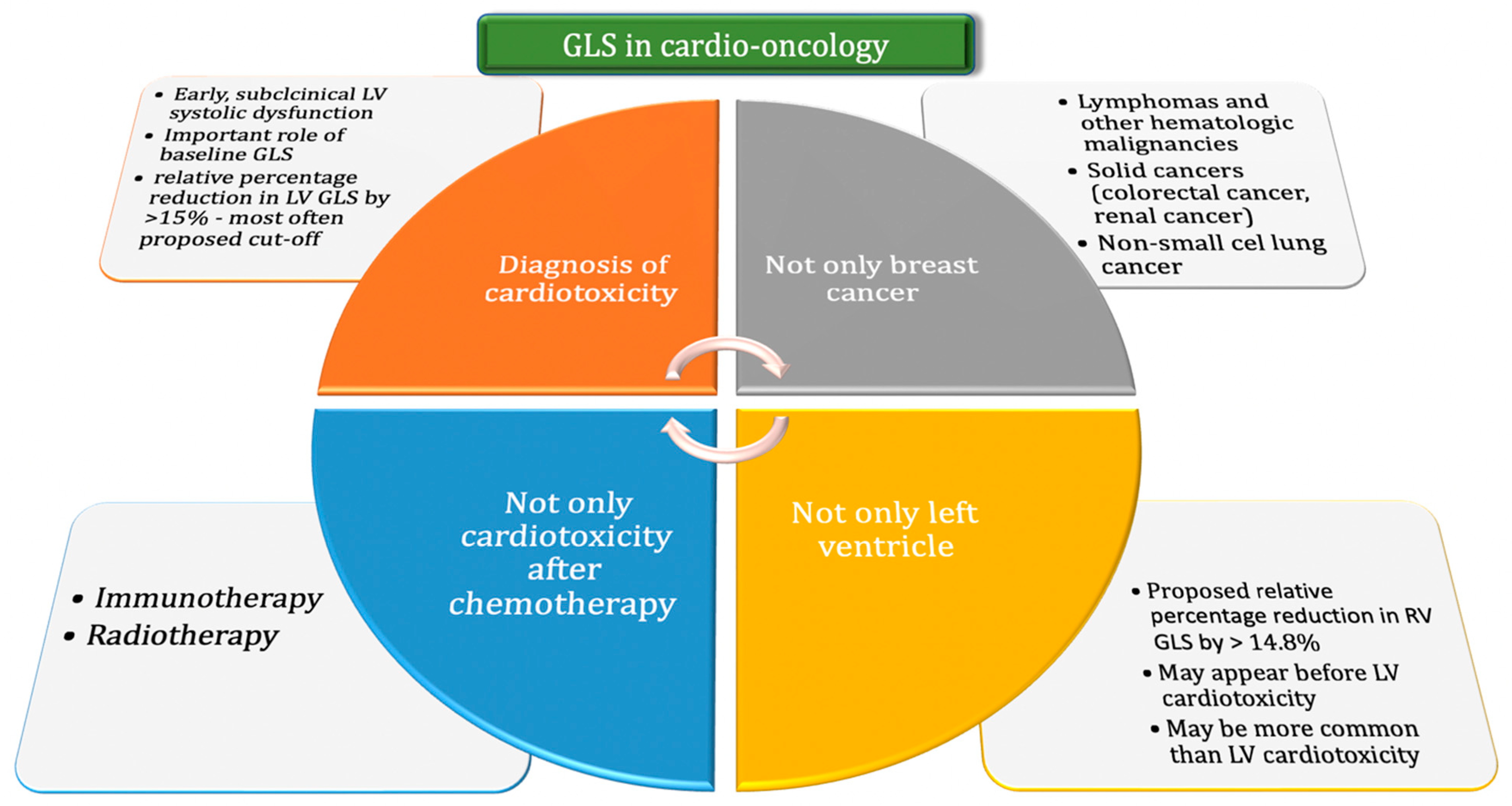
The GLS is valuable in diagnosing patients at risk for LV systolic dysfunction and in monitoring patients undergoing potentially cardiotoxic anticancer therapy. Due to its simplicity and repeatability, the GLS assessment has been rapidly incorporated into cardio-oncology (Figure 1). A relative decrease in GLS of >15% compared to baseline during cancer treatment is the recommended cut-off point for suspecting subclinical cardiac dysfunction and predicting a significant LVEF decrease in the future. An early diagnosis of asymptomatic cardiotoxicity enables the quick implementation of a cardioprotective treatment and reduces the risk of interruptions or changes in the oncological treatment, which may affect the survival of patients. Additional research is needed to precisely assess both the LV GLS and RV GLS in the constantly evolving field of cardio-oncology.
Author Contributions
Conceptualization, G.S.; writing—original draft preparation, G.S. and M.H.; writing—review and editing, I.N.-T., A.L.-S. and E.L.; supervision, E.L. All authors have read and agreed to the published version of the manuscript.
Funding
This research received no external funding.
Institutional Review Board Statement
Not applicable.
Informed Consent Statement
Not applicable.
Data Availability Statement
No new data were created or analyzed in this study. Data sharing is not applicable to this article.
Conflicts of Interest
The authors declare no conflict of interest.
Abbreviations
| BNP | brain natriuretic peptide |
| cTnT | cardiac troponin T |
| GLS | global longitudinal strain |
| HSCT | hematopoietic stem cell transplantation |
| LVEF | left ventricular ejection fraction |
| MACE | major adverse cardiac events |
| NT-proBNP | N-terminal pro B-type natriuretic peptide |
| RV | right ventricle |
| SR | strain rate |
| STE | speckle tracking echocardiography |
References
- Bohdan, M.; Kowalczys, A.; Mickiewicz, A.; Gruchała, M.; Lewicka, E. Cancer Therapy-Related Cardiovascular Complications in Clinical Practice: Current Perspectives. Clin. Med. 2021, 10, 1647. [Google Scholar] [CrossRef] [PubMed]
- Medvedofsky, D.; Maffessanti, F.; Weinert, L.; Tehrani, D.M.; Narang, A.; Addetia, K.; Mediratta, A.; Besser, S.A.; Maor, M.; Patel, A.R.; et al. 2D and 3D Echocardiography-Derived Indices of Left Ventricular Function and Shape: Relationship with Mortality. JACC Cardiovasc. Imaging 2018, 11, 1569–1579. [Google Scholar] [PubMed]
- Flachskampf, F.A.; Blankstein, R.; Grayburn, P.A.; Kramer, C.M.; Kwong, R.Y.; Marwick, T.H.; Nagel, E.; Sengupta, P.P.; Zoghbi, W.A.; Chandrashekhar, Y. Global Longitudinal Shortening: A Positive Step Towards Reducing Confusion Surrounding Global Longitudinal Strain. JACC Cardiovasc. Imaging 2019, 12, 1566–1567. [Google Scholar] [CrossRef] [PubMed]
- Karlsen, S.; Dahlslett, T.; Grenne, B.; Sjøli, B.; Smiseth, O.; Edvardsen, T.; Brunvand, H. Global longitudinal strain is a more reproducible measure of left ventricular function than ejection fraction regardless of echocardiographic training. Cardiovasc. Ultrasound 2019, 17, 18. [Google Scholar] [CrossRef]
- Belghitia, H.; Brette, S.; Lafitte, S.; Reant, P.; Picard, F.; Serri, K.; Courregelongue, M.; Dos Santos, P.; Douard, H.; Roudaut, R.; et al. Automated function imaging: A new operator-independent strain method for assessing left ventricular function. Arch. Cardiovasc. Dis. 2008, 101, 163–169. [Google Scholar]
- Krishnasamy, R.; Isbel, N.M.; Hawley, C.M.; Pascoe, E.M.; Burrage, M.K.; Leano, R.; Haluska, B.A.; Marwick, T.H.; Stanton, T. Left Ventricular Global Longitudinal Strain (GLS) Is a Superior Predictor of All-Cause and Cardiovascular Mortality When Compared to Ejection Fraction in Advanced Chronic Kidney Disease. PLoS ONE 2015, 10, e0127044. [Google Scholar]
- D’Elia, N.; Caselli, S.; Kosmala, W.; Lancellotti, P.; Morris, D.; Muraru, D.; Takeuchi, M.; van den Bosch, A.; van Grootel, R.W.J.; Villarraga, H.; et al. Normal Global Longitudinal Strain: An Individual Patient Meta-Analysis. JACC Cardiovasc. Imaging 2020, 13, 167–169. [Google Scholar]
- Dobson, R.; Ghosh, A.K.; Ky, B.; Marwick, T.; Stout, M.; Harkness, A.; Steeds, R.; Robinson, S.; Oxborough, D.; Adlam, D.; et al. BSE and BCOS Guideline for Transthoracic Echocardiographic Assessment of Adult Cancer Patients Receiving Anthracyclines and/or Trastuzumab. JACC CardioOncol. 2021, 3, 1–16. [Google Scholar]
- Plana, J.C.; Galderisi, M.; Barac, A.; Ewer, M.S.; Ky, B.; Scherrer-Crosbie, M.; Ganame, J.; Sebag, I.A.; Agler, D.A.; Badano, L.P.; et al. Expert consensus for multimodality imaging evaluation of adult patients during and after cancer therapy: A report from the American Society of Echocardiography and the European Association of Cardiovascular Imaging. J. Am. Soc. Echocardiogr. 2014, 27, 911–939. [Google Scholar] [CrossRef]
- Lyon, A.R.; López-Fernández, T.; Couch, L.; Asteggiano, R.; Aznar, M.; Bergler-Klein, J.; Boriani, G.; Cardinale, D.; Cordoba, R.; Cosyns, B.; et al. 2022 ESC Guidelines on cardio-oncology developed in collaboration with the European Hematology Association (EHA), the European Society for Therapeutic Radiology and Oncology (ESTRO) and the International Cardio-Oncology Society (IC-OS): Developed by the task force on cardio-oncology of the European Society of Cardiology (ESC). Eur. Heart J. 2022, 41, 4229–4361. [Google Scholar]
- Gripp, E.A.; Oliveira, G.E.; Feijó, L.A.; Garcia, M.I.; Xavier, S.S.; Sousa, A.S. Global Longitudinal Strain Accuracy for Cardiotoxicity Prediction in a Cohort of Breast Cancer Patients During Anthracycline and/or Trastuzumab Treatment. Arq. Bras. Cardiol. 2018, 110, 140–150. [Google Scholar] [CrossRef] [PubMed]
- Oikonomou, E.K.; Kokkinidis, D.G.; Kampaktsis, P.N.; Amir, E.A.; Marwick, T.H.; Gupta, D.; Thavendiranathan, P. Assessment of Prognostic Value of Left Ventricular Global Longitudinal Strain for Early Prediction of Chemotherapy-Induced Cardiotoxicity: A Systematic Review and Meta-analysis. JAMA Cardiol. 2019, 4, 1007–1018. [Google Scholar] [CrossRef] [PubMed]
- Cocco, L.D.; Chiaparini, A.F.; Saffi, M.A.L.; Leiria, T.L.L. Global Longitudinal Strain for the Early Detection of Chemotherapy-Induced Cardiotoxicity: A Systematic Review and Meta-analysis. Clin. Oncol. (R. Coll. Radiol.) 2022, 34, 514–525. [Google Scholar] [CrossRef]
- Araujo-Gutierrez, R.; Chitturi, K.R.; Xu, J.; Wang, Y.; Kinder, E.; Senapati, A.; Chebrolu, B.L.; Kassi, M.; Trachtenberg, B.H. Baseline global longitudinal strain predictive of anthracycline-induced cardiotoxicity. Cardiooncology 2021, 7, 4. [Google Scholar]
- Laufer-Perl, M.; Arnold, J.H.; Mor, L.; Amrami, N.; Derakhshesh, M.; Moshkovits, Y.; Sadeh, B.; Arbel, Y.; Topilsky, Y.; Rozenbaum, Z. The association of reduced global longitudinal strain with cancer therapy-related cardiac dysfunction among patients receiving cancer therapy. Clin. Res. Cardiol. 2020, 109, 255–262. [Google Scholar] [CrossRef]
- Negishi, T.; Thavendiranathan, P.; Negishi, K.; Marwick, T.H.; SUCCOUR Investigators. Rationale and Design of the Strain Surveillance of Chemotherapy for Improving Cardiovascular Outcomes: The SUCCOUR Trial. JACC Cardiovasc. Imaging 2018, 11, 1098–1105. [Google Scholar] [CrossRef]
- Thavendiranathan, P.; Negishi, T.; Somerset, E.; Negishi, K.; Penicka, M.; Lemieux, J.; Aakhus, S.; Miyazaki, S.; Shirazi, M.; Galderisi, M.; et al. Strain-Guided Management of Potentially Cardiotoxic Cancer Therapy. J. Am. Coll. Cardiol. 2021, 77, 392–401. [Google Scholar]
- Jordan, J.H.; Sukpraphrute, B.; Meléndez, G.C.; Jolly, M.P.; D’Agostino RBJr Hundley, W.G. Early Myocardial Strain Changes During Potentially Cardiotoxic Chemotherapy May Occur as a Result of Reductions in Left Ventricular End-Diastolic Volume: The Need to Interpret Left Ventricular Strain With Volumes. Circulation 2017, 135, 2575–2577. [Google Scholar] [CrossRef]
- Mele, D.; Malagutti, P.; Indelli, M.; Ferrari, L.; Casadei, F.; Da Ros, L.; Pollina, A.; Fiorencis, A.; Frassoldati, A.; Ferrari, R. Reversibility of Left Ventricle Longitudinal Strain Alterations Induced by Adjuvant Therapy in Early Breast Cancer Patients. Ultrasound Med. Biol. 2016, 42, 125–132. [Google Scholar] [CrossRef]
- Avila, M.; Alves, M.; Ayub-Ferreira, S.; Junior, M.R.D.B.W.; Cruz, F.D.D.; Brandão, S.M.G.; Hajjar, L.A.; Filho, R.K.; Cruz, C.B.B.V.; Abduch, A.M.C.; et al. Global Longitudinal Strain as a Predictor of Chemotherapy-Induced Cardiotoxicity. Authorea 2021, 35, eabc340. [Google Scholar]
- Sulaiman, L.; Hesham, D.; Abdel Hamid, M.; Youssef, G. The combined role of NT-proBNP and LV-GLS in the detection of early subtle chemotherapy-induced cardiotoxicity in breast cancer female patients. Egypt. Heart J. 2021, 73, 20. [Google Scholar]
- Kang, Y.; Xu, X.; Cheng, L.; Li, L.; Sun, M.; Chen, H.; Pan, C.; Shu, X. Two-dimensional speckle tracking echocardiography combined with high-sensitive cardiac troponin T in early detection and prediction of cardiotoxicity during epirubicine-based chemotherapy. Eur. J. Heart Fail. 2014, 16, 300–308. [Google Scholar] [PubMed]
- Öztürk, C.; Validyev, D.; Becher, U.M.; Weber, M.; Nickenig, G.; Tiyerili, V. A novel scoring system to estimate chemotherapy-induced myocardial toxicity: Risk assessment prior to non-anthracycline chemotherapy regimens. Int. J. Cardiol. Heart Vasc. 2021, 33, 100751. [Google Scholar] [CrossRef] [PubMed]
- Thavendiranathan, P.; Negishi, T.; Coté, M.A.; Penicka, M.; Massey, R.; Cho, G.-Y.; Hristova, K.; Vinereanu, D.; Popescu, B.A.; Izumo, M.; et al. Single Versus Standard Multiview Assessment of Global Longitudinal Strain for the Diagnosis of Cardiotoxicity During Cancer Therapy. JACC Cardiovasc. Imaging 2018, 11, 1109–1118. [Google Scholar] [CrossRef] [PubMed]
- Portugal, G.; Branco, L.M.; Galrinho, A.; Carmo, M.M.; Timoteo, A.T.; Feliciano, J.; Abreu, J.; Oliveira, S.D.; Batarda, L.; Ferreira, R.C. Global and regional patterns of longitudinal strain in screening for chemotherapy-induced cardiotoxicity. Importância da deformação longitudinal na deteção da cardiotoxicidade induzida por quimioterapia e na identificação de padrões específicos de afetação segmentar. Rev. Port. Cardiol. 2017, 36, 9–15. [Google Scholar] [PubMed]
- Astuti, A.; Erwinanto, E.; Akbar, M.R.; Martanto, E.; Badudu, D.F. Global and Regional Longitudinal Strain Reduction in Breast Cancer Patients After First Chemotherapy Cycle With Fluorouracil, Adriamycin, and Cyclophosphamide Regimen. Cardiol. Res. 2021, 12, 238–243. [Google Scholar] [CrossRef]
- Mahjoob, M.P.; Sheikholeslami, S.A.; Dadras, M.; Mansouri, H.; Haghi, M.; Naderian, M.; Sadeghi, L.; Tabary, M.; Khaheshi, I. Prognostic Value of Cardiac Biomarkers Assessment in Combination with Myocardial 2D Strain Echocardiography for Early Detection of Anthracycline-Related Cardiac Toxicity. Cardiovasc. Hematol. Disord.-Drug Targets 2020, 20, 74–83. [Google Scholar]
- Arciniegas Calle, M.C.; Sandhu, N.P.; Xia, H.; Cha, S.S.; Pellikka, P.A.; Ye, Z.; Villarraga, H.R. Two-dimensional speckle tracking echocardiography predicts early subclinical cardiotoxicity associated with anthracycline-trastuzumab chemotherapy in patients with breast cancer. BMC Cancer 2018, 18, 1037. [Google Scholar] [CrossRef]
- Chang, H.Y.; Lee, C.H.; Su, P.L.; Li, S.S.; Chen, M.Y.; Chen, Y.P.; Hsu, Y.T.; Tsai, W.C.; Liu, P.Y.; Chen, T.Y.; et al. Subtle cardiac dysfunction in lymphoma patients receiving low to moderate dose chemotherapy. Sci. Rep. 2021, 11, 7100. [Google Scholar]
- Hatazawa, K.; Tanaka, H.; Nonaka, A.; Takada, H.; Soga, F.; Hatani, Y.; Matsuzoe, H.; Shimoura, H.; Ooka, J.; Sano, H.; et al. Baseline Global Longitudinal Strain as a Predictor of Left Ventricular Dysfunction and Hospitalization for Heart Failure of Patients With Malignant Lymphoma After Anthracycline Therapy. Circ. J. 2018, 82, 2566–2574. [Google Scholar] [CrossRef]
- Gonzalez-Manzanares, R.; Castillo, J.C.; Molina, J.R.; Ruiz-Ortiz, M.; Mesa, D.; Ojeda, S.; Anguita, M.; Pan, M. Automated Global Longitudinal Strain Assessment in Long-Term Survivors of Childhood Acute Lymphoblastic Leukemia. Cancers 2022, 14, 1513. [Google Scholar]
- Oka, T.; Tada, Y.; Oboshi, M.; Kamada, R.; Yasui, T.; Shioyama, W.; Nishikawa, T.; Hino, A.; Ishikawa, J.; Fujita, M. Serial Changes in Cardiac Strain and Contractility after Hematopoietic Stem Cell Transplantation in Patients with Hematologic Malignancies. Int. Heart J. 2021, 62, 575–583. [Google Scholar] [PubMed]
- Sonaglioni, A.; Albini, A.; Fossile, E.; Pessi, M.A.; Nicolosi, G.L.; Lombardo, M.; Anzà, C.; Ambrosio, G. Speckle-Tracking Echocardiography for Cardioncological Evaluation in Bevacizumab-Treated Colorectal Cancer Patients. Cardiovasc. Toxicol. 2020, 20, 581–592. [Google Scholar] [CrossRef] [PubMed]
- Mingrone, G.; Astarita, A.; Airale, L.; Maffei, I.; Cesareo, M.; Crea, T.; Bruno, G.; Leone, D.; Avenatti, E.; Catarinella, C.; et al. Effects of Carfilzomib Therapy on Left Ventricular Function in Multiple Myeloma Patients. Front. Cardiovasc. Med. 2021, 8, 645678. [Google Scholar]
- Iannaccone, A.; Bruno, G.; Ravera, A.; Gay, F.; Salvini, M.; Bringhen, S.; Sabia, L.; Avenatti, E.; Veglio, F.; Milan, A. Evaluation of Cardiovascular Toxicity Associated with Treatments Containing Proteasome Inhibitors in Multiple Myeloma Therapy. High Blood Press. Cardiovasc. Prev. 2018, 25, 209–218. [Google Scholar] [PubMed]
- Liu, Z.; Zhang, L.; Liu, M.; Wang, F.; Xiong, Y.; Tang, Z.; Li, Q.; Lu, Q.; Liang, S.; Niu, T.; et al. Myocardial Injury in Multiple Myeloma Patients With Preserved Left Ventricular Ejection Fraction: Noninvasive Left Ventricular Pressure-Strain Myocardial Work. Front. Cardiovasc. Med. 2022, 8, 782580. [Google Scholar] [CrossRef]
- Chen, L.; Ta, S.; Wu, W.; Wang, C.; Zhang, Q. Prognostic and Added Value of Echocardiographic Strain for Prediction of Adverse Outcomes in Patients with Locally Advanced Non-Small Cell Lung Cancer after Radiotherapy. Ultrasound Med. Biol. 2019, 45, 98–107. [Google Scholar] [CrossRef]
- Lee Chuy, K.; Drill, E.; Yang, J.C.; Landau, H.; Hassoun, H.; Nahhas, O.; Chen, C.L.; Yu, A.F.; Steingart, R.M.; Liu, J.E. Incremental Value of Global Longitudinal Strain for Predicting Survival in Patients With Advanced AL Amyloidosis. JACC CardioOncol. 2020, 2, 223–231. [Google Scholar] [CrossRef]
- Awadalla, M.; Mahmood, S.S.; Groarke, J.D.; Hassan, M.Z.; Nohria, A.; Rokicki, A.; Murphy, S.P.; Mercaldo, N.D.; Zhang, L.; Zlotoff, D.A.; et al. Global Longitudinal Strain and Cardiac Events in Patients With Immune Checkpoint Inhibitor-Related Myocarditis. J. Am. Coll. Cardiol. 2020, 75, 467–478. [Google Scholar] [CrossRef]
- Mincu, R.I.; Pohl, J.; Mrotzek, S.; Michel, L.; Hinrichs, L.; Lampe, L.; Rassaf, T.; Totzeck, M. Left ventricular global longitudinal strain reduction in patients with melanoma and extra-cardiac immune-related adverse events during immune checkpoint inhibitor therapy. Eur. Heart J. 2020, 41, ehaa946.3261. [Google Scholar] [CrossRef]
- Tamura, Y.; Tamura, Y.; Takemura, R.; Yamada, K.; Taniguchi, H.; Iwasawa, J.; Yada, H.; Kawamura, A. Longitudinal Strain and Troponin I Elevation in Patients Undergoing Immune Checkpoint Inhibitor Therapy. JACC CardioOncol. 2022, 4, 673–685. [Google Scholar] [CrossRef] [PubMed]
- Fourati, N.; Charfeddine, S.; Chaffai, I.; Dhouib, F.; Farhat, L.; Boukhris, M.; Abid, L.; Kammoun, S.; Mnejja, W.; Daoud, J. Subclinical left ventricle impairment following breast cancer radiotherapy: Is there an association between segmental doses and segmental strain dysfunction? Int. J. Cardiol. 2021, 345, 130–136. [Google Scholar] [PubMed]
- Walker, V.; Lairez, O.; Fondard, O.; Pathak, A.; Pinel, B.; Chevelle, C.; Franck, D.; Jimenez, G.; Camilleri, J.; Panh, L.; et al. Early detection of subclinical left ventricular dysfunction after breast cancer radiation therapy using speckle-tracking echocardiography: Association between cardiac exposure and longitudinal strain reduction (BACCARAT study). Radiat. Ther. 2019, 14, 204. [Google Scholar]
- Xu, X.; Wang, D.; Yin, Y.; Dai, H.; Chen, L.; Xue, J.; Li, Q. Role of global longitudinal strain in evaluating radiotherapy-induced early cardiotoxicity in breast cancer: A meta-analysis. Kardiol. Pol. 2022; ahead of print. [Google Scholar]
- Keramida, K.; Farmakis, D.; Bingcang, J.; Sulemane, S.; Sutherland, S.; Bingcang, R.A.; Ramachandran, K.; Tzavara, C.; Charalampopoulos, G.; Filippiadis, D.; et al. Longitudinal changes of right ventricular deformation mechanics during trastuzumab therapy in breast cancer patients. Eur. J. Heart Fail. 2019, 21, 529–535. [Google Scholar] [PubMed]
- Laufer-Perl, M.; Perelman-Gvili, M.; Sirota Dorfman, S.; Sulemane, S.; Sutherland, S.; Bingcang, R.A.; Ramachandran, K.; Tzavara, C.; Charalampopoulos, G.; Filippiadis, D.; et al. Prevalence of Right Ventricle Strain Changes following Anthracycline Therapy. Life 2022, 12, 291. [Google Scholar]
- Khairat, I.; Khalfallah, M.; Shaban, A.; Farag, I.A.; Elkady, A. Right ventricular 2D speckle-tracking echocardiography in children with osteosarcoma under chemotherapy. Egypt. Heart J. 2019, 71, 23. [Google Scholar]
- Curigliano, G.; Lenihan, D.; Fradley, M.; Ganatra, S.; Barac, A.; Blaes, A.; Herrmann, J.; Porter, C.; Lyon, A.R.; Lancellotti, P.; et al. Management of cardiac disease in cancer patients throughout oncological treatment: ESMO consensus recommendations. Ann. Oncol. 2020, 31, 171–190. [Google Scholar]
- Čelutkienė, J.; Pudil, R.; Lopez-Fernandez, T.; Grapsa, J.; Nihoyannopoulos, P.; Bergler-Klein, J.; Cohen-Solal, A.; Farmakis, D.; Tocchetti, C.G.; von Haehling, S.; et al. Role of cardiovascular imaging in cancer patients receiving cardiotoxic therapies: A position statement on behalf of the Heart Failure Association (HFA), the European Association of Cardiovascular Imaging (EACVI) and the Cardio-Oncology Council of the European Society of Cardiology (ESC) [published correction appears in Eur. J. Heart Fail. 2021, 23, 345]. Eur. J. Heart Fail. 2020, 22, 1504–1524. [Google Scholar]
- Virani, S.A.; Dent, S.; Brezden-Masley, C.; Clarke, B.; Davis, M.K.; Jassal, D.S.; Johnson, C.; Lemieux, J.; Paterson, I.; Sebag, I.A.; et al. Canadian Cardiovascular Society Guidelines for Evaluation and Management of Cardiovascular Complications of Cancer Therapy. Can. J. Cardiol. 2016, 32, 831–841. [Google Scholar]
- Zamorano, J.L.; Lancellotti, P.; Rodriguez Muñoz, D.; Aboyans, V.; Asteggiano, R.; Galderisi, M.; Habib, G.; Lenihan, D.J.; Lip, G.Y.H.; Lyon, A.R.; et al. 2016 ESC Position Paper on cancer treatments and cardiovascular toxicity developed under the auspices of the ESC Committee for Practice Guidelines: The Task Force for cancer treatments and cardiovascular toxicity of the European Society of Cardiology (ESC) [published correction appears in Eur. Heart J. 24 December 2016]. Eur. Heart J. 2016, 37, 2768–2801. [Google Scholar]
- Di Lisi, D.; Madaudo, C.; Di Fazio, L.; Gulotta, A.; Triolo, O.F.; Galassi, A.R.; Incorvaia, L.; Russo, A.; Novo, G. Higher Incidence of Cancer Therapy-Related Cardiac Dysfunction in the COVID-19 Era: A Single Cardio-Oncology Center Experience. J. Cardiovasc. Dev. Dis. 2023, 10, 23. [Google Scholar] [PubMed]
- Piveta, R.B.; Rodrigues, A.C.T.; Vieira, M.L.C.; Fischer, C.H.; Afonso, T.R.; Daminello, E.; Cruz, F.M.; Galvão, T.F.G.; Filho, E.B.L.; Katz, M.; et al. Early Change in Area Strain Detected by 3D Speckle Tracking Is Associated With Subsequent Cardiotoxicity in Patients Treated With Low Doses of Anthracyclines. Front. Cardiovasc. Med. 2022, 9, 842532. [Google Scholar]
- Chen, H.; Ouyang, D.; Baykaner, T.; Jamal, F.; Cheng, P.; Rhee, J.W. Artificial intelligence applications in cardio-oncology: Leveraging high dimensional cardiovascular data. Front. Cardiovasc. Med. 2022, 9, 941148. [Google Scholar] [CrossRef] [PubMed]
- Kar, J.; Cohen, M.V.; McQuiston, S.A.; Malozzi, C.M. Can global longitudinal strain (GLS) with magnetic resonance prognosticate early cancer therapy-related cardiac dysfunction (CTRCD) in breast cancer patients, a prospective study? [published online ahead of print, 2022 Dec 26]. Magn. Reson. Imaging 2022, 97, 68–81. [Google Scholar] [CrossRef] [PubMed]
- Kosmala, W.; Negishi, T.; Thavendiranathan, P.; Penicka, M.; De Blois, J.; Murbræch, K.; Miyazaki, S.; Shirazi, M.; Santoro, C.; Vinereanu, D.; et al. Incremental Value of Myocardial Work over Global Longitudinal Strain in the Surveillance for Cancer-Treatment-Related Cardiac Dysfunction: A Case-Control Study. J. Clin. Med. 2022, 11, 912. [Google Scholar] [CrossRef]
- Iida, N.; Tajiri, K.; Ishizu, T.; Sasamura-Koshizuka, R.; Nakajima, H.; Kawamatsu, N.; Sato, K.; Yamamoto, M.; Machino-Ohtsuka, T.; Bando, H.; et al. Echocardiography image quality of global longitudinal strain in cardio-oncology: A prospective real-world investigation. J. Echocardiogr. 2022, 20, 159–165. [Google Scholar] [PubMed]
- Marwick, T.H. Global Longitudinal Strain Monitoring to Guide Cardioprotective Medications During Anthracycline Treatment. Curr. Oncol. Rep. 2022, 24, 687–694. [Google Scholar] [CrossRef]
- Moslehi, J.J.; Witteles, R.M. Global Longitudinal Strain in Cardio-Oncology. J. Am. Coll. Cardiol. 2021, 77, 402–404. [Google Scholar] [CrossRef]
- Negishi, T.; Thavendiranathan, P.; Penicka, M.; Lemieux, J.; Aakhus, S.; Miyazaki, S.; Shirazi, M.; Galderisi, M.; Cho, G.Y.; Popescu, B.A.; et al. Cardioprotection Using Strain-Guided Management of Potentially Cardiotoxic Cancer Therapy: 3-Year Results of the SUCCOUR Trial [published online ahead of print, 16 November 2022]. JACC Cardiovasc. Imaging 2022, S1936-878X(22)00616-7. [Google Scholar]
Figure 1.
The use of GLS in cardio-oncology, taking into account the role of GLS in the diagnosis of cardiotoxicity, the possibility of assessing this parameters in many types of cancer and for many types of oncological treatment techniques, and the possibility of assessment also for the right ventricle. Abbreviations: GLS = global longitudinal strain; LV = left ventricular; RV = right ventricular.


Table 1.
Recommendations of scientific societies in recent years regarding the frequency of performing an echocardiographic examination with a GLS assessment.
Table 1.
Recommendations of scientific societies in recent years regarding the frequency of performing an echocardiographic examination with a GLS assessment.
| Guidelines, Year | Anthracyclines | HER2-Targeted Therapies |
|---|---|---|
| 2022 ESC Guidelines on cardio-oncology developed in collaboration with the European Hematology Association (EHA), the European Society for Therapeutic Radiology and Oncology (ESTRO), and the International Cardio-Oncology Society (IC-OS): Developed by the task force on cardio-oncology of the European Society of Cardiology (ESC) [10] | Low baseline cardiotoxicity risk: at baseline and 12 months post-treatment (may be also considered after the fourth cycle) Moderate risk: at baseline and 12 months posttreatment (should be also considered after the fourth cycle). High and very high risk: at baseline, after 2nd, 4th, and 6th cycles and also 3 and 12 months after treatment | Low and moderate risk: at baseline; after 3, 6, 9, and 12 months; and then 12 months post-treatment High and very high risk: at baseline; after 3, 6, 9, and 12 months; and then 3 and 12 months post-treatment |
| 2021 British Society for Echocardiography and British Cardio-Oncology Society guideline for transthoracic echocardiographic assessment of adult cancer patients receiving anthracyclines and/or trastuzumab [8]. | Every 3 months during chemotherapy, and 3–12 months after its termination | Every 3 months during the therapy and 3–12 months after the end of therapy |
| 2020 Management of cardiac disease in cancer patients throughout oncological treatment: ESMO consensus recommendations [48]. | After a cumulative dose of 250 mg/m2 doxorubicin or equivalent. Next after each additional 100 mg/m2 | Every 3 months |
| 2020 Role of cardiovascular imaging in cancer patients receiving cardiotoxic therapies: a position statement on behalf of the Heart Failure Association (HFA), the European Association of Cardiovascular Imaging (EACVI), and the Cardio-Oncology Council of the European Society of Cardiology (ESC) [49]. | Depending on the risk calculated according to the planned therapy and patient-related risk factors, including age, comorbidities, and cardiovascular (CV) risk factors | Risk is calculated according to the planned therapy and patient-related factors, including age, comorbidities, and CV risk factors (range from 6 to 12 weeks) |
| 2016 Canadian Cardiovascular Society Guidelines for Evaluation and Management of Cardiovascular Complications of Cancer Therapy [50]. | No recommendations | Every 3 months |
| 2016 ESC Position Paper on cancer treatments and cardiovascular toxicity developed under the auspices of the ESC Committee for Practice Guidelines: The Task Force for cancer treatments and cardiovascular toxicity of the European Society of Cardiology (ESC) [51]. | After a cumulative dose of 200 mg/m2 doxorubicin or equivalent | Every 4 cycles |
Disclaimer/Publisher’s Note: The statements, opinions and data contained in all publications are solely those of the individual author(s) and contributor(s) and not of MDPI and/or the editor(s). MDPI and/or the editor(s) disclaim responsibility for any injury to people or property resulting from any ideas, methods, instructions or products referred to in the content. |
© 2023 by the authors. Licensee MDPI, Basel, Switzerland. This article is an open access article distributed under the terms and conditions of the Creative Commons Attribution (CC BY) license (https://creativecommons.org/licenses/by/4.0/).
Share and Cite
MDPI and ACS Style
Sławiński, G.; Hawryszko, M.; Liżewska-Springer, A.; Nabiałek-Trojanowska, I.; Lewicka, E. Global Longitudinal Strain in Cardio-Oncology: A Review. Cancers 2023, 15, 986. https://doi.org/10.3390/cancers15030986
AMA Style
Sławiński G, Hawryszko M, Liżewska-Springer A, Nabiałek-Trojanowska I, Lewicka E. Global Longitudinal Strain in Cardio-Oncology: A Review. Cancers. 2023; 15(3):986. https://doi.org/10.3390/cancers15030986
Chicago/Turabian StyleSławiński, Grzegorz, Maja Hawryszko, Aleksandra Liżewska-Springer, Izabela Nabiałek-Trojanowska, and Ewa Lewicka. 2023. "Global Longitudinal Strain in Cardio-Oncology: A Review" Cancers 15, no. 3: 986. https://doi.org/10.3390/cancers15030986
Note that from the first issue of 2016, this journal uses article numbers instead of page numbers. See further details here.