Article

Characteristics and Lenvatinib Treatment Response of Unresectable Hepatocellular Carcinoma with Iso-High Intensity in the Hepatobiliary Phase of EOB-MRI

1
Departments of Gastroenterology and Hepatology, Graduate School of Medicine, Hokkaido University, Sapporo 060-8589, Japan
2
Laboratory of Molecular and Cellular Medicine, Faculty of Pharmaceutical Sciences, Hokkaido University, Sapporo 060-8589, Japan
*
Author to whom correspondence should be addressed.
The authors equally contributed to this study.
Cancers 2021, 13(14), 3633; https://doi.org/10.3390/cancers13143633
Submission received: 5 July 2021 / Revised: 10 July 2021 / Accepted: 16 July 2021 / Published: 20 July 2021
(This article belongs to the Section Cancer Biomarkers)

Abstract

:

Simple Summary

HCC with alterations in CTNNB1 (which encodes β-catenin) is resistant to immune checkpoint inhibitors and is associated with HCC with iso-high intensity in the hepatobiliary phase of EOB-MRI in resectable HCC. However, the prevalence, characteristics, mutation profile, and treatment response in unresectable HCC with iso-high intensity in the hepatobiliary phase of EOB-MRI are not well clarified. In this study, we showed that the prevalence was 13%, and the response to lenvatinib does not differ between HCC with and without iso-high intensity in the hepatobiliary phase of EOB-MRI. We analyzed CTNNB-1 mutations using cell-free DNA, providing support for their association with iso-high intensity in the hepatobiliary phase of EOB-MRI.

Abstract

In hepatocellular carcinoma (HCC), CTNNB-1 mutations, which cause resistance to immune checkpoint inhibitors, are associated with HCC with iso-high intensity in the hepatobiliary phase of gadoxetic acid-enhanced magnetic resonance imaging (EOB-MRI) in resectable HCC; however, analyses on unresectable HCC are lacking. This study analyzed the prevalence, characteristics, response to lenvatinib, and CTNNB-1 mutation frequency in unresectable HCC with iso-high intensity in the hepatobiliary phase of EOB-MRI. In 52 patients with unresectable HCC treated with lenvatinib, the prevalence of iso-high intensity in the hepatobiliary phase of EOB-MRI was 13%. All patients had multiple HCCs, and 3 patients had multiple HCCs with iso-high intensity in the hepatobiliary phase of EOB-MRI. Lenvatinib response to progression-free survival and overall survival were similar between patients with or without iso-high intensity in the hepatobiliary phase of EOB-MRI. Seven patients (three and four patients who had unresectable HCC with or without iso-high intensity in the hepatobiliary phase of EOB-MRI, respectively) underwent genetic analyses. Among these, two (67%, 2/3) who had HCC with iso-high intensity in the hepatobiliary phase of EOB-MRI carried a CTNNB-1 mutation, while all four patients who had HCC without iso-high intensity in the hepatobiliary phase of EOB-MRI did not carry the CTNNB-1 mutation. This study’s findings have clinical implications for the detection and treatment of HCC with iso-high intensity in the hepatobiliary phase of EOB-MRI.

1. Introduction

Recent advances have dramatically changed the hepatocellular carcinoma (HCC) treatment landscape. The development of immune checkpoint inhibitors (ICIs), which target negative anti-cancer immune response regulators, has drastically improved anti-cancer therapy. Various clinical trials and real-world data have revealed the efficacy of ICIs, conferring a durable response, which is not observed in conventional anti-cancer therapy, in various malignancies [1,2,3,4,5]. A clinical trial on HCC has revealed that bevacizumab plus atezolizumab has been associated with better overall survival (OS) in patients with unresectable tumors than sorafenib, which was the standard therapy for patients with unresectable HCC [6].
However, some patients did not experience sufficient treatment response. Thus, predictive factors for response to ICIs have been an important clinical issue and identified in various malignancies, including PD-L1 expression, immune cell infiltration, mutation burden, T cell receptor repertoires, and the gut microbiome [7]. Recently, Harding et al. reported that in patients with advanced HCC carrying WNT/β-catenin mutations, ICI treatment results in a lower disease control rate and shorter median OS than those for HCC without WNT/β-catenin mutations [8]. Thus, the presence of WNT/β-catenin mutations in advanced HCC might be an effective predictive factor for resistance to ICI therapy. However, analyses of genetic alterations in advanced HCC are difficult because biopsy of advanced HCC sometimes causes tumor dissemination and bleeding. Thus, alternative methods for the detection of WNT/β-catenin mutations in advanced HCC are required. HCC imaging using gadoxetic acid-enhanced magnetic resonance imaging (EOB-MRI) could be a candidate approach. In the hepatobiliary phase of EOB-MRI, HCC typically shows low intensity, while HCC with the WNT/β-catenin mutation shows iso-high intensity because WNT/β-catenin mutations result in higher organic anion transporter 1B3 (OATP1B3) expression than in surrounding non-HCC hepatocytes [9,10,11]. In resectable HCC, the reported frequency of cases with iso-high intensity in the hepatobiliary phase of EOB-MRI is 12% (22/180) to 20% (5/25) [12,13,14,15]. However, the prevalence of unresectable HCC with iso-high intensity in the hepatobiliary phase of EOB-MRI, requiring systemic therapy, has not been clarified.
Analyses of cell-free DNA (cfDNA) are promising for detecting genetic alterations in HCC [16,17] and may provide insight into the frequent mutations in unresectable HCC with high intensity in the hepatobiliary phase of EOB-MRI.
Lenvatinib is a recently developed multi-kinase inhibitor that targets VEGFR1–3, FGFR1–4, RET c-Kit, and PDGFR-α mediated signaling [18,19]. The REFLECT trial, which is a phase 3 clinical trial regarding lenvatinib for patients with unresectable HCC, revealed that lenvatinib has similar OS compared with sorafenib, while lenvatinib has superior progression-free survival than sorafenib [20]. Real-world data validated the efficacy and safety of lenvatinib for patients with unresectable HCC [21,22]. Thus, in patients with unresectable HCC, who might have a poor response to ICIs, lenvatinib could be alternative first-line therapy. However, the efficacy and safety of lenvatinib in patients with unresectable HCC with iso-high intensity in the hepatobiliary phase of EOB-MRI have not been clarified.
Therefore, in this study, we analyzed (a) the prevalence of unresectable HCC with iso-high intensity in the hepatobiliary phase of EOB-MRI in patients with unresectable HCC who were treated with lenvatinib, (b) the response to lenvatinib, and (c) the relationship between the presence of unresectable HCC with iso-high intensity in the hepatobiliary phase of EOB-MRI and β-Catenin (CTNNB-1) mutations determined by a cfDNA analysis.

2. Materials and Methods

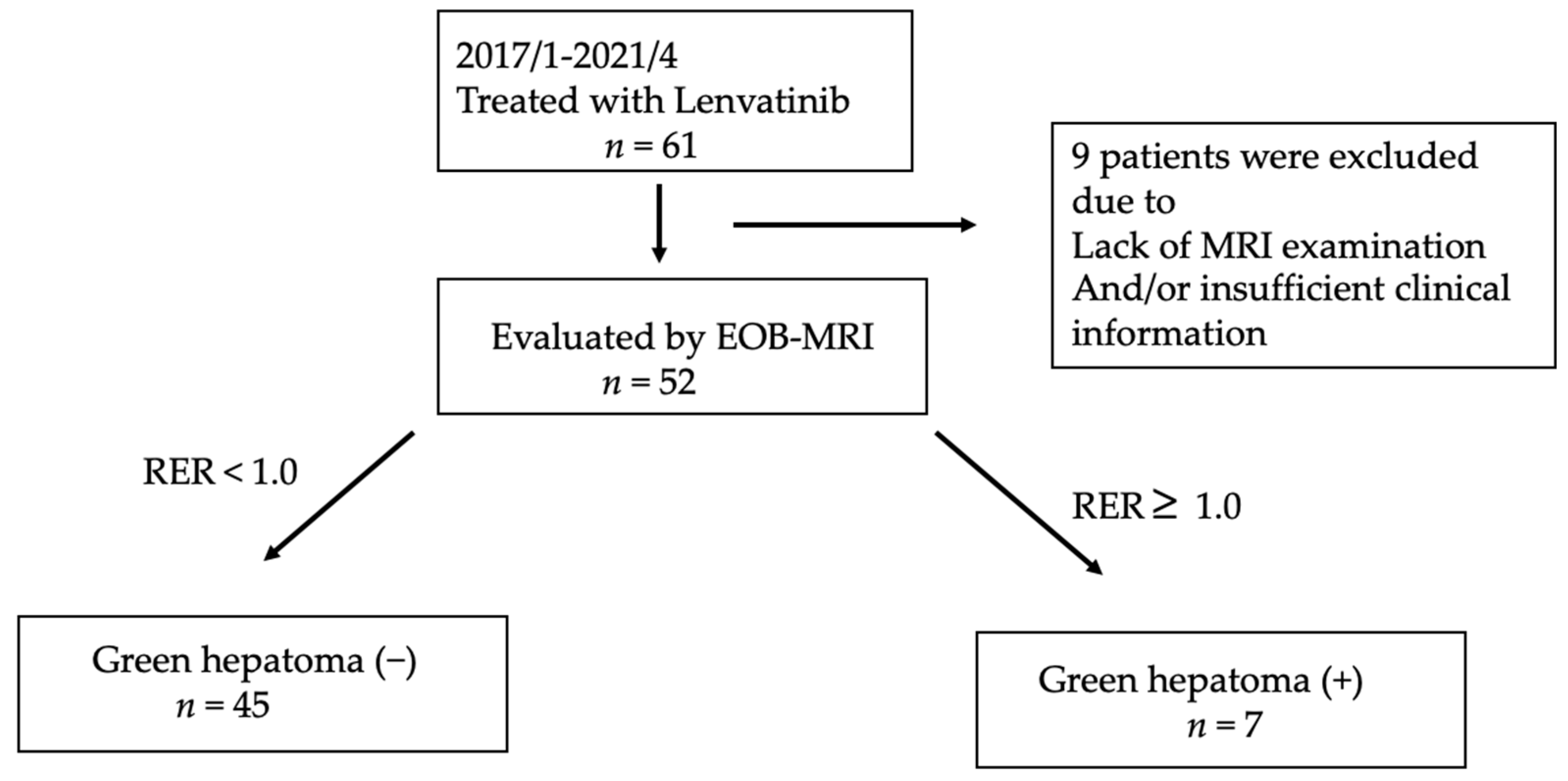
2.1. Patients and Study Design

Patients with unresectable HCC for whom lenvatinib treatment was initiated at Hokkaido University Hospital between April 2018 and April 2021 were screened in this retrospective study. We included patients if they had an EOB-MRI examination before lenvatinib treatment and were evaluated after treatment using dynamic computed tomography (CT) or EOB-MRI at baseline and every 2–3 months. Patients were excluded if they had decompensated liver cirrhosis, had insufficient clinical data or no EOB-MRI examination, had a treatment duration of less than 1 week, or were not evaluated for treatment responses by dynamic CT or MRI.
Attending physicians routinely assessed patients using laboratory tests and physical examinations every 2 weeks and using enhanced CT or EOB-MRI every 2–3 months after treatment initiation.

2.2. Treatment Protocol

Patients were administrated lenvatinib according to the package insert as follows: once-daily doses of 8 and 12 mg for patients weighing <60 and ≥60 kg, respectively.

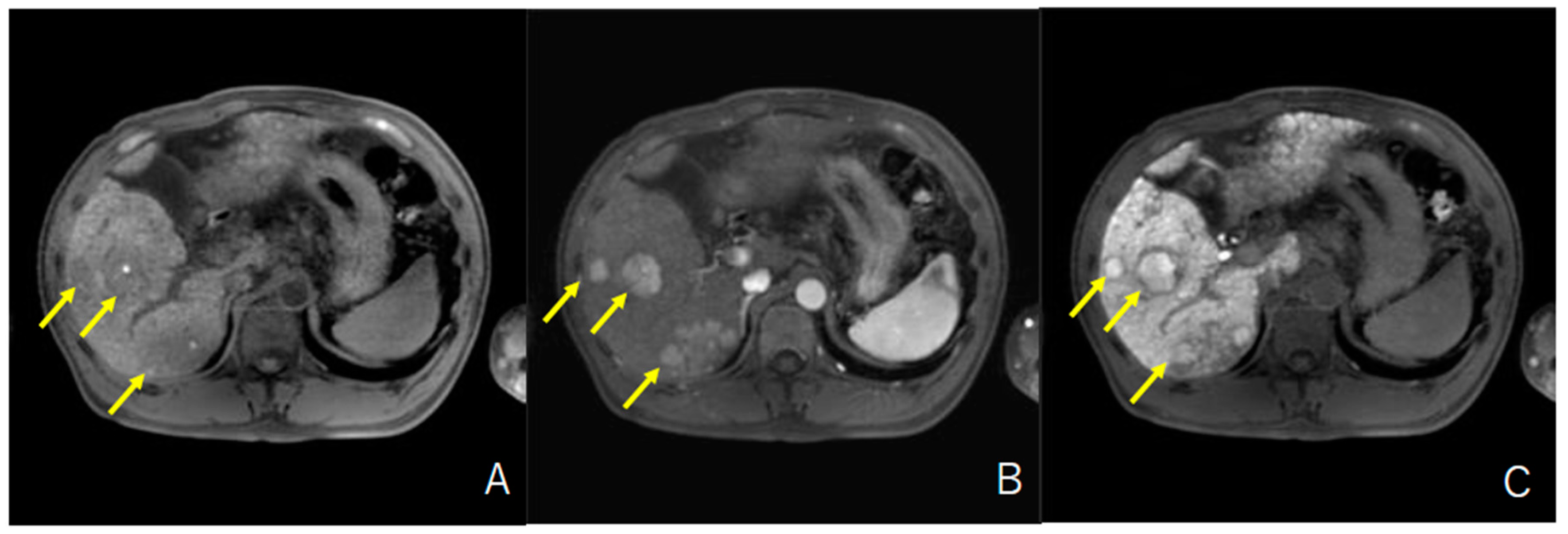
2.3. Evaluation of HCC with Iso-High Intensity in the Hepatobiliary Phase of EOB-MRI

Two MRI systems, Achieva 1.5 T dStream (Philips N. V., Tokyo, Japan) and TRILLIUM–OVAL 3.0 T (HITACHI, Tokyo, Japan), were utilized for the evaluation of HCC according to previously described methods [10] by two hepatologists. The signal intensities (SIs) of the tumor and surrounding non-tumor liver tissues were measured by defining regions of interest (ROIs). The enhancement in HCC relative to liver parenchyma was calculated according to a previously described method [15] as follows. Relative intensity ratio (RIR) = SI (nodule)/SI (parenchyma). Relative enhancement ratio (RER) = RIR (in hepatobiliary-phase images)/RIR (precontract RIR). Tumor enhancement was defined as iso-high when RER ≥ 1.0.

2.4. Evaluation of the Response to Lenvatinib

Treatment responses were evaluated every 2–3 months using dynamic CT based on mRECIST criteria [23].

2.5. CTNNB1 Mutation Detection in Cell-Free DNA

Eleven of the 52 patients included had cfDNA at baseline and/or within 1 month after treatment initiation. Two of these patients had HCC with iso-high intensity in the hepatobiliary phase of EOB-MRI. Moreover, four patients without HCC with iso-high intensity in the hepatobiliary phase of EOB-MRI who initiated lenvatinib in June 2019 and agreed to cfDNA collection were selected for an analysis of CTNNB1 alterations. In addition, CTNNB1 alterations in cfDNA were analyzed in another patient who had unresectable HCC with iso-high intensity in the hepatobiliary phase of EOB-MRI and had cfDNA.
For the extraction of cfDNA, 4 mL of plasma was collected, and cfDNA was extracted using the AVENIO cfDNA Isolation Kit (Roche Diagnostics K. K., Tokyo, Japan) according to the manufacturer’s protocol at Tsukuba i-Laboratory LLP (Tsukuba, Ibaraki, Japan) and stored at −80 °C. The concentration of cfDNA was determined using the Qubit dsDNA HS Assay Kit (Thermo Fisher Scientific, Waltham, MA, USA), and fragment sizes were examined using the Agilent 2200 TapeStation System with High Sensitivity D5000 ScreenTape Assay (Agilent, Santa Clara, CA, USA).
We conducted library preparation and sequencing according to the manufacturer’s (Thermo Fisher Scientific) methods. We utilized an Oncomine Colon cfDNA for PCR amplification of target regions, and library preparation for sequencing reads was mapped using Torrent Suite 5.6.0 (Thermo Fisher Scientific). Variant calling was performed using Oncomine Colon Liquid Biopsy-w2.1-DNA—Single Sample workflow in Ion Reporter version 5.6 (Thermo Fisher Scientific). Modified Oncomine_Colon_cfDNA.03062017, a BED file, was used for variant calling at hotspot sites. Annotations of called variants were based on dbSNP151, CCDS (NCBI, Release 15), RefSeq (UCSC Genome Browser, Nov 2018), Gencode (UCSC Genome Browser, ver. 19), and 1000 Genomes (phase3 release v5).

2.6. Statistical Analysis

Categorical variables were analyzed using the chi-square and Fisher’s exact tests. Continuous variables were analyzed using the Mann–Whitney U-test. In this study, we set statistical significance at p < 0.05.
Progression-free survival (PFS) and overall survival (OS) were defined as follows; the time from the initiation of lenvatinib to the day of disease progression or last follow-up and death, respectively. PFS and OS curves were compared using the log-rank test. The association of etiology and overall survival was analyzed by Cox regression analysis. Statistical analyses were performed using IBM SPSS Statistics 22.0 (IBM Corp., Armonk, NY, USA).

3. Results

3.1. Enrolled Patients and Baseline Characteristics

Between April 2018 and April 2021, a total of 61 patients with unresectable HCC were treated with lenvatinib at Hokkaido University Hospital. Among these patients, nine were excluded owing to a lack of EOB-MRI examination and/or insufficient treatment duration. Finally, 52 patients with unresectable HCC were included in the study (Figure 1).
Table 1 shows the baseline characteristics of patients. The median age of patients with HCC with iso-high intensity in the hepatobiliary phase of EOB-MRI was 70 years and that of patients without was 71 years. A total of 88% of patients (46/52) were males. Forty-one and 11 patients had Child–Pugh grades A and B, respectively. Twenty-seven and 25 patients had BCLC stages B and C, respectively. The median serum AFP and PIVKA-II levels were 56.7 IU/mL (range, 1.6–449,909 IU/mL) and 404.0 mAU/mL (range, 13–416,670 mAU/mL), respectively. Regarding disease etiology, 14, 8, and 30 patients had HBV infection, HCV infection, and non-B non-C liver disease.

3.2. Prevalence and Characteristics of HCC with Iso-High Intensity in the Hepatobiliary Phase of EOB-MRI in Patients with Unresectable HCC Who Were Treated with Lenvatinib

Overall, among 52 patients, 7 (13%) had HCC with high intensity in the hepatobiliary phase of EOB-MRI. Typical MR images are shown in Figure 2. As summarized in Table 1, the median age, sex, BCLC stage, and tumor markers were similar in patients with unresectable HCC with or without iso-high intensity in the hepatobiliary phase of EOB-MRI. Table 2 presents the characteristics of patients with unresectable HCC with iso-high intensity in the hepatobiliary phase of EOB-MRI. All patients had multiple HCCs and had both HCCs with or without iso-high intensity in the hepatobiliary phase of EOB-MRI simultaneously. Three patients (42.9%) had multiple HCCs with iso-high intensity in the hepatobiliary phase of EOB-MRI.

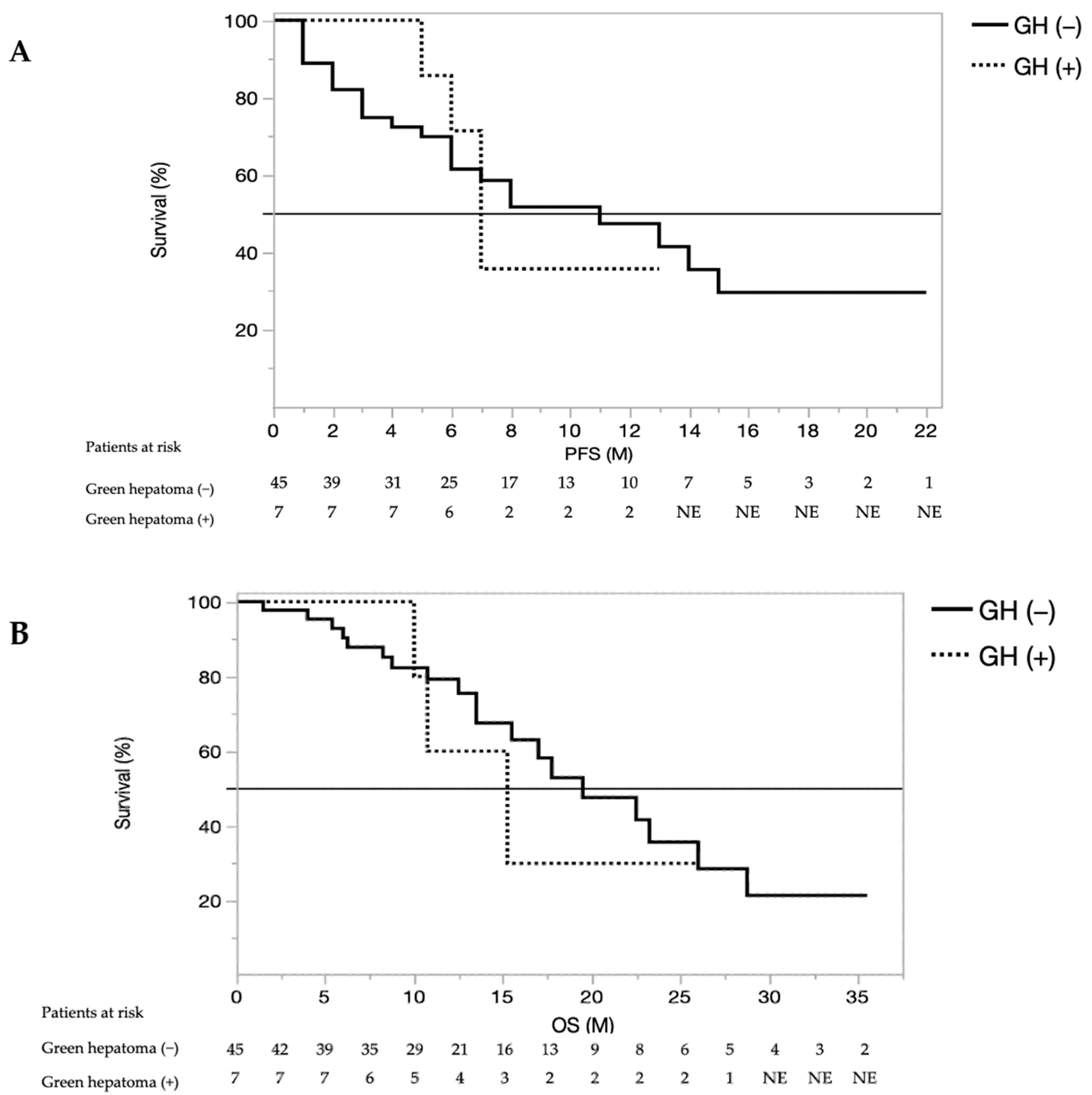
3.3. Treatment Response, Overall Survival, and Progression-Free Survival

Subsequently, we analyzed the response to lenvatinib, PFS, and OS in patients with unresectable HCC. As shown in Table 3, treatment responses were similar between patients with or without HCC with iso-high intensity in the hepatobiliary phase of EOB-MRI; the objective response rates (ORR) were 42.8% (3/7) and 48.8% (22/45), and the disease control rates (DCR) were 100% (7/7) and 86.6% (39/45) in patients with or without HCC with iso-high intensity in the hepatobiliary phase of EOB-MRI, respectively.
Comparison of OS and PFS in patients with HCC with or without iso-high intensity in the hepatobiliary phase of EOB-MRI treated with lenvatinib are shown in Figure 3. PFS rates were not significantly different between patients with or without HCC with iso-high intensity in the hepatobiliary phase of EOB-MRI (median PFS; 7.0 and 8.3 months, respectively; HR = 1.041074, 95% CI: 0.863166–1.171921, p = 0.8456). Similarly, OS rates were not significantly different between patients with or without HCC with iso-high intensity in the hepatobiliary phase of EOB-MRI (median OS; 17.5 and 19.5 months, respectively; HR = 1.1449, 95% CI: 0.892235–1.090671, p = 0.8315). In addition, as shown in the Supplementary Table S1, cox regression analysis revealed that the presence of HCC with iso-high intensity in the hepatobiliary phase of EOB-MRI did not affect OS (HR 0.95, 95% CI 0.153–4.849, p = 0.9613).

3.4. Relationship between HCC with Iso-High Intensity in the Hepatobiliary Phase of EOB-MRI and WNT/β-Catenin Mutations Diagnosed by Cell-Free DNA

Finally, we analyzed the CTNNB1 mutation profile in cfDNA in one patient with unresectable HCC with iso-high intensity in the hepatobiliary phase of EOB-MRI and three patients without HCC with iso-high intensity in the hepatobiliary phase of EOB-MRI who were treated with lenvatinib.
As shown in Table 4, of three the patient with HCC with iso-high intensity in the hepatobiliary phase of EOB-MRI, two (66.7% 2/3) showed CTNNB-1 alterations (case 1; CTNNB-1 T41A, variant frequency 30/13,721; CTNNB-1 S45F, variant frequency 28/13,723, case 2; CTNNB-1 T41A, variant frequency 196/12,329) in cfDNA.
In the four patients with HCC without iso-high intensity in the hepatobiliary phase of EOB-MRI, CTNNB-1 genetic alterations were not detected in cfDNA.

4. Discussion

In this study, we revealed that the prevalence of HCC with high intensity in the hepatobiliary phase of EOB-MRI was 13.4% (7/52) among patients with unresectable HCC. All 7 patients had multiple HCCs and had both HCCs with or without iso-high intensity in the hepatobiliary phase of EOB-MRI simultaneously. Only 3 patients had multiple HCCs with iso-high intensity in the hepatobiliary phase of EOB-MRI. The response to lenvatinib, PFS, and OS was similar between patients with or without iso-high intensity in the hepatobiliary phase of EOB-MRI. Furthermore, our results supported the relationship between iso-high intensity in the hepatobiliary phase of EOB-MRI and CTNNB-1 mutations detected in cfDNA.
The wnt/β-catenin pathway is known to be involved in the development of HCC. Hoshida et al. reported that HCC with WNT/β-catenin mutations is associated with good survival, pathologically well-differentiated tumors, smaller tumor sizes, and the S3 subclass [24]. Similarly, Zucman-Rossi et al. reported that HCC with WNT/β-catenin mutations is pathologically well-differentiated, lacks inflammatory infiltrates, and is classified as G5 and G6 subclass [25]. CTNNB1 gene alterations in HCC are associated with less lymphocytic infiltration [25] and decreased levels of CCL5, which recruits antigen-specific CD8+ T cells, resulting in poor immunogenicity and resistance to ICI therapy [11,26]. Thus, HCC with CTNNB1 alterations is called “cold tumors” or “immune desert tumors” [27]. Importantly, a recent report has revealed that HCC with mutant CTNNB-1 has a poor response to ICI treatment, resulting in a poor prognosis [8].
However, because tumor biopsy is an invasive procedure in patients with unresectable HCC, it is difficult to obtain samples for genetic analyses. Thus, EOB-MRI is a potential alternative. In HCC, WNT/β-catenin mutations induce the upregulation of OATP1B3, which transports Gd-EOBDTPA [10], resulting in high intensity in the hepatobiliary phase of EOB-MRI [10]. Thus, HCC with iso-high intensity in the hepatobiliary phase of EOB-MRI indicates the presence of WNT/β-catenin mutations.
A previous study revealed that the prevalence of HCC with iso-high intensity in the hepatobiliary phase of EOB-MRI is 12% (22/180) to 20% (5/25) in typical resectable HCC [12,13,14,15]. To the best of our knowledge, this is the first report of the prevalence in patients with unresectable HCC who are adapted to systemic chemotherapy. Of 52 patients with unresectable HCC treated with lenvatinib, a total of 13% of patients had HCC with iso-high intensity in the hepatobiliary phase of EOB-MRI. This prevalence was similar to that reported in resectable HCC. However, all patients had multiple HCCs with and without iso-high intensity in the hepatobiliary phase of EOB-MRI. Only three patients had multiple HCCs with iso-high intensity in the hepatobiliary phase of EOB-MRI. This result might reflect the nature of multicentric carcinogenesis in HCC. This should be considered when evaluating the results of this study.
Comparison between patients with or without HCCs with iso-high intensity in the hepatobiliary phase of EOB-MRI revealed that in patients with HCCs with iso-high intensity in the hepatobiliary phase of EOB-MRI, the prevalence of HCC etiology of HBV had a low tendency (0% vs. 26.9% p = 0.0702), while the prevalence of HCC etiology of non-B non-C had a high tendency (85.7% vs. 53.3%, p = 0.0864). This result might be consistent with the previous report that the nonproliferation class of HCC had a high prevalence of WNT/b-catenin activation and etiology of HCV and alcohol, not that of HBV [28].
In this study, the response to lenvatinib (as evaluated by the DCR and ORR) was similar for patients with iso-high intensity in the hepatobiliary phase of EOB-MRI and without. In addition, OS and PFS were similar between patients with or without HCC with iso-high intensity in the hepatobiliary phase of EOB-MRI. These results are consistent with those of a previous report showing that sorafenib is similarly effective for patients with or without advanced HCC harboring a WNT/β-catenin mutation and that PFS and median OS after ICI treatment are lower in HCC with mutant WNT/β-catenin than with wild-type WNT/β-catenin [8]. Thus, in patients with unresectable HCC, EOB-MRI might be useful for selecting appropriate therapies. However, further studies are required, especially studies of the responses of unresectable HCC with iso-high intensity in the hepatobiliary phase of EOB-MRI to atezolizumab plus bevacizumab.
Finally, our analysis supported the relationship between unresectable HCC with high intensity in the hepatobiliary phase of EOB-MRI and CTNNB-1 mutations determined by cfDNA analysis. The analysis of cfDNA is an effective method for the detection of genetic alterations in HCC [16,29,30], and it is less impacted by intratumoral heterogeneity than are analyses of single tumor tissue samples [31]. In three patients with unresectable HCC with high intensity in the hepatobiliary phase of EOB-MRI, CTNNB1 alterations in cfDNA were observed in two patients (66.7%, 2/3), while all four patients without HCC with iso-high intensity in the hepatobiliary phase of EOB-MRI lacked mutations. Thus, CTNNB1 alterations may be associated with HCC with iso-high intensity in the hepatobiliary phase of EOB-MRI in patients with resectable and unresectable HCC. In one patient with HCC with high intensity in the hepatobiliary phase of EOB-MRI, the CTNNB1 alteration in cfDNA was not detected. The reason why CTNNB1 alteration in cfDAA was not detected has not been clarified. Sensitivity of NGS analysis, quality of sample, or existence of another gene alteration of WNT/b-catenin pathway such as AXIN 1 may have been the reasons. Additionally, the number of patients was limited; thus, further studies of a larger number of patients are required.
Moreover, the prevalence of CTNNB1 alterations in advanced HCC is reported to be 35.7% [8]. Thus, patients without HCC, iso-high intensity in the hepatobiliary phase of EOB-MRI might have CTNNB1 alterations. Additional analyses of cfDNA for the detection of CTNNB1 alteration are needed. However, in advanced HCC, multicentric carcinogenesis is usually observed, resulting in different genetic alterations within the same patient [32,33]. Thus, it is not clear whether genetic alterations in cfDNA represent all HCCs, and the results of genetic analyses should be interpreted with caution.
There are several limitations in this study. It was a retrospective single-center study and included a relatively small sample size, especially for patients who had HCC with iso-high intensity in the hepatobiliary phase of EOB-MRI. Moreover, the number of patients with an analysis for the genetic alteration of CTNNB-1 in cfDNA was quite limited. Thus, those limitations should be considered when interpreting the results. However, this study is the first to estimate the prevalence of unresectable HCC with iso-high intensity in the hepatobiliary phase of EOB-MRI and its association with CTNNB1 mutations and the response to lenvatinib. Therefore, future prospective multicenter studies, including large sample sizes, should be required to validate the results of this study.

5. Conclusions

We revealed that the prevalence of HCC with iso-high intensity in the hepatobiliary phase of EOB-MRI in patients with unresectable HCC is 13.4% (7/52). Furthermore, we found that unresectable HCC with iso-high intensity in the hepatobiliary phase of EOB-MRI is related to CTNNB-1 mutations in cfDNA, consistent with previous results for resectable HCC. The response to lenvatinib, PFS, and OS were similar in patients with or without iso-high intensity in the hepatobiliary phase of EOB-MRI.

Supplementary Materials

The following are available online at https://www.mdpi.com/article/10.3390/cancers13143633/s1: Table S1; Cox regression analysis of association between clinical factors and overall survival.

Author Contributions

A.K. and G.S. contributed equally to this work. A.K. and G.S. designed this study, performed the statistical analyses, and wrote the manuscript. M.K., O.M., Y.T., T.K., M.O., R.Y., T.S. (Taku Shigesawa), K.S., N.K., M.N. (Masato Nakai), T.S. (Takuya Sho), M.N. (Mitsuteru Natsuizaka), K.M., K.O. and S.O. collected the data. S.O. and N.S. provided hepatological advice and edited the manuscript. N.S. revised the manuscript for important intellectual content. All authors have read and agreed to the published version of the manuscript.

Funding

This study was supported in part by grants from the Japan Agency for Medical Research and Development (AMED; grant number JP20fk0210072, JP20fk0210064, JP20fk0210056, JP20fk0310101, JP20fk0210047, JP20fk0210048: JP20fk0210058, JP20fk0210066, JP20fk0210067) and SPS KAKENHI (grant No. 19K08458).

Institutional Review Board Statement

The study protocol conformed to the ethical guidelines of the Declaration of Helsinki and was approved by the Hokkaido University Hospital ethics committee (Approval No. 017-0521).

Informed Consent Statement

All included patients provided informed consent to participate in this study.

Data Availability Statement

The data that support the findings of this study are available from the corresponding author upon reasonable request.

Acknowledgments

The authors want to thank all patients and their families, as well as the investigators and staff of the participating institution.

Conflicts of Interest

Naoya Sakamoto has received lecture fees from Bristol Myers Squibb and Pharmaceutical K.K., grants and endowments from MSD, K.K., and Chugai Pharmaceutical Co., Ltd., and a research grant from Gilead Sciences Inc. and AbbVie GK. Goki Suda received research grants from Merck & Co., Gilead Sciences Inc., and Bristol Myers Squibb. The other authors have no conflicts of interest to disclose.

References

  1. Roth, D.; Nelson, D.R.; Bruchfeld, A.; Liapakis, A.; Silva, M.; Monsour, H., Jr.; Martin, P.; Pol, S.; Londono, M.C.; Hassanein, T.; et al. Grazoprevir plus elbasvir in treatment-naive and treatment-experienced patients with hepatitis C virus genotype 1 infection and stage 4–5 chronic kidney disease (the C-SURFER study): A combination phase 3 study. Lancet 2015, 386, 1537–1545. [Google Scholar] [CrossRef]
  2. Mazza, C.; Escudier, B.; Albiges, L. Nivolumab in renal cell carcinoma: Latest evidence and clinical potential. Ther. Adv. Med. Oncol. 2017, 9, 171–181. [Google Scholar] [CrossRef]
  3. Fehrenbacher, L.; Spira, A.; Ballinger, M.; Kowanetz, M.; Vansteenkiste, J.; Mazieres, J.; Park, K.; Smith, D.; Artal-Cortes, A.; Lewanski, C.; et al. Atezolizumab versus docetaxel for patients with previously treated non-small-cell lung cancer (POPLAR): A multicentre, open-label, phase 2 randomised controlled trial. Lancet 2016, 387, 1837–1846. [Google Scholar] [CrossRef]
  4. Gandhi, L.; Rodriguez-Abreu, D.; Gadgeel, S.; Esteban, E.; Felip, E.; De Angelis, F.; Domine, M.; Clingan, P.; Hochmair, M.J.; Powell, S.F.; et al. Pembrolizumab plus Chemotherapy in Metastatic Non-Small-Cell Lung Cancer. N. Engl. J. Med. 2018, 378, 2078–2092. [Google Scholar] [CrossRef] [PubMed]
  5. Motzer, R.J.; Tannir, N.M.; McDermott, D.F.; Aren Frontera, O.; Melichar, B.; Choueiri, T.K.; Plimack, E.R.; Barthelemy, P.; Porta, C.; George, S.; et al. Nivolumab plus Ipilimumab versus Sunitinib in Advanced Renal-Cell Carcinoma. N. Engl. J. Med. 2018, 378, 1277–1290. [Google Scholar] [CrossRef] [PubMed]
  6. Finn, R.S.; Qin, S.; Ikeda, M.; Galle, P.R.; Ducreux, M.; Kim, T.Y.; Kudo, M.; Breder, V.; Merle, P.; Kaseb, A.O.; et al. Atezolizumab plus Bevacizumab in Unresectable Hepatocellular Carcinoma. N. Engl. J. Med. 2020, 382, 1894–1905. [Google Scholar] [CrossRef]
  7. Nakamura, Y. Biomarkers for Immune Checkpoint Inhibitor-Mediated Tumor Response and Adverse Events. Front. Med. 2019, 6, 119. [Google Scholar] [CrossRef] [PubMed] [Green Version]
  8. Harding, J.J.; Nandakumar, S.; Armenia, J.; Khalil, D.N.; Albano, M.; Ly, M.; Shia, J.; Hechtman, J.F.; Kundra, R.; El Dika, I.; et al. Prospective Genotyping of Hepatocellular Carcinoma: Clinical Implications of Next-Generation Sequencing for Matching Patients to Targeted and Immune Therapies. Clin. Cancer Res. 2019, 25, 2116–2126. [Google Scholar] [CrossRef] [PubMed] [Green Version]
  9. Kitao, A.; Matsui, O.; Yoneda, N.; Kozaka, K.; Kobayashi, S.; Sanada, J.; Koda, W.; Minami, T.; Inoue, D.; Yoshida, K.; et al. Hepatocellular Carcinoma with beta-Catenin Mutation: Imaging and Pathologic Characteristics. Radiology 2015, 275, 708–717. [Google Scholar] [CrossRef]
  10. Ueno, A.; Masugi, Y.; Yamazaki, K.; Komuta, M.; Effendi, K.; Tanami, Y.; Tsujikawa, H.; Tanimoto, A.; Okuda, S.; Itano, O.; et al. OATP1B3 expression is strongly associated with Wnt/beta-catenin signalling and represents the transporter of gadoxetic acid in hepatocellular carcinoma. J. Hepatol. 2014, 61, 1080–1087. [Google Scholar] [CrossRef] [PubMed]
  11. Kudo, M. Gd-EOB-DTPA-MRI Could Predict WNT/beta-Catenin Mutation and Resistance to Immune Checkpoint Inhibitor Therapy in Hepatocellular Carcinoma. Liver Cancer 2020, 9, 479–490. [Google Scholar] [CrossRef] [PubMed]
  12. Narita, M.; Hatano, E.; Arizono, S.; Miyagawa-Hayashino, A.; Isoda, H.; Kitamura, K.; Taura, K.; Yasuchika, K.; Nitta, T.; Ikai, I.; et al. Expression of OATP1B3 determines uptake of Gd-EOB-DTPA in hepatocellular carcinoma. J. Gastroenterol. 2009, 44, 793–798. [Google Scholar] [CrossRef] [PubMed] [Green Version]
  13. Kitao, A.; Zen, Y.; Matsui, O.; Gabata, T.; Kobayashi, S.; Koda, W.; Kozaka, K.; Yoneda, N.; Yamashita, T.; Kaneko, S.; et al. Hepatocellular carcinoma: Signal intensity at gadoxetic acid-enhanced MR Imaging—correlation with molecular transporters and histopathologic features. Radiology 2010, 256, 817–826. [Google Scholar] [CrossRef] [Green Version]
  14. Kitao, A.; Matsui, O.; Yoneda, N.; Kozaka, K.; Kobayashi, S.; Koda, W.; Gabata, T.; Yamashita, T.; Kaneko, S.; Nakanuma, Y.; et al. Hypervascular hepatocellular carcinoma: Correlation between biologic features and signal intensity on gadoxetic acid-enhanced MR images. Radiology 2012, 265, 780–789. [Google Scholar] [CrossRef] [PubMed]
  15. Tsuboyama, T.; Onishi, H.; Kim, T.; Akita, H.; Hori, M.; Tatsumi, M.; Nakamoto, A.; Nagano, H.; Matsuura, N.; Wakasa, K.; et al. Hepatocellular carcinoma: Hepatocyte-selective enhancement at gadoxetic acid-enhanced MR imaging--correlation with expression of sinusoidal and canalicular transporters and bile accumulation. Radiology 2010, 255, 824–833. [Google Scholar] [CrossRef]
  16. Chen, V.L.; Xu, D.; Wicha, M.S.; Lok, A.S.; Parikh, N.D. Utility of Liquid Biopsy Analysis in Detection of Hepatocellular Carcinoma, Determination of Prognosis, and Disease Monitoring: A Systematic Review. Clin. Gastroenterol. Hepatol. 2020, 18, 2879–2902.e2879. [Google Scholar] [CrossRef]
  17. Cai, Z.; Chen, G.; Zeng, Y.; Dong, X.; Li, Z.; Huang, Y.; Xin, F.; Qiu, L.; Xu, H.; Zhang, W.; et al. Comprehensive Liquid Profiling of Circulating Tumor DNA and Protein Biomarkers in Long-Term Follow-Up Patients with Hepatocellular Carcinoma. Clin. Cancer Res. 2019, 25, 5284–5294. [Google Scholar] [CrossRef] [PubMed]
  18. Yamamoto, Y.; Matsui, J.; Matsushima, T.; Obaishi, H.; Miyazaki, K.; Nakamura, K.; Tohyama, O.; Semba, T.; Yamaguchi, A.; Hoshi, S.S.; et al. Lenvatinib, an angiogenesis inhibitor targeting VEGFR/FGFR, shows broad antitumor activity in human tumor xenograft models associated with microvessel density and pericyte coverage. Vasc. Cell 2014, 6, 18. [Google Scholar] [CrossRef] [Green Version]
  19. Eso, Y.; Marusawa, H. Novel approaches for molecular targeted therapy against hepatocellular carcinoma. Hepatol. Res. 2018, 48, 597–607. [Google Scholar] [CrossRef]
  20. Kudo, M.; Finn, R.S.; Qin, S.; Han, K.H.; Ikeda, K.; Piscaglia, F.; Baron, A.; Park, J.W.; Han, G.; Jassem, J.; et al. Lenvatinib versus sorafenib in first-line treatment of patients with unresectable hepatocellular carcinoma: A randomised phase 3 non-inferiority trial. Lancet 2018, 391, 1163–1173. [Google Scholar] [CrossRef] [Green Version]
  21. Sho, T.; Suda, G.; Ogawa, K.; Shigesawa, T.; Suzuki, K.; Nakamura, A.; Ohara, M.; Umemura, M.; Kawagishi, N.; Natsuizaka, M.; et al. Lenvatinib in patients with unresectable hepatocellular carcinoma who do not meet the REFLECT trial eligibility criteria. Hepatol. Res. 2020, 50, 966–977. [Google Scholar] [CrossRef] [PubMed]
  22. Sho, T.; Suda, G.; Ogawa, K.; Kimura, M.; Shimazaki, T.; Maehara, O.; Shigesawa, T.; Suzuki, K.; Nakamura, A.; Ohara, M.; et al. Early response and safety of lenvatinib for patients with advanced hepatocellular carcinoma in a real-world setting. JGH Open 2020, 4, 54–60. [Google Scholar] [CrossRef] [PubMed] [Green Version]
  23. Lencioni, R.; Llovet, J.M. Modified RECIST (mRECIST) assessment for hepatocellular carcinoma. Semin. liver Dis. 2010, 30, 52–60. [Google Scholar] [CrossRef] [PubMed] [Green Version]
  24. Hoshida, Y.; Nijman, S.M.; Kobayashi, M.; Chan, J.A.; Brunet, J.P.; Chiang, D.Y.; Villanueva, A.; Newell, P.; Ikeda, K.; Hashimoto, M.; et al. Integrative transcriptome analysis reveals common molecular subclasses of human hepatocellular carcinoma. Cancer Res. 2009, 69, 7385–7392. [Google Scholar] [CrossRef] [Green Version]
  25. Calderaro, J.; Couchy, G.; Imbeaud, S.; Amaddeo, G.; Letouze, E.; Blanc, J.F.; Laurent, C.; Hajji, Y.; Azoulay, D.; Bioulac-Sage, P.; et al. Histological subtypes of hepatocellular carcinoma are related to gene mutations and molecular tumour classification. J. Hepatol. 2017, 67, 727–738. [Google Scholar] [CrossRef]
  26. Llovet, J.M.; Montal, R.; Sia, D.; Finn, R.S. Molecular therapies and precision medicine for hepatocellular carcinoma. Nat. Rev. Clin. Oncol. 2018, 15, 599–616. [Google Scholar] [CrossRef]
  27. Sia, D.; Jiao, Y.; Martinez-Quetglas, I.; Kuchuk, O.; Villacorta-Martin, C.; Castro de Moura, M.; Putra, J.; Camprecios, G.; Bassaganyas, L.; Akers, N.; et al. Identification of an Immune-specific Class of Hepatocellular Carcinoma, Based on Molecular Features. Gastroenterology 2017, 153, 812–826. [Google Scholar] [CrossRef] [Green Version]
  28. Zucman-Rossi, J.; Villanueva, A.; Nault, J.C.; Llovet, J.M. Genetic Landscape and Biomarkers of Hepatocellular Carcinoma. Gastroenterology 2015, 149, 1226–1239.e1224. [Google Scholar] [CrossRef] [Green Version]
  29. Jiao, J.; Watt, G.P.; Stevenson, H.L.; Calderone, T.L.; Fisher-Hoch, S.P.; Ye, Y.; Wu, X.; Vierling, J.M.; Beretta, L. Telomerase reverse transcriptase mutations in plasma DNA in patients with hepatocellular carcinoma or cirrhosis: Prevalence and risk factors. Hepatol. Commun. 2018, 2, 718–731. [Google Scholar] [CrossRef]
  30. Xu, R.H.; Wei, W.; Krawczyk, M.; Wang, W.; Luo, H.; Flagg, K.; Yi, S.; Shi, W.; Quan, Q.; Li, K.; et al. Circulating tumour DNA methylation markers for diagnosis and prognosis of hepatocellular carcinoma. Nat. Mater. 2017, 16, 1155–1161. [Google Scholar] [CrossRef] [PubMed]
  31. Dhayat, S.A.; Yang, Z. Impact of circulating tumor DNA in hepatocellular and pancreatic carcinomas. J. Cancer Res. Clin. Oncol. 2020, 146, 1625–1645. [Google Scholar] [CrossRef] [PubMed] [Green Version]
  32. Fujimoto, A.; Totoki, Y.; Abe, T.; Boroevich, K.A.; Hosoda, F.; Nguyen, H.H.; Aoki, M.; Hosono, N.; Kubo, M.; Miya, F.; et al. Whole-genome sequencing of liver cancers identifies etiological influences on mutation patterns and recurrent mutations in chromatin regulators. Nat. Genet. 2012, 44, 760–764. [Google Scholar] [CrossRef] [PubMed]
  33. Nakagawa, H.; Shibata, T. Comprehensive genome sequencing of the liver cancer genome. Cancer Lett. 2013, 340, 234–240. [Google Scholar] [CrossRef] [PubMed]
Figure 1. Study Flow.
Cancers 13 03633 g001
Figure 2. Typical MR images of unresectable HCC with iso-high intensity in the hepatobiliary phase. (A) Plain phase, (B) Arterial phase, (C) Hepatobiliary phase.
Cancers 13 03633 g002
Figure 3. Kaplan-Meier estimates of (A) overall survival (OS) and (B) progression-free survival (PFS) in patients with unresectable HCC with or without iso-high intensity in the hepatobiliary phase of EOB-MRI. M, month; NE, not evaluated; GH, Green hepatoma.
Cancers 13 03633 g003
Table 1. Characteristics of patients with unresectable HCC with or without iso-high intensity in the hepatobiliary phase of EOB-MRI.
Clinical CharacteristicsPatients with HCC Treated by Lenvatinib (n = 52)Green Hepatoma (n = 7)Green Hepatoma (−) (n = 45)p-Value
Baseline characteristics
Age (yr)70 (45–83)70 (55–79)71 (45–83)0.9357
Sex (Male/Female)46/67/039/60.3043
Etiology, No. (%)
HBV14 (26.9%)0 (0%)14 (31.1%)0.0702
HCV8 (15.3%)1 (14.3%)7 (15.5%)0.9310
NBNC30 (57.6%)6 (85.7%)24 (53.3%)0.0864
Vascular invasion, No. (%)16 (30.7%)0 (0.0%)16 (35.5%)0.1006
Extrahepatic extension, No. (%)16 (30.7%)0 (0.0%)16 (35.5%)0.0580
BCLC stage, No. (%)
B27 (51.9%)5 (71.4%)22 (48.8%)0.2669
C25 (48.0%)2 (28.6.%)23 (51.1%)
Child–Pugh class, No. (%)
A41 (78.8%)4 (57.1%)37 (82.2%)0.1307
B11 (21.1%)3 (42.8%)8 (17.7%)
Biochemical analysis
Albumin, g/dL3.7 (2.8–4.6)3.2 (2.9–4.6)3.8 (2.8–4.6)0.4354
Total bilirubin, mg/dL0.7 (0.3–3.1)1.2 (0.9–3.1)0.7 (0.3–1.9)0.0197
Prothrombin time, %88.3 (40.7–117.7)83.9 (46.6–107.6)88.4 (40.7–117.7)0.1637
Platelet, ×104/μL16.1 (4.4–51.7)13.4 (4.4–51.7)17.3 (6.5–51.7)0.4448
Alpha-fetoprotein, ng/mL78.3 (2.0–449,909)2.2 (2.2–852.5)78.3 (2.0–449,909)0.4759
AFP, L3%19.2 (0–99.5)41.1 (0.5–80.4)19.2 (0–99.5)0.4273
PIVKA-II, mAU/mL345 (13–93,644)266 (26–29,756)381 (13–93,644)0.3961
RER0.785 (0.446–1.446)1.110 (1.018–1.446)0.7526 (0.446–0.969)<0.001
yr, years; HBV, hepatitis B virus; HCV, hepatitis C virus; NBNC, non-B non-C; BCLC, Barcelona Clinic Liver Cancer; AFP, alpha-fetoprotein; PIVKA-II, protein induced by vitamin K absence or antagonist-II; RER, relative enhancement ratio of nodule; Green hepatoma, HCC with iso-high intensity in the hepatobiliary phase of EOB-MRI.
Table 2. Characteristics of patients with HCC with iso-high intensity in the hepatobiliary phase of EOB-MRI.
Pt No.Age (years)SexHCCs in Liver
(n)
Green Hepatoma (n)Size of Green Hepatoma (mm)RER
(Median)
Etiology
155M6312.6, 33.3, 32.51.155HCV
279M5211.1, 19.11.174NBNC
370M3216.6, 39.11.191NBNC
473M31101.073NBNC
567M4118.41.085NBNC
683M5119.81.105NBNC
757M4123.61.004NBNC
RER, relative enhancement ratio of nodule; yr, years; M, Male; HCV, hepatitis C virus; NBNC, non-B non-C.
Table 3. Comparison of the response to lenvatinib between patients with or without unresectable HCC with or without iso-high intensity in the hepatobiliary phase of EOB-MRI imaging.
Treatment ResponseGreen HepatomaGreen Hepatoma (−)p-Value
n = 7n = 45
CR/PR/SD/PD0/3/4/04/19/16/60.5087
ORR3 (42.8%)22 (48.8%)0.5901
DCR7 (100%)39(86.6%)0.3416
CR, complete response; PR, partial response; SD, stable disease; PD, progressive disease; ORR, objective response rate; DCR, disease control rate.
Table 4. CTNNB-1 gene alterations in cell-free DNA in five patients with or without iso-high intensity in the hepatobiliary phase of EOB-MRI.
CaseGreen HepatomaAge (Years)SexEtiologyChild–PughBCLCCTNNB-1Mutation
1+70MNBNC6BpositiveT41AS45F
2+72MNBNC6BpositiveT41A
3+83MNBNC6Bnegative
470MNBNC5Bnegative
562MNBNC6Bnegative
670MNBNC7Bnegative
770FNBNC5Bnegative
BCLC, Barcelona clinic liver cancer; NBNC, non-B-non-C.
Publisher’s Note: MDPI stays neutral with regard to jurisdictional claims in published maps and institutional affiliations.

