Review

Anti-PD-1/PD-L1 Based Combination Immunotherapy to Boost Antigen-Specific CD8+ T Cell Response in Hepatocellular Carcinoma

by
Julia Peña-Asensio
1,2,
Henar Calvo
1,3,
Miguel Torralba
1,4,5,
Joaquín Miquel
1,3,
Eduardo Sanz-de-Villalobos
1,3 and
Juan-Ramón Larrubia
1,3,5,*
1
Translational Hepatology Unit, Guadalajara University Hospital, 19002 Gudalajara, Spain
2
Department of Biology of Systems, University of Alcalá, 28871 Alcalá de Henares, Spain
3
Section of Gastroenterology & Hepatology, Guadalajara University Hospital, 19002 Guadalajara, Spain
4
Service of Internal Medicine, Guadalajara University Hospital, 19002 Guadalajara, Spain
5
Department of Medicine & Medical Specialties, University of Alcalá, 28871 Alcalá de Henares, Spain
*
Author to whom correspondence should be addressed.
Cancers 2021, 13(8), 1922; https://doi.org/10.3390/cancers13081922
Submission received: 1 March 2021 / Revised: 22 March 2021 / Accepted: 14 April 2021 / Published: 16 April 2021

Abstract

:

Simple Summary

The cytotoxic T cell response against hepatocellular carcinoma antigens is exhausted and fails in its task of deleting tumoral cells. These cells are featured by the expression of negative immune checkpoints that can be modulated to restore T cell function. The blockade of the PD-1/PD-L1 pathway has shown promising results in rescuing hepatocellular carcinoma-specific CD8 T cells but only a reduced group of cases is sensitive to this treatment and the effect is usually temporary. Therefore, new anti-PD-1 based combinatory strategies are underway to increase the response by adding the effect of blocking neo-angiogenesis and other negative immune checkpoints, boosting positive immune checkpoints, blocking suppressive cytokines, or inducing the expression of tumoral neoantigens. The restoration of T cell responses with these anti-PD-1 based combinatory therapies will change the outcome of advanced hepatocellular carcinoma.

Abstract

Thirty to fifty percent of hepatocellular carcinomas (HCC) display an immune class genetic signature. In this type of tumor, HCC-specific CD8 T cells carry out a key role in HCC control. Those potential reactive HCC-specific CD8 T cells recognize either HCC immunogenic neoantigens or aberrantly expressed host’s antigens, but they become progressively exhausted or deleted. These cells express the negative immunoregulatory checkpoint programmed cell death protein 1 (PD-1) which impairs T cell receptor signaling by blocking the CD28 positive co-stimulatory signal. The pool of CD8 cells sensitive to anti-PD-1/PD-L1 treatment is the PD-1dim memory-like precursor pool that gives rise to the effector subset involved in HCC control. Due to the epigenetic imprints that are transmitted to the next generation, the effect of PD-1 blockade is transient, and repeated treatments lead to tumor resistance. During long-lasting disease, besides the TCR signaling impairment, T cells develop other failures that should be also set-up to increase T cell reactivity. Therefore, several PD-1 blockade-based combinatory therapies are currently under investigation such as adding antiangiogenics, anti-TGFβ1, blockade of other negative immune checkpoints, or increasing HCC antigen presentation. The effect of these combinations on CD8+ T cells is discussed in this review.

1. Introduction

Hepatocellular carcinoma (HCC) is the second leading cause of cancer-related death worldwide and has an incidence of approximately 850,000 new cases per year. HCC accounts for approximately 90% of malignant liver tumors and is one of the most aggressive types of cancer, with few and unsatisfactory therapeutic options [1]. Since current therapies are limited and recurrence of HCC is common in patients treated with the standard therapeutic arsenal, immunotherapy presents itself as a promising possible response to the real need for new treatments [2]. The new emergence of treatments based on the modulation of inhibitory immune checkpoint (IC) opens new opportunities for the control of hepatocarcinoma [3,4,5]. Blockade of the programmed cell death protein 1 (PD-1)/PD-ligand(L)1 pathway is the paradigm of this type of treatment to counteract T cell response exhaustion [6,7], but we still do not have predictive variables to forecast a favorable response, and in many cases the initial response disappears [8,9]. The HCC-specific CD8 T cell response can be essential in HCC control due to its ability to recognize tumor cells and destroy them by cytolytic and no cytolytic mechanisms [10]. Nevertheless, these cells become exhausted during cancer progression [11,12] but can be temporally restored by immune checkpoint inhibitors (ICI). To enhance and extend the effect of this approach, the synergy of this therapy with other strategies acting at different levels (blocking other negative ICs, triggering positive co-stimulatory pathways, blocking vascular endothelial growth factor (VEGF) pathway, reprogramming mitochondrial metabolism, or adding loco-regional therapies) is being explored [13]. This work revises the types of HCC potentially sensitive to immunotherapy, the role of PD-1 expressing CD8 T cells in HCC treatment, the target population for CD8 T cell immunotherapy and the effect of PD-1 based treatment combinations on the restauration of HCC-specific cytotoxic T cell response.

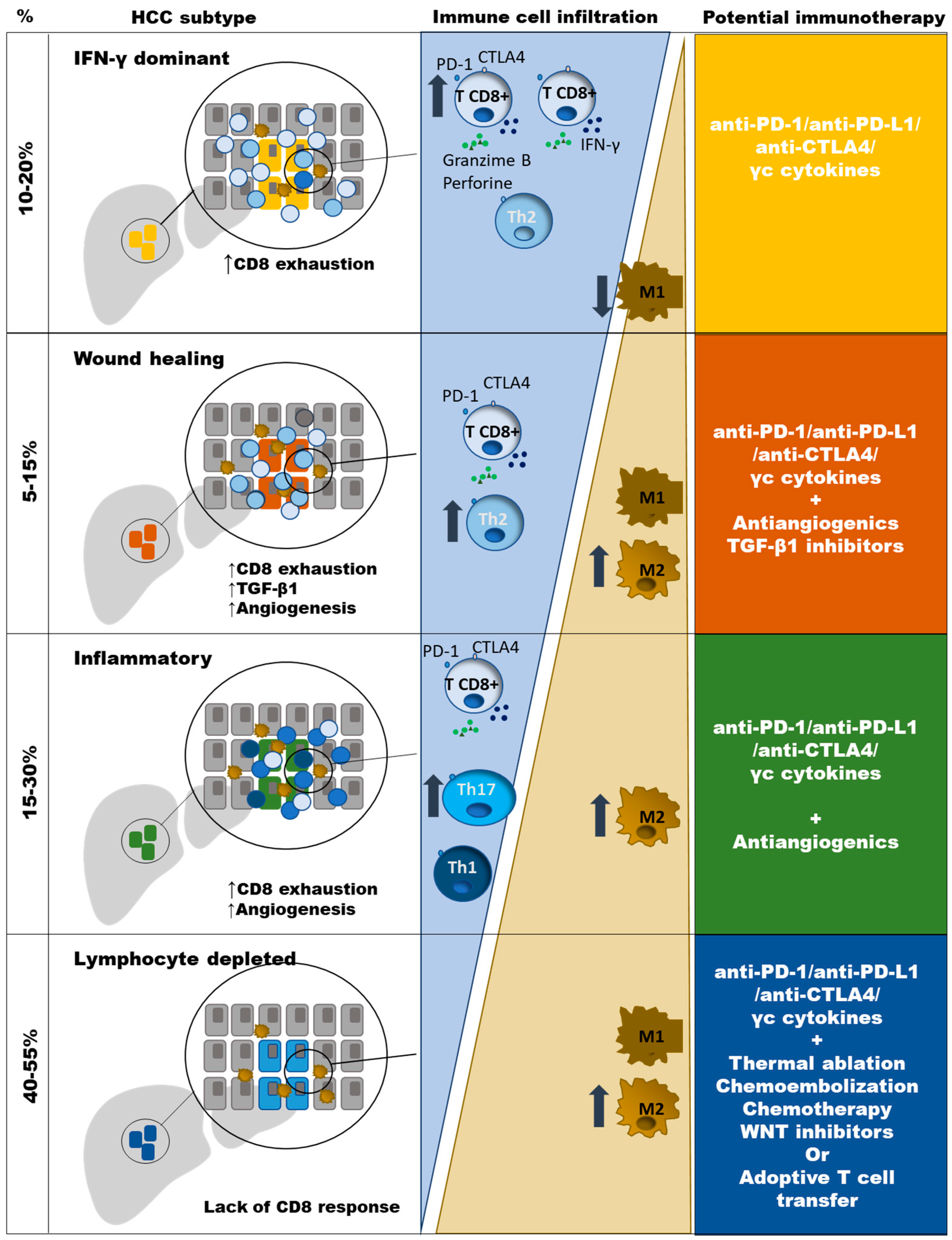
2. Types of HCC According to Potential Sensitivity to PD-1 Based Immunotherapy

HCC is an inflammation-driven disease induced by chronic viral hepatitis (hepatitis B (HBV) and C (HCV) viruses) and non-viral “sterile” inflammation (alcohol abuse, non-alcoholic steatohepatitis (NASH) and other rare metabolic disorders, such as hemochromatosis or α-1 antitrypsin deficiency) [14]. Despite the different etiologies, the immune compositions are similar between HBV/HCV infected and virus negative tumors, apart from activated M2 macrophages that are observed mainly in virus-related HCC [15]. Nevertheless, the HCC-specific CD8 T cell response could be weaker in NASH-related HCC than in HCC associated with hepatitis virus infection [16], which could impact on immunotherapeutic strategies.
Previous extensive immunogenomic analysis of different type of tumors have shown diverse immune signatures. Based on these clusters, HCC can be classified according to the features of the induced immune response. Those non-immunogenic tumors are characterized by lymphocyte depletion or being immunologically quiet. Immunotolerant cancers are featured by the phenotypes “wound healing”, inflammatory and tumor growth factor (TGF)-β1 dominant. The immunogenic tumors comprise those cases that are interferon (IFN)-γ dominant [17]. The lymphocyte depleted cluster includes around 40–55% of HCC cases and are featured by M2 macrophage infiltration, moderate genomic heterogenicity, and high level of mutations in the catenin–β1 gene (CTNNB1). The “wound healing” HCCs are around 5–15% of cases and have a balance between macrophages and lymphocytes, high genomic heterogenicity, and mutations in the tumor suppressor gene TP53. The IFNγ dominant HCCs comprise around 10–20% of cases and are rich in CD8+ T cells and M1 macrophages with high PD-1 expression and T cell receptor (TCR) diversity. The HCC inflammatory type includes around 15–30% of cases and is featured by T helper (Th)17 infiltration, high Th1 response, low genomic heterogenicity, and high PD-L1 expression. Finally, the TGF-β1 dominant and the immunologically quiet clusters are poorly represented in HCC [17,18]. These different phenotypes should be considered to adapt the strategies to boost the immune response, as will be discussed at the end of this review (Figure 1). The detection of immunogenic HCC could be a key step to select those cases prone to respond to ICI. In those non-immune cases, strategies to induce a CD8+ T cell tumoral infiltration should be taken into consideration.
The liver is an immunotolerant environment due to its need to deal with the exposure to many gut-derived exoantigens [19]. This special liver characteristic could facilitate the immune evasion in HCC, inducing either a defective T cell response or its physical deletion [20], linked to the induction of inhibitory IC such as PD-1 and its ligands [21,22]. Nevertheless, at HCC diagnosis not all tumors show data of complete HCC immune suppression. Trying to stratify the type of tumor that could respond to PD-1/PDL-1 manipulation, researchers have described another classification based on four scenarios according to PD-L1 expression. Type I tumors display tumor infiltrating lymphocytes (TIL) and PD-L1 expression on tumor cells. These features would suggest that an adaptive immune resistance is ongoing. Type II cases neither express PD-L1 nor have TIL, which indicates lack of immune response. Type III tumors express PD-L1 but without TIL, indicating an intrinsic induction of PD-L1. Finally, type IV tumors are PD-L1 negative but with TIL, suggesting that in these cases other mechanisms are involved in the development of intra-tumoral T cell exhaustion [23]. HCC patients with PD-L1 expression in the tumor have poorer overall survival (OS) than PD-L1 negative cases [24], but this can also be a marker of higher CD8 infiltration and higher survival after treatment [25]. In a recent metanalysis, PD-L1 positive patients had a risk of death higher than the double with respect to PD-L1 negative cases [26]. Around 20% of HCC expresses PD-L1 in more than 1% of tumor cells, and this correlates with a worse progression [27]. Fortunately, in the anti-PD-1 clinical trials with nivolumab and pembrolizumab (ChekMate-40 and KEYNOTE-224), those cases with higher PD-L1 expression had a higher response rate with a longer OS, but some patients with lower PD-L1 expression also responded to this treatment [28,29,30]. This response in negative cases could be due to biopsy sampling error because of the heterogenicity of PD-L1 expression in the HCC, since in resected HCC, PD-L1 detection has been reported in more than 80% of cases [31]. Consequently, this variable tested in a liver biopsy could not be useful to decide which patients should receive treatment to block the PD1/PD-L1 pathway, although it could be used as a prognosis marker. Consequently, high PD-L1 expression could be a marker of worse progression but also a prognosis factor for better treatment response. In any case, the role of PD-L1 expression to plot a therapy based on anti-PD-1 treatment is not clear yet and it must be clarified in future studies.
Another specific analysis of gene expression profiles from tumor, stromal, and immune cells in a big cohort of HCC showed that 25% of patients have a genetic signature related to the induction of an immune response [32]. These cases displayed a high level of PD-1 expression and markers of cytolytic activity. Within this “Immune Class” HCC, two different groups were described; those characterized by an effective response and those in whom there is an exhausted immune response that expressed many genes regulated by TGF-β1 [32]. These immune HCC types could correspond to the IFNγ dominant and the immunotolerant groups previously discussed in this section [17]. These distinct immunologic signatures correlate with different HCC outcomes. Those cases with an active immune response phenotype had a longer OS than those with an exhausted or non-detectable response [32]. The PD-1 up-regulation on this “Immune class” tumors could make them sensitive to ICI.
An important issue in HCC-specific CD8 T cell response induction is the immunogenic capacity of the potential HCC antigens. The T cell response against aberrant expression of host’s antigen, such as glypican-3, NY-ESO-1, MAGE-A1, and MAGE-A3 are not commonly efficiently induced in HCC and subsequently cannot be considered the backbone for immunotherapy [33]. Nevertheless, intrahepatic T cells targeting immunogenic tumoral neo-antigens display an exhausted phenotype that suggests tumor recognition [34]. Therefore, immunotherapeutic rescue of T cells targeting tumor neo-antigens could be the best approach to improve tumor control [35]. HCC has an intermediate level of neo-antigen expression, which could be an advantage for immunotherapeutic strategies. Consequently, the existence of neo-antigens could be a premise for the presence of sensitive HCC-specific CD8 T cells to ICI. Therefore, tumor mutational burden (TMB) could be a predictive biomarker for the efficacy of ICI therapy [36]. Specifically, HCC develops regularly neoantigens although less frequently than other tumors, such as melanoma or lung cancer [37]. Nevertheless, in HCC the frequency of cases with high TMB is low and does not correlate with the rate of predicted neo-antigens [38]. Therefore, the role of TMB as predictor of response to ICI is not clear yet.
According to these data, the analysis of the immune response could be a predictive factor for ICI success. In fact, in those cases with a more immunosuppressive microenvironment such as in virus-associated HCC, a higher PD-1 expression is found, and this could impact on ICI response [39]. Moreover, in the nivolumab trial, those cases with higher T cell reactive liver inflammation correlated with better response and longer OS [32]. We could summarize that a good candidate to restore HCC-specific CD8 T cell response by PD-1/PD-L1 blockade could be considered those tumors with high grade of IFN-γ-secreting TIL, high PD-L1 expression, low M2 macrophage infiltration, and presence of HCC neo-antigens.

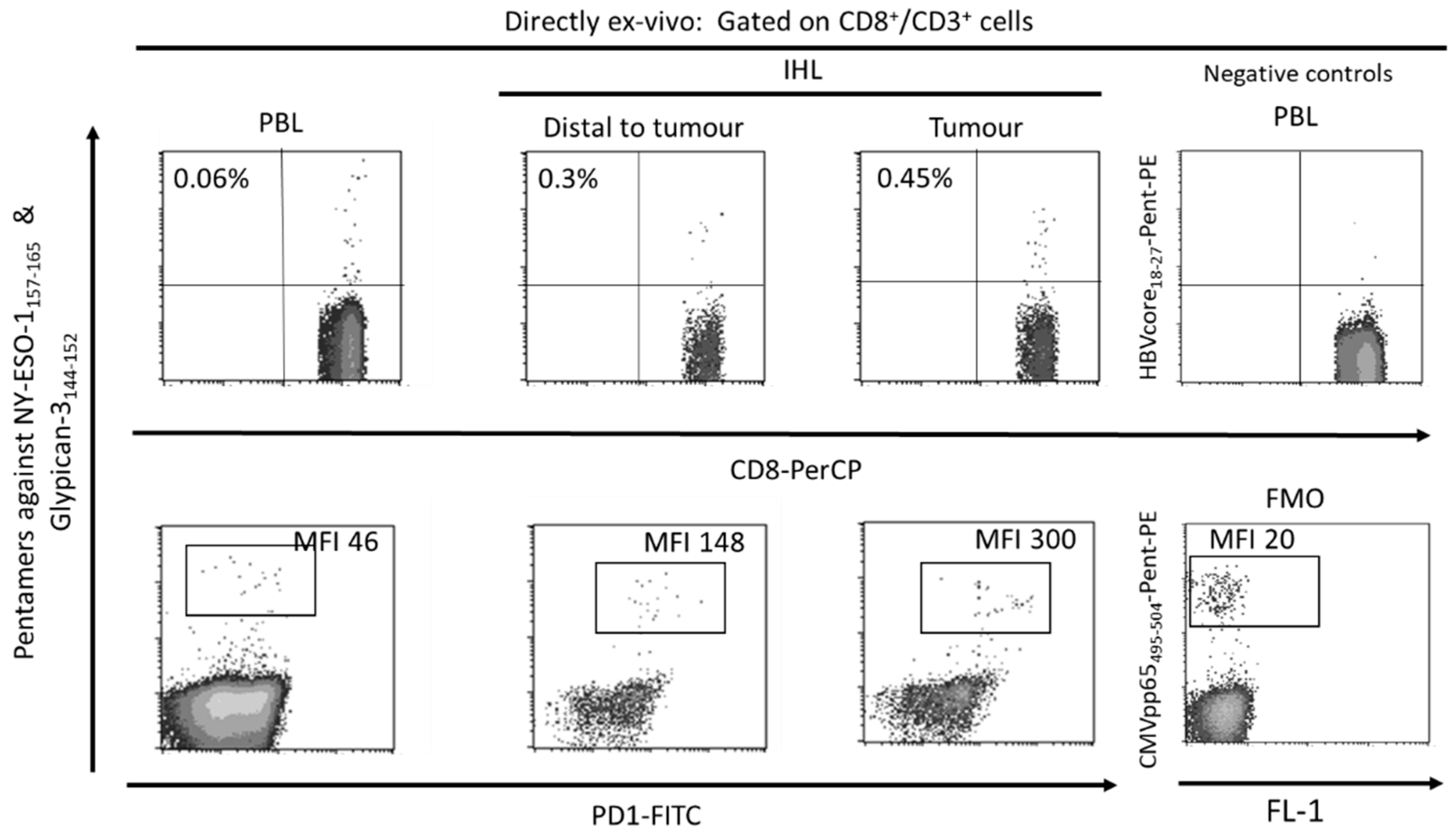
3. Role of PD-1-Expressing HCC-Specific CD8 T Cell Response in HCC Control

The tumor-associated antigen-specific CD8+ T cell response is essential for the control of solid tumors due to their ability to recognize tumor cells and to destroy them [40,41]. Exhausted CD8+ T cells are the main subset of TILs that perform anti-tumor effector functions [10,42]. Specifically, in HCC it is possible to find a high number of T cells infiltrating the tumor (Figure 2) and there is a correlation between the density of infiltrating lymphocytes and the prognosis of the disease [43]. Furthermore, after loco-regional treatment of HCC, a longer survival has been observed in those subjects in whom a specific CD8+ T response against HCC associated antigens is detected [44]. Therefore, large numbers of CD8+ TILs in HCC correlate with improved OS, longer relapse-free survival, and diminished disease progression [10,45,46]. HCC associated antigens comprise a heterogeneous set of autologous proteins that are rendered immunogenic in tumors by mutation [35] or aberrant expression [10,47] (Figure 2). The development of an effector cytotoxic T cell immune response against these antigens may provide a barrier to tumor progression. Nevertheless, most of the T cells targeting autologous aberrantly expressed epitopes are deleted or in a tolerogenic state [33,48], although some studies have shown a positive effect after PD-1 blockade in T cells targeting these kind of antigens [7]. Anyhow, the response to ICI could be more effective in T cells targeting HCC neo-antigens. A previous study has suggested that searching for host’s human leukocyte antigen (HLA) restricted tumoral neoantigens will permit to find T cells that after the appropriate treatment will exert tumor control [35]. HCC will commit mutations that will give rise to new epitopes that will be recognized as exogen antigens by T cells. Unfortunately, these cells will become exhausted during HCC progression, not being able to keep the tumor under control. These exhausted CD8+ T cells show a gene signature that is completely different from those of memory and effector T cells [49,50,51]. During the exhaustion process, T cell response progressively loses the effector functions and finally undergoes apoptosis [11,52]. The exhausted CD8 T cells are featured by the expression of different inhibitory IC, such as PD-1, cytotoxic T lymphocyte antigen-4 (CTLA-4), T cell immunoglobulin domain and mucin domain (Tim3), and lymphocyte-activation gene 3 (LAG3) that can be modulated to rescue T cell reactivity [21,53]. The ligands of these IC are expressed by resident liver cells. In fact, PD-L1 is expressed on hepatocytes [22], hepatic stellate cells [54], liver sinusoidal endothelial cell (LSEC) [55], and Kupffer cells [56,57], while PD-L2 expression is restricted to dendritic cells [58]. PD-1 and PD-L1 upregulation promotes CD8+ T-cell apoptosis and postoperative recurrence in hepatocellular carcinoma patients [59]. The in-vitro treatment of these T cells with antibodies against these myriad of inhibitory IC increases T cell proliferation and cytokine secretion.
Nevertheless, these exhausted CD8+ cells develop epigenetic imprints that steadily maintain the functional impairment, which is transmitted to the progeny, making temporary the effects of immunomodulatory treatments [49]. Even in the context of a PD-1 knockdown HCC-specific CD8 T cell model, although a tumor killing enhancement is initially observed, this effect is limited by compensatory engagement of alternative co-inhibitory and senescence programs upon repetitive antigen stimulation [60]. These epigenetic imprints induce the expression of specific transcription factors, such as the thymocyte selection-associated high mobility group box protein (TOX) that is up-regulated on CD8+ T cells in HCC and promotes their exhaustion by regulating endocytic recycling of PD-1 [61]. Additionally, the up-regulation of the ligands of these negative IC is induced in the HCC microenvironment. The regulation of PD-L1 expression could be related with the level of M2 macrophages that are recruited by the tumor cell-intrinsic osteopontin secretion. In HCC animal models, the level of M2 macrophages decreases after osteopintin blockade, which correlates with an increased CD8 effector response to PD-L1 blockade [62]. M2-like macrophages favor an immunosuppressive landscape by depleting CD8 T cells and inducing CD4+ T regulatory cells (Tregs) [63]. Additionally, M1 macrophages can induce PD-L1 expression on hepatocarcinoma cells by IL-1β effect [64]. These data suggest an important role of macrophages in modulating CD8 T cell exhaustion in HCC by promoting a immunotolerant environment [65] and by up-regulating themselves both PD-L1 and PD-L2 expression [66]. However, PD-L1 expression on macrophages could also have a positive input because it correlates with CD8 T cell infiltration and increased OS after treatment [67], probably in the case of M1-like macrophage infiltration [17]. Moreover, HCC-specific CD8 T cell itself induces PD-L1 up-regulation on hepatocytes, linked to a subsequent impairment of IFN-γ secretion by T cells [68]. There is a gradient of PD-1 expression on intra-tumoral CD8+ cells, which probably defines the progenitor (PD-1dim) and effector (PD-1high) pools [69]. The PD-1high population can co-express positive co-stimulatory checkpoints, such as 4-1BB, that can be triggered to rescue these cells from exhaustion [70,71]. Nevertheless, to target the PD-1dim subset could be more operative to get a more efficient response to ICI. The PD-1 level of the intrahepatic resident CD8+ cells inversely correlates with the expression of the transcription factor T-bet [72], which has been correlated with a late dysfunctional phenotype in the PD-1high pool, since the effector late dysfunctional pool expresses the transcription factor Eomes and loses T-bet expression [73]. On the contrary, PD-1dim expression is a marker of the progenitor pool, which is the subset that provides the proliferative burst after anti-PD-1 treatment [69]. Therefore, T cell restoration should focus on PD-1dim CD8+ T cells. In order to improve both the precursor and the effector pools, searching for PD-1/PD-L1 based combinatory therapies, such as modulation of suppressive soluble mediators (interleukin (IL)-10, IL-17, TGF-β1), blocking suppressive cells (Tregs, myeloid derived suppressor cells (MDSC), M2 tumor associated macrophages (TAM)), triggering positive co-stimulation (tumor necrosis factor receptor superfamily member 9, 4-1BB), mitochondrial metabolic reprogramming, impairing neo-angiogenesis, inducing expression of HCC neo-antigens or epigenetic modulation by gamma-chain (γc) cytokines [11,12,14,74] could improve the response to PD-1/PD-L1 blocking monotherapy. Figure 3 summarizes the potential mechanisms involved in HCC-specific CD8 T cell impairment.

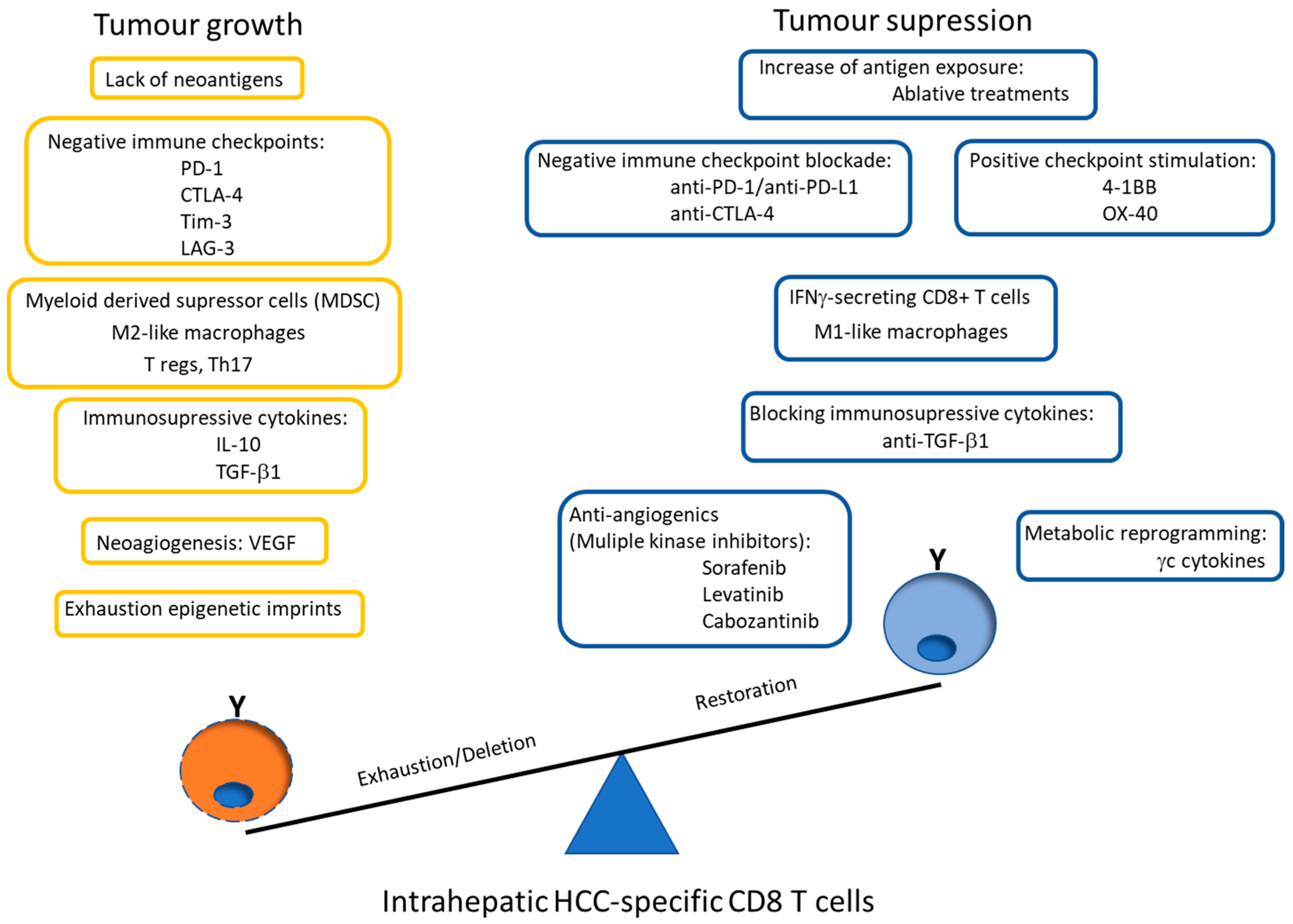
4. Targeting the Progenitor HCC-Specific CD8 T Cell Pool for Immunomodulation

Unlocking the potential of immunotherapy requires the design of strategies that induce a potent functional memory T cell pool capable of protecting from recurrence by producing a non-exhausted effector subset [75]. The exhausted progenitor pool gives rise to the progeny probably by asymmetric cell division [76,77]. The progeny comprises two sub-populations: effector-like transitory cells and late dysfunctional pool [73]. The effector-like transitory cells are characterized by the expression certain IC, such as PD-1 and Tim-3, and are able to sustain certain degree of tumor control. These cells will differentiate into the late dysfunctional pool, which expresses high levels of multiple negative IC, lacks proliferative capacity, displays low polyfunctionality, but retains some killing capability and expresses the transcription factor Eomes. Due to the high PD-1 expression on these cells, they are not suitable for anti-PD-1 therapy [73]. The progenitor pool is featured by the expression of the transcription factor TCF-1 and it is comprised by the stem-like precursor and the precursor subsets. The stem-like precursor pool is tissue resident and expresses the chemokine receptor CXCR5. CD8+ CXCR5+ T cells strongly infiltrate HCC, and their infiltration has been suggested to predict a better prognosis linked to HCC cell death [78,79]. CXCR5 also mediates homing of B cells [80], which are also enriched in HCC and interacts with T cells, enhancing immune activation, and this finding is linked to better outcome [81]. Therefore, tertiary lymphoid structures enriched in CXCR5+ B cells and TCF1+ stem-like CD8+ T cells correlates with responses to immunotherapy [82]. The stem-like precursor pool differentiates into the precursor pool that is also TCF-1 positive and can dedifferentiate into the stem-like precursor but also give rise to the effector progeny. These precursors pools have self-renewal potential and a catabolic mitochondrial metabolism, based on fatty acid oxidation and oxidative phosphorylation which promotes long-survival of these T cells [83,84]. They express memory markers such as IL-7 receptor (CD127), and lower levels of PD-1. It is noteworthy to know that these PD-1dim precursor pool is the subset sensitive to the immunoregulatory strategies based on PD-1 blockade [85]. During persistent antigen stimulation, this subset develops an epigenetic signature to adapt to this high pressive antigenic landscape [50,51]. Although PD-1 based therapy should target the progenitor pool, this approach should be combined with the modulation of epigenetic changes [86] to improve current anti-PD-1/PD-L1 immunotherapies, since PD-1 blocking alone does not remodel the exhausted epigenetic profile [49], which would make temporary the response to anti-PD-1 treatment [60]. In this sense, γc cytokines could play a role in epigenetic and metabolic remodeling that should be explored [74,87].

5. PD-1 Mechanisms Involved in CD8 T Cell Exhaustion

ICI therapy has signified an outstanding breakthrough in Oncology in the last decade, with objective response rates of 15–20% in more than 10 different kind of tumors [88,89]. Blocking PD-1/PD-L1 pathway is the prototype of this therapy and it is the backbone of different combination therapies including other IC, cytotoxic chemotherapy, ablatives strategies, or molecular targeted therapy [13]. Recently, the mechanism of action of this inhibitory IC has been described [90,91]. PD-1 is a member of the CD28 family that, unlike the other CD28 family members, generates negative signals after TCR triggering [92]. PD-1 negatively signals by preferentially dephosphorylating CD28 by PD-1 recruited Shp2 phosphatases. In fact, CD28 expression is necessary for the recovery of T cells subjected to anti-PD-1 immunotherapy. PD-1/PD-L1 interaction dephosphorylates CD28 but not TCR. Therefore, the TCR triggering impairment observed in exhausted T cells during persistent antigenic stimulation is due to the loss of the positive CD28 co-stimulation. CD28 is a positive co-stimulatory signal involved in naïve and memory T cell activation upon TCR triggering. The PD-1dim stem-like precursor T cells express CD28, making these cells sensitive to PD-1 modulation while the generated progeny lacks the expression of this receptor, making less likely the response to PD-1 blocking [93]. In a cancer animal model, PD1-1 blockade did not restore anti-tumor T cell function in presence of CD28 blocking antibodies, linked to a tumor growth similar to the not-treated control group [90]. Besides this CD28 dependent PD-1 inhibitory mechanism, it has been recently reported that PD-1 can also inhibit T cell activation upon TCR triggering in absence of CD28 co-stimulation [94]. Therefore, PD-1 blocking could be also useful as a short-term strategy to rescue CD28 negative effector cells but, to obtain a more persistent response, it will probably be necessary to restore the functionality of CD28 positive memory T cells. All these data explain the basic mechanisms involved in the impairment of the reactivity in the exhausted T cell response after T cell triggering. Nevertheless, the exhaustion profile is linked to other T cell changes that should also be rescued in addition to restoring the TCR signaling [95]. This issue renders the possibility of searching for synergic treatments in combination with anti-PD-1 therapy to increase the OS in HCC.

6. Results of PD-1 Blocking Based Combination Therapy on CD8+ Cells in HCC

To personalize the immunotherapy, it is necessary to know the quality of the immune response that is fighting against HCC. As discussed in Section 2, around 10–20% of HCC has reactive CD8 T cells prone to respond to ICI (IFNγ dominant cluster). Another 40% displays an inflammatory immunotolerant phenotype (Wound healing and inflammatory clusters) featured by an imbalance between pro-tumoral and antitumoral responses (Figure 1). These cases could need other treatments besides ICI to boost immune response such as anti-angiogenics or TGF-β inhibitors. Finally, around 40–45% of cases are not immunoactive (Lymphocyte depleted cluster). This last group could need synergic strategies to favor intra-tumoral trafficking of CD8+ T cells. To reach this goal, ablation therapy, chemo/radioembolization, and chemotherapy are being tested to increase antigen exposure, and WNT inhibitors besides ICI [17,18], because these cold tumors are characterized by CTNNB1 and AXIN1 mutations in the WNT-β catenin pathway [32]. All these strategies are currently being explored, but in the future a more specific approach should be tried to focus these actions on the CD8+ progenitor pool to make the response sustainable. Consequently, these approaches should be also combined with therapies involved in dealing with the metabolic impairments linked to the exhaustion imprints of the progenitor tumor specific CD8+ T cells [96,97]. Finally, anti-PD-1/PD-L1 treatment can be applied simultaneously with these combinations to fulfil these objectives but can also be clinically useful as neoadjuvant therapy to increase the response to systemic therapies [98].

6.1. PD-1/PD-L1 Blockade Monotherapy

Nivolumab and pembrolizumab are the first humanized monoclonal antibodies (mAb) against PD-1 tested in HCC patients [99]. The initial data on objective response rate (ORR) and median OS for both nivolumab and pembrolizumab in patients previously treated with sorafenib were around 15% and 1 year, respectively, in the initial clinical trials (CheckMate 040 and KEYNOTE-224) [28,29]. This response rate would correspond with the reported frequency of cases with IFNγ dominant HCC cluster [17]. Nevertheless, subsequent phase 3 clinical trials, nivolumab vs. sorafenib as first line (CheckMate 459) and pembrolizumab vs. placebo as second line (KEYNOTE-240) did not demonstrate improvement in OS [100]. This lack of benefit could be due to the selection of patients that could include more cases with non-immune-type HCC but also could be the result of tumor resistance to PD-1 treatment in the initial responders after repetitive cycles of treatment. Although anti-PD-1/PD-L1 therapy changes the tumor microenvironment by increasing the infiltrating CD8 T cells with cytotoxic activity [30,101] and can improve CD8 T cell primming by dendritic cells [102], these effects could be temporary due to the induction of compensatory inhibitory pathways [60]. Other anti-PD-1/PD-L1 monotherapies as second-line after sorafenib failure have shown also promising results, such as durvalumab (anti-PD-L1) and cemiplimab (anti-PD-1) [18]. These PD-1/PD-L1 monotherapy protocols could be effective in those HCC case with an IFN-γ dominant CD8 T cell infiltrate but those cases without an immune-class phenotype could also benefit from ICI after additional synergic therapies. Moreover, in the initial responders the role of epigenetic imprints in the exhausted T cells should be considered, which could lead to the induction of compensatory inhibitory mechanisms and resistance during long-lasting therapy. Therefore, to increase the number of responders and to avoid the development of resistance to treatment, several combinatory strategies with synergic actions are currently underway.

6.2. PD-1/PD-L1 Blockade Plus Antiangiogenics

The angiogenesis inhibitors are the current basic treatment in advanced HCC [103]. Multi-kinase inhibitors (MKIs) may modulate antitumor immunity through both angiogenesis-dependent and independent mechanisms (Table 1). Potentially beneficial effects on anti-tumor immunity may result from increased M1 polarization of macrophages and stimulation of CD8 T cell function. Nevertheless, high dosage of the MKI may contribute to immune suppression in the tumor microenvironment [104].
The wound healing HCC type is very rich in the expression of angiogenic genes, which could be a rationale to use anti-angiogenics in combination with ICI to change the balance between CD8 response and HCC in this type of tumor. The tumor hypoxic environment promotes high VEGF intra-tumoral level, which induces immunosuppressive effects by primming negative immunomodulatory inflammatory cells and by impairing the maturation of dendritic cells [105,106]. Clinical trials combining these drugs with anti-PD-1/PD-L1 are ongoing [18]. VEGF inhibition or blocking its receptors increases intra-tumoral infiltration and survival of cytotoxic T lymphocytes and decreases regulatory T lymphocyte recruitment, resulting in a more favorable immune microenvironment for ICI antitumoral activity [107,108]. These data highlight the important correlation of HCC neo-angiogenesis and the induction of immunosuppression produced by the hypoxic intra-tumoral environment. Levatinib is a MKI targeting VEGFR 1–3, fibroblast growth factor receptor 1–4, platelet derived growth factor receptor, RET and KIT with similar anti-tumor activity to sorafenib [103]. In an HCC animal model, levatinib shows an immunomodulatory activity featured by increasing CD8+ cell tumor infiltration and by decreasing intra-tumoral M2 TAM population [109]. This effect leads to a synergy with nivolumab treatment, enhancing tumor regression, which was attenuated by CD8+ T cell depletion, highlighting the role of CD8+ T cells in the response to the combination levatinib plus nivolumab [110]. Levatinib plus pembrolizumab combination have been also been tested in a phase I clinical trial with tumor reduction in most patients [111] and, according to this encouraging result, a new phase 3 clinical trial is underway, comparing levatinib vs. levatinib plus pembrolizumab in naïve patients (NCT03713593). This positive synergic effect has also been described combining durvalumab (anti-PD-L1) with ramucirumab (anti-VEGFR 2), which displayed a particularly high anti-tumor response in patients with high PD-L1 expression [112]. In the same line, the IMbrave 150 trial has tested the combination of atezolimumab (anti-PD-L1) plus bevacizumab (anti-VEGF mAb) with one third of cases with radiological response that were maintained by most of the responders during follow up [113]. Cabozantinib is another MKI of c-Met and VEGFR-2, and also inhibits AXL and RET [103]. The treatment with cabozantinib increases the expression of HLA-I molecules on tumor cells, leading to a greater sensitivity to killing by specific CD8 T cells. Besides this effect, cabozantinib also decreases the tumor infiltration by Tregs and MDSCs and increases the frequency of M1 TAM, which all together will improve the CD8+ T cell response [13]. Sorafenib is the first MKI clinically used in HCC that acts on RAF and VEGFR-2 and -3. This drug in a low dose promotes the intra-tumoral CD8+ T cell response and increases the M1 polarization of TAM, but in a high dose sorafenib displays the opposite effect [104,114,115]. The dose-effect on T cell response with MKI must be addressed in the future to obtain the best synergy with anti-PD-1 treatment. The potential immunological effects of MKI are summarized in Table 1. To sum up, based on all these clinical and experimental data, several clinical trials testing the combination of antiangiogenic plus PD-1/PD-L1 inhibitor are underway [116]. These combinations yield synergic effect and they could be specially interesting in “Immune class” tumors without reactive IFN-γ secreting CD8 T cells (wound healing and inflammatory clusters).

6.3. PD-1/PD-L1 Blockade Plus Anti-TGF-β1

TGF-β1 promotes tumor cell proliferation and invasion [117] and attenuates tumor response to PD-L1 blockade by excluding T cells [118]. In half of the lymphocyte rich tumors with exhausted T cells, the TFG-β1 induced genes are up-regulated [18,32]. Although the TGF-β1 dominant cluster is poorly represented in HCC [17], TGF-β1 plays a role in HCC pathogenesis. Galunisertib is TGF-β1R inhibitor that is been tested as a single drug and in combination with sorafenib in a phase I-II clinical trial in HCC [119,120]. This molecule has also tested in combination with stereotactic body radiotherapy to promote T-cell antitumor immunity, showing an anti-HCC effect featured by an increase in the CD8 PD1+ TIGIT+ population [121]. This drug could be an attractive strategy to increase the effect of PD-1/PD-L1 blockade on exhausted T cells after showing ability in increasing intratumoral infiltration of PD-1-expressing cytotoxic T cells. In fact, fusion proteins targeting both TGF-β1 and anti-PD-L1 are underway [122].

6.4. PD-1/PD-L1 Blockade Plus Other Immune Checkpoint Modulators

HCC-specific CD8 T cells can express other inhibitory checkpoints that are also up-regulated as exhaustion compensatory mechanism after anti-PD-1/PD-L1 treatment. Therefore, strategies to combined blockade of different ICs could be effective to rescue these cells from exhaustion but increasing the risk of adverse events such as autoimmune phenomenon. CTLA-4 competes with more avidity than CD28 for the same ligands CD80 and CD86, transmitting an inhibitory signal to the cell, instead of the positive co-stimulation mediated by CD28. CTLA-4 is highly expressed by tumor infiltrating HCC-specific CD8 T cells and the level of expression inversely correlates with the effector function. The CTLA-4 blockade with blocking anti-CTLA4 mAb improved CD8 T cell reactivity [16]. Based on these translational data, CTLA-4 blockade with tremilimumab has been tested in clinical trials as monotherapy or combined with ablation therapy in pre-treated patients with an elevated disease control rate [123,124]. Another anti-CTLA-4 inhibitory mAb, ipilumumab, has been tested in the phase II CheckMate040 clinical trial in sorafenib pre-treated patients to enhance the response against PD-1/PD-L1 blockade with a median OS of 24 months and with a 34% objective response rate and 5% cases with complete response, but has increased the rate of adverse events with respect to the arms without anti-CTLA-4 [125].
The effect of in-vitro blocking of other inhibitory ICs on HCC-specific CD8 T cells have already been tested with encouraging results. Blocking mAbs against TIM3 and LAG3 in association with anti-PD-L1 restore HCC-specific CD8 T cell response with synergic effect [21,53,121]. Nevertheless, these combinations have not been assayed in-vivo. Another strategy to boost HCC-specific CD8 T cells could be to associate the stimulation of positive co-stimulatory receptors, such as 4-1BB. In this line, a fusion protein containing PD1 and 4-1BBL has shown to block the PD-1/PD-L1 interaction and to trigger 4-1BB in HCC-specific CD8+ T cells in an HCC animal model, enhancing antitumor immune response and long-lasting tumor regression [71]. This same strategy has been applied in a transgenic HCC mouse model combining anti-PD-1 blockade with the synergic stimulation of two positive co-stimulatory molecules (4-1BB and tumor necrosis factor receptor superfamily, member 4 (OX40)). The triple combination extended the survival of mice bearing hepatocellular carcinoma in a CD8-dependent fashion [126].

6.5. PD-1/PD-L1 Blockade Plus Strategies to Increase HCC Antigen Presentation

In those lymphocyte depleted HCCs, strategies to release neo-antigens could have a positive effect in the induction of an HCC-specific CD8 T cell response. To achieve this goal, systemic chemotherapy, thermal ablation, or chemoembolization could be used. In an animal HCC model the combination of microwave ablation plus anti-PD-1/anti-CTLA-4 enhanced intra-tumoral infiltration of cytotoxic T lymphocytes and extended survival [127]. Although chemotherapy agents are generally considered immunosuppressive, these treatments through tumor cell death promotes the releasing of HCC neo-antigens that could let the priming of HCC-specific CD8 T cell response. The combination of cytotoxic agents (FOLFOX4 or GEMOX) plus anti-PD-1 (camrelizumab) in naïve HCC patients showed an overall rate response of 26.5% [128]. Currently, clinical trials to check anti-PD1 plus chemoembolization (NCT03572582), radioembolization (NCT03033446, NCT03099564), and stereotactic body radiation therapy (NCT03316872) are underway.

6.6. PD-1/PD-L1 Blockade Plus γc Cytokines

Along with these synergic strategies to increase the number of sensitive cases and to enhance the response to ICI, other approaches must be considered to direct these therapies to improve the HCC-specific progenitor pool. The addition of survival cytokines involved in memory differentiation and with the ability of metabolism reprogramming [129] would favor a healthier precursor pool to sustain a long-lasting efficient effector subset, able to keep HCC under control. IL-15 and IL-21 have shown promising in vitro results, favoring the development of HCC-specific stem-cell memory and central memory cells [130]. Additionally, IL-15 in a HCC animal model promotes tumor control, based on the long-term expansion of tumor-specific CD8 T cells, increasing IFNγ secretion and cytotoxicity, and also decreasing the expression of co-inhibitory molecules on dendritic cells [131]. Intra-tumoral IL-15 can also enable T cells to eliminate cancer cells lacking cognate antigens, which could be especially interesting in those lymphocyte-depleted HCCs [132]. Moreover, those HCCs with IL-21-secreting CD8 CXCR5 T cells develop a better outcome than cases without this type of cell [78]. The combination of PD-1/PD-L1 blockade with these γ-chain cytokines involved in precursor CD8 T cell generation [133] has not yet been explored in HCC but it has shown promising results in a cancer animal model [134].

6.7. PD-1/PD-L1 Blockade Plus Other Immunotherapies

Adoptive T cell transfer (ACT) therapy of TCR-engineered T cells [135,136], chimeric antigen receptor T cells (CAR-T) [137], cytokine-induced killer cells (CIK) [138], or ex-vivo restored T cells [35] could benefit from combined anti-PD-1 treatment to sustain the response of the transferred anti-tumoral T cells. The anti-HCC vaccines, both peptide- and dendritic cell-based vaccines, could also benefit from anti-PD-1 combined therapy [139]. Finally, the oncolytic virotherapy could also improves its results by adding anti-PD-1 treatment. This therapy is a novel approach based in the lyse of tumor cells by a vaccina virus that promotes the release of tumor antigens that will trigger an anti-tumoral immune response, which could be resistant to exhaustion by blocking the PD-1/PD-L1 pathway [140].

7. Conclusions

The antigen-specific CD8 T cell response against HCC-neoantigens carries out a key role in HCC control. Nevertheless, these cells become exhausted, being unable to keep tumor cells under control. These exhausted cells are featured by the expression of several negative regulatory checkpoints. PD-1 is the prototype of negative immune checkpoint and its blockade has been shown to rescue T cells from exhaustion in chronic viral infections and tumors. The HCC-specific CD8 T cells can be either exhausted in different grade or being inexistent according to the HCC immune class. The IFN-γ dominant HCC cluster displays exhausted T cells prone to respond to single PD-1/PD-L1 blockade. The immunotolerant types (wound healing and inflammatory clusters) will need the combination of PD-1/PD-L1 blocking plus other strategies to change the balance between pro-tumoral and anti-tumoral responses. The combination with anti-angiogenic drugs has shown promising results in this setting. Finally, the lymphocyte depleted tumors could need the association of lytic treatments to increase the exposure to neo-antigens to induce an HCC-specific CD8+ T cell response. To get this goal, the use of ablative treatments or chemotherapy could have an important role. Besides these strategies, new approaches focused on directing these treatments to improve the precursor pool of HCC-specific CD8 T cells could make long-lasting the response to these PD-1 based combination therapies. The progressive better understanding of the immune tumoral microenvironment and the mechanisms involved in the CD8 T cell exhaustion will probably permit soon to improve the overall survival of advanced HCC patients. Probably, PD-1/PD-L1 blockade-based combination therapies will be the backbone of these strategies to increase duration of response and number of responders.

Author Contributions

The following tasks were carried out as follows: Conceptualization J.P.-A. and J.-R.L.; writing—original draft preparation, J.-R.L.; writing—review and editing, J.P.-A., H.C., M.T., J.M., E.S.-d.-V., J.P.-A. and J.-R.L.; funding acquisition, J.-R.L. All authors have read and agreed to the published version of the manuscript.

Funding

This work was sponsored by the “Instituto de Salud Carlos III” (ISCIII), Spain, through the “State Plan for Scientific and Technical Research and Innovation 2017–2020”, grant PI19/00206 (JRL), co-funded by the European Regional Development Fund (ERDF), E.U. (A way to make Europe) and by the Gilead Fellowship Programme, grants GLD14/00217 (JRL) and GLD16/00014 (JRL).

Conflicts of Interest

The authors declare no conflict of interest. The funders had no role in the design of the study; in the collection, analyses, or interpretation of data; in the writing of the manuscript, or in the decision to publish the results.

References

  1. European Association for the Study of the Liver. Electronic address eee, European Association for the Study of the L. EASL Clinical Practice Guidelines: Management of hepatocellular carcinoma. J. Hepatol. 2018, 69, 182–236. [Google Scholar] [CrossRef] [Green Version]
  2. Hartke, J.; Johnson, M.; Ghabril, M. The diagnosis and treatment of hepatocellular carcinoma. Semin. Diagn. Pathol. 2017, 34, 153–159. [Google Scholar] [CrossRef]
  3. Li, S.; Yang, F.; Ren, X. Immunotherapy for hepatocellular carcinoma. Drug Discov. Ther. 2015, 9, 363–371. [Google Scholar] [CrossRef]
  4. Mellman, I.; Coukos, G.; Dranoff, G. Cancer immunotherapy comes of age. Nature 2011, 480, 480–489. [Google Scholar] [CrossRef] [PubMed]
  5. El Dika, I.; Khalil, D.N.; Abou-Alfa, G.K. Immune checkpoint inhibitors for hepatocellular carcinoma. Cancer 2019, 125, 3312–3319. [Google Scholar] [CrossRef] [PubMed]
  6. Freeman, G.J.; Wherry, E.J.; Ahmed, R.; Sharpe, A.H. Reinvigorating exhausted HIV-specific T cells via PD-1–PD-1 ligand blockade. J. Exp. Med. 2006, 203, 2223–2227. [Google Scholar] [CrossRef] [Green Version]
  7. Guo, X.; Jiang, H.; Shi, B.; Zhou, M.; Zhang, H.; Shi, Z.; Du, G.; Luo, H.; Wu, X.; Wang, Y.; et al. Disruption of PD-1 Enhanced the Anti-tumor Activity of Chimeric Antigen Receptor T Cells Against Hepatocellular Carcinoma. Front. Pharmacol. 2018, 9, 1118. [Google Scholar] [CrossRef] [PubMed]
  8. Topalian, S.L.; Hodi, F.S.; Brahmer, J.R.; Gettinger, S.N.; Smith, D.C.; McDermott, D.F.; Powderly, J.D.; Carvajal, R.D.; Sosman, J.A.; Atkins, M.B.; et al. Safety, Activity, and Immune Correlates of Anti–PD-1 Antibody in Cancer. N. Engl. J. Med. 2012, 366, 2443–2454. [Google Scholar] [CrossRef]
  9. Brahmer, J.R.; Tykodi, S.S.; Chow, L.Q.M.; Hwu, W.-J.; Topalian, S.L.; Hwu, P.; Drake, C.G.; Camacho, L.H.; Kauh, J.; Odunsi, K.; et al. Safety and Activity of Anti–PD-L1 Antibody in Patients with Advanced Cancer. N. Engl. J. Med. 2012, 366, 2455–2465. [Google Scholar] [CrossRef] [Green Version]
  10. Flecken, T.; Schmidt, N.; Hild, S.; Gostick, E.; Drognitz, O.; Zeiser, R.; Schemmer, P.; Bruns, H.; Eiermann, T.; Price, D.A.; et al. Immunodominance and functional alterations of tumor-associated antigen-specific CD8 + T-cell responses in hepatocellular carcinoma. Hepatology 2014, 59, 1415–1426. [Google Scholar] [CrossRef] [Green Version]
  11. McLane, L.M.; Abdel-Hakeem, M.S.; Wherry, E.J. CD8 T Cell Exhaustion During Chronic Viral Infection and Cancer. Annu. Rev. Immunol. 2019, 37, 457–495. [Google Scholar] [CrossRef] [Green Version]
  12. Zarour, H.M. Reversing T-cell Dysfunction and Exhaustion in Cancer. Clin. Cancer Res. 2016, 22, 1856–1864. [Google Scholar] [CrossRef] [PubMed] [Green Version]
  13. Cheng, A.-L.; Hsu, C.; Chan, S.L.; Choo, S.-P.; Kudo, M. Challenges of combination therapy with immune checkpoint inhibitors for hepatocellular carcinoma. J. Hepatol. 2020, 72, 307–319. [Google Scholar] [CrossRef] [PubMed] [Green Version]
  14. Ringelhan, M.; Pfister, D.; O’Connor, T.; Pikarsky, E.; Heikenwalder, M. The immunology of hepatocellular carcinoma. Nat. Immunol. 2018, 19, 222–232. [Google Scholar] [CrossRef]
  15. Cancer Genome Atlas Research Network. Electronic address wbe, Cancer Genome Atlas Research N. Comprehensive and Integrative Genomic Characterization of Hepatocellular Carcinoma. Cell 2017, 169, 1327–1341. [Google Scholar] [CrossRef] [PubMed] [Green Version]
  16. Inada, Y.; Mizukoshi, E.; Seike, T.; Tamai, T.; Iida, N.; Kitahara, M.; Yamashita, T.; Arai, K.; Terashima, T.; Fushimi, K.; et al. Characteristics of Immune Response to Tumor-Associated Antigens and Immune Cell Profile in Patients with Hepatocellular Carcinoma. Hepatology 2019, 69, 653–665. [Google Scholar] [CrossRef] [PubMed] [Green Version]
  17. Thorsson, V.; Gibbs, D.L.; Brown, S.; Wolf, D.; Bortone, D.S.; Ouyang, T.-H.; Porta-Pardo, E.; Gao, G.F.; Plaisier, C.L.; Eddy, J.A.; et al. The Immune Landscape of Cancer. Immunity 2018, 48, 812–830.e14. [Google Scholar] [CrossRef] [PubMed] [Green Version]
  18. Hilmi, M.; Neuzillet, C.; Calderaro, J.; Lafdil, F.; Pawlotsky, J.-M.; Rousseau, B. Angiogenesis and immune checkpoint inhibitors as therapies for hepatocellular carcinoma: Current knowledge and future research directions. J. Immunother. Cancer 2019, 7, 1–13. [Google Scholar] [CrossRef]
  19. Roth, G.S.; Decaens, T. Liver immunotolerance and hepatocellular carcinoma: Patho-physiological mechanisms and therapeutic perspectives. Eur. J. Cancer 2017, 87, 101–112. [Google Scholar] [CrossRef]
  20. Jiang, Y.; Li, Y.; Zhu, B. T-cell exhaustion in the tumor microenvironment. Cell Death Dis. 2015, 6, e1792. [Google Scholar] [CrossRef] [PubMed] [Green Version]
  21. Kim, H.-D.; Song, G.-W.; Park, S.; Jung, M.K.; Kim, M.H.; Kang, H.J.; Yoo, C.; Yi, K.; Kim, K.H.; Eo, S.; et al. Association between expression level of PD1 by tumor-infiltrating CD8+ T cells and features of hepatocellular carcinoma. Gastroenterology 2018, 155, 1936.e17–1950.e17. [Google Scholar] [CrossRef]
  22. Mühlbauer, M.; Fleck, M.; Schütz, C.; Weiss, T.; Froh, M.; Blank, C.; Schölmerich, J.; Hellerbrand, C. PD-L1 is induced in hepatocytes by viral infection and by interferon-α and -γ and mediates T cell apoptosis. J. Hepatol. 2006, 45, 520–528. [Google Scholar] [CrossRef] [PubMed]
  23. Teng, M.W.L.; Ngiow, S.F.; Ribas, A.; Smyth, M.J. Classifying Cancers Based on T-cell Infiltration and PD-L1. Cancer Res. 2015, 75, 2139–2145. [Google Scholar] [CrossRef] [PubMed] [Green Version]
  24. Gao, Q.; Wang, X.-Y.; Qiu, S.-J.; Yamato, I.; Sho, M.; Nakajima, Y.; Zhou, J.; Li, B.-Z.; Shi, Y.-H.; Xiao, Y.-S.; et al. Overexpression of PD-L1 Significantly Associates with Tumor Aggressiveness and Postoperative Recurrence in Human Hepatocellular Carcinoma. Clin. Cancer Res. 2009, 15, 971–979. [Google Scholar] [CrossRef] [Green Version]
  25. Gabrielson, A.; Wu, Y.; Wang, H.; Jiang, J.; Kallakury, B.; Gatalica, Z.; Reddy, S.; Kleiner, D.; Fishbein, T.; Johnson, L.; et al. Intratumoral CD3 and CD8 T-cell Densities Associated with Relapse-Free Survival in HCC. Cancer Immunol. Res. 2016, 4, 419–430. [Google Scholar] [CrossRef] [Green Version]
  26. Gao, X.-S.; Gu, X.; Xiong, W.; Guo, W.; Han, L.; Bai, Y.; Peng, C.; Cui, M.; Xie, M. Increased programmed death ligand-1 expression predicts poor prognosis in hepatocellular carcinoma patients. OncoTargets Ther. 2016, 9, 4805–4813. [Google Scholar] [CrossRef] [Green Version]
  27. Gandini, S.; Massi, D.; Mandalà, M. PD-L1 expression in cancer patients receiving anti PD-1/PD-L1 antibodies: A systematic review and meta-analysis. Crit. Rev. Oncol. 2016, 100, 88–98. [Google Scholar] [CrossRef] [PubMed]
  28. El-Khoueiry, A.B.; Sangro, B.; Yau, T.; Crocenzi, T.S.; Kudo, M.; Hsu, C.; Kim, T.-Y.; Choo, S.-P.; Trojan, J.; Welling, T.H.; et al. Nivolumab in patients with advanced hepatocellular carcinoma (CheckMate 040): An open-label, non-comparative, phase 1/2 dose escalation and expansion trial. Lancet 2017, 389, 2492–2502. [Google Scholar] [CrossRef]
  29. Zhu, A.X.; Finn, R.S.; Edeline, J.; Cattan, S.; Ogasawara, S.; Palmer, D.; Verslype, C.; Zagonel, V.; Fartoux, L.; Vogel, A.; et al. Pembrolizumab in patients with advanced hepatocellular carcinoma previously treated with sorafenib (KEYNOTE-224): A non-randomised, open-label phase 2 trial. Lancet Oncol. 2018, 19, 940–952. [Google Scholar] [CrossRef]
  30. Sangro, B.; Melero, I.; Wadhawan, S.; Finn, R.S.; Abou-Alfa, G.K.; Cheng, A.-L.; Yau, T.; Furuse, J.; Park, J.-W.; Boyd, Z.; et al. Association of inflammatory biomarkers with clinical outcomes in nivolumab-treated patients with advanced hepatocellular carcinoma. J. Hepatol. 2020, 73, 1460–1469. [Google Scholar] [CrossRef]
  31. Yarchoan, M.; Xing, D.; Luan, L.; Xu, H.; Sharma, R.B.; Popovic, A.; Pawlik, T.M.; Kim, A.K.; Zhu, Q.; Jaffee, E.M.; et al. Characterization of the Immune Microenvironment in Hepatocellular Carcinoma. Clin. Cancer Res. 2017, 23, 7333–7339. [Google Scholar] [CrossRef] [Green Version]
  32. Sia, D.; Jiao, Y.; Martinez-Quetglas, I.; Kuchuk, O.; Villacorta-Martin, C.; de Moura, M.C.; Putra, J.; Camprecios, G.; Bassaganyas, L.; Akers, N.; et al. Identification of an Immune-specific Class of Hepatocellular Carcinoma, Based on Molecular Features. Gastroenterology 2017, 153, 812–826. [Google Scholar] [CrossRef] [PubMed] [Green Version]
  33. Tauber, C.; Schultheiss, M.; De Luca, R.; Buettner, N.; Llewellyn-Lacey, S.; Emmerich, F.; Zehe, S.; Price, D.A.; Neumann-Haefelin, C.; Schmitt-Graeff, A.; et al. Inefficient induction of circulating TAA-specific CD8+ T-cell responses in hepatocellular carcinoma. Oncotarget 2019, 10, 5194–5206. [Google Scholar] [CrossRef] [PubMed] [Green Version]
  34. Ma, J.; Zheng, B.; Goswami, S.; Meng, L.; Zhang, D.; Cao, C.; Li, T.; Zhu, F.; Ma, L.; Zhang, Z.; et al. PD1Hi CD8+ T cells correlate with exhausted signature and poor clinical outcome in hepatocellular carcinoma. J. Immunother. Cancer 2019, 7, 1–15. [Google Scholar] [CrossRef] [PubMed]
  35. Tran, E.; Turcotte, S.; Gros, A.; Robbins, P.F.; Lu, Y.-C.; Dudley, M.E.; Wunderlich, J.R.; Somerville, R.P.; Hogan, K.; Hinrichs, C.S.; et al. Cancer Immunotherapy Based on Mutation-Specific CD4+ T Cells in a Patient with Epithelial Cancer. Science 2014, 344, 641–645. [Google Scholar] [CrossRef]
  36. Chan, T.; Yarchoan, M.; Jaffee, E.; Swanton, C.; Quezada, S.; Stenzinger, A.; Peters, S. Development of tumor mutation burden as an immunotherapy biomarker: Utility for the oncology clinic. Ann. Oncol. 2019, 30, 44–56. [Google Scholar] [CrossRef]
  37. Alexandrov, L.B.; Nik-Zainal, S.; Wedge, D.C.; Aparicio, S.A.J.R.; Behjati, S.; Biankin, A.V.; Bignell, G.R.; Bolli, N.; Borg, A.; Børresen-Dale, A.-L.; et al. Signatures of mutational processes in human cancer. Nature 2013, 500, 415–421. [Google Scholar] [CrossRef] [Green Version]
  38. Luchini, C.; Bibeau, F.; Ligtenberg, M.; Singh, N.; Nottegar, A.; Bosse, T.; Miller, R.; Riaz, N.; Douillard, J.-Y.; Andre, F.; et al. ESMO recommendations on microsatellite instability testing for immunotherapy in cancer, and its relationship with PD-1/PD-L1 expression and tumour mutational burden: A systematic review-based approach. Ann. Oncol. 2019, 30, 1232–1243. [Google Scholar] [CrossRef] [Green Version]
  39. Lim, C.J.; Lee, Y.H.; Pan, L.; Lai, L.; Chua, C.; Wasser, M.; Lim, T.K.H.; Yeong, J.; Toh, H.C.; Lee, S.Y.; et al. Multidimensional analyses reveal distinct immune microenvironment in hepatitis B virus-related hepatocellular carcinoma. Gut 2019, 68, 916–927. [Google Scholar] [CrossRef] [Green Version]
  40. Martínez-Lostao, L.; Anel, A.; Pardo, J. How Do Cytotoxic Lymphocytes Kill Cancer Cells? Clin. Cancer Res. 2015, 21, 5047–5056. [Google Scholar] [CrossRef] [Green Version]
  41. Eynde, B.J.V.D.; van der Bruggen, P. T cell defined tumor antigens. Curr. Opin. Immunol. 1997, 9, 684–693. [Google Scholar] [CrossRef]
  42. Zheng, C.; Zheng, L.; Yoo, J.-K.; Guo, H.; Zhang, Y.; Guo, X.; Kang, B.; Hu, R.; Huang, J.Y.; Zhang, Q.; et al. Landscape of Infiltrating T Cells in Liver Cancer Revealed by Single-Cell Sequencing. Cell 2017, 169, 1342–1356.e16. [Google Scholar] [CrossRef] [PubMed] [Green Version]
  43. Wada, Y.; Nakashima, O.; Kutami, R.; Yamamoto, O.; Kojiro, M. Clinicopathological study on hepatocellular carcinoma with lymphocytic infiltration. Hepatology 1998, 27, 407–414. [Google Scholar] [CrossRef] [PubMed]
  44. Mizukoshi, E.; Yamashita, T.; Arai, K.; Sunagozaka, H.; Ueda, T.; Arihara, F.; Kagaya, T.; Yamashita, T.; Fushimi, K.; Kaneko, S. Enhancement of tumor-associated antigen-specific T cell responses by radiofrequency ablation of hepatocellular carcinoma. Hepatology 2013, 57, 1448–1457. [Google Scholar] [CrossRef] [Green Version]
  45. Kang, T.-W.; Yevsa, T.; Woller, N.; Hoenicke, L.; Wuestefeld, T.; Dauch, D.; Hohmeyer, A.; Gereke, M.; Rudalska, R.; Potapova, A.; et al. Senescence surveillance of pre-malignant hepatocytes limits liver cancer development. Nature 2011, 479, 547–551. [Google Scholar] [CrossRef]
  46. Chang, B.; Shen, L.; Wang, K.; Jin, J.; Huang, T.; Chen, Q.; Li, W.; Wu, P. High number of PD-1 positive intratumoural lymphocytes predicts survival benefit of cytokine-induced killer cells for hepatocellular carcinoma patients. Liver Int. 2018, 38, 1449–1458. [Google Scholar] [CrossRef] [Green Version]
  47. Schmidt, N.; Flecken, T.; Thimme, R. Tumor-associated antigen specific CD8+T cells in hepatocellular carcinoma-a promising target for immunotherapy. OncoImmunology 2014, 3, e954919. [Google Scholar] [CrossRef] [Green Version]
  48. Gehring, A.J.; Ho, Z.Z.; Tan, A.T.; Aung, M.O.; Lee, K.H.; Tan, K.C.; Lim, S.G.; Bertoletti, A. Profile of Tumor Antigen-Specific CD8 T Cells in Patients with Hepatitis B Virus-Related Hepatocellular Carcinoma. Gastroenterology 2009, 137, 682–690. [Google Scholar] [CrossRef]
  49. Pauken, K.E.; Sammons, M.A.; Odorizzi, P.M.; Manne, S.; Godec, J.; Khan, O.; Drake, A.M.; Chen, Z.; Sen, D.R.; Kurachi, M.; et al. Epigenetic stability of exhausted T cells limits durability of reinvigoration by PD-1 blockade. Science 2016, 354, 1160–1165. [Google Scholar] [CrossRef] [Green Version]
  50. Jadhav, R.R.; Im, S.J.; Hu, B.; Hashimoto, M.; Li, P.; Lin, J.-X.; Leonard, W.J.; Greenleaf, W.J.; Ahmed, R.; Goronzy, J.J. Epigenetic signature of PD-1+ TCF1+ CD8 T cells that act as resource cells during chronic viral infection and respond to PD-1 blockade. Proc. Natl. Acad. Sci. USA 2019, 116, 14113–14118. [Google Scholar] [CrossRef] [Green Version]
  51. Sen, D.R.; Kaminski, J.; Barnitz, R.A.; Kurachi, M.; Gerdemann, U.; Yates, K.B.; Tsao, H.-W.; Godec, J.; LaFleur, M.W.; Brown, F.D.; et al. The epigenetic landscape of T cell exhaustion. Science 2016, 354, 1165–1169. [Google Scholar] [CrossRef] [Green Version]
  52. Wherry, E.J.; Blattman, J.N.; Murali-Krishna, K.; Van Der Most, R.; Ahmed, R. Viral Persistence Alters CD8 T-Cell Immunodominance and Tissue Distribution and Results in Distinct Stages of Functional Impairment. J. Virol. 2003, 77, 4911–4927. [Google Scholar] [CrossRef] [Green Version]
  53. Zhou, G.; Sprengers, D.; Boor, P.P.; Doukas, M.; Schutz, H.; Mancham, S.; Pedroza-Gonzalez, A.; Polak, W.G.; de Jonge, J.; Gaspersz, M.; et al. Antibodies Against Immune Checkpoint Molecules Restore Functions of Tumor-Infiltrating T Cells in Hepatocellular Carcinomas. Gastroenterology 2017, 153, 1107–1119.e10. [Google Scholar] [CrossRef]
  54. Yu, M.-C.; Chen, C.-H.; Liang, X.; Wang, L.; Gandhi, C.R.; Fung, J.J.; Lu, L.; Qian, S. Inhibition of T-cell responses by hepatic stellate cells via B7-H1-mediated T-cell apoptosis in mice. Hepatology 2004, 40, 1312–1321. [Google Scholar] [CrossRef]
  55. Diehl, L.; Schurich, A.; Grochtmann, R.; Hegenbarth, S.; Chen, L.; Knolle, P.A. Tolerogenic maturation of liver sinusoidal endothelial cells promotes B7-homolog 1-dependent CD8+ T cell tolerance. Hepatology 2007, 47, 296–305. [Google Scholar] [CrossRef]
  56. Kassel, R.; Cruise, M.W.; Iezzoni, J.C.; Taylor, N.A.; Pruett, T.L.; Hahn, Y.S. Chronically inflamed livers up-regulate expression of inhibitory B7 family members. Hepatology 2009, 50, 1625–1637. [Google Scholar] [CrossRef] [PubMed] [Green Version]
  57. Wu, K.; Kryczek, I.; Chen, L.; Zou, W.; Welling, T.H. Kupffer Cell Suppression of CD8+ T Cells in Human Hepatocellular Carcinoma Is Mediated by B7-H1/Programmed Death-1 Interactions. Cancer Res. 2009, 69, 8067–8075. [Google Scholar] [CrossRef] [Green Version]
  58. Mocan, T.; Sparchez, Z.; Craciun, R.; Bora, C.N.; Leucuta, D.C. Programmed cell death protein-1 (PD-1)/programmed death-ligand-1 (PD-L1) axis in hepatocellular carcinoma: Prognostic and therapeutic perspectives. Clin. Transl. Oncol. 2019, 21, 702–712. [Google Scholar] [CrossRef]
  59. Shi, F.; Shi, M.; Zeng, Z.; Qi, R.-Z.; Liu, Z.-W.; Zhang, J.-Y.; Yang, Y.-P.; Tien, P.; Wang, F.-S. PD-1 and PD-L1 upregulation promotes CD8+ T-cell apoptosis and postoperative recurrence in hepatocellular carcinoma patients. Int. J. Cancer 2010, 128, 887–896. [Google Scholar] [CrossRef]
  60. Otano, I.; Escors, D.; Schurich, A.; Singh, H.; Robertson, F.; Davidson, B.R.; Fusai, G.; Vargas, F.A.; Tan, Z.M.; Aw, J.Y.; et al. Molecular Recalibration of PD-1+ Antigen-Specific T Cells from Blood and Liver. Mol. Ther. 2018, 26, 2553–2566. [Google Scholar] [CrossRef] [Green Version]
  61. Wang, X.; He, Q.; Shen, H.; Xia, A.; Tian, W.; Yu, W.; Sun, B. TOX promotes the exhaustion of antitumor CD8+ T cells by preventing PD1 degradation in hepatocellular carcinoma. J. Hepatol. 2019, 71, 731–741. [Google Scholar] [CrossRef]
  62. Zhu, Y.; Yang, J.; Xu, D.; Gao, X.-M.; Zhang, Z.; Hsu, J.L.; Li, C.-W.; Lim, S.-O.; Sheng, Y.-Y.; Zhang, Y.; et al. Disruption of tumour-associated macrophage trafficking by the osteopontin-induced colony-stimulating factor-1 signalling sensitises hepatocellular carcinoma to anti-PD-L1 blockade. Gut 2019, 68, 1653–1666. [Google Scholar] [CrossRef] [PubMed]
  63. Freitas-Lopes, M.A.; Mafra, K.; David, B.A.; Carvalho-Gontijo, R.; Menezes, G.B. Differential Location and Distribution of Hepatic Immune Cells. Cells 2017, 6, 48. [Google Scholar] [CrossRef] [Green Version]
  64. Zong, Z.; Zou, J.; Mao, R.; Ma, C.; Li, N.; Wang, J.; Wang, X.; Zhou, H.; Zhang, L.; Shi, Y. M1 Macrophages Induce PD-L1 Expression in Hepatocellular Carcinoma Cells Through IL-1β Signaling. Front. Immunol. 2019, 10, 1643. [Google Scholar] [CrossRef] [PubMed] [Green Version]
  65. Yun, J.; Yu, G.; Hu, P.; Chao, Y.; Li, X.; Chen, X.; Wei, Q.; Wang, J. PD-1 expression is elevated in monocytes from hepatocellular carcinoma patients and contributes to CD8 T cell suppression. Immunol. Res. 2020, 68, 436–444. [Google Scholar] [CrossRef]
  66. Yasuoka, H.; Asai, A.; Ohama, H.; Tsuchimoto, Y.; Fukunishi, S.; Higuchi, K. Increased both PD–L1 and PD–L2 expressions on monocytes of patients with hepatocellular carcinoma was associated with a poor prognosis. Sci. Rep. 2020, 10, 10377. [Google Scholar] [CrossRef] [PubMed]
  67. Liu, C.-Q.; Xu, J.; Zhou, Z.-G.; Jin, L.-L.; Yu, X.-J.; Xiao, G.; Lin, J.; Zhuang, S.-M.; Zhang, Y.-J.; Zheng, L. Expression patterns of programmed death ligand 1 correlate with different microenvironments and patient prognosis in hepatocellular carcinoma. Br. J. Cancer 2018, 119, 80–88. [Google Scholar] [CrossRef] [Green Version]
  68. Huang, C.-Y.; Wang, Y.; Luo, G.-Y.; Han, F.; Li, Y.-Q.; Zhou, Z.-G.; Xu, G.-L. Relationship Between PD-L1 Expression and CD8+ T-cell Immune Responses in Hepatocellular Carcinoma. J. Immunother. 2017, 40, 323–333. [Google Scholar] [CrossRef]
  69. Im, S.J.; Hashimoto, M.; Gerner, M.Y.; Lee, J.; Kissick, H.T.; Burger, M.C.; Shan, Q.; Hale, J.S.; Lee, J.; Nasti, T.H.; et al. Defining CD8+ T cells that provide the proliferative burst after PD-1 therapy. Nat. Cell Biol. 2016, 537, 417–421. [Google Scholar] [CrossRef]
  70. Kim, H.; Park, S.; Jeong, S.; Lee, Y.J.; Lee, H.; Kim, C.G.; Kim, K.H.; Hong, S.; Lee, J.; Kim, S.; et al. 4-1BB Delineates Distinct Activation Status of Exhausted Tumor-Infiltrating CD8 + T Cells in Hepatocellular Carcinoma. Hepatology 2020, 71, 955–971. [Google Scholar] [CrossRef] [Green Version]
  71. Zhang, Y.; Zhang, H.; Wei, M.; Mou, T.; Shi, T.; Ma, Y.; Cai, X.; Li, Y.; Dong, J.; Wei, J. Recombinant Adenovirus Expressing a Soluble Fusion Protein PD-1/CD137L Subverts the Suppression of CD8+ T Cells in HCC. Mol. Ther. 2019, 27, 1906–1918. [Google Scholar] [CrossRef]
  72. Chew, V.; Lai, L.; Pan, L.; Lim, C.J.; Li, J.; Chong, T.H.; Chua, C.; Leong, J.Y.; Lim, K.H.; Toh, H.C.; et al. Delineation of an immunosuppressive gradient in hepatocellular carcinoma using high-dimensional proteomic and transcriptomic analyses. Proc. Natl. Acad. Sci. USA 2017, 114, E5900–E5909. [Google Scholar] [CrossRef] [Green Version]
  73. Escobar, G.; Mangani, D.; Anderson, A.C. T cell factor 1: A master regulator of the T cell response in disease. Sci. Immunol. 2020, 5, eabb9726. [Google Scholar] [CrossRef]
  74. Abdelsamed, H.A.; Zebley, C.C.; Youngblood, B. Epigenetic Maintenance of Acquired Gene Expression Programs during Memory CD8 T Cell Homeostasis. Front. Immunol. 2018, 9, 6. [Google Scholar] [CrossRef] [PubMed]
  75. Reading, J.L.; Gálvez-Cancino, F.; Swanton, C.; Lladser, A.; Peggs, K.S.; Quezada, S.A. The function and dysfunction of memory CD8+T cells in tumor immunity. Immunol. Rev. 2018, 283, 194–212. [Google Scholar] [CrossRef]
  76. Borsa, M.; Barnstorf, I.; Baumann, N.S.; Pallmer, K.; Yermanos, A.; Gräbnitz, F.; Barandun, N.; Hausmann, A.; Sandu, I.; Barral, Y.; et al. Modulation of asymmetric cell division as a mechanism to boost CD8+ T cell memory. Sci. Immunol. 2019, 4, eaav1730. [Google Scholar] [CrossRef]
  77. Chen, Y.-H.; Kratchmarov, R.; Lin, W.-H.W.; Rothman, N.J.; Yen, B.; Adams, W.C.; Nish, S.A.; Rathmell, J.C.; Reiner, S.L. Asymmetric PI3K Activity in Lymphocytes Organized by a PI3K-Mediated Polarity Pathway. Cell Rep. 2018, 22, 860–868. [Google Scholar] [CrossRef] [Green Version]
  78. Ye, L.; Li, Y.; Tang, H.; Liu, W.; Chen, Y.; Dai, T.; Liang, R.; Shi, M.; Yi, S.; Chen, G.; et al. CD8+CXCR5+T cells infiltrating hepatocellular carcinomas are activated and predictive of a better prognosis. Aging 2019, 11, 8879–8891. [Google Scholar] [CrossRef]
  79. Jin, Y.; Lang, C.; Tang, J.; Geng, J.; Song, H.K.; Sun, Z.; Wang, J. CXCR5 + CD8 + T cells could induce the death of tumor cells in HBV-related hepatocellular carcinoma. Int. Immunopharmacol. 2017, 53, 42–48. [Google Scholar] [CrossRef]
  80. Panjideh, H.; Müller, G.; Koch, M.; Wilde, F.; Scheu, S.; Moldenhauer, G.; Lipp, M. Immunotherapy of B-cell non-Hodgkin lymphoma by targeting the chemokine receptor CXCR5 in a preclinical mouse model. Int. J. Cancer 2014, 135, 2623–2632. [Google Scholar] [CrossRef]
  81. Garnelo, M.; Tan, A.; Her, Z.; Yeong, J.; Lim, C.J.; Chen, J.; Lim, K.H.; Weber, A.; Chow, P.; Chung, A.; et al. Interaction between tumour-infiltrating B cells and T cells controls the progression of hepatocellular carcinoma. Gut 2017, 66, 342–351. [Google Scholar] [CrossRef] [Green Version]
  82. Cabrita, R.; Lauss, M.; Sanna, A.; Donia, M.; Larsen, M.S.; Mitra, S.; Johansson, I.; Phung, B.; Harbst, K.; Vallon-Christersson, J.; et al. Tertiary lymphoid structures improve immunotherapy and survival in melanoma. Nature 2020, 577, 561–565. [Google Scholar] [CrossRef]
  83. Adams, W.C.; Chen, Y.-H.; Kratchmarov, R.; Yen, B.; Nish, S.A.; Lin, W.-H.W.; Rothman, N.J.; Luchsinger, L.L.; Klein, U.; Busslinger, M.; et al. Anabolism-Associated Mitochondrial Stasis Driving Lymphocyte Differentiation over Self-Renewal. Cell Rep. 2016, 17, 3142–3152. [Google Scholar] [CrossRef]
  84. Lin, W.-H.W.; Nish, S.A.; Yen, B.; Chen, Y.-H.; Adams, W.C.; Kratchmarov, R.; Rothman, N.J.; Bhandoola, A.; Xue, H.-H.; Reiner, S.L. CD8 + T Lymphocyte Self-Renewal during Effector Cell Determination. Cell Rep. 2016, 17, 1773–1782. [Google Scholar] [CrossRef] [Green Version]
  85. Paley, M.A.; Kroy, D.C.; Odorizzi, P.M.; Johnnidis, J.B.; Dolfi, D.V.; Barnett, B.E.; Bikoff, E.K.; Robertson, E.J.; Lauer, G.M.; Reiner, S.L.; et al. Progenitor and Terminal Subsets of CD8+ T Cells Cooperate to Contain Chronic Viral Infection. Science 2012, 338, 1220–1225. [Google Scholar] [CrossRef] [Green Version]
  86. Ghoneim, H.E.; Fan, Y.; Moustaki, A.; Abdelsamed, H.A.; Dash, P.; Dogra, P.; Carter, R.; Awad, W.; Neale, G.; Thomas, P.G.; et al. De Novo Epigenetic Programs Inhibit PD-1 Blockade-Mediated T Cell Rejuvenation. Cell 2017, 170, 142–157.e19. [Google Scholar] [CrossRef] [Green Version]
  87. Van Der Windt, G.J.W.; Everts, B.; Chang, C.-H.; Curtis, J.D.; Freitas, T.C.; Amiel, E.; Pearce, E.J.; Pearce, E.L. Mitochondrial Respiratory Capacity Is a Critical Regulator of CD8+ T Cell Memory Development. Immunity 2012, 36, 68–78. [Google Scholar] [CrossRef] [Green Version]
  88. Callahan, M.K.; Postow, M.A.; Wolchok, J.D. Targeting T Cell Co-receptors for Cancer Therapy. Immunity 2016, 44, 1069–1078. [Google Scholar] [CrossRef]
  89. Hoos, A. Development of immuno-oncology drugs-from CTLA4 to PD1 to the next generations. Nat. Rev. Drug Discov. 2016, 15, 235–247. [Google Scholar] [CrossRef]
  90. Kamphorst, A.O.; Wieland, A.; Nasti, T.; Yang, S.; Zhang, R.; Barber, D.L.; Konieczny, B.T.; Daugherty, C.Z.; Koenig, L.; Yu, K.; et al. Rescue of exhausted CD8 T cells by PD-1–targeted therapies is CD28-dependent. Science 2017, 355, 1423–1427. [Google Scholar] [CrossRef] [Green Version]
  91. Hui, E.; Cheung, J.; Zhu, J.; Su, X.; Taylor, M.J.; Wallweber, H.A.; Sasmal, D.K.; Huang, J.; Kim, J.M.; Mellman, I.; et al. T cell costimulatory receptor CD28 is a primary target for PD-1–mediated inhibition. Science 2017, 355, 1428–1433. [Google Scholar] [CrossRef]
  92. Okazaki, T.; Chikuma, S.; Iwai, Y.; Fagarasan, S.; Honjo, T. A rheostat for immune responses: The unique properties of PD-1 and their advantages for clinical application. Nat. Immunol. 2013, 14, 1212–1218. [Google Scholar] [CrossRef] [Green Version]
  93. Tomiyama, H.; Matsuda, T.; Takiguchi, M. Differentiation of Human CD8+T Cells from a Memory to Memory/Effector Phenotype. J. Immunol. 2002, 168, 5538–5550. [Google Scholar] [CrossRef] [Green Version]
  94. Mizuno, R.; Sugiura, D.; Shimizu, K.; Maruhashi, T.; Watada, M.; Okazaki, I.-M.; Okazaki, T. PD-1 Primarily Targets TCR Signal in the Inhibition of Functional T Cell Activation. Front. Immunol. 2019, 10, 630. [Google Scholar] [CrossRef] [Green Version]
  95. Teijeira, A.; Garasa, S.; Etxeberria, I.; Gato-Cañas, M.; Melero, I.; Delgoffe, G.M. Metabolic Consequences of T-cell Costimulation in Anticancer Immunity. Cancer Immunol. Res. 2019, 7, 1564–1569. [Google Scholar] [CrossRef]
  96. Scharping, N.E.; Menk, A.V.; Moreci, R.S.; Whetstone, R.D.; Dadey, R.E.; Watkins, S.C.; Ferris, R.L.; Delgoffe, G.M. The Tumor Microenvironment Represses T Cell Mitochondrial Biogenesis to Drive Intratumoral T Cell Metabolic Insufficiency and Dysfunction. Immunity 2016, 45, 374–388. [Google Scholar] [CrossRef] [Green Version]
  97. Menk, A.V.; Scharping, N.E.; Rivadeneira, D.B.; Calderon, M.J.; Watson, M.J.; Dunstane, D.; Watkins, S.C.; Delgoffe, G.M. 4-1BB costimulation induces T cell mitochondrial function and biogenesis enabling cancer immunotherapeutic responses. J. Exp. Med. 2018, 215, 1091–1100. [Google Scholar] [CrossRef] [PubMed] [Green Version]
  98. Kaseb, A.O.; Pestana, R.C.; Vence, L.M.; Blando, J.M.; Singh, S.; Ikoma, N.; Vauthey, J.-N.; Allison, J.P.; Sharma, P. Randomized, open-label, perioperative phase II study evaluating nivolumab alone versus nivolumab plus ipilimumab in patients with resectable HCC. J. Clin. Oncol. 2019, 37, 185. [Google Scholar] [CrossRef]
  99. Moreno-Cubero, E.; Larrubia, J.-R. Specific CD8+T cell response immunotherapy for hepatocellular carcinoma and viral hepatitis. World J. Gastroenterol. 2016, 22, 6469–6483. [Google Scholar] [CrossRef]
  100. Finn, R.S.; Ryoo, B.-Y.; Merle, P.; Kudo, M.; Bouattour, M.; Lim, H.Y.; Breder, V.; Edeline, J.; Chao, Y.; Ogasawara, S.; et al. Pembrolizumab As Second-Line Therapy in Patients With Advanced Hepatocellular Carcinoma in KEYNOTE-240: A Randomized, Double-Blind, Phase III Trial. J. Clin. Oncol. 2020, 38, 193–202. [Google Scholar] [CrossRef]
  101. Kaseb, A.O.; Vence, L.; Blando, J.; Yadav, S.S.; Ikoma, N.; Pestana, R.C.; Vauthey, J.N.; Allison, J.P.; Sharma, P. Immunologic Correlates of Pathologic Complete Response to Preoperative Immunotherapy in Hepatocellular Carcinoma. Cancer Immunol. Res. 2019, 7, 1390–1395. [Google Scholar] [CrossRef]
  102. Zhang, W.; Song, Z.; Xiao, J.; Liu, X.; Luo, Y.; Yang, Z.; Luo, R.; Li, A. Blocking the PD-1/PD-L1 axis in dendritic cell-stimulated Cytokine-Induced Killer Cells with pembrolizumab enhances their therapeutic effects against hepatocellular carcinoma. J. Cancer 2019, 10, 2578–2587. [Google Scholar] [CrossRef] [PubMed] [Green Version]
  103. Berretta, M.; Rinaldi, L.; Di Benedetto, F.; Lleshi, A.; De Re, V.; Facchini, G.; De Paoli, P.; Di Francia, R. Angiogenesis Inhibitors for the Treatment of Hepatocellular Carcinoma. Front. Pharmacol. 2016, 7, 428. [Google Scholar] [CrossRef] [Green Version]
  104. Lin, Y.-Y.; Tan, C.-T.; Chen, C.-W.; Ou, D.-L.; Cheng, A.-L.; Hsu, C. Immunomodulatory Effects of Current Targeted Therapies on Hepatocellular Carcinoma: Implication for the Future of Immunotherapy. Semin. Liver Dis. 2018, 38, 379–388. [Google Scholar] [CrossRef] [PubMed]
  105. Terme, M.; Colussi, O.; Marcheteau, E.; Tanchot, C.; Tartour, E.; Taieb, J. Modulation of Immunity by Antiangiogenic Molecules in Cancer. Clin. Dev. Immunol. 2012, 2012, 492920. [Google Scholar] [CrossRef] [PubMed]
  106. Rivera, L.B.; Meyronet, D.; Hervieu, V.; Frederick, M.J.; Bergsland, E.; Bergers, G. Intratumoral Myeloid Cells Regulate Responsiveness and Resistance to Antiangiogenic Therapy. Cell Rep. 2015, 11, 577–591. [Google Scholar] [CrossRef] [PubMed] [Green Version]
  107. Motz, G.T.; Santoro, S.P.; Wang, L.-P.; Garrabrant, T.; Lastra, R.R.; Hagemann, I.S.; Lal, P.; Feldman, M.D.; Benencia, F.; Coukos, G. Tumor endothelium FasL establishes a selective immune barrier promoting tolerance in tumors. Nat. Med. 2014, 20, 607–615. [Google Scholar] [CrossRef]
  108. Manning, E.A.; Ullman, J.G.; Leatherman, J.M.; Asquith, J.M.; Hansen, T.R.; Armstrong, T.D.; Hicklin, D.J.; Jaffee, E.M.; Emens, L.A. A Vascular Endothelial Growth Factor Receptor-2 Inhibitor Enhances Antitumor Immunity through an Immune-Based Mechanism. Clin. Cancer Res. 2007, 13, 3951–3959. [Google Scholar] [CrossRef] [Green Version]
  109. Kato, Y.; Tabata, K.; Kimura, T.; Yachie-Kinoshita, A.; Ozawa, Y.; Yamada, K.; Ito, J.; Tachino, S.; Hori, Y.; Matsuki, M.; et al. Lenvatinib plus anti-PD-1 antibody combination treatment activates CD8+ T cells through reduction of tumor-associated macrophage and activation of the interferon pathway. PLoS ONE 2019, 14, e0212513. [Google Scholar] [CrossRef]
  110. Kimura, T.; Kato, Y.; Ozawa, Y.; Kodama, K.; Ito, J.; Ichikawa, K.; Yamada, K.; Hori, Y.; Tabata, K.; Takase, K.; et al. Immunomodulatory activity of lenvatinib contributes to antitumor activity in the Hepa1-6 hepatocellular carcinoma model. Cancer Sci. 2018, 109, 3993–4002. [Google Scholar] [CrossRef]
  111. Ikeda, M.; Sung, M.W.; Kudo, M.; Kobayashi, M.; Baron, A.D.; Finn, R.S.; Kaneko, S.; Zhu, A.X.; Kubota, T.; Kraljevic, S.; et al. A phase 1b trial of lenvatinib (LEN) plus pembrolizumab (PEM) in patients (pts) with unresectable hepatocellular carcinoma (uHCC). J. Clin. Oncol. 2018, 36, 4076. [Google Scholar] [CrossRef]
  112. Bang, Y.-J.; Golan, T.; Dahan, L.; Fu, S.; Moreno, V.; Park, K.; Geva, R.; De Braud, F.; Wainberg, Z.A.; Reck, M.; et al. Ramucirumab and durvalumab for previously treated, advanced non–small-cell lung cancer, gastric/gastro-oesophageal junction adenocarcinoma, or hepatocellular carcinoma: An open-label, phase Ia/b study (JVDJ). Eur. J. Cancer 2020, 137, 272–284. [Google Scholar] [CrossRef]
  113. Finn, R.S.; Ducreux, M.; Qin, S.; Galle, P.R.; Zhu, A.X.; Ikeda, M.; Kim, T.-Y.; Xu, D.-Z.; Verret, W.; Liu, J.; et al. IMbrave150: A randomized phase III study of 1L atezolizumab plus bevacizumab vs sorafenib in locally advanced or metastatic hepatocellular carcinoma. J. Clin. Oncol. 2018, 36, TPS4141. [Google Scholar] [CrossRef]
  114. Farsaci, B.; Donahue, R.N.; Coplin, M.A.; Grenga, I.; Lepone, L.M.; Molinolo, A.A.; Hodge, J.W. Immune Consequences of Decreasing Tumor Vasculature with Antiangiogenic Tyrosine Kinase Inhibitors in Combination with Therapeutic Vaccines. Cancer Immunol. Res. 2014, 2, 1090–1102. [Google Scholar] [CrossRef] [PubMed] [Green Version]
  115. Sprinzl, M.F.; Reisinger, F.; Puschnik, A.; Ringelhan, M.; Ackermann, K.; Hartmann, D.; Schiemann, M.; Weinmann, A.; Galle, P.R.; Schuchmann, M.; et al. Sorafenib perpetuates cellular anticancer effector functions by modulating the crosstalk between macrophages and natural killer cells. Hepatology 2013, 57, 2358–2368. [Google Scholar] [CrossRef] [PubMed]
  116. Faivre, S.; Rimassa, L.; Finn, R.S. Molecular therapies for HCC: Looking outside the box. J. Hepatol. 2020, 72, 342–352. [Google Scholar] [CrossRef] [PubMed] [Green Version]
  117. Huang, J.; Qiu, M.; Wan, L.; Wang, G.; Huang, T.; Chen, Z.; Jiang, S.; Li, X.; Xie, L.; Cai, L. TGF-β1 Promotes Hepatocellular Carcinoma Invasion and Metastasis via ERK Pathway-Mediated FGFR4 Expression. Cell. Physiol. Biochem. 2018, 45, 1690–1699. [Google Scholar] [CrossRef]
  118. Mariathasan, S.; Turley, S.J.; Nickles, D.; Castiglioni, A.; Yuen, K.; Wang, Y.; Kadel III, E.E.; Koeppen, H.; Astarita, J.L.; Cubas, R.; et al. TGFβ attenuates tumour response to PD-L1 blockade by contributing to exclusion of T cells. Nature 2018, 554, 544–548. [Google Scholar] [CrossRef]
  119. Faivre, S.; Santoro, A.; Kelley, R.K.; Gane, E.; Costentin, C.E.; Gueorguieva, I.; Smith, C.; Cleverly, A.; Lahn, M.M.; Raymond, E.; et al. Novel transforming growth factor beta receptor I kinase inhibitor galunisertib (LY2157299) in advanced hepatocellular carcinoma. Liver Int. 2019, 39, 1468–1477. [Google Scholar] [CrossRef] [PubMed]
  120. Ikeda, M.; Morimoto, M.; Tajimi, M.; Inoue, K.; Benhadji, K.A.; Lahn, M.M.F.; Sakai, D. A phase 1b study of transforming growth factor-beta receptor I inhibitor galunisertib in combination with sorafenib in Japanese patients with unresectable hepatocellular carcinoma. Investig. New Drugs 2019, 37, 118–126. [Google Scholar] [CrossRef] [PubMed] [Green Version]
  121. Kim, K.; Lee, H.W.; Seong, J. Combination therapy with anti-T-cell immunoglobulin and mucin-domain containing molecule 3 and radiation improves antitumor efficacy in murine hepatocellular carcinoma. J. Gastroenterol. Hepatol. 2020. [Google Scholar] [CrossRef]
  122. Lan, Y.; Zhang, D.; Xu, C.; Hance, K.W.; Marelli, B.; Qi, J.; Yu, H.; Qin, G.; Sircar, A.; Hernández, V.M.; et al. Enhanced preclinical antitumor activity of M7824, a bifunctional fusion protein simultaneously targeting PD-L1 and TGF-β. Sci. Transl. Med. 2018, 10, eaan5488. [Google Scholar] [CrossRef] [Green Version]
  123. Sangro, B.; Gomez-Martin, C.; de la Mata, M.; Iñarrairaegui, M.; Garralda, E.; Barrera, P.; Riezu-Boj, J.I.; Larrea, E.; Alfaro, C.; Sarobe, P.; et al. A clinical trial of CTLA-4 blockade with tremelimumab in patients with hepatocellular carcinoma and chronic hepatitis C. J. Hepatol. 2013, 59, 81–88. [Google Scholar] [CrossRef]
  124. Duffy, A.G.; Ulahannan, S.V.; Makorova-Rusher, O.; Rahma, O.; Wedemeyer, H.; Pratt, D.; Davis, J.L.; Hughes, M.S.; Heller, T.; ElGindi, M.; et al. Tremelimumab in combination with ablation in patients with advanced hepatocellular carcinoma. J. Hepatol. 2017, 66, 545–551. [Google Scholar] [CrossRef] [PubMed] [Green Version]
  125. Yau, T.; Kang, Y.-K.; Kim, T.-Y.; El-Khoueiry, A.B.; Santoro, A.; Sangro, B.; Melero, I.; Kudo, M.; Hou, M.-M.; Matilla, A.; et al. Nivolumab (NIVO) + ipilimumab (IPI) combination therapy in patients (pts) with advanced hepatocellular carcinoma (aHCC): Results from CheckMate 040. J. Clin. Oncol. 2019, 37, 4012. [Google Scholar] [CrossRef]
  126. Morales-Kastresana, A.; Sanmamed, M.F.; Rodriguez, I.; Palazon, A.; Martinez-Forero, I.; Labiano, S.; Hervas-Stubbs, S.; Sangro, B.; Ochoa, C.; Rouzaut, A.; et al. Combined Immunostimulatory Monoclonal Antibodies Extend Survival in an Aggressive Transgenic Hepatocellular Carcinoma Mouse Model. Clin. Cancer Res. 2013, 19, 6151–6162. [Google Scholar] [CrossRef] [Green Version]
  127. Duan, X.; Wang, M.; Han, X.; Ren, J.; Huang, G.; Ju, S.; Zhang, Q. Combined use of microwave ablation and cell immunotherapy induces nonspecific immunity of hepatocellular carcinoma model mice. Cell Cycle 2020, 19, 3595–3607. [Google Scholar] [CrossRef]
  128. Qin, S.; Chen, Z.; Liu, Y.; Xiong, J.; Ren, Z.; Meng, Z.; Gu, S.; Wang, L.; Zou, J. A phase II study of anti–PD-1 antibody camrelizumab plus FOLFOX4 or GEMOX systemic chemotherapy as first-line therapy for advanced hepatocellular carcinoma or biliary tract cancer. J. Clin. Oncol. 2019, 37, 4074. [Google Scholar] [CrossRef]
  129. Buck, M.D.; O’Sullivan, D.; Geltink, R.I.K.; Curtis, J.D.; Chang, C.-H.; Sanin, D.E.; Qiu, J.; Kretz, O.; Braas, D.; Van Der Windt, G.J.; et al. Mitochondrial Dynamics Controls T Cell Fate through Metabolic Programming. Cell 2016, 166, 63–76. [Google Scholar] [CrossRef] [PubMed] [Green Version]
  130. Batra, S.A.; Rathi, P.; Guo, L.; Courtney, A.N.; Fleurence, J.; Balzeau, J.; Shaik, R.S.; Nguyen, T.P.; Wu, M.-F.; Bulsara, S.; et al. Glypican-3–Specific CAR T Cells Coexpressing IL15 and IL21 Have Superior Expansion and Antitumor Activity against Hepatocellular Carcinoma. Cancer Immunol. Res. 2020, 8, 309–320. [Google Scholar] [CrossRef] [PubMed]
  131. Cheng, L.; Du, X.; Wang, Z.; Ju, J.; Jia, M.; Huang, Q.; Xing, Q.; Xu, M.; Tan, Y.; Liu, M.; et al. Hyper-IL-15 suppresses metastatic and autochthonous liver cancer by promoting tumour-specific CD8+ T cell responses. J. Hepatol. 2014, 61, 1297–1303. [Google Scholar] [CrossRef] [Green Version]
  132. Liu, R.B.; Engels, B.; Schreiber, K.; Ciszewski, C.; Schietinger, A.; Jabri, B. IL-15 in tumor microenvironment causes rejection of large established tumors by T cells in a noncognate T cell receptor-dependent manner. Proc. Natl. Acad. Sci. USA 2013, 110, 8158–8163. [Google Scholar] [CrossRef] [Green Version]
  133. Pilipow, K.; Roberto, A.; Roederer, M.; Waldmann, T.A.; Mavilio, D.; Lugli, E. IL15 and T-cell Stemness in T-cell–Based Cancer Immunotherapy. Cancer Res. 2015, 75, 5187–5193. [Google Scholar] [CrossRef] [Green Version]
  134. Kowalsky, S.J.; Liu, Z.; Feist, M.; Berkey, S.E.; Ma, C.; Ravindranathan, R.; Dai, E.; Roy, E.J.; Guo, Z.S.; Bartlett, D.L. Superagonist IL-15-Armed Oncolytic Virus Elicits Potent Antitumor Immunity and Therapy That Are Enhanced with PD-1 Blockade. Mol. Ther. 2018, 26, 2476–2486. [Google Scholar] [CrossRef] [PubMed] [Green Version]
  135. Dargel, C.; Bassani-Sternberg, M.; Hasreiter, J.; Zani, F.; Bockmann, J.-H.; Thiele, F.; Bohne, F.; Wisskirchen, K.; Wilde, S.; Sprinzl, M.F.; et al. T Cells Engineered to Express a T-Cell Receptor Specific for Glypican-3 to Recognize and Kill Hepatoma Cells In Vitro and in Mice. Gastroenterology 2015, 149, 1042–1052. [Google Scholar] [CrossRef] [Green Version]
  136. Gehring, A.J.; Xue, S.-A.; Ho, Z.Z.; Teoh, D.; Ruedl, C.; Chia, A.; Koh, S.; Lim, S.G.; Maini, M.K.; Stauss, H.; et al. Engineering virus-specific T cells that target HBV infected hepatocytes and hepatocellular carcinoma cell lines. J. Hepatol. 2011, 55, 103–110. [Google Scholar] [CrossRef] [PubMed]
  137. Zhai, B.; Shi, D.; Gao, H.; Qi, X.; Jiang, H.; Zhang, Y.; Chi, J.; Ruan, H.; Wang, H.; Ru, Q.C.; et al. A phase I study of anti-GPC3 chimeric antigen receptor modified T cells (GPC3 CAR-T) in Chinese patients with refractory or relapsed GPC3+ hepatocellular carcinoma (r/r GPC3+ HCC). J. Clin. Oncol. 2017, 35, 3049. [Google Scholar] [CrossRef]
  138. Yu, X.; Zhao, H.; Liu, L.; Cao, S.; Ren, B.; Zhang, N.; An, X.; Yu, J.; Li, H.; Ren, X. A randomized phase II study of autologous cytokine-induced killer cells in treatment of hepatocelluar carcinoma. J. Clin. Immunol. 2013, 34, 194–203. [Google Scholar] [CrossRef]
  139. Sawada, Y.; Yoshikawa, T.; Shimomura, M.; Iwama, T.; Endo, I.; Nakatsura, T. Programmed death-1 blockade enhances the antitumor effects of peptide vaccine-induced peptide-specific cytotoxic T lymphocytes. Int. J. Oncol. 2014, 46, 28–36. [Google Scholar] [CrossRef] [PubMed] [Green Version]
  140. Heo, J.; Reid, T.; Ruo, L.; Breitbach, C.J.; Rose, S.; Bloomston, M.; Cho, M.; Lim, H.Y.; Chung, H.C.; Kim, C.W.; et al. Randomized dose-finding clinical trial of oncolytic immunotherapeutic vaccinia JX-594 in liver cancer. Nat. Med. 2013, 19, 329–336. [Google Scholar] [CrossRef] [PubMed]
Figure 1. Type of anti-PD-1/PD-L1 based combination therapy, according to immune HCC subtypes. Progressive impairment in the balance between CD8 T cells and M2 TAM is observed in different HCC clusters. Neo-angiogenesis is induced in “Wound healing” and “Inflammatory” types. TGF-b1 genes are up-regulated in “Wound healing” HCC type. Absence of CD8 T cells in the “Lymphocyte depleted” type could be due to the lack of neoantigens or T cell deletion. TGF-β1 dominant and the immunologically quiet clusters are poorly represented in HCC.
Cancers 13 01922 g001
Figure 2. Flow-cytometric analysis of peripheral blood and intrahepatic lymphocytes in a patient with a Barcelona Clinic Liver Cancer Stage B hepatocellular carcinoma with hepatitis C cirrhosis, HBV negative, CMV positive. The ex-vivo CD8+ T cell response against the HLA-A2 restricted HCC epitopes Glypican-3144–152 and NY-ESO-1157–165 was tested by staining with CD8, CD3 and HLA-I pentameric complexes loaded with specific peptides. A tumoral sequestration of HCC-specific CD8 T cells and a PD-1 up-regulation gradient between peripheral blood and tumor was observed. PBL: peripheral blood lymphocytes, IHL: intrahepatic lymphocytes, MFI: mean fluorescence intensity, FMO: fluorescence minus one.
Cancers 13 01922 g002
Figure 3. Scheme showing the potential mechanisms involved in HCC-specific CD8 T cell impairment. The graph also highlights the possible PD-1/PD-L1 blockade-based combination therapies to rescue effector and precursor HCC-specific cytotoxic T cell response. PD-1: programmed cell death protein 1, CTLA-4: cytotoxic T lymphocyte antigen-4, Tim-3: T cell immunoglobulin domain and mucin domain, LAG-3: lymphocyte-activation gene 3, T regs: CD4 T regulatory cells, Th: T helper, IL: interleukin, TFG: tumor growth factor, VEGF: vascular endothelial growth factor, 4-1BB: tumor necrosis factor receptor superfamily member 9, OX-40: Tumor necrosis factor receptor superfamily member 4, γc: gamma-chain.
Cancers 13 01922 g003
Table 1. Immunological effects of multi tyrosine kinase inhibitors in hepatocellular carcinoma [13]. TAM: tumor associated macrophages, MDSC: myeloid derived suppressor cells, Treg: CD4 T regulatory cell, DC: dendritic cell, NK: natural killer cell.
Multi Tyrosine Kinase InhibitorM1 TAMM2 TAMMDSCCD8TregDCNK
Sorafenib (low dose)↑↑ ↑↑
Regorafenib
Cabozantinib
Lenvatinib
↑: moderate increase, ↓: decrease, ↑↑: high increase.
Publisher’s Note: MDPI stays neutral with regard to jurisdictional claims in published maps and institutional affiliations.

