Article

ERBB2 mRNA Expression and Response to Ado-Trastuzumab Emtansine (T-DM1) in HER2-Positive Breast Cancer

by
Gaia Griguolo
1,2,†,
Fara Brasó-Maristany
3,4,†,
Blanca González-Farré
4,5,
Tomás Pascual
3,4,6,
Núria Chic
3,4,
Tamara Saurí
3,4,
Ronald Kates
7,
Oleg Gluz
7,8,9,
Débora Martínez
3,4,
Laia Paré
6,
Vassilena Tsvetkova
2,
David Pesantez
3,4,
Maria Vidal
3,4,6,
Barbara Adamo
3,4,
Montserrat Muñoz
3,4,6,
Patricia Galván
3,4,
Laura Barberá
3,4,
Miriam Cuatrecasas
5,
Mathias Christgen
10,
Hans Kreipe
10,
Inés Monge-Escartín
11,
Patricia Villagrasa
6,
Dolors Soy
11,
Tommaso Giarratano
1,
Maria Vittoria Dieci
1,2,
Pierfranco Conte
1,2,
Nadia Harbeck
12,
Valentina Guarneri
1,2 and
Aleix Prat
3,4,6,*
add Show full author list remove Hide full author list
1
Division of Medical Oncology 2, Istituto Oncologico Veneto IOV—IRCCS, 35128 Padova, Italy
2
Department of Surgery, Oncology and Gastroenterology, University of Padova, 35124 Padova, Italy
3
Department of Medical Oncology, Hospital Clínic de Barcelona, 08036 Barcelona, Spain
4
Translational Genomics and Targeted Therapeutics in Solid Tumors, August Pi i Sunyer Biomedical Research Institute (IDIBAPS), 08036 Barcelona, Spain
5
Department of Pathology, Hospital Clínic de Barcelona, 08036 Barcelona, Spain
6
SOLTI breast cancer cooperative group, 08008 Barcelona, Spain
7
The West German Study Group, 41061 Mönchengladbach, Germany
8
Ev. Hospital Bethesda, Breast Center Niederrhein, 41061 Mönchengladbach, Germany
9
University Clinics Cologne, 50937 Cologne, Germany
10
Medical School Hannover, Institute of Pathology, 30625 Hannover, Germany
11
Pharmacy Department, Division of Medicines, Hospital Clínic de Barcelona, 08036 Barcelona, Spain
12
Breast Center, Department of Gynecology and Obstetrics, University of Munich (LMU) and CCCLMU, 80337 Munich, Germany
*
Author to whom correspondence should be addressed.
These authors contributed equally to this work.
Cancers 2020, 12(7), 1902; https://doi.org/10.3390/cancers12071902
Submission received: 13 June 2020 / Revised: 6 July 2020 / Accepted: 8 July 2020 / Published: 14 July 2020

Abstract

:
Trastuzumab emtansine (T-DM1) is approved for the treatment of human epidermal growth factor receptor 2 (HER2)-positive (HER2+) metastatic breast cancer (BC) and for residual disease after neoadjuvant therapy; however, not all patients benefit. Here, we hypothesized that the heterogeneity in the response seen in patients is partly explained by the levels of human epidermal growth factor receptor 2 gene (ERBB2) mRNA. We analyzed ERBB2 expression using a clinically applicable assay in formalin-fixed paraffin-embedded (FFPE) tumors (primary or metastatic) from a retrospective series of 77 patients with advanced HER2+ BC treated with T-DM1. The association of ERBB2 levels and response was further validated in 161 baseline tumors from the West German Study (WGS) Group ADAPT phase II trial exploring neoadjuvant T-DM1 and 9 in vitro BC cell lines. Finally, ERBB2 expression was explored in 392 BCs from an in-house dataset, 368 primary BCs from The Cancer Genome Atlas (TCGA) dataset and 10,071 tumors representing 33 cancer types from the PanCancer TCGA dataset. High ERBB2 mRNA was found associated with better response and progression-free survival in the metastatic setting and higher rates of pathological complete response in the neoadjuvant setting. ERBB2 expression also correlated with in vitro response to T-DM1. Finally, our assay identified 0.20–8.41% of tumors across 15 cancer types as ERBB2-high, including gastric and esophagus adenocarcinomas, urothelial carcinoma, cervical squamous carcinoma and pancreatic cancer. In particular, we identified high ERBB2 mRNA in a patient with HER2+ advanced gastric cancer who achieved a long-lasting partial response to T-DM1. Our study demonstrates that the heterogeneity in response to T-DM1 is partly explained by ERBB2 levels and provides a clinically applicable assay to be tested in future clinical trials of breast cancer and other cancer types.

1. Introduction

Trastuzumab emtansine (T-DM1) is an antibody–drug conjugate (ADC) linking the anti-HER2 (human epidermal growth factor receptor 2) monoclonal antibody trastuzumab to a microtubule inhibitor, DM1. T-DM1 is approved in several countries as single-agent treatment for HER2+ metastatic breast cancer (BC) patients previously treated with trastuzumab and a taxane. In the phase III randomized EMILIA trial, T-DM1 was compared with capecitabine plus lapatinib in previously treated (trastuzumab and taxane) HER2+ metastatic BC patients [1]. In the phase III randomized TH3RESA trial, enrolling patients previously treated with trastuzumab and lapatinib in the advanced setting, T-DM1 was compared with treatment of physician’s choice [2]. Treatment with T-DM1 was associated with a significant improvement in both progression-free survival (PFS) and overall survival (OS) in both trials. Recently, trastuzumab emtansine (T-DM1) has also been approved (Food and Drug Administration and European Medicines Agency) for the treatment of residual invasive disease after neoadjuvant treatment for HER2+ BC. In fact, the phase III randomized KATHERINE trial, which enrolled HER2+ BC who were found to have residual invasive disease (breast or axilla) after receiving neoadjuvant therapy containing at least a taxane and trastuzumab, demonstrated a clear improvement in invasive disease-free survival for patients who were switched to T-DM1 instead of continuing trastuzumab [3].
HER2-positivity is currently defined by semi-quantitative methods such as immunohistochemistry (IHC) and in situ hybridization (ISH). This definition was originally designed to predict benefit from trastuzumab in advanced and adjuvant trials [4,5], and has remained the definition for the development of the rest of anti-HER2 therapies, including T-DM1. However, the classic definition of HER2-positive breast cancer has been recently been challenged. For example, the presence of HER2 intratumor heterogeneity plays a significant role in modulating response to anti-HER2 treatments and is associated with worse patient outcomes, in terms of shorter disease free survival and overall survival [6,7,8], an aspect scarcely accounted for by semi-quantitative evaluations; at the same time, new HER2-targeted ADCs approved [9] or currently under investigation in phase III trials for HER2+ advanced BC have been reported to be active in HER2-low BC (defined as IHC 1+ or 2+/non amplified) [10,11,12], thus raising the question if different definitions of HER2-positivity should be adopted for each HER2-targeted agent.
Here, we hypothesized that quantitative measurements of HER2, such as ERBB2 mRNA expression, might further help better identify within HER2+ metastatic BC patients those who will benefit from T-DM1.

2. Results

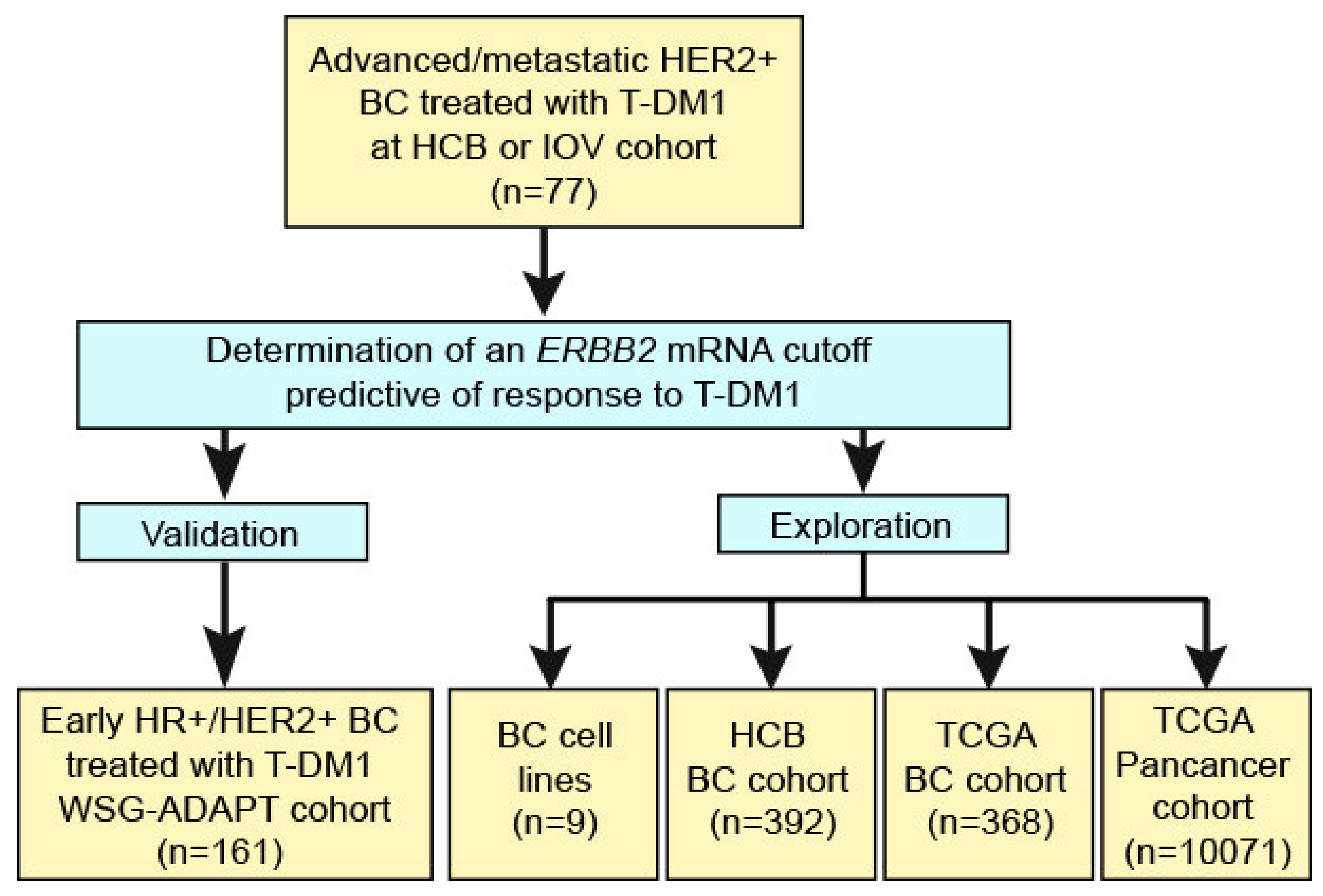
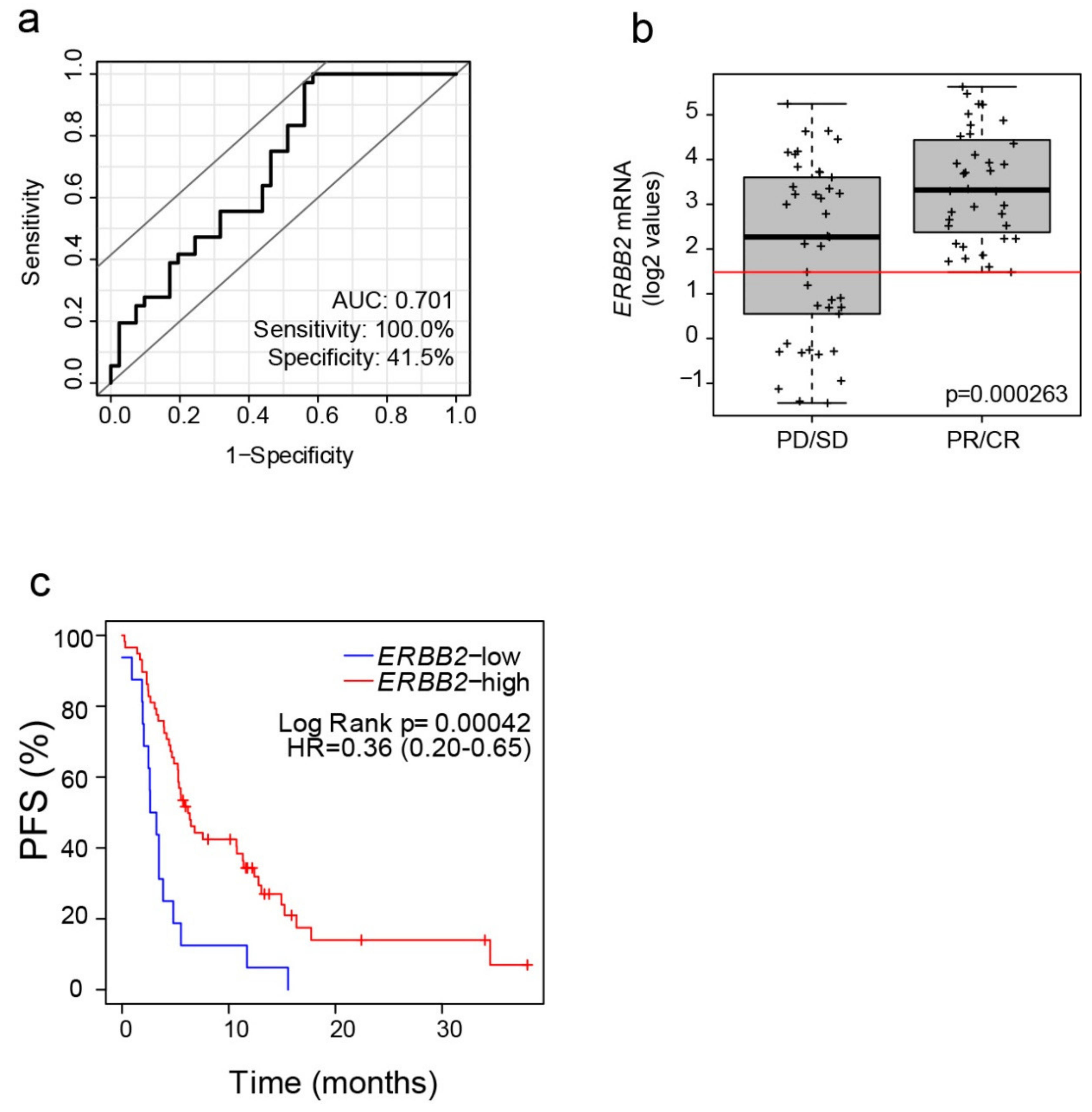
2.1. ERBB2 mRNA in Advanced HER2+ BC Treated with T-DM1

Seventy-seven consecutive patients diagnosed with HER2+ advanced BC and treated with T-DM1 at Hospital Clínic of Barcelona (HCB) and Istituto Oncologico Veneto (IOV) in Padova were evaluated. Demographic and disease characteristics of these patients are presented in Table 1. Briefly, all patients were pre-treated with trastuzumab in the (neo)adjuvant or metastatic setting and most had received at least 1 line of HER2-targeted treatment for metastatic disease (median 1, range 0–4). In addition, twenty-eight patients (36%) presented brain metastases at time of initiation of T-DM1. Regarding T-DM1 efficacy, overall response rate (ORR) was 47% (6 complete and 30 partial responses) and median progression-free survival (PFS) was 5.3 months (95% CI 4.4–10.7).
ERBB2 mRNA expression was assessed in 77 tumor samples from patients with advanced HER2+ BC treated with T-DM1. After determination of an ERBB2 mRNA cutoff predictive of response to T-DM1, several cohorts were evaluated. On one hand, the ERBB2 mRNA assay was validated in 161 patients recruited in the WSG-ADAPT phase II neoadjuvant trial of T-DM1. On the other, ERBB2 mRNA expression and response was evaluated in 9 in vitro BC cell lines. Finally, ERBB2 mRNA expression was explored in 392 BCs from an in-house dataset, 368 primary BCs from the TCGA BC dataset, and 10,071 tumors representing 33 cancer types from the PanCancer TCGA dataset (Figure 1).
A large range of ERBB2 mRNA expression was observed (log2 median 2.98; interquantile range 1.60–3.91). As expected, the expression of ERBB2 varied according to HER2 IHC expression (0, 1+, 2+ and 3+) (Figure S1). Compared to 0–2+, the expression in 3+ tumor samples was increased by 5.7-fold. Of note, although all patients were classified as HER2+ tumors by guidelines [4], based on clinical history and previous assessment of HER2 status on tumor samples, and were eligible for treatment with T-DM1, 8 of the 74 samples that were re-analyzed for HER2 had an IHC HER2 result of 0 (n = 5) or 1+ (n = 3). Four of these 8 samples were tested for HER2 amplification by ISH, and HER2 was found amplified in 3 cases and non-amplified in 1 sample.
The clinicopathological variables associated with response (i.e., partial and complete) to T-DM1 were: negative hormone-receptor status, lower number of prior lines of HER2-targeted therapy in the metastatic setting, higher HER2 IHC expression (i.e., 3+ vs. 0–2+) and higher ERBB2 mRNA as a continuous variable (Table 2). The overall response rate in HER2 3+ was 62.0% compared to 20.8% in the 0–2+ group (odds ratio = 1.84, 95% confidence interval [CI] 1.26–2.69, p = 0.002). However, only ERBB2 mRNA expression (as a continuous variable) and number of prior HER2-targeted lines, but not HER2 IHC expression or hormone-receptor status, were found independently associated with response (Table 2).

2.2. Identification of an Optimized ERBB2 mRNA Cutoff

Using T-DM1 response data in advanced HER2+ BC (i.e., PR and CR vs. stable disease [SD] and progression of disease [PD]), an optimized ERBB2 mRNA cutoff was identified based on Fisher’s exact test. This cutoff maximized the area under the curve (AUC) of the receiver operating characteristic (ROC) curve (AUC = 0.701, sensitivity: 100%, specificity: 41.5%) (Figure 2a). The cutoff was ERBB2 log2 value = 1.483 and it classified 60 tumors (77.9%) as ERBB2-high and 17 (22.1%) as ERBB2-low (Figure 2b). As expected, the ERBB2-high group had a response rate of 60%, whereas the response rate in the ERBB2-low group was 0%. ERBB2-high, as compared to ERBB2-low, was significantly associated with higher response rates, both in the HER2 IHC 3+ subgroup (67% vs. 0%, Fisher exact p = 0.017) and in the HER2 IHC 0–2+ subgroup (42% vs. 0%, Fisher exact p = 0.037).
Finally, the ERBB2-high group had a better PFS compared to the ERBB2-low group (median PFS 6.2 months vs. 2.93 months; hazard ratio = 0.36, 95% CI 0.20–0.65, p = 0.001) (Figure 2c), even after correction by number of prior lines of HER2-targeted therapy in the metastatic setting (hazard ratio = 0.38, 95% CI 0.21–0.70, p = 0.002).

2.3. Validation of ERBB2 mRNA Expression in Early-Stage HER2+ BC Treated with Neoadjuvant T-DM1

To further validate the ERBB2-based assay as a predictor of response to T-DM1 in the neoadjuvant setting, we assessed gene expression and pCR data from 158 patients treated with T-DM1 (alone or in combination with endocrine therapy) in the WSG-ADAPT HER2+/hormone receptor-positive (HR+) Phase II Trial [13]. Since gene expression in the WSG-ADAPT trial was determined using a different nCounter CodeSet and house-keeping gene list, we estimated where our ERBB2 cutoff would fall. To accomplish this, we first determined the percentile of ERBB2 mRNA expression corresponding to our ERBB2 cutoff (i.e., 1.483) in 77 HER2+/HR+ primary tumors from our previously published PAMELA trial [14], since this cohort is similar to the WSG-ADAPT cohort. Our ERBB2 cutoff corresponded to the 50th percentile in primary HER2+/HR+ tumors.
We then applied the 50th percentile of ERBB2 mRNA expression as the cutpoint to define ERBB2-high from ERBB2-low in the 161 patients of WSG-ADAPT trial. The overall pCR rate was 34.8% (56/161). When the ERBB2 cutoff was evaluated, the pCR rate in the ERBB2-high group was 42.9% (33/77) and in the ERBB2-low group was 27.4% (23/84) (sensitivity = 58.9%; specificity = 58.1%; odds ratio = 2.0; p-value = 0.041). Altogether, these results confirmed a significant association between ERBB2 mRNA levels and response to neoadjuvant T-DM1.

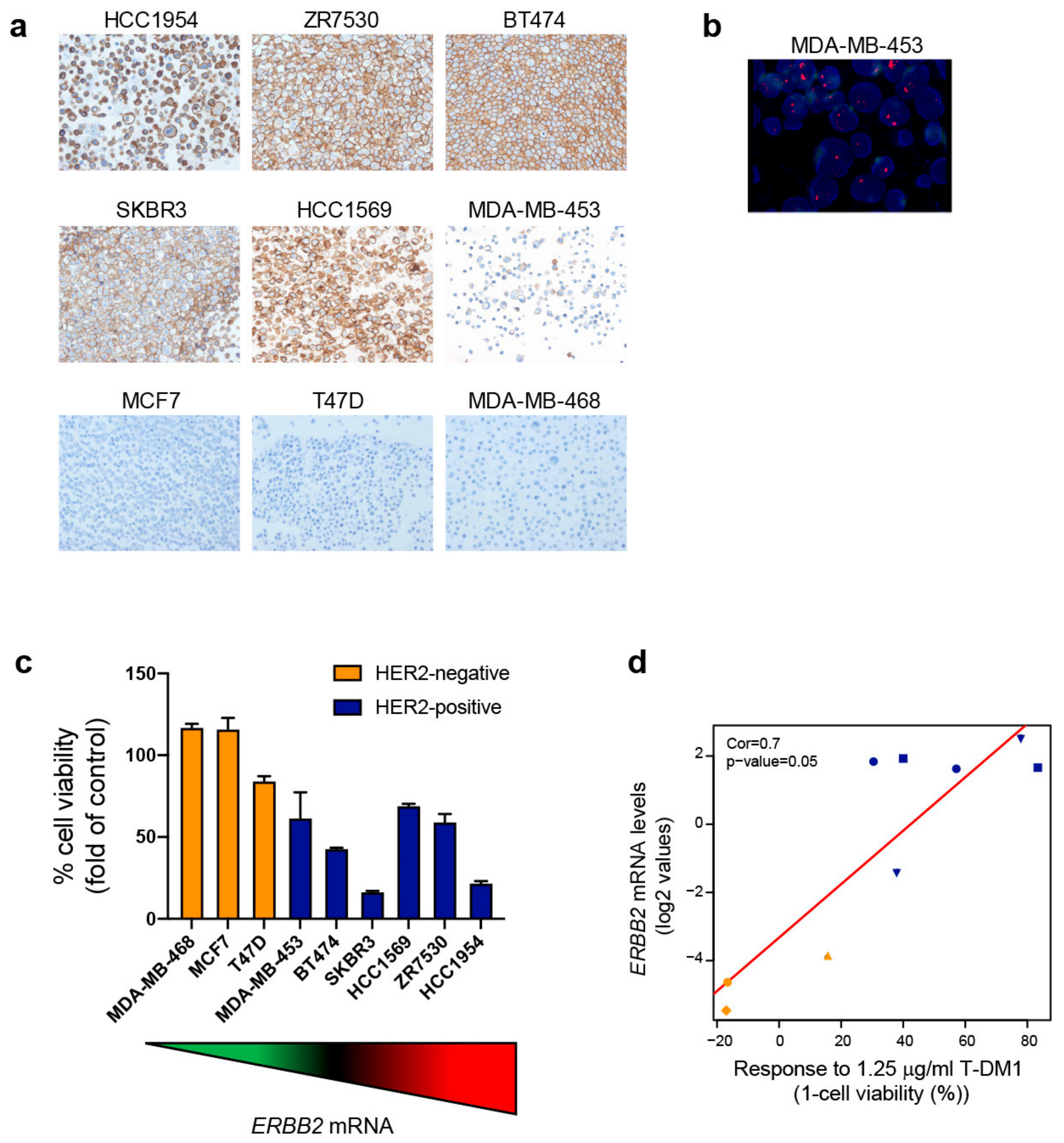
2.4. Exploring ERBB2 mRNA Expression and In Vitro Response to T-DM1

Next, we evaluated the expression of ERBB2 and the effects of T-DM1 across 6 HER2+ (HCC1954, ZR-75-30, BT-474, SK-BR3, HCC1569 and MDA-MB-453) and 3 HER2-negative BC cell lines (MCF7, T-47D and MDA-MB-468) (Figure 3a,b). ERBB2 mRNA expression varied substantially across cell lines and similarly to patient’s tumors (interquartile range of 6.16 across all cell lines and 0.60 within the HER2+ cell lines). As expected, HER2+ cell lines showed higher ERBB2 expression than the HER2-negative cell lines (mean 1.39 vs. −4.64, p = 0.0002) (Figure S2A). HER2+ cell lines were also more responsive to T-DM1 than HER2-negative cell lines (Figure 3c and Figure S2B), consistently with findings reported by others [15,16]. Response was defined as the decrease in cell viability (%) at 72 h of T-DM1 treatment. HER2+ cell lines showed greater response to T-DM1 than HER2-negative cell lines (mean response 54.71% vs. 5.24%, p = 0.008) (Figure S2C). Importantly, we observed response to T-DM1 in all cell lines with ERBB2 mRNA levels above the cutoff. In addition, correlation between ERBB2 mRNA and T-DM1 response was observed across the 9 cell lines (coefficient = 0.7, p = 0.05) (Figure 3d). This correlation coefficient suggests that 76.6% of the differences in response across cell lines may be explained by ERBB2 levels.

2.5. ERBB2 mRNA Expression in BC across the HER2 IHC-Based Groups

To determine the proportion of ERBB2-high tumors across the HER2 IHC groups, we analyzed a retrospective dataset of 392 tumor samples from HCB with both HER2 IHC status and gene expression data. As HER2 protein increased, ERBB2 mRNA levels increased as well, and all possible comparisons (except for 1+ vs. 2+/non-amplified) of ERBB2 expression between groups were statistically significant. According to our pre-established cutoff, the proportion of ERBB2-high across 0, 1+, 2+/ISH-negative, 2+/ISH-positive and 3+ was 0%, 1.1%, 0%, 9.38% and 76.17%, respectively (Figure 4a).
To provide more evidence of the association of ERBB2 and HER2 IHC expression in BC, we explored 368 BCs from the TCGA dataset including ERBB2 mRNA expression and HER2 IHC. Since the methodology to assess ERBB2 mRNA expression in our dataset (nCounter) was different to the TCGA dataset (RNAseq), the range of ERBB2 mRNA expression was different for each cohort. Therefore, we estimated where the pre-established cutoff would fall in the TCGA cohort. To do so, we calculated the median ERBB2 mRNA log2 values for each IHC group in HCB and TCGA cohorts (Figure S3A) and the Pearson correlation between the two cohorts (Figure S3B). The proportion of ERBB2-high across 0, 1+, 2+/ISH-negative, 2+/ISH-positive and 3+ was 0%, 0.59%, 0%, 25% and 74.04%, respectively (Figure 4b). The correlation coefficient between these ERBB2-high proportions and the proportions found in our in-house dataset was 0.975.

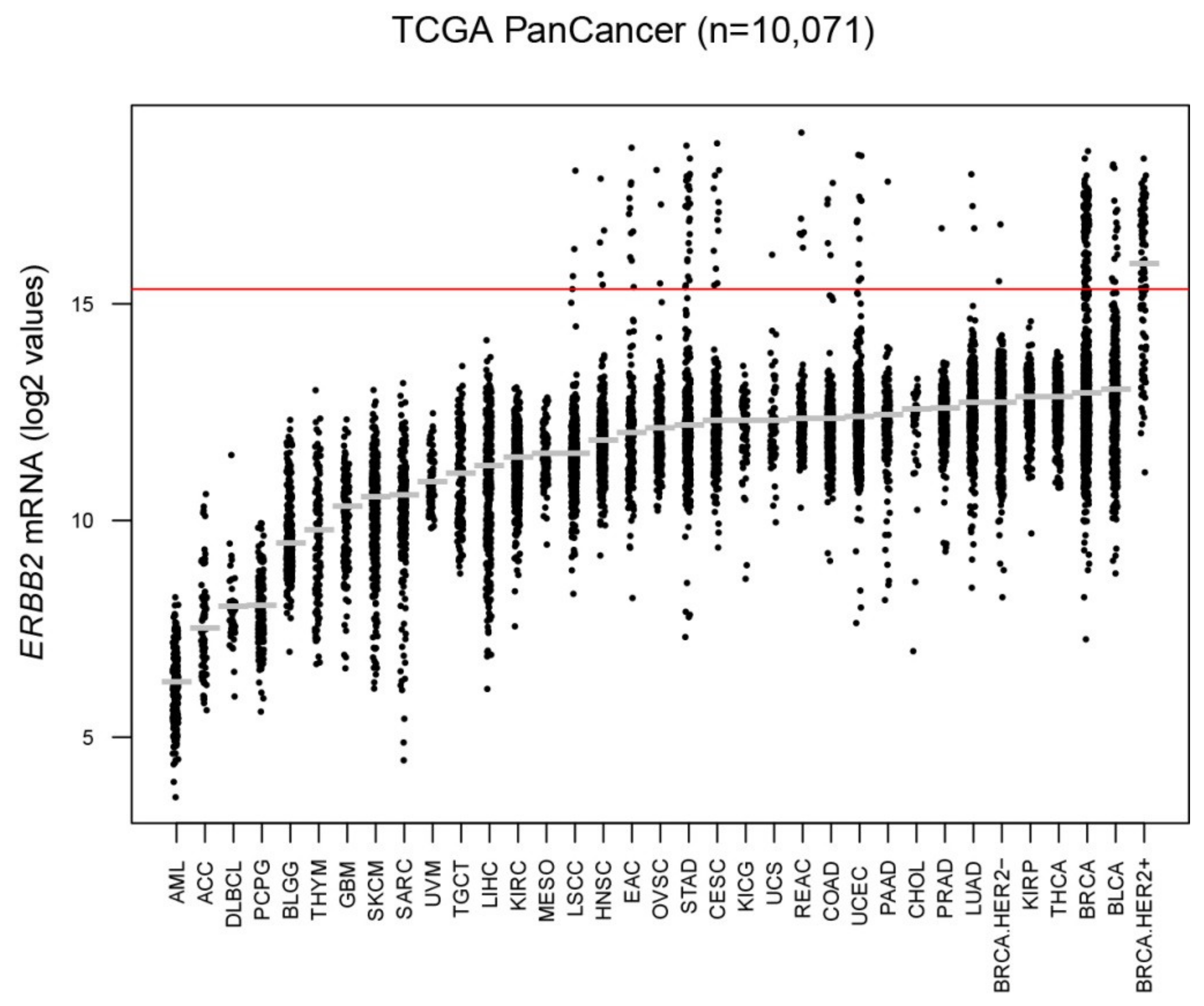
2.6. ERBB2 mRNA Expression across Cancer Types

In order to determine the proportion of ERBB2-high tumors in other cancer types, we explored ERBB2 expression for 10,071 tumors of different origins. According to our pre-established cutoff, ERBB2-high tumors were identified in 15 cancer types including: prostate cancer (0.2%), lung adenocarcinoma (0.59%), lung squamous cell carcinoma (0.83%), head and neck squamous cell carcinoma (0.97%), ovarian serous cystadenocarcinoma (1%), colon adenocarcinoma (1.14%), uterine carcinosarcoma (1.75%), uterine corpus endometrial carcinoma (2.09%), pancreatic adenocarcinoma (3.39%), rectum adenocarcinoma (3.9%), bladder urothelial carcinoma (3.93%), cervical squamous cell carcinoma (4.08%), esophageal adenocarcinoma (6.08%), stomach adenocarcinoma (6.32%) and breast cancer (9.41%) (Figure 5 and Table S1). As expected, we identified a lower proportion of ERBB2-high tumors in each cancer type as compared to standard IHC/ISH definition of HER2-positivity, thus potentially selecting tumors sensitive to T-DM1 treatment even in cancer types generally not considered amenable to treatment with this agent.

2.7. ERBB2 mRNA Expression in HER2+ Gastric Cancer Treated with T-DM1

We retrospectively studied the case of a 42-year-old male with a gastroesophageal adenocarcinoma diagnosed at HCB. In September 2011, a total esophagectomy and cervical esophago-gastric anastomosis was performed. Ascites and pleural effusion positive cytology adenocarcinoma were observed after surgery, and the patient was diagnosed with a HER2 3+ pT2pN3pM1 stage IV gastroesophageal junction adenocarcinoma. The patient subsequently received 6 cycles of first-line treatment with cisplatin plus 5-fluorouracil and trastuzumab achieving a radiological CR.
After 10 months, the patient presented with lung and bone PD and was enrolled in the GATSBY phase II/III trial [17] and treated with T-DM1 (2.4 mg/kg weekly) monotherapy. Since July 2013, he received 8 cycles of T-DM1 and obtained a PR (Table 3 and Figure S4) and a time-to-progression of 5.4 months. Of note, median PFS and ORR in GATSBY’s T-DM1 arm was 2.7 months and 20.6%, respectively [17]. Concordant with the efficacy results obtained in our patient, ERBB2 mRNA levels measured in the primary tumor were high (i.e., 2.99). Upon PD, the patient received third-line docetaxel monotherapy achieving SD until July 2014 when he presented with a central nervous system PD. The patient was lost to follow-up in November 2014.

3. Discussion

As an increasing number of HER2-targeted agents are becoming available in clinical practice, biomarkers are increasingly needed that can predict the response to specific anti-HER2 agents beyond the classic IHC/ISH definition of HER2-positivity. In this context, HER2 3+ tumors have been reported to benefit more from T-DM1 than other IHC groups in retrospective and prospective studies [7,18]. Moreover, benefit associated with the use of post-neoadjuvant T-DM1 as compared to trastuzumab in the KATHERINE trial appeared to more marked in HER2 3+ than in HER2 2+ tumors [19]. Greater benefit to T-DM1 in HER2 3+ tumors has also been reported in the KATE2 trial [20] and other cancer types [21,22].
ERBB2 mRNA expression has been previously associated with a more pronounced T-DM1 benefit in several randomized clinical trials which tested the use of T-DM1 as compared to other HER2-targeted treatments in HER2+ metastatic BC [23,24,25]. In the randomized EMILIA trial, which compared T-DM1 and capecitabine-lapatinib (CL) for pretreated metastatic HER2+ BC patients, patients with tumor ERBB2 mRNA levels above median showed a greater benefit from the use of T-DM1 in terms of ORR and overall survival (OS). However, T-DM1 treatment, compared with CL, reduced the risk of PD to a similar degree regardless of tumor ERBB2 mRNA levels and tests for interaction between treatment and ERBB2 mRNA levels were not statistically significant (p = 0.07). However, tests were exploratory and not powered to detect an interaction [23].
Moreover, data from the randomized TH3RESA trial, which compared T-DM1 vs. treatment of physician choice for pretreated metastatic HER2+ BC patients, confirmed that patients with higher ERBB2 mRNA levels benefited more from T-DM1 than patients with lower ERBB2 mRNA levels [24]. Similar results confirming an association between higher ERBB2 mRNA levels and increased T-DM1 benefit, both in terms of ORR and PFS, were also reported for several randomized phase II trials [25,26,27]. Furthermore, the impact of ERBB2 mRNA levels on T-DM1 benefit has been reported in other cancers beyond BC. A translational study [21] evaluating gastric cancer samples of the GATSBY trial [17] demonstrated more benefit to T-DM1 in terms of PFS in patients with tumors with higher ERBB2 mRNA levels [21].
In addition, recently presented biomarker data from the post-neoadjuvant KATHERINE trial have been reported showing that patients with high ERBB2 mRNA levels (above median) at surgery have a worse outcome than patients with low ERBB2 mRNA levels when treated with adjuvant trastuzumab, but not when treated with adjuvant T-DM1. In fact, while both patients with ERBB2 mRNA levels above and below median levels benefited from switching to T-DM1, those with higher ERBB2 mRNA levels (above median) benefited more from T-DM1 than those with lower ERBB2 mRNA levels, potentially questioning the use of median mRNA expression value as cutoff in this setting [28].
Our results not only confirm the association between ERBB2 mRNA expression and T-DM1 benefit in a more heterogeneous real-world setting, but also highlight the relevance of a quantitative method as a better method to predict response to T-DM1 by proposing a cutoff for selecting patients responsive to T-DM1 both in the metastatic and neoadjuvant settings. Other HER2-targeted ADCs are entering clinical practice, for instance DS-8201, which has shown activity in HER2-low advanced BC [12,29]. Therefore, we might expect that different ERBB2 cutoffs will be needed for different ADCs. In this context, the use of a quantitative method such as ERBB2 mRNA expression, which offers the opportunity to identify different cutoffs, might potentially improve treatment personalization. Moreover, a quantitative method as ERBB2 mRNA expression might recapitulate tumor heterogeneity in a single, easily manageable assay.
Our study has several limitations. First, the study cohort is retrospective and only involved a limited number of patients who were treated according to everyday clinical practice, thus being heterogeneous, both in previous lines of treatment received and in the kind of histological samples available (primary tumor vs. metastasis). Despite this, our analysis was able to clearly identify ERBB2 mRNA expression as the main predictor of responsiveness together with number of previous lines of HER2-targeted treatment. Moreover, we validated ERBB2 mRNA as a predictor of response in the neoadjuvant setting. This highlights the clinical importance of ERBB2 mRNA expression. Second, we could not address if the biomarker works better when primary or metastatic tumor samples are used. Third, our data in non-BC-types is currently in the hypothesis-generating stage.

4. Materials and Methods

4.1. Patient Datasets and Tumor Samples

This study analyzed a retrospective cohort of 77 HER2+ (as defined by standard guidelines [4]) advanced/metastatic BC patients treated with T-DM1 between January 2013 and November 2019 in two independent institutions: Hospital Clínic of Barcelona (HCB) (n = 26) and Istituto Oncologico Veneto (IOV) in Padova (n = 51). One formalin-fixed paraffin-embedded (FFPE) tumor sample per patient was selected: if available, a biopsy of metastatic site nearest in time to start of T-DM1 was preferred (n = 38); otherwise primary tumor sample was used (n = 39), favouring pre-treatment biopsy over surgical sample for patients treated with neoadjuvant therapy. Gene expression was also assessed in 77 primary HER2+/HR+ BC of the PAMELA trial [14], and gene expression and pathological complete response (pCR) data was evaluated in 161 HER2+ primary samples of the T-DM1 arms (A&B) of the WSG-ADAPT HER2+/HR+ Phase II Trial [13]. In addition, we evaluated 392 primary BCs from HCB with available HER2 IHC status and gene expression data evaluated at the nCounter platform; 368 primary BCs from The Cancer Genome Atlas (TCGA) with HER2 IHC status and ERBB2 RNASeqv2 data; and 10,071 TCGA pan-cancers with ERBB2 RNASeqv2 data. Finally, we analyzed a primary tumor sample of a patient with advanced gastric cancer treated with T-DM1 in the GATSBY trial [17] at HCB.

4.2. In Vitro Cell Lines and T-DM1

The BC cell lines BT-474, HCC1569, HCC1954, MCF7, MDA-MB-453, MDA-MB-468, SK-BR3, T-47D and ZR-75-30 were purchased from the American Type Culture Collection. All cell lines were maintained as recommended by the suppliers. T-DM1 was provided as remnant of the product used in common clinical practice by the oncology pharmacy Service at HCB.

4.3. HER2 Immunohistochemistry and Fluorescent In Situ Hybridization

HER2 status was re-assessed in 74 of 77 FFPE tumors of the T-DM1 HCB/IOV cohort and FFPE BC cell line pellets by either IHC and/or in situ hybridization (ISH) according to the American Society of Clinical Oncologists (ASCO)/College of American Pathologists (CAP) guidelines. IHC was performed on 2-μm-thick sections using anti-HER-2/neu (4B5) Rabbit Monoclonal Primary Antibody kit (Ventana Medical Systems Inc., Oro Valley, AZ, USA) and ISH for HER2 was performed on 4-μm-thick sections using the FDA-approved XL ERBB2 (HER2/NEU) AMP (MetaSystems Probes, Altlußheim, Germany) according to manufacturer’s instructions.

4.4. In Vitro Cell Viability Assay

BC cell lines were plated in triplicate at 4000 cells/well in 96-well plates. Cells were then treated with 1.25 μg/mL T-DM1. Cell viability was determined 72 h after treatment using CellTiter 96 AQueous One Solution Cell Proliferation Assay (MTS) (Promega Corporation, Madison, Wisconsin, USA) following the manufacturer’s instructions, and quantified using the Gen5 Microplate Reader and Imager Software (BioTek, Winooski, VT, USA). Data were analyzed using GraphPad Prism 5 software (GraphPad, San Diego, CA, USA).

4.5. RNA Extraction

RNA samples were extracted from biopsy and surgical tumor FFPE material using the High Pure FFPET RNA isolation kit (Roche) following manufacturer’s protocol. FFPE slides with at least 10% tumor cells and 4 mm2 of tissue were used for each tumor specimen, and macrodissection was performed to avoid contamination with normal breast tissue if needed. Cell line RNA samples were extracted using the RNeasy Mini Kit (Qiagen, Hilden, Alemanya). RNA samples were quantified at the NanoDrop spectrophotometer (Thermo Fisher Scientific, Waltham, MA, USA).

4.6. Gene Expression Analysis

The nCounter platform (NanoString Technologies, Seattle, WA, USA) was used to analyze RNA samples from tumors and cell lines. A minimum of 100 ng of total RNA was used to measure the expression of 50 genes of the PAM50 intrinsic subtype predictor assay and 5 housekeeping genes (ACTB, MRPL19, PSMC4, RPLP0, and SF3A1). Expression counts were then normalized using the nSolver 4.0 software (nanoString, Seattle, WA, USA) and custom scripts in R 3.4.3 (R Foundation, Vienna, Austria) [30].

4.7. Statistical Analysis

Univariate and multivariable logistic regression analyses were used to investigate the association of each variable with overall response. Odds ratios and 95% confidence intervals (CIs) were calculated for each variable. An optimized cutoff of gene expression was identified to predict overall response. Univariate and multivariable Cox proportional hazard regression analyses were performed to investigate the association of each variable with PFS. All statistical analyses were carried out using the R software version 3.4.3.

5. Conclusions

To conclude, our study presents a clinically applicable assay to help identify patients most likely to benefit from T-DM1, regardless of HER2 status. In addition, the assay could help identify patients most likely to benefit from other HER2-targeted ADCs across cancer types.

Supplementary Materials

The following are available online at https://www.mdpi.com/2072-6694/12/7/1902/s1, Table S1: Proportions of ERBB2-high tumors across cancer types, Table S2: Table of abbreviations, Figure S1: Distribution of ERBB2 mRNA levels across HER2 IHC subgroups of tumors of the T-DM1 cohort, Figure S2: ERBB2 mRNA levels and T-DM1 response in BC cell lines, Figure S3: Determination of an ERBB2 mRNA cutoff for the TCGA dataset, Figure S4: Partial response to T-DM1 in gastric cancer patient.

Author Contributions

Experimental study design: G.G., F.B.-M. and A.P. Data acquisition and analysis: G.G., F.B.-M., B.G.-F., T.P., N.C., T.S., R.K., O.G., D.M, V.T., D.P., P.G., L.B., M.C. (Miriam Cuatrecasas), M.C. (Mathias Christgen), H.K., T.G. Data interpretation: G.G., F.B.-M., B.G.-F., T.P., N.C., T.S., R.K., O.G., D.M., L.P., V.T., D.P., M.V.D., B.A., M.M., P.G., L.B., M.C. (Miriam Cuatrecasas), M.C. (Mathias Christgen), H.K., I.M.-E., P.V., D.S., T.G., M.V.D., P.C., N.H., V.G., A.P.; Writing of the manuscript: G.G., F.B-M. and A.P. Review of the manuscript: all authors. All authors have read and agreed to the published version of the manuscript.

Funding

This study has received funding from Instituto de Salud Carlos III—PI16/00904 and PI19/01846 (to A.P.), Breast Cancer Now—2018NOVPCC1294 (to A.P.), Breast Cancer Research Foundation-AACR Career Development Awards for Translational Breast Cancer Research 19-20-26-PRAT (to A.P.), Fundació La Marató TV3 201935-30 (to A.P.), the European Union’s Horizon 2020 research and innovation programme H2020-SC1-BHC-2018-2020 (to A.P.), Pas a Pas (to A.P.), Save the Mama (to A.P.), Fundación Científica Asociación Española Contra el Cáncer AECC_Postdoctoral17-1062 (to F. B-M), Generalitat de Catalunya Peris PhD4MD 2019 SLT008/18/00122 (to N.C.), DiSCOG—University of Padova DOR 1721185/17 and DOR 1830512/18 (to MV.D.), Conquer Cancer Foundation/Shanken Family Foundation -YIA in Breast Cancer 2019 (to G.G.).

Conflicts of Interest

Potential conflicts of interest are the following: A.P. reports consulting fees from Nanostring Technologies Roche, Pfizer, Novartis and Daiichi Sankyo outside the submitted work. A.P. is listed as an inventor on a patent application on HER2 as a predictor of response to dual HER2 blockade in the absence of cytotoxic therapy (WO2018/103834A1). MV.D. reports lecture fees and honoraria for participation on advisory boards from Roche, Genomic Health, Eli Lilly, and Celgene outside the submitted work. PF.C. reports honoraria for participation on advisory boards from Eli Lilly, Novartis, AstraZeneca, Tesaro, Roche Genentech, Daiichi Sankyo, and BMS, and research grants to the Institution from Novartis, Roche Genentech, Merck KGaA, and BMS outside the submitted work. V.G. reports lecture fees and honoraria for participation on advisory boards from Eli Lilly, Roche Genentech and Novartis, honoraria for participation on Speakers bureau from Eli Lilly and Novartis outside the submitted work. N.H. reports fees for consulting and/or lectures Novartis and Roche outside the submitted work and minority ownership in WSG (Westdeutsche Studiengruppe).

References

  1. Verma, S.; Miles, D.; Gianni, L.; Krop, I.E.; Welslau, M.; Baselga, J.; Pegram, M.; Oh, D.-Y.; Diéras, V.; Guardino, E.; et al. Trastuzumab emtansine for HER2-positive advanced breast cancer. New Engl. J. Med. 2012, 367, 1783–1791. [Google Scholar] [CrossRef] [PubMed] [Green Version]
  2. Krop, I.E.; Kim, S.-B.; González-Martín, A.; Lorusso, P.M.; Ferrero, J.-M.; Smitt, M.; Yu, R.; Leung, A.C.F.; Wildiers, H. Trastuzumab emtansine versus treatment of physician’s choice for pretreated HER2-positive advanced breast cancer (TH3RESA): A randomised, open-label, phase 3 trial. Lancet Oncol. 2014, 15, 689–699. [Google Scholar] [CrossRef]
  3. Von Minckwitz, G.; Huang, C.-S.; Mano, M.S.; Loibl, S.; Mamounas, E.P.; Untch, M.; Wolmark, N.; Rastogi, P.; Schneeweiss, A.; Redondo, A.; et al. Trastuzumab Emtansine for Residual Invasive HER2-Positive Breast Cancer. New Engl. J. Med. 2019, 380, 617–628. [Google Scholar] [CrossRef] [PubMed]
  4. Wolff, A.C.; Hammond, M.E.H.; Hicks, D.G.; Dowsett, M.; McShane, L.M.; Allison, K.H.; Allred, D.C.; Bartlett, J.; Bilous, M.; Fitzgibbons, P.; et al. Recommendations for Human Epidermal Growth Factor Receptor 2 Testing in Breast Cancer: American Society of Clinical Oncology/College of American Pathologists Clinical Practice Guideline Update. J. Clin. Oncol. 2013, 31, 3997–4013. [Google Scholar] [CrossRef]
  5. Slamon, D.J.; Leyland-Jones, B.; Shak, S.; Fuchs, H.; Paton, V.; Bajamonde, A.; Fleming, T.; Eiermann, W.; Wolter, J.; Pegram, M.D.; et al. Use of Chemotherapy plus a Monoclonal Antibody against HER2 for Metastatic Breast Cancer That Overexpresses HER2. New Engl. J. Med. 2001, 344, 783–792. [Google Scholar] [CrossRef]
  6. Lee, H.J.; Na Seo, A.; Kim, E.J.; Jang, M.H.; Suh, K.J.; Ryu, H.S.; Kim, Y.J.; Kim, J.H.; Im, S.-A.; Gong, G.; et al. HER2 Heterogeneity Affects Trastuzumab Responses and Survival in Patients With HER2-Positive Metastatic Breast Cancer. Am. J. Clin. Pathol. 2014, 142, 755–766. [Google Scholar] [CrossRef] [Green Version]
  7. Filho, O.M.; Viale, G.; Trippa, L.; Li, T.; Yardley, D.A.; Mayer, I.A.; Abramson, V.G.; Arteaga, C.L.; Spring, L.; Waks, A.G.; et al. HER2 heterogeneity as a predictor of response to neoadjuvant T-DM1 plus pertuzumab: Results from a prospective clinical trial. J. Clin. Oncol. 2019, 37, 502. [Google Scholar] [CrossRef]
  8. Hou, Y.; Nitta, H.; Wei, L.; Banks, P.M.; Portier, B.; Parwani, A.V.; Li, Z. HER2 intratumoral heterogeneity is independently associated with incomplete response to anti-HER2 neoadjuvant chemotherapy in HER2-positive breast carcinoma. Breast Cancer Res. Treat. 2017, 166, 447–457. [Google Scholar] [CrossRef]
  9. Modi, S.; Saura, C.; Yamashita, T.; Park, Y.H.; Kim, S.-B.; Tamura, K.; Andre, F.; Iwata, H.; Ito, Y.; Tsurutani, J.; et al. Trastuzumab Deruxtecan in Previously Treated HER2-Positive Breast Cancer. New Engl. J. Med. 2019, 382, 610–621. [Google Scholar] [CrossRef]
  10. Shitara, K.; Iwata, H.; Takahashi, S.; Tamura, K.; Park, H.; Modi, S.; Tsurutani, J.; Kadowaki, S.; Yamaguchi, K.; Iwasa, S.; et al. Trastuzumab deruxtecan (DS-8201a) in patients with advanced HER2-positive gastric cancer: a dose-expansion, phase 1 study. Lancet Oncol. 2019, 20, 827–836. [Google Scholar] [CrossRef]
  11. Banerji, U.; Van Herpen, C.M.L.; Saura, C.; Thistlethwaite, F.; Lord, S.; Moreno, V.; MacPherson, I.R.; Boni, V.; Rolfo, C.; E De Vries, E.G.; et al. Trastuzumab duocarmazine in locally advanced and metastatic solid tumours and HER2-expressing breast cancer: a phase 1 dose-escalation and dose-expansion study. Lancet Oncol. 2019, 20, 1124–1135. [Google Scholar] [CrossRef] [Green Version]
  12. Modi, S.; Park, H.; Murthy, R.K.; Iwata, H.; Tamura, K.; Tsurutani, J.; Moreno-Aspitia, A.; Doi, T.; Sagara, Y.; Redfern, C.; et al. Antitumor Activity and Safety of Trastuzumab Deruxtecan in Patients With HER2-Low-Expressing Advanced Breast Cancer: Results From a Phase Ib Study. J. Clin. Oncol. 2020. [Google Scholar] [CrossRef] [PubMed]
  13. Harbeck, N.; Gluz, O.; Christgen, M.; Kates, R.E.; Braun, M.; Küemmel, S.; Schumacher, C.; Potenberg, J.; Kraemer, S.; Kleine-Tebbe, A.; et al. De-Escalation Strategies in Human Epidermal Growth Factor Receptor 2 (HER2)–Positive Early Breast Cancer (BC): Final Analysis of the West German Study Group Adjuvant Dynamic Marker-Adjusted Personalized Therapy Trial Optimizing Risk Assessment and Therapy Response Prediction in Early BC HER2- and Hormone Receptor–Positive Phase II Randomized Trial—Efficacy, Safety, and Predictive Markers for 12 Weeks of Neoadjuvant Trastuzumab Emtansine With or Without Endocrine Therapy (ET) Versus Trastuzumab Plus ET. J. Clin. Oncol. 2017, 35, 3046–3054. [Google Scholar] [CrossRef] [PubMed]
  14. Llombart, A.; Cortés, J.; Paré, L.; Galván, P.; Bermejo, B.; Martínez, N.; Vidal, M.; Pernas, S.; López, R.; Muñoz, M.; et al. HER2-enriched subtype as a predictor of pathological complete response following trastuzumab and lapatinib without chemotherapy in early-stage HER2-positive breast cancer (PAMELA): an open-label, single-group, multicentre, phase 2 trial. Lancet Oncol. 2017, 18, 545–554. [Google Scholar] [CrossRef]
  15. Lambert, J.M.; Chari, R.V. Ado-trastuzumab Emtansine (T-DM1): An Antibody–Drug Conjugate (ADC) for HER2-Positive Breast Cancer. J. Med. Chem. 2014, 57, 6949–6964. [Google Scholar] [CrossRef]
  16. Phillips, G.D.L.; Li, G.; Dugger, D.L.; Crocker, L.M.; Parsons, K.L.; Mai, E.; Lambert, J.M.; Chari, R.V.; Lutz, R.J.; Wong, W.L.T.; et al. Targeting HER2-Positive Breast Cancer with Trastuzumab-DM1, an Antibody-Cytotoxic Drug Conjugate. Cancer Res. 2008, 68, 9280–9290. [Google Scholar] [CrossRef] [Green Version]
  17. Thuss-Patience, P.C.; A Shah, M.; Ohtsu, A.; Van Cutsem, E.; Ajani, J.A.; Castro, H.; Mansoor, W.; Chung, H.C.; Bodoky, G.; Shitara, K.; et al. Trastuzumab emtansine versus taxane use for previously treated HER2-positive locally advanced or metastatic gastric or gastro-oesophageal junction adenocarcinoma (GATSBY): an international randomised, open-label, adaptive, phase 2/3 study. Lancet Oncol. 2017, 18, 640–653. [Google Scholar] [CrossRef]
  18. Yazaki, S.; Hashimoto, J.; Ogita, S.; Nakano, E.; Yamauchi, T. Lower response to T-DM1 in metastatic breast cancer patients with HER2 IHC score of 2 and FISH positive compared with IHC score of 3. Ann. Oncol. 2017, 28, v102–v103. [Google Scholar] [CrossRef] [Green Version]
  19. Geyer, C.; Huang, C.-S.; Mano, M.; Loibl, S.; Mamounas, E.; Untch, M.; Wolmark, N.; Rastogi, P.; Fischer, H.; Redondo, A.; et al. Abstract GS1-10: Phase III study of trastuzumab emtansine (T-DM1) vs trastuzumab as adjuvant therapy in patients with HER2-positive early breast cancer with residual invasive disease after neoadjuvant chemotherapy and HER2-targeted therapy including trastuzumab: Primary results from KATHERINE. Gen. Sess. Abstr. 2019, 79, GS1–GS10. [Google Scholar] [CrossRef]
  20. Emens, L.; Esteva, F.; Beresford, M.; Saura, C.; De Laurentiis, M.; Kim, S.-B.; Im, S.-A.; Patre, M.; Wang, Y.; Mani, A.; et al. Abstract PD3-01: Results from KATE2, a randomized phase 2 study of atezolizumab (atezo)+trastuzumab emtansine (T-DM1) vs placebo (pbo)+T-DM1 in previously treated HER2+ advanced breast cancer (BC). Poster Discuss. Abstr. 2019, 79. [Google Scholar] [CrossRef]
  21. Shah, M.A.; Kang, Y.-K.; Thuss-Patience, P.C.; Ohtsu, A.; Ajani, J.A.; Van Cutsem, E.; Hoersch, S.; Harle-Yge, M.-L.; De Haas, S.L. Biomarker analysis of the GATSBY study of trastuzumab emtansine versus a taxane in previously treated HER2-positive advanced gastric/gastroesophageal junction cancer. Gastric Cancer 2019, 22, 803–816. [Google Scholar] [CrossRef] [PubMed] [Green Version]
  22. Peters, S.; Stahel, R.A.; Bubendorf, L.; Bonomi, P.; Villegas, A.; Kowalski, D.M.; Baik, C.S.; Isla, D.; Carpeño, J.D.C.; Garrido, P.; et al. Trastuzumab Emtansine (T-DM1) in Patients with Previously Treated HER2-Overexpressing Metastatic Non–Small Cell Lung Cancer: Efficacy, Safety, and Biomarkers. Clin. Cancer Res. 2018, 25, 64–72. [Google Scholar] [CrossRef] [PubMed] [Green Version]
  23. Baselga, J.; Phillips, G.D.L.; Verma, S.; Ro, J.; Huober, J.; Guardino, A.E.; Samant, M.K.; Olsen, S.; De Haas, S.L.; Pegram, M.D. Relationship between Tumor Biomarkers and Efficacy in EMILIA, a Phase III Study of Trastuzumab Emtansine in HER2-Positive Metastatic Breast Cancer. Clin. Cancer Res. 2016, 22, 3755–3763. [Google Scholar] [CrossRef] [PubMed] [Green Version]
  24. Kim, S.-B.; Wildiers, H.; Krop, I.E.; Smitt, M.; Yu, R.; De Haas, S.L.; Gonzalez-Martin, A. Relationship between tumor biomarkers and efficacy in TH3RESA, a phase III study of trastuzumab emtansine (T-DM1) versus treatment of physician’s choice in previously treated HER2-positive advanced breast cancer. Int. J. Cancer 2016, 139, 2336–2342. [Google Scholar] [CrossRef]
  25. A Perez, E.; Hurvitz, S.A.; Amler, L.C.; E Mundt, K.; Ng, V.W.; Guardino, E.; Gianni, L. Relationship between HER2 expression and efficacy with first-line trastuzumab emtansine compared with trastuzumab plus docetaxel in TDM4450g: a randomized phase II study of patients with previously untreated HER2-positive metastatic breast cancer. Breast Cancer Res. 2014, 16, R50. [Google Scholar] [CrossRef] [PubMed] [Green Version]
  26. Krop, I.E.; Lorusso, P.; Miller, K.D.; Modi, S.; Yardley, D.; Rodriguez, G.; Guardino, E.; Lu, M.; Zheng, M.; Girish, S.; et al. A Phase II Study of Trastuzumab Emtansine in Patients With Human Epidermal Growth Factor Receptor 2–Positive Metastatic Breast Cancer Who Were Previously Treated With Trastuzumab, Lapatinib, an Anthracycline, a Taxane, and Capecitabine. J. Clin. Oncol. 2012, 30, 3234–3241. [Google Scholar] [CrossRef]
  27. Burris, H.A.; Rugo, H.S.; Vukelja, S.J.; Vogel, C.L.; Borson, R.A.; Limentani, S.; Tan-Chiu, E.; Krop, I.E.; Michaelson, R.A.; Girish, S.; et al. Phase II Study of the Antibody Drug Conjugate Trastuzumab-DM1 for the Treatment of Human Epidermal Growth Factor Receptor 2 (HER2) –Positive Breast Cancer After Prior HER2-Directed Therapy. J. Clin. Oncol. 2011, 29, 398–405. [Google Scholar] [CrossRef]
  28. Denkert, C.; Lambertini, C.; Fasching, P.A.; Pogue-Geile, K.L.; Mano, M.S.; Untch, M.; Wolmark, N.; Huang, C.-S.; Loibl, S.; Mamounas, E.P.; et al. Biomarker data from KATHERINE: A phase III study of adjuvant trastuzumab emtansine (T-DM1) versus trastuzumab (H) in patients with residual invasive disease after neoadjuvant therapy for HER2-positive breast cancer. J. Clin. Oncol. 2020, 38, 502. [Google Scholar] [CrossRef]
  29. Doi, T.; Shitara, K.; Naito, Y.; Shimomura, A.; Fujiwara, Y.; Yonemori, K.; Shimizu, C.; Shimoi, T.; Kuboki, Y.; Matsubara, N.; et al. Safety, pharmacokinetics, and antitumour activity of trastuzumab deruxtecan (DS-8201), a HER2-targeting antibody–drug conjugate, in patients with advanced breast and gastric or gastro-oesophageal tumours: a phase 1 dose-escalation study. Lancet Oncol. 2017, 18, 1512–1522. [Google Scholar] [CrossRef]
  30. Cejalvo, J.M.; De Dueñas, E.M.; Galvan, P.; García-Recio, S.; Gasión, O.B.; Paré, L.; Antolin, S.; Martinello, R.; Blancas, I.; Adamo, B.; et al. Intrinsic Subtypes and Gene Expression Profiles in Primary and Metastatic Breast Cancer. Cancer Res. 2017, 77, 2213–2221. [Google Scholar] [CrossRef] [Green Version]
Figure 1. Workflow of the study.
Cancers 12 01902 g001
Figure 2. ERBB2 mRNA expression predicts T-DM1 response and survival in metastatic HER2+ BC. (a) ROC curve to identify ERBB2 mRNA cutoff of response to T-DM1. (b) ERBB2 mRNA levels in patients with progressive disease (PD) or stable disease (SD) (n = 41) vs. patients achieving complete response (CR) or partial response (PR) (n = 36). p-value was determined using a two-tailed unpaired t-test. The ERBB2 mRNA cutoff is shown as a red line. (c) Kaplan–Meier estimate of progression-free survival (PFS) using the ERBB2 mRNA cutoff.
Cancers 12 01902 g002
Figure 3. ERBB2 mRNA expression correlates with response to T-DM1 in cell lines. (a) Images of HER2 expression by IHC in 9 BC cell lines (40×). (b) Image of HER2 amplification by ISH (100×). (c) Cell viability of 9 BC cell lines upon 72 h of treatment with 1.25 μg/mL T-DM1. Data points represent the mean; error bars represent the standard error of the mean of 3 independent experiments. (d) Spearman correlation between ERBB2 mRNA expression and response to 1.25 μg/mL T-DM1 expressed as 1-cell viability (%).
Cancers 12 01902 g003
Figure 4. ERBB2 mRNA expression in BC across the HER2 IHC groups. Distribution of ERBB2 mRNA levels across HER2 IHC subgroups of the primary BC (a) HCB and (b) TCGA datasets. The proportion of ERBB2-high tumors is indicated as defined by the ERBB2 mRNA cutoff (shown as a red line). p-values were determined using one-way analysis of variance.
Cancers 12 01902 g004
Figure 5. ERBB2 mRNA expression across cancer types. Distribution of ERBB2 mRNA levels across cancer-types TCGA datasets. The ERBB2 mRNA cutoff is shown as a red line. Abbreviations are shown in Table S2.
Cancers 12 01902 g005
Table 1. Patient characteristics.
Characteristicsn = 77
Median age at BC diagnosis, years (range)49 (27–88)
Median age at start of T-DM1, years (range)51 (35–93)
Histology: Ductal71 (92%)
      Lobular/other5 (6%)
      NA1 (1%)
Histologic Grade: G12 (3%)
         G210 (13%)
        G328 (36%)
        NA37 (48%)
Hormone-receptor: positive46 (60%)
         negative31 (40%)
HER2 IHC status: IHC 05 (6%)
        IHC 1+3 (4%)
        HC 2+16 (21%)
        IHC 3+50 (65%)
           NA *3 (4%)
HER2 ISH status in HER2 IHC 2+ cases: Amplified 15 (94%)
                      Not evaluable *1 (6%)
HER2 ISH status in HER2 IHC 0/1+ cases: Amplified 3 (38%)
                      Non-amplified *1 (12%)
                      Not available *4 (50%)
Previous (neo)adjuvant treatment44 (57%)
Median number previous lines HER2-targeted 1 (0–4)
treatment for metastatic disease (range)
Previously received:
  Pertuzumab-trastuzumab31 (40%)
  Trastuzumab 41 (53%)
  Lapatinib14 (24%)
Visceral metastases at start of T-DM166 (86%)
Brain metastases at start of T-DM128 (36%)
Concomitant endocrine treatment during T-DM117 (22%)
* These cases were confirmed to be HER2-positive in other tumor samples and treated with T-DM1 according to clinical practice.
Table 2. Univariable and multivariable logistic regression analyses of overall response.
Clinicopathological VariableUnivariateMultivariable
Odds Ratio (95%CI)pOdds Ratio (95%CI)p
Hormone-receptor statusnegativeref0.038ref0.152
positive0.37 (0.14–0.95)0.39 (0.11–1.41)
De-novo metastatic diseasenoref0.990
yes0.99 (0.40–2.49)
Visceral diseasenoref0.577
yes0.69 (0.19–2.50)
Brain involvementnoref0.966
yes0.98 (0.39–2.49)
HER2 IHC≤2+ref0.002ref0.257
3+1.84 (1.26–2.69)1.32 (0.82–2.13)
ERBB2 (continuous)1.73 (1.25–2.39)0.0011.95 (1.22–3.12)0.006
Prior lines HER2-targeted therapy0–1ref0.009ref0.002
≥20.06 (0.01-0.50)0.02 (0.002-0.23)
Table 3. Response evaluation criteria in solid tumors (RECIST) table for a gastric cancer case before and during T-DM1 treatment.
Target LesionScreeningPre Cycle 3Pre Cycle 5Pre Cycle 7Pre Cycle 9
Right upper lobe lung metastasis14 mm8 mm8 mm8 mm10 mm
Left upper
lobe lung metastasis
10 mm10 mm10 mm10 mm12 mm
Mesentheric adenopathy17 mm11 mm11 mm11 mm18 mm
Retroperitoneal adenopathy18 mm9 mm9 mm9 mm17 mm
Total59 mm38 mm38 mm38 mm57 mm
ResponseNA39% reduction39% reduction39% reduction36% increase
NAPRMaintained PRMaintained PRPD

