Article

Colorectal Cancer Presentation and Survival in Young Individuals: A Retrospective Cohort Study

1
Department of Internal Medicine, Reno School of Medicine, University of Nevada, Reno, NV 89557, USA
2
Renown Institute for Cancer, 1155 Mill Street, W-11, Reno, NV 89502, USA
*
Author to whom correspondence should be addressed.
Cancers 2018, 10(12), 472; https://doi.org/10.3390/cancers10120472
Submission received: 3 November 2018 / Revised: 22 November 2018 / Accepted: 24 November 2018 / Published: 28 November 2018

Abstract

:
Emerging evidence suggests that the incidence of colorectal cancer is increasing among individuals under the age of 50 years. However, the pattern of disease presentation in young patients remains under investigation. This is a retrospective cohort study of patients diagnosed with colorectal cancer (CRC) between 2004 and 2015. Data was acquired from the Surveillance, Epidemiology, and End Results 18 program registries. A total of 269,398 patients who met the inclusion criteria were included in the final analysis. The primary outcomes were the likelihood of metastatic disease at diagnosis and survival. Of the 269,389 patients diagnosed with CRC, 11.8% of the patients were young (20 to 49 years), 45.6% were middle-aged (50 to 69 years), and 42.6% were elderly (70 years or older). Individuals in the middle-aged and elderly cohorts were significantly less likely to present with metastatic disease as compared to the young cohort (middle-aged adjusted odds ratio (aOR) = 0.73, 95% confidence interval (CI) = 0.70 to 0.75, elderly aOR = 0.49, 95% CI = 0.47 to 0.50). However, overall survival was longest in the young cohort. We conclude that young individuals with colorectal cancer have an increased risk of presenting with distant metastases as compared to the middle-aged and elderly, but, nevertheless, exhibit prolonged survival.

1. Introduction

Colorectal cancer represents the third most common malignancy in the United States and emerging evidence suggests that the incidence of colorectal cancer (CRC) is increasing among individuals under the age of 50 years. However, the pattern of disease presentation in young patients remains under investigation.
The incidence of CRC in young individuals has increased by 2% to 8% annually over the past two decades [1,2]. CRC is now one of the 10 most common causes of death among individuals between the ages of 20 and 49 years [3]. Adults aged 50 years and older have had a decline in CRC. This has been attributed primarily to widespread implementation of screening guidelines [4]. Several retrospective studies have shown that younger patients have an increased risk of presenting with advanced stage disease as compared to older adults [5]. However, some investigators have reported contradictory results showing that younger patients are more likely to present with less-advanced stages of CRC [6,7]. Furthermore, the relationship between patient age at CRC diagnosis and survival remains under investigation. We, therefore, evaluated the National Cancer Institute’s Surveillance, Epidemiology, and End Results (SEER) 18 registries to determine if young patients with CRC are more likely to present with metastatic disease and to ascertain survival among this cohort. Age was grouped into 20–49 years (young), 50–69 years (middle age), and 70 years or older.

2. Results

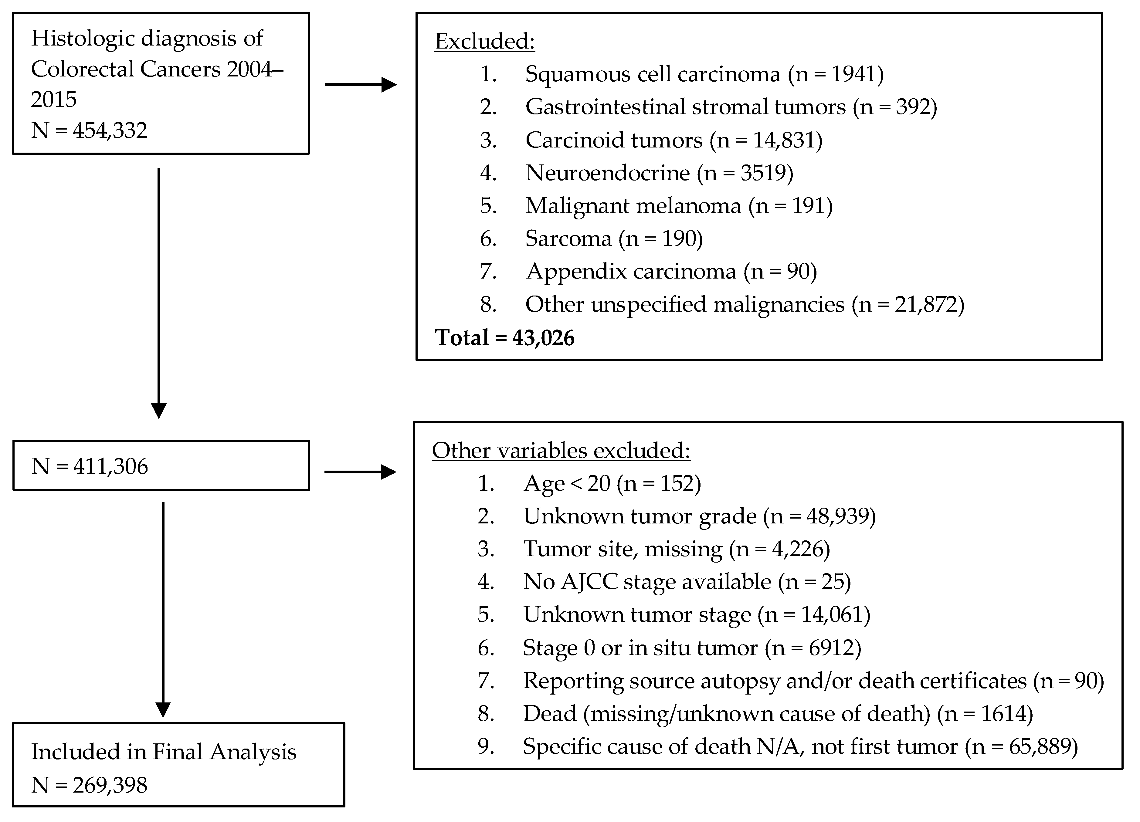
2.1. Study Population

A total of 269,398 patients diagnosed with CRC are included in the final analysis. In addition, 129,538 (48.0%) were female and 139,860 (51.9%) were male. Overall, 11.8% of the patients were young (20–49 years), 45.6% were middle-aged (50–69 years), and 42.6% were elderly (70 years or older). Patients were further stratified into two groups based on year of diagnosis: EraA (2004–2009) and EraB (2010–2015). A significantly higher proportion of young patients were diagnosed in EraB as compared to EraA (p < 0.05). In addition, EraB had a significantly higher proportion of middle-aged (47.9%), female (52.0%), white (66.6%), and insured (81.6%-excluding Medicaid) patients.

2.2. Patient Characteristics

Baseline demographic, tumor, clinical, and treatment characteristics are presented in Table 1. Furthermore, 111,935 (41.6%) patients died over the study period in which 75,398 (67.4%) were in EraA and 36,537 (32.6%) were in EraB (p < 0.001). A total of 48,559 (18.0%) patients had metastatic CRC at diagnosis. Among these, 16.0% were young, 48.9% were middle-aged, and 35.1% were elderly. In contrast, a diagnosis of either stage 1, 2, or 3 CRC was established in 10.8% of young, 44.9% of middle-aged, and 44.3% of elderly individuals (Figure S1).

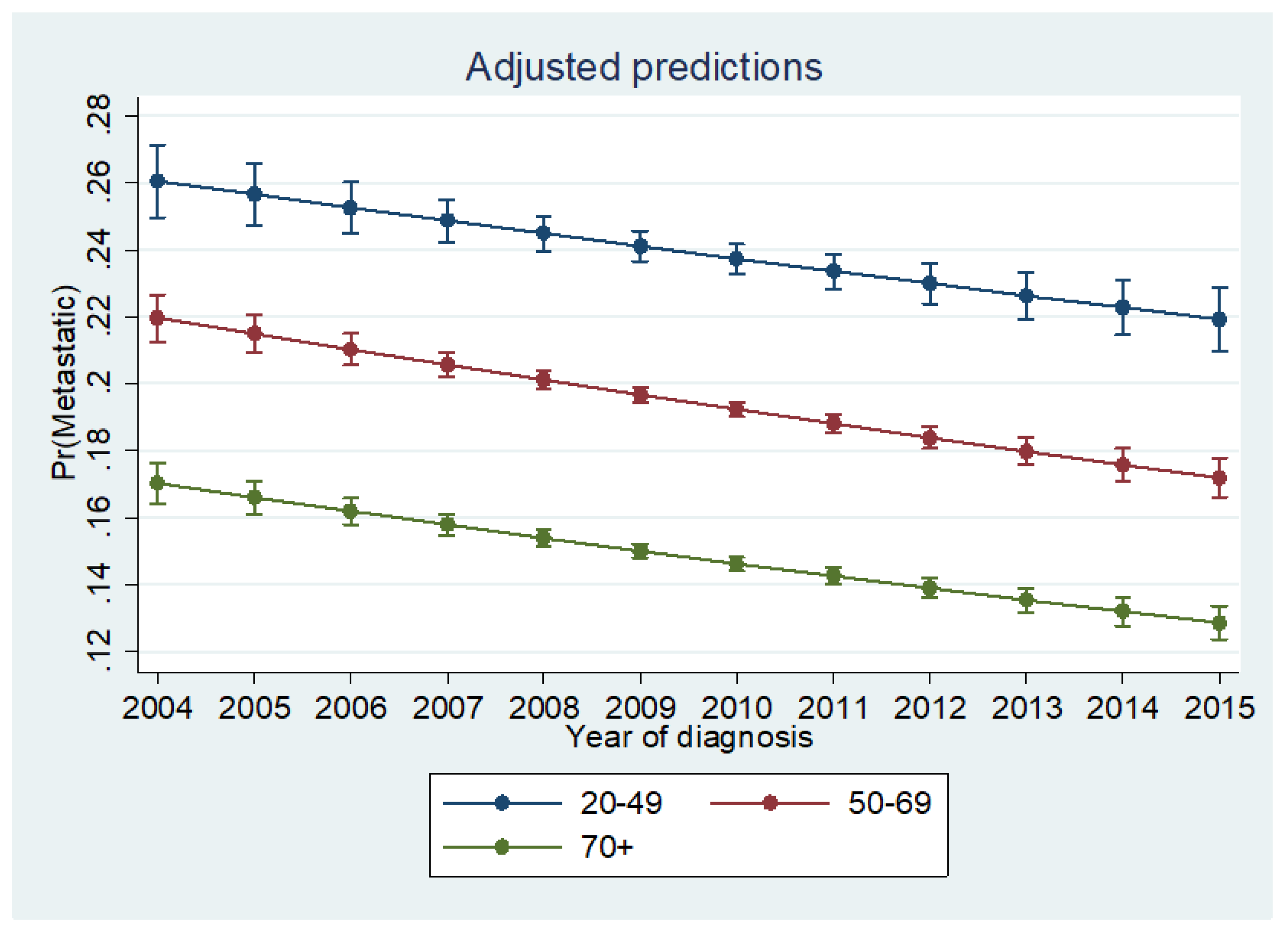
2.3. Likelihood of Metastatic Disease

In a logistic multivariable model for demographic, tumor, and clinical characteristics, middle-aged (adjusted odds ratio (aOR: 0.73, 95% CI, 0.70–0.75, p < 0.001) and elderly (aOR: 0.49, 95% CI, 0.47–0.50, p < 0.001) patients were less likely to present with metastatic disease as compared to the young cohort. In univariate analysis, the middle-aged cohort was less likely to present with high-grade cancer (OR: 0.79, 95% CI, 0.77–0.81, p < 0.001), but no differences in grade at presentation was noted between the young and elderly cohorts (p = 0.80). Patients who did not undergo surgery were more likely to have metastatic disease at presentation (aOR: 23.1, p < 0.001). The other independent predictors of metastatic disease of CRC at diagnosis are shown in Table 2. Patients in EraB had increased likelihood of presenting with metastatic disease on univariate analysis (OR = 1.03, 95% CI, 1.01–1.05, p = 0.001). In adjusted analysis, however, there was decreased likelihood of stage IV disease in EraB (aOR = 0.93, 95% CI, 0.91–0.95, p < 0.001).

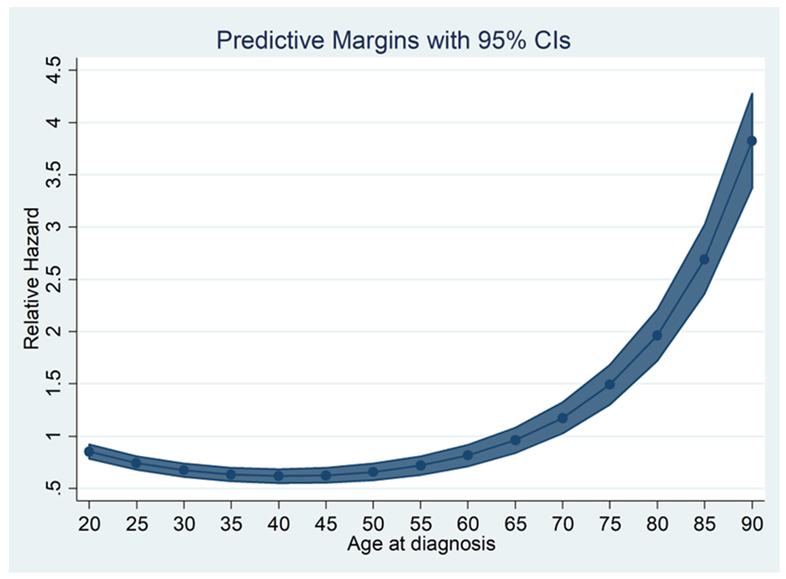
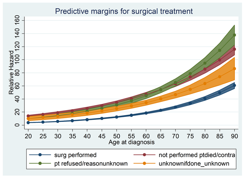
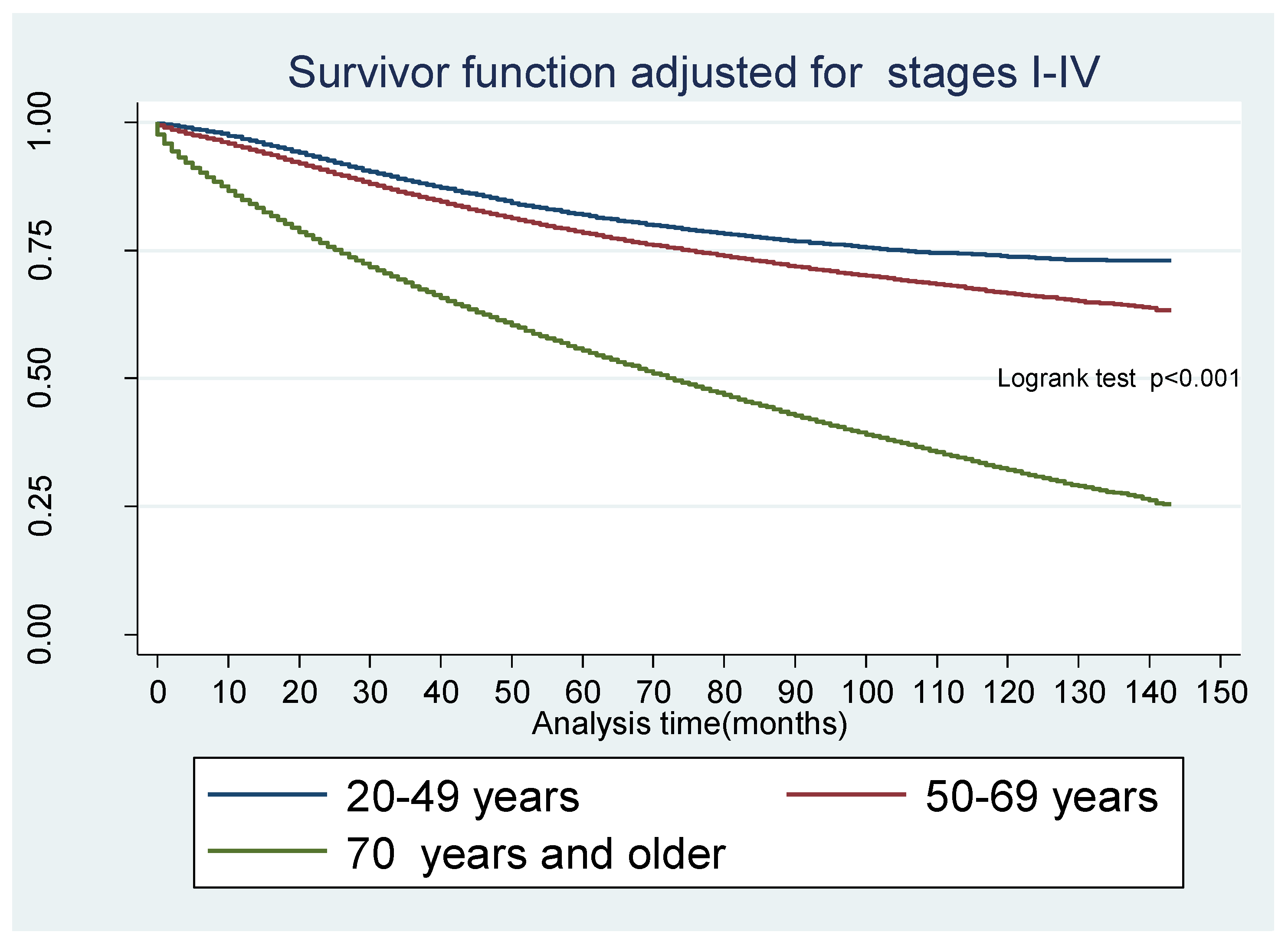
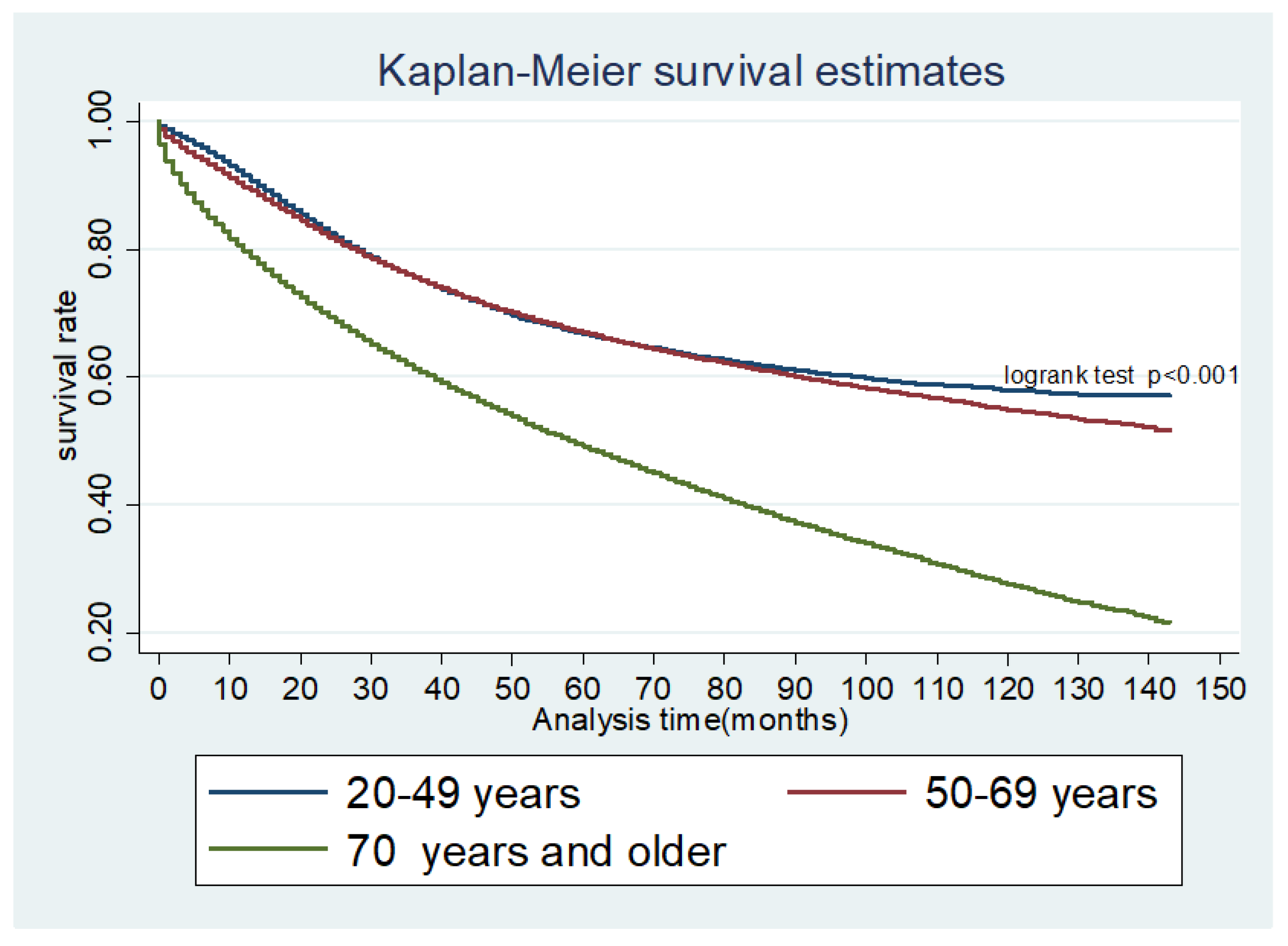
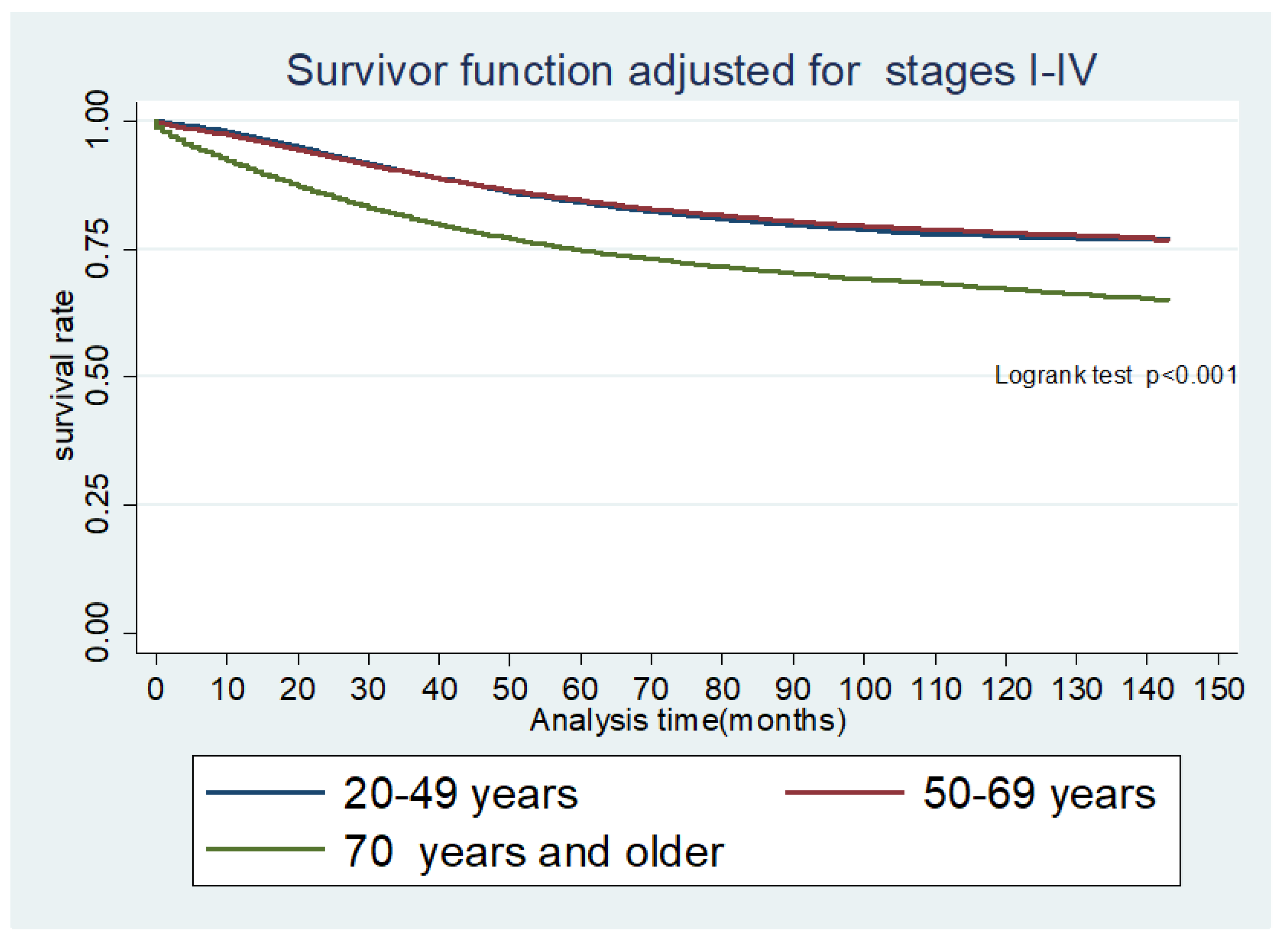
2.4. Overall and CRC-Specific Survival

Of the 269,398 patients, 111,935 (41.6%) died over the study period of which 75,398 (67.4%) and 36,537 (32.6%) were in 2004–2009 and 2010–2015 respectively; p < 0.001. The median overall survival (OS) was not reached in young and middle-aged people over the study period but was 59 months for the elderly. However, OS was 121, 93, and 18 months for AJCC stages II, III, and IV, respectively. Median CRC-specific survival (CSS) was not reached in each age group. However, CSS by AJCC stage IV was 19 months. The 5-year OS for young individuals was 66.9% (95% CI, (66.3–67.5%)), middle aged, 67.2% (66.9–67.5%), and elderly, 49.5% (49.2–49.8%). In univariate analysis, middle-aged and elderly patients had inferior long-term survival. This persisted in an adjusted Cox proportional hazard model (aHR: 1.21, 95% CI, 1.19–1.24, p < 0.001 and aHR: 2.63, 95% CI, 2.57–2.70, p < 0.001). There was improved OS for all three age cohorts in EraB as compared to EraA (aHR: 0.96, 95% CI: 0.95–0.97, p < 0.001). The other independent predictors for survival are shown in Table 3.
The adjusted relative hazard (RH) of death as a function of age and as a continuous variable is shown in Figure 1. Overall survival was used to determine RH. We observed that there was evidence of a higher RH among younger individuals in the cohort. This seemingly plateaued until about 50 years at which point the RH increased dramatically. On stratified analysis by surgical intervention, the trend was reversed for younger patients but was similar for the rest of the age cohorts with an upward trend (Figure 2). For example, a 20-year-old patient who had surgical intervention has an RH of death of 3.8 (p < 0.001) compared to 15.2 (p < 0.001) for a 55-year-old patient. A similar trend was noted when stratified for the stage of CRC.

3. Discussion

Our results show that young individuals have a higher likelihood of presenting with metastatic CRC but have superior OS and CSS (Figure 3, Figure 4 and Figure 5). Although an increased incidence of CRC among young individuals has been all but established, the relationship between age and prognosis is not yet fully understood. Our results are consistent with a retrospective cohort study using SEER data collected between 1991 and 1999, which revealed that young patients (age 20 to 40 years) with CRC were more likely to present with later-stage and higher-grade tumors than their older (age 60 to 80 years) counterparts [8]. The authors showed that, despite presenting with more advanced cancers, overall five-year survival was only slightly lower in the young cohort when compared to the older cohort. Among those with stage IV cancer, 5-year survival was markedly longer in young versus older patients (18% and 6%, respectively).
Investigators hypothesized that young individuals with stage IV CRC demonstrated prolonged survival because they were more likely to be treated with adjuvant chemotherapy than older patients. Although there is no SEER data pertaining to specific chemotherapeutic regimens, young patients with stage IV CRC can often tolerate the toxic effects associated with FOLFOX-4 (oxaliplatin, folinic acid, and fluorouracil) and CLF-1 (irinotekan, folinic acid, and fluorouracil) that frequently require discontinuation of therapy in the elderly [9]. Manjelievskaia et al. evaluated data from the Central Cancer Registry of the United States Department of Defense and found that young (18 to 49 years) and middle-aged (50 to 64 years) patients were two to eight times more likely to receive systemic postoperative chemotherapy when compared to older patients (65 to 75 years) across all tumor stages [10]. However, there was no corresponding improvement in survival. Limitations of the study included a higher proportion of unknown chemotherapy regimens among the older group as compared to the younger age group as well as a small sample size for stratified analysis.
It is conceivable that evidence of superior OS and CSS in our study among young aged individuals was related to their ability to receive adjuvant and neoadjuvant chemotherapy. Young patients were also more likely than other age groups to have surgery in our data, which could have contributed to prolonged OS and CSS.
Recently, Chou et al. published a retrospective study evaluating the relationship between age and CRC prognosis [11]. Over 60,000 patients with CRC in Taiwan were divided into six age groups: age 40 or younger, 41 to 50 years, 51 to 60 years, 61 to 70 years, 71 to 80 years, and over 80 years. Individuals in the youngest age cohort had poorer overall survival than those in middle-aged patients (41 to 50 years and 61 to 70 years) and were more likely to present with tumors that exhibited aggressive histopathologic features. Investigators postulated that poor overall survival among young individuals with CRC is due to late detection. CRC screening before the age of 50 is uncommon and, thus, CRC is typically identified in young patients only after it has become symptomatic. However, despite a higher prevalence of aggressive tumors such as signet-ring cell carcinoma and mucinous adenocarcinoma among the youngest cohort, young individuals demonstrated superior overall survival as compared to those in the oldest cohorts (71 to 80 years and over 80 years). The authors attributed this finding to a higher rate of non-CRC-related death among those aged 71 or older. Limitations of that study include the absence of surgical treatment information and AJCC tumor staging data. Our data showed that both the middle-aged and elderly cohorts had poorer overall survival for all histologic CRC as well as shorter CSS for adenocarcinoma not otherwise specified and signet-ring cell carcinoma as compared to young patients.
To further examine these findings by Chou et al., we calculated RH of death as a function of age analyzed as a continuous variable (Figure 1). Using overall survival data, we calculated adjusted RH for increasing age. We observed that there was evidence of a higher RH for young individuals in the cohort, which seemingly plateaued until about 50 years of age at which point the RH increased dramatically. We then looked at RH stratified by surgical intervention and noted that this trend was reversed among younger patients but was similar for the rest of the age cohort with uptrend (Figure 2). Our analysis mirrors that of a 2014 study by Lieu et al. in which authors reviewed data on 20,023 patients with metastatic CRC and identified poorer OS among the youngest and oldest individuals who underwent treatment [12]. Investigators, therefore, concluded that younger and older patients might represent higher-risk populations. Our adjusted analysis for RH is consistent with the Lieu group. However, our data showed a relatively smaller RH among the young age group (Figure 1).
Our study contributes to the growing body of evidence indicating an increased risk of metastatic disease at presentation among young individuals. Those aged 20 to 49 years are significantly more likely than middle-aged and elderly individuals to have distant metastases at diagnosis (Figure 6) across the study period. However, young patients demonstrated the longest overall and CRC-specific survival among the three cohorts. Our findings are consistent with those published in a recent study by Rodriguez et al. who reviewed data from the Ontario Cancer Registry and found that patients under the age of 40 years exhibited improved overall survival as compared to older patients despite presenting with more advanced and aggressive disease [13].
There are several hypotheses to explain why young people are more likely to present with metastatic disease. First, CRC screening typically does not begin until the age of 50 years. While the American Cancer Society (ACS) guidelines were recently revised, the majority of practitioners have yet to implement earlier screening and, thus, late detection in the young is common. The United States Preventive Services Task Force (USPSTF), the American College of Physicians, and the American Society for Gastrointestinal Endoscopy have long recommended that individuals with an average risk for colorectal cancer (CRC) begin screening at the age of 50 [14,15,16], which most practitioners adhere to. In 2017, however, the American Cancer Society (ACS) published new guidelines suggesting that CRC screening start as early as the age of 45. The ACS cited a landmark study by Siegel et al. in which investigators had analyzed data from nearly half a million patients and noted a marked increase in the annual incidence of CRC in young individuals since the mid-1980s [17]. In their examination of CRC incidence trends in Surveillance, Epidemiology, and End Results (SEER) areas from 1974 to 2013, the Siegel group discovered that CRC incidence rates had consistently increased by 1% to 2.4% annually among adults age 20 to 39 years. A similar but slightly smaller increase in incidence was observed in those aged 40 to 54 years. Recently, Vuik et al. analyzed data collected over a 25-year period from 20 European national cancer registries to study trends in incidence rates of young adults with CRC. Investigators found that the incidence of colon cancer increased by 1.5% per year between 1990–2008 and by 7.4% annually between 2008–2016 [18]. Further reports confirm a similar incidence increase in Australia and New Zealand [19], China [20], Germany [21], and Pakistan [22].
Second, younger patients frequently delay seeking medical attention for CRC-associated symptoms [23]. Furthermore, young patients are more likely to tolerate chemotherapy regimens as compared to middle-aged and older adults. As described by Kneuertz et al., young patients are more likely to have a low Charlson-Deyo Comorbidity Index-a weighted score calculated based on the number of pre-existing comorbid conditions-when compared to individuals of a more advanced age [24]. These factors could have contributed to superior survival in this age category. Third, signet-ring cell carcinoma - an aggressive subtype of CRC that spreads rapidly and is characterized by late symptom manifestation - disproportionately affects young individuals [25]. Stratification of our data by histologic subtype revealed that young individuals had an increased risk of signet-ring cell carcinoma as compared to the older cohorts (Figure S2, Table S1).
The mechanisms underlying superior survival among young individuals with CRC have yet to be fully elucidated. It is conceivable that intrinsic immunologic differences between young and old patients contributes to prolonged survival in the former group. Age-related immunosenescence, T-cell dysregulation, and systemic inflammation have been postulated to play a role in oncogenesis [26].
In addition to investigating the effect of age on CRC presentation and survival, we examined the relationship between race and CRC. Patients were stratified into six distinct ethnic groups: white (71.2%), African American (11.2%), Hispanic (9.2%), Asian (7.6%), Native American (0.6%), and unknown (0.2%). African Americans and Native Americans as compared to Hispanics were found to have an increased risk of metastatic disease at presentation and poorer survival, but OS was superior and likelihood of stage 4 disease was more common among whites and Asians. The association between race and CRC has been previously studied by Tammana and Laiyemo who identified several factors such as differences in diet and lifestyle, limited access to healthcare, and distinct tumor biology to account for the increased rate of advanced CRC and CRC-related mortality in some minority communities [27]. However, the most significant contributory factor may be poor patient education and insurance status. A 2015 study by May et al. concluded that racial minorities are less likely than whites to receive physician recommendations for CRC screening [28]. These findings suggest that establishing CRC screening guidelines by race is unlikely to have a significant effect on mortality in the absence of improved patient communication.
We also found that the chance of metastatic disease at presentation was lower in EraB (Figure S3). The recent decrease in metastatic disease warrants further investigation but may be attributed toward improved patient education and increased CRC screening. The percentage of adults who underwent CRC screening increased from 34.8% to 66.1% between 1987 and 2010 [29]. It appears likely that CRC is now more frequently detected at an early stage, which results in relatively lower number of patients presenting with advanced disease.
The increasing incidence of CRC in young individuals parallels that of obesity. As recently as 30 years ago, obesity was relatively rare among young and middle-aged adults. Today, however, approximately one-quarter to one-third of adults are affected [30]. Obesity has been identified as an independent risk factor for the development of CRC. Metabolic abnormalities, inappropriate insulin signaling, and activation of inflammatory pathways have been proposed as obesity-mediated mechanisms of oncogenesis [31]. Inadequate fiber intake is also common among overweight individuals and represents another obesity-associated risk factor for CRC [32]. Furthermore, a recent study by Hidayat et al. showed that body fatness at an early age increases the risk of CRC later in life [33]. Obesity in children and young adults is a relatively recent phenomenon. It is, therefore, possible that the increased incidence and presentation with distant metastasis of CRC in young people can be attributed in part to the rapid and unprecedented rise of obesity.
Our study has some limitations. First, we could not assess causation and subject it to selection bias. One would expect young patients to present with symptoms while older patients are diagnosed mostly through screening. Second, although multivariable analysis was used to control for potential confounders, there still remains the risk of residual confounding. Third, we did not have baseline data on comorbidities, which greatly influence treatment decision-making such as who undergoes surgeries. In addition, due to inaccurate reporting, SEER database has no information on non-surgical cancer directed therapies such as chemotherapy, which affects survival. Lastly, the SEER program collects data from population-based cancer registries, which covers about 28% of the USA population, and the results may not be generalizable to the entire USA population. However, this was minimized by adjusting for geographic regions. Despite these limitations, this study adds and provides a unique perspective on the stages of presentation and survival for CRC across age groups.

4. Materials and Methods

4.1. Study Design and Study Population

This is a retrospective cohort study using the SEER database for identification of CRC from all the registries captured in the SEER 18 program (San Francisco, Connecticut, Detroit, California, Kentucky, Louisiana, New Jersey, Greater Georgia, Hawaii, Iowa, New Mexico, Seattle, Utah, Alaska, San Jose-Monterey, Los Angeles, Rural Georgia, and Metropolitan Atlanta) who had a histologic diagnosis of colorectal cancers. SEER histology codes 8140, 8141, 8143, 8147, 8260, 8261, 8262, 8263, 8265, 8210, 8211, 8213, 8480, 8481, 8490, 8510, 8560, 8562, 8220, and 8221 were used for CRC diagnosed between 2004 and 2015. Patients were categorized into two groups based on the year of diagnosis: EraA (2004–2009) and EraB (2010–2015). An index registry was used to classify patients into various geographic regions: Midwestern (Detroit and Iowa), Western (California, Los Angeles, San Francisco, Hawaii, New Mexico, Seattle, Utah, Alaska, and San Jose-Monterey), Southern (Rural Georgia, Kentucky, Louisiana, Metropolitan Atlanta, and Greater Georgia), and North Eastern (New Jersey and Connecticut). The SEER registries continuously code and submit American Joint Committee on Cancer (AJCC) 6th and 7th Edition stages for all cancers diagnosed in 2010 and beyond. Patients diagnosed before 2010 are staged using the AJCC 6th edition only. The AJCC 6th edition was used in order to include all patients diagnosed between 2004 and 2015. Exclusion criteria included: (1) age younger than 20 years, (2) stage 0 or in situ tumor, (3) unknown tumor grade, (4) unknown tumor stage, (5) unknown site of primary tumor, (6) unavailable staging data, (7) patient deceased and cause of death unknown, and (8) history of previous cancer (Figure 7). This study did not require IRB approval since this is a publically available de-identified data and was granted access on the application request.

4.2. Data Source

The SEER database is comprised of data collected by the National Cancer Institute (https://www.cancer.gov). The SEER program collects and publishes cancer incidence and survival data using population-based cancer registries that include approximately 28% of the population of the United States. The program routinely collects data on patient demographics, tumor sites, tumor morphology, staging, surgical treatment, and follow-up.

4.3. Main Outcome Measures

Our primary outcome of interest was the likelihood of metastatic (stage IV) CRC at the time of diagnosis as compared to non-metastatic (stages I, II, or III) CRC. The secondary outcomes of interest were overall survival (OS) and CRC-specific survival (CSS). We estimated OS in months from the date of diagnosis to the date of death for non-survivors. The end of the follow-up period was used to ascertain OS for survivors. Patients were stratified into three groups based on age: young (20–49 years), middle-aged (50–69 years), and elderly (70 years or older).

4.4. Statistical Analysis

The baseline characteristics and group differences were compared by using Pearson’s Chi square (X2) test for proportions. Non-parametric variables were compared using the Mann Whitney-U test. The Kaplan-Meier method was used for survival analysis and the log rank test for equality of survival functions including assessing survival differences between patients diagnosed in EraA and EraB and among age groups. Continuous variables were analyzed with the student t-test. Stepwise multivariable Logistic and Cox regression models were built by using the forward method and adjusting for baseline demographics, treatment, and tumor characteristics. Variables included in the adjusted models had a p-value < 0.05 for the outcome of interest in the univariate analysis. These variables remained in the final model if they were still significant at p < 0.05 in the final adjusted model since a p-value < 0.05 was deemed statistically significant in this study. All statistical analyses were performed using Stata version 14.2 (StataCorp, College Station, TX, USA).

5. Conclusions

Young individuals with CRC have an increased likelihood of presenting with distant metastases as compared to the middle-aged and elderly. Yet, nevertheless, these individuals exhibit prolonged OS and CSS survival. The likelihood of metastatic disease at diagnosis for all age groups has been decreasing over time and has been persistently higher for young individuals over the study period. Screening for colorectal cancer is typically initiated at the age of 50. These findings support the new American Cancer Society (ACS) recommendations and suggest that earlier screening may improve colorectal cancer-associated morbidity and mortality.

Supplementary Materials

The following are available online at https://www.mdpi.com/2072-6694/10/12/472/s1. Figure S1: CRC stages and distribution by age group, Figure S2: Predicted probabilities for age groups by CRC histology type, Figure S3: Adjusted Predicted probabilities of metastatic disease by age for EraA and EraB, Table S1: Hazard ratios (HR) of all-cause and CRC specific mortality in different age groups stratify by histology.

Author Contributions

M.B.U., B.D.B., and S.A. conceived and planned the work. B.D.B. wrote the initial manuscript. M.R., C.O., D.R.P., N.G. and M.B.U. supported the writing of the manuscript. B.D.B., S.A., D.R.P. and N.G. proofread the manuscript. M.B.U. performed the computations. S.A., D.R.P. and N.G. supervised the findings of this project. All authors discussed the results and contributed to the final manuscript.

Funding

This research received no external funding.

Acknowledgments

We are thankful to Wei Yang, Executive Director, Nevada Center for Surveys, Evaluation and Statistics, for his suggestions and support. We also thank Pascal Apanga, University of Nevada, Reno for helping us extracting the SEER data using SAS and into more usable STATA format. We are grateful to you. Your contributions are appreciated.

Conflicts of Interest

The authors have no relationship with industry and no potential financial conflicts of interest relevant to the submitted manuscript.

References

  1. O’Connell, J.B.; Maggard, M.A.; Liu, J.H.; Etzioni, D.A.; Livingston, E.H.; Ko, C.Y. Rates of colon and rectal cancers are increasing in young adults. Am. Surg. 2003, 69, 866–872. [Google Scholar] [PubMed]
  2. Siegel, R.L.; Jemal, A.; Ward, E.M. Increase in incidence of colorectal cancer among young men and women in the United States. Cancer Epidemiol. Biomark. Prev. 2009, 18, 1695–1698. [Google Scholar] [CrossRef] [PubMed]
  3. Fairley, T.L.; Cardinez, C.J.; Martin, J.; Alley, L.; Friedman, C.; Edwards, B.; Jamison, P. Colorectal cancer in U.S. adults younger than 50 years of age, 1998–2001. Cancer 2006, 107, 1153–1161. [Google Scholar] [CrossRef] [PubMed] [Green Version]
  4. Wang, R.; Wang, M.J.; Ping, J. Clinicopathological Features and Survival Outcomes of Colorectal Cancer in Young Versus Elderly: A Population-Based Cohort Study of SEER 9 Registries Data (1988–2011). Medicine 2015, 94, e1402. [Google Scholar] [CrossRef] [PubMed]
  5. Zbuk, K.; Sidebotham, E.L.; Bleyer, A.; La Quaglia, M.P. Colorectal cancer in young adults. Semin. Oncol. 2009, 36, 439–450. [Google Scholar] [CrossRef] [PubMed]
  6. Paraf, F.; Jothy, S. Colorectal cancer before the age of 40: A case-control study. Dis. Colon Rectum. 2000, 43, 1222–1226. [Google Scholar] [CrossRef] [PubMed]
  7. Chung, Y.F.; Eu, K.W.; Machin, D.; Ho, J.M.; Nyam, D.C.; Leong, A.F.; Ho, Y.H.; Seow-Choen, F. Young age is not a poor prognostic marker in colorectal cancer. Br. J. Surg. 1998, 85, 1255–1259. [Google Scholar] [CrossRef] [PubMed]
  8. O’Connell, J.B.; Maggard, M.A.; Liu, J.H.; Etzioni, D.A.; Livingston, E.H.; Ko, C.Y. Do young colon cancer patients have worse outcomes? World J. Surg. 2004, 28, 558–562. [Google Scholar] [CrossRef] [PubMed]
  9. Wiela-Hojenska, A.; Kowalska, T.; Filipczyk-Cisarz, E.; Łapiński, Ł.; Nartowski, K. Evaluation of the toxicity of anticancer chemotherapy in patients with colon cancer. Adv. Clin. Exp. Med. 2015, 24, 103–111. [Google Scholar] [CrossRef] [PubMed]
  10. Manjelievskaia, J.; Brown, D.; McGlynn, K.A. Chemotherapy Use and Survival Among Young and Middle-Aged Patients with Colon Cancer. JAMA Surg. 2017, 152, 452–459. [Google Scholar] [CrossRef] [PubMed]
  11. Chou, C.L.; Tseng, C.J.; Shiue, Y.L. The impact of young age on the prognosis for colorectal cancer: A population-based study in Taiwan. Jpn. J. Clin. Oncol. 2017, 47, 1010–1018. [Google Scholar] [CrossRef] [PubMed]
  12. Lieu, C.H.; Renfro, L.A.; de Gramont, A.; Maughan, T.S.; Seymour, M.T.; Saltz, L.; Goldberg, R.M.; Sargent, D.J.; Eckhardt, S.G.; et al. Association of age with survival in patients with metastatic colorectal cancer: Analysis from the ARCAD Clinical Trials Program. J. Clin. Oncol. 2014, 32, 2975–2984. [Google Scholar] [CrossRef] [PubMed]
  13. Rodriguez, L.; Brennan, K.; Karim, S.; Nanji, S.; Patel, S.V.; Booth, C.M. Disease Characteristics, Clinical Management, and Outcomes of Young Patients with Colon Cancer: A Population-based Study. Clin. Colorectal Cancer 2018, 17, e651–e661. [Google Scholar] [CrossRef] [PubMed]
  14. Lin, J.S.; Piper, M.A.; Perdue, L.A.; Rutter, C.M.; Webber, E.M.; O’Connor, E.; Smith, N.; Whitlock, E.P. Screening for Colorectal Cancer: Updated Evidence Report and Systematic Review for the US Preventive Services Task Force. JAMA 2016, 315, 2576–2594. [Google Scholar] [CrossRef] [PubMed]
  15. Wilt, T.J.; Harris, R.P.; Qaseem, A. Screening for cancer: Advice for high-value care from the American College of Physicians. Ann. Intern. Med. 2015, 162, 718–725. [Google Scholar] [CrossRef] [PubMed]
  16. Rex, D.K.; Boland, C.R.; Dominitz, J.A.; Giardiello, F.M.; Johnson, D.A.; Kaltenbach, T.; Levin, T.R.; Lieberman, D.; Robertson, D.J. Colorectal Cancer Screening: Recommendations for Physicians and Patients From the U.S. Multi-Society Task Force on Colorectal Cancer. Gastroenterology 2017, 153, 307–323. [Google Scholar] [CrossRef] [PubMed]
  17. Siegel, R.L.; Fedewa, S.A.; Anderson, W.F.; Miller, K.D.; Ma, J.; Rosenberg, P.S.; Jemal, A. Colorectal Cancer Incidence Patterns in the United States, 1974–2013. J. Natl. Cancer Inst. 2017, 109. [Google Scholar] [CrossRef] [PubMed] [Green Version]
  18. Vuik, F.E.; Bardou, M.; Kuipers, E.J.; Spaander, M. Increasing incidence of colorectal cancer in young adults in Europe. Presented at UEG Week Vienna, Vienna, Austria, 20–24 October 2018. [Google Scholar]
  19. Troeung, L.; Sodhi-Berry, N.; Martini, A.; Malacova, E.; Ee, H.; O’Leary, P.; Lansdorp-Vogelaar, I.; Preen, D.B. Increasing Incidence of Colorectal Cancer in Adolescents and Young Adults Aged 15–39 Years in Western Australia 1982–2007: Examination of Colonoscopy History. Front. Public Health 2017, 5, 179. [Google Scholar] [CrossRef] [PubMed]
  20. Li, Z.; Yang, L.; Du, C.; Fang, X.; Wang, N.; Gu, J. Characteristics and comparison of colorectal cancer incidence in Beijing with other regions in the world. Oncotarget 2017, 8, 24593–24603. [Google Scholar] [CrossRef] [PubMed] [Green Version]
  21. Ambe, P.C.; Jansen, S.; Zirngibl, H. New trend in colorectal cancer in Germany: Are young patients at increased risk for advanced colorectal cancer? World J. Surg. Oncol. 2017, 15, 159. [Google Scholar] [CrossRef] [PubMed]
  22. Pirzada, M.T.; Ahmed, M.J.; Muzzafar, A.; Nasir, I.U.I.; Shah, M.F.; Khattak, S.; Syed, A.A. Rectal Carcinoma: Demographics and Clinicopathological Features from Pakistani Population Perspective. Cureus 2017, 9, e1375. [Google Scholar] [CrossRef] [PubMed]
  23. Scott, R.B.; Rangel, L.E.; Osler, T.M.; Hyman, N.H. Rectal cancer in patients under the age of 50 years: The delayed diagnosis. Am. J. Surg. 2016, 211, 1014–1018. [Google Scholar] [CrossRef] [PubMed]
  24. Kneuertz, P.J.; Chang, G.J.; Hu, C.Y.; Rodriguez-Bigas, M.A.; Eng, C.; Vilar, E.; Skibber, J.M.; Feig, B.W.; Cormier, J.N.; You, Y.N. Overtreatment of young adults with colon cancer: More intense treatments with unmatched survival gains. JAMA Surg. 2015, 150, 402–409. [Google Scholar] [CrossRef] [PubMed]
  25. Gopalan, V.; Smith, R.A.; Ho, Y.H.; Lam, A.K. Signet-ring cell carcinoma of the colorectum—Current perspectives and molecular biology. Int. J. Colorectal Dis. 2011, 26, 127–133. [Google Scholar] [CrossRef] [PubMed]
  26. Foster, A.D.; Sivarapatna, A.; Gress, R.E. The aging immune system and its relationship with cancer. Aging Health 2011, 7, 707–718. [Google Scholar] [CrossRef] [PubMed] [Green Version]
  27. Tammana, V.S.; Laiyemo, A.O. Colorectal cancer disparities: Issues, controversies and solutions. World J. Gastroenterol. 2014, 20, 869–876. [Google Scholar] [CrossRef] [PubMed]
  28. May, F.P.; Almario, C.V.; Ponce, N.; Spiegel, B.M. Racial minorities are more likely than whites to report lack of provider recommendation for colon cancer screening. Am. J. Gastroenterol. 2015, 110, 1388–1394. [Google Scholar] [CrossRef] [PubMed]
  29. Yang, D.X.; Gross, C.P.; Soulos, P.R.; Yu, J.B. Estimating the magnitude of colorectal cancers prevented during the era of screening: 1976 to 2009. Cancer 2014, 120, 2893–2901. [Google Scholar] [CrossRef] [PubMed]
  30. Flegal, K.M.; Carroll, D.M.; Ogden, C.L.; Curtin, L.R. Prevalence and trends in obesity among US adults, 1999–2008. JAMA 2010, 303, 235–241. [Google Scholar] [CrossRef] [PubMed]
  31. Frezza, E.E.; Wachtel, M.S.; Chiriva-Internati, M. Influence of obesity on the risk of developing colon cancer. Gut 2006, 55, 285–291. [Google Scholar] [CrossRef] [PubMed] [Green Version]
  32. Linde, J.A.; Utter, J.; Jeffery, R.W.; Sherwood, N.E.; Pronk, N.P.; Boyle, R.G. Specific food intake, fat and fiber intake, and behavioral correlates of BMI among overweight and obese members of a managed care organization. Int. J. Behav. Nutr. Phys. Act. 2006, 3, 42. [Google Scholar] [CrossRef] [PubMed]
  33. Hidayat, K.; Yang, C.M.; Shi, B.M. Body fatness at an early age and risk of colorectal cancer. Int. J. Cancer 2018, 142, 729–740. [Google Scholar] [CrossRef] [PubMed]
Figure 1. Relative Hazard (RH) of death as a nonlinear function of age at diagnosis. The RH is adjusted for other confounders. RH is high at a younger age and begins to decrease until age 50 at which point there is a gradual then sharp increase in the RH of death. RH was determined from overall survival (OS). CIs = Confidence intervals.
Cancers 10 00472 g001
Figure 2. Relative Hazard (RH) of death as a nonlinear function of age at diagnosis, stratified for surgical intervention and non-surgical intervention. The RH is adjusted for other confounders. RH determined from overall survival (OS). Surg performed = those who had surgical intervention; Pt refused/reason unknown = patient refused or no reason given; not performed pt died/contra = no surgery performed or patient died; Unknownifdone_unknown = unknown if surgery was done.
Cancers 10 00472 g002
Figure 3. Kaplan Meier Survivor function for overall survival for age group adjusted for colorectal cancer stages. Colorectal cancer (CRC) stages and distribution by age group. Stage1, stage 2, stage 3, and stage 4 represent cancer stages at diagnosis. 20–49 = young age group 20 to 49, 50–69 = middle age group 50 to 69 and 70+ = 70 years and older. Stage 1 = stage I, Stage 2 = stage II, Stage 3 = stage III, Stage 4 = stage IV, I–IV = CRC stages I to IV.
Cancers 10 00472 g003
Figure 4. Kaplan Meier Survivor function: OS (overall survival) for age groups.
Cancers 10 00472 g004
Figure 5. Kaplan Meier survivor function for Median CRC-specific survival (CSS) for an age group adjusted for CRC stages.
Cancers 10 00472 g005
Figure 6. Predicted probabilities across age groups presenting with Stage IV disease for the follow-up period. 20–49 = age from 20 to 49, 50–69 = age from 50 to 69, 70+ = age 70 years and older; Pr (Metastatic) = probability of presenting with metastatic CRC.
Cancers 10 00472 g006
Figure 7. Patients’ cohort flow chart showing exclusion criteria.
Cancers 10 00472 g007
Table 1. Demographic and tumor characteristic.
VariablesEraA (2004–2009)
N = 138,650 (%)
EraB (2010–2015)
N = 130,748 (%)
p-Value
Age <0.001
20–4915,502 (11.2)16,166(12.4)
50–6960,288 (43.5)62,620 (47.9)
70+62,860 (45.3)51,962 (39.7)
Age (SD)66.8 ± 13.865.5 ± 13.8<0.001
Gender <0.001
Female67,377 (48.6)62,161 (47.5)
Male71,273 (51.4)68,587 (52.5)
Race <0.001
Hispanic12,691 (9.2)15,430 (11.8)
American Indian or Alaska Native824 (0.6)992 (0.8)
Asian or Pacific Islanda10,555 (7.6)11,483 (8.8)
Black15,581 (11.2)15,121 (11.6)
Unknown338 (0.2)631 (0.5)
White98,661 (71.2)87,091 (66.6)
Health Insurance <0.001
Uninsured2193 (1.6)4576 (3.6)
Insured57,776 (41.7)106,695 (81.6)
Medicaid7320 (5.3)16,993 (13.0)
Unknown71,361 (51.5)2484 (1.9)
Marital Status <0.001
Married76,867 (55.4)68,775 (52.6)
Divorced12,204 (8.8)12,529 (9.6)
Separated/domestic partner1269 (0.9)1691 (1.3)
Single17,950 (13.0)21,537 (16.5)
Unknown5087 (3.7)7153 (5.5)
Widowed25,273 (18.2)19,063 (14.6)
Geographic Region <0.001
Northeastern23,289 (16.8)15.54 (15.4)
Midwestern13,851 (10.0)12,023 (9.2)
Western69,131 (49.9)66,137 (50.6)
Southern32,379 (23.4)32,273 (24.7)
Tumor Site <0.001
Right sided49,460 (35.7)46,018 (35.2)
Left sided52,435 (37.8)47,508 (36.3)
Transverse9416 (6.8)9042 (6.9)
Rectum25,355 (18.3)26,164 (20.0)
Large intestine NOS1984 (1.4)2016 (1.5)
Tumor Grade <0.001
Low110,871 (80.0)106,357 (81.4)
High27,779 (20.0)24,391 (18.7)
Tumor Histology <0.001
Mucinous adenocarcinoma12,134 (8.8)9106 (7.0)
Adenocarcinoma124,564 (89.8)119,334 (91.3)
Signet ring cell adenocarcinoma1367 (1.0)1303 (1.0)
Other (mixed, medullary, adenosquamous)585 (0.4)1005 (0.8)
Treatment by Surgery <0.001
Surgery performed129,337 (93.3)118,092 (90.3)
No surgery7709 (5.6)10,699 (8.2)
Patient Refused1167 (0.8)1206 (0.9)
Unknown437 (0.3)751 (0.6)
AJCC Clinical Tumor Stage 6th Edition <0.001
I34,181 (24.7)30,909 (23.6)
II39,495 (28.5)35,993 (27.5)
III40,319 (29.1)39,942 (30.6)
IV24,655 (17.8)23,904 (18.3)
I–IV represent AJCC tumor stages, NOS = Non-Otherwise Specified, AJCC = American Joint Commission on Cancer.
Table 2. Independent predictors for metastatic disease at diagnosis.
VariablesOdds Ratio95% Confidence Intervalp-Value
AgeRef (20–49)
50–690.730.70–0.75<0.001
70 and older0.490.47–0.50<0.001
Age (continuous)
Male1.041.02–1.07<0.001
RaceRef (Hispanic)
American Indian/Alaska Native1.100.96–1.260.162
Asian or Pacific Islander1.040.99–1.090.156
Black1.261.20–1.32<0.001
Unknown0.220.16–0.29<0.001
White1.041.01–1.080.028
Health InsuranceRef (Uninsured)
Insured0.780.73–0.83<0.001
Medicaid0.960.89–1.030.203
Unknown0.860.80–0.92<0.001
Marital StatusRef (Married)
Divorced1.091.05–1.14<0.001
Separated domestic partner0.990.89–1.090.818
Single1.061.02–1.090.001
Unknown0.770.73–0.82<0.001
Widowed0.930.90–0.97<0.001
EraARef (EraA)
EraB0.930.91–0.95<0.001
Geographic Region (Northeastern)Ref (Northeastern)
Midwestern1.020.98–1.070.356
Western1.020.99–1.050.237
Southern1.051.02–1.090.005
Tumor SiteRef (Right sided)
Left sided1.061.03–1.09<0.001
Transverse0.960.91–1.000.055
Rectum0.500.48–0.52<0.001
Large intestine NOS1.631.51–1.77<0.001
Tumor GradeRef (Low)
High2.122.07–2.18<0.001
Tumor HistologyRef (Mucinous adenocarcinoma)
Adenocarcinoma0.840.81–0.87<0.001
Signet ring cell adenocarcinoma1.131.02–1.240.019
Other (mixed, medullary, adenosquamous)0.990.87–1.120.846
Treatment by SurgeryRef (Surgery performed)
No surgery23.1422.27–24.05<0.001
Patient Refused6.726.16–7.33<0.001
Unknown4.544.00–5.16<0.001
Ref = reference.
Table 3. Independent predictors of survival.
VariablesHazard Ratio95% Confidence Intervalp-Value
AgeRef (20–49)
50–691.211.19–1.24<0.001
70 and older2.632.57–2.69<0.001
Male1.201.18–1.22<0.001
RaceRef (Hispanic)
American Indian/Alaska Native1.091.00–1.180.040
Asian or Pacific Islander0.900.88–0.93<0.001
Black1.171.14–1.21<0.001
Unknown0.230.17–0.29<0.001
White1.041.02–1.07<0.001
Health InsuranceRef (Uninsured)
Insured0.780.75–0.82<0.001
Medicaid1.061.01–1.110.016
Unknown0.860.82–0.89<0.001
Marital StatusRef (Married)
Divorced1.241.22–1.27<0.001
Separated Domestic partner1.261.19–1.34<0.001
Single1.331.30–1.35<0.001
Unknown1.091.05–1.12<0.001
Widowed1.461.43–1.49<0.001
Era (EraA)Ref (EraA)
EraB (2010–2015)0.960.95–0.98<0.001
Geographic RegionRef (Northeastern)
Midwestern1.041.02–1.07<0.001
Western1.031.01–1.050.002
Southern1.121.10–1.14<0.001
Tumor SiteRef (Right sided)
Left sided0.910.90–0.92<0.001
Transverse0.990.97–1.020.543
Rectum0.900.89–0.92<0.001
Large intestine NOS1.141.09–1.20<0.001
Tumor GradeRef (low)
High1.441.42–1.46<0.001
Tumor HistologyRef (Mucinous adenocarcinoma)
Adenocarcinoma0.860.84–0.88<0.001
Signet ring cell adenocarcinoma1.341.27–1.41<0.001
Other (mixed, medullary, adenosquamous)1.121.04–1.200.004
Treatment by SurgeryRef (Surgery performed)
No surgery2.422.37–2.47<0.001
Patient refused2.542.41–2.67<0.001
Unknown1.771.63–1.92<0.001
AJCC Clinical Tumor Stage 6th EditionRef (stage 1–3)
Stage IV5.075.00–5.15<0.001
Ref = reference.

