Advances in Detection of Antibiotic Pollutants in Aqueous Media Using Molecular Imprinting Technique—A Review

 , and
, and
Abstract
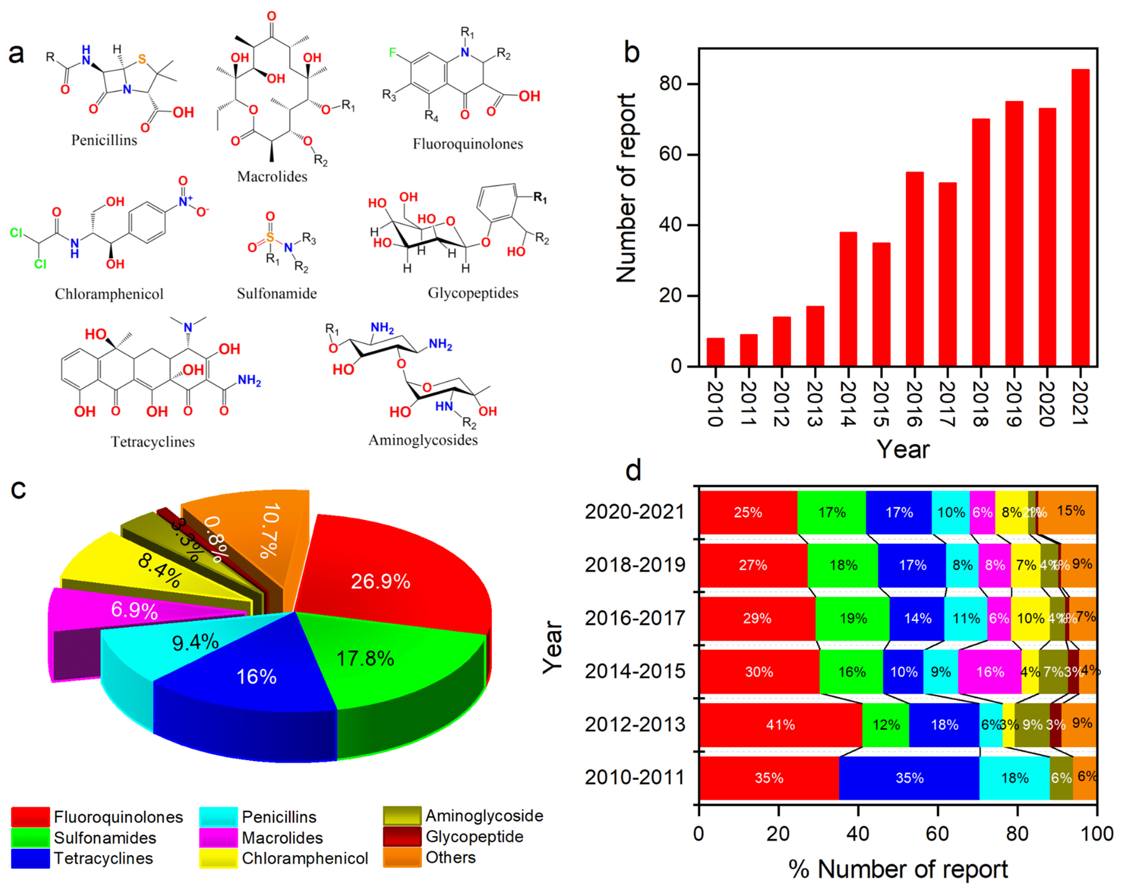
:1. Introduction
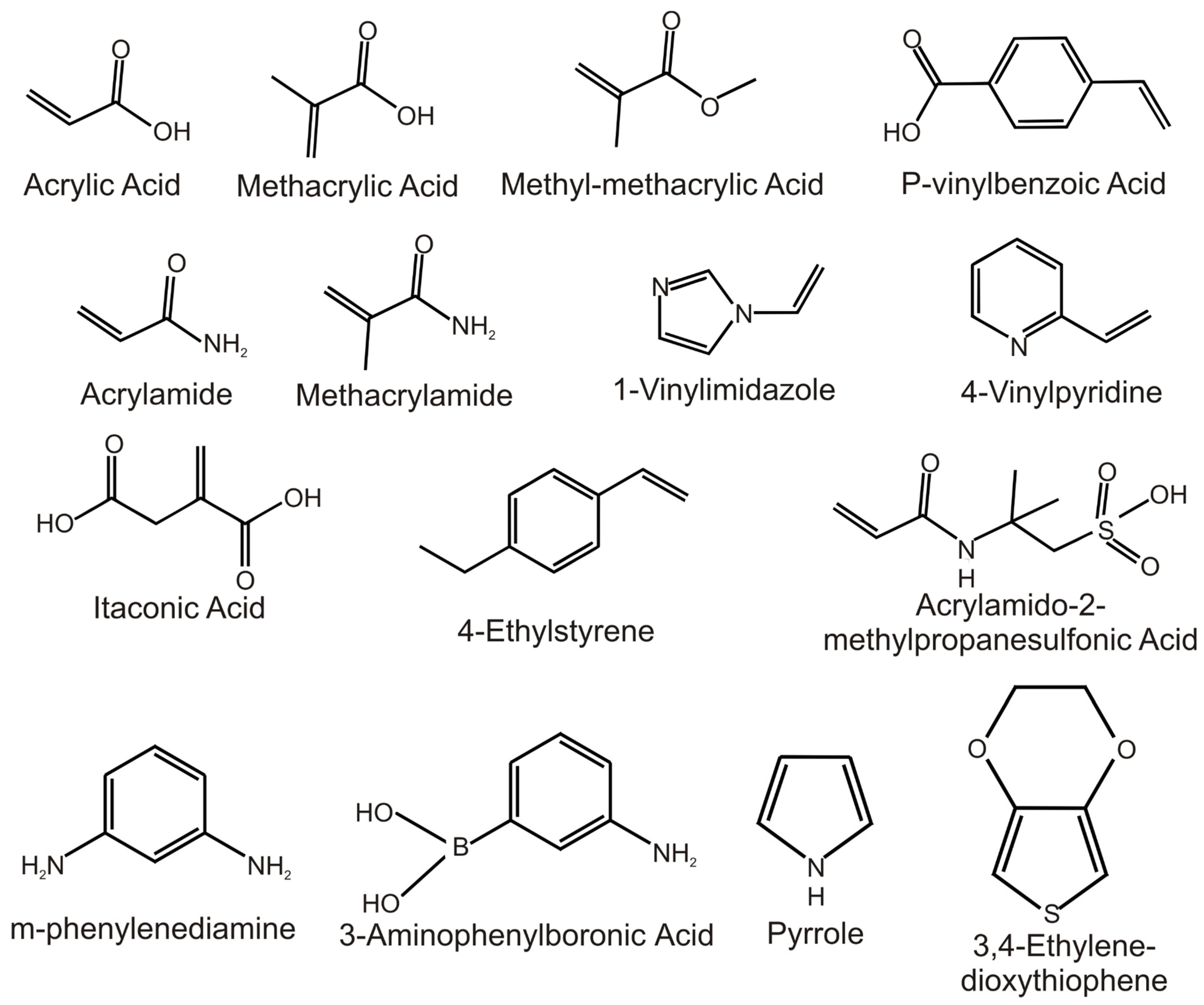
2. Rational Design of Antibiotic MIPs

| Computational Approach | Template | Monomers Studied | Binding Energy (kJ/mol) | Selected Monomer | KD (μM) | QMAX(MIP)/QMAX(NIP) | LOD (nM) | Media | Ref. |
|---|---|---|---|---|---|---|---|---|---|
| QCC | Sulfanilamide | Pyrrole | 18.86 | Pyrrole | - | 20 | Ground water | [67] | |
| Furan | 15.11 | ||||||||
| Thiophene | 8.62 | ||||||||
| 1-Methylthiophene | 7.63 | ||||||||
| Methylpyrrole | 7.13 | ||||||||
| Flumequine | Pyrrole | 34.98 | Pyrrole | 20 | - | 1000 | Aquaculture water | [73] | |
| Sulfadiazine and sulfamerazine | AA | 557.727 552.58 | AA | 0.19 | >3.0 | water | [74] | ||
| Sulfamethizole | mPD Pyrrole EDOT | 181.20 80.26 30.10 | mPD | 47.2 | 8.24 | 2 | Tap water | [75] | |
| Amoxicillin | mPD EDOT Pyrrole | 273.05 8.40 63.01 | mPD | 28.8 | 7.2 | 0.2 | PBS | [68] | |
| Azithromycin | 4-ABA Phenol Pyrrole Aniline Thiophene | 71.55 69.41 68.93 45.08 31.96 | 4-ABA | - | - | 80 | River water | [76] | |
| Molecular Docking (CDOCKER) | Norfloxacin | AM AA MAM MAA N-iAA PVP | 82.22 54.27 58.95 100.33 61.04 76.32 | MAA | 0.004 | 4.3 | 31 | Waste water | [72] |
| Norfloxacin | MAA AA MAM AM 4-VP | 87.45 64.68 53.97 76.65 56.48 | MAA | 2.06 | 2.44 | 16 | Lake water | [71] | |
| Molecular Docking (SYBYL) | Cefquinome sulphate | Pyrrole-2-carboxylic acid Pyrrolidine-2-carbohydrazide 4-ABA oPD 4-ATP | 603.09 485.79 435.47 325.77 271.94 | 4-ABA | - | 3.5 | - | PBS | [77] |
| Nafcillin | oPD Proline Aniline Pyrrolidine-2-carbohydrazide 4-Aminobenzoic acid | 163.55 159.41 152.67 149.33 135.23 | oPD | - | - | 80 | River water | [78] | |
| Chloramphenicol | AA, MMA | 75 | AA | - | - | 1.5 | Tap water | [79] | |
| Molecular Dynamic | Norfloxacin | MAA/EGDMA ratio is optimised | - | MAA, EGDMA | 374; 279 772; 481 | High adsorption capacity (29.35 mg/g) | - | Water | [80] |
| Sulfamethoxazole | APTES/TEOS ratio optimised | - | APTES, TEOS | - | - | 60 | Lake water | [81] | |
| Molecular Mechanic | Penicillin G | CMA and CSEV compatibility | - | MAA, TRIM | - | 6.03–6.69 | - | Water | [82] |
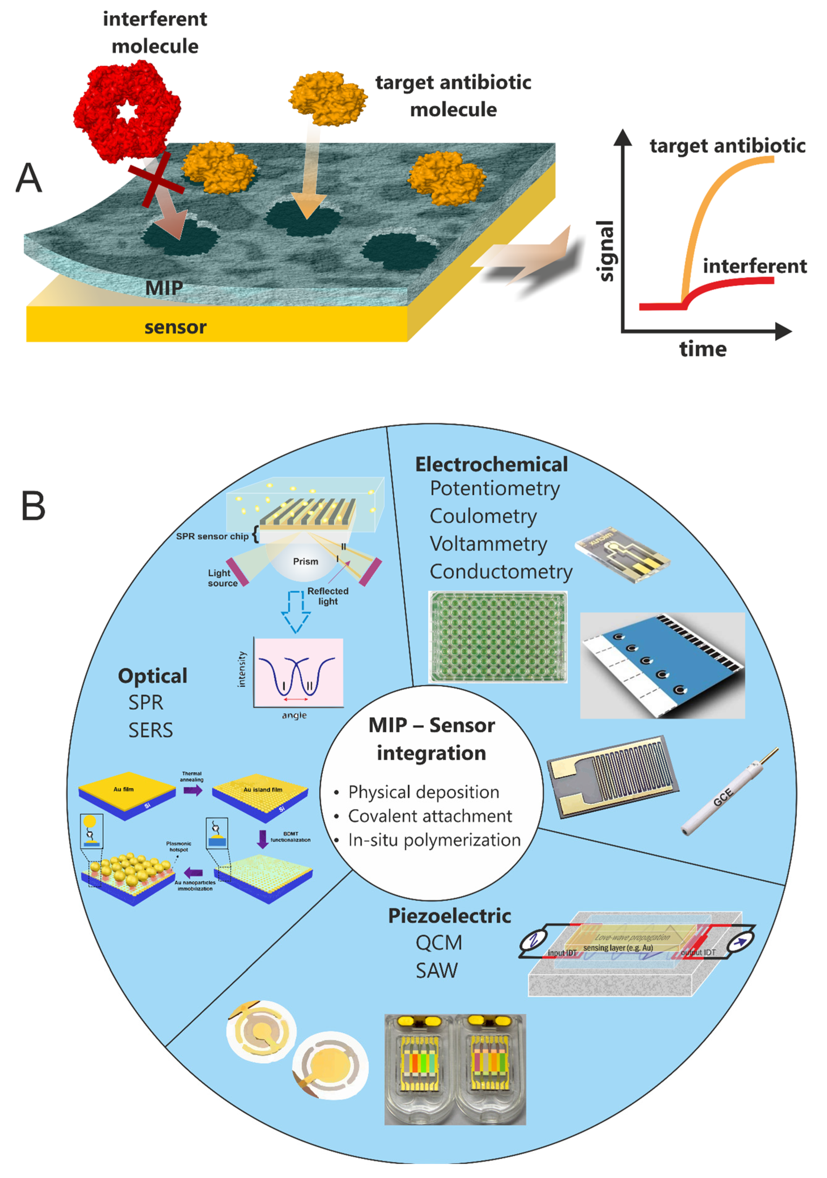
3. Label-Free Detection of Antibiotics
3.1. Optical Sensor Platforms
3.1.1. Surface Plasmon Resonance
3.1.2. Surface-Enhanced Raman Spectroscopy
3.2. Piezoelectric Sensor Platforms
3.2.1. Quartz Crystal Microbalance
3.2.2. Surface Acoustic Wave
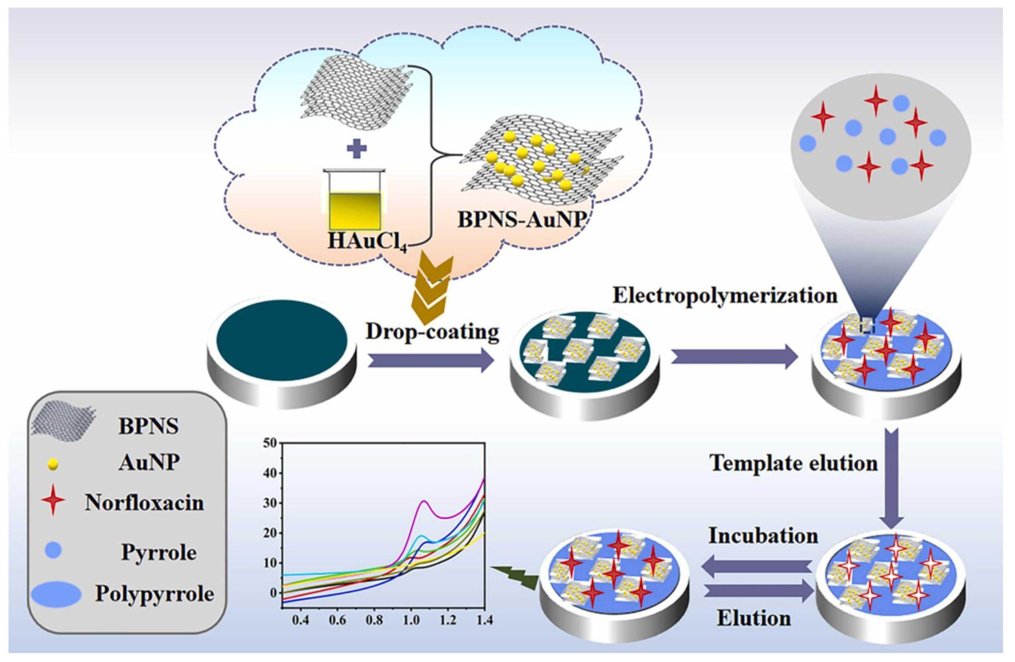
3.3. Electrochemical Sensors
4. Signal Amplification Approaches for Antibiotic MIP-Based Sensors
5. Practical Challenges of Antibiotic MIP
6. Conclusions and Future Perspectives
Author Contributions
Funding
Institutional Review Board Statement
Informed Consent Statement
Data Availability Statement
Conflicts of Interest
References
- Gothwal, R.; Shashidhar, T. Antibiotic Pollution in the Environment: A Review. CLEAN—Soil Air Water 2015, 43, 479–489. [Google Scholar] [CrossRef]
- Tong, L.; Huang, S.; Wang, Y.; Liu, H.; Li, M. Occurrence of Antibiotics in the Aquatic Environment of Jianghan Plain, Central China. Sci. Total Environ. 2014, 497–498, 180–187. [Google Scholar] [CrossRef] [PubMed]
- Dong, H.; Yuan, X.; Wang, W.; Qiang, Z. Occurrence and Removal of Antibiotics in Ecological and Conventional Wastewater Treatment Processes: A Field Study. J. Environ. Manag. 2016, 178, 11–19. [Google Scholar] [CrossRef] [PubMed]
- Jin, Y.; Wu, S.; Zeng, Z.; Fu, Z. Effects of Environmental Pollutants on Gut Microbiota. Environ. Pollut. 2017, 222, 1–9. [Google Scholar] [CrossRef]
- Xu, Y.; Guo, C.; Luo, Y.; Lv, J.; Zhang, Y.; Lin, H.; Wang, L.; Xu, J. Occurrence and Distribution of Antibiotics, Antibiotic Resistance Genes in the Urban Rivers in Beijing, China. Environ. Pollut. 2016, 213, 833–840. [Google Scholar] [CrossRef] [PubMed]
- Almeida, A.; Duarte, S.; Nunes, R.; Rocha, H.; Pena, A.; Meisel, L. Human and Veterinary Antibiotics Used in Portugal—A Ranking for Ecosurveillance. Toxics 2014, 2, 188–225. [Google Scholar] [CrossRef]
- Fent, K.; Weston, A.; Caminada, D. Ecotoxicology of Human Pharmaceuticals. Aquat. Toxicol. 2006, 76, 122–159. [Google Scholar] [CrossRef]
- Garoma, T.; Umamaheshwar, S.K.; Mumper, A. Removal of Sulfadiazine, Sulfamethizole, Sulfamethoxazole, and Sulfathiazole from Aqueous Solution by Ozonation. Chemosphere 2010, 79, 814–820. [Google Scholar] [CrossRef]
- Golovko, O.; Kumar, V.; Fedorova, G.; Randak, T.; Grabic, R. Seasonal Changes in Antibiotics, Antidepressants/Psychiatric Drugs, Antihistamines and Lipid Regulators in a Wastewater Treatment Plant. Chemosphere 2014, 111, 418–426. [Google Scholar] [CrossRef]
- Senta, I.; Terzic, S.; Ahel, M. Occurrence and Fate of Dissolved and Particulate Antimicrobials in Municipal Wastewater Treatment. Water Res. 2013, 47, 705–714. [Google Scholar] [CrossRef] [Green Version]
- Sukul, P.; Spiteller, M. Fluoroquinolone Antibiotics in the Environment. In Reviews of Environmental Contamination and Toxicology; Reviews of Environmental Contamination and Toxicology; Springer: New York, NY, USA, 2007; Volume 191, pp. 131–162. ISBN 978-0-387-69162-6. [Google Scholar]
- Ding, C.; He, J. Effect of Antibiotics in the Environment on Microbial Populations. Appl. Microbiol. Biotechnol. 2010, 87, 925–941. [Google Scholar] [CrossRef] [PubMed]
- Fatta-Kassinos, D.; Meric, S.; Nikolaou, A. Pharmaceutical Residues in Environmental Waters and Wastewater: Current State of Knowledge and Future Research. Anal. Bioanal. Chem. 2011, 399, 251–275. [Google Scholar] [CrossRef] [PubMed]
- Kümmerer, K. The Presence of Pharmaceuticals in the Environment Due to Human Use—Present Knowledge and Future Challenges. J. Environ. Manag. 2009, 90, 2354–2366. [Google Scholar] [CrossRef]
- Martinez, J.L. Environmental Pollution by Antibiotics and by Antibiotic Resistance Determinants. Environ. Pollut. 2009, 157, 2893–2902. [Google Scholar] [CrossRef] [PubMed]
- WHO. Ten Threats to Global Health in 2019. Available online: https://www.who.int/news-room/feature-stories/ten-threats-to-global-health-in-2019 (accessed on 23 April 2020).
- OECD. Health and Economic Burden of Antimicrobial Resistance. In Stemming the Superbug Tide: Just a Few Dollars More; OECD Publishing: Paris, France, 2018. [Google Scholar]
- Cassini, A.; Högberg, L.D.; Plachouras, D.; Quattrocchi, A.; Hoxha, A.; Simonsen, G.S.; Colomb-Cotinat, M.; Kretzschmar, M.E.; Devleesschauwer, B.; Cecchini, M.; et al. Attributable Deaths and Disability-Adjusted Life-Years Caused by Infections with Antibiotic-Resistant Bacteria in the EU and the European Economic Area in 2015: A Population-Level Modelling Analysis. Lancet Infect. Dis. 2019, 19, 56–66. [Google Scholar] [CrossRef] [Green Version]
- Badal, S.; Her, Y.F.; Maher, L.J. Nonantibiotic Effects of Fluoroquinolones in Mammalian Cells. J. Biol. Chem. 2015, 290, 22287–22297. [Google Scholar] [CrossRef] [Green Version]
- Dong, L.; Gao, J.; Xie, X.; Zhou, Q. DNA Damage and Biochemical Toxicity of Antibiotics in Soil on the Earthworm Eisenia Fetida. Chemosphere 2012, 89, 44–51. [Google Scholar] [CrossRef]
- Hillis, D.G.; Fletcher, J.; Solomon, K.R.; Sibley, P.K. Effects of Ten Antibiotics on Seed Germination and Root Elongation in Three Plant Species. Arch. Environ. Contam. Toxicol. 2011, 60, 220–232. [Google Scholar] [CrossRef] [Green Version]
- Kalghatgi, S.; Spina, C.S.; Costello, J.C.; Liesa, M.; Morones-Ramirez, J.R.; Slomovic, S.; Molina, A.; Shirihai, O.S.; Collins, J.J. Bactericidal Antibiotics Induce Mitochondrial Dysfunction and Oxidative Damage in Mammalian Cells. Sci. Transl. Med. 2013, 5, 192ra85. [Google Scholar] [CrossRef] [Green Version]
- Li, Z.-J.; Xie, X.-Y.; Zhang, S.-Q.; Liang, Y.-C. Wheat Growth and Photosynthesis as Affected by Oxytetracycline as a Soil Contaminant. Pedosphere 2011, 21, 244–250. [Google Scholar] [CrossRef]
- Chow, L.K.M.; Ghaly, T.M.; Gillings, M.R. A Survey of Sub-Inhibitory Concentrations of Antibiotics in the Environment. J. Environ. Sci. 2021, 99, 21–27. [Google Scholar] [CrossRef]
- Chemicals—Water Pollution—Environment—European Commission. Available online: https://ec.europa.eu/environment/water/water-dangersub/index.htm#surface_water_watch_list (accessed on 25 April 2022).
- Parthasarathy, R.; Monette, C.E.; Bracero, S.; Sabrina Saha, M. Methods for Field Measurement of Antibiotic Concentrations: Limitations and Outlook. FEMS Microbiol. Ecol. 2018, 94, fiy105. [Google Scholar] [CrossRef]
- Mosbach, K. Molecular Imprinting. Trends Biochem. Sci. 1994, 19, 9–14. [Google Scholar] [CrossRef]
- Saylan, Y.; Akgönüllü, S.; Yavuz, H.; Ünal, S.; Denizli, A. Molecularly Imprinted Polymer Based Sensors for Medical Applications. Sensors 2019, 19, 1279. [Google Scholar] [CrossRef] [Green Version]
- Rutkowska, M.; Płotka-Wasylka, J.; Morrison, C.; Wieczorek, P.P.; Namieśnik, J.; Marć, M. Application of Molecularly Imprinted Polymers in Analytical Chiral Separations and Analysis. TrAC Trends Anal. Chem. 2018, 102, 91–102. [Google Scholar] [CrossRef] [Green Version]
- Hu, T.; Chen, R.; Wang, Q.; He, C.; Liu, S. Recent Advances and Applications of Molecularly Imprinted Polymers in Solid-phase Extraction for Real Sample Analysis. J. Sep. Sci. 2021, 44, 274–309. [Google Scholar] [CrossRef]
- Ahmad, O.S.; Bedwell, T.S.; Esen, C.; Garcia-Cruz, A.; Piletsky, S.A. Molecularly Imprinted Polymers in Electrochemical and Optical Sensors. Trends Biotechnol. 2019, 37, 294–309. [Google Scholar] [CrossRef]
- Zaidi, S.A. Latest Trends in Molecular Imprinted Polymer Based Drug Delivery Systems. RSC Adv. 2016, 6, 88807–88819. [Google Scholar] [CrossRef]
- Pan, J.; Chen, W.; Ma, Y.; Pan, G. Molecularly Imprinted Polymers as Receptor Mimics for Selective Cell Recognition. Chem. Soc. Rev. 2018, 47, 5574–5587. [Google Scholar] [CrossRef]
- Ayankojo, A.G.; Boroznjak, R.; Reut, J.; Öpik, A.; Syritski, V. Molecularly Imprinted Polymer Based Electrochemical Sensor for Quantitative Detection of SARS-CoV-2 Spike Protein. Sens. Actuators B Chem. 2022, 353, 131160. [Google Scholar] [CrossRef]
- Kadhem, A.J.; Gentile, G.J.; Fidalgo de Cortalezzi, M.M. Molecularly Imprinted Polymers (MIPs) in Sensors for Environmental and Biomedical Applications: A Review. Molecules 2021, 26, 6233. [Google Scholar] [CrossRef]
- Selvolini, G.; Marrazza, G. MIP-Based Sensors: Promising New Tools for Cancer Biomarker Determination. Sensors 2017, 17, 718. [Google Scholar] [CrossRef] [Green Version]
- Yarman, A.; Kurbanoglu, S.; Zebger, I.; Scheller, F.W. Simple and Robust: The Claims of Protein Sensing by Molecularly Imprinted Polymers. Sens. Actuators B Chem. 2021, 330, 129369. [Google Scholar] [CrossRef]
- Becskereki, G.; Horvai, G.; Tóth, B. The Selectivity of Molecularly Imprinted Polymers. Polymers 2021, 13, 1781. [Google Scholar] [CrossRef]
- Lowdon, J.W.; Diliën, H.; Singla, P.; Peeters, M.; Cleij, T.J.; van Grinsven, B.; Eersels, K. MIPs for Commercial Application in Low-Cost Sensors and Assays—An Overview of the Current Status Quo. Sens. Actuators B Chem. 2020, 325, 128973. [Google Scholar] [CrossRef]
- Nicholls, I.A.; Golker, K.; Olsson, G.D.; Suriyanarayanan, S.; Wiklander, J.G. The Use of Computational Methods for the Development of Molecularly Imprinted Polymers. Polymers 2021, 13, 2841. [Google Scholar] [CrossRef]
- Sharma, P.S.; Pietrzyk-Le, A.; D’Souza, F.; Kutner, W. Electrochemically Synthesized Polymers in Molecular Imprinting for Chemical Sensing. Anal. Bioanal. Chem. 2012, 402, 3177–3204. [Google Scholar] [CrossRef] [Green Version]
- Kidakova, A.; Reut, J.; Boroznjak, R.; Öpik, A.; Syritski, V. Advanced Sensing Materials Based on Molecularly Imprinted Polymers towards Developing Point-of-Care Diagnostics Devices. Proc. Est. Acad. Sci. 2019, 68, 158. [Google Scholar] [CrossRef]
- Sharma, P.S.; Dąbrowski, M.; D’Souza, F.; Kutner, W. Surface Development of Molecularly Imprinted Polymer Films to Enhance Sensing Signals. TrAC Trends Anal. Chem. 2013, 51, 146–157. [Google Scholar] [CrossRef]
- Altintas, Z. Surface Plasmon Resonance Based Sensor for the Detection of Glycopeptide Antibiotics in Milk Using Rationally Designed NanoMIPs. Sci. Rep. 2018, 8, 11222. [Google Scholar] [CrossRef]
- Munawar, A.; Tahir, M.A.; Shaheen, A.; Lieberzeit, P.A.; Khan, W.S.; Bajwa, S.Z. Investigating Nanohybrid Material Based on 3D CNTs@Cu Nanoparticle Composite and Imprinted Polymer for Highly Selective Detection of Chloramphenicol. J. Hazard. Mater. 2018, 342, 96–106. [Google Scholar] [CrossRef]
- Benachio, I.; Lobato, A.; Gonçalves, L.M. Employing Molecularly Imprinted Polymers in the Development of Electroanalytical Methodologies for Antibiotic Determination. J. Mol. Recognit. 2021, 34, e2878. [Google Scholar] [CrossRef]
- Bitas, D.; Samanidou, V. Molecularly Imprinted Polymers as Extracting Media for the Chromatographic Determination of Antibiotics in Milk. Molecules 2018, 23, 316. [Google Scholar] [CrossRef] [Green Version]
- Jamieson, O.; Mecozzi, F.; Crapnell, R.D.; Battell, W.; Hudson, A.; Novakovic, K.; Sachdeva, A.; Canfarotta, F.; Herdes, C.; Banks, C.E.; et al. Approaches to the Rational Design of Molecularly Imprinted Polymers Developed for the Selective Extraction or Detection of Antibiotics in Environmental and Food Samples. Phys. Status Solidi A 2021, 218, 2100021. [Google Scholar] [CrossRef]
- Mohsenzadeh, M.S.; Mohammadinejad, A.; Mohajeri, S.A. Simple and Selective Analysis of Different Antibiotics in Milk Using Molecularly Imprinted Polymers: A Review. Food Addit. Contam. Part A 2018, 35, 1959–1974. [Google Scholar] [CrossRef]
- Tarannum, N.; Khatoon, S.; Dzantiev, B.B. Perspective and Application of Molecular Imprinting Approach for Antibiotic Detection in Food and Environmental Samples: A Critical Review. Food Control 2020, 118, 107381. [Google Scholar] [CrossRef]
- Tarannum, N.; Hendrickson, O.D.; Khatoon, S.; Zherdev, A.V.; Dzantiev, B.B. Molecularly Imprinted Polymers as Receptors for Assays of Antibiotics. Crit. Rev. Anal. Chem. 2019, 50, 291–310. [Google Scholar] [CrossRef]
- Singhal, A.; Sadique, M.A.; Kumar, N.; Yadav, S.; Ranjan, P.; Parihar, A.; Khan, R.; Kaushik, A.K. Multifunctional Carbon Nanomaterials Decorated Molecularly Imprinted Hybrid Polymers for Efficient Electrochemical Antibiotics Sensing. J. Environ. Chem. Eng. 2022, 10, 107703. [Google Scholar] [CrossRef]
- Chen, L.; Wang, X.; Lu, W.; Wu, X.; Li, J. Molecular Imprinting: Perspectives and Applications. Chem. Soc. Rev. 2016, 45, 2137–2211. [Google Scholar] [CrossRef]
- Qu, S.; Wang, X.; Tong, C.; Wu, J. Metal Ion Mediated Molecularly Imprinted Polymer for Selective Capturing Antibiotics Containing Beta-Diketone Structure. J. Chromatogr. A 2010, 1217, 8205–8211. [Google Scholar] [CrossRef]
- Matsui, J.; Nicholls, I.A.; Takeuchi, T.; Mosbach, K.; Karube, I. Metal Ion Mediated Recognition in Molecularly Imprinted Polymers. Anal. Chim. Acta 1996, 335, 71–77. [Google Scholar] [CrossRef]
- Lay, S.; Ni, X.; Yu, H.; Shen, S. State-of-the-Art Applications of Cyclodextrins as Functional Monomers in Molecular Imprinting Techniques: A Review. J. Sep. Sci. 2016, 39, 2321–2331. [Google Scholar] [CrossRef]
- Wagner, R.; Wan, W.; Biyikal, M.; Benito-Peña, E.; Moreno-Bondi, M.C.; Lazraq, I.; Rurack, K.; Sellergren, B. Synthesis, Spectroscopic, and Analyte-Responsive Behavior of a Polymerizable Naphthalimide-Based Carboxylate Probe and Molecularly Imprinted Polymers Prepared Thereof. J. Org. Chem. 2013, 78, 1377–1389. [Google Scholar] [CrossRef]
- Wang, L.; Lin, Q.; Zhang, Y.; Liu, Y.; Yasin, A.; Zhang, L. Design and Synthesis of Supramolecular Functional Monomers Bearing Urea and Norbornene Motifs. RSC Adv. 2019, 9, 20058–20064. [Google Scholar] [CrossRef] [PubMed] [Green Version]
- Xu, S.; Chen, L.; Li, J.; Guan, Y.; Lu, H. Novel Hg2+-Imprinted Polymers Based on Thymine–Hg2+–Thymine Interaction for Highly Selective Preconcentration of Hg2+ in Water Samples. J. Hazard. Mater. 2012, 237–238, 347–354. [Google Scholar] [CrossRef]
- Zhang, X.; Shen, F.; Zhang, Z.; Xing, Y.; Ren, X. Synthesis of a Novel Cross-Linker Doubles as a Functional Monomer for Preparing a Water Compatible Molecularly Imprinted Polymer. Anal. Methods 2014, 6, 9483–9489. [Google Scholar] [CrossRef]
- Gao, M.; Gao, Y.; Chen, G.; Huang, X.; Xu, X.; Lv, J.; Wang, J.; Xu, D.; Liu, G. Recent Advances and Future Trends in the Detection of Contaminants by Molecularly Imprinted Polymers in Food Samples. Front. Chem. 2020, 8, 616326. [Google Scholar] [CrossRef]
- Muhammad, T.; Nur, Z.; Piletska, E.V.; Yimit, O.; Piletsky, S.A. Rational Design of Molecularly Imprinted Polymer: The Choice of Cross-Linker. Analyst 2012, 137, 2623–2628. [Google Scholar] [CrossRef]
- Zhang, Y.; Song, D.; Lanni, L.M.; Shimizu, K.D. Importance of Functional Monomer Dimerization in the Molecular Imprinting Process. Macromolecules 2010, 43, 6284–6294. [Google Scholar] [CrossRef]
- Azimi, A.; Javanbakht, M. Computational Prediction and Experimental Selectivity Coefficients for Hydroxyzine and Cetirizine Molecularly Imprinted Polymer Based Potentiometric Sensors. Anal. Chim. Acta 2014, 812, 184–190. [Google Scholar] [CrossRef]
- Dong, C.; Li, X.; Guo, Z.; Qi, J. Development of a Model for the Rational Design of Molecular Imprinted Polymer: Computational Approach for Combined Molecular Dynamics/Quantum Mechanics Calculations. Anal. Chim. Acta 2009, 647, 117–124. [Google Scholar] [CrossRef] [PubMed]
- Grabowski, S.J. Chapter 1 Hydrogen Bond—Definitions, Criteria of Existence and Various Types. In Understanding Hydrogen Bonds: Theoretical and Experimental Views; The Royal Society of Chemistry: London, UK, 2021; pp. 1–40. ISBN 978-1-78801-479-3. [Google Scholar]
- Tadi, K.K.; Motghare, R.V.; Ganesh, V. Electrochemical Detection of Sulfanilamide Using Pencil Graphite Electrode Based on Molecular Imprinting Technology. Electroanalysis 2014, 26, 2328–2336. [Google Scholar] [CrossRef]
- Ayankojo, A.G.; Reut, J.; Boroznjak, R.; Öpik, A.; Syritski, V. Molecularly Imprinted Poly(Meta-Phenylenediamine) Based QCM Sensor for Detecting Amoxicillin. Sens. Actuators B-Chem. 2018, 258, 766–774. [Google Scholar] [CrossRef]
- Xi, S.; Zhang, K.; Xiao, D.; He, H. Computational-Aided Design of Magnetic Ultra-Thin Dummy Molecularly Imprinted Polymer for Selective Extraction and Determination of Morphine from Urine by High-Performance Liquid Chromatography. J. Chromatogr. A 2016, 1473, 1–9. [Google Scholar] [CrossRef]
- Zhang, K.; Zou, W.; Zhao, H.; Dramou, P.; Pham-Huy, C.; He, J.; He, H. Adsorption Behavior of a Computer-Aid Designed Magnetic Molecularly Imprinted Polymer via Response Surface Methodology. RSC Adv. 2015, 5, 61161–61169. [Google Scholar] [CrossRef]
- Fizir, M.; Wei, L.; Muchuan, N.; Itatahine, A.; mehdi, Y.A.; He, H.; Dramou, P. QbD Approach by Computer Aided Design and Response Surface Methodology for Molecularly Imprinted Polymer Based on Magnetic Halloysite Nanotubes for Extraction of Norfloxacin from Real Samples. Talanta 2018, 184, 266–276. [Google Scholar] [CrossRef]
- Niu, M.; Sun, C.; Zhang, K.; Li, G.; Meriem, F.; Pham-Huy, C.; Hui, X.; Shi, J.; He, H. A Simple Extraction Method for Norfloxacin from Pharmaceutical Wastewater with a Magnetic Core–Shell Molecularly Imprinted Polymer with the Aid of Computer Simulation. New J. Chem. 2017, 41, 2614–2624. [Google Scholar] [CrossRef]
- Ktari, N.; Fourati, N.; Zerrouki, C.; Ruan, M.; Seydou, M.; Barbaut, F.; Nal, F.; Yaakoubi, N.; Chehimi, M.M.; Kalfat, R. Design of a Polypyrrole MIP-SAW Sensor for Selective Detection of Flumequine in Aqueous Media. Correlation between Experimental Results and DFT Calculations. RSC Adv. 2015, 5, 88666–88674. [Google Scholar] [CrossRef]
- Chen, L.; Lee, Y.K.; Manmana, Y.; Tay, K.S.; Lee, V.S.; Rahman, N.A. Synthesis, Characterization, and Theoretical Study of an Acrylamide-Based Magnetic Molecularly Imprinted Polymer for the Recognition of Sulfonamide Drugs. E-Polymers 2015, 15, 141–150. [Google Scholar] [CrossRef]
- Ayankojo, A.G.; Tretjakov, A.; Reut, J.; Boroznjak, R.; Öpik, A.; Rappich, J.; Furchner, A.; Hinrichs, K.; Syritski, V. Molecularly Imprinted Polymer Integrated with a Surface Acoustic Wave Technique for Detection of Sulfamethizole. Anal. Chem. 2016, 88, 1476–1484. [Google Scholar] [CrossRef]
- Rebelo, P.; Pacheco, J.G.; Cordeiro, M.N.D.S.; Melo, A.; Delerue-Matos, C. Azithromycin Electrochemical Detection Using a Molecularly Imprinted Polymer Prepared on a Disposable Screen-Printed Electrode. Anal. Methods 2020, 12, 1486–1494. [Google Scholar] [CrossRef]
- Moro, G.; Bottari, F.; Sleegers, N.; Florea, A.; Cowen, T.; Moretto, L.M.; Piletsky, S.; De Wael, K. Conductive Imprinted Polymers for the Direct Electrochemical Detection of β-Lactam Antibiotics: The Case of Cefquinome. Sens. Actuators B Chem. 2019, 297, 126786. [Google Scholar] [CrossRef]
- Bottari, F.; Moro, G.; Sleegers, N.; Florea, A.; Cowen, T.; Piletsky, S.; van Nuijs, A.L.N.; De Wael, K. Electropolymerized O-Phenylenediamine on Graphite Promoting the Electrochemical Detection of Nafcillin. Electroanalysis 2020, 32, 135–141. [Google Scholar] [CrossRef]
- Sai, N.; Wu, Y.; Yu, G.; Sun, Z.; Huang, G. A Novel Enrichment Imprinted Crystalline Colloidal Array for the Ultratrace Detection of Chloramphenicol. Talanta 2016, 161, 1–7. [Google Scholar] [CrossRef]
- Kong, Y.; Wang, N.; Ni, X.; Yu, Q.; Liu, H.; Huang, W.; Xu, W. Molecular Dynamics Simulations of Molecularly Imprinted Polymer Approaches to the Preparation of Selective Materials to Remove Norfloxacin. J. Appl. Polym. Sci. 2016, 133, 42817. [Google Scholar] [CrossRef]
- Xu, W.; Wang, Y.; Huang, W.; Yu, L.; Yang, Y.; Liu, H.; Yang, W. Computer-Aided Design and Synthesis of CdTe@SiO2 Core-Shell Molecularly Imprinted Polymers as a Fluorescent Sensor for the Selective Determination of Sulfamethoxazole in Milk and Lake Water. J. Sep. Sci. 2017, 40, 1091–1098. [Google Scholar] [CrossRef]
- Kempe, H.; Kempe, M. QSRR Analysis of β-Lactam Antibiotics on a Penicillin G Targeted MIP Stationary Phase. Anal. Bioanal. Chem. 2010, 398, 3087–3096. [Google Scholar] [CrossRef] [Green Version]
- Homola, J. Surface Plasmon Resonance Sensors for Detection of Chemical and Biological Species. Chem. Rev. 2008, 108, 462–493. [Google Scholar] [CrossRef]
- Shankaran, D.R.; Gobiand, K.V.; Miura, N. Recent Advancements in Surface Plasmon Resonance Immunosensors for Detection of Small Molecules of Biomedical, Food and Environmental Interest. Sens. Actuators B Chem. 2007, 121, 158–177. [Google Scholar] [CrossRef]
- Homola, J.; Yee, S.S.; Gauglitz, G. Surface Plasmon Resonance Sensors: Review. Sens. Actuators B Chem. 1999, 54, 3–15. [Google Scholar] [CrossRef]
- Lotierzo, M.; Henry, O.Y.F.; Piletsky, S.; Tothill, I.; Cullen, D.; Kania, M.; Hock, B.; Turner, A.P.F. Surface Plasmon Resonance Sensor for Domoic Acid Based on Grafted Imprinted Polymer. Biosens. Bioelectron. 2004, 20, 145–152. [Google Scholar] [CrossRef] [PubMed] [Green Version]
- Matsui, J.; Akamatsu, K.; Hara, N.; Miyoshi, D.; Nawafune, H.; Tamaki, K.; Sugimoto, N. SPR Sensor Chip for Detection of Small Molecules Using Molecularly Imprinted Polymer with Embedded Gold Nanoparticles. Anal. Chem. 2005, 77, 4282–4285. [Google Scholar] [CrossRef]
- Hinman, S.S.; McKeating, K.S.; Cheng, Q. Surface Plasmon Resonance: Material and Interface Design for Universal Accessibility. Anal. Chem. 2018, 90, 19–39. [Google Scholar] [CrossRef]
- Frasconi, M.; Tel-Vered, R.; Riskin, M.; Willner, I. Surface Plasmon Resonance Analysis of Antibiotics Using Imprinted Boronic Acid-Functionalized Au Nanoparticle Composites. Anal. Chem. 2010, 82, 2512–2519. [Google Scholar] [CrossRef]
- Shrivastav, A.M.; Mishra, S.K.; Gupta, B.D. Localized and Propagating Surface Plasmon Resonance Based Fiber Optic Sensor for the Detection of Tetracycline Using Molecular Imprinting. Mater. Res. Express 2015, 2, 035007. [Google Scholar] [CrossRef]
- Ayankojo, A.G.; Reut, J.; Öpik, A.; Furchner, A.; Syritski, V. Hybrid Molecularly Imprinted Polymer for Amoxicillin Detection. Biosens. Bioelectron. 2018, 118, 102–107. [Google Scholar] [CrossRef]
- Sari, E.; Üzek, R.; Duman, M.; Denizli, A. Detection of Ciprofloxacin through Surface Plasmon Resonance Nanosensor with Specific Recognition Sites. J. Biomater. Sci. Polym. Ed. 2018, 29, 1302–1318. [Google Scholar] [CrossRef]
- Ivleva, N.P.; Wagner, M.; Horn, H.; Niessner, R.; Haisch, C. Raman Microscopy and Surface-Enhanced Raman Scattering (SERS) for in Situ Analysis of Biofilms. J. Biophotonics 2010, 3, 548–556. [Google Scholar] [CrossRef]
- Kostrewa, S.; Emgenbroich, M.; Klockow, D.; Wulff, G. Surface-Enhanced Raman Scattering on Molecularly Imprinted Polymers in Water. Macromol. Chem. Phys. 2003, 204, 481–487. [Google Scholar] [CrossRef]
- Ashley, J.; Wu, K.; Hansen, M.F.; Schmidt, M.S.; Boisen, A.; Sun, Y. Quantitative Detection of Trace Level Cloxacillin in Food Samples Using Magnetic Molecularly Imprinted Polymer Extraction and Surface-Enhanced Raman Spectroscopy Nanopillars. Anal. Chem. 2017, 89, 11484–11490. [Google Scholar] [CrossRef] [Green Version]
- Guo, X.; Li, J.; Arabi, M.; Wang, X.; Wang, Y.; Chen, L. Molecular-Imprinting-Based Surface-Enhanced Raman Scattering Sensors. ACS Sens. 2020, 5, 601–619. [Google Scholar] [CrossRef] [PubMed]
- Carrasco, S.; Benito-Peña, E.; Navarro-Villoslada, F.; Langer, J.; Sanz-Ortiz, M.N.; Reguera, J.; Liz-Marzán, L.M.; Moreno-Bondi, M.C. Multibranched Gold–Mesoporous Silica Nanoparticles Coated with a Molecularly Imprinted Polymer for Label-Free Antibiotic Surface-Enhanced Raman Scattering Analysis. Chem. Mater. 2016, 28, 7947–7954. [Google Scholar] [CrossRef]
- Sari, E.; Üzek, R.; Duman, M.; Denizli, A. Fabrication of Surface Plasmon Resonance Nanosensor for the Selective Determination of Erythromycin via Molecular Imprinted Nanoparticles. Talanta 2016, 150, 607–614. [Google Scholar] [CrossRef]
- Sui, G.; Yang, X.; Li, H.; Li, Y.; Li, L.; Shao, J.; Li, Y.; Wang, J.; Xue, Y.; Zhang, J.; et al. Synthesis of SERS Imprinted Membrane Based on Ag/ESM with Different Morphologies for Selective Detection of Antibiotics in Aqueous Sample. Opt. Mater. 2021, 121, 111581. [Google Scholar] [CrossRef]
- Sullivan, M.V.; Henderson, A.; Hand, R.A.; Turner, N.W. A Molecularly Imprinted Polymer Nanoparticle-Based Surface Plasmon Resonance Sensor Platform for Antibiotic Detection in River Water and Milk. Anal. Bioanal. Chem. 2022, 414, 3687–3696. [Google Scholar] [CrossRef]
- Wang, J.; Li, X.; Zhang, R.; Fu, B.; Chen, M.; Ye, M.; Liu, W.; Xu, J.; Pan, G.; Zhang, H. A Molecularly Imprinted Antibiotic Receptor on Magnetic Nanotubes for the Detection and Removal of Environmental Oxytetracycline. J. Mater. Chem. B 2022. [Google Scholar] [CrossRef]
- Huang, Q.-D.; Lv, C.-H.; Yuan, X.-L.; He, M.; Lai, J.-P.; Sun, H. A Novel Fluorescent Optical Fiber Sensor for Highly Selective Detection of Antibiotic Ciprofloxacin Based on Replaceable Molecularly Imprinted Nanoparticles Composite Hydrogel Detector. Sens. Actuators B Chem. 2021, 328, 129000. [Google Scholar] [CrossRef]
- Li, H.; Zhang, J.; Wang, D.; Wang, J.; Jiang, W.; Zhou, T.; Liu, C.; Che, G. Synthesization of Flexible SERS Imprinted Sensor Based on Ag/GO Composites and Selective Detection of Antibiotic in Aqueous Sample. Adv. Powder Technol. 2021, 32, 3405–3411. [Google Scholar] [CrossRef]
- Zhang, F.; Liu, E.; Zheng, X.; Yu, L.; Yan, Y. A Flexible Imprinted Photonic Resin Film Templated by Nanocrystalline Cellulose for Naked-Eye Recognition of Sulfonamides. J. Ind. Eng. Chem. 2018, 58, 172–178. [Google Scholar] [CrossRef]
- Cao, Y.; Liu, G.; Zheng, B.; Wang, X.; Li, H.; Wang, G.; Zhao, L.; Wang, Y. A Sulfamethoxazole Molecularly Imprinted Two-Dimensional Photonic Crystal Hydrogel Sensor. Soft Matter 2021, 17, 4969–4978. [Google Scholar] [CrossRef]
- Zheng, B.; Liu, G.; Zhao, L.; Wang, G.; Wang, Y. Levofloxacin Molecularly Imprinted Two Dimensional Photonic Crystal Hydrogel Sensor. Polymer 2022, 239, 124448. [Google Scholar] [CrossRef]
- Li, H.; Jia, X.; Jiang, W.; Zhou, T.; He, J.; Luan, Y.; Shang, Y.; Liu, C.; Che, G. Magnetically Assisted Imprinted Sensor for Selective Detection Antibiotics in River Based on Surface-Enhanced Raman Scattering. Opt. Mater. 2020, 108, 110200. [Google Scholar] [CrossRef]
- Wei, X.; Zhang, S.; Wang, T.; Chen, A.; Guo, J.; Bai, B.; Yang, S.; Li, Y.; Wang, X.; Liu, X. Molecularly Imprinted CsPbBr3 Quantum Dot-Based Fluorescent Sensor for Trace Tetracycline Detection in Aqueous Environments. J. Mater. Chem. C 2022, 10, 8432–8440. [Google Scholar] [CrossRef]
- Chandra Sekhar, B.; Dhanalakshmi, B.; Srinivasa Rao, B.; Ramesh, S.; Venkata Prasad, K.; Subba Rao, P.S.V.; Parvatheeswara Rao, B. Piezoelectricity and Its Applications. In Multifunctional Ferroelectric Materials; Ranjan Sahu, D., Ed.; IntechOpen: London, UK, 2021; ISBN 978-1-83968-991-8. [Google Scholar]
- Cooper, M.A.; Singleton, V.T. A Survey of the 2001 to 2005 Quartz Crystal Microbalance Biosensor Literature: Applications of Acoustic Physics to the Analysis of Biomolecular Interactions. J. Mol. Recognit. 2007, 20, 154–184. [Google Scholar] [CrossRef]
- Acharya, B.; Sidheswaran, M.A.; Yungk, R.; Krim, J. Quartz Crystal Microbalance Apparatus for Study of Viscous Liquids at High Temperatures. Rev. Sci. Instrum. 2017, 88, 025112. [Google Scholar] [CrossRef]
- Wachiralurpan, S.; Chansiri, K.; Lieberzeit, P.A. Direct Detection of Listeria Monocytogenes DNA Amplification Products with Quartz Crystal Microbalances at Elevated Temperatures. Sens. Actuators B Chem. 2020, 308, 127678. [Google Scholar] [CrossRef]
- Ávila, M.; Zougagh, M.; Ríos, Á.; Escarpa, A. Molecularly Imprinted Polymers for Selective Piezoelectric Sensing of Small Molecules. TrAC Trends Anal. Chem. 2008, 27, 54–65. [Google Scholar] [CrossRef]
- Uludağ, Y.; Piletsky, S.A.; Turner, A.P.F.; Cooper, M.A. Piezoelectric Sensors Based on Molecular Imprinted Polymers for Detection of Low Molecular Mass Analytes: Detection of Low Molecular Mass Analytes. FEBS J. 2007, 274, 5471–5480. [Google Scholar] [CrossRef]
- Emir Diltemiz, S.; Keçili, R.; Ersöz, A.; Say, R. Molecular Imprinting Technology in Quartz Crystal Microbalance (QCM) Sensors. Sensors 2017, 17, 454. [Google Scholar] [CrossRef]
- Haupt, K.; Noworyta, K.; Kutner, W. Imprinted Polymer-Based Enantioselective Acoustic Sensor Using a Quartz Crystal Microbalance. Anal. Commun. 1999, 36, 391–393. [Google Scholar] [CrossRef]
- Pan, M.; Gu, Y.; Zhang, M.; Wang, J.; Yun, Y.; Wang, S. Reproducible Molecularly Imprinted QCM Sensor for Accurate, Stable, and Sensitive Detection of Enrofloxacin Residue in Animal-Derived Foods. Food Anal. Methods 2018, 11, 495–503. [Google Scholar] [CrossRef]
- Afzal, A.; Iqbal, N.; Mujahid, A.; Schirhagl, R. Advanced Vapor Recognition Materials for Selective and Fast Responsive Surface Acoustic Wave Sensors: A Review. Anal. Chim. Acta 2013, 787, 36–49. [Google Scholar] [CrossRef]
- Ferrari, V.; Lucklum, R. Overview of Acoustic-Wave Microsensors. In Piezoelectric Transducers and Applications; Vives, A.A., Ed.; Springer: Berlin/Heidelberg, Germany, 2008; pp. 39–62. ISBN 978-3-540-77507-2. [Google Scholar]
- Du, J.; Harding, G.L.; Ogilvy, J.A.; Dencher, P.R.; Lake, M. A Study of Love-Wave Acoustic Sensors. Sens. Actuators A Phys. 1996, 56, 211–219. [Google Scholar] [CrossRef]
- Kovacs, G.; Vellekoop, M.J.; Haueis, R.; Lubking, G.W.; Venema, A. A Love Wave Sensor for (Bio)Chemical Sensing in Liquids. Sens. Actuators A Phys. 1994, 43, 38–43. [Google Scholar] [CrossRef]
- Devkota, J.; Ohodnicki, P.; Greve, D. SAW Sensors for Chemical Vapors and Gases. Sensors 2017, 17, 801. [Google Scholar] [CrossRef] [Green Version]
- Tretjakov, A.; Syritski, V.; Reut, J.; Boroznjak, R.; Öpik, A. Molecularly Imprinted Polymer Film Interfaced with Surface Acoustic Wave Technology as a Sensing Platform for Label-Free Protein Detection. Anal. Chim. Acta 2016, 902, 182–188. [Google Scholar] [CrossRef]
- Okan, M.; Duman, M. Functional Polymeric Nanoparticle Decorated Microcantilever Sensor for Specific Detection of Erythromycin. Sens. Actuators B Chem. 2018, 256, 325–333. [Google Scholar] [CrossRef]
- Haghdoust, S.; Arshad, U.; Mujahid, A.; Schranzhofer, L.; Lieberzeit, P.A. Development of a MIP-Based QCM Sensor for Selective Detection of Penicillins in Aqueous Media. Chemosensors 2021, 9, 362. [Google Scholar] [CrossRef]
- Shaheen, A.; Taj, A.; Liberzeit, P.A.; Mujahid, A.; Hameed, S.; Yu, H.; Mahmood, A.; Webster, T.J.; Rashid, M.H.; Khan, W.S.; et al. Design of Heterostructured Hybrids Comprising Ultrathin 2D Bismuth Tungstate Nanosheets Reinforced by Chloramphenicol Imprinted Polymers Used as Biomimetic Interfaces for Mass-Sensitive Detection. Colloids Surf. B Biointerfaces 2020, 188, 110775. [Google Scholar] [CrossRef]
- Shaheen, A.; Taj, A.; Jameel, F.; Tahir, M.A.; Mujahid, A.; Butt, F.K.; Khan, W.S.; Bajwa, S.Z. Synthesis of Graphitic Carbon Nitride Nanosheets-Layered Imprinted Polymer System as a Nanointerface for Detection of Chloramphenicol. Appl. Nanosci. 2022, 12, 139–150. [Google Scholar] [CrossRef]
- Qian, L.; Durairaj, S.; Prins, S.; Chen, A. Nanomaterial-Based Electrochemical Sensors and Biosensors for the Detection of Pharmaceutical Compounds. Biosens. Bioelectron. 2021, 175, 112836. [Google Scholar] [CrossRef] [PubMed]
- Wang, Q.; Xue, Q.; Chen, T.; Li, J.; Liu, Y.; Shan, X.; Liu, F.; Jia, J. Recent Advances in Electrochemical Sensors for Antibiotics and Their Applications. Chin. Chem. Lett. 2021, 32, 609–619. [Google Scholar] [CrossRef]
- Li, G.; Qi, X.; Zhang, G.; Wang, S.; Li, K.; Wu, J.; Wan, X.; Liu, Y.; Li, Q. Low-Cost Voltammetric Sensors for Robust Determination of Toxic Cd(II) and Pb(II) in Environment and Food Based on Shuttle-like α-Fe2O3 Nanoparticles Decorated β-Bi2O3 Microspheres. Microchem. J. 2022, 179, 107515. [Google Scholar] [CrossRef]
- Grieshaber, D.; MacKenzie, R.; Vörös, J.; Reimhult, E.; Grieshaber, D.; MacKenzie, R.; Vörös, J.; Reimhult, E. Electrochemical Biosensors—Sensor Principles and Architectures. Sensors 2008, 8, 1400–1458. [Google Scholar] [CrossRef]
- Sharma, P.S.; Garcia-Cruz, A.; Cieplak, M.; Noworyta, K.R.; Kutner, W. ‘Gate Effect’ in Molecularly Imprinted Polymers: The Current State of Understanding. Curr. Opin. Electrochem. 2019, 16, 50–56. [Google Scholar] [CrossRef]
- Zamora-Gálvez, A.; Ait-Lahcen, A.; Mercante, L.A.; Morales-Narváez, E.; Amine, A.; Merkoçi, A. Molecularly Imprinted Polymer-Decorated Magnetite Nanoparticles for Selective Sulfonamide Detection. Anal. Chem. 2016, 88, 3578–3584. [Google Scholar] [CrossRef]
- Zhao, Y.; Yuan, F.; Quan, X.; Yu, H.; Chen, S.; Zhao, H.; Liu, Z.; Hilal, N. An Electrochemical Sensor for Selective Determination of Sulfamethoxazole in Surface Water Using a Molecularly Imprinted Polymer Modified BDD Electrode. Anal. Methods 2015, 7, 2693–2698. [Google Scholar] [CrossRef]
- Beytur, M.; Kardaş, F.; Akyıldırım, O.; Özkan, A.; Bankoğlu, B.; Yüksek, H.; Yola, M.L.; Atar, N. A Highly Selective and Sensitive Voltammetric Sensor with Molecularly Imprinted Polymer Based Silver@gold Nanoparticles/Ionic Liquid Modified Glassy Carbon Electrode for Determination of Ceftizoxime. J. Mol. Liq. 2018, 251, 212–217. [Google Scholar] [CrossRef]
- Li, G.; Qi, X.; Wu, J.; Xu, L.; Wan, X.; Liu, Y.; Chen, Y.; Li, Q. Ultrasensitive, Label-Free Voltammetric Determination of Norfloxacin Based on Molecularly Imprinted Polymers and Au Nanoparticle-Functionalized Black Phosphorus Nanosheet Nanocomposite. J. Hazard. Mater. 2022, 436, 129107. [Google Scholar] [CrossRef]
- Li, J.; Huang, X.; Ma, J.; Wei, S.; Zhang, H. A Novel Electrochemical Sensor Based on Molecularly Imprinted Polymer with Binary Functional Monomers at Fe-Doped Porous Carbon Decorated Au Electrode for the Sensitive Detection of Lomefloxacin. Ionics 2020, 26, 4183–4192. [Google Scholar] [CrossRef]
- Ayankojo, A.G.; Reut, J.; Ciocan, V.; Öpik, A.; Syritski, V. Molecularly Imprinted Polymer-Based Sensor for Electrochemical Detection of Erythromycin. Talanta 2020, 209, 120502. [Google Scholar] [CrossRef] [PubMed]
- Chu, T.-X.; Vu, V.-P.; Tran, H.-T.; Tran, T.-L.; Tran, Q.-T.; Le Manh, T. Molecularly Imprinted Polyaniline Nanowire-Based Electrochemical Biosensor for Chloramphenicol Detection: A Kinetic Study of Aniline Electropolymerization. J. Electrochem. Soc. 2020, 167, 027527. [Google Scholar] [CrossRef]
- Bi, H.; Wu, Y.; Wang, Y.; Liu, G.; Ning, G.; Xu, Z. A Molecularly Imprinted Polymer Combined with Dual Functional Au@Fe3O4 Nanocomposites for Sensitive Detection of Kanamycin. J. Electroanal. Chem. 2020, 870, 114216. [Google Scholar] [CrossRef]
- Peng, Y.; Ji, Q. A Sensitive and Selective Electrochemical Sensor for Sulfadimethoxine Based on Electropolymerized Molecularly Imprinted Poly (o-Aminophenol) Film. Curr. Anal. Chem. 2020, 16, 413–420. [Google Scholar] [CrossRef]
- De Faria, L.V.; Lisboa, T.P.; Matias, T.A.; de Sousa, R.A.; Matos, M.A.C.; Munoz, R.A.A.; Matos, R.C. Use of Reduced Graphene Oxide for Sensitive Determination of Sulfanilamide in Synthetic Biological Fluids and Environmental Samples by Batch Injection Analysis. J. Electroanal. Chem. 2021, 892, 115298. [Google Scholar] [CrossRef]
- Surya, S.G.; Khatoon, S.; Ait Lahcen, A.; Nguyen, A.T.H.; Dzantiev, B.B.; Tarannum, N.; Salama, K.N. A Chitosan Gold Nanoparticles Molecularly Imprinted Polymer Based Ciprofloxacin Sensor. RSC Adv. 2020, 10, 12823–12832. [Google Scholar] [CrossRef]
- Wang, D.; Jiang, S.; Liang, Y.; Wang, X.; Zhuang, X.; Tian, C.; Luan, F.; Chen, L. Selective Detection of Enrofloxacin in Biological and Environmental Samples Using a Molecularly Imprinted Electrochemiluminescence Sensor Based on Functionalized Copper Nanoclusters. Talanta 2022, 236, 122835. [Google Scholar] [CrossRef]
- Chen, J.; Tan, L.; Qu, K.; Cui, Z.; Wang, J. Novel Electrochemical Sensor Modified with Molecularly Imprinted Polymers for Determination of Enrofloxacin in Marine Environment. Microchim. Acta 2022, 189, 95. [Google Scholar] [CrossRef]
- Hammam, M.A.; Wagdy, H.A.; El Nashar, R.M. Moxifloxacin Hydrochloride Electrochemical Detection Based on Newly Designed Molecularly Imprinted Polymer. Sens. Actuators B Chem. 2018, 275, 127–136. [Google Scholar] [CrossRef]
- Pan, Y.; Shan, D.; Ding, L.; Yang, X.; Xu, K.; Huang, H.; Wang, J.; Ren, H. Developing a Generally Applicable Electrochemical Sensor for Detecting Macrolides in Water with Thiophene-Based Molecularly Imprinted Polymers. Water Res. 2021, 205, 117670. [Google Scholar] [CrossRef]
- López, R.; Khan, S.; Wong, A.; Sotomayor, M.D.P.T.; Picasso, G. Development of a New Electrochemical Sensor Based on Mag-MIP Selective Toward Amoxicillin in Different Samples. Front. Chem. 2021, 9, 615602. [Google Scholar] [CrossRef] [PubMed]
- Rebelo, P.; Pacheco, J.G.; Voroshylova, I.V.; Melo, A.; Cordeiro, M.N.D.S.; Delerue-Matos, C. Rational Development of Molecular Imprinted Carbon Paste Electrode for Furazolidone Detection: Theoretical and Experimental Approach. Sens. Actuators B Chem. 2021, 329, 129112. [Google Scholar] [CrossRef]
- Jamieson, O.; Soares, T.C.C.; de Faria, B.A.; Hudson, A.; Mecozzi, F.; Rowley-Neale, S.J.; Banks, C.E.; Gruber, J.; Novakovic, K.; Peeters, M.; et al. Screen Printed Electrode Based Detection Systems for the Antibiotic Amoxicillin in Aqueous Samples Utilising Molecularly Imprinted Polymers as Synthetic Receptors. Chemosensors 2019, 8, 5. [Google Scholar] [CrossRef] [Green Version]
- Tavares, A.P.; de Sá, M.H.; Sales, M.G.F. Innovative Screen-Printed Electrodes on Cork Composite Substrates Applied to Sulfadiazine Electrochemical Sensing. J. Electroanal. Chem. 2021, 880, 114922. [Google Scholar] [CrossRef]
- Wei, C.; Zhou, H.; Liu, Q. PCN-222 MOF Decorated Conductive PEDOT Films for Sensitive Electrochemical Determination of Chloramphenicol. Mater. Chem. Phys. 2021, 270, 124831. [Google Scholar] [CrossRef]
- Zhang, Z.; Wu, T.; Zhou, H.; Jiang, C.; Wang, Y. 3D Flower-Shaped BiOI Encapsulated in Molecularly Imprinted Polymer for Hypersensitivity to Norfloxacin. Microchem. J. 2021, 164, 106017. [Google Scholar] [CrossRef]
- Bai, X.; Zhang, Y.; Gao, W.; Zhao, D.; Yang, D.; Jia, N. Hollow ZnS–CdS Nanocage Based Photoelectrochemical Sensor Combined with Molecularly Imprinting Technology for Sensitive Detection of Oxytetracycline. Biosens. Bioelectron. 2020, 168, 112522. [Google Scholar] [CrossRef]
- Zhang, Z.; Zhou, H.; Jiang, C.; Wang, Y. Molecularly Imprinted Polymer Functionalized Flower-like BiOBr Microspheres for Photoelectrochemical Sensing of Chloramphenicol. Electrochim. Acta 2020, 344, 136161. [Google Scholar] [CrossRef]
- Xiao, F.; Zhao, F.; Li, J.; Yan, R.; Yu, J.; Zeng, B. Sensitive Voltammetric Determination of Chloramphenicol by Using Single-Wall Carbon Nanotube–Gold Nanoparticle–Ionic Liquid Composite Film Modified Glassy Carbon Electrodes. Anal. Chim. Acta 2007, 596, 79–85. [Google Scholar] [CrossRef]
- Ebarvia, B.S.; Ubando, I.E.; Sevilla, F.B. Biomimetic Piezoelectric Quartz Crystal Sensor with Chloramphenicol-Imprinted Polymer Sensing Layer. Talanta 2015, 144, 1260–1265. [Google Scholar] [CrossRef]
- Ebarvia, B.S.; Ubando, I.E. Molecularly Imprinted Polymer Sensing Layer for Tetracycline Chemical Sensor Based on Piezoelectric Quartz Crystal Transducer. Sens. Transducers 2018, 28, 7–11. [Google Scholar]
- Ayankojo, A.G.; Reut, J.; Öpik, A.; Tretjakov, A.; Syritski, V. Enhancing Binding Properties of Imprinted Polymers for the Detection of Small Molecules. Proc. Est. Acad. Sci. 2018, 67, 138–146. [Google Scholar] [CrossRef]
- Karaseva, N.; Ermolaeva, T.; Mizaikoff, B. Piezoelectric Sensors Using Molecularly Imprinted Nanospheres for the Detection of Antibiotics. Sens. Actuators B Chem. 2016, 225, 199–208. [Google Scholar] [CrossRef]
- Haasnoot, W.; Bienenmann-Ploum, M.; Kohen, F. Biosensor Immunoassay for the Detection of Eight Sulfonamides in Chicken Serum. Anal. Chim. Acta 2003, 483, 171–180. [Google Scholar] [CrossRef]
- Huang, C.-F.; Yao, G.-H.; Liang, R.-P.; Qiu, J.-D. Graphene Oxide and Dextran Capped Gold Nanoparticles Based Surface Plasmon Resonance Sensor for Sensitive Detection of Concanavalin A. Biosens. Bioelectron. 2013, 50, 305–310. [Google Scholar] [CrossRef]
- Wang, Q.; Zhao, W.-M. Optical Methods of Antibiotic Residues Detections: A Comprehensive Review. Sens. Actuators B Chem. 2018, 269, 238–256. [Google Scholar] [CrossRef]
- Wen, Y.; Liao, X.; Deng, C.; Liu, G.; Yan, Q.; Li, L.; Wang, X. Imprinted Voltammetric Streptomycin Sensor Based on a Glassy Carbon Electrode Modified with Electropolymerized Poly(Pyrrole-3-Carboxy Acid) and Electrochemically Reduced Graphene Oxide. Microchim. Acta 2017, 184, 935–941. [Google Scholar] [CrossRef]
- Bu, L.; Chen, X.; Song, Q.; Jiang, D.; Shan, X.; Wang, W.; Chen, Z. Supersensitive Detection of Chloramphenicol with an EIS Method Based on Molecularly Imprinted Polypyrrole at UiO-66 and CDs Modified Electrode. Microchem. J. 2022, 179, 107459. [Google Scholar] [CrossRef]
- Devkota, L.; Nguyen, L.T.; Vu, T.; Piro, B. Electrochemical Determination of Tetracycline Using AuNP-Coated Molecularly Imprinted Overoxidized Polypyrrole Sensing Interface. Electrochim. Acta 2018, 270, 535–542. [Google Scholar] [CrossRef]
- Yang, G.; Zhao, F. Molecularly Imprinted Polymer Grown on Multiwalled Carbon Nanotube Surface for the Sensitive Electrochemical Determination of Amoxicillin. Electrochim. Acta 2015, 174, 33–40. [Google Scholar] [CrossRef]
- Lian, W.; Liu, S.; Wang, L.; Liu, H. A Novel Strategy to Improve the Sensitivity of Antibiotics Determination Based on Bioelectrocatalysis at Molecularly Imprinted Polymer Film Electrodes. Biosens. Bioelectron. 2015, 73, 214–220. [Google Scholar] [CrossRef] [PubMed]
- Amjadi, M.; Jalili, R.; Manzoori, J.L. A Sensitive Fluorescent Nanosensor for Chloramphenicol Based on Molecularly Imprinted Polymer-Capped CdTe Quantum Dots: A Fluorescent Nanosensor for Chloramphenicol. Luminescence 2016, 31, 633–639. [Google Scholar] [CrossRef] [PubMed]
- Bougrini, M.; Florea, A.; Cristea, C.; Sandulescu, R.; Vocanson, F.; Errachid, A.; Bouchikhi, B.; El Bari, N.; Jaffrezic-Renault, N. Development of a Novel Sensitive Molecularly Imprinted Polymer Sensor Based on Electropolymerization of a Microporous-Metal-Organic Framework for Tetracycline Detection in Honey. Food Control 2016, 59, 424–429. [Google Scholar] [CrossRef]
- Jafari, S.; Dehghani, M.; Nasirizadeh, N.; Azimzadeh, M. An Azithromycin Electrochemical Sensor Based on an Aniline MIP Film Electropolymerized on a Gold Nano Urchins/Graphene Oxide Modified Glassy Carbon Electrode. J. Electroanal. Chem. 2018, 829, 27–34. [Google Scholar] [CrossRef]
- Roushani, M.; Rahmati, Z.; Hoseini, S.J.; Hashemi Fath, R. Impedimetric Ultrasensitive Detection of Chloramphenicol Based on Aptamer MIP Using a Glassy Carbon Electrode Modified by 3-Ampy-RGO and Silver Nanoparticle. Colloids Surf. B Biointerfaces 2019, 183, 110451. [Google Scholar] [CrossRef] [PubMed]
- Ansari, S.; Karimi, M. Recent Progress, Challenges and Trends in Trace Determination of Drug Analysis Using Molecularly Imprinted Solid-Phase Microextraction Technology. Talanta 2017, 164, 612–625. [Google Scholar] [CrossRef] [PubMed]
- Vasapollo, G.; Del Sole, R.; Mergola, L.; Lazzoi, M.R.; Scardino, A.; Scorrano, S.; Mele, G.; Vasapollo, G.; Del Sole, R.; Mergola, L.; et al. Molecularly Imprinted Polymers: Present and Future Prospective. Int. J. Mol. Sci. 2011, 12, 5908–5945. [Google Scholar] [CrossRef] [Green Version]
- BelBruno, J.J. Molecularly Imprinted Polymers. Chem. Rev. 2019, 119, 94–119. [Google Scholar] [CrossRef]
- Rodriguez-Mozaz, S.; Vaz-Moreira, I.; Varela Della Giustina, S.; Llorca, M.; Barceló, D.; Schubert, S.; Berendonk, T.U.; Michael-Kordatou, I.; Fatta-Kassinos, D.; Martinez, J.L.; et al. Antibiotic Residues in Final Effluents of European Wastewater Treatment Plants and Their Impact on the Aquatic Environment. Environ. Int. 2020, 140, 105733. [Google Scholar] [CrossRef]


| Sensor Platform | Target Antibiotic | MIP Format | Media | LOD (nM) | Ref. |
|---|---|---|---|---|---|
| SPR | Kanamycin, streptomycin and neomycin | Film | - | 0.2 × 10⁻³−2 × 10⁻³ | [89] |
| Tetracycline | Film | - | 2.2 | [90] | |
| Ciprofloxacin | Nanoparticle | Synthetic wastewater | 3.21 and 7.1 ppb | [92] | |
| Erythromycin | Nanoparticle | Water | 400 | [98] | |
| Amoxicillin | Film | Tap water | 0.073 | [91] | |
| Spiramycin | Membrane | Water | 0.027 | [99] | |
| Ciprofloxacin, moxifloxacin, ofloxacin | Nanoparticle | River water | 55, 1, 12.5 | [100] | |
| Oxytetracycline | Magnetic halloysite nanotube | Aquaculture wastewater, river water | 8.1 | [101] | |
| Ciprofloxacin | Nanoparticle | Fish pond and river water | 6860 | [102] | |
| SERS | [94] | ||||
| Enrofloxacin | Nanoparticle | - | 1.5 | [97] | |
| Cloxacillin | Nanoparticle | - | 0.0078 | [95] | |
| Enrofloxacin hydrochloride | Nanoparticle | Water | 0.0078 | [103] | |
| Optical nanocrystalline cellulose | Sulfanilamide | Film | Water | - | [104] |
| Photonic crystal | Sulfamethoxazole | Film | Tap water | 1 × 10⁻⁷ | [105] |
| Levofloxacin | Film | Tap water | 1 × 10⁻³ | [106] | |
| UV—Vis | Enrofloxacin hydrochloride | Nanoparticle | Water | 0.012 | [107] |
| Tetracycline | Nanoparticle | Water | 28 | [108] |
| Sensor Platform | Target Antibiotic | MIP Format | Media | LOD (nM) | Ref. |
|---|---|---|---|---|---|
| QCM | Amoxicillin | Film | Tap water | 0.2 | [68] |
| Enrofloxacin | Nanoparticle | Food | 147.5 | [117] | |
| Penicillin and amoxicillin | Film | Water | 0.25 × 10⁶−0.30 × 10⁶ | [125] | |
| Chloramphenicol | Film | - | 0.74 × 10³ | [126] | |
| Chloramphenicol | Nanoparticle | - | 177 × 103 | [127] | |
| SAW | Flumequine | Film | - | 1000 | [73] |
| Sulfamethizole | Film | Tap water | 0.9 | [75] | |
| Microcanti- lever mass sensor | Erythromycin | Nanoparticle | 1000 | [124] |
| Sensor Platform | Target Antibiotic | MIP Format | Media | LOD (nM) | Ref. |
|---|---|---|---|---|---|
| Au electrode | Lomefloxacin | Film | River and lake water | 0.2 | [137] |
| Erythromycin | Film | Tap water | 0.1 | [138] | |
| Chloramphenicol | Film | Aquaculture water | 1.24 | [139] | |
| GCE | Ceftizoxime | Film | Water | 2.0 × 10⁻³ | [135] |
| Kanamycin | Nanoparticle | Tap water, ground water | 1.87 | [140] | |
| Sulfadimethoxine | Film | Aquaculture water | 40 | [141] | |
| Sulfanilamide | Film | Lake water | 2.30 × 10³ | [142] | |
| Ciprofloxacin | Nanoparticle | Tap water | 0.21 × 10³ | [143] | |
| Enrofloxacin | Nanoparticle | Lake water | 0.027 | [144] | |
| Enrofloxacin | Nanoparticle | Water | 0.9 × 10⁻³ | [145] | |
| Polyvinyl chloride (PVC) membrane | Moxifloxacin | Nanoparticle | Aqueous media | - | [146] |
| Boron doped diamond electrode | Sulfamethoxazole | Film | Lake water | 24.1 | [134] |
| Carbon electrode | Sulfonamide | Nanoparticle | Sea water | 10−3 | [133] |
| Azithromycin | Film | Sewage/ wastewater | 0.12 × 10³ | [147] | |
| Amoxicillin | Nanoparticle | River water | 0.75 × 10³ | [148] | |
| Furazolidone | Film | River and tap water | 0.03 × 10³ | [149] | |
| Azithromycin | Film | River and tap water | 0.08 × 10³ | [76] | |
| Amoxicillin | Film | - | 0.54 | [150] | |
| Handmade with carbon ink | Sulfadiazine | Film | Water | 4.22 × 10³ | [151] |
| Indium tin oxide electrode | Chloramphenicol | Film | Tap water | 1.8 | [152] |
| Norfloxacin | Film | Tap water | 0.04 | [153] | |
| Fluorine-doped tin oxide | Oxytetracycline | Nanoparticle | River and tap water | 0.1 | [154] |
| Chloramphenicol | Film | Tap water | 9.35 × 10⁻³ | [155] | |
| Black phosphorus nanosheet nanocomposite | Norfloxacin | Film | Water | 0.012 | [136] |
Publisher’s Note: MDPI stays neutral with regard to jurisdictional claims in published maps and institutional affiliations. |
© 2022 by the authors. Licensee MDPI, Basel, Switzerland. This article is an open access article distributed under the terms and conditions of the Creative Commons Attribution (CC BY) license (https://creativecommons.org/licenses/by/4.0/).
Share and Cite
Ayankojo, A.G.; Reut, J.; Nguyen, V.B.C.; Boroznjak, R.; Syritski, V. Advances in Detection of Antibiotic Pollutants in Aqueous Media Using Molecular Imprinting Technique—A Review. Biosensors 2022, 12, 441. https://doi.org/10.3390/bios12070441
Ayankojo AG, Reut J, Nguyen VBC, Boroznjak R, Syritski V. Advances in Detection of Antibiotic Pollutants in Aqueous Media Using Molecular Imprinting Technique—A Review. Biosensors. 2022; 12(7):441. https://doi.org/10.3390/bios12070441
Chicago/Turabian StyleAyankojo, Akinrinade George, Jekaterina Reut, Vu Bao Chau Nguyen, Roman Boroznjak, and Vitali Syritski. 2022. "Advances in Detection of Antibiotic Pollutants in Aqueous Media Using Molecular Imprinting Technique—A Review" Biosensors 12, no. 7: 441. https://doi.org/10.3390/bios12070441