Review

Competitive Endogenous RNA Network Involving miRNA and lncRNA in Non-Hodgkin Lymphoma: Current Advances and Clinical Perspectives

by
Mara Fernandes
1,2,3,
Herlander Marques
4,5,6,7,
Ana Luísa Teixeira
1,8 and
Rui Medeiros
1,2,3,8,9,*
1
Molecular Oncology and Viral Pathology Group, Research Center of IPO Porto (CI-IPOP)/RISE@CI-IPOP (Health Research Network), Portuguese Oncology Institute of Porto (IPO Porto)/Porto Comprehensive Cancer Center (Porto.CCC), 4200-072 Porto, Portugal
2
Research Department of the Portuguese League against Cancer Regional Nucleus of the North (LPCC-NRN), 4200-177 Porto, Portugal
3
Faculty of Medicine, University of Porto (FMUP), 4200-319 Porto, Portugal
4
Life and Health Sciences Research Institute (ICVS), School of Medicine, Campus de Gualtar, University of Minho, 4710-057 Braga, Portugal
5
ICVS/3B’s–PT Government Associate Laboratory, 4805-017 Braga/Guimarães, Portugal
6
Department of Oncology, Hospital de Braga, 4710-243 Braga, Portugal
7
CINTESIS, Center for Health Technology and Services Research, Faculty of Medicine, University of Porto, 4200-450 Porto, Portugal
8
ICBAS—Instituto de Ciências Biomédicas Abel Salazar, Universidade do Porto, 4050-513 Porto, Portugal
9
Biomedical Research Center (CEBIMED), Faculty of Health Sciences of Fernando Pessoa University (UFP), 4249-004 Porto, Portugal
*
Author to whom correspondence should be addressed.
Biomedicines 2021, 9(12), 1934; https://doi.org/10.3390/biomedicines9121934
Submission received: 15 November 2021 / Revised: 14 December 2021 / Accepted: 16 December 2021 / Published: 17 December 2021
(This article belongs to the Special Issue MicroRNA in Solid Tumor and Hematological Diseases 2.0)

Abstract

:
Non-Hodgkin lymphoma (NHL) is a heterogeneous malignancy with variable patient outcomes. There is still a lack of understanding about the different players involved in lymphomagenesis, and the identification of new diagnostic and prognostic biomarkers is urgent. MicroRNAs and long non-coding RNAs emerged as master regulators of B-cell development, and their deregulation has been associated with the initiation and progression of lymphomagenesis. They can function by acting alone or, as recently proposed, by creating competing endogenous RNA (ceRNA) networks. Most studies have focused on individual miRNAs/lncRNAs function in lymphoma, and there is still limited data regarding their interactions in lymphoma progression. The study of miRNAs’ and lncRNAs’ deregulation in NHL, either alone or as ceRNAs networks, offers new insights into the molecular mechanisms underlying lymphoma pathogenesis and opens a window of opportunity to identify potential diagnostic and prognostic biomarkers. In this review, we summarized the current knowledge regarding the role of miRNAs and lncRNAs in B-cell lymphoma, including their interactions and regulatory networks. Finally, we summarized the studies investigating the potential of miRNAs and lncRNAs as clinical biomarkers, with a special focus on the circulating profiles, to be applied as a non-invasive, easy-to-obtain, and reproducible liquid biopsy for dynamic management of NHL patients.

1. Introduction

Non-Hodgkin lymphomas (NHL) are a very heterogeneous group of lymphoproliferative malignancies characterized by the infiltration of lymphoid tissues [1]. The majority of NHL are derived from B cells (85% to 90%), while the remaining are derived from T cells or NK cells [1]. The most common NHL subtypes are the aggressive diffuse large B-cell lymphoma (DLBCL) (~30%) and the indolent follicular lymphoma (FL) (~20%) [1]. Over the years, with the advance of gene-expression profiling and next-generation sequencing, new evidence has provided substantial insights into the etiology and the molecular alterations of the different entities comprising NHL; however, several questions remain unanswered; therefore, it is crucial to deepen our understanding of the pathogenesis of NHL in order to adopt a more personalized therapeutic approach for these patients.
In general, treatment modalities of NHL encompass chemotherapy, immunochemotherapy, and/or radiation therapy. In fact, the addition of rituximab to the anthracycline-containing chemotherapy regimen (cyclophosphamide, doxorubicin, vincristine, prednisone–R-CHOP) was a major turning point in NHL patients’ management, significantly improving the outcomes. However, a significant percentage of patients (approximately 20–50%) are refractory ab initio or ultimately relapse, presenting only a 20–40% 2-year overall survival rate [2,3,4]. These raise the urgent need to broaden our knowledge concerning new molecular and biological biomarkers with predictive and prognostic potential, and ultimately, develop new therapeutic agents.
Recently, non-coding RNAs (ncRNAs), which were once thought to be “junk RNA”, have emerged as essential players in the molecular events of normal B-cell development and in lymphomagenesis [5]. MicroRNAs (miRNAs) are undoubtedly the class of ncRNAs most studied over the years, especially due to their relevant biological function in gene regulation [6]. MiRNAs are characterized as small ncRNAs with ~22 nucleotides in length, present in all eukaryotic cells, and highly conserved. They function as gene regulators at a post-transcriptional level through binding to the 3′ untranslated region (UTR) of a target mRNA, which results in their repression or degradation [7]. Recently, the role of miRNAs as regulatory players in B-cell lymphomas is being unveiled, and they have been proposed as potential biomarkers for the diagnosis, prognosis, and prediction of therapy response [8]. To date, it is established that miRNAs can be found in circulation, not only in its cell-free form but also encapsulated in extracellular vesicles (such as exosomes), which permit them to function in a paracrine manner during lymphoma development and progression (reviewed by Fernandes et al. [9]).
Recent studies have shown that another class of ncRNAs, known as long non-coding RNAs (lncRNAs), are also master regulators of multiple protein-coding genes and are involved in all cancer hallmarks [10,11]. LncRNAs are characterized for being more than 200 nucleotides long and can be further classified based on their biogenesis loci in intronic, exonic, intergenic, or overlapping sense/antisense lncRNAs, and divergent/convergent lncRNAs (Figure 1) [12]. These molecules exhibit relatively low expression but high tissue and disease-specific expression patterns [13]. Among the different functions of lncRNAs in gene expression regulation is the remarkable interplay between lncRNAs and miRNAs, which has the ability to balance miRNA function as miRNA sponges/decoys, creating a competitive endogenous RNA (ceRNA) network [14,15]. LncRNAs can sequester miRNAs by presenting biding sequences for miRNAs and impairing their functional interaction with mRNA [16]. Moreover, one lncRNA has the ability to sponge various miRNAs through different biding sites, as seen, for example, for lncRNA MALAT1, which was demonstrated to target miR-101, miR-129, and miR-199a [17,18,19,20]. Therefore, the miRNA regulatory network is more intricate than previously thought by adding another regulatory layer to the network involving lncRNAs. Recent studies have shown that lncRNAs regulate cell differentiation, and their deregulation plays a key role in the pathogenesis of cancer [21,22]. In fact, some studies have been analyzing the expression pattern of lncRNAs in the different B-cell lymphoma subtypes. However, compared to solid tumors, there is still a limited number of studies analyzing the role of lncRNAs during normal B-cell development and as key players in B-cell malignancies.
In this review, we will address the current knowledge of the biological function of miRNAs and lncRNAs in B-cell development and how this intricate regulatory network involving the two ncRNAs plays a central role in lymphomagenesis. Moreover, we attempt to shed light on the potential of miRNAs and lncRNAs as biomarkers to be used in the liquid biopsy-based clinical management of NHL patients in order to potentiate treatment efficacy.

2. MiRNAs and lncRNAs Deregulation in Lymphomagenesis

2.1. Evidence Acquisition

The literature search was performed in the PubMed database and Google Academic to identify articles published between January 2000 and July 2021 that analyzed the role of miRNAs and lncRNAs in B-cell lymphoma using the following search terms: ((“Non-coding RNA”) OR (“microRNA” OR “miRNA” OR “miR”) OR (“lncRNA” OR “long noncoding RNA”) AND (“lymphoma” OR “B-cell Lymphoma” OR “NHL”). In order to search for studies investigating miRNAs and lncRNAs as a diagnosis, subtype, treatment response, or prognosis biomarkers, with special focus in circulating biomarkers, the following search terms were used: (“B-cell Lymphoma” OR “Lymphoma Non-Hodgkin” OR “NHL”) AND ((miRNA OR microRNA OR miR) OR (“long non-coding RNA” OR lncRNA)) AND (Biomarker OR Biomarkers) AND (circulating OR “peripheral blood” OR “cell-free” OR free OR plasma OR serum). We excluded articles not published in English, reviews, case reports, opinion articles, as well as those studies carried out on other diseases.

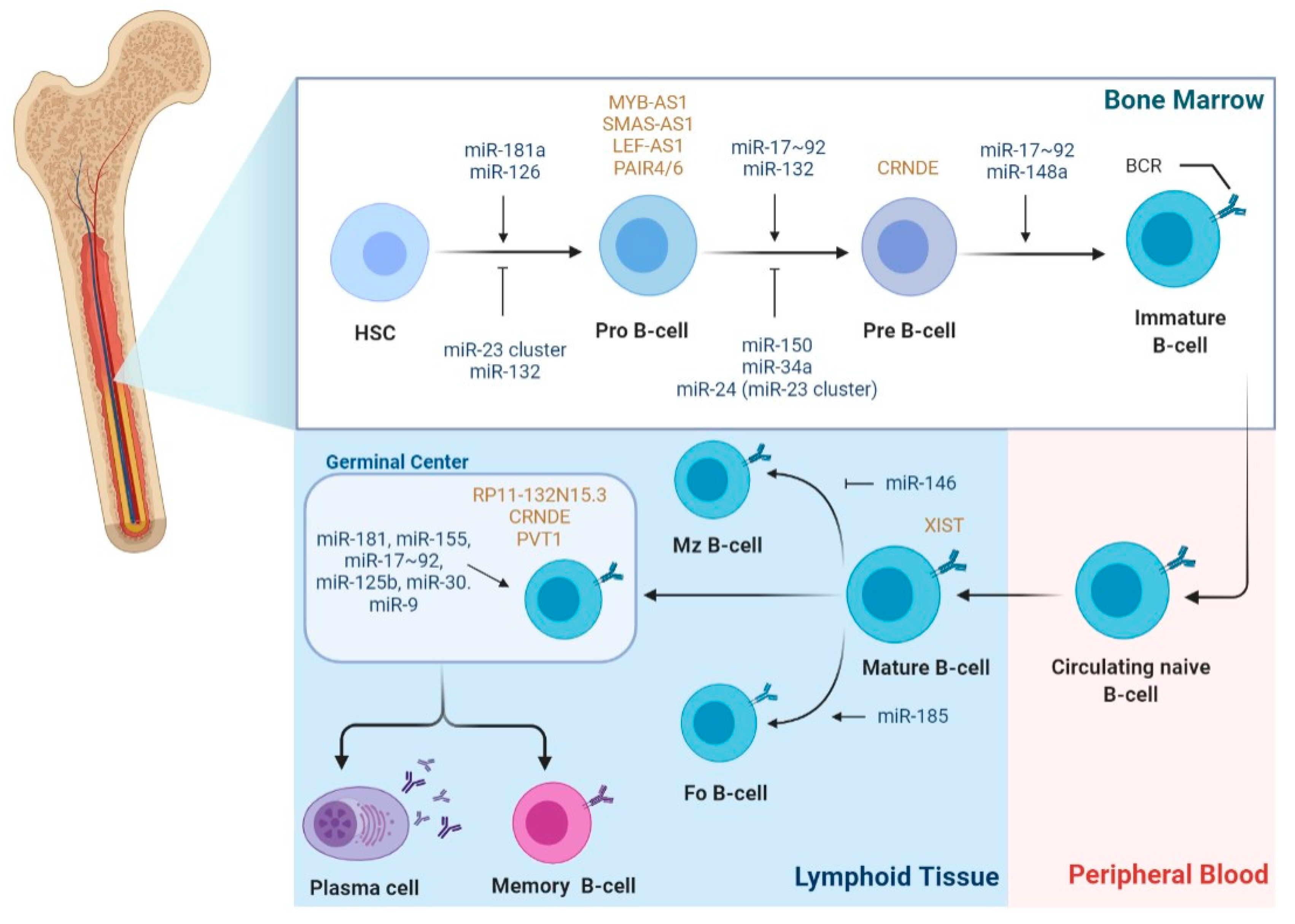
2.2. The Role of miRNAs in B-Cell Lymphomagenesis

Considering that B-cell development is a highly regulated process, it is not surprising that miRNAs have been implicated in the regulation of most of the stages comprising this process (Figure 2). Interestingly, during B-cell development, most miRNAs show a stage-specific expression pattern, highlighting their stage-specific function [23]. The process involving B-cell differentiation seems to be prone to malignant transformation, with increasing evidence showing that disruption of the miRNA network takes part in the initiation and maintenance of lymphomagenesis.
Given the regulatory role of the miR-17~92 cluster (comprising miR-17, miR-18a, miR-19a, miR-19b-1, miR-20a, and miR-92-1) during the stages of bone marrow (BM) B-cell development and central tolerance, its deregulated expression was shown to have oncogenic potential [24]. In fact, the 13q31.3 locus, which encodes this cluster, is often found amplified in B-cell malignancies, including DLBCL, mantle cell lymphoma (MCL), and FL [25,26,27]. The overexpression of the miR-17~92 cluster, concomitantly with the expression of oncogene Myc, promoted the onset and increased tumor growth in a mouse model of B-cell lymphoma [28]. Interestingly, Xiao et al. generated mice expressing two copies of human miR-17~92 in the B and T-cell compartments and observed that these mice developed lymphoproliferative disease and autoimmunity, showing elevated autoantibody and lymphocyte infiltration into non-lymphoid organs. The effects of miR-17~92 upregulation seemed to be associated with decreased levels of BIM and PTEN (both miR-17~92 targets), resulting in increased proliferation and resistance to cell death [29]. Moreover, a study identified miR-19a and miR-19b as the key oncogenic components of the miR-17~92 cluster. MiR-19 was shown to target PTEN, resulting in the activation of the AKT1-MTOR pathway and ultimately promoting cell survival in vitro and boosting tumor formation in vivo [30]. A B-cell-specific miR-17~92 transgenic mouse developed lymphomas with high penetrance, phenotypically resembling human lymphomas, including DLBCL, showing the role of the miR-17~92 cluster as a driver of B-cell lymphomagenesis [31]. The cell-cycle regulators E2F3 and BIM were identified as direct targets of the miR-17~92 cluster; therefore, they are involved in the mediation of B-cells’ proliferation and survival. Additionally, miR-17~92-driven lymphoma cells exhibited constitutive activation of PI3K due to PTEN and PHLPP2 repression, and of the NF-κB pathway, due to inhibition of NF-kB deubiquitinases, CYLD, and A20 [31]. Lastly, chemical inhibition of PI3K (AZD8055) or NF-kB (BMS-345541) resulted in reduced tumor size and prolonged survival of lymphoma-bearing mice, highlighting the importance of the miR-17~92 cluster as a driver of both tumor development and maintenance [31].
Another central miRNA during B-cell development, which is also aberrantly expressed in different B-cell lymphomas, is miR-155. MiR-155-5p controls plasma-cell production by inhibiting transcription factor SPI1, culminating in the downregulation of PAX5, which is required for normal terminal B-cell differentiation [32]. In vitro and in vivo inhibition of miR-155 was shown to diminish cell survival and cell proliferation and reduce tumor growth in mice [33]. Thai et al. observed a reduction in the total number of germinal center (GC) B cells in miR-155-deficient mice and, conversely, an increase in knock-in mice, probably as a result of targeting the transcription factor SPI1 [34,35]. MiR-155 was reported to be involved in the modulation of the PIK3CA-AKT1 pathway by directly targeting the negative regulator PIK3R1 (p85α) in DLBCL cells [36]. According to Zhu et al., miR-155 seems to modulate cell proliferation, cell cycle, and apoptosis of DLBCL cells by targeting transforming growth factor-β receptor 2 (TGFBR2) [37]. Several studies have been focusing on unveiling the miR-155 oncogenic role in B-cell lymphomas, reporting that its targets NIAM, histone deacetylase 4 (HDAC4), and SHIP1 [38,39,40]. Interestingly, Rai et al. reported a unique mechanism in DLBCL cells involving miR-155 overexpression, which renders lymphoma cells the ability to escape TGF-β’s growth-inhibitory effects. They reported that miR-155 directly targets the bone morphogenetic protein (BMP)-responsive transcriptional factor SMAD5 and that TGF-β1 also activated SMAD5 in DLBCL cells. Thus, the overexpression of miR-155 resulted in resistance to the cytostatic effects derived from both BMPs and TGF-β1 due to impaired activation of p21 and impaired cell cycle arrest [41]. During follicular B-cell maturation, miRNAs firmly regulate class-switch recombination, which includes further Ig gene rearrangements. miR-125b-5p targets transcription factors PRDM1 and IRF4, both involved in the activation of class-switch recombination, making its repression required for the normal B-cell development; otherwise, the development of B-cell malignancies may occur [42,43,44]. Moreover, similar results were obtained by other miRNAs, namely miR-30b-5p, miR-30d-5p, and miR-9-5p, by also targeting PRDM1 [45].
In GC, miR-21 promotes B-cell activation and proliferation, driving GC B-cell expansion and response [46]. The role of miR-21 as oncomiRNA in lymphoma was demonstrated using a mouse model overexpressing miR-21, which resulted in the spontaneous development of a pre-B-cell lymphoma. Inversely, tumor regression was observed after miR-21 repression due to increased apoptosis and proliferation arrest [47]. Moreover, PTEN and PDCD4 have been indicated as miR-21 targets and regulators of the PI3K/AKT and MAPK signaling pathways, which in turn mediate proliferation and apoptosis in lymphoma cells [48,49]. Conversely, miR-21 expression was shown to be transcriptionally activated by NF-κB [50]. Another described oncomiRNA in B-cell lymphomas is miR-217, whose expression is found upregulated as a result of B-cell stimulation in the context of the GC reaction. GC reaction is induced by miR-217 due to stabilization of Bcl-6 expression, resulting in enhanced production of class-switched antibodies and frequency of somatic hypermutation [51]. Importantly, Yébenes et al. showed that miR-217 overexpression also promotes mature B-cell lymphomagenesis. These results suggest that both miR-217 and Bcl-6 participate in the same network that makes GC cells permissive to genomic instability and prone to malignant transformation [51].
On the other hand, the deregulation of tumor-suppressive miRNAs is also frequently implicated in both B-cell lymphoma development and progression. In this instance, miR-34a has emerged as a tumor suppressor in lymphoma, linked to the p53 network. After p53 stabilization, miR-34a is upregulated in B cells, leading to a direct decrease of B-MYB and indirect decrease of E2F1 and the induction of cell-cycle arrest [52]. Moreover, p53 FOXP1 has also been described as a miR-34a target, where miR-34a downregulation results in higher levels of FOXP1 and BCL6, which in turn supports DLBCL proliferation [53,54,55]. Furthermore, miR-34a was also shown to target AXL, a receptor tyrosine kinase that activates PI3K/AKT signaling, and to be overexpressed in chronic lymphocytic leukemia (CLL) [56]. Cluster 15/16 miRNAs’ implication in the pathogenesis of B-cell lymphomas was first demonstrated when Calin et al. reported that miR-15/16 downregulation is due to the loss of the 13q14 locus, where the cluster is encoded in CLL [5]. Subsequent studies demonstrated that their tumor suppressor function was, at least in part, through the activation of cell apoptosis by targeting BCL2, MCL1, and CDK6 [57]. Moreover, miR-15/16 were shown to be part of a positive feedback loop involving TP53, which is a direct target of the cluster in CLL [58]. The deregulation of the miR-15/16 cluster has also been linked with the pathogenesis of other types of lymphoma, such as MCL, where their downregulation was induced by c-Myc and histone deacetylase [59].
Another MYC-regulated miRNA that has been implicated in B-cell pathogenesis is miR-150. Presenting a stage-specific expression during B-cell development, miR-150-5p prevents pro to pre-B-cell transition when overexpressed prematurely [60,61]. By downregulating miR-150, MYC induces the expression of FOXP1 and GAB1, regulators of cell survival and B-cell receptors, and of NF-κB signaling in malignant B cells [62]. The deregulation of miRNAs has also been implicated in other signaling pathways of B-cell lymphomas, such as mitogen-activated protein kinase (MAPK) and Wnt/β-catenin signaling pathways. For example, miR-101 was shown to regulate cell proliferation and apoptosis by targeting mitogen-activated protein kinase 1 (MEK1), an upstream protein kinase of the ERK/MAPK pathway, in DLBCL [63].
Taken together, these studies have been confirming the causative role of deregulated miRNA expression in B-cell lymphomagenesis, identifying potential clinically relevant miRNAs for profiling studies and relevant targets for miRNA-based therapies.

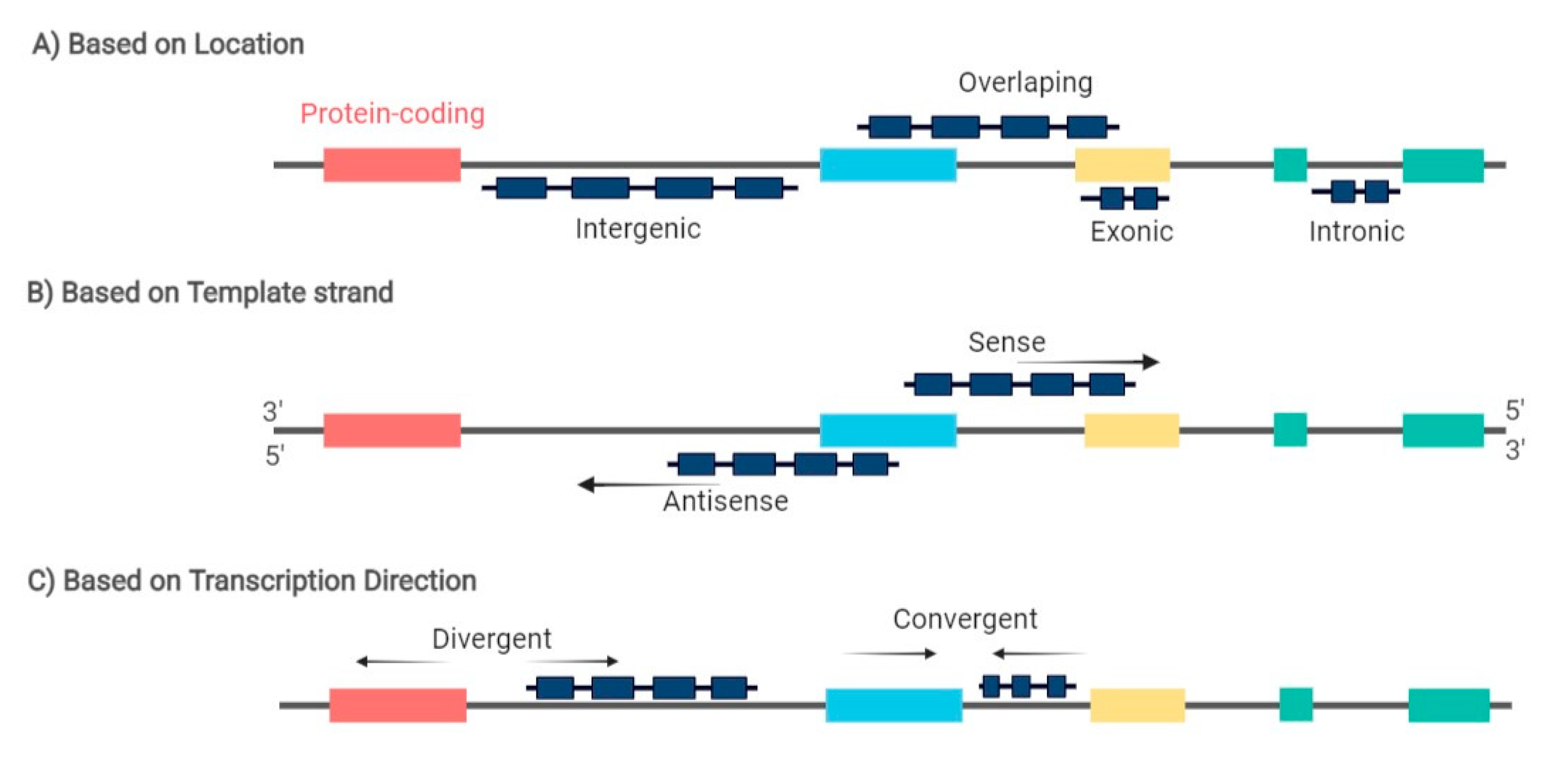
2.3. The Role of LncRNAs in B-Cell Lymphomagenesis

Regarding the regulatory role of lncRNA during the different stages of B-cell development and as drivers of B-cell malignancies, there is still scarcer information when compared to miRNAs. LncRNA expression profiling studies have reported that lncRNA exhibits cell-type-specific expression patterns during the different stages of B-cell differentiation (Figure 2) [64,65,66,67]. Consequently, each B-cell subset can be differentiated using its unique lncRNA expression profile [67]. Petri et al., using a guilt-by-association method, analyzed lncRNAs originated from protein-coding genes with known functions in B-cell development and identified antisense lncRNAs, such as MYB-AS1, SMAS-AS1, and LEF-AS1, with roles in early B cells, associated with RAG2, VPREB1, DNTT, LEF1, SMAD1, and MYB expression [66]. On the other hand, lncRNA colorectal neoplasia differentially expressed (CRNDE) showed high expression during the proliferating stages, such as pre-B cells and centroblasts in the GC, which was tightly associated with the expression of mitotic cell cycle genes [66]. In fact, CRNDE was previously demonstrated to be linked to cell-cycle and metabolic changes during proliferation [68,69]. Brazão et al. identified the expression of PVT1 and some uncharacterized lincRNAs, such as LINC00487, LINC00877, and RP11-132N15.3, associated with the expression of AID and SERPINA9, both specifically expressed in GC centroblasts and centrocytes. Of note, RP11-132N15.3 is described to be encoded approximately 240 kilobases upstream of BCL6 [65,66]. Additionally, based on mice models, several lncRNAs demonstrated a PAX5-dependent expression, a transcription factor involved in B-cell commitment, which were shown to be bound by PAX5 and to have human orthologs previously described [65]. Verma-Gaur et al. proposed that germ-line-transcribed lncRNAs regulate V(D)J recombination during the pro-B stage by assisting locus compaction, which brings heavy chain genes close to undergoing efficient gene rearrangements. Remarkably, the most expressed transcripts were PAX5-activated intergenic repeat (PAIR) elements, PAIR4 and PAIR6, whose transcription is antisense to PAX5 [70]. Moreover, they demonstrated that YY1−/− pro-B cells, a key player in distal VH gene rearrangement, presented a considerable reduction in antisense transcription between the PAIR promoters and the intronic enhancer, and consequently an impaired distal VH gene rearrangement, reinforcing the hypothesis of PAIR4 and PAIR6 as central regulators of the V(D)J recombination [70]. Later on, Syrett et al. proposed that YY1 binds and relocates lncRNA Xist to the inactive X-chromosome during B-cell stimulation, modulating the X-linked gene regulation from antigen naïve B cells to activated B cells [71]. According to Tayari et al., naïve and memory B-cell subsets exhibit analogous lncRNA expression patterns but are distinct from highly proliferating GC-B cells, which was corroborated in two other profiling studies [64,65,67].
Non-coding antisense transcripts of PU.1 were reported to inhibit the expression of PU.1 at a translation level, which could indicate its pivotal role in lymphomagenesis given the regulatory function of PU.1 in B-cell differentiation [72,73].
On the other hand, over the past few years, some studies have been trying to unveil the mechanistic pathways associated with the deregulation of lncRNAs during lymphomagenesis. In this instance, TP53 has been linked to the expression of some lncRNAs in different lymphoma subtypes. In fact, Blume et al. demonstrated, for the first time, the association of lncRNAs and the p53 pathway in CLL and lymphoma by inducing a p53-dependent DNA damage response, which resulted in increased expression of two lncRNAs, NEAT1 and lincRNA-p21, regulating apoptosis or cell-cycle arrest and DNA repair [74]. In DLBCL, p53 can directly bind to the promoter region of the lncRNA PANDA, which inactivates the MAPK/ERK signaling pathway, suppressing the proliferation of DLBCL cells by a G0/G1 cell-cycle arrest [75]. Peng et al. demonstrated that lncRNA HULC regulates DLBCL cell apoptosis and cell proliferation via the upregulation of antiapoptotic BCL2 protein and cyclin D1 [76]. The Wnt/β-catenin signaling pathway was shown to be activated by lncRNA FIRRE through promoting the nuclear translocation of β-catenin [77]. LncRNA DBH-AS1, found to be upregulated in DLBCL, was identified as a positive regulator of cell proliferation, migration, and invasion via binding to the RNA-binding protein BUD13 homolog (BUD13), which in turn regulates fibronectin 1 expression [78]. Cheng et al. observed suppression of DLBCL cell proliferation and tumor growth after TUG1 knockdown, promoting the ubiquitination of MET and the subsequent degradation [79]. Similarly, the knockdown of HOTAIR expression resulted in growth inhibition, cell-cycle arrest, and apoptosis, possibly involving the regulation of PI3K/AKT/NF-κB pathways [80]. Moreover, the HOTAIR function was associated with epigenetic regulation by recruiting polycomb repressive complex 2 (PRC2) proteins (EZH2, SUZ12, and EED), which induces H3K27me3, strongly related with aggressive DLBCL [81]. Sehgal et al. described an alternative splicing mechanism of Fas, firmly regulated by its antisense lncRNA FAS-AS via binding to RBM5, which induces Fas-mediated apoptosis in lymphoma. Conversely, FAS-AS1 expression is regulated by EZH2, which hyper-methylates the FAS-AS1 promoter, suppressing its expression [82]. In MCL, EZH2 has also been associated with the lncRNA MALAT1. Specifically, Wang et al. demonstrated that the knockdown of MALAT1 resulted in reduced levels of EZH2 and H3K27me3, while the expression levels of CDKs inhibitors p21 and p27 increased, resulting in cell-cycle arrest at the G1/S transition [83]. Similarly, a study by Li et al. reported silencing MALAT1 in DLBCL cells results in decreased cell proliferation and invasion and increased cell apoptosis. Concomitantly, they observed an increase in the expression levels of two autophagy indicators, LC3-II/LC3-I proteins and p62, reflecting the increase in autophagy in DLBCL cells [84]. A previously identified NOTCH1-regulated lncRNA in T-ALL, LUNAR1, was shown to regulate cell cycles and proliferation in lymphoma cells. LUNAR1 downregulation resulted in the inhibition of cell proliferation via E2F1, cyclin D1, and p21 [85,86].
Recently, emerging evidence has been demonstrating that a large group of lncRNAs exert their function as ceRNAs to fine-tune the miRNA-mediated regulation of protein abundance in lymphoma development.

2.4. The Role of LncRNAs as ceRNAs in B-Cell Lymphomagenesis

Karreth et al. demonstrated that lncRNA BRAFP1, which is aberrantly expressed in B-cell lymphomas, acts as a ceRNA with BRAF mRNA, increasing its stability and BRAF levels by sequestering specific BRAF-targeting miRNAs, such as miR-134, miR-543, and miR-653. Consequently, BRAF activates MAPK signaling, resulting in DLBCL cells’ proliferation [87]. In fact, NEAT1 was identified as an MYC-regulated transcript promoting DLBCL cells proliferation and lymphomagenesis by regulating the miR-34b-5p-GLI1 pathway [88]. Interestingly, NEAT1, along with LincRNA-p21, were also identified as p53-dependent DNA damage response machinery in lymphoma and CLL [74]. Zhao et al. reported that lncRNA PEG10 exhibits an oncogenic role in DLBCL progression by sponging miR-101-3p, which directly targets KIF2A. Moreover, abrogation of PEG10 or KIF2A expression resulted in the inhibition of cell proliferation, migration, and invasion, and promotion of cell apoptosis [89]. Recently, Tian et al. reported the upregulation of PCAT1, a proven key carcinogenic driver of hepatocellular carcinoma, in DLBCL [90]. Moreover, they investigated the mechanism behind PCAT1 action in DLBCL cells and observed an interplay between this lncRNA and miR-508-3p, which the inhibition of results in upregulation of NFIB, inducing DLBCL cell proliferation, migration, and invasion [90]. Similarly, lncRNA UCA1 was found upregulated in DLBCL, being involved in the regulation of cell proliferation, migration, and invasion by competitively binding with miR-331-3p [91].
Another reported upregulated lncRNA in DLBCL is MALAT1, whose ceRNA function is through sponging miR-195, resulting in the activation of the immune checkpoint molecule PD-L1 and consequently promoting cell proliferation, migration, and immune escape. Moreover, MALAT1 can induce CD8+ T cell apoptosis and epithelial–mesenchymal transition (EMT)-like processes by regulating the Ras/ERK signaling pathway [92]. In MCL, the knockdown of MALAT1 resulted in cell-cycle arrest and impaired proliferation due to the upregulation of p21 and p27 by EZH2 [83]. MiR-423-5p was reported to be involved in a ceRNA network with lncRNA FOXP4-AS1 in MCL cells. Mechanistically, FOXP4-AS1 acts as a sponge to miR-423-5p, upregulating the expression of NACC1, which results in MCL cell proliferation, migration, and invasion [93].
The induction of proliferation and cell-cycle progression accompanied with inhibition of cell apoptosis was observed as a result of lncRNA LINC01857 upregulation through targeting miR-141-3p/MAP4K4 axis in DLBCL. The biological effect of LINC01857 was associated with the activation of the PI3K/mTOR pathway and the induction of the EMT process [94]. Huang et al. reported that lncRNA LINC00857 regulates the miR-370-3p/CBX3 axis by competing for miR-370-3p binding, resulting in the modulation of DLBCL cell proliferation and apoptosis [95]. The silencing of lncRNA SBF2-AS1 resulted in decreased cell viability and growth via directly targeting miR-494-3p and, consequently, resulting in decreased FGFR2 expression [96]. Recently, Xu et al. reported that transcription factor PAX5 activates lncRNA ARRDC1-AS1, which functions as a sponge for miR-2355-5p to upregulate ATG5, resulting in increased autophagy and progression of DLBCL [97]. Li et al. demonstrated the oncogenic function of TUC338 in DLBCL by sponging miR-28-5p, which leads to the activation of the EGFR/PI3K/AKT pathway [98]. Another recently identified oncogenic lncRNA in DLBCL is LINC00908, which, when silenced, results in the inhibition of cell proliferation and invasion. Moreover, LINC00908 function seems to be through directly binding and inhibiting miR-671-5p [99].
On the other hand, SMAD5-AS1 was reported to be downregulated in DLBCL since it inhibits DLBCL proliferation by sponging miR-135b-5p to ultimately upregulate APC expression and inhibit the Wnt/β-catenin pathway [100]. LINC00963 was also described as having a tumor-suppressive role in DLBCL cells since overexpression of this lncRNA resulted in the inhibition of cell proliferation in vitro and tumor growth in vivo. LINC00963 directly binds to miR-320a, regulating endoplasmic reticulum stress and autophagy via XBP-1 targeting [101].
An emerging class of lncRNA, known as small nucleolar RNA host genes (SNHGs), has been proposed to also function as miRNA sponges in B cell lymphoma. Chen et al. observed that SNHG12 knockdown inhibited cell growth, migration, and invasion in vitro and in vivo by sequestering miR-195 [102]. SNHG14 was reported to promote DLBCL progression and immune evasion by a positive feedback loop involving the SNHG14/miR-5590-3p/ZEB1 axis. Specifically, SNHG14 sequesters miR-5590-3p, upregulating ZEB1, which, in turn, transcriptionally activates SNHG14 and PD-L1, thus modulating the PD-1/PD-L1 checkpoint [103]. Similarly, another study also reported the role of SNHG14 in promoting both DLBCL progression and immune evasion via the PD-1/PD-L1 checkpoint, but in this instance by sponging miR-152-3p [104]. Zhu et al. demonstrated that mechanistically SNHG16 acts as a ceRNA by sequestering miR-497-5p, which targets PIM1 resulting in the promotion of proliferation and inhibition of apoptosis in DLBCL [105]. Moreover, lncRNA SNHG8 was described as having a cancer-promoting effect on DLBCL by directly targeting miR-335-5p, which promotes proliferation while inhibiting the apoptosis of DLBCL cells [106].
The recent emerging data concerning the action of lncRNAs as miRNAs sponges highlights the complex post-transcriptional balance involved in gene-regulation networks. Altogether, these studies show the significant role of ceRNAs in lymphoma pathogenesis, which needs further characterization.A better understanding of ceRNA interactions may open the opportunity to exploit this crosstalk for biomarkers discovery and precise RNA-based therapies in lymphoma.

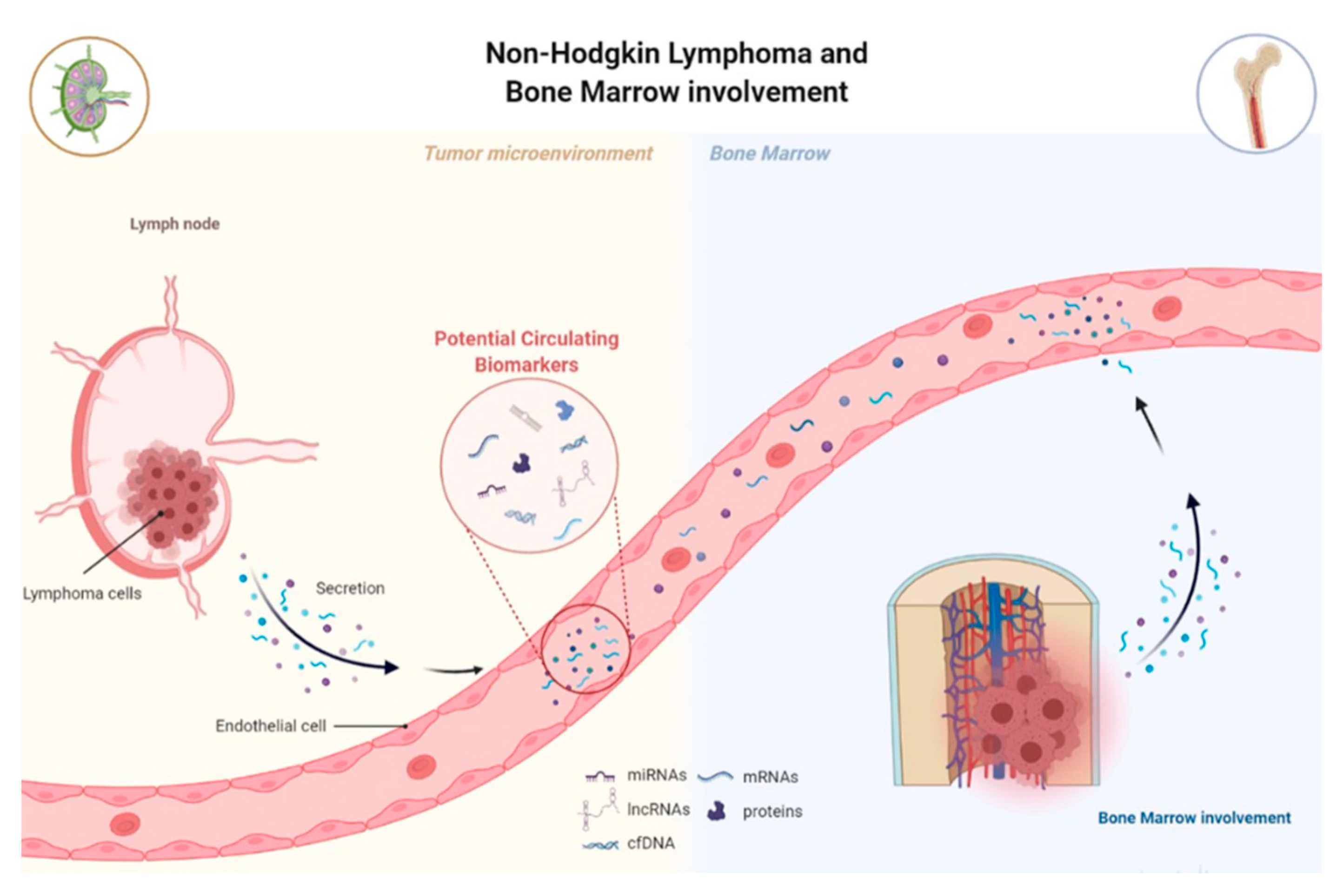
3. MiRNAs and lncRNAs as Potential Biomarkers for NHL

The current gold standard for NHL diagnosis and classification remains tissue biopsies of an involved lymph node (or a tumor in another organ) and BM aspiration and biopsy to establish BM involvement [1]. However, these procedures are not only invasive but also do not reflect tumor heterogeneity and clinical behavior. BM biopsy raises the question of its routine use, especially in patients with a low probability of bone marrow involvement, but also, being the collection sites only the sternum or ilium; they do not necessarily reflect the overall condition of the BM. Lastly, biopsies are unsuitable for dynamic monitoring during treatment and follow-up. The presence of circulating tumor-associated components, known as “tumor circulome”, which can be easily assessed, appears as a potential option as cancer biomarkers for liquid biopsies (Figure 3) [107]. One of the major components of “tumor circulome”, highly present in circulation, are the miRNAs [108]. MiRNAs emerged as excellent biomarker candidates due to their high stability in biological samples and their high specificity and sensitivity (Table 1) [8]. Similarly, increasing evidence has proposed lncRNAs are promising cancer diagnostic and prognostic biomarkers, especially given their high cell type, tissue, and disease type-specific expression (Table 2). Moreover, lncRNAs have been considered stable and can also be detected in circulation [109]. However, the majority of studies analyzing deregulated lncRNAs in lymphoma have been performed on tissue samples and cell lines.

3.1. MiRNAs and lncRNAs as Non-Invasive Diagnostic Biomarkers

A current clinical problem in the context of NHL remains the late and imprecise diagnosis, which in turn negatively conditions patient morbidity and mortality. Several authors have proposed and showed the potential of both individual miRNAs and lncRNAs or panels of these ncRNAs, as biomarkers for diagnosis and subclassification of NHL. Regarding the diagnostic potential, studies have analyzed the expression of miRNAs by comparing NHL cases to healthy controls in order to identify a specific miRNA signature. Lawrie et al.’s study showed, for the first time, that tumor-associated miRNAs, miR-155, miR-210, and miR-21 expression levels were upregulated in DLBCL patients’ serum compared to healthy controls [110]. Beheshti et al. first established a miRNA expression profile associated with DLBCL development based on cell lines and patient-derived xenograft models, which was then validated in patients’ serum. In the validation study, their profile was reduced to five miRNAs, let-7b, let-7c, miR-18a, miR-24, and miR-15a, which had the ability to discriminate DLBCL patients from healthy donors, with an accuracy of 91% [116,142]. Another study showed a 4-miRNA panel, comprising miR-15a, miR-16-1, miR-29c, and miR-155, significantly elevated in DLBCL serum and one downregulated, miR-34a, when compared to healthy controls [111]. More recently, Yuan et al. demonstrated a correlation between circulating levels of eight miRNAs and their matched formalin-fixed, paraffin-embedded (FFPE) samples [124]. Inada et al. demonstrated that the expression levels of circulating miR-15a-3p, miR-21-5p, miR-181a-5p, and miR-210-5p were significantly different between DLBCL patients and healthy controls, and specially miR-15a-3p, and miR-21-5p showed higher power to discriminate patients from controls [115]. MiR-34a-5p, miR31-5p, miR-155-5p, miR-150-5p, miR-15a-3p, and miR-29a-3p were shown to be elevated in CLL patients compared with healthy individuals and seemed to be associated with B-cell differentiation and homeostasis [127]. Most recently, Jørgensen et al. addressed the question of if it is possible to detect specific circulating miRNAs years before the diagnosis of B-cell lymphoma. Firstly, they identified the plasma miRNA candidates in pretreatment samples of DLBCL patients, showing miR-326 and miR-375 as the best markers differentiating B-cell lymphomas from controls. Subsequently, they validated the identified miRNA signature in a confirmatory cohort comprising samples from blood donors, who later developed different types of B-cell lymphoma to test pre-diagnostic plasma samples for early diagnosis. In the end, they verified that both miR-326 and miR199a-5p were upregulated months to years before diagnosis, which showed that changes in plasma miRNAs may precede the development of symptoms of NHL [126].
Circulating miRNAs have also been studied as refiners of the World Health Organization (WHO) classification, leading the way towards a molecular classification of NHL. In fact, the current system of differential diagnosis between the different types of B-cell NHL remains ineffective. For example, Chen et al. demonstrated that plasma levels of miR-21 were higher in DLBCL activated B-cell-like (ABC) compared to the germinal center B-cell-like (GCB), while Bedewy et al. reported higher expression of miR-155 in non-GCB compared to the GCB subtype [112,118]. Moreover, although involving the analysis of FFPE tumor tissues, a study by Leich et al. showed that miRNA expression was different between t(14;18)–negative FLs (without high expression of BCL2) and t(14;18)–positive FL patients. The most robust expression change was verified in miR-16-5p, which was significantly downregulated in t(14;18)–negative FL patients [143]. Similarly, BCL2+/BCL6+ FL and BCL2/BCL6+ FL patients showed upregulation of 21 miRNAs and downregulation of 12 miRNAs compared to BCL6 patients [144]. A major study by Lisio et al. performed a comparative analysis that included all major B-cell NHL types and identified a 128-miRNA signature capable of characterized lymphoma neoplasms, reflecting the lymphoma type, cell of origin, and/or discrete oncogene alterations [145]. Even though these studies do not involve the analysis of circulating miRNAs, they underline the differential molecular background of these NHL subtypes and pave the way to future analysis and validation of these results in the context of liquid biopsies in a larger patient cohort.
Concerning the deregulated expression of lncRNAs in NHL, the vast majority of studies were performed on tissue samples; however, the found tissue-derived deregulated lncRNAs could represent promising circulating biomarkers to be analyzed in future studies. Verma et al. performed a large RNA-seq study of 116 DLBCL tissue samples and identified 2,632 novel lncRNAs, two-thirds of which were only expressed in tumor cells. Moreover, more than one-third of these lncRNAs were found to be differently expressed between ABC and GCB subtypes [146]. LncRNAs PEG10 and LUNAR1 were found to be upregulated in DBCL patients compared to healthy individuals, with areas under the ROC curve (AUC) up to 0.8228 and 0.9420, respectively, indicating their potential diagnostic value [86,128]. Other reported upregulated lncRNAs in DLBCL tissue and cell lines were the lncRNAs FIRRE, HULC, LINC01857, and OR3A4 [76,77,94,129]. A 5-lncRNA signature (upregulated ENST00000424690, ENST00000425358, and NR_026892, and downregulated ENST00000464929 and ENST00000475089) was validated by Gao et al., which distinguished GCB-DLBCL tissue samples from reactive lymph nodes [130]. Moreover, Zhou et al. defined a 17-lncRNA signature, termed SubSigLnc-17, comprising ENTPD1-AS1, SACS- AS1, SH3BP5-AS1, RP11-101C11.1, AC009892.10, RP1- 68D18.4, MIR600HG, RP11-278 J6.4, RP11-203B7.2, CSMD2-AS1, CTC-467 M3.1, RP4-788P17.1, RP11-553 L6.5, CRNDE, RP11-519G16.3, RP11-21 L19.1, and MME- AS1, which permitted the discrimination between GCB and ABC subtypes with high accuracy [131]. In this manner, NONHSAG026900 showed higher levels in the GCB subtype compared to the non-GCB subtype, also indicating the diagnostic potential of these lncRNA [132]. In Deng et al.’s study, the NEAT1_1 expression level was found to increase in DLBCL compared to lymphadenitis [133]. Dousti et al. performed an in silico analysis and identified 189 deregulated lncRNAs in DLBCL, highlighting GAS5, MIR17HG, HULC, and PCA3 with the greatest deregulation score [134]. Pan et al. performed a microarray analysis to identify the expression profile of FL compared to reactive lymphatic nodes. They found 10 altered lncRNAs, with RP11-625 L16.3 being the most significantly upregulated [137]. On the other hand, Roisman et al. performed an RNA-seq analysis in different histological variants of FL and observed that FL3B-DLBCL/tDLBCL showed a higher number of differentially expressed lncRNAs and reported lncRNA RP4-694A7.2 as the most deregulated [138].
Regarding circulating NHL-associated lncRNAs, there are still few studies, even though their diagnostic and prognostic potential hs been shown in several solid tumors [147,148]. The first study on circulating lncRNAs in B-cell lymphoma was performed by Isin et al., which showed downregulation of lincRNA-p21 in CLL patients, upregulation of TUG1 and downregulation of MALAT1, HOTAIR, and GAS5 in MM patients [141]. On the other hand, Wang et al. investigated the circulating lncRNA levels in DLBCL and reported lower levels of lncRNA PANDA in patients’ plasma samples, whereas TUG1 was found upregulated compared to healthy controls [75]. In MCL, LINK-A lncRNA plasma levels were found to significantly increase while lncRNA GATA6-AS was downregulated, allowing to sensitively distinguished patients from healthy controls (AUC of 0.8338 and 0.9195, respectively) [139,149]. Recently, Senousy et al. reported that plasma levels of HOTAIR and XIST were significantly upregulated, whereas GAS5 were downregulated when comparing DLBCL patients to healthy individuals [135]. Interestingly, Tang et al. conjugated the analysis of two circulating ncRNAs, miR-16 and lncRNA MORT, and showed that not only the expression levels of miR-16 and MORT were significantly lower in patients with early-stage MCL compared to controls, but they also showed a regulatory association between the two ncRNA involved in cell proliferation and apoptosis [140].

3.2. MiRNAs and lncRNAs as Prognostic Biomarkers

Several studies have been exploring the value of circulating miRNAs as prognostic markers for NHL. In 2008, Lawrie et al. were the first to report that high serum levels of miR-21 were associated with relapse-free survival (RFS) in DLBCL patients, which were later on supported in other studies [110,118]. Similarly, high serum levels of miR-22 at diagnosis in DLBCL were associated with a worse progression-free survival (PFS), independently of the currently used clinical prognostic index [121]. The upregulation of circulating miR-155 and miR-125b was associated with shorter overall survival (OS) of DLBCL patients, while miR-20a/b, miR-93, and miR-106a/b plasma profiles were associated with higher mortality in DLBCL [113,117,124]. Song et al.’s study reported that elevated levels of miR-224, miR-520d-3p, and miR-1236 and lower levels of miR-33a and miR-455-3p were associated with lower medium remission time, and consequently, higher probability of remission, independently of IPI score [122]. A 4-miRNA expression profile (higher levels of miR-21, miR-130b, miR-155; lower levels of miR-28) was shown to be associated with relapse, as well as inferior PFS and OS after R-CHOP, independently of IPI score [114].
The 6-lncRNA signature defined by Sun et al. was shown to be associated with patients OS, independently of standard clinical factors, and permitted to stratify DLBCL patients in high and low-risk groups, improving survival prediction [136]. Moreover, the SubSigLnc-17 profile was not only able to discriminate clinically molecular DLBCL subtypes but also was shown to be significantly associated with patients’ OS and PFS [131]. The expression levels of lncRNA HOTAIR were not only associated with tumor size and clinical stage but also with the presence of B symptoms and IPI score. In fact, higher levels of HOTAIR were associated with better patients’ prognoses, being characterized as an independent predictive biomarker for DLBCL [80,135]. Peng et al. showed in different studies that lncRNAs HULC, LUNAR1, and PEG10 could represent potential prognostic biomarkers of DLBCL. Specifically, HULC and LUNAR1 expression levels were associated with poor prognosis and represented an independent factor of OS and PFS, while PEG10 showed potential as an independent predictor of poor OS of DLBCL [76,86,128]. Moreover, the upregulation of lncRNAs FIRRE and OR3A4 in DLBCL tissue samples is associated with decreased OS [77,129]. In MCL, the tissue-derived MALAT1 and plasma-derived FOXP4-AS1 levels were found correlated with higher MIPI scores and unfavorable patients’ OS and disease-free survival (DFS) [83,93]. Conversely, lower PANDA expression levels, either in plasma or in tissue samples, were associated with inferior RFS and OS of DLBCL patients, being an independent prognostic factor [75]. Moreover, patients expressing high levels of LincRNA-p21 and NONHSAG026900 were found to have a favorable prognosis, with longer OS and PFS, and NONHSAG026900 act as an independent factor by also enhancing the predictive ability of the IPI score [132,150].
The assessment of patients’ prognosis and decisions on treatment alteration are mostly based on imaging PET-CT and clinical evaluations [151]. However, due to the insufficient sensitivity and specificity of these tools, there is an impending need to identify new predictors that permit the early identification of patients with inherent or acquired the refractory disease during treatment. Detection of miRNAs or lncRNAs can lower the detection limit of disease beyond the capabilities of current methods and create a “window of opportunity” for intervention prior to clinical relapse. Earlier initiation of second-line therapy at a point of minimal tumor burden may improve patients’ outcomes. Song et al. identified a 5-miRNA profile (miR-224, miR-455-3p, miR-1236, miR-33a, and miR-520d-3p) associated with R-CHOP response in DLBCL patients, being a significant predictor of response, independent from the IPI score [122]. Specifically, high levels of miR-455-3p and miR-33a were associated with chemosensitivity, while high levels of miR-224, miR-1236, and miR-520d-3p were associated with chemoresistance [122]. Dynamic monitorization of the levels of two potential drug-resistant miRNAs, miR-125b and miR-130a, showed that they are involved not only in recurrence and disease progression but also in chemoresistance in DLBCL patients [124]. Another study took a different approach by analyzing the circulating kinetics of two miRNAs, miR-494 and miR-21, in comparison to interim-PET/CT scans. In this study, they reported that both miRNAs were upregulated in untreated patients and that their levels decreased in patients that achieved interim-PET/CT negativity after four cycles of R-CHOP compared to those that presented a positive interim-PET/CT [119]. Bouvy et al. analyzed the circulating miRNA profile during the treatment course and found higher levels of miR-21 and miR-197 in patients unresponsive to treatment, and high levels of miR-19b, miR-20a, and miR-451 in patients with complete remission, which allowed the differentiation of patients with residual disease from those with complete remission during follow-up [152]. Recently, Fajardo-Ramirez et al. reported a miRNA signature composed of nine upregulated and six downregulated associated with chemoresistance to the R–CHOP regime [153]. Considering these results, circulating miRNAs have a strong potential to be used as predictive and monitoring biomarkers of therapy response in NHL patients. However, further investigation is needed in order to obtain more reliable data.
Regarding lncRNAs as treatment response biomarkers, Senousy et al. observed that pretreatment circulating levels of HOTAIR were higher, whereas GAS5 were lower in non-responders compared to responders to R-CHOP. Moreover, when performing multivariate analysis, HOTAIR appeared as an independent predictor of R-CHOP failure [135]. DLBCL patients with higher expression levels of NONHSAG026900 were shown to have a better response to chemotherapy compared to patients with lower levels [132]. Currently, it is established that treatment failure and consequent relapse can be due to the existence of drug-resistance cancer cell subpopulations [2]. In this instance, chemotherapy-resistant lymphoma cell lines show higher levels of lncRNA MALAT1, which seems to be associated with the inhibition of the autophagy signaling pathway [84]. Using RNA sequencing, Karstensen et al. analyzed the transcription profile of rituximab-sensitive and resistant DLBCL cell lines. They observed a differential expression of lncRNA between sensitive and resistant cells, comprising 54 up and 69 downregulated lncRNAs [154]. In MCL cells, a study by Mourtada-Maarabouni et al. demonstrated that lncRNA GAS5 regulates the effect of known mTOR inhibitors, such as rapamycin, everolimus, and temsirolimus. Particularly, the inhibition of GAS5 substantially decreased the effect of each rapalogue on MCL cells [155]. In Burkitt lymphoma cells, the upregulation of lncRNA MCM3AP-AS1 induced chemotherapy resistance to doxorubicin treatment by regulating the expression of antiapoptotic factors via the miR-15a/EIF4E axis [156]. There are still few studies analyzing lncRNAs as potential biomarkers for treatment response in B-cell lymphomas compared to some solid tumors, with only one studying a circulating lncRNA profile differentiating responders from non-responders.

3.3. Clinical Trials for Potential miRNA and lncRNA Biomarkers

Since 2008, when Lawrie et al. demonstrated for the first time the potential of miRNAs as biomarkers in DLBCL patients, a considerable number of clinical trials have been registered to clinically validate them [110]. A clinical trial (NCT01505699) involving 186 patients with B-cell acute lymphoblastic leukemia analyzed microRNA signature levels and their association with different clinical outcomes [157]. On the other hand, clinical trial NCT01057199 specifically tested miR-34a and miR-194 as biomarkers in cell samples from patients with acute myeloid leukemia [158]. Moreover, clinical trial NCT01541800 especially focused on pediatric cancers (central nervous system tumors, leukemia, and lymphoma), evaluated the presence of circulating miRNAs in the blood and cerebrospinal fluid of patients under chemotherapy [159]. Similarly, clinical trial NCT02791217 was developed to identify circulating miRNAs associated with early diagnosis of aggressive hematological malignancies (including B-cell lymphoma) in order to ultimately improve patients’ prognosis and increase survival rates [160]. Another example is the clinical trial NCT03340155, where miRNA expression levels, including miR-155, were tested in the serum of patients diagnosed with skin diseases, such as cutaneous T cell lymphoma (CTCL), and treated with photo(chemo)therapy [161]. One of their objectives was to correlate miRNA levels with clinical responses to treatment at different time points and ultimately identify potential predictive biomarkers. Lastly, clinical trial NCT01606605 included a retrospective and observational analysis of 350 patients diagnosed with DLBCL to study miRNA expression patterns and their correlation with clinical outcome, relapse, and disease progression [162].
Currently, there is no clinical trial exploring the possible application of lncRNAs as lymphoma biomarkers; however, there are some lncRNAs undergoing clinical trials or being patented for solid tumors, such as lung, colorectal, thyroid, breast, and gynecologic cancers [163,164]. To date, the lncRNA PCA3 was the first and only lncRNA approved for clinical practice as an early diagnostic biomarker for prostate cancer [165]. There is only one registered clinical trial, currently active, for hematological cancer to study the correlation between lncRNA XIST and immunophenotyping AML patients (NCT04288739) [166]. Moreover, few clinical trials have been exploring the possible application of combined ncRNA profiles, especially miRNAs and lncRNAs, as biomarkers for the diagnosis and prognosis of cancer [164].

4. Conclusions

B-cell NHLs are characterized as a highly heterogeneous group, which is reflected by variability in prognosis and treatment responses between the different subgroups. Therefore, it is crucial to focus on elucidating the molecular background and pathogenesis underlying these malignancies. As previously discussed, in the last years, miRNAs and lncRNAs have emerged as powerful regulators of lymphomagenesis; however, our current knowledge is still limited since it derives essentially from individual research studies of miRNA and lncRNA expression profiles. Additional research is needed to unravel the complex functional network surrounding the miRNAs and lncRNAs and their potential regulatory interactions involved in the process of B-cell lymphomagenesis.
The better understanding we have of the involved molecular mechanisms, the faster we will open clinical avenues toward personalized medicine and the development of new effective therapies, with great clinical benefit for the patient. Moreover, the introduction of miRNAs and lncRNAs as a liquid biopsy strategy provides a great opportunity for improving the diagnostic accuracy and for dynamically monitoring NHL patients in a non-invasive and reproducible manner. Ideally, the expression analysis of a given miRNA/lncRNA, miRNA or lncRNA panel, or even a combination of miRNAs and lncRNAs, should be able to molecularly diagnose and classify patients or predict survival, relapse, remission, and even responsiveness to treatment upfront.
To date, the majority of the studies focus on miRNAs, and there is still a lack of studies on lncRNAs in lymphomas, especially circulating lncRNAs, which opens an interesting opportunity to invest in further understanding their biology and function and their association with miRNAs, and the contribution of lncRNAs profiling in the diagnosis and prognostication of lymphomas.

Author Contributions

Conceptualization, M.F.; writing—original draft preparation, M.F.; writing—review and editing, M.F., A.L.T. and H.M.; supervision, R.M. All authors have read and agreed to the published version of the manuscript.

Funding

M.F. is recipient of a research scholarship awarded by LPCC-NRN (Portuguese League Against Cancer—Northern Branch).

Institutional Review Board Statement

Not applicable.

Informed Consent Statement

Not applicable.

Acknowledgments

We would like to thank Liga Portuguesa Contra o Cancro—Núcleo Regional do Norte—LPCC-NRN (Portuguese League Against Cancer).

Conflicts of Interest

The authors declare no conflict of interest.

References

  1. Armitage, J.O.; Gascoyne, R.D.; Lunning, M.A.; Cavalli, F. Non-Hodgkin lymphoma. Lancet 2017, 390, 298–310. [Google Scholar] [CrossRef]
  2. Klener, P.; Klanova, M. Drug Resistance in Non-Hodgkin Lymphomas. Int. J. Mol. Sci. 2020, 21, 2081. [Google Scholar] [CrossRef] [Green Version]
  3. Rovira, J.; Valera, A.; Colomo, L.; Setoain, X.; Rodríguez, S.; Martínez-Trillos, A.; Giné, E.; Dlouhy, I.; Magnano, L.; Gaya, A. Prognosis of patients with diffuse large B cell lymphoma not reaching complete response or relapsing after frontline chemotherapy or immunochemotherapy. Ann. Hematol. 2015, 94, 803–812. [Google Scholar] [CrossRef] [PubMed] [Green Version]
  4. Crump, M.; Neelapu, S.S.; Farooq, U.; Van Den Neste, E.; Kuruvilla, J.; Westin, J.; Link, B.K.; Hay, A.; Cerhan, J.R.; Zhu, L. Outcomes in refractory diffuse large B-cell lymphoma: Results from the international SCHOLAR-1 study. Blood 2017, 130, 1800–1808. [Google Scholar] [CrossRef] [PubMed]
  5. Calin, G.A.; Dumitru, C.D.; Shimizu, M.; Bichi, R.; Zupo, S.; Noch, E.; Aldler, H.; Rattan, S.; Keating, M.; Rai, K.; et al. Frequent deletions and down-regulation of micro- RNA genes miR15 and miR16 at 13q14 in chronic lymphocytic leukemia. Proc. Natl. Acad. Sci. USA 2002, 99, 15524–15529. [Google Scholar] [CrossRef] [Green Version]
  6. O’Brien, J.; Hayder, H.; Zayed, Y.; Peng, C. Overview of MicroRNA Biogenesis, Mechanisms of Actions, and Circulation. Front. Endocrinol. 2018, 9, 402. [Google Scholar] [CrossRef] [PubMed] [Green Version]
  7. Bartel, D.P. MicroRNAs: Genomics, biogenesis, mechanism, and function. Cell 2004, 116, 281–297. [Google Scholar] [CrossRef] [Green Version]
  8. Condrat, C.E.; Thompson, D.C.; Barbu, M.G.; Bugnar, O.L.; Boboc, A.; Cretoiu, D.; Suciu, N.; Cretoiu, S.M.; Voinea, S.C. miRNAs as Biomarkers in Disease: Latest Findings Regarding Their Role in Diagnosis and Prognosis. Cells 2020, 9, 276. [Google Scholar] [CrossRef] [Green Version]
  9. Fernandes, M.; Teixeira, A.L.; Medeiros, R. The opportunistic effect of exosomes on Non-Hodgkin Lymphoma microenvironment modulation. Crit. Rev. Oncol. Hematol. 2019, 144, 102825. [Google Scholar] [CrossRef] [PubMed]
  10. Fernandes, J.C.R.; Acuña, S.M.; Aoki, J.I.; Floeter-Winter, L.M.; Muxel, S.M. Long Non-Coding RNAs in the Regulation of Gene Expression: Physiology and Disease. Non-Coding RNA 2019, 5, 17. [Google Scholar] [CrossRef] [PubMed] [Green Version]
  11. Schmitt, A.M.; Chang, H.Y. Long noncoding RNAs in cancer pathways. Cancer Cell 2016, 29, 452–463. [Google Scholar] [CrossRef] [PubMed] [Green Version]
  12. Kopp, F.; Mendell, J.T. Functional Classification and Experimental Dissection of Long Noncoding RNAs. Cell 2018, 172, 393–407. [Google Scholar] [CrossRef] [PubMed] [Green Version]
  13. Derrien, T.; Johnson, R.; Bussotti, G.; Tanzer, A.; Djebali, S.; Tilgner, H.; Guernec, G.; Martin, D.; Merkel, A.; Knowles, D.G. The GENCODE v7 catalog of human long noncoding RNAs: Analysis of their gene structure, evolution, and expression. Genome Res. 2012, 22, 1775–1789. [Google Scholar] [CrossRef] [Green Version]
  14. Paraskevopoulou, M.D.; Hatzigeorgiou, A.G. Analyzing MiRNA–LncRNA Interactions. In Long Non-Coding RNAs: Methods and Protocols; Feng, Y., Zhang, L., Eds.; Springer: New York, NY, USA, 2016; pp. 271–286. [Google Scholar]
  15. Chi, Y.; Wang, D.; Wang, J.; Yu, W.; Yang, J. Long Non-Coding RNA in the Pathogenesis of Cancers. Cells 2019, 8, 1015. [Google Scholar] [CrossRef] [PubMed] [Green Version]
  16. Salmena, L.; Poliseno, L.; Tay, Y.; Kats, L.; Pandolfi, P.P. A ceRNA hypothesis: The Rosetta Stone of a hidden RNA language? Cell 2011, 146, 353–358. [Google Scholar] [CrossRef] [PubMed] [Green Version]
  17. Si, Y.; Yang, Z.; Ge, Q.; Yu, L.; Yao, M.; Sun, X.; Ren, Z.; Ding, C. Long non-coding RNA Malat1 activated autophagy, hence promoting cell proliferation and inhibiting apoptosis by sponging miR-101 in colorectal cancer. Cell. Mol. Biol. Lett. 2019, 24, 50. [Google Scholar] [CrossRef] [PubMed]
  18. Fu, Z.; Luo, W.; Wang, J.; Peng, T.; Sun, G.; Shi, J.; Li, Z.; Zhang, B. Malat1 activates autophagy and promotes cell proliferation by sponging miR-101 and upregulating STMN1, RAB5A and ATG4D expression in glioma. Biochem. Biophys. Res. Commun. 2017, 492, 480–486. [Google Scholar] [CrossRef]
  19. Xiong, Z.; Wang, L.; Wang, Q.; Yuan, Y. LncRNA MALAT1/miR-129 axis promotes glioma tumorigenesis by targeting SOX2. J. Cell. Mol. Med. 2018, 22, 3929–3940. [Google Scholar] [CrossRef]
  20. Liao, K.; Lin, Y.; Gao, W.; Xiao, Z.; Medina, R.; Dmitriev, P.; Cui, J.; Zhuang, Z.; Zhao, X.; Qiu, Y.; et al. Blocking lncRNA MALAT1/miR-199a/ZHX1 Axis Inhibits Glioblastoma Proliferation and Progression. Mol. Ther. Nucleic Acids 2019, 18, 388–399. [Google Scholar] [CrossRef] [Green Version]
  21. Fatica, A.; Bozzoni, I. Long non-coding RNAs: New players in cell differentiation and development. Nat. Rev. Genet. 2014, 15, 7–21. [Google Scholar] [CrossRef] [PubMed]
  22. Greene, J.; Baird, A.-M.; Brady, L.; Lim, M.; Gray, S.G.; McDermott, R.; Finn, S.P. Circular RNAs: Biogenesis, function and role in human diseases. Front. Mol. Biosci. 2017, 4, 38. [Google Scholar] [CrossRef]
  23. Spierings, D.C.; McGoldrick, D.; Hamilton-Easton, A.M.; Neale, G.; Murchison, E.P.; Hannon, G.J.; Green, D.R.; Withoff, S. Ordered progression of stage-specific miRNA profiles in the mouse B2 B-cell lineage. Blood J. Am. Soc. Hematol. 2011, 117, 5340–5349. [Google Scholar] [CrossRef] [PubMed] [Green Version]
  24. Benhamou, D.; Labi, V.; Novak, R.; Dai, I.; Shafir-Alon, S.; Weiss, A.; Gaujoux, R.; Arnold, R.; Shen-Orr, S.S.; Rajewsky, K.; et al. A c-Myc/miR17-92/Pten Axis Controls PI3K-Mediated Positive and Negative Selection in B Cell Development and Reconstitutes CD19 Deficiency. Cell Rep. 2016, 16, 419–431. [Google Scholar] [CrossRef] [PubMed] [Green Version]
  25. Neat, M.J.; Foot, N.; Jenner, M.; Goff, L.; Ashcroft, K.; Burford, D.; Dunham, A.; Norton, A.; Lister, T.A.; Fitzgibbon, J. Localisation of a novel region of recurrent amplification in follicular lymphoma to an∼6.8 Mb region of 13q32-33. Genes Chromosomes Cancer 2001, 32, 236–243. [Google Scholar] [CrossRef] [PubMed]
  26. Monni, O.; Oinonen, R.; Elonen, E.; Franssila, K.; Teerenhovi, L.; Joensuu, H.; Knuutila, S. Gain of 3q and deletion of 11q22 are frequent aberrations in mantle cell lymphoma. Genes Chromosomes Cancer 1998, 21, 298–307. [Google Scholar] [CrossRef]
  27. Rao, P.H.; Houldsworth, J.; Dyomina, K.; Parsa, N.Z.; Cigudosa, J.C.; Louie, D.C.; Popplewell, L.; Offit, K.; Jhanwar, S.C.; Chaganti, R.S.K. Chromosomal and Gene Amplification in Diffuse Large B-Cell Lymphoma. Blood 1998, 92, 234–240. [Google Scholar] [CrossRef]
  28. He, L.; Thomson, J.M.; Hemann, M.T.; Hernando-Monge, E.; Mu, D.; Goodson, S.; Powers, S.; Cordon-Cardo, C.; Lowe, S.W.; Hannon, G.J. A microRNA polycistron as a potential human oncogene. Nature 2005, 435, 828–833. [Google Scholar] [CrossRef] [PubMed]
  29. Xiao, C.; Srinivasan, L.; Calado, D.P.; Patterson, H.C.; Zhang, B.; Wang, J.; Henderson, J.M.; Kutok, J.L.; Rajewsky, K. Lymphoproliferative disease and autoimmunity in mice with increased miR-17-92 expression in lymphocytes. Nat. Immunol. 2008, 9, 405–414. [Google Scholar] [CrossRef] [PubMed] [Green Version]
  30. Mu, P.; Han, Y.-C.; Betel, D.; Yao, E.; Squatrito, M.; Ogrodowski, P.; de Stanchina, E.; D’Andrea, A.; Sander, C.; Ventura, A. Genetic dissection of the miR-17∼92 cluster of microRNAs in Myc-induced B-cell lymphomas. Genes Dev. 2009, 23, 2806–2811. [Google Scholar] [CrossRef] [PubMed] [Green Version]
  31. Jin, H.Y.; Oda, H.; Lai, M.; Skalsky, R.L.; Bethel, K.; Shepherd, J.; Kang, S.G.; Liu, W.-H.; Sabouri-Ghomi, M.; Cullen, B.R.; et al. MicroRNA-17∼92 plays a causative role in lymphomagenesis by coordinating multiple oncogenic pathways. EMBO J. 2013, 32, 2377–2391. [Google Scholar] [CrossRef] [Green Version]
  32. Lu, D.; Nakagawa, R.; Lazzaro, S.; Staudacher, P.; Abreu-Goodger, C.; Henley, T.; Boiani, S.; Leyland, R.; Galloway, A.; Andrews, S. The miR-155–PU.1 axis acts on Pax5 to enable efficient terminal B cell differentiation. J. Exp. Med. 2014, 211, 2183–2198. [Google Scholar] [CrossRef] [Green Version]
  33. Zhang, Y.; Roccaro, A.M.; Rombaoa, C.; Flores, L.; Obad, S.; Fernandes, S.M.; Sacco, A.; Liu, Y.; Ngo, H.; Quang, P. LNA-mediated anti–miR-155 silencing in low-grade B-cell lymphomas. Blood J. Am. Soc. Hematol. 2012, 120, 1678–1686. [Google Scholar] [CrossRef] [Green Version]
  34. Vigorito, E.; Perks, K.L.; Abreu-Goodger, C.; Bunting, S.; Xiang, Z.; Kohlhaas, S.; Das, P.P.; Miska, E.A.; Rodriguez, A.; Bradley, A.; et al. microRNA-155 regulates the generation of immunoglobulin class-switched plasma cells. Immunity 2007, 27, 847–859. [Google Scholar] [CrossRef] [PubMed] [Green Version]
  35. Thai, T.-H.; Calado, D.P.; Casola, S.; Ansel, K.M.; Xiao, C.; Xue, Y.; Murphy, A.; Frendewey, D.; Valenzuela, D.; Kutok, J.L.; et al. Regulation of the Germinal Center Response by MicroRNA-155. Science 2007, 316, 604–608. [Google Scholar] [CrossRef]
  36. Huang, X.; Shen, Y.; Liu, M.; Bi, C.; Jiang, C.; Iqbal, J.; McKeithan, T.W.; Chan, W.C.; Ding, S.-J.; Fu, K. Quantitative proteomics reveals that miR-155 regulates the PI3K-AKT pathway in diffuse large B-cell lymphoma. Am. J. Pathol. 2012, 181, 26–33. [Google Scholar] [CrossRef] [PubMed] [Green Version]
  37. Zhu, F.-Q.; Zeng, L.; Tang, N.; Tang, Y.-P.; Zhou, B.-P.; Li, F.-F.; Wu, W.-G.; Zeng, X.-B.; Peng, S.-S. MicroRNA-155 downregulation promotes cell cycle arrest and apoptosis in diffuse large B-cell lymphoma. Oncol. Res. Featur. Preclin. Clin. Cancer Ther. 2016, 24, 415–427. [Google Scholar] [CrossRef] [PubMed]
  38. Slezak-Prochazka, I.; Kluiver, J.; de Jong, D.; Smigielska-Czepiel, K.; Kortman, G.; Winkle, M.; Rutgers, B.; Koerts, J.; Visser, L.; Diepstra, A. Inhibition of the miR-155 target NIAM phenocopies the growth promoting effect of miR-155 in B-cell lymphoma. Oncotarget 2016, 7, 2391. [Google Scholar] [CrossRef] [PubMed] [Green Version]
  39. Sandhu, S.K.; Volinia, S.; Costinean, S.; Galasso, M.; Neinast, R.; Santhanam, R.; Parthun, M.R.; Perrotti, D.; Marcucci, G.; Garzon, R. miR-155 targets histone deacetylase 4 (HDAC4) and impairs transcriptional activity of B-cell lymphoma 6 (BCL6) in the Eµ-miR-155 transgenic mouse model. Proc. Natl. Acad. Sci. USA 2012, 109, 20047–20052. [Google Scholar] [CrossRef] [Green Version]
  40. Pedersen, I.M.; Otero, D.; Kao, E.; Miletic, A.V.; Hother, C.; Ralfkiaer, E.; Rickert, R.C.; Gronbaek, K.; David, M. Onco-miR-155 targets SHIP1 to promote TNFα-dependent growth of B cell lymphomas. EMBO Mol. Med. 2009, 1, 288–295. [Google Scholar] [CrossRef]
  41. Rai, D.; Kim, S.W.; McKeller, M.R.; Dahia, P.L.; Aguiar, R.C. Targeting of SMAD5 links microRNA-155 to the TGF-β pathway and lymphomagenesis. Proc. Natl. Acad. Sci. USA 2010, 107, 3111–3116. [Google Scholar] [CrossRef] [Green Version]
  42. Gururajan, M.; Haga, C.L.; Das, S.; Leu, C.-M.; Hodson, D.; Josson, S.; Turner, M.; Cooper, M.D. MicroRNA 125b inhibition of B cell differentiation in germinal centers. Int. Immunol. 2010, 22, 583–592. [Google Scholar] [CrossRef] [PubMed]
  43. Li, G.; So, A.Y.-L.; Sookram, R.; Wong, S.; Wang, J.K.; Ouyang, Y.; He, P.; Su, Y.; Casellas, R.; Baltimore, D. Epigenetic silencing of miR-125b is required for normal B-cell development. Blood 2018, 131, 1920–1930. [Google Scholar] [CrossRef] [PubMed] [Green Version]
  44. Enomoto, Y.; Kitaura, J.; Hatakeyama, K.; Watanuki, J.; Akasaka, T.; Kato, N.; Shimanuki, M.; Nishimura, K.; Takahashi, M.; Taniwaki, M.; et al. Eμ/miR-125b transgenic mice develop lethal B-cell malignancies. Leukemia 2011, 25, 1849–1856. [Google Scholar] [CrossRef] [PubMed] [Green Version]
  45. Zhang, J.; Jima, D.D.; Jacobs, C.; Fischer, R.; Gottwein, E.; Huang, G.; Lugar, P.L.; Lagoo, A.S.; Rizzieri, D.A.; Friedman, D.R.; et al. Patterns of microRNA expression characterize stages of human B-cell differentiation. Blood 2009, 113, 4586–4594. [Google Scholar] [CrossRef] [Green Version]
  46. Schell, S.L.; Bricker, K.N.; Fike, A.J.; Chodisetti, S.B.; Domeier, P.P.; Choi, N.M.; Fasnacht, M.J.; Luckenbill, S.A.; Ziegler, S.F.; Rahman, Z.S.M. Context-Dependent miR-21 Regulation of TLR7-Mediated Autoimmune and Foreign Antigen–Driven Antibody-Forming Cell and Germinal Center Responses. J. Immunol. 2021, 206, 2803–2818. [Google Scholar] [CrossRef]
  47. Medina, P.P.; Nolde, M.; Slack, F.J. OncomiR addiction in an in vivo model of microRNA-21-induced pre-B-cell lymphoma. Nature 2010, 467, 86–90. [Google Scholar] [CrossRef] [PubMed]
  48. Song, J.; Shao, Q.; Li, C.; Liu, H.; Li, J.; Wang, Y.; Song, W.; Li, L.; Wang, G.; Shao, Z.; et al. Effects of microRNA-21 on apoptosis by regulating the expression of PTEN in diffuse large B-cell lymphoma. Medicine 2017, 96, e7952. [Google Scholar] [CrossRef] [PubMed]
  49. Gu, L.; Song, G.; Chen, L.; Nie, Z.; He, B.; Pan, Y.; Xu, Y.; Li, R.; Gao, T.; Cho, W.C.; et al. Inhibition of miR-21 Induces Biological and Behavioral Alterations in Diffuse Large B-Cell Lymphoma. Acta Haematol. 2013, 130, 87–94. [Google Scholar] [CrossRef]
  50. Bai, H.; Wei, J.; Deng, C.; Yang, X.; Wang, C.; Xu, R. MicroRNA-21 regulates the sensitivity of diffuse large B-cell lymphoma cells to the CHOP chemotherapy regimen. Int. J. Hematol. 2013, 97, 223–231. [Google Scholar] [CrossRef]
  51. de Yébenes, V.G.; Bartolomé-Izquierdo, N.; Nogales-Cadenas, R.; Pérez-Durán, P.; Mur, S.M.; Martínez, N.; Di Lisio, L.; Robbiani, D.F.; Pascual-Montano, A.; Cañamero, M.; et al. miR-217 is an oncogene that enhances the germinal center reaction. Blood 2014, 124, 229–239. [Google Scholar] [CrossRef]
  52. Zauli, G.; Voltan, R.; di Iasio, M.G.; Bosco, R.; Melloni, E.; Sana, M.E.; Secchiero, P. miR-34a induces the downregulation of both E2F1 and B-Myb oncogenes in leukemic cells. Clin. Cancer Res. 2011, 17, 2712–2724. [Google Scholar] [CrossRef] [PubMed] [Green Version]
  53. Craig, V.J.; Cogliatti, S.B.; Imig, J.; Renner, C.; Neuenschwander, S.; Rehrauer, H.; Schlapbach, R.; Dirnhofer, S.; Tzankov, A.; Müller, A. Myc-mediated repression of microRNA-34a promotes high-grade transformation of B-cell lymphoma by dysregulation of FoxP1. Blood 2011, 117, 6227–6236. [Google Scholar] [CrossRef]
  54. Rizzo, M.; Mariani, L.; Cavallini, S.; Simili, M.; Rainaldi, G. The over-expression of miR-34a fails to block DoHH2 lymphoma cell proliferation by reducing p53 via c-MYC down-regulation. Nucleic Acid Ther. 2012, 22, 283–288. [Google Scholar] [CrossRef]
  55. Marques, S.C.; Ranjbar, B.; Laursen, M.B.; Falgreen, S.; Bilgrau, A.E.; Bødker, J.S.; Jørgensen, L.K.; Primo, M.N.; Schmitz, A.; Ettrup, M.S.; et al. High miR-34a expression improves response to doxorubicin in diffuse large B-cell lymphoma. Exp. Hematol. 2016, 44, 238–246.e2. [Google Scholar] [CrossRef] [PubMed]
  56. Boysen, J.; Sinha, S.; Price-Troska, T.; Warner, S.L.; Bearss, D.J.; Viswanatha, D.; Shanafelt, T.D.; Kay, N.E.; Ghosh, A.K. The tumor suppressor axis p53/miR-34a regulates Axl expression in B-cell chronic lymphocytic leukemia: Implications for therapy in p53-defective CLL patients. Leukemia 2014, 28, 451–455. [Google Scholar] [CrossRef] [Green Version]
  57. Cimmino, A.; Calin, G.A.; Fabbri, M.; Iorio, M.V.; Ferracin, M.; Shimizu, M.; Wojcik, S.E.; Aqeilan, R.I.; Zupo, S.; Dono, M. miR-15 and miR-16 induce apoptosis by targeting BCL2. Proc. Natl. Acad. Sci. USA 2005, 102, 13944–13949. [Google Scholar] [CrossRef] [Green Version]
  58. Fabbri, M.; Bottoni, A.; Shimizu, M.; Spizzo, R.; Nicoloso, M.S.; Rossi, S.; Barbarotto, E.; Cimmino, A.; Adair, B.; Wojcik, S.E.; et al. Association of a microRNA/TP53 feedback circuitry with pathogenesis and outcome of B-cell chronic lymphocytic leukemia. JAMA 2011, 305, 59–67. [Google Scholar] [CrossRef] [PubMed]
  59. Zhang, X.; Chen, X.; Lin, J.; Lwin, T.; Wright, G.; Moscinski, L.; Dalton, W.; Seto, E.; Wright, K.; Sotomayor, E. Myc represses miR-15a/miR-16-1 expression through recruitment of HDAC3 in mantle cell and other non-Hodgkin B-cell lymphomas. Oncogene 2012, 31, 3002–3008. [Google Scholar] [CrossRef] [Green Version]
  60. Xiao, C.; Calado, D.P.; Galler, G.; Thai, T.-H.; Patterson, H.C.; Wang, J.; Rajewsky, N.; Bender, T.P.; Rajewsky, K. MiR-150 Controls B Cell Differentiation by Targeting the Transcription Factor c-Myb. Cell 2007, 131, 146–159. [Google Scholar] [CrossRef] [PubMed] [Green Version]
  61. Zhou, B.; Wang, S.; Mayr, C.; Bartel, D.P.; Lodish, H.F. miR-150, a microRNA expressed in mature B and T cells, blocks early B cell development when expressed prematurely. Proc. Natl. Acad. Sci. USA 2007, 104, 7080–7085. [Google Scholar] [CrossRef] [Green Version]
  62. Musilova, K.; Devan, J.; Cerna, K.; Seda, V.; Pavlasova, G.; Sharma, S.; Oppelt, J.; Pytlik, R.; Prochazka, V.; Prouzova, Z.; et al. miR-150 downregulation contributes to the high-grade transformation of follicular lymphoma by upregulating FOXP1 levels. Blood 2018, 132, 2389–2400. [Google Scholar] [CrossRef] [PubMed] [Green Version]
  63. Huang, Y.; Zou, Y.; Lin, L.; Ma, X.; Zheng, R. miR-101 regulates the cell proliferation and apoptosis in diffuse large B-cell lymphoma by targeting MEK1 via regulation of the ERK/MAPK signaling pathway. Oncol. Rep. 2019, 41, 377–386. [Google Scholar] [CrossRef]
  64. Tayari, M.M.; Winkle, M.; Kortman, G.; Sietzema, J.; de Jong, D.; Terpstra, M.; Mestdagh, P.; Kroese, F.G.; Visser, L.; Diepstra, A. Long noncoding RNA expression profiling in normal B-cell subsets and Hodgkin lymphoma reveals hodgkin and reed-sternberg cell–specific long noncoding RNAs. Am. J. Pathol. 2016, 186, 2462–2472. [Google Scholar] [CrossRef] [Green Version]
  65. Brazão, T.F.; Johnson, J.S.; Müller, J.; Heger, A.; Ponting, C.P.; Tybulewicz, V.L. Long noncoding RNAs in B-cell development and activation. Blood J. Am. Soc. Hematol. 2016, 128, e10–e19. [Google Scholar] [CrossRef] [PubMed] [Green Version]
  66. Petri, A.; Dybkaer, K.; Bøgsted, M.; Thrue, C.A.; Hagedorn, P.H.; Schmitz, A.; Bødker, J.S.; Johnsen, H.E.; Kauppinen, S. Long noncoding RNA expression during human B-cell development. PLoS ONE 2015, 10, e0138236. [Google Scholar] [CrossRef] [Green Version]
  67. Casero, D.; Sandoval, S.; Seet, C.S.; Scholes, J.; Zhu, Y.; Ha, V.L.; Luong, A.; Parekh, C.; Crooks, G.M. Long non-coding RNA profiling of human lymphoid progenitor cells reveals transcriptional divergence of B cell and T cell lineages. Nat. Immunol. 2015, 16, 1282–1291. [Google Scholar] [CrossRef] [PubMed]
  68. Meng, Y.B.; He, X.; Huang, Y.F.; Wu, Q.N.; Zhou, Y.C.; Hao, D.J. Long Noncoding RNA CRNDE Promotes Multiple Myeloma Cell Growth by Suppressing miR-451. Oncol. Res. 2017, 25, 1207–1214. [Google Scholar] [CrossRef]
  69. Ellis, B.C.; Graham, L.D.; Molloy, P.L. CRNDE, a long non-coding RNA responsive to insulin/IGF signaling, regulates genes involved in central metabolism. Biochim. Biophys. Acta Mol. Cell Res. 2014, 1843, 372–386. [Google Scholar] [CrossRef] [Green Version]
  70. Verma-Gaur, J.; Torkamani, A.; Schaffer, L.; Head, S.R.; Schork, N.J.; Feeney, A.J. Noncoding transcription within the Igh distal VH region at PAIR elements affects the 3D structure of the Igh locus in pro-B cells. Proc. Natl. Acad. Sci. USA 2012, 109, 17004–17009. [Google Scholar] [CrossRef] [Green Version]
  71. Syrett, C.M.; Sindhava, V.; Hodawadekar, S.; Myles, A.; Liang, G.; Zhang, Y.; Nandi, S.; Cancro, M.; Atchison, M.; Anguera, M.C. Loss of Xist RNA from the inactive X during B cell development is restored in a dynamic YY1-dependent two-step process in activated B cells. PLoS Genet. 2017, 13, e1007050. [Google Scholar] [CrossRef] [Green Version]
  72. Ebralidze, A.K.; Guibal, F.C.; Steidl, U.; Zhang, P.; Lee, S.; Bartholdy, B.; Jorda, M.A.; Petkova, V.; Rosenbauer, F.; Huang, G. PU.1 expression is modulated by the balance of functional sense and antisense RNAs regulated by a shared cis-regulatory element. Genes Dev. 2008, 22, 2085–2092. [Google Scholar] [CrossRef] [PubMed] [Green Version]
  73. DeKoter, R.P.; Singh, H. Regulation of B lymphocyte and macrophage development by graded expression of PU. 1. Science 2000, 288, 1439–1441. [Google Scholar] [CrossRef] [PubMed]
  74. Blume, C.J.; Hotz-Wagenblatt, A.; Hüllein, J.; Sellner, L.; Jethwa, A.; Stolz, T.; Slabicki, M.; Lee, K.; Sharathchandra, A.; Benner, A.; et al. p53-dependent non-coding RNA networks in chronic lymphocytic leukemia. Leukemia 2015, 29, 2015–2023. [Google Scholar] [CrossRef] [PubMed]
  75. Wang, Y.; Zhang, M.; Xu, H.; Wang, Y.; Li, Z.; Chang, Y.; Wang, X.; Fu, X.; Zhou, Z.; Yang, S.; et al. Discovery and validation of the tumor-suppressive function of long noncoding RNA PANDA in human diffuse large B-cell lymphoma through the inactivation of MAPK/ERK signaling pathway. Oncotarget 2017, 8, 72182–72196. [Google Scholar] [CrossRef] [PubMed] [Green Version]
  76. Peng, W.; Wu, J.; Feng, J. Long noncoding RNA HULC predicts poor clinical outcome and represents pro-oncogenic activity in diffuse large B-cell lymphoma. Biomed. Pharmacother. 2016, 79, 188–193. [Google Scholar] [CrossRef]
  77. Shi, X.; Cui, Z.; Liu, X.; Wu, S.; Wu, Y.; Fang, F.; Zhao, H. LncRNA FIRRE is activated by MYC and promotes the development of diffuse large B-cell lymphoma via Wnt/β-catenin signaling pathway. Biochem. Biophys. Res. Commun. 2019, 510, 594–600. [Google Scholar] [CrossRef]
  78. Song, Y.; Gao, F.; Peng, Y.; Yang, X. Long non-coding RNA DBH-AS1 promotes cancer progression in diffuse large B-cell lymphoma by targeting FN1 via RNA-binding protein BUD13. Cell Biol. Int. 2020, 44, 1331–1340. [Google Scholar] [CrossRef]
  79. Cheng, H.; Yan, Z.; Wang, X.; Cao, J.; Chen, W.; Qi, K.; Zhou, D.; Xia, J.; Qi, N.; Li, Z.; et al. Downregulation of long non-coding RNA TUG1 suppresses tumor growth by promoting ubiquitination of MET in diffuse large B-cell lymphoma. Mol. Cell. Biochem. 2019, 461, 47–56. [Google Scholar] [CrossRef]
  80. Yan, Y.; Han, J.; Li, Z.; Yang, H.; Sui, Y.; Wang, M. Elevated RNA expression of long non-coding HOTAIR promotes cell proliferation and predicts a poor prognosis in patients with diffuse large B cell lymphoma. Mol. Med. Rep. 2016, 13, 5125–5131. [Google Scholar] [CrossRef] [PubMed] [Green Version]
  81. Oh, E.J.; Kim, S.H.; Yang, W.I.; Ko, Y.H.; Yoon, S.O. Long Non-coding RNA HOTAIR Expression in Diffuse Large B-Cell Lymphoma: In Relation to Polycomb Repressive Complex Pathway Proteins and H3K27 Trimethylation. J. Pathol. Transl. Med. 2016, 50, 369–376. [Google Scholar] [CrossRef] [PubMed]
  82. Sehgal, L.; Mathur, R.; Braun, F.K.; Wise, J.F.; Berkova, Z.; Neelapu, S.; Kwak, L.W.; Samaniego, F. FAS-antisense 1 lncRNA and production of soluble versus membrane Fas in B-cell lymphoma. Leukemia 2014, 28, 2376–2387. [Google Scholar] [CrossRef] [PubMed]
  83. Wang, X.; Sehgal, L.; Jain, N.; Khashab, T.; Mathur, R.; Samaniego, F. LncRNA MALAT1 promotes development of mantle cell lymphoma by associating with EZH2. J. Transl. Med. 2016, 14, 346. [Google Scholar] [CrossRef] [PubMed] [Green Version]
  84. Li, L.-J.; Chai, Y.; Guo, X.-J.; Chu, S.-L.; Zhang, L.-S. The effects of the long non-coding RNA MALAT-1 regulated autophagy-related signaling pathway on chemotherapy resistance in diffuse large B-cell lymphoma. Biomed. Pharmacother. 2017, 89, 939–948. [Google Scholar] [CrossRef] [PubMed]
  85. Trimarchi, T.; Bilal, E.; Ntziachristos, P.; Fabbri, G.; Dalla-Favera, R.; Tsirigos, A.; Aifantis, I. Genome-wide Mapping and Characterization of Notch-Regulated Long Noncoding RNAs in Acute Leukemia. Cell 2014, 158, 593–606. [Google Scholar] [CrossRef] [PubMed] [Green Version]
  86. Peng, W.; Feng, J. Long noncoding RNA LUNAR1 associates with cell proliferation and predicts a poor prognosis in diffuse large B-cell lymphoma. Biomed. Pharmacother. 2016, 77, 65–71. [Google Scholar] [CrossRef] [PubMed]
  87. Florian, A.K.; Reschke, M.; Ruocco, A.; Ng, C.; Chapuy, B.; Léopold, V.; Sjoberg, M.; Thomas, M.K.; Verma, A.; Ala, U.; et al. The BRAF Pseudogene Functions as a Competitive Endogenous RNA and Induces Lymphoma In Vivo. Cell 2015, 161, 319–332. [Google Scholar] [CrossRef] [Green Version]
  88. Qian, C.-S.; Li, L.-J.; Huang, H.-W.; Yang, H.-F.; Wu, D.-P. MYC-regulated lncRNA NEAT1 promotes B cell proliferation and lymphomagenesis via the miR-34b-5p-GLI1 pathway in diffuse large B-cell lymphoma. Cancer Cell Int. 2020, 20, 87. [Google Scholar] [CrossRef] [Green Version]
  89. Zhao, J.; Su, L.; Jiang, J. Long Non-Coding RNA Paternally Expressed Imprinted Gene 10 (PEG10) Elevates Diffuse Large B-Cell Lymphoma Progression by Regulating Kinesin Family Member 2A (KIF2A) via Targeting MiR-101-3p. Med. Sci. Monit. 2020, 26, e922810. [Google Scholar] [CrossRef] [PubMed]
  90. Tian, M.; Li, Y.; Zheng, W.; Liu, Q.Q.; Zhang, X.L.; Liu, J.L.; Zhang, S.; Sheng, Y.X.; Fan, C.B.; Zhang, W.L. LncRNA PCAT1 enhances cell proliferation, migration and invasion by miR-508-3p/NFIB axis in diffuse large B-cell lymphoma. Eur. Rev. Med. Pharmacol. Sci. 2021, 25, 2567–2576. [Google Scholar] [CrossRef] [PubMed]
  91. Zhang, M.; Du, Y.; Shang, J.; Zhang, D.; Dong, X.; Chen, H. Knockdown of UCA1 restrains cell proliferation and metastasis of diffuse large B-cell lymphoma by counteracting miR-331-3p expression. Oncol. Lett. 2021, 21, 39. [Google Scholar] [CrossRef] [PubMed]
  92. Wang, Q.M.; Lian, G.Y.; Song, Y.; Huang, Y.F.; Gong, Y. LncRNA MALAT1 promotes tumorigenesis and immune escape of diffuse large B cell lymphoma by sponging miR-195. Life Sci. 2019, 231, 116335. [Google Scholar] [CrossRef] [PubMed]
  93. Tao, H.F.; Shen, J.X.; Hou, Z.W.; Chen, S.Y.; Su, Y.Z.; Fang, J.L. lncRNA FOXP4-AS1 predicts poor prognosis and accelerates the progression of mantle cell lymphoma through the miR-423-5p/NACC1 pathway. Oncol. Rep. 2021, 45, 469–480. [Google Scholar] [CrossRef] [PubMed]
  94. Li, Q.; Li, B.; Lu, C.-L.; Wang, J.-Y.; Gao, M.; Gao, W. LncRNA LINC01857 promotes cell growth and diminishes apoptosis via PI3K/mTOR pathway and EMT process by regulating miR-141-3p/MAP4K4 axis in diffuse large B-cell lymphoma. Cancer Gene Ther. 2020. [Google Scholar] [CrossRef] [PubMed]
  95. Huang, Y.; Lin, Y.; Song, X.; Wu, D. LINC00857 contributes to proliferation and lymphomagenesis by regulating miR-370-3p/CBX3 axis in diffuse large B-cell lymphoma. Carcinogenesis 2021, 42, 733–741. [Google Scholar] [CrossRef]
  96. Fu, D.-W.; Liu, A.-C. LncRNA SBF2-AS1 Promotes Diffuse Large B-Cell Lymphoma Growth by Regulating FGFR2 via Sponging miR-494-3p. Cancer Manag. Res. 2021, 13, 571–578. [Google Scholar] [CrossRef]
  97. Xu, H.; Yu, X.; Yang, Z.; Song, Q.; Cheng, S.; He, Z.; Dai, L. PAX5-activated lncRNA ARRDC1-AS1 accelerates the autophagy and progression of DLBCL through sponging miR-2355-5p to regulate ATG5. Life Sci. 2021, 119932. [Google Scholar] [CrossRef] [PubMed]
  98. Li, Y.; Jia, Z.; Zhao, H.; Liu, X.; Luo, J.; Cui, G.; Kong, X. TUC338 Promotes Diffuse Large B Cell Lymphoma Growth via Regulating EGFR/PI3K/AKT Signaling Pathway. J. Oncol. 2021, 2021, 5593720. [Google Scholar] [CrossRef] [PubMed]
  99. Zeng, H.; Wei, Y.; Wei, X.; Feng, R. LINC00908 Promotes Diffuse Large B-Cell Lymphoma Development by Down-Regulating miR-671-5p. Cancer Manag. Res. 2021, 13, 3589–3599. [Google Scholar] [CrossRef] [PubMed]
  100. Zhao, C.C.; Jiao, Y.; Zhang, Y.Y.; Ning, J.; Zhang, Y.R.; Xu, J.; Wei, W.; Kang-Sheng, G. Lnc SMAD5-AS1 as ceRNA inhibit proliferation of diffuse large B cell lymphoma via Wnt/β-catenin pathway by sponging miR-135b-5p to elevate expression of APC. Cell Death Dis. 2019, 10, 252. [Google Scholar] [CrossRef] [PubMed]
  101. Cui, Y.; Xu, H.; Yang, Y.; Zhao, D.; Wen, Y.; Lv, C.; Qiu, H.; Wang, C. The regulation of miR-320a/XBP1 axis through LINC00963 for endoplasmic reticulum stress and autophagy in diffuse large B-cell lymphoma. Cancer Cell Int. 2021, 21, 305. [Google Scholar] [CrossRef]
  102. Chen, L.Y.; Zhang, X.M.; Han, B.Q.; Dai, H.B. Long Noncoding RNA SNHG12 Indicates the Prognosis and Accelerates Tumorigenesis of Diffuse Large B-Cell Lymphoma Through Sponging microR-195. OncoTargets Ther. 2020, 13, 5563–5574. [Google Scholar] [CrossRef]
  103. Zhao, L.; Liu, Y.; Zhang, J.; Liu, Y.; Qi, Q. LncRNA SNHG14/miR-5590-3p/ZEB1 positive feedback loop promoted diffuse large B cell lymphoma progression and immune evasion through regulating PD-1/PD-L1 checkpoint. Cell Death Dis. 2019, 10, 731. [Google Scholar] [CrossRef]
  104. Tian, Y.; Li, L.; Lin, G.; Wang, Y.; Wang, L.; Zhao, Q.; Hu, Y.; Yong, H.; Wan, Y.; Zhang, Y. lncRNA SNHG14 promotes oncogenesis and immune evasion in diffuse large-B-cell lymphoma by sequestering miR-152-3p. Leuk. Lymphoma 2021, 1–15. [Google Scholar] [CrossRef]
  105. Zhu, Q.; Li, Y.; Guo, Y.; Hu, L.; Xiao, Z.; Liu, X.; Wang, J.; Xu, Q.; Tong, X. Long non-coding RNA SNHG16 promotes proliferation and inhibits apoptosis of diffuse large B-cell lymphoma cells by targeting miR-497-5p/PIM1 axis. J. Cell. Mol. Med. 2019, 23, 7395–7405. [Google Scholar] [CrossRef] [PubMed]
  106. Yu, B.; Wang, B.; Wu, Z.; Wu, C.; Ling, J.; Gao, X.; Zeng, H. LncRNA SNHG8 Promotes Proliferation and Inhibits Apoptosis of Diffuse Large B-Cell Lymphoma via Sponging miR-335-5p. Front. Oncol. 2021, 11, 650287. [Google Scholar] [CrossRef] [PubMed]
  107. De Rubis, G.; Rajeev Krishnan, S.; Bebawy, M. Liquid Biopsies in Cancer Diagnosis, Monitoring, and Prognosis. Trends Pharmacol. Sci. 2019, 40, 172–186. [Google Scholar] [CrossRef]
  108. Arroyo, J.D.; Chevillet, J.R.; Kroh, E.M.; Ruf, I.K.; Pritchard, C.C.; Gibson, D.F.; Mitchell, P.S.; Bennett, C.F.; Pogosova-Agadjanyan, E.L.; Stirewalt, D.L.; et al. Argonaute2 complexes carry a population of circulating microRNAs independent of vesicles in human plasma. Proc. Natl. Acad. Sci. USA 2011, 108, 5003–5008. [Google Scholar] [CrossRef] [PubMed] [Green Version]
  109. Shi, T.; Gao, G.; Cao, Y. Long Noncoding RNAs as Novel Biomarkers Have a Promising Future in Cancer Diagnostics. Dis. Markers 2016, 2016, 9085195. [Google Scholar] [CrossRef] [PubMed] [Green Version]
  110. Lawrie, C.H.; Gal, S.; Dunlop, H.M.; Pushkaran, B.; Liggins, A.P.; Pulford, K.; Banham, A.H.; Pezzella, F.; Boultwood, J.; Wainscoat, J.S.; et al. Detection of elevated levels of tumour-associated microRNAs in serum of patients with diffuse large B-cell lymphoma. Br. J. Haematol. 2008, 141, 672–675. [Google Scholar] [CrossRef]
  111. Fang, C.; Zhu, D.-X.; Dong, H.-J.; Zhou, Z.-J.; Wang, Y.-H.; Liu, L.; Fan, L.; Miao, K.-R.; Liu, P.; Xu, W.; et al. Serum microRNAs are promising novel biomarkers for diffuse large B cell lymphoma. Ann. Hematol. 2012, 91, 553–559. [Google Scholar] [CrossRef]
  112. Bedewy, A.M.L.; Elmaghraby, S.M.; Shehata, A.A.; Kandil, N.S. Prognostic Value of miRNA-155 Expression in B-Cell Non-Hodgkin Lymphoma. Turk. J. Haematol. 2017, 34, 207–212. [Google Scholar] [CrossRef] [PubMed]
  113. Ahmadvand, M.; Eskandari, M.; Pashaiefar, H.; Yaghmaie, M.; Manoochehrabadi, S.; Khakpour, G.; Sheikhsaran, F.; Montazer Zohour, M. Over expression of circulating miR-155 predicts prognosis in diffuse large B-cell lymphoma. Leuk. Res. 2018, 70, 45–48. [Google Scholar] [CrossRef]
  114. Sun, R.; Zheng, Z.; Wang, L.; Cheng, S.; Shi, Q.; Qu, B.; Fu, D.; Leboeuf, C.; Zhao, Y.; Ye, J.; et al. A novel prognostic model based on four circulating miRNA in diffuse large B-cell lymphoma: Implications for the roles of MDSC and Th17 cells in lymphoma progression. Mol. Oncol. 2021, 15, 246–261. [Google Scholar] [CrossRef]
  115. Inada, K.; Okoshi, Y.; Cho, Y.; Saito, H.; Iijima, T.; Hori, M.; Kojima, H. Availability of Circulating microRNAs as a Biomarker for the Early Diagnosis of Diffuse Large B-Cell Lymphoma. Blood 2014, 124, 2988. [Google Scholar] [CrossRef]
  116. Beheshti, A.; Stevenson, K.; Vanderburg, C.; Ravi, D.; McDonald, J.T.; Christie, A.L.; Shigemori, K.; Jester, H.; Weinstock, D.M.; Evens, A.M. Identification of Circulating Serum Multi-MicroRNA Signatures in Human DLBCL Models. Sci. Rep. 2019, 9, 17161. [Google Scholar] [CrossRef]
  117. Khare, D.; Goldschmidt, N.; Bardugo, A.; Gur-Wahnon, D.; Ben-Dov, I.Z.; Avni, B. Plasma microRNA profiling: Exploring better biomarkers for lymphoma surveillance. PLoS ONE 2017, 12, e0187722. [Google Scholar] [CrossRef] [Green Version]
  118. Chen, W.; Wang, H.; Chen, H.; Liu, S.; Lu, H.; Kong, D.; Huang, X.; Kong, Q.; Lu, Z. Clinical significance and detection of microRNA-21 in serum of patients with diffuse large B-cell lymphoma in Chinese population. Eur. J. Haematol. 2014, 92, 407–412. [Google Scholar] [CrossRef]
  119. Cui, Q.; Vari, F.; Cristino, A.S.; Salomon, C.; Rice, G.E.; Sabdia, M.B.; Guanzon, D.; Palma, C.; Mathew, M.; Talaulikar, D.; et al. Circulating cell-free miR-494 and miR-21 are disease response biomarkers associated with interim-positron emission tomography response in patients with diffuse large B-cell lymphoma. Oncotarget 2018, 9, 34644–34657. [Google Scholar] [CrossRef] [PubMed] [Green Version]
  120. Li, J.; Fu, R.; Yang, L.; Tu, W. miR-21 expression predicts prognosis in diffuse large B-cell lymphoma. Int. J. Clin. Exp. Pathol. 2015, 8, 15019–15024. [Google Scholar]
  121. Marchesi, F.; Regazzo, G.; Palombi, F.; Terrenato, I.; Sacconi, A.; Spagnuolo, M.; Donzelli, S.; Marino, M.; Ercolani, C.; Di Benedetto, A.; et al. Serum miR-22 as potential non-invasive predictor of poor clinical outcome in newly diagnosed, uniformly treated patients with diffuse large B-cell lymphoma: An explorative pilot study. J. Exp. Clin. Cancer Res. 2018, 37, 95. [Google Scholar] [CrossRef] [PubMed] [Green Version]
  122. Song, G.; Gu, L.; Li, J.; Tang, Z.; Liu, H.; Chen, B.; Sun, X.; He, B.; Pan, Y.; Wang, S.; et al. Serum microRNA expression profiling predict response to R-CHOP treatment in diffuse large B cell lymphoma patients. Ann. Hematol. 2014, 93, 1735–1743. [Google Scholar] [CrossRef]
  123. Ohyashiki, K.; Umezu, T.; Yoshizawa, S.-i.; Ito, Y.; Ohyashiki, M.; Kawashima, H.; Tanaka, M.; Kuroda, M.; Ohyashiki, J.H. Clinical Impact of Down-Regulated Plasma miR-92a Levels in Non-Hodgkin’s Lymphoma. PLoS ONE 2011, 6, e16408. [Google Scholar] [CrossRef] [Green Version]
  124. Yuan, W.X.; Gui, Y.X.; Na, W.N.; Chao, J.; Yang, X. Circulating microRNA-125b and microRNA-130a expression profiles predict chemoresistance to R-CHOP in diffuse large B-cell lymphoma patients. Oncol. Lett. 2016, 11, 423–432. [Google Scholar] [CrossRef] [PubMed] [Green Version]
  125. Meng, Y.; Quan, L.; Liu, A. Identification of key microRNAs associated with diffuse large B-cell lymphoma by analyzing serum microRNA expressions. Gene 2018, 642, 205–211. [Google Scholar] [CrossRef]
  126. Jørgensen, S.; Paulsen, I.W.; Hansen, J.W.; Tholstrup, D.; Hother, C.; Sørensen, E.; Petersen, M.S.; Nielsen, K.R.; Rostgaard, K.; Larsen, M.A.H.; et al. The value of circulating microRNAs for early diagnosis of B-cell lymphoma: A case-control study on historical samples. Sci. Rep. 2020, 10, 9637. [Google Scholar] [CrossRef] [PubMed]
  127. Filip, A.A.; Grenda, A.; Popek, S.; Koczkodaj, D.; Michalak-Wojnowska, M.; Budzyński, M.; Wąsik-Szczepanek, E.; Zmorzyński, S.; Karczmarczyk, A.; Giannopoulos, K. Expression of circulating miRNAs associated with lymphocyte differentiation and activation in CLL—another piece in the puzzle. Ann. Hematol. 2017, 96, 33–50. [Google Scholar] [CrossRef] [Green Version]
  128. Peng, W.; Fan, H.; Wu, G.; Wu, J.; Feng, J. Upregulation of long noncoding RNA PEG10 associates with poor prognosis in diffuse large B cell lymphoma with facilitating tumorigenicity. Clin. Exp. Med. 2016, 16, 177–182. [Google Scholar] [CrossRef] [PubMed]
  129. Meng, H.; Zhao, B.; Wang, Y. FOXM1-induced upregulation of lncRNA OR3A4 promotes the progression of diffuse large B-cell lymphoma via Wnt/β-catenin signaling pathway. Exp. Mol. Pathol. 2020, 115, 104451. [Google Scholar] [CrossRef]
  130. Gao, H.-Y.; Wu, B.; Yan, W.; Gong, Z.-M.; Sun, Q.; Wang, H.-H.; Yang, W. Microarray expression profiles of long non-coding RNAs in germinal center-like diffuse large B-cell lymphoma. Oncol. Rep. 2017, 38, 1363–1372. [Google Scholar] [CrossRef] [Green Version]
  131. Zhou, M.; Zhao, H.; Xu, W.; Bao, S.; Cheng, L.; Sun, J. Discovery and validation of immune-associated long non-coding RNA biomarkers associated with clinically molecular subtype and prognosis in diffuse large B cell lymphoma. Mol. Cancer 2017, 16, 16. [Google Scholar] [CrossRef] [Green Version]
  132. Zhao, S.; Fang, S.; Liu, Y.; Li, X.; Liao, S.; Chen, J.; Liu, J.; Zhao, L.; Li, H.; Zhou, W.; et al. The long non-coding RNA NONHSAG026900 predicts prognosis as a favorable biomarker in patients with diffuse large B-cell lymphoma. Oncotarget 2017, 8, 34374. [Google Scholar] [CrossRef] [PubMed] [Green Version]
  133. Deng, L.; Jiang, L.; Tseng, K.F.; Liu, Y.; Zhang, X.; Dong, R.; Lu, Z.; Wang, X. Aberrant NEAT1_1 expression may be a predictive marker of poor prognosis in diffuse large B cell lymphoma. Cancer Biomark. 2018, 23, 157–164. [Google Scholar] [CrossRef] [PubMed]
  134. Dousti, F.; Shahrisa, A.; Ansari, H.; Hajjari, M.; Tahmasebi Birgani, Y.; Mohammadiasl, J.; Tahmasebi Birgani, M. Long non-coding RNAs expression levels in diffuse large B-cell lymphoma: An in silico analysis. Pathol. Res. Pract. 2018, 214, 1462–1466. [Google Scholar] [CrossRef]
  135. Senousy, M.A.; El-Abd, A.M.; Abdel-Malek, R.R.; Rizk, S.M. Circulating long non-coding RNAs HOTAIR, Linc-p21, GAS5 and XIST expression profiles in diffuse large B-cell lymphoma: Association with R-CHOP responsiveness. Sci. Rep. 2021, 11, 2095. [Google Scholar] [CrossRef] [PubMed]
  136. Sun, J.; Cheng, L.; Shi, H.; Zhang, Z.; Zhao, H.; Wang, Z.; Zhou, M. A potential panel of six-long non-coding RNA signature to improve survival prediction of diffuse large-B-cell lymphoma. Sci. Rep. 2016, 6, 27842. [Google Scholar] [CrossRef] [Green Version]
  137. Pan, Y.; Li, H.; Guo, Y.; Luo, Y.; Li, H.; Xu, Y.; Deng, J.; Sun, B. A pilot study of long noncoding RNA expression profiling by microarray in follicular lymphoma. Gene 2016, 577, 132–139. [Google Scholar] [CrossRef]
  138. Roisman, A.; Castellano, G.; Navarro, A.; Gonzalez-Farre, B.; Pérez-Galan, P.; Esteve-Codina, A.; Dabad, M.; Heath, S.; Gut, M.; Bosio, M.; et al. Differential expression of long non-coding RNAs are related to proliferation and histological diversity in follicular lymphomas. Br. J. Haematol. 2019, 184, 373–383. [Google Scholar] [CrossRef] [Green Version]
  139. Zhang, Y.; Lu, P.; Du, H.; Zhang, L. LINK-A lncRNA Promotes Proliferation and Inhibits Apoptosis of Mantle Cell Lymphoma Cell by Upregulating Survivin. Med. Sci. Monit. 2019, 25, 365–370. [Google Scholar] [CrossRef]
  140. Tang, X.; Long, Y.; Xu, L.; Yan, X. LncRNA MORT Inhibits Cancer Cell Proliferation and Promotes Apoptosis in Mantle Cell Lymphoma by Upregulating miRNA-16. Cancer Manag. Res. 2020, 12, 2119–2125. [Google Scholar] [CrossRef] [Green Version]
  141. Isin, M.; Ozgur, E.; Cetin, G.; Erten, N.; Aktan, M.; Gezer, U.; Dalay, N. Investigation of circulating lncRNAs in B-cell neoplasms. Clin. Chim. Acta 2014, 431, 255–259. [Google Scholar] [CrossRef]
  142. Beheshti, A.; Vanderburg, C.; McDonald, J.T.; Ramkumar, C.; Kadungure, T.; Zhang, H.; Gartenhaus, R.B.; Evens, A.M. A Circulating microRNA Signature Predicts Age-Based Development of Lymphoma. PLoS ONE 2017, 12, e0170521. [Google Scholar] [CrossRef]
  143. Leich, E.; Zamo, A.; Horn, H.; Haralambieva, E.; Puppe, B.; Gascoyne, R.D.; Chan, W.C.; Braziel, R.M.; Rimsza, L.M.; Weisenburger, D.D.; et al. MicroRNA profiles of t(14;18)-negative follicular lymphoma support a late germinal center B-cell phenotype. Blood 2011, 118, 5550–5558. [Google Scholar] [CrossRef]
  144. Gebauer, N.; Gollub, W.; Stassek, B.; Bernard, V.; Rades, D.; Feller, A.C.; Thorns, C. MicroRNA signatures in subtypes of follicular lymphoma. Anticancer Res. 2014, 34, 2105–2111. [Google Scholar] [PubMed]
  145. Di Lisio, L.; Sánchez-Beato, M.; Gómez-López, G.; Rodríguez, M.E.; Montes-Moreno, S.; Mollejo, M.; Menárguez, J.; Martínez, M.A.; Alves, F.J.; Pisano, D.G.; et al. MicroRNA signatures in B-cell lymphomas. Blood Cancer J. 2012, 2, e57. [Google Scholar] [CrossRef] [PubMed] [Green Version]
  146. Verma, A.; Jiang, Y.; Du, W.; Fairchild, L.; Melnick, A.; Elemento, O. Transcriptome sequencing reveals thousands of novel long non-coding RNAs in B cell lymphoma. Genome Med. 2015, 7, 110. [Google Scholar] [CrossRef] [Green Version]
  147. Qi, P.; Zhou, X.-y.; Du, X. Circulating long non-coding RNAs in cancer: Current status and future perspectives. Mol. Cancer 2016, 15, 39. [Google Scholar] [CrossRef] [Green Version]
  148. Diez-Fraile, A.; Ceulaer, J.D.; Derpoorter, C.; Spaas, C.; Backer, T.D.; Lamoral, P.; Abeloos, J.; Lammens, T. Circulating Non-Coding RNAs in Head and Neck Cancer: Roles in Diagnosis, Prognosis, and Therapy Monitoring. Cells 2021, 10, 48. [Google Scholar] [CrossRef]
  149. Fan, Z.; Wang, X.; Li, P.; Mei, C.; Zhang, M.; Zhao, C. Overexpression of lncRNA GATA6-AS inhibits cancer cell proliferation in mantle cell lymphoma by downregulating GLUT1. Oncol. Lett. 2019, 18, 2443–2447. [Google Scholar] [CrossRef]
  150. Peng, W.; Wu, J.; Feng, J. LincRNA-p21 predicts favorable clinical outcome and impairs tumorigenesis in diffuse large B cell lymphoma patients treated with R-CHOP chemotherapy. Clin. Exp. Med. 2017, 17, 1–8. [Google Scholar] [CrossRef] [PubMed]
  151. Adams, H.J.A.; Nievelstein, R.A.J.; Kwee, T.C. Prognostic value of interim and end-of-treatment FDG-PET in follicular lymphoma: A systematic review. Ann. Hematol. 2016, 95, 11–18. [Google Scholar] [CrossRef] [PubMed] [Green Version]
  152. Bouvy, C.; Wannez, A.; George, F.; Graux, C.; Chatelain, C.; Dogné, J.M. Circulating MicroRNAs as Biomarkers in Diffuse Large B-cell Lymphoma: A Pilot Prospective Longitudinal Clinical Study. Biomark. Cancer 2018, 10, 13. [Google Scholar] [CrossRef] [PubMed] [Green Version]
  153. Fajardo-Ramirez, O.R.; Villela, L.; Campa-Carranza, J.N.; Perez-Maya, A.A.; Borrego-Soto, G.; Wah-Suarez, M.I.; Rodríguez-Sánchez, I.P.; Zapata-Morin, P.A.; Ortiz-Lopez, R.; Treviño, V.M.; et al. miRNA signature associated with R–CHOP refractoriness in patients diagnosed with diffuse large B cell lymphoma. Non-Coding RNA Res. 2020, 5, 185–190. [Google Scholar] [CrossRef] [PubMed]
  154. Karstensen, K.T.; Schein, A.; Petri, A.; Bøgsted, M.; Dybkær, K.; Uchida, S.; Kauppinen, S. Long Non-Coding RNAs in Diffuse Large B-Cell Lymphoma. Non-Coding RNA 2021, 7, 1. [Google Scholar] [CrossRef] [PubMed]
  155. Mourtada-Maarabouni, M.; Williams, G.T. Role of GAS5 Noncoding RNA in Mediating the Effects of Rapamycin and Its Analogues on Mantle Cell Lymphoma Cells. Clin. Lymphoma Myeloma Leuk. 2014, 14, 468–473. [Google Scholar] [CrossRef] [PubMed]
  156. Guo, C.; Gong, M.; Li, Z. Knockdown of lncRNA MCM3AP-AS1 Attenuates Chemoresistance of Burkitt Lymphoma to Doxorubicin Treatment via Targeting the miR-15a/EIF4E Axis. Cancer Manag. Res. 2020, 12, 5845–5855. [Google Scholar] [CrossRef] [PubMed]
  157. Study Biomarkers in Samples From Patients With B-Cell Acute Lymphoblastic Leukemia. Available online: https://ClinicalTrials.gov/show/NCT01505699 (accessed on 3 June 2021).
  158. Studying Biomarkers in Cell Samples From Patients With Acute Myeloid Leukemia. Available online: https://ClinicalTrials.gov/show/NCT01057199 (accessed on 3 June 2021).
  159. Circulating microRNAs as Disease Markers in Pediatric Cancers. Available online: https://ClinicalTrials.gov/show/NCT01541800 (accessed on 3 June 2021).
  160. Identification of Hematological Malignancies and Therapy Predication Using microRNAs as a Diagnostic Tool. Available online: https://ClinicalTrials.gov/show/NCT02791217 (accessed on 3 June 2021).
  161. Mechanisms of Action of Photo(Chemo)Therapy in Skin Diseases. Available online: https://ClinicalTrials.gov/show/NCT03340155 (accessed on 3 June 2021).
  162. Treatment Resistance Related With Gene Expression Profile of Diffuse Large B-cell Lymphoma. Available online: https://ClinicalTrials.gov/show/NCT01606605 (accessed on 3 June 2021).
  163. Qian, Y.; Shi, L.; Luo, Z. Long Non-coding RNAs in Cancer: Implications for Diagnosis, Prognosis, and Therapy. Front. Med. 2020, 7, 612393. [Google Scholar] [CrossRef] [PubMed]
  164. ClinicalTrials.gov. 2021. Available online: https://clinicaltrials.gov/ct2/about-site/new (accessed on 3 June 2021).
  165. Pal, R.P.; Maitra, N.U.; Mellon, J.K.; Khan, M.A. Defining prostate cancer risk before prostate biopsy. Urol. Oncol. 2013, 31, 1408–1418. [Google Scholar] [CrossRef]
  166. Immunophenotyping and Xist Gene in AML. Available online: https://ClinicalTrials.gov/show/NCT04288739 (accessed on 3 June 2021).
Figure 1. LncRNA can be classified based on: (A) the genomic location between two coding genes in: intronic, exonic, intergenic, and overlapping lncRNA; (B) the template strand from which they are transcribed in: sense and antisense lncRNA; and (C) the direction of lncRNA transcription in: divergent and convergentlncRNA. Arrows indicate the transcription direction. Red, blue, yellow, and green boxes represent exons from different coding genes.
Biomedicines 09 01934 g001
Figure 2. miRNA and lncRNA expression during the different stages of B-cell development. During B-cell development, miRNAs and lncRNAs show a stage-specific expression pattern. For example, miR-181a-5p, miR-150-5p, miR-132-3p, and miR-126-3p were shown to be differentially expressed during the development stages of B cells; in particular, miR-181a-5p ectopic overexpression in common lymphoid progenitors results in an increasing total number of B cells. Conversely, overexpression of miR-23a-5p in HSCs results in the inhibition of B-cell development. MiRNAs are involved in the modulation of the checkpoint of pro to pre-B-cell transition. MiR-132-3p shows a stage-specific and BCR-dependent expression, being normally expressed after the pro-B stage; miR-24-3p, miR-34a, and miR-150-5p, when overexpressed, block the transition at pro to pre-B-cell. In secondary lymphoid tissues, miR-155 and miR-181b are highly expressed in activated B-cells in germinal centers. miR-155 and miR-181b-deficient B cells have defective antibody class switching and differentiation into plasma cells; both miRNAs target activation-induced cytidine deaminase (AID) and PU.1, which promote antibody class switching and antibody production. Other miRNAs, e.g., miR-9, miR-125b, and the miR-30 family, are expressed in GC B cells and enhance plasma-cell differentiation. Concerning lncRNAs regulation of B-cell development, lncRNAs MYB-AS1, SMAS-AS1, and LEF-AS1 were found to play a role in early B cells; CRNDE is overexpressed during proliferating stages, such as pre-B-cells and centroblasts in the GC. LncRNA XIST modulates the X-linked gene regulation from antigen naïve B-cells to activated B-cells during B-cell stimulation. Expression of lncRNAs PVT1 and RP11-132N15.3 were associated with the expression of AID in the GC. (Abbreviations: B-cell receptor (BCR); Follicular B cells (FO B-cells); Hematopoietic stem cells (HSCs); Marginal zone B-cells (MZ B-cells)).
Biomedicines 09 01934 g002
Figure 3. Lymphoma-related circulating-free DNA, RNA, or proteins are released by lymphoma cells into circulation, known as tumor circuloma. Analysis of the tumor circuloma can provide a non-invasive approach to screen, diagnose, and surveillance patients during the course of the disease. Moreover, the use of liquid biopsy in substitution of BM biopsy to detect BM infiltration by lymphoma opens the need to readdress the value of the routine standard analysis.
Biomedicines 09 01934 g003
Table 1. Circulating miRNAs as Potential Diagnostic and Prognostic Biomarkers of B-NHL.
NHLmiRNAExpressionBiomarker UtilitySource
Material
Refs.
DLBCLmiR-155UpregulatedDiagnosticSerum[110,111]
SubclassificationSerum[112]
Prognostic of OS, PFS and RFSPlasma,
serum
[113,114]
miR-210UpregulatedDiagnosticSerum[110,115]
let-7b/cUpregulatedDiagnosticSerum[116]
miR-15aUpregulatedDiagnosticSerum[111,115,116]
miR-16-1UpregulatedDiagnosticSerum[111]
miR-18aUpregulatedDiagnosticSerum[116]
miR-20a/bUpregulatedPrognostic of OSSerum[117]
miR-21UpregulatedDiagnosticSerum[110,115]
SubclassificationSerum[118]
MonitoringPlasma[119]
Prognostic of OS, PFS and RFSSerum[110,114,118,120]
miR-22UpregulatedPrognostic of PFSSerum[121]
miR-24UpregulatedDiagnosticSerum[116]
miR-28DownregulatedPrognostic of OS, PFS and RFSSerum[114]
miR-29cUpregulatedDiagnosticSerum[111]
miR-33aDownregulatedPrognostic of RFSSerum[122]
miR-34DownregulatedDiagnosticSerum[111]
miR-92aDownregulatedDiagnosticPlasma[123]
Monitoring
miR-93UpregulatedPrognostic of OSSerum[117]
miR-106a/bUpregulatedPrognostic of OSSerum[117]
miR-125bUpregulationPrognostic of OSSerum[124]
miR-130aUpregulatedMonitoringSerum[124]
miR-130bupregulationPrognostic of OS, PFS and RFSSerum[114]
miR-181-5pDownregulatedSubclassificationSerum[112]
miR-199-5pUpregulatedPrognostic of OSPlasma[113]
miR-224UpregulatedPrognostic of RFSSerum[122]
miR-323bDownregulatedDiagnosticSerum[125]
miR-326UpregulatedDiagnosticSerum[126]
miR-375DownregulatedDiagnosticSerum[126]
miR-431DownregulatedDiagnosticSerum[125]
miR-455-3pdownregulatedPrognostic of RFSSerum[122]
miR-494upregulatedMonitoringPlasma[119]
miR-520d-3pUpregulatedPrognostic of RFSSerum[122]
miR-1236UpregulatedPrognostic of RFSSerum[122]
CLLmiR-34aUpregulatedDiagnosticSerum[127]
miR-31-5pUpregulatedDiagnosticSerum[127]
miR-150-5pUpregulatedDiagnosticSerum[127]
miR-155-5pUpregulatedDiagnosticSerum[127]
miR-15a-3pUpregulatedDiagnosticSerum[127]
miR-29a-3pUpregulatedDiagnosticSerum[127]
Abbreviations: CLL—Chronic lymphocytic leukemia; DLBCL—Diffuse large B-cell lymphoma; OS—Overall Survival; PFS—Progression-free Survival; RFS—Relapse-free Survival.
Table 2. LncRNAs as Potential Diagnostic and Prognostic Biomarkers of NHL.
NHLLncRNAExpressionBiomarker UtilitySource
Material
Refs.
DLBCLPEG10UpregulatedDiagnosticTissue
Cell lines
[128]
Prognostic of OS
LUNAR1UpregulatedDiagnosticTissue
Cell lines
[86]
Prognostic of OS and PFS
FIRREUpregulatedDiagnosticTissue
Cell lines
[77]
Prognostic of OS
HULCUpregulatedDiagnosticTissue
Cell lines
[76]
Prognostic of OS and PFS
LINC01857UpregulatedDiagnosticTissue
Cell lines
[94]
OR3A4UpregulatedDiagnosticTissue
Cell lines
[129]
Prognostic of OS
ENST00000424690UpregulatedDiagnosticTissue
Cell lines
[130]
ENST00000425358
NR_026892
ENST00000464929Downregulated
ENST00000475089
SubSigLnc-17-DiagnosticTissue
Cell lines
[131]
Subclassification
Prognostic of OS and PFS
NONHSAG026900UpregulatedDiagnosticTissue
Cell lines
[132]
Prognostic of OS and PFS
NEAT1_1UpregulatedDiagnosticTissue
Cell lines
[133]
Prognostic of OS
GAS5UpregulatedDiagnosticTissue
Cell lines
[134]
MIR17HGUpregulatedDiagnosticTissue
Cell lines
HULCUpregulationDiagnosticTissue
Cell lines
PCA3UpregulatedDiagnosticTissue
Cell lines
PANDADownregulationDiagnosticPlasma
Tissue
[75]
Prognostic of OS and RFS
TUG1UpregulatedDiagnosticPlasma[75]
HOTAIRUpregulatedDiagnosticPlasma
Tissue
[80,135]
Predictive of Treatment
response
Prognostic of
OS
XISTUpregulatedDiagnosticPlasma[135]
GAS5DownregulatedDiagnosticPlasma
Predictive of Treatment
response
6-lncRNA signature-Prognostic of OSTissue[136]
FLRP11-625 L16.3UpregulatedDiagnosticTissue[137]
RP4-694A7.2UpregulatedDiagnostic and subclassificationTissue[138]
MCLLINK-AUpregulatedDiagnosticPlasma[139]
GATA6-ASDownregulatedDiagnosticPlasma
MALAT1UpregulatedPrognostic of OS and DFSTissue
Cell lines
[83]
FOXP4-AS1UpregulatedPrognostic of OS and DFSPlasma[93]
MORTDownregulatedDiagnosticPlasma[140]
CLLlincRNA-p21DownregulatedDiagnosticPlasma[141]
MMTUG1UpregulatedDiagnosticPlasma
MALAT1DownregulatedDiagnosticPlasma
HOTAIRDiagnosticPlasma
GAS5DiagnosticPlasma
Abbreviations: CLL—Chronic lymphocytic leukemia; DFS—Disease-free Survival; DLBCL—Diffuse large B-cell lymphoma; FL—Follicular lymphoma; MCL—Mantle cell lymphoma; MM—Multiple Myeloma; OS—Overall Survival; PFS—Progression-free Survival; RFS—Relapse-free Survival.
Publisher’s Note: MDPI stays neutral with regard to jurisdictional claims in published maps and institutional affiliations.

