
Heat Shock Factor 1 as a Prognostic and Diagnostic Biomarker of Gastric Cancer


1
Department of Integrative Biological Sciences & BK21 FOUR Educational Research Group for Age-Associated Disorder Control Technology, Chosun University, Gwangju 61452, Korea
2
Department of Biomedical Science, College of Natural Sciences, Chosun University, Gwangju 61452, Korea
*
Author to whom correspondence should be addressed.
Biomedicines 2021, 9(6), 586; https://doi.org/10.3390/biomedicines9060586
Submission received: 4 May 2021
/
Revised: 18 May 2021
/
Accepted: 19 May 2021
/
Published: 21 May 2021
(This article belongs to the Special Issue Gastrointestinal Cancers: Molecular Mechanisms and Therapies)
Abstract
:Identification of effective prognostic and diagnostic biomarkers is needed to improve the diagnosis and treatment of gastric cancer. Early detection of gastric cancer through diagnostic markers can help establish effective treatments. Heat shock factor 1 (HSF1), presented in this review, is known to be regulated by a broad range of transcription factors, including those characterized in various malignant tumors, including gastric cancer. Particularly, it has been demonstrated that HSF1 regulation in various cancers is correlated with different processes, such as cell death, proliferation, and metastasis. Due to the effect of HSF1 on the initiation, development, and progression of various tumors, it is considered as an important gene for understanding and treating tumors. Additionally, HSF1 exhibits high expression in various cancers, and its high expression adversely affects the prognosis of various cancer patients, thereby suggesting that it can be used as a novel, predictive, prognostic, and diagnostic biomarker for gastric cancer. In this review, we discuss the literature accumulated in recent years, which suggests that there is a correlation between the expression of HSF1 and prognosis of gastric cancer patients through public data. Consequently, this evidence also indicates that HSF1 can be established as a powerful biomarker for the prognosis and diagnosis of gastric cancer.
1. Introduction
According to the GLOBOCAN 2018 database, gastric cancer is considered the third leading cause of cancer-related deaths worldwide, following lung and colorectal cancer, in overall mortality [1]. Moreover, over a million new patients with gastric cancer are diagnosed every year [2]. Interestingly, the incidence of gastric cancer has steadily decreased over the past 50 years. This decline can be attributed to the rapid development of novel therapeutic techniques. Notably, the survival rates with gastric cancer have constantly improved due to early detection and successful treatment [1,3]. Moreover, due to early diagnosis and treatment of gastric cancer, the 5-year survival rate is approximately over 90% [4]. Therefore, it is important and necessary to identify effective prognostic and diagnostic biomarkers and therapeutic targets of gastric cancer. However, in the case of gastric cancer, further research on the molecular biomarker is still needed because the understanding of molecular diagnostic and prognostic biomarkers remains poor [5,6,7,8]. Biomarkers are used as indicators of normal or abnormal states [9,10,11]. Despite the advances in research on cancer biomarkers, many biomarkers still lack detection, specificity, and sensitivity in gastric cancer [12]. Biomarkers can be classified based on the purpose of their use. Diagnostic biomarkers are used for early detection of disease [13,14]. Prognostic biomarkers provide information regarding the clinical or biological characteristics of the patients [13,15].
Heat shock factor 1 (HSF1) is an evolutionarily and indispensable component of transcription factor complex that maintains proteome homeostasis (proteostasis) in cells [16,17,18]. The cellular proteome homeostasis is vital to regulating protein synthesis, folding, maintenance, and degradation [19,20,21]. If various cellular or environmental cues that induce stress are stimulated in cells, the state of proteome homeostasis is disrupted [19]. In stress conditions, HSF1 acts as a key regulator that induces transcription of heat shock proteins (HSPs) [16,22]. HSPs induce the expression of chaperone genes and control cellular proteome homeostasis, such as folding of proteins and refolding of misfolded proteins [19,23,24,25]. Similarly, HSF1 can mediate protein stability through maintaining cell functionality and viability during stress [26]. In addition, numerous reports demonstrated that the expression of HSF1 was notably elevated in various human cancers [27,28,29,30,31,32,33,34,35]. In cancer cells, protein synthesis is elevated upon increased ribosomal biogenesis [36,37]. HSF1 transcriptional activity is closely related to protein translation, and thus, it mediates protein synthesis and induces remodeling to support the tumor state [38]. Moreover, HSF1 is not only involved in proteome homeostasis but also in cell proliferation, invasion, migration, and metabolism [29,31,39,40]. Recently, studies on the molecular mechanism of HSF1 have been conducted in gastric cancer [41,42,43]. In gastric cancer cells, the presence of HSF1 promotes the effect of tumorigenesis. Additionally, it is one of the most studied potential molecular biomarkers of gastric cancer. In this review, we summarize the important studies on the activation and role of HSF1 in gastric cancer. Moreover, we suggest a role of HSF1 as a potential biomarker of gastric cancer.
2. Structure and Function of HSF1
2.1. The Discovery of HSF1 and Its Structure
Heat shock response was initially discovered in 1962 [44], and HSF1 and various other HSFs have also been discovered [45,46,47]. The molecular structure of HSF1 is highly conserved from yeast to mammals. HSF1 consists of an N-terminal, helix-turn-helix DNA-binding domain (DBD), and an oligomerization domain that comprises hydrophobic heptad repeats (HR-A/B) [48,49]. Oligomerization of HSF1 is mediated by the interaction of coiled-coil and HR-A/B domains, and consequently, HSF1 binds to DNA via DBD as a trimer [50,51,52]. Since HSF1 possesses the inverted nGAAn pentamer sequences, the trimer recognizes a specific nGAAn sequence in DNA [53,54,55]. Spontaneous oligomerization of HSF1 is regulated by the C-terminal heptad repeat domain (HR-C). The HR-C domain regulates the trimerization of HSF1 through suppressing the intracellular interaction with the HR-A/B domain [51]. Additionally, HSF1 possesses a transactivation domain (TAD) located between the HR-A/B and HR-C domains. TAD is targeted by several proteins to regulate the extent of HSF1 activation and direct HSF1 to the specific target genes [56,57].
2.2. HSF1 in Cancers
In cancer, various environmental stressors, such as genotoxic, oxidative, inflammatory, and metabolic, induce tumor progression [58,59,60,61]. Under these stress conditions, HSF1 is elevated and activated, and thus, high HSF1 expression and activation is observed in various cancers. Moreover, cancer cells are easily mutated and exhibit high expression and activity of HSF1 [62]. Accumulating studies have demonstrated that the activation of HSF1 in cancers facilitates oncogenesis. The studies conducted by Dai and Min revealed that the silencing of HSF1 in mice that are deficient in p53 protected them from tumorigenesis [63,64]. Subsequently, the presence of HSF1 has been shown to induce tumor growth, invasion, and metastasis in melanoma and hepatocellular carcinoma (HCC) [39,65,66]. Conversely, the overexpression of HSF1 promotes tumor growth and metastasis in cooperation with the RAS/MAP kinase pathway [65,67]. Moreover, a relation between HSF1 expression and translation has also been recognized. HSF1 plays a role in stabilizing cancer proteome, and its silencing in cancer cells suppresses oncogenic proteins [68,69,70]. HSF1 facilitates the expression of HSPs, and it also supports tumorigenesis and tumor progression [27]. Interestingly, HSF1 deficiency has been shown to suppress the expression of HSPs, thereby mediating the inhibition of crucial signaling molecules and tumorigenesis [39]. Additionally, HSF1 silencing regulates ribosomal proteins, RPL13 and RPL17 [67]. These effects of HSF1 are associated with prognostic markers in various cancers [35]. These findings suggest that HSF1 induces tumorigenesis through the activation of growth, invasion, and metastasis, and it also mediates protein synthesis and stabilization of oncogenic proteins.
3. Role of HSF1 in Gastric Cancer
As mentioned earlier, the accumulating evidence in cancers for HSF1 activation suggests that it acts as a potent carcinogen in various cancers [22,26,71,72,73]. The role of HSF1 in these cancers is the same as in gastric cancer, which is tumor progression, including cell proliferation, invasion, and metastasis [41]. In the next section, we describe the biological role of HSF1 in the development of gastric cancer.
3.1. Proliferation and Apoptosis
It has been confirmed that HSF1 is highly expressed in various rapidly proliferating carcinomas [16,27,28]. This phenomenon also suggests that HSF1 is associated with a variety of biological functions in promoting tumor progression. Particularly, high expression of HSF1 exerts the same effect in the proliferation of gastric cancer cells [41,43]. Conversely, it has also been shown that HSF1 silencing inhibits the proliferation of gastric cancer cells [41,43]. The cell cycle is very important in regulating cell proliferation; therefore, we searched for reports on HSF1 and cell cycle in the literature. When S216 of HSF1 is phosphorylated, it was confirmed that HSF1 was degraded and promoted cell cycle arrest [74]. As a result, it was confirmed that the expression of HSF1 is associated with cell cycle regulation, as well as the resulting cell proliferation.
Additionally, under stress, HSF1 plays a fundamental role in improving cell survival through inhibiting apoptosis and cell death [71]. A study conducted by Aziz showed that HSF1, which was inhibited in gastric cancer cells, induced apoptosis through activating apoptosis-related proteins, such as Bax, caspase-3, -8, -9, and PARP [42]. HSP maintains not only the functional configuration and stability of cellular proteins but also the role of tumor-related proteins. HSF1 has been reported to act as a transcription factor that regulates the HSP genes [16,22]. A study conducted by Arora demonstrated that the HSP70 gene plays an important role in cell death in gastric cancer [75]. Additionally, it has been reported in several carcinomas that HSF1 regulates HSP70 [76,77]. As a result, it was confirmed that HSF1 regulates genes related to apoptosis and the expression of HSPs to modulate cell proliferation and cell death.
3.2. Invasion and Metastasis
In cancer patients, the spreading of cancer from the tissue of origin, and its continued growth in other organs, is a major life-threatening factor [78]. Therefore, the investigations of invasion and metastasis are important in cancer research.
Many studies indicated that cell motility for invasion and migration is promoted upon high expression of HSF1 in various cancer cells, such as breast cancer, hepatocellular cancer, and ovarian cancer [29,66,79]. Following this, Kim and Tong suggested that the presence of HSF1 promoted the invasion and migration activities in gastric cancer cells [41,43]. Among the evidence related to cytoskeleton and cell motility, the evidence that HSF1 regulates cell invasion and migration in gastric cancer suggests that HSF1 binds to the ArgBP2 promoter containing the sequence nGAAn [43]. The interaction of HSF1 with MORC2 mediates the invasion and migration of gastric cancer cells through inhibiting ArgBP2, which is an important regulator of cytoskeleton and cellular motility [43]. According to these findings, the presence of HSF1 mediates cell motility, thereby effecting invasion and migration in gastric cancer cells, which needs to be explored further in vivo.
3.3. Others
HSF1 plays a more diverse role in gastric cancer. A study conducted by Grunberg suggested that HSF1 upregulated inhibin subunit beta A (INHBA) and thrombospondin 2 (THBS2), which are involved in tumor progression [80]. INHBA and THBS2 from cancer-associated fibroblasts are packaged into extracellular vesicles and secreted into the tumor microenvironment to promote gastric cancer [80]. Abnormal regulation of metabolism affects the development and progression of cancer cells. In chemo-resistant gastric cancer cells, HSF1 promotes the transcription of pyruvate dehydrogenase kinase 3 (PDK3). Thereafter, PDK3 stimulates glycolysis through inhibiting pyruvate dehydrogenase (PDH). Furthermore, PDK3 prevents ubiquitination-dependent degradation of HSF1. Therefore, HSF1 and PDK3 promote glycolysis and chemoresistance in gastric cancer through a positive feedback loop [81]. Helicobacter pylori (H. pylori) is a gastric bacterial pathogen, and its infection is a major risk factor associated with gastric cancer [82,83,84]. Cytotoxin-associated gene A (CagA) is a virulence factor of H. pylori, and it plays an important role in the development of gastric cancer [85]. Gastric cancer cells treated with H. pylori CagA inhibited apoptosis through stimulating FUT4 fucosylation, which activated HSF1 transcription [42]. Collectively, these findings reinforce the fact that HSF1 aptly orchestrates an extensive network of cellular functions to facilitate robust oncogenesis systemically.
4. Clinicopathological Characteristics of HSF1 in Gastric Cancer
4.1. HSF1 Expression in Gastric Cancer
HSF1 is aberrantly expressed in various cancers [27,28,67]. As previously mentioned, HSF1 is a transcription factor that is mainly expressed inside the cytoplasm and nucleus, but it is also linked to the extracellular secretion [16,17,18]. Therefore, HSF1 expression is assessed for tissue-specific detection in gastric cancer, but it exhibits the advantage of predicting the expression patterns of various genes associated with gastric cancer progression. The study conducted by Tong revealed that the gene expression of HSF1 was high in 21 out of 32 gastric cancer tissues [43]. Through the seven public GEO datasets, HSF1 in gastric cancer tissues exhibited high expression, which was more than that in normal tissue (Table 1). Additionally, TCGA data also revealed the overexpression of HSF1 in gastric cancer tissues [86]. Moreover, both gene and protein expression levels of HSF1 have also been reported to be elevated in tumor tissues of gastric patients [41,43,86]. Although HSF1 levels have not been compared at different cancer stages, they are shown to be elevated in gastric cancer patient tissues and are proposed to exhibit a potential role in diagnosing gastric cancer.
4.2. Impact of HSF1 on the Survival of Gastric Cancer Patients
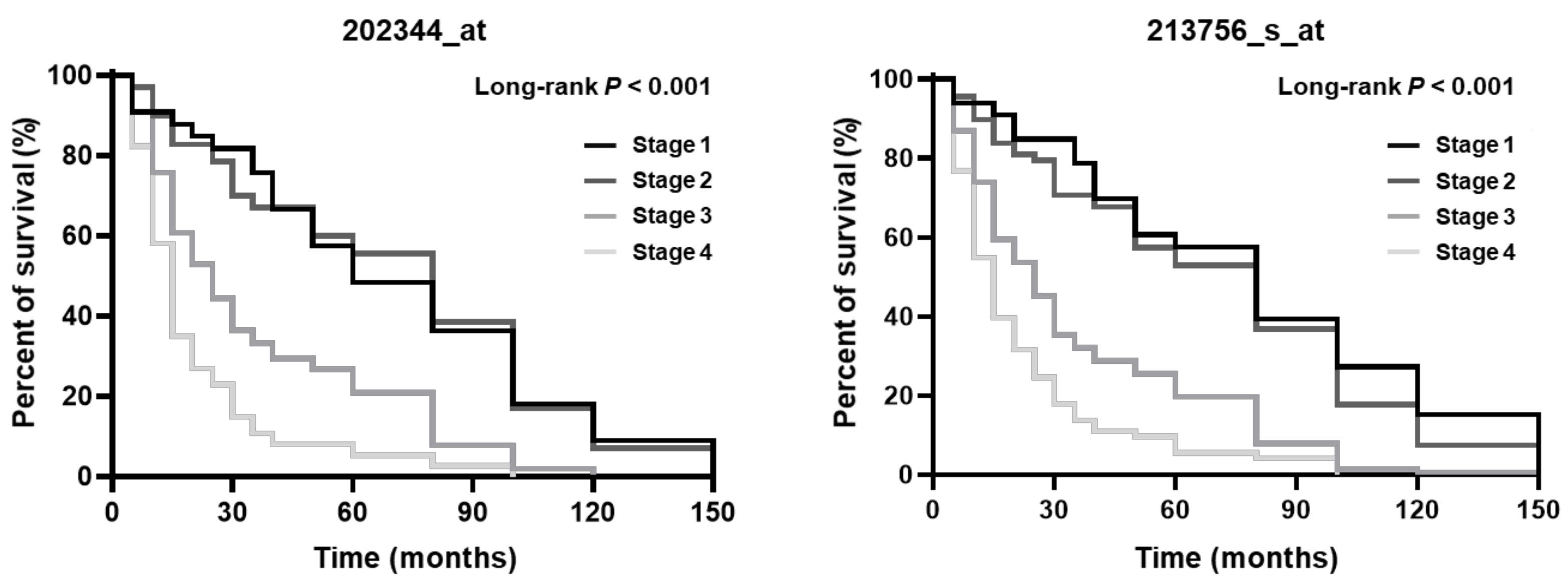
High HSF1 expression has been associated with poor survival in cancer patients [87,88,89]. The silencing of HSF1 reduced the incidence of tumors and increased the effects on long-term survival in mice [63]. In advanced stages of other cancers, the expression and activation of HSF1 has been shown to be elevated [31,66,90]. HSF1 has been suggested to play a role as a potential prognostic biomarker in other cancers. The studies conducted by Kim, Dai, and Grunberg revealed that high HSF1 expression also contributes to poor survival in gastric cancer patients [41,80,86]. Therefore, we confirmed the overall survival of gastric cancer patients with HSF1 expression using Kmplot public data (https://kmplot.com/analysis/, accessed on 2 January 2021). HSF1 overexpression in early (I and II) and advanced (III and IV) stages was associated with poor survival [86]. Additionally, for the overall survival, high HSF1 expression levels in gastric cancer patients led to poor survival in advanced cancer stages (Affy ID: 202344_at and 213756_s_at) (Figure 1, Table 2 and Table 3). Therefore, high HSF1 levels are associated with poor survival and worse long-term survival in advanced gastric cancer stages.
5. HSF1 as a Biomarker in Gastric Cancer
5.1. HSF1 as a Therapeutic Target
As a therapeutic target of gastric cancer, it is important to study the transcription factors involved in tumorigenesis and tumor progression [71]. Recent studies have demonstrated the potential of HSF1 as a target for gastric cancer therapy through the inhibition of the transcriptional activity of HSF1. Rocaglamide A and rohinitib (analogs to RocA) were found to disrupt HSF1 binding to the target genes and act as HSF1 inhibitors [91]. The studies conducted by Yoon suggested that KRIBB11 inhibits the transcriptional activity of HSF1 through disrupting the HSF1-dependent binding of p-TEFb (positive transcription elongation factor b) and hsp70 promoter [92]. Triptolide is derived from T. wilfordii, and it inhibits the trimerization of HSF1 complexes that bind to the endogenous HSP70 promoter [93]. Ginsenoside Rg3, the major compound in ginseng, was found to induce apoptosis through FUT4 inhibition via SP1 and HSF1 transcription regulation in gastric cancer cells with H. pylori CagA [42]. The inhibitory activities of triptolide [94,95,96,97] and KRIBB11 and KNK437 [81] were also confirmed in gastric cancer cell lines. Although not yet identified in gastric cancer, many HSF1 mediated inhibitors are being studied. Cantharidin [81,98] and Cardenolide CL-43 [99] have been shown to inhibit HSF1 transcriptional activity in cancer cells. In addition, PW3405 [100] and Compound 1 [101] were discovered as inhibitors of HSF1 by inhibition of phosphorylation. Quercetin [102,103,104] and Fisetin [76] are flavonoids identified as suppressors of the HSF1 binding to HSE. Additional HSF1-mediated inhibitors are summarized in Table 4. HSF1 inhibitors in gastric cancer are still in a preclinical stage; however, this evidence suggests that HSF1 acts as a potent target for gastric cancer therapy.
5.2. HSF1 Expression Level as a Prognostic and Diagnostic Biomarker
Biomarkers can be used for risk assessment of cancer and also for assisting in cancer staging for initial therapy [105]. Prognostic markers provide the information regarding the onset of cancer and can help identify the cancer patients requiring treatment. In cancer patients, the diagnostic biomarker can delay cancer progression through clinical management and suitable preventive interventions. Recent studies presented the possibility of employing HSF1 as a prognostic and diagnostic biomarker of gastric cancer. In gastric cancer tissue, mRNA and/or protein expression levels of HSF1 are significantly higher than those in normal tissue. Furthermore, HSF1 promotes the proliferation, invasion, and migration of gastric cancer cells [41,43,86]. Additionally, the expression level of HSF1 is associated with advanced tumor progression in gastric cancer patients. Kaplan–Meier analysis in patients with gastric cancer revealed that the high expression levels of HSF1 were associated with poor prognosis [41]. Dai et al. analyzed the prognostic value of HSF1 expression in early and advanced gastric cancer using TNM classification. It was confirmed that gastric cancer patients with higher HSF1 expression had worse overall survival rates and recurrence-free survival than those with low expression in early and advanced stages [43]. Altogether, HSF1 is associated with tumor progression and poor prognosis in gastric cancer, and it can serve as a prognostic and diagnostic biomarker of gastric cancer.
6. Conclusions
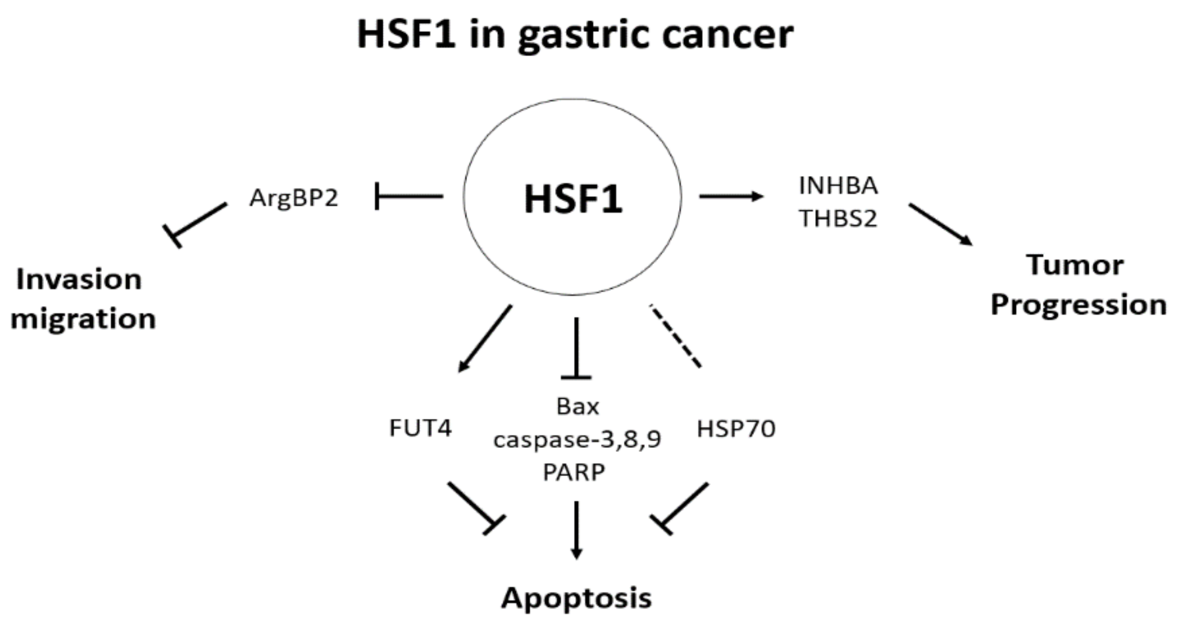
As endoscopy and imaging technology developed for the diagnosis of gastric cancer, the detection of early gastric cancer-related lesions has also been improved; however, it is still important to identify additional biomarkers and study the mechanisms to assist in diagnosing gastric cancer at early stages. This review suggested that HSF1 can be employed as a diagnostic and prognostic biomarker of gastric cancer. Using several reports, we showed that HSF1 is a powerful multifaceted regulator of various cancers, including gastric cancer. Particularly, it was confirmed that the expression of HSF1 is increased in various cancers, including gastric cancer, and an increased HSF1 expression is involved in cell death through regulating genes (Bax, caspase-3, 8, 9, and PARP) related to apoptosis (Figure 2). It was confirmed that HSF1 exerts an anti-apoptotic effect. Additionally, it was confirmed that HSF1 regulates HSPs as a transcription factor of HSPs, thereby also affecting cell proliferation (Figure 2).
Additionally, HSF1 has been confirmed to exert an effect on cell migration and metastasis. Particularly, in gastric cancer, it has been reported to regulate cell migration through binding to ArgBP2, and HSF1 has been shown to regulate cell migration and metastasis (Figure 2). Moreover, it was confirmed that HSF1 is involved in the proliferation and metastasis of gastric cancer cells via an association with CagA, a major factor of H. pylori, which is known as a high-risk factor associated with gastric cancer (Figure 2). HSF1 expression has also been reported to be increased in various cancers as previously mentioned, and the increased expression of HSF1 in gastric cancer was confirmed through public data analysis (Table 1 and Figure 1). Based on these results, HSF1 has been suggested as an important regulator of gastric cancer cells, and its association with various signaling and target genes has been confirmed. These results confirm that HSF1 can be considered as an important biomarker for diagnosing gastric cancer and as a potential target for future drug development.
Author Contributions
Conceptualization, W.K. and S.-J.K.; investigation, W.K. and S.-J.K.; writing—original draft preparation, W.K. and S.-J.K.; writing—review and editing, W.K. and S.-J.K.; visualization, W.K. and S.-J.K.; supervision, S.-J.K.; project administration, S.-J.K.; funding acquisition, S.-J.K. All authors have read and agreed to the published version of the manuscript.
Funding
This work was supported by the grants from National Research Foundation (NRF) of Korea funded by the Korean government (MSIP, No. NRF-2020R1A2C1100078) and Chosun University, 1 May 2021.
Institutional Review Board Statement
Not applicable.
Informed Consent Statement
Not applicable.
Data Availability Statement
Not applicable.
Conflicts of Interest
The authors declare no conflict of interest. The funders had no role in the design of the study; in the collection, analyses, or interpretation of data; in the writing of the manuscript, or in the decision to publish the results.
References
- Rawla, P.; Barsouk, A. Epidemiology of gastric cancer: Global trends, risk factors and prevention. Gastroenterol. Rev. 2019, 14, 26–38. [Google Scholar] [CrossRef] [PubMed]
- Xia, Y.; Wu, Q.; Wang, H.; Zhang, S.; Jiang, Y.; Gong, T.; Xu, X.; Chang, Q.; Niu, K.; Zhao, Y. Global, regional and national burden of gout, 1990–2017: A systematic analysis of the Global Burden of Disease Study. Rheumatology 2019, 59, 1529–1538. [Google Scholar] [CrossRef] [PubMed]
- Balakrishnan, M.; George, R.; Sharma, A.; Graham, D.Y. Changing Trends in Stomach Cancer Throughout the World. Curr. Gastroenterol. Rep. 2017, 19, 1–10. [Google Scholar] [CrossRef] [PubMed]
- Song, Z.; Wu, Y.; Yang, J.; Yang, D.; Fang, X. Progress in the treatment of advanced gastric cancer. Tumor Biol. 2017, 39, 1010428317714626. [Google Scholar] [CrossRef] [Green Version]
- Herrero, R.; Park, J.Y.; Forman, D. The fight against gastric cancer—The IARC Working Group report. Best Pr. Res. Clin. Gastroenterol. 2014, 28, 1107–1114. [Google Scholar] [CrossRef]
- Ryu, J.W.; Kim, H.J.; Lee, Y.S.; Myong, N.H.; Hwang, C.H.; Lee, G.S.; Yom, H.C. The Proteomics Approach to Find Biomarkers in Gastric Cancer. J. Korean Med. Sci. 2003, 18, 505–509. [Google Scholar] [CrossRef] [Green Version]
- Szász, A.M.; Lánczky, A.; Nagy, A.; Förster, S.; Hark, K.; Green, J.E.; Boussioutas, A.; Busuttil, R.; Szabó, A.; Győrffy, B. Cross-validation of survival associated biomarkers in gastric cancer using transcriptomic data of 1,065 patients. Oncotarget 2016, 7, 49322–49333. [Google Scholar] [CrossRef] [Green Version]
- Abbas, M.; Faggian, A.; Sintali, D.N.; Khan, G.J.; Naeem, S.; Shi, M.; Dingding, C. Current and future biomarkers in gastric cancer. Biomed. Pharmacother. 2018, 103, 1688–1700. [Google Scholar] [CrossRef]
- Goossens, N.; Nakagawa, S.; Sun, X.; Hoshida, Y. Cancer biomarker discovery and validation. Transl. Cancer Res. 2015, 4, 256–269. [Google Scholar]
- A Mamas, M.; Dunn, W.; Neyses, L.; Goodacre, R. The role of metabolites and metabolomics in clinically applicable biomarkers of disease. Arch. Toxicol. 2011, 85, 5–17. [Google Scholar] [CrossRef]
- Ballard-Barbash, R.; Friedenreich, C.M.; Courneya, K.S.; Siddiqi, S.M.; McTiernan, A.; Alfano, C.M. Physical Activity, Biomarkers, and Disease Outcomes in Cancer Survivors: A Systematic Review. J. Natl. Cancer Inst. 2012, 104, 815–840. [Google Scholar] [CrossRef] [Green Version]
- Matsuoka, T.; Yashiro, M. Biomarkers of gastric cancer: Current topics and future perspective. World J. Gastroenterol. 2018, 24, 2818–2832. [Google Scholar] [CrossRef]
- Carlomagno, N.; Incollingo, P.; Tammaro, V.; Peluso, G.; Rupealta, N.; Chiacchio, G.; Sotelo, M.L.S.; Minieri, G.; Pisani, A.; Riccio, E.; et al. Diagnostic, Predictive, Prognostic, and Therapeutic Molecular Biomarkers in Third Millennium: A Breakthrough in Gastric Cancer. BioMed Res. Int. 2017, 2017, 1–11. [Google Scholar] [CrossRef] [Green Version]
- Azuaje, F.; Devaux, Y.; Wagner, D. Challenges and Standards in Reporting Diagnostic and Prognostic Biomarker Studies. Clin. Transl. Sci. 2009, 2, 156–161. [Google Scholar] [CrossRef] [Green Version]
- Italiano, A. Prognostic or Predictive? It’s Time to Get Back to Definitions! J. Clin. Oncol. 2011, 29, 4718. [Google Scholar] [CrossRef]
- Vihervaara, A.; Sistonen, L. HSF1 at a glance. J. Cell Sci. 2014, 127, 261–266. [Google Scholar] [CrossRef] [Green Version]
- Barna, J.; Csermely, P.; Vellai, T. Roles of heat shock factor 1 beyond the heat shock response. Cell. Mol. Life Sci. 2018, 75, 2897–2916. [Google Scholar] [CrossRef]
- Gomez-Pastor, R.; Burchfiel, E.T.; Thiele, D.J. Regulation of heat shock transcription factors and their roles in physiology and disease. Nat. Rev. Mol. Cell Biol. 2018, 19, 4–19. [Google Scholar] [CrossRef]
- Labbadia, J.; Morimoto, R.I. The Biology of Proteostasis in Aging and Disease. Annu. Rev. Biochem. 2015, 84, 435–464. [Google Scholar] [CrossRef] [Green Version]
- Grandi, P.; Bantscheff, M. Advanced proteomics approaches to unravel protein homeostasis. Drug Discov. Today Technol. 2019, 31, 99–108. [Google Scholar] [CrossRef]
- Van Drie, J.H. Protein folding, protein homeostasis, and cancer. Chin. J. Cancer 2011, 30, 124–137. [Google Scholar] [CrossRef] [Green Version]
- Wang, G.; Cao, P.; Fan, Y.; Tan, K. Emerging roles of HSF1 in cancer: Cellular and molecular episodes. Biochim. Biophys. Acta (BBA) Bioenerg. 2020, 1874, 188390. [Google Scholar] [CrossRef] [PubMed]
- Doyle, S.M.; Genest, O.; Wickner, S. Protein rescue from aggregates by powerful molecular chaperone machines. Nat. Rev. Mol. Cell Biol. 2013, 14, 617–629. [Google Scholar] [CrossRef] [PubMed]
- Brandvold, K.R.; Morimoto, R.I. The Chemical Biology of Molecular Chaperones—Implications for Modulation of Proteostasis. J. Mol. Biol. 2015, 427, 2931–2947. [Google Scholar] [CrossRef] [PubMed] [Green Version]
- Smith, H.L.; Li, W.; Cheetham, M.E. Molecular chaperones and neuronal proteostasis. Semin. Cell Dev. Biol. 2015, 40, 142–152. [Google Scholar] [CrossRef] [PubMed] [Green Version]
- Dong, B.; Jaeger, A.M.; Thiele, D.J. Inhibiting Heat Shock Factor 1 in Cancer: A Unique Therapeutic Opportunity. Trends Pharmacol. Sci. 2019, 40, 986–1005. [Google Scholar] [CrossRef] [PubMed]
- Ciocca, D.R.; Arrigo, A.P.; Calderwood, S.K. Heat shock proteins and heat shock factor 1 in carcinogenesis and tumor development: An update. Arch. Toxicol. 2013, 87, 19–48. [Google Scholar] [CrossRef] [PubMed] [Green Version]
- Jego, G.; Hazoumé, A.; Seigneuric, R.; Garrido, C. Targeting heat shock proteins in cancer. Cancer Lett. 2013, 332, 275–285. [Google Scholar] [CrossRef]
- Powell, C.D.; Paullin, T.R.; Aoisa, C.; Menzie, C.J.; Ubaldini, A.; Westerheide, S.D. The Heat Shock Transcription Factor HSF1 Induces Ovarian Cancer Epithelial-Mesenchymal Transition in a 3D Spheroid Growth Model. PLoS ONE 2016, 11, e0168389. [Google Scholar] [CrossRef] [Green Version]
- Engerud, H.; Tangen, I.L.; Berg, A.; Kusonmano, K.; Halle, M.K.; Øyan, A.M.; Kalland, K.H.; Stefansson, I.; Trovik, J.; Salvesen, H.B.; et al. High level of HSF1 associates with aggressive endometrial carcinoma and suggests potential for HSP90 inhibitors. Br. J. Cancer 2014, 111, 78–84. [Google Scholar] [CrossRef] [Green Version]
- Chen, K.; Qian, W.; Li, J.; Jiang, Z.; Cheng, L.; Yan, B.; Cao, J.; Sun, L.; Zhou, C.; Lei, M.; et al. Loss of AMPK activation promotes the invasion and metastasis of pancreatic cancer through an HSF1-dependent pathway. Mol. Oncol. 2017, 11, 1475–1492. [Google Scholar] [CrossRef]
- Björk, J.K.; Ahonen, I.; Mirtti, T.; Erickson, A.; Rannikko, A.; Bützow, A.; Nordling, S.; Lundin, J.; Lundin, M.; Sistonen, L.; et al. Increased HSF1 expression predicts shorter disease-specific survival of prostate cancer patients following radical prostatectomy. Oncotarget 2018, 9, 31200–31213. [Google Scholar] [CrossRef] [Green Version]
- Heimberger, T.; Andrulis, M.; Riedel, S.; Stühmer, T.; Schraud, H.; Bumm, T.; Bargou, R.C.; Chatterjee, M.; Beilhack, A.; Bogen, B.; et al. The heat shock transcription factor 1 as a potential new therapeutic target in multiple myeloma. Br. J. Haematol. 2012, 160, 465–476. [Google Scholar] [CrossRef]
- Cigliano, A.; Wang, C.; Pilo, M.G.; Szydlowska, M.; Brozzetti, S.; Latte, G.; Pes, G.M.; Pascale, R.M.; Seddaiu, M.A.; Vidili, G.; et al. Inhibition of HSF1 suppresses the growth of hepatocarcinoma cell lines in vitro and AKT-driven hepatocarcinogenesis in mice. Oncotarget 2017, 8, 54149–54159. [Google Scholar] [CrossRef] [Green Version]
- Mendillo, M.; Santagata, S.; Koeva, M.; Bell, G.W.; Hu, R.; Tamimi, R.M.; Fraenkel, E.; Ince, T.A.; Whitesell, L.; Lindquist, S. HSF1 Drives a Transcriptional Program Distinct from Heat Shock to Support Highly Malignant Human Cancers. Cell 2012, 150, 549–562. [Google Scholar] [CrossRef] [Green Version]
- White, R.J. RNA polymerases I and III, growth control and cancer. Nat. Rev. Mol. Cell Biol. 2005, 6, 69–78. [Google Scholar] [CrossRef]
- Stumpf, C.R.; Ruggero, D. The cancerous translation apparatus. Curr. Opin. Genet. Dev. 2011, 21, 474–483. [Google Scholar] [CrossRef] [Green Version]
- De Billy, E.; Clarke, P.A.; Workman, P. HSF1 in Translation. Cancer Cell 2013, 24, 147–149. [Google Scholar] [CrossRef] [Green Version]
- Xi, C.; Hu, Y.; Buckhaults, P.; Moskophidis, D.; Mivechi, N.F. Heat Shock Factor Hsf1 Cooperates with ErbB2 (Her2/Neu) Protein to Promote Mammary Tumorigenesis and Metastasis. J. Biol. Chem. 2012, 287, 35646–35657. [Google Scholar] [CrossRef] [Green Version]
- Lee, Y.J.; Lee, H.J.; Lee, J.S.; Jeoung, D.; Kang, C.M.; Bae, S.; Lee, S.J.; Kwon, S.H.; Kang, D. A novel function for HSF1-induced mitotic exit failure and genomic instability through direct interaction between HSF1 and Cdc20. Oncogene 2007, 27, 2999–3009. [Google Scholar] [CrossRef] [Green Version]
- Kim, S.-J.; Lee, S.-C.; Kang, H.-G.; Gim, J.; Lee, K.-H.; Lee, S.-H.; Chun, K.-H. Heat Shock Factor 1 Predicts Poor Prognosis of Gastric Cancer. Yonsei Med. J. 2018, 59, 1041–1048. [Google Scholar] [CrossRef] [PubMed]
- Aziz, F.; Wang, X.; Liu, J.; Yan, Q. Ginsenoside Rg3 induces FUT4-mediated apoptosis in H. pylori CagA-treated gastric cancer cells by regulating SP1 and HSF1 expressions. Toxicol. Vitr. 2016, 31, 158–166. [Google Scholar] [CrossRef]
- Tong, Y.; Li, Y.; Gu, H.; Wang, C.; Liu, F.; Shao, Y.; Li, F. HSF1, in association with MORC2, downregulates ArgBP2 via the PRC2 family in gastric cancer cells. Biochim. Biophys. Acta (BBA) Mol. Basis Dis. 2018, 1864, 1104–1114. [Google Scholar] [CrossRef] [PubMed]
- Ritossa, F. A new puffing pattern induced by temperature shock and DNP in drosophila. Cell. Mol. Life Sci. 1962, 18, 571–573. [Google Scholar] [CrossRef]
- Schuetz, T.J.; Gallo, G.J.; Sheldon, L.; Tempst, P.; Kingston, R.E. Isolation of a cDNA for HSF2: Evidence for two heat shock factor genes in humans. Proc. Natl. Acad. Sci. USA 1991, 88, 6911–6915. [Google Scholar] [CrossRef] [PubMed] [Green Version]
- Nakai, A.; I Morimoto, R. Characterization of a novel chicken heat shock transcription factor, heat shock factor 3, suggests a new regulatory pathway. Mol. Cell. Biol. 1993, 13, 1983–1997. [Google Scholar] [CrossRef]
- Nakai, A.; Tanabe, M.; Kawazoe, Y.; Inazawa, J.; I Morimoto, R.; Nagata, K. HSF4, a new member of the human heat shock factor family which lacks properties of a transcriptional activator. Mol. Cell. Biol. 1997, 17, 469–481. [Google Scholar] [CrossRef] [Green Version]
- Fujimoto, M.; Nakai, A. The heat shock factor family and adaptation to proteotoxic stress. FEBS J. 2010, 277, 4112–4125. [Google Scholar] [CrossRef]
- Nover, L.; Bharti, K.; Döring, P.; Mishra, S.K.; Ganguli, A.; Scharf, K.-D. Arabidopsis and the heat stress transcription factor world: How many heat stress transcription factors do we need? Cell Stress Chaperon 2001, 6, 177–189. [Google Scholar] [CrossRef]
- Peteranderl, R.; Nelson, H.C.M. Trimerization of the heat shock transcription factor by a triple-stranded alpha-helical coiled-coil. Biochemistry 1992, 31, 12272–12276. [Google Scholar] [CrossRef]
- Rabindran, S.K.; I Haroun, R.; Clos, J.; Wisniewski, J.; Wu, C. Regulation of heat shock factor trimer formation: Role of a conserved leucine zipper. Science 1993, 259, 230–234. [Google Scholar] [CrossRef]
- Westwood, J.T.; Wu, C. Activation of Drosophila heat shock factor: Conformational change associated with a monomer-to-trimer transition. Mol. Cell. Biol. 1993, 13, 3481–3486. [Google Scholar] [CrossRef] [Green Version]
- Amin, J.; Ananthan, J.; Voellmy, R. Key features of heat shock regulatory elements. Mol. Cell. Biol. 1988, 8, 3761–3769. [Google Scholar] [CrossRef]
- Pelham, H.R. A regulatory upstream promoter element in the Drosophila Hsp 70 heat-shock gene. Cell 1982, 30, 517–528. [Google Scholar] [CrossRef]
- Sorger, P.K.; Pelham, H.R. Yeast heat shock factor is an essential DNA-binding protein that exhibits temperature-dependent phosphorylation. Cell 1988, 54, 855–864. [Google Scholar] [CrossRef]
- Anckar, J.; Sistonen, L. Regulation of HSF1 Function in the Heat Stress Response: Implications in Aging and Disease. Annu. Rev. Biochem. 2011, 80, 1089–1115. [Google Scholar] [CrossRef]
- Eastmond, D.L.; Nelson, H.C.M. Genome-wide Analysis Reveals New Roles for the Activation Domains of the Saccharomyces cerevisiae Heat Shock Transcription Factor (Hsf1) during the Transient Heat Shock Response. J. Biol. Chem. 2006, 281, 32909–32921. [Google Scholar] [CrossRef] [Green Version]
- Hanahan, D.; Weinberg, R.A. Hallmarks of Cancer: The Next Generation. Cell 2011, 144, 646–674. [Google Scholar] [CrossRef] [Green Version]
- Kong, M.; Reid, M.A. Dealing with hunger: Metabolic stress responses in tumors. J. Carcinog. 2013, 12, 17. [Google Scholar] [CrossRef]
- Sosa, V.; Moliné, T.; Somoza, R.; Paciucci, R.; Kondoh, H.; Lleonart, M.E. Oxidative stress and cancer: An overview. Ageing Res. Rev. 2013, 12, 376–390. [Google Scholar] [CrossRef]
- Gaillard, H.; García-Muse, T.; Aguilera, A. Replication stress and cancer. Nat. Rev. Cancer 2015, 15, 276–289. [Google Scholar] [CrossRef] [PubMed]
- Solimini, N.L.; Luo, J.; Elledge, S.J. Non-Oncogene Addiction and the Stress Phenotype of Cancer Cells. Cell 2007, 130, 986–988. [Google Scholar] [CrossRef] [PubMed] [Green Version]
- Dai, C.; Whitesell, L.; Rogers, A.B.; Lindquist, S. Heat Shock Factor 1 Is a Powerful Multifaceted Modifier of Carcinogenesis. Cell 2007, 130, 1005–1018. [Google Scholar] [CrossRef] [Green Version]
- Min, J.-N.; Huang, L.; Zimonjic, D.B.; Moskophidis, D.; Mivechi, N.F. Selective suppression of lymphomas by functional loss of Hsf1 in a p53-deficient mouse model for spontaneous tumors. Oncogene 2007, 26, 5086–5097. [Google Scholar] [CrossRef] [PubMed] [Green Version]
- Scott, K.L.; Nogueira, C.; Heffernan, T.P.; van Doorn, R.; Dhakal, S.; Hanna, J.A.; Min, C.; Jaskelioff, M.; Xiao, Y.; Wu, C.-J.; et al. Proinvasion Metastasis Drivers in Early-Stage Melanoma Are Oncogenes. Cancer Cell 2011, 20, 92–103. [Google Scholar] [CrossRef] [PubMed] [Green Version]
- Fang, F.; Chang, R.; Yang, L. Heat shock factor 1 promotes invasion and metastasis of hepatocellular carcinoma in vitro and in vivo. Cancer 2011, 118, 1782–1794. [Google Scholar] [CrossRef]
- Tang, Z.; Dai, S.; He, Y.; Doty, R.A.; Shultz, L.D.; Sampson, S.B.; Dai, C. MEK Guards Proteome Stability and Inhibits Tumor-Suppressive Amyloidogenesis via HSF1. Cell 2015, 160, 729–744. [Google Scholar] [CrossRef] [Green Version]
- Dai, C.; Santagata, S.; Tang, Z.; Shi, J.; Cao, J.; Kwon, H.; Bronson, R.T.; Whitesell, L.; Lindquist, S. Loss of tumor suppressor NF1 activates HSF1 to promote carcinogenesis. J. Clin. Investig. 2012, 122, 3742–3754. [Google Scholar] [CrossRef] [Green Version]
- Fujimoto, M.; Takaki, E.; Takii, R.; Tan, K.; Prakasam, R.; Hayashida, N.; Iemura, S.-I.; Natsume, T.; Nakai, A. RPA Assists HSF1 Access to Nucleosomal DNA by Recruiting Histone Chaperone FACT. Mol. Cell 2012, 48, 182–194. [Google Scholar] [CrossRef] [Green Version]
- Li, D.; Yallowitz, A.; Ozog, L.; Marchenko, N. A gain-of-function mutant p53–HSF1 feed forward circuit governs adaptation of cancer cells to proteotoxic stress. Cell Death Dis. 2014, 5, e1194. [Google Scholar] [CrossRef] [Green Version]
- Carpenter, R.L. HSF1 as a Cancer Biomarker and Therapeutic Target. Curr. Cancer Drug Targets 2019, 19, 515–524. [Google Scholar] [CrossRef]
- Prince, T.L.; Lang, B.J.; Guerrero-Gimenez, M.E.; Fernandez-Muñoz, J.M.; Ackerman, A.; Calderwood, S.K. HSF1: Primary Factor in Molecular Chaperone Expression and a Major Contributor to Cancer Morbidity. Cells 2020, 9, 1046. [Google Scholar] [CrossRef]
- Verma, R.; Sharma, P.C. Exploring the Role of Heat Shock Proteins in the Development of Gastric Cancer. Heat Shock Proteins Human Dis. 2020, 21, 1–23. [Google Scholar]
- Lee, Y.-J.; Kim, E.-H.; Lee, J.S.; Jeoung, D.; Bae, S.; Kwon, S.H.; Lee, Y.-S. HSF1 as a Mitotic Regulator: Phosphorylation of HSF1 by Plk1 Is Essential for Mitotic Progression. Cancer Res. 2008, 68, 7550–7560. [Google Scholar] [CrossRef] [Green Version]
- Arora, N.; Alsaied, O.; Dauer, P.; Majumder, K.; Modi, S.; Giri, B.; Dudeja, V.; Banerjee, S.; Von Hoff, D.; Saluja, A. Downregulation of Sp1 by Minnelide leads to decrease in HSP70 and decrease in tumor burden of gastric cancer. PLoS ONE 2017, 12, e0171827. [Google Scholar] [CrossRef]
- Kim, J.A.; Lee, S.; Kim, D.-E.; Kim, M.; Kwon, B.-M.; Han, D.C. Fisetin, a dietary flavonoid, induces apoptosis of cancer cells by inhibiting HSF1 activity through blocking its binding to the hsp70 promoter. Carcinogenesis 2015, 36, 696–706. [Google Scholar] [CrossRef]
- Workman, P. Reflections and Outlook on Targeting HSP90, HSP70 and HSF1 in Cancer: A Personal Perspective. Adv. Exp. Med. Biol. 2020, 1243, 163–179. [Google Scholar] [CrossRef]
- Sahai, E. Mechanisms of cancer cell invasion. Curr. Opin. Genet. Dev. 2005, 15, 87–96. [Google Scholar] [CrossRef]
- Meng, L.; Gabai, V.L.; Sherman, M.Y. Heat-shock transcription factor HSF1 has a critical role in human epidermal growth factor receptor-2-induced cellular transformation and tumorigenesis. Oncogene 2010, 29, 5204–5213. [Google Scholar] [CrossRef] [Green Version]
- Grunberg, N.; Pevsner-Fischer, M.; Goshen-Lago, T.; Diment, J.; Stein, Y.; Lavon, H.; Mayer, S.; Levi-Galibov, O.; Friedman, G.; Ofir-Birin, Y.; et al. Cancer-Associated Fibroblasts Promote Aggressive Gastric Cancer Phenotypes via Heat Shock Factor 1–Mediated Secretion of Extracellular Vesicles. Cancer Res. 2021, 81, 1639–1653. [Google Scholar] [CrossRef]
- Xu, J.; Shi, Q.; Xu, W.; Zhou, Q.; Shi, R.; Ma, Y.; Chen, D.; Zhu, L.; Feng, L.; Cheng, A.S.-L.; et al. Metabolic enzyme PDK3 forms a positive feedback loop with transcription factor HSF1 to drive chemoresistance. Theranostics 2019, 9, 2999–3013. [Google Scholar] [CrossRef] [PubMed]
- Uemura, N.; Okamoto, S.; Yamamoto, S.; Matsumura, N.; Yamaguchi, S.; Yamakido, M.; Taniyama, K.; Sasaki, N.; Schlemper, R.J. Helicobacter pylori Infection and the Development of Gastric Cancer. N. Engl. J. Med. 2001, 345, 784–789. [Google Scholar] [CrossRef] [PubMed]
- Rieder, G.; Hofmann, J.A.; Hatz, R.A.; Stolte, M.; Enders, G.A. Up-regulation of inducible nitric oxide synthase in Helicobacter pylori-associated gastritis may represent an increased risk factor to develop gastric carcinoma of the intestinal type. Int. J. Med. Microbiol. 2003, 293, 403–412. [Google Scholar] [CrossRef] [PubMed]
- Polk, D.B.; Peek, R.M., Jr. Helicobacter pylori: Gastric cancer and beyond. Nat. Rev. Cancer 2010, 10, 403–414. [Google Scholar] [CrossRef] [Green Version]
- Zhu, Y.; Zhong, X.; Zheng, S.; Du, Q.; Xu, W. Transformed immortalized gastric epithelial cells by virulence factor CagA of Helicobacter pylori through Erk mitogen-activated protein kinase pathway. Oncogene 2005, 24, 3886–3895. [Google Scholar] [CrossRef] [Green Version]
- Dai, W.; Ye, J.; Zhang, Z.; Yang, L.; Ren, H.; Wu, H.; Chen, J.; Ma, J.; Zhai, E.; Cai, S.; et al. Increased expression of heat shock factor 1 (HSF1) is associated with poor survival in gastric cancer patients. Diagn. Pathol. 2018, 13, 80. [Google Scholar] [CrossRef]
- Liao, Y.; Xue, Y.; Zhang, L.; Feng, X.; Liu, W.; Zhang, G. Higher heat shock factor 1 expression in tumor stroma predicts poor prognosis in esophageal squamous cell carcinoma patients. J. Transl. Med. 2015, 13, 1–13. [Google Scholar] [CrossRef] [Green Version]
- Tsukao, Y.; Yamasaki, M.; Miyazaki, Y.; Makino, T.; Takahashi, T.; Kurokawa, Y.; Miyata, H.; Nakajima, K.; Takiguchi, S.; Mimori, K.; et al. Overexpression of heat-shock factor 1 is associated with a poor prognosis in esophageal squamous cell carcinoma. Oncol. Lett. 2017, 13, 1819–1825. [Google Scholar] [CrossRef]
- Zhou, Z.; Li, Y.; Jia, Q.; Wang, Z.; Wang, X.; Hu, J.; Xiao, J. Heat shock transcription factor 1 promotes the proliferation, migration and invasion of osteosarcoma cells. Cell Prolif. 2017, 50, e12346. [Google Scholar] [CrossRef]
- Hoang, A.T.; Huang, J.; Rudra-Ganguly, N.; Zheng, J.; Powell, W.C.; Rabindran, S.K.; Wu, C.; Roy-Burman, P. A Novel Association between the Human Heat Shock Transcription Factor 1 (HSF1) and Prostate Adenocarcinoma. Am. J. Pathol. 2000, 156, 857–864. [Google Scholar] [CrossRef] [Green Version]
- Santagata, S.; Mendillo, M.; Tang, Y.-C.; Subramanian, A.; Perley, C.C.; Roche, S.P.; Wong, B.; Narayan, R.; Kwon, H.; Koeva, M.I.; et al. Tight Coordination of Protein Translation and HSF1 Activation Supports the Anabolic Malignant State. Science 2013, 341, 1238303. [Google Scholar] [CrossRef] [Green Version]
- Yoon, Y.J.; Kim, J.A.; Shin, K.D.; Shin, D.-S.; Han, Y.M.; Lee, Y.J.; Lee, J.S.; Kwon, B.-M.; Han, D.C. KRIBB11 Inhibits HSP70 Synthesis through Inhibition of Heat Shock Factor 1 Function by Impairing the Recruitment of Positive Transcription Elongation Factor b to the hsp70 Promoter. J. Biol. Chem. 2011, 286, 1737–1747. [Google Scholar] [CrossRef] [Green Version]
- Westerheide, S.D.; Kawahara, T.L.; Orton, K.; Morimoto, R.I. Triptolide, an Inhibitor of the Human Heat Shock Response That Enhances Stress-induced Cell Death. J. Biol. Chem. 2006, 281, 9616–9622. [Google Scholar] [CrossRef] [Green Version]
- Chang, H.J.; Kim, M.H.; Baek, M.K.; Park, J.S.; Chung, I.J.; Shin, B.A.; Ahn, B.W.; Jung, Y.D. Triptolide inhibits tumor promoter-induced upar expression via blocking nf-κb signaling in human gastric ags cells. Anticancer Res. 2007, 27, 3411–3417. [Google Scholar]
- Xie, M.; Wu, J.; Ji, L.; Jiang, X.; Zhang, J.; Ge, M.; Cai, X. Development of Triptolide Self-Microemulsifying Drug Delivery System and Its Anti-tumor Effect on Gastric Cancer Xenografts. Front. Oncol. 2019, 9, 978. [Google Scholar] [CrossRef] [Green Version]
- Wang, B.-Y.; Cao, J.; Chen, J.-W.; Liu, Q.-Y. Triptolide induces apoptosis of gastric cancer cells via inhibiting the overexpression of MDM2. Med. Oncol. 2014, 31, 270. [Google Scholar] [CrossRef]
- Li, C.-J.; Chu, C.-Y.; Huang, L.-H.; Wang, M.-H.; Sheu, L.-F.; Yeh, J.-I.; Hsu, H.-Y. Synergistic anticancer activity of triptolide combined with cisplatin enhances apoptosis in gastric cancer in vitro and in vivo. Cancer Lett. 2012, 319, 203–213. [Google Scholar] [CrossRef]
- Xu, M.-D.; Liu, L.; Wu, M.-Y.; Jiang, M.; Shou, L.-M.; Wang, W.-J.; Wu, J.; Zhang, Y.; Gong, F.-R.; Chen, K.; et al. The combination of cantharidin and antiangiogenic therapeutics presents additive antitumor effects against pancreatic cancer. Oncogenesis 2018, 7, 1–15. [Google Scholar] [CrossRef]
- Nikotina, A.D.; Koludarova, L.; Komarova, E.Y.; Mikhaylova, E.R.; Aksenov, N.D.; Suezov, R.; Kartzev, V.G.; Margulis, B.A.; Guzhova, I.V. Discovery and optimization of cardenolides inhibiting HSF1 activation in human colon HCT-116 cancer cells. Oncotarget 2018, 9, 27268–27279. [Google Scholar] [CrossRef]
- Raj, L.; Ide, T.; Gurkar, A.U.; Foley, M.A.; Schenone, M.; Li, X.; Tolliday, N.J.; Golub, T.R.; Carr, S.A.; Shamji, A.F.; et al. Selective killing of cancer cells by a small molecule targeting the stress response to ROS. Nat. Cell Biol. 2011, 475, 231–234. [Google Scholar] [CrossRef]
- Au, Q.; Zhang, Y.; Barber, J.R.; Ng, S.C.; Zhang, B. Identification of Inhibitors of HSF1 Functional Activity by High-Content Target-Based Screening. J. Biomol. Screen. 2009, 14, 1165–1175. [Google Scholar] [CrossRef] [PubMed] [Green Version]
- Nagai, N.; Nakai, A.; Nagata, K. Quercetin Suppresses Heat Shock Response by Down-Regulation of HSF1. Biochem. Biophys. Res. Commun. 1995, 208, 1099–1105. [Google Scholar] [CrossRef] [PubMed]
- Yang, W.; Cui, M.; Lee, J.; Gong, W.; Wang, S.; Fu, J.; Wu, G.; Yan, K. Heat shock protein inhibitor, quercetin, as a novel adjuvant agent to improve radiofrequency ablation-induced tumor destruction and its molecular mechanism. Chin. J. Cancer Res. 2016, 28, 19–28. [Google Scholar] [PubMed]
- Propper, D.; Han, H.; Von Hoff, D.; Borazanci, E.; Reya, T.; Ghergurovich, J.; Pshenichnaya, I.; Antal, C.; Condjella, R.; Sharma, S.; et al. Abstract CT165: Phase II open label trial of minnelide™ in patients with chemotherapy refractory metastatic pancreatic cancer. Clin. Trials 2019. [Google Scholar] [CrossRef]
- Ludwig, J.A.; Weinstein, J.N. Biomarkers in Cancer Staging, Prognosis and Treatment Selection. Nat. Rev. Cancer 2005, 5, 845–856. [Google Scholar] [CrossRef]
Figure 1.
HSF1 expression was correlated with the overall survival of gastric cancer patients. Two probes (Affy ID: 202344_at, and Affy ID: 213756_s_at) revealed that high HSF1 expression leads to poor survival depending on gastric cancer patient stage. The Kaplan–Meier survival curves were generated using the KM-plotter online analysis tool.

Figure 2.
Various roles of HSF1 in gastric cancer. HSF1 has been shown to be involved in cell growth, apoptosis, migration, and invasion in gastric cancer.
Figure 2.
Various roles of HSF1 in gastric cancer. HSF1 has been shown to be involved in cell growth, apoptosis, migration, and invasion in gastric cancer.


Table 1.
HSF1 expression profile in GEO datasets.
| Datasets | Country | Year | Platform | Normal Tissue | Cancer Tissue | p-Value | ||||
|---|---|---|---|---|---|---|---|---|---|---|
| Number | Mean | SD | Number | Mean | SD | |||||
| GSE2685 | Japan | 2005 | GPL80 | 8 | 7.57 | 0.83 | 22 | 8.20 | 0.24 | 0.0026 |
| GSE13861 | USA | 2008 | GPL6884 | 19 | 5.39 | 0.10 | 71 | 5.53 | 0.17 | 0.0017 |
| GSE13911 | Italy | 2008 | GPL570 | 31 | 7.40 | 1.16 | 38 | 8.82 | 0.91 | <0.001 |
| GSE29272 | USA | 2011 | GPL96 | 134 | 5.68 | 0.41 | 134 | 5.84 | 0.53 | 0.0065 |
| GSE54129 | China | 2014 | GPL570 | 21 | 6.58 | 0.14 | 111 | 7.40 | 0.35 | <0.001 |
| GSE81948 | Italy | 2016 | GPL6244 | 5 | 8.46 | 0.12 | 15 | 8.80 | 0.32 | 0.0368 |
| GSE109476 | China | 2018 | GPL24530 | 5 | 13.57 | 0.58 | 5 | 14.34 | 0.40 | 0.0398 |
Table 2.
Overall survival of gastric cancer patients with high HSF1 expression according to the diseases stage (HSF1 probe: Affy ID: 202344_at).
Table 2.
Overall survival of gastric cancer patients with high HSF1 expression according to the diseases stage (HSF1 probe: Affy ID: 202344_at).
| Month (m) | Tumor Stage | |||||||
|---|---|---|---|---|---|---|---|---|
| Stage I | Stage II | Stage III | Stage IV | |||||
| Number of Patients (%) | Number of Patients (%) | Number of Patients (%) | Number of Patients (%) | |||||
| 0 | 33 | (100.0) | 70 | (100.0) | 153 | (100.0) | 74 | (100.0) |
| 5 | 30 | (90.9) | 68 | (97.1) | 139 | (90.8) | 61 | (82.4) |
| 10 | 30 | (90.9) | 63 | (90.0) | 116 | (75.8) | 43 | (58.1) |
| 20 | 28 | (84.8) | 58 | (82.6) | 81 | (52.9) | 20 | (27.0) |
| 40 | 22 | (66.7) | 47 | (67.1) | 45 | (29.4) | 6 | (8.1) |
| 60 | 16 | (48.5) | 39 | (55.7) | 32 | (20.9) | 4 | (5.4) |
| 80 | 12 | (36.4) | 27 | (38.6) | 12 | (7.8) | 2 | (2.7) |
| 100 | 6 | (19.2) | 12 | (17.1) | 3 | (2.0) | 0 | (0.0) |
| 120 | 3 | (9.1) | 5 | (7.1) | 1 | (0.7) | 0 | (0.0) |
| 150 | 0 | (0.0) | 1 | (1.4) | ||||
Table 3.
Overall survival of gastric cancer patients with high HSF1 expression according to the diseases stage (HSF1 probe: Affy ID: 213756_s_at).
Table 3.
Overall survival of gastric cancer patients with high HSF1 expression according to the diseases stage (HSF1 probe: Affy ID: 213756_s_at).
| Month (m) | Tumor Stage | |||||||
|---|---|---|---|---|---|---|---|---|
| Stage I | Stage II | Stage III | Stage IV | |||||
| Number of Patients (%) | Number of Patients (%) | Number of Patients (%) | Number of Patients (%) | |||||
| 0 | 33 | (100.0) | 68 | (100.0) | 153 | (100.0) | 73 | (100.0) |
| 5 | 31 | (93.9) | 65 | (95.6) | 133 | (86.9) | 56 | (76.7) |
| 10 | 31 | (93.9) | 61 | (89.7) | 113 | (73.9) | 40 | (54.8) |
| 20 | 28 | (84.8) | 55 | (80.9) | 82 | (53.6) | 23 | (31.5) |
| 40 | 23 | (69.7) | 46 | (67.6) | 44 | (28.8) | 8 | (11.0) |
| 60 | 19 | (57.6) | 36 | (52.9) | 30 | (19.6) | 4 | (5.5) |
| 80 | 13 | (39.4) | 25 | (36.8) | 12 | (7.8) | 3 | (4.1) |
| 100 | 8 | (27.3) | 12 | (17.6) | 2 | (1.3) | 0 | (0.0) |
| 120 | 5 | (15.2) | 5 | (7.4) | 1 | (0.7) | 0 | (0.0) |
| 150 | 0 | (0.0) | 1 | (1.5) | ||||
Table 4.
HSF1 inhibitors in cancer.
| Inhibitors | Mechanism | References |
|---|---|---|
| Rocaglamide A | Inhibition of HSF1 binding to HSE | [91] |
| Rohintib | Inhibition of HSF1 binding to HSE | [91] |
| KRIBB11 | Inhibition of HSF1 transcriptional activity | [81,92] |
| KNK437 | Inhibition of HSF1 transcriptional activity | [81] |
| Ginsenoside Rg3 | Inhibition of HSF1 transcriptional activity | [42] |
| Cantharidin | Inhibition of HSF1 transcriptional activity | [81,98] |
| Cardenolide CL-43 | Inhibition of HSF1 transcriptional activity | [99] |
| PW3405 | Inhibition of HSF1 phosphorylation | [100] |
| Compound 1 | Inhibition of HSF1 phosphorylation | [101] |
| Quercetin | Inhibition of HSF1 binding to HSE | [102,103,104] |
| Fisetin | Inhibition of HSF1 binding to HSE | [76] |
Publisher’s Note: MDPI stays neutral with regard to jurisdictional claims in published maps and institutional affiliations. |
© 2021 by the authors. Licensee MDPI, Basel, Switzerland. This article is an open access article distributed under the terms and conditions of the Creative Commons Attribution (CC BY) license (https://creativecommons.org/licenses/by/4.0/).
Share and Cite
MDPI and ACS Style
Kim, W.; Kim, S.-J. Heat Shock Factor 1 as a Prognostic and Diagnostic Biomarker of Gastric Cancer. Biomedicines 2021, 9, 586. https://doi.org/10.3390/biomedicines9060586
AMA Style
Kim W, Kim S-J. Heat Shock Factor 1 as a Prognostic and Diagnostic Biomarker of Gastric Cancer. Biomedicines. 2021; 9(6):586. https://doi.org/10.3390/biomedicines9060586
Chicago/Turabian StyleKim, Woong, and Seok-Jun Kim. 2021. "Heat Shock Factor 1 as a Prognostic and Diagnostic Biomarker of Gastric Cancer" Biomedicines 9, no. 6: 586. https://doi.org/10.3390/biomedicines9060586
Note that from the first issue of 2016, this journal uses article numbers instead of page numbers. See further details here.