Article

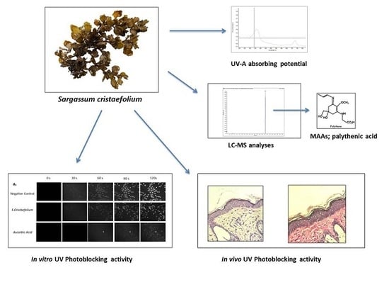
UVA Photoprotective Activity of Brown Macroalgae Sargassum cristafolium

by
Eka Sunarwidhi Prasedya
1,
Sundari Maulinda Syafitri
1,
Brigitta A. F. D. Geraldine
1,
Candra Dwipayana Hamdin
2,
Andri Frediansyah
3,4,
Masao Miyake
5,
Daisuke Kobayashi
5,
Akihiro Hazama
5 and
Haji Sunarpi
1,*
1
Bioscience and Biotechnology Research Centre, Department of Biology, Faculty of Mathematics and Natural Sciences, University of Mataram, Mataram 83115, Indonesia
2
Department of Pharmacy, Medical Faculty, University of Mataram, Mataram 83115, Indonesia
3
Pharmaceutical Institute, University of Tuebingen, 72074 Tuebingen, Germany
4
Research Unit for Natural Product (BPTBA), Indonesian Institute of Sciences (LIPI), Wonosari 55861, Indonesia
5
Department of Cellular and Integrative Physiology, Fukushima Medical University, Fukushima 960-1247, Japan
*
Author to whom correspondence should be addressed.
Biomedicines 2019, 7(4), 77; https://doi.org/10.3390/biomedicines7040077
Submission received: 23 August 2019 / Revised: 15 September 2019 / Accepted: 25 September 2019 / Published: 27 September 2019
(This article belongs to the Section Drug Discovery and Development)

Abstract

:
Sunscreens today contain several synthetic UV (Ultraviolet) filter molecules to protect the skin epidermis from UV radiation damage. However, these molecules may create several negative effects on human skin. Due to this condition, there is an increase in the development of natural products to replace uses of these synthetic chemicals. Brown macroalgae Sargassum has been recently studied for its photoprotective activities. The purpose of this study is to investigate photoprotective activity of one of most abundant Sargassum species in Lombok coast; Sargassum cristaefolium. Spectrophotometry analysis with UV-VIS revealed the UV spectra absorbing capability of Sargassum cristaefolium (SC) in the UVA spectrum range (314–400 nm). Furthermore, spectrometry analyses with LC-MS revealed the existence of UV absorbing compound MAA-palythene. In correlation, SC ethanol extracts also demonstrate that it could protect DNA from UVA irradiation as analyzed in vitro in HeLa cell model. The effects of SC on UVA exposed-dorsal mice skin have also shown interesting results, as mice pretreated with SC before UVA exposure showed protective activity on the epidermal integrity similar as positive control. Whereas, UV exposed mice without SC or commercial products resulted in increased epidermal thickness, which is the common parameter of skin photoaging. In addition, pretreated mice with SC also show protective effects in the formation of collagen connective tissues. Overall, current results show promising photoprotective activity of SC against UV radiation. More advanced investigations of SC as a potential photoprotective agent would be reasonable for development of macroalgae-based natural skin protection products.

Graphical Abstract

1. Introduction

Repeated ultraviolet (UV) exposure to human skin can cause severe skin damage, including photo-aging, inflammation, and photo-carcinogenesis [1,2]. UV light radiation is classified into three categories depending on wavelength ranges; UVA (320–400 nm), UVB (290–320 nm), and UVC (200–290 nm). Both UVA and UVB can injure the human skin, resulting in sunburns, actinic keratosis, photo-aging, and severe harms such as skin cancers [3,4].
The use of sunscreen as photoprotecting agents against UV radiation is becoming very popular. However, most sunscreens are composed of inorganic filters, such as Zinc Oxide (ZnO) and Titanium dioxide (TiO) which could cause negative effects in the human skin [5]. Hence, several studies suggest that skin protectors derived from natural products are potentially more suitable for the human skin [6]. Hence, the concept of complementary or alternative medicine has become more widely accepted, and there is a corresponding interest in natural remedies.
Marine life is probably the most affected environment by UV radiation. Marine organisms such as algae, corals, and crustaceans must find a way to survive such extreme environmental conditions. Hence, marine organisms are forced to develop a metabolic response as a self-defense mechanism; for example, by producing secondary metabolites that allow them to protect themselves against external factors [7]. Macroalgae are one of the most considered organisms to have huge potential as a natural source of important bioactive compounds, which have several bioactivities, such as antitumor, anti-inflammatory, antioxidant, antimicrobial, and UV radiation protective activities [8,9]. Brown macroalgae Sargassum has frequently been reported to exhibit anti-UV absorbing compounds [10,11]. Because of its natural environment, which is mostly exposed to UV light, macroalgae Sargassum produces secondary metabolites for its survival [12]. Indonesia is endowed with abundance of macroalgae such as Sargassum species; particularly Sargassum cristaefolium, which are commonly found in most Indonesian coastal areas. The present study therefore investigates the UV photoprotective potential of Indonesian macroalgae Sargassum cristaefolium ethanol extract against UV radiation. Results from this research would provide basic information regarding potentials of Lombok coast, Indonesia macroalgae for development of natural UV skin protection products.

2. Materials and Methods

2.1. Sample Collection and Extraction

Macroalgae S. cristaefolium were sampled from Batu Layar Coast, West Lombok (8°29′54.251″ S and 116°4′36.664″ E). The macroalgae was identified with reference to algae electronic database [13]. The macroalgae samples were rinsed with freshwater to remove sand debris. Samples were then air dried for ±5 days. After constant dry weight of the samples was obtained, samples were then ground into powder. Macroalgae powder samples were mixed with absolute ethanol solvent with 5× volume of the sample weight (w/v). The seaweed samples were washed with freshwater to remove adhering debris. The collected samples were then dried and powdered. Powder samples were mixed with absolute ethanol solvent with 5× volume of sample weight (w/v). Mixed solutions were incubated at room temperature for 48 maceration process. Every 24 h, mixed solutions were filtered with Whatman grade 1 filter papers. Furthermore, macroalgae extract was subjected to rotary evaporator to evaporate the remaining ethanol solutions. Obtained filtrates were then used as macroalgae ethanol extracts for further experiments.

2.2. UVA–UVB Absorbing Activity

SC extract was diluted to 0.1% concentration for evaluation of its UV light absorbing capability. Extract was added in to a quartz cuvette, and its absorption spectra (wavelengths 200–800 nm) were acquired using a Nanodrop 2000c UV-Visible spectrophotometer (Thermofisher Scientific, DE, USA) against a blank containing ethanol.

2.3. Liquid Chromatography-Mass Spectrometry (LC-MS)

Screening of bioactive compounds in macroalgae ethanol extracts were performed by LC-MS analyses on a LCMS-Q-TQF system (Dionex, Ultimate 3000, Bruker, mi-crOTOF Q) (Bruker Daltonik GmbH, Bremen, Germany). Liquid chromatography separation was achieved, followed by 20 μL injection of extract on a C18 column (250 × 4.6 mm, 5 μm) at a flow rate of 1.4 mL·min−1 with a total run of 45 min. The mobile phase consisted of solvent A, methanol/water/acetonitrile 51/13/36 with 0.3 M ammonium acetate, and solvent B was methanol. The source conditions were the following: nebulizer 40 pst, dry gas 91 min−4, and temperature 200 °C with a scan range of 50–1000 m/z.

2.4. Cell Culture

HeLa cell line (DMEM, Wako, Japan) was used as in vitro cell model to evaluate macroalgae cytoprotective activity. Cells were cultured in Dulbecco’s Modified Eagle’s Medium (DMEM, Wako, Japan) supplemented with 10% Fetal Bovine Serum (FBS) in CO2 humidified incubator (37 °C; 5% CO2). Cells were grown until 80–90% confluency before used in cytoprotective assay. Cell images were obtained from phase-contrast microscopy by BZ-9000 microscope (Keyence, Osaka, Japan).

2.5. Cytoprotective Activity of SC Ethanol Extract against UV-A Irradiation

Human cell model HeLa cells were plated in 35 mm dishes (1 × 10⁵ cells/dish). The DMEM media was removed 24 h after seeding (except for the negative control group) and the cells were exposed SC ethanol extract (0.01%) and Ascorbic acid (0.01%) in 10% FBS supplemented DMEM medium as positive control. After this treatment, the cells were stained with DAPI (1 μg/mL). UV-A radiation was applied to the cells simultaneously with photos taken every 30 s under fluorescence microscope. Number of dead cells (excited by DAPI) were counted and analyzed with ImageJ.

2.6. Animals

For this experiment, a total of 30 female BALBL/c mice, 6–8-week-old and weighing 25–30 g were used. The mice had free access to food and water in a temperature (24–25 °C) and humidity (50%) controlled room. Animals were also subjected to a 12 h light/dark cycle. All experimental procedures were in compliance with the Health guidelines for Care and Use of Laboratory Animals and were approved on 13 June 2019 by the Bioethics Committee of Medical Faculty University of Mataram (Approval code: 117/UN.18.F7/E7/ETIK/2019).

2.7. In Vivo UV-A Irradiation

Mice were randomly divided into 4 groups (n = 6 per group) that represented treatments and controls. These were: normal healthy controls with no UV treatment (Control − UV), normal healthy controls with UV treatment (Control + UV), UV irradiation with 0.1% SC treatment (SC + UV), and UV irradiation with commercial product (CP + UV). Before experimentation, mice were anesthetized by intraperitoneal injection of 1% sodium pentobarbital, and hair was removed from the back skin of mice using hair removal wax.
Parallel UV lamps Reptile UV150 PT2188 13W (Exo Terra, Winchester, MA, USA) were used for UV-A irradiation treatment. Distance of UV lamp to mice was 20 cm to obtain UVA spectral radiance of 300 μW/cm2. Animals were irradiated for 7 days, 2 h/day, with a daily dose of 2.16 J/cm2.

2.8. Histological Measurements

Skin specimens were soaked in 10% formalin and embedded in paraffin. Approximately five micrometer thin sections of the paraffin-embedded skin were cut and subjected to hematoxylin eosin (HE) and Van Gieson (VG) histochemical methods. Morphological analysis of the histological sections was performed by bright field microscopy (Zeiss, Axio Observer D1, Oberkochen, Germany).

2.9. Statistical Analysis

Data were expressed as mean ± standard error of the mean (SEM) or ± standard deviation (SD). The data obtained were subjected to one-way analysis of variance (ANOVA), followed by Tukey’s test. Data analyses were conducted with GraphPad Prism (ver 5.0, San Diego, CA, USA) and Kaleidagraph (ver 4.5.2, Reading, PA, USA). Differences were considered significant if p < 0.05.

3. Results

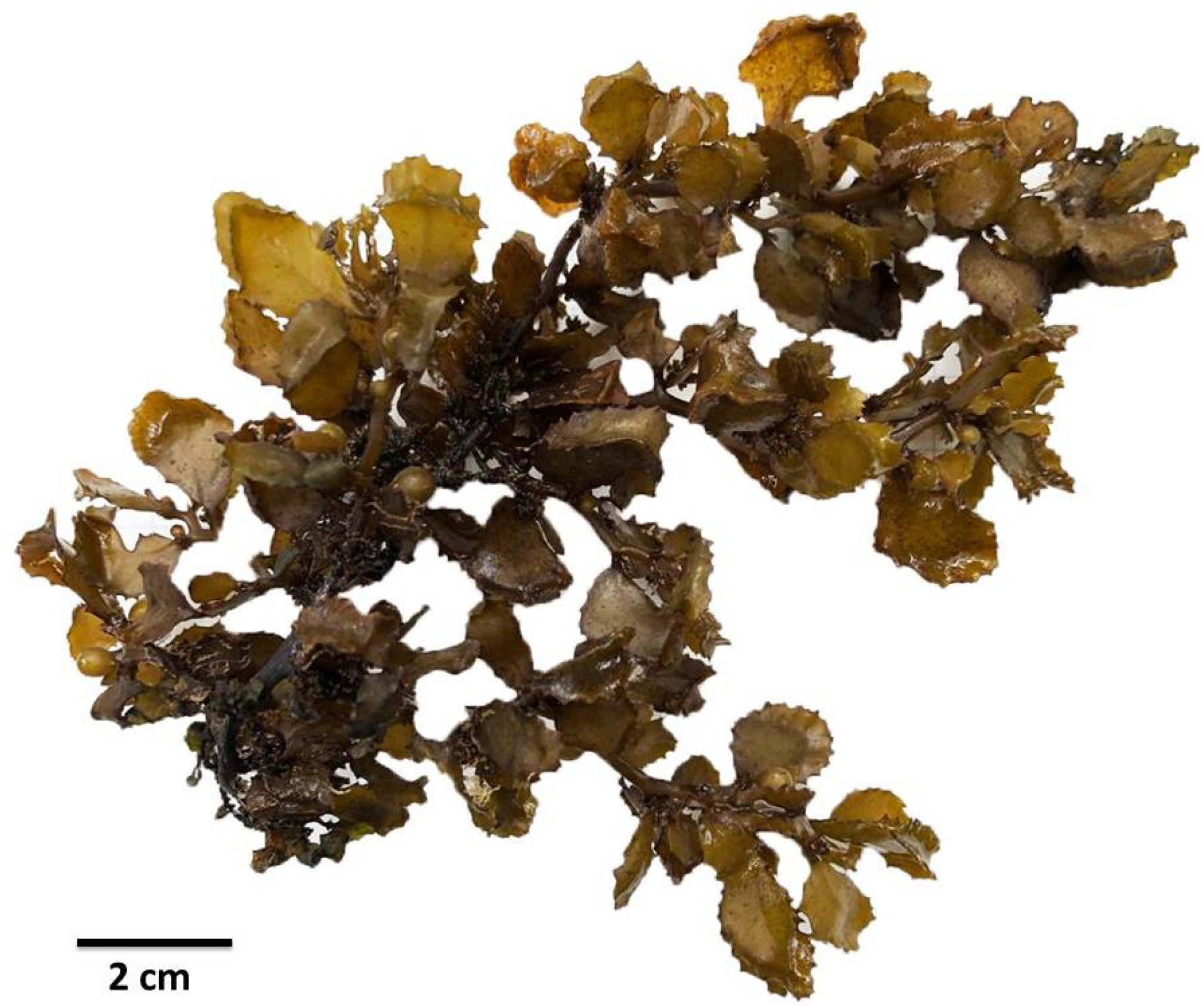
3.1. Collection of Macroalgae Sargassum cristaefolium

Macroalgae S. cristaefolium were collected from North West Lombok coastal area (8°24′11.7396″ S, 116°4′1.9056″ E), West Nusa Tenggara Province, Indonesia in the early 2019 (Figure 1). Macroalgae specimens were identified according to electronic algae database [13].

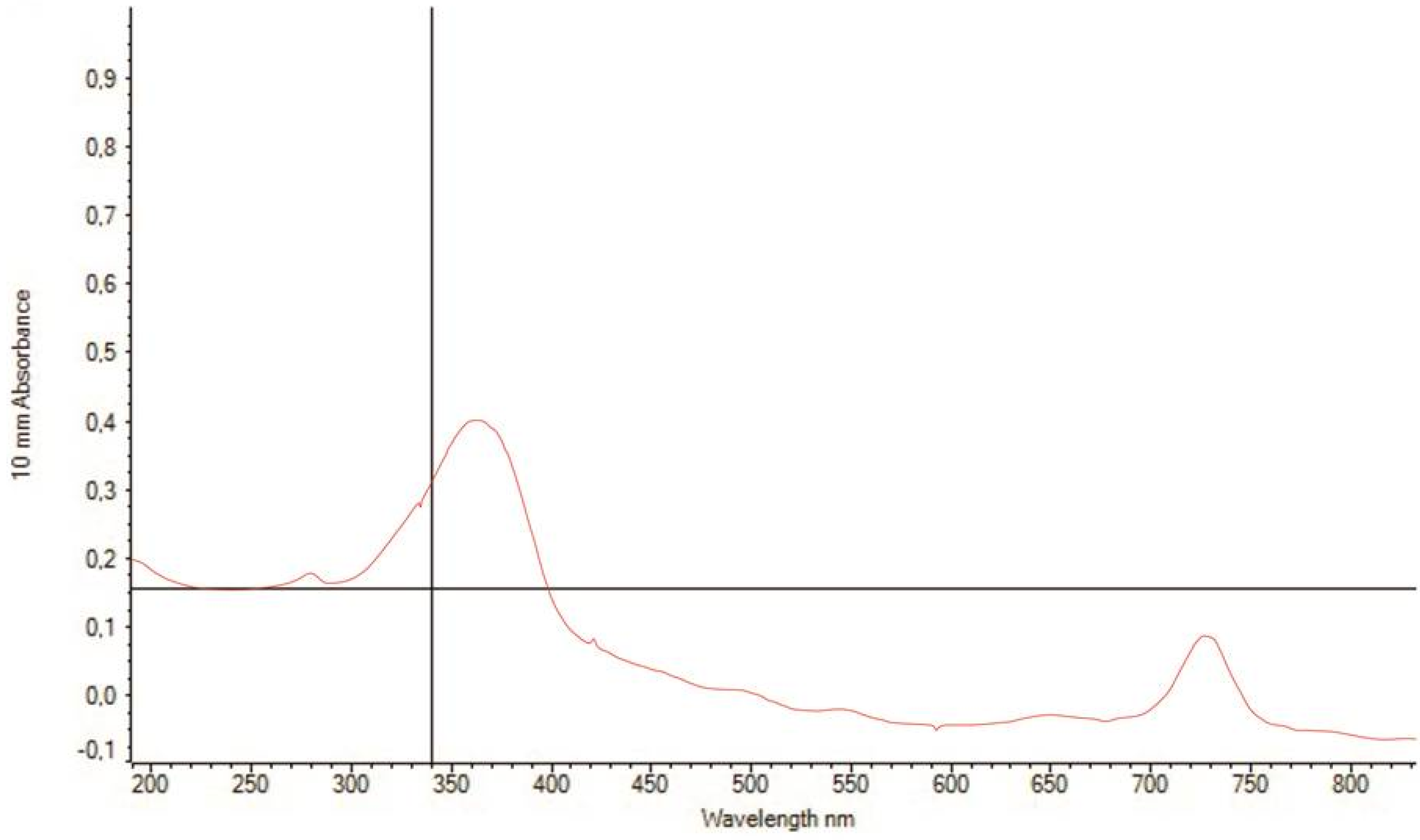
3.2. UV Spectra Absorbance of SC Extract

The UV-Vis absorbance spectra of Sargassum cristaefolium (SC) ethanol extract were evaluated within the range of 200–700 nm. Figure 2 shows the UV-Vis absorbance spectra of SC shown UV absorbing capability in UV-A spectra region (340–400 nm). This result implies that brown macroalgae S. cristaefolium potentially exhibits UV-A light absorbing activity.

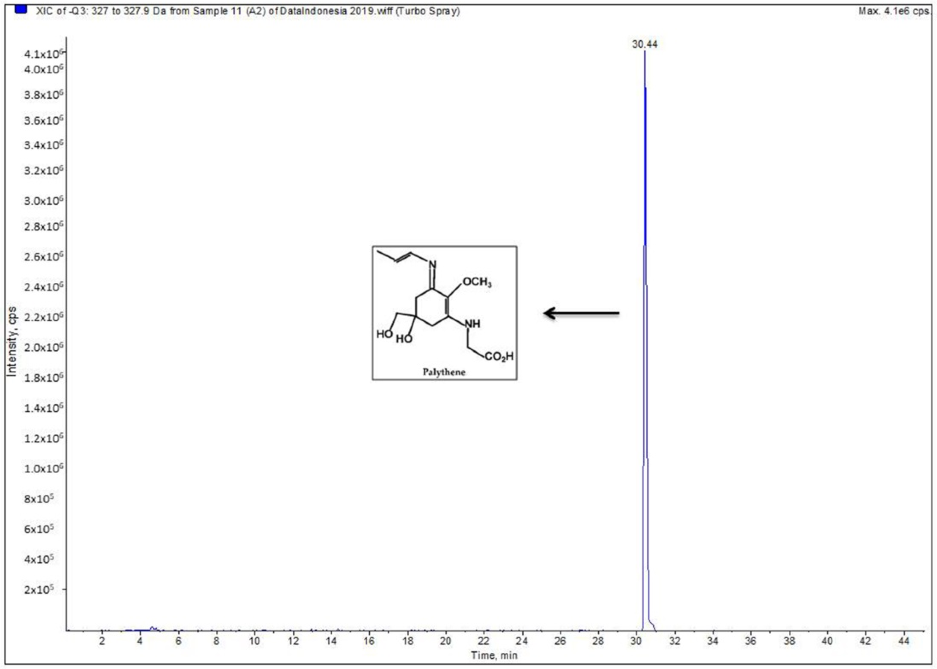
3.3. LC-MS, Detection of Potential UV-Absorbing Compounds

The SC ethanol extract was subjected to LC-MS analysis. The mass spectrum revealed one dominant charged ion peak (Figure 3). The detected peak showed a large absorption in the region of Microsporine-like Amino Acids (MAAs), and the ion of (M-H) of 327.2 m/z corresponded to the monoisotopic molecular mass of the proton adduct of palythene [14].

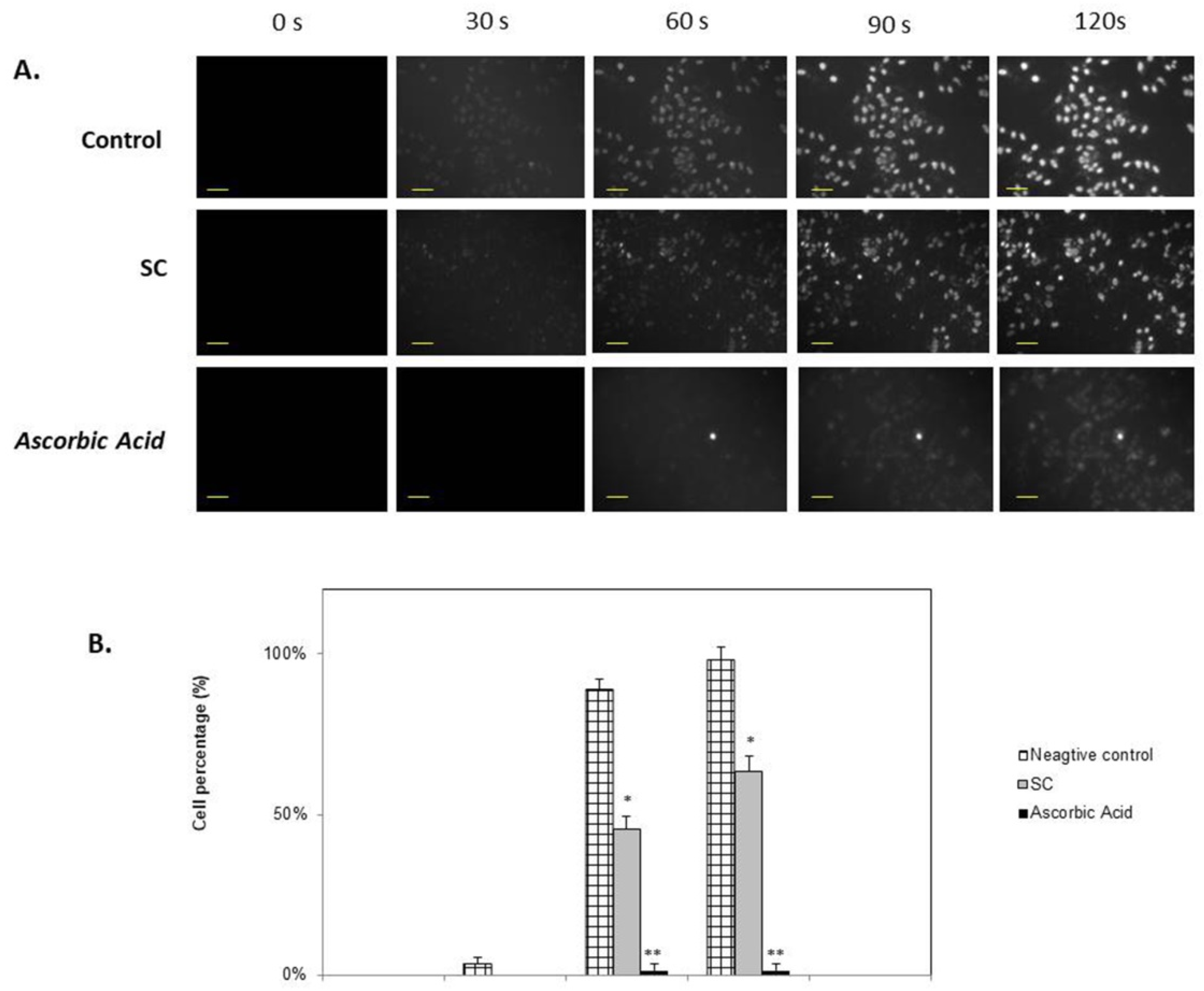
3.4. DAPI Fluorescence Staining

Nucleic staining with DAPI was conducted to estimate cytoprotective activity of Sargassum crassifolium against UV radiation treatment. Control cells were incubated with no SC extracts, and these cells showed more intense excitation of UV light compared to SC and Ascorbic acid treated cells (Figure 4). Cells treated with SC were seen to show lower exitation of DAPI fluorescence staining when exposed to UV light. This difference was signifcantly observable at 90 s exposure compared to control, indicating photoprotective activity of SC against UVA light exposure.

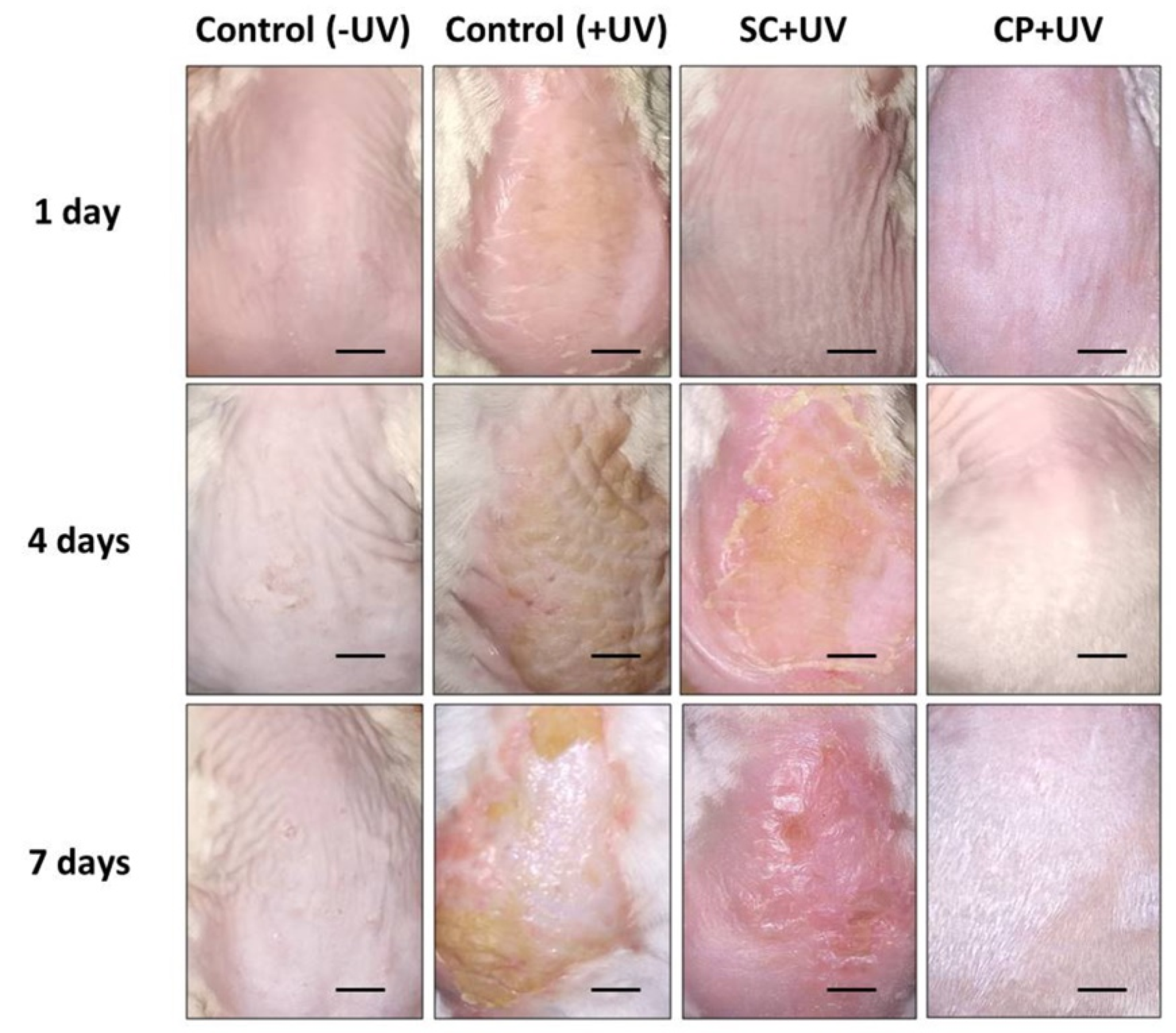
3.5. SC Protects Skin from UVA-Induced Damage

We have verified the photoprotective activity of SC ethanol extract against UV damage in vivo. The results show that the skin in the SC group formed an oily layer that was absorbed by the skin approximately after 1 day treatment (Figure 5). There were redness and inflammation effects observed on the fourth day of treatment. However, the UVA induced damage was seen to heal on the seventh day of the treatment. Compared to the control group (Control + UV) exposed by UV, the skin was rougher and also had scaling, also there was mild congestive inflammation. Commercial product with 30 SPF (CP + UV) group shows impressing photoprotective activity, as pathological changes of the skin were almost absent and similar to control group (Control − UV) not treated with UV. Furthermore, we also observed the increase growth of hair in the dorsal area of the CP + UV group.

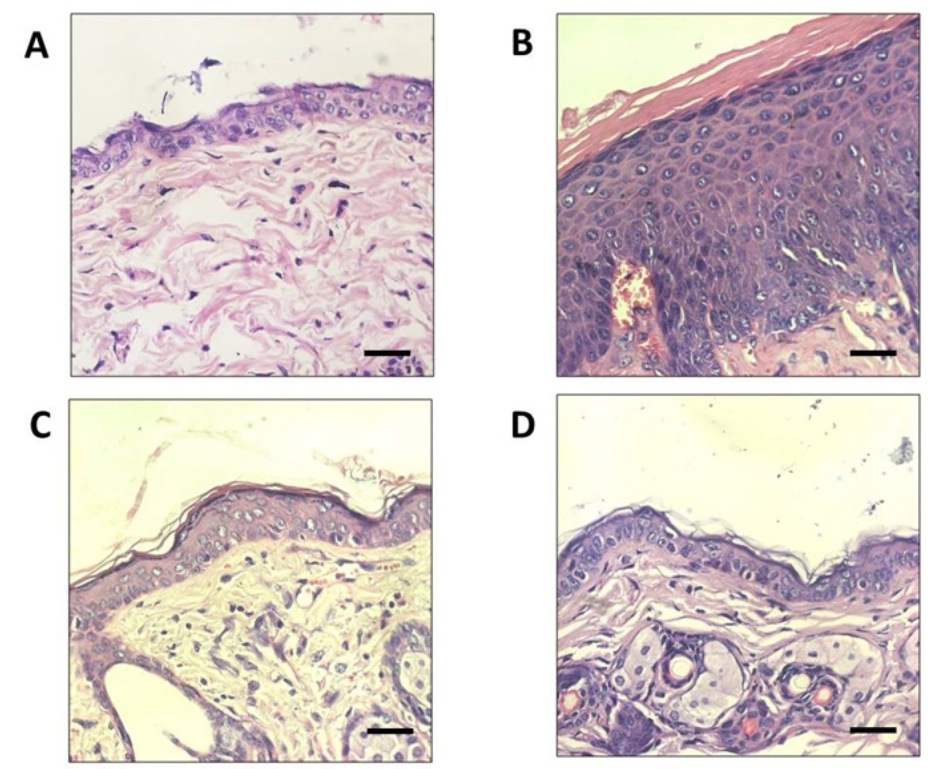
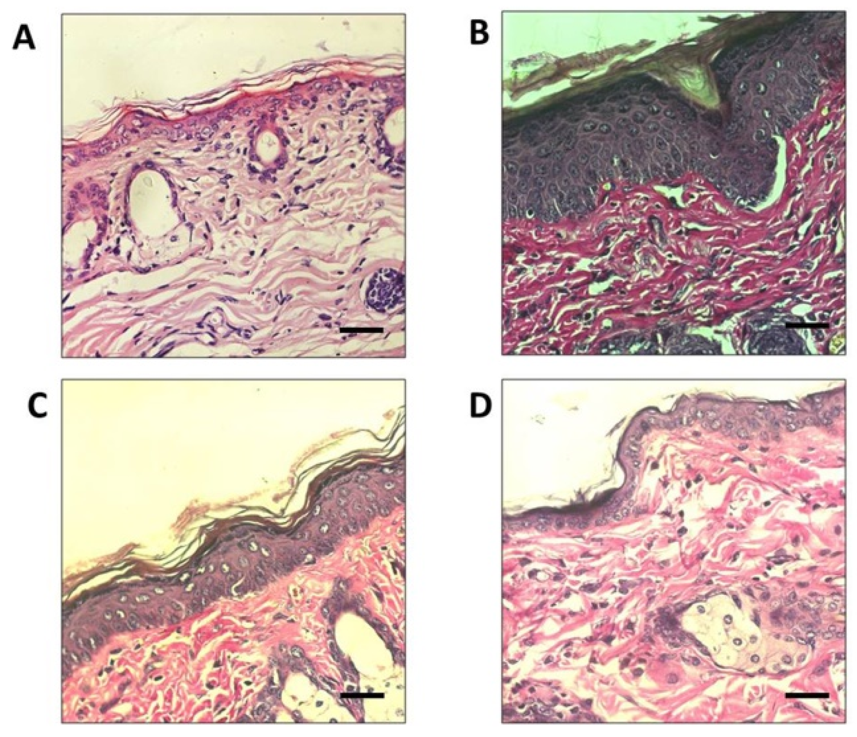
3.6. Pathological Changes of UVA-Irradiated Mice Skin Treated with SC

Pathological changes, such as epidermal thickness, could be used to evaluate effects on skin inflammation and photo-aging. In this study, the protective effects of SC were evaluated by analysis of epidermal thickness of UVA-irradiated mice (SC + UV) skin and compared with unexposed mice (Control − UV) skin. Histological analysis of dorsal skin with Hematoxylin and Eosin (H&E) staining revealed that the epidermal thickness was increased in the UVA-irradiated dorsal skin of the control group (Control + UV) exposed to UVA without SC or CP treatments (Figure 6). UV exposed groups: SC + UV and CP + UV groups showed considerable photoprotective activity, as both could maintain the integrity of the skin epidermal layer. The tissue sections were also subjected to Van Gieson staining to visualize changes in collagen fiber formation in the dermal areas of the UV-A exposed dorsal skin (Figure 7). Notably, the collagen fibers in the Control + UV group were disorganized and mostly were degraded non-functional fibers. Compared to SC + UV and CP + UV treated group, the collagen formation were neatly and densely arranged, similar to Control − UV group.

4. Discussion

UVA radiation effects are often overlooked, yet it produces the same hazardous effects on the skin, including sunburn, photo-aging, and skin cancer as UVB [15,16,17]. Skin protection products such as sunscreens have become an alternative to reduce damaging effects of UV radiation. However, most skin protection products rely on photoprotective activity of synthetic compounds, which potentially induces negative side effects [18]. Hence, there is an increasing interest in developing skin protection products derived from natural resources. Many studies have reported the potential of marine algae as a principle source of various bioactive substances with significant medicinal and nutritional value, such as brown macroalgae Sargassum, which are commonly exposed to extensive amount of UV radiation throughout the year. In this study, we evaluated the photoprotective potential of the abundant Sargassum species in Lombok coastal area; Sargassum cristaefolium.
Its capability to survive in such extreme conditions is reportedly due to the existence of broad and diverse group secondary metabolites [19]. Furthermore, Macroalgae are well reported to exhibit several UV-absorbing compounds [20], such as phlorotannins, a marine algal polyphenol which has demonstrated anti-UV activity [21]. Another common UV-absorbing compound found in marine macroalgae are the Microsporin-like Amino Acids (MAAs) [22]. Based on UV-VIS and LC-MS spectrometry analyses, brown macroalgae S. cristaefolium ethanol extract potentially contains MAAs Palythene (Figure 3). MAAs such as Porphyra-334, palythine, palythene, and shinorine exist in various seaweeds [23]. The presence of this compound could be the answer to the photoprotective activity of S. cristaefolium. Overall, macroalgae S. polycystum exhibits a similar qualitative phytochemical profile to S. crassifolium. Numerous studies have shown that MAAs prevent UVR-induced damage at the cytoplasmic level. Cells with high concentrations of MAA are likely to be more resistant to UVR than those with low concentrations [24].
Low concentration of fluorescence stain DAPI is semi-permeable to the cells. Hence, it would mostly bind to the nuclear DNA of dead cells [25]. Treatment of SC in cells resulted in significantly lower DAPI excited cells compared to untreated cells (control + UV), as shown in Figure 4. This indicates potential UVA photoprotective activity of SC ethanol extracts. Macroalgae Sargassum muticum have demonstrated cellular DNA protective activity against UV-A radiation [11]. Other considerable studies have also stated the photoprotective activity of brown macroaglae Sargassum species [26,27,28]. However, to our knowledge, this is the first study to state the photoprotective activity of Sargassum cristaefolium.
Continuous exposure of UV light could induce skin photo-aging, which is characterized by pathological changes in the components of the dermal connective tissue extracellular matrix, such as wrinkles and reduced amounts of collagen content [29]. Previous studies have revealed that photodamaged skin is also associated with increased epidermal thickness and dryness [30]. Within the dermal ECM, aging is associated with a thickening of collagen connective tissues and disorganization of total collagen content, mainly due to decreased collagen synthesis and increased fibril fragmentation [31]. In the present study, collagen fiber formations were seen to be restored in UVA-treated SC-pretreated skin, compared with the damaged skin of the UVA-treated CP-pretreated skin. Furthermore, on the seventh day, the SC pretreated mice group showed healing activity in UVA irradiates skin area. This result implies that SC photoprotective activity could protect collagen connective tissue from UV radiation damage.

5. Conclusions

In conclusion, the results of the current study suggest the cytoprotective effects of macroalgae Sargassum cristaefolium (SC) in the region of UV-A spectrum. Cytoprotective mechanisms of action of SC ethanol extract may correlate to presence of MAA-palythene, which probably contributes in inhibition of cellular DNA damage by UVA radiation. Furthermore, SC pretreated mice skin resulted in less pathological changes compared to untreated mice after UVA radiation. SC pretreated mice show less epidermal thickening and protects collagen connective tissue formations. Nevertheless, it could be concluded that SC may be a suitable candidate as a source of photoprotective agents with cosmeceutical and pharmaceutical value.

Author Contributions

E.S.P. conducted the whole experiment, analysis, and original manuscript draft, B.A.F.D.G. contributed the UV absorbing activity analyses, S.M.S. conducted the in vivo treatment, C.D.H. assisted in preparation of histological specimens, A.F. provided the LC-MS data analysis, M.M. and D.K. assisted the in vitro treatment, A.H. and H.S. reviewed the experiment design and manuscript draft.

Funding

We thank Indonesian Ministry of Research, Technology, and Higher Education for funding this research through International Collaborative Research Grant scheme (LN18/2019).

Acknowledgments

The authors are thankful to the Bioscience and Biotechnology Research Centre, University of Mataram for providing all the laboratory facilities. The authors are also thankful to all staff members of Dept. of Cellular and Integrative Physiology, Fukushima Medical University for their kind help in the experiment.

Conflicts of Interest

The authors declare no conflict of interest. The funders had no role in the design of the study; in the collection, analyses, or interpretation of data; in the writing of the manuscript, or in the decision to publish the results.

References

  1. Sjerobabski Masnec, I.; Poduje, S. Photoaging. Coll. Antropol. 2008, 32, 177–180. [Google Scholar] [PubMed]
  2. Damiani, E.; Ullrich, S.E. Understanding the connection between platelet-activating factor, a UV-induced lipid mediator of inflammation, immune suppression and skin cancer. Prog. Lipid Res. 2016, 63, 14–27. [Google Scholar] [CrossRef] [PubMed] [Green Version]
  3. Tuchinda, C.; Srivannaboon, S.; Lim, W.H. Photoprotection by window glass, automobile glass and sunglasses. J. Am. Acad. Dermatol. 2006, 54, 845–854. [Google Scholar] [CrossRef] [PubMed]
  4. Kullavanijaya, P.; Henry, W.; Lim, H.W. Photoprotection. J. Am. Acad. Dermatol. 2005, 52, 959–961. [Google Scholar] [CrossRef] [PubMed]
  5. Barone, A.N.; Hayes, C.E.; Kerr, J.J.; Lee, R.C.; Flaherty, D.B. Acute toxicity testing of TiO2-based vs. oxybenzone-based sunscreens on clownfish (Amphiprion ocellaris). Environ. Sci. Pollut. Res. Int. 2019, 26, 14513–14520. [Google Scholar] [CrossRef] [PubMed]
  6. Saewan, N.; Jimtaisong, A. Natural products as photoprotection. J. Cosmet. Dermatol. 2015, 14, 47–63. [Google Scholar] [CrossRef]
  7. Núñez-Pons, L.; Avila, C.; Romano, G.; Verde, C.; Giordano, D. UV-Protective Compounds in Marine Organisms from the Southern Ocean. Mar. Drugs 2018, 16, 336. [Google Scholar] [CrossRef] [PubMed]
  8. Pangestuti, R.; Siahaan, E.A.; Kim, S.-K. Photoprotective Substances Derived from Marine Algae. Mar. Drugs 2018, 16, 399. [Google Scholar] [CrossRef]
  9. Prasedya, E.S.; Ariyana, M.; Hamdin, C.D.; Nikmatullah, A.; Yoshie, S.; Miyake, M.; Kobayashi, D.; Hazama, A.; Sunarpi, H. Evaluation of Indonesian selected macroalgae for their antitumor and cytoprotective activity. J. Appl. Pharm. Sci. 2018, 8, 123–130. [Google Scholar] [Green Version]
  10. Lee, C.; Park, G.H.; Ahn, E.M.; Park, C.-I.; Jang, J.-H. Sargassum fulvellum protects HaCaT cells and BALB/c mice from UVB-induced proinflammatory responses. Evid.-Based Complement. Altern. Med. 2013, 2013. [Google Scholar] [CrossRef]
  11. Song, J.H.; Piao, M.J.; Han, X.; Kang, K.A.; Kang, H.K.; Yoon, W.J.; Ko, M.H.; Lee, N.H.; Lee, M.Y.; Chae, S.; et al. Anti-wrinkle effects of Sargassum muticum ethyl acetate fraction on ultraviolet B-irradiated hairless mouse skin and mechanistic evaluation in the human HaCaT keratinocyte cell line. Mol. Med. Rep. 2016, 14, 2937–2944. [Google Scholar] [CrossRef] [PubMed] [Green Version]
  12. Xiao, X.; de Bettignies, T.; Olsen, Y.S.; Agusti, S.; Duarte, C.M.; Wernberg, T. Sensitivity and Acclimation of Three Canopy-Forming Seaweeds to UVB Radiation and Warming. PLoS ONE 2015, 10, e0143031. [Google Scholar] [CrossRef] [PubMed]
  13. Guiry, M.D. How many species of algae are there? J. Phycol. 2012, 48, 1057–1063. [Google Scholar] [CrossRef]
  14. Bedoux, G.; Hardouin, K.; Marty, C.; Taupin, L.; Vandanjon, L.; Bourgougnon, N. Chemical characterization and photoprotective activity measurement of extracts from the red macroalga Solieria chordalis. Bot. Mar. 2014, 57, 291–301. [Google Scholar] [CrossRef]
  15. Chen, J.; Luo, J.; Tan, Y.; Wang, M.; Liu, Z.; Yang, T.; Lei, X. Effects of low-dose ALA-PDT on fibroblast photoaging induced by UVA irradiation and the underlying mechanisms. Photodiagn. Photodyn. 2019, 27, 79–84. [Google Scholar] [CrossRef]
  16. Zawrotniak, M.; Bartnicka, D.; Rapala-Kozik, M. UVA and UVB radiation induce the formation of neutrophil extracellular traps by human polymorphonuclear cells. J. Photochem. Photobiol. B 2019, 196. [Google Scholar] [CrossRef]
  17. Hiramoto, K.; Kasahara, E. Long-term UVA eye irradiation causes decreased learning ability in mice. Photodermatol. Photoimmunol. Photomed. 2016, 32, 129–135. [Google Scholar] [CrossRef]
  18. Pandika, M. Looking to Nature for New Sunscreens. ACS Cent. Sci. 2018, 4, 788–790. [Google Scholar] [CrossRef] [Green Version]
  19. Máximo, P.; Ferreira, L.M.; Branco, P.; Lima, P.; Lourenço, A. Secondary Metabolites and Biological Activity of Invasive Macroalgae of Southern Europe. Mar. Drugs 2018, 16, 265. [Google Scholar] [CrossRef]
  20. Schmitz, C.; Ramlov, F.; de Lucena, L.A.F.; Uarrota, V.; Batista, M.B.; Sissini, M.N.; Oliveira, I.; Briani, B.; Martins, C.D.L.; de Castro Nunes, J.M.; et al. UVR and PAR absorbing compounds of marine brown macroalgae along a latitudinal gradient of the Brazilian coast. J. Photochem. Photobiol. B 2018, 178, 165–174. [Google Scholar] [CrossRef]
  21. Flores-Molina, M.R.; Rautenberger, R.; Muñoz, P.; Huovinen, P.; Gómez, I. Stress Tolerance of the Endemic Antarctic Brown Alga Desmarestia anceps to UV Radiation and Temperature is Mediated by High Concentrations of Phlorotannins. Photochem. Photobiol. 2016, 92, 455–466. [Google Scholar] [CrossRef] [PubMed]
  22. Lalegerie, F.; Lajili, S.; Bedoux, G.; Taupin, L.; Stiger-Pouvreau, V.; Connan, S. Photo-protective compounds in red macroalgae from Brittany: Considerable diversity in mycosporine-like amino acids (MAAs). Mar. Environ. Res. 2019, 147, 37–48. [Google Scholar] [CrossRef] [PubMed] [Green Version]
  23. Athukorala, Y.; Trang, S.; Kwok, C.; Yuan, Y.V. Antiproliferative and antioxidant activities and mycrosporine-like amino acid profiles of wild-harvested and cultivated edible Canadian marine red macroalgae. Molecules 2016, 21, 119. [Google Scholar] [CrossRef] [PubMed]
  24. Lawrence, K.P.; Long, P.F.; Young, A.R. Mycosporine-Like Amino Acids for Skin Photoprotection. Curr. Med. Chem. 2018, 25, 5512–5527. [Google Scholar] [CrossRef] [PubMed]
  25. Manzini, G.; Xodo, L.; Barcellona, M.L.; Quadrifoglio, F. Interaction of DAPI with double-stranded ribonucleic acids. Nucleic Acids Res. 1985, 13, 8955–8967. [Google Scholar] [CrossRef] [PubMed] [Green Version]
  26. Ayyad, S.-E.N.; Ezmirly, S.T.; Basaif, S.A.; Alarif, W.M.; Badria, A.F.; Badria, F.A. Antioxidant, cytotoxic, antitumor, and protective DNA damage metabolites from the red sea brown alga Sargassum sp. Pharmacogn. Res. 2011, 3, 160–165. [Google Scholar] [CrossRef] [PubMed]
  27. Piao, M.J.; Yoon, W.J.; Kang, H.K.; Yoo, E.S.; Koh, Y.S.; Kim, D.S.; Lee, N.H.; Hyun, J.W. Protective Effect of the Ethyl Acetate Fraction of Sargassum muticum against Ultraviolet B–Irradiated Damage in Human Keratinocytes. Int. J. Mol. Sci. 2011, 12, 8146–8160. [Google Scholar] [CrossRef] [PubMed]
  28. Quah, C.C.; Kim, K.H.; Lau, M.S.; Kim, W.R.; Cheah, S.H.; Gundamaraju, R. Pigmentation and Dermal Conservative Effects of the Astonishing Algae Sargassum Polycystum and Padina Tenuis on Guinea Pigs, Human Epidermal Melanocytes (HEM) and Chang Cells. Afr. J. Tradit. Complement. Altern. Med. 2014, 11, 77–83. [Google Scholar] [CrossRef] [PubMed] [Green Version]
  29. Prasanth, M.I.; Santoshram, G.S.; Bhaskar, J.P.; Balamurugan, K. Ultraviolet-A triggers photoaging in model nematode Caenorhabditis elegans in a DAF-16 dependent pathway. Age 2016, 38, 27. [Google Scholar] [CrossRef]
  30. Mechanistic Effects of Long-Term Ultraviolet B Irradiation Induce Epidermal and Dermal Changes in Human Skin Xenografts. Available online: https://www.ncbi.nlm.nih.gov/pmc/articles/PMC2630550/ (accessed on 23 August 2019).
  31. Elicited ROS Scavenging Activity, Photoprotective, and Wound-Healing Properties of Collagen-Derived Peptides from the Marine Sponge Chondrosia reniformis. Available online: https://www.ncbi.nlm.nih.gov/pmc/articles/PMC6316299/ (accessed on 23 August 2019).
Figure 1. Brown macroalgae Sargassum cristaefolium.
Biomedicines 07 00077 g001
Figure 2. Ultraviolet Absorption spectrum of the ethanol extract of Sargassum crassifolium (0.1%).
Biomedicines 07 00077 g002
Figure 3. LC/MS analysis of Sargassum cristaefolium ethanol extract (0.1%).
Biomedicines 07 00077 g003
Figure 4. Cytoprotective analysis of macroalgae extracts (0.01%) against UV-A irradiation in HeLa cells for 2.5 min irradiation. (A) Nucleic staining with DAPI was conducted to determine cellular damage. Scale bar: 200 µm (B) Dead cell percentage of HeLa cells irradiated with UVA. * is considered significantly different to control (p < 0.05); ** is considered highly significantly different compared to control (p < 0.01).
Biomedicines 07 00077 g004
Figure 5. Effect of Sargassum cristaefolium extract (SC) on UV-irradiation skin lesion in mice for seven days. Scale bar: 1 cm.
Biomedicines 07 00077 g005
Figure 6. Histological analyses of mice skin irradiated by UV for two hours every day for seven days. (A) Control not irradiated with UV (Control − UV) (B) Control irradiated with UV (Control + UV) (C) Irradiated skin treated with 0.1% SC (SC + UV) (D) Irradiated skin treated with commercial product 30 SPF (CP + UV). Skin sections were stained with Hematoxylin and Eosin (H&E). Scale bars: 20 μm.
Biomedicines 07 00077 g006
Figure 7. Histological analyses of mice skin irradiated by UV for two hours every day for seven days. (A) Control not irradiated with UV (Control − UV) (B) Control irradiated with UV (Control + UV) (C) Irradiated skin treated with 0.1% SC (SC + UV) (D) Irradiated skin treated with commercial product 30 SPF (CP + UV). Skin sections were stained with Van Gieson. Scale bars: 20 μm.
Biomedicines 07 00077 g007

