Article

Clinical Significance of Preoperative Hematological Parameters in Patients with D2-Resected, Node-Positive Stomach Cancer

1
Department of Radiation Oncology, Chungnam National University Sejong Hospital, Chungnam National University School of Medicine, Sejong 30099, Korea
2
Department of Radiation Oncology, Samsung Medical Center, Sungkyunkwan University School of Medicine, Seoul 06351, Korea
3
Department of Radiation Oncology, Kangbuk Samsung Hospital, Sungkyunkwan University School of Medicine, Seoul 03181, Korea
4
Department of Medicine, Division of Hematology-Oncology, Samsung Medical Center, Sungkyunkwan University School of Medicine, Seoul 06351, Korea
5
Department of Surgery, Samsung Medical Center, Sungkyunkwan University School of Medicine, Seoul 06351, Korea
*
Author to whom correspondence should be addressed.
Biomedicines 2022, 10(7), 1565; https://doi.org/10.3390/biomedicines10071565
Submission received: 15 April 2022 / Revised: 22 June 2022 / Accepted: 29 June 2022 / Published: 30 June 2022
(This article belongs to the Special Issue Gastric Cancer: From Mechanisms to Therapeutic Approaches)

Abstract

:
The purpose of the present study was to investigate the clinical significance of preoperative hematological parameters in patients with advanced stomach cancer, and to explore who might benefit from adjuvant concurrent chemoradiotherapy (CCRT) compared to chemotherapy alone. Among 1032 patients with node-positive stomach cancer who had a confirmed diagnosis after complete D2 resection, and who received adjuvant chemotherapy alone or CCRT, a total of 692 patients was selected using propensity score matching. Among absolute neutrophil count, absolute lymphocyte count (ALC), absolute monocyte count (AMC), platelet count, neutrophil-to-lymphocyte ratio, lymphocyte-to-monocyte ratio, and platelet-to-lymphocyte ratio, AMC was the most relevant prognostic factor for overall survival and recurrence-free survival (hazard ratio (HR) 1.674, 95% confidence interval (CI) 1.180–2.376; HR 1.908, 95% CI 1.650–2.695, respectively). In a subgroup with a high ALC, patients treated with adjuvant CCRT had a favorable recurrence-free survival (HR 0.620, 95% CI 0.393–0.980) compared to those treated with chemotherapy alone. Further study is needed to confirm our findings and to develop tailored adjuvant treatment.

1. Introduction

Stomach cancer is the fifth most common cancer (5.6% of all cases) and the fourth leading cause of death (7.7% of all cancer-related deaths) based on the Global Cancer Statistics 2020 [1]. The incidence of stomach cancer is the highest in Eastern Asia—especially in Japan and Mongolia—and Eastern Europe.
Based on the results of two large phase III randomized controlled trials (RCTs) conducted on Asian patients, the Korean Gastric Cancer Association recommends adjuvant chemotherapy with S-1 alone or capecitabine plus oxaliplatin for patients with pathological stage II or III stomach cancer [2,3]. The survival benefit from both chemotherapy regimens was confirmed by assessing the 5-year follow-up data [4,5]. On the other hand, the role and necessity of adjuvant concurrent chemoradiotherapy (CCRT) in completely D2-resected gastric cancer remains controversial. The Intergroup Study 0116 (INT-0116), which showed that adjuvant CCRT improved overall survival (OS) and relapse-free survival when compared with surgery alone, was criticized because only 10% of patients had undergone a D2 resection, which is considered a standard surgical procedure for advanced stomach cancer in Asian countries [6]. Additionally, a phase III RCT conducted in Korea compared the treatment outcomes between adjuvant chemotherapy alone and CCRT after D2 resection. CCRT reduced locoregional recurrence, but there was no benefit of OS in the CCRT group [7,8,9].
Since blood testing is an essential examination before gastric surgery, preoperative hematological parameters are easily available, and might be useful for predicting prognosis along with various tumor characteristics. There is a growing body of evidence demonstrating the prognostic value of hematological parameters—including neutrophils, lymphocytes, and monocytes—as well as the ratios of these cells—including neutrophil-to-lymphocyte ratio (NLR), lymphocyte-to-monocyte ratio (LMR), and platelet-to-lymphocyte ratio (PLR)—in various cancers [10,11,12,13]. These hematological parameters are related to the innate immune system or adaptive immune response against pathogens, and are involved in the prevention, carcinogenesis, exacerbation, and metastasis of cancer. The association between these hematological parameters and the extent of cancer and its biology suggests the possibility of an association with the prognosis as well as the risk and pattern of recurrence in several cancers [14,15,16]. Therefore, it is possible to provide important information for implementing customized adjuvant treatment in patients with completely resected gastric cancer.
In this study, we aimed to investigate the prognostic significance of preoperative hematological parameters in patients with D2-resected, node-positive stomach cancer. We also explored who benefited more from adjuvant CCRT than from chemotherapy alone.

2. Materials and Methods

2.1. Patients

Between April 2008 and February 2013, a total of 1032 patients with D2-resected, node-positive stomach cancer received either adjuvant chemotherapy or adjuvant chemoradiotherapy (CCRT) at Samsung Medical Center. Most of these patients (n = 1005) received adjuvant treatment according to the protocol of the Adjuvant Chemotherapy Trial of TS-1 for Gastric Cancer (ACTS-GC) and the Intergroup Study 0116 (INT-0116) [2,6]. The other 21 patients received adjuvant chemotherapy according to the protocol of the capecitabine and oxaliplatin adjuvant study in stomach cancer (CLASSIC), and 6 patients received off-protocol chemotherapy. This study included patients who received adjuvant treatment according to the ACTS-GC or INT-0116 protocols.

2.2. Treatment

Adjuvant treatment, which can be CCRT for 5 months or chemotherapy alone for 1 year, was determined based on patient preference, with informed consent. Patients who chose chemotherapy alone followed the ACTS-GC protocol, and received two oral doses of 40 mg/m2 S−1 per day for 4 weeks, followed by 2 weeks of rest [2]. This 6-week cycle of chemotherapy was repeated eight times during the first year after surgery. CCRT was performed according to the INT-0116 study protocol [6]. Six cycles of chemotherapy consisted of 425 mg/m2 fluorouracil per day and 20 mg/m2 leucovorin per day, taken for 5 days. The second and third cycles were administered on the first four days and the last three days of radiotherapy, respectively, and the fluorouracil dose was reduced to 400 mg/m2 per day. A total radiation dose of 45 Gy was delivered in daily fractions of 1.8 Gy over a duration of 5 weeks. The radiation field encompassed the tumor bed, anastomosis site, duodenal stump, regional lymph nodes, and 2 cm beyond the proximal and distal resection margins. The remnant stomach was not routinely encompassed in the radiation field.

2.3. Preoperative Blood Cell Counts and Ratios

Complete blood counts with a differential count within 60 days before surgery were used to calculate blood cell ratios. Seven patients who had unavailability of data were excluded from this study. NLR was defined as the neutrophil count divided by the lymphocyte count. LMR was defined as the lymphocyte count divided by the monocyte count. PLR was defined as the platelet count divided by the lymphocyte count. Continuous variables were converted into categorical variables based on the median values.

2.4. Evaluation and Statistical Analysis

The chi-squared test, independent t-test, and one-way analysis of variance were used to compare patient characteristics between the adjuvant treatment groups. OS was defined as the time from the date of surgery to the date of death from any cause. Recurrence-free survival (RFS) was defined as the time from the date of surgery to the date of recurrence or death—whichever occurred first. Kaplan–Meier survival curves were constructed and compared using the log-rank test to investigate the association between patient characteristics and OS and RFS. We also conducted a Cox regression analysis using forward stepwise regression to evaluate the effects of patient characteristics on OS and RFS. Statistically significant covariates in the univariate analyses and the type of adjuvant treatment, which was our interest, were included in the multivariate analysis. Statistical significance was set at p < 0.05. Statistical analyses were performed using SPSS, version 26 (IBM, Armonk, NY, USA). Propensity score matching (PSM) was performed between the CCRT group and the chemotherapy-alone group using R software, version 4.1.0 (Foundation for Statistical computing, Vienna, Austria). Age, sex, T stage, N stage, stage group, Lauren classification, and surgical extent were included in the calculation of propensity scores.

2.5. Ethical Statement

This study was approved by the Institutional Review Board of Samsung Medical Center (SMC 2022-03-104-001).

3. Results

3.1. Patient Characteristics

After PSM matching, a total of 692 patients was included in the study, and their characteristics are shown in Table 1. The mean patient age was 55.0 years (95% confidence interval (CI) 54.2–55.9 years old). All patients underwent D2 resection with negative margins, and had lymph node metastases. The mean number of harvested lymph nodes was 47 (range, 16–119). No patient received neoadjuvant treatment prior to surgery. The proportions of patients with stages I, II, and III were 10.1%, 42.2%, and 47.7%, respectively. Stage was classified according to the seventh edition of the American Joint Committee on Cancer’s staging system. The mean interval between surgery and initiation of adjuvant treatment was 5 weeks (range: 2–15 weeks). Patients’ characteristics before matching are presented in Supplementary Table S1.

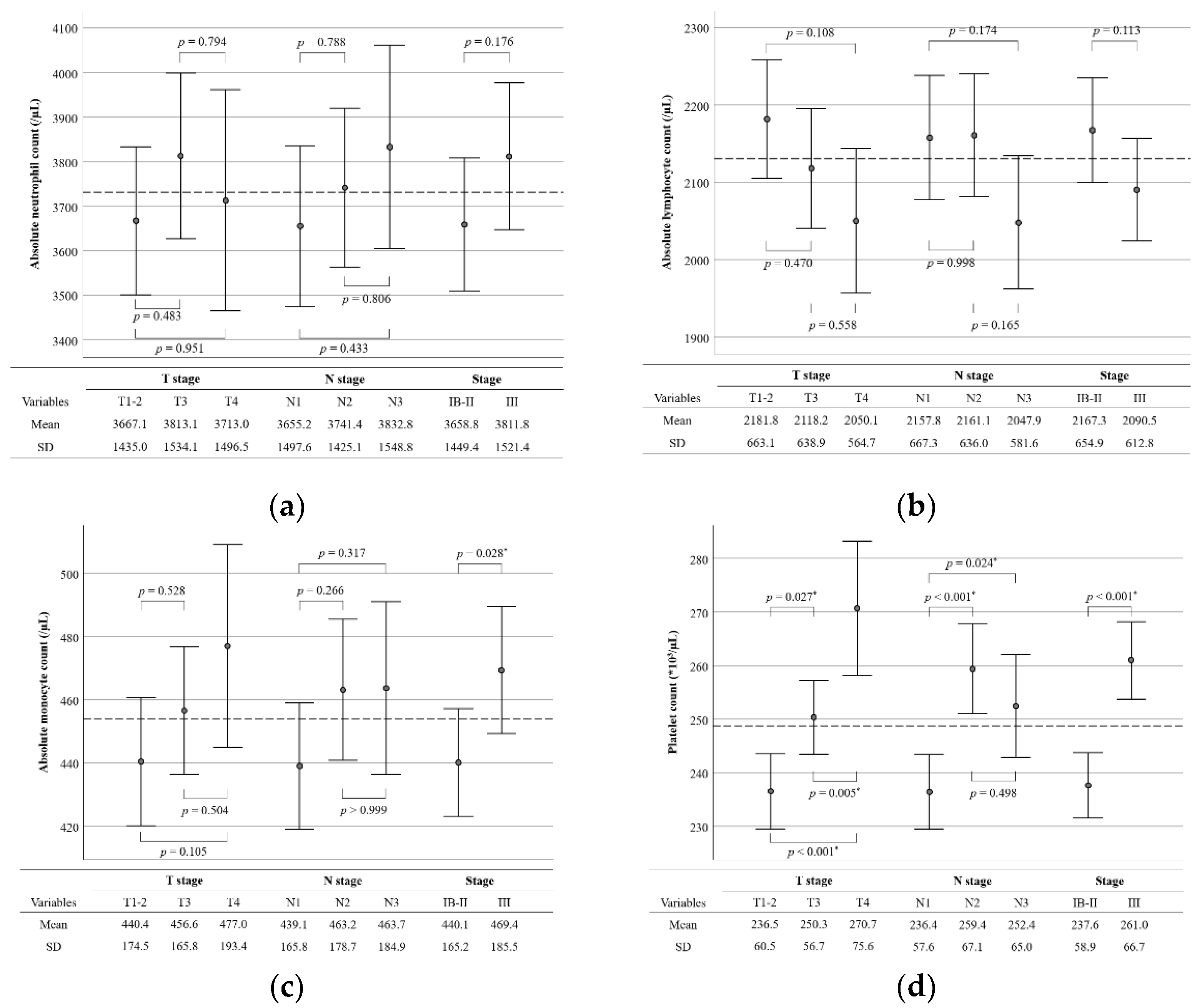
3.2. Preoperative Blood Cell Counts and Ratios

The mean values of absolute neutrophil count (ANC), absolute lymphocyte count (ALC), absolute monocyte count (AMC), platelet count (PC), NLR, LMR, and PLR were 3731.8/µL (95% CI 3620.9–3842.6/µL), 2130.7/µL (95% CI 2083.2–2178.2/µL), 454.0/µL (95% CI 440.9–467.1/µL), 248.8 × 10³/µL (95% CI 244.0–253.5 × 10³/µL), 1.920 (95% CI 1.824–2.015), 5.184 (95% CI 5.034–5.334), and 128.0 (95% CI 123.7–132.3), respectively. The CCRT group had a higher mean ALC (2188.7/µL versus 2072.7/µL, p = 0.016), a lower NLR (1.796 versus 2.044, p = 0.004), and a lower PLR (122.6 versus 133.4, p = 0.002) than the chemotherapy-alone group. The differences in ANC, AMC, PC, and LMR between the two groups were not statistically significant. The median values of ANC, ALC, AMC, PC, NLR, LMR, and PLR were 3448.5/µL, 2074.4/µL, 422.5/µL, 240.5 × 10³/µL, 1.657, 4.956, and 117.6, respectively. Based on the median values, the CCRT group had more patients with low NLR and high LMR compared to the chemotherapy-alone group (Table 1).
Figure 1 shows the differences in the mean values of hematological parameters according to the disease stage. The mean values of ANC and NLR tended to be higher and the mean ALC tended to be lower in patients with an advanced disease stage, but the differences were not statistically significant. The mean values of AMC, PC, and PLR were higher in patients with stage III disease than in those with stage IB–II disease (469.4/µL versus 440.1/µL, p = 0.028; 261.0 × 10³/µL versus 237.6 × 10³/µL, p < 0.001; 137.5 versus 119.3, p < 0.001, respectively). On the other hand, the mean LMR was lower in patients with stage III disease than in those with stage IB–II disease (4.944 versus 5.403, p = 0.003).

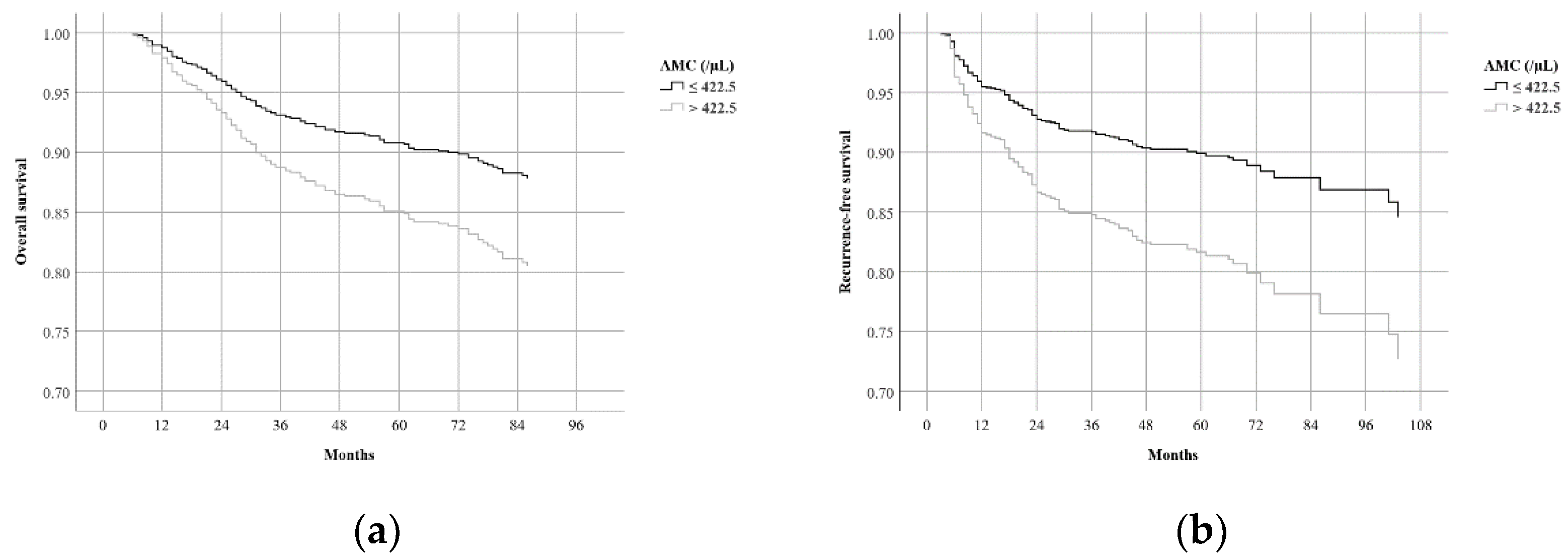
3.3. Survival Results

The median follow-up time was 79 months (IQR 60–95 months). The mean OS and RFS were 103.3 months (95% CI 100.6–106.1 months) and 98.7 months (95% CI 95.4–102.0 months), respectively. The 5-year OS and RFS were 83.4% and 80.6%, respectively. According to the results of the log-rank test, age > 65 years, advanced T stage, advanced N stage, advanced stage group, TG, high AMC, and low LMR were associated with worse OS (Table 2). In terms of RFS, age > 65 years, advanced T stage, advanced N stage, advanced stage group, TG, high AMC, and low LMR were associated with worse RFS. The type of adjuvant treatment did not affect OS or RFS.
We evaluated the effects of patient characteristics on OS and RFS using univariate Cox regression analysis, and the results are demonstrated in Table 3. Multivariate Cox regression analysis showed that age >65 years (HR 2.190, 95% CI 1.531–3.132), advanced T stage (HR 2.099, 95% CI 1.261–3.492 for T3; HR 4.346, 95% CI 2.587–7.300 for T4), advanced N stage (HR 1.770, 95% CI 1.081–2.896 for N2; HR 2.340, 95% CI 1.436-3.814 for N3), TG (HR 1.548, 95% CI 1.094–2.191), and AMC > 422.5/µL (HR 1.674, 95% CI 1.180–2.376) were significant prognostic factors for worse OS (Table 4). Age >65 years (HR 1.444, 95% CI 1.001–2.084), advanced T stage (HR 2.509, 95% CI 1.521–4.141 for T3; HR 4.717, 95% CI 2.829–7.865 for T4), advanced N stage (HR 2.496, 95% CI 1.500–4.155 for N2; HR 3.591, 95% CI 2.150–5.999 for N3), and AMC > 422.5/µL (HR 1.908, 95% CI 1.650–2.695) were significant prognostic factors for worse RFS. While LMR and AMC were both closely associated with OS and RFS in univariate analysis, only AMC remained significant in multivariate analysis. Figure 2 shows the adjusted OS and RFS curves according to AMC.

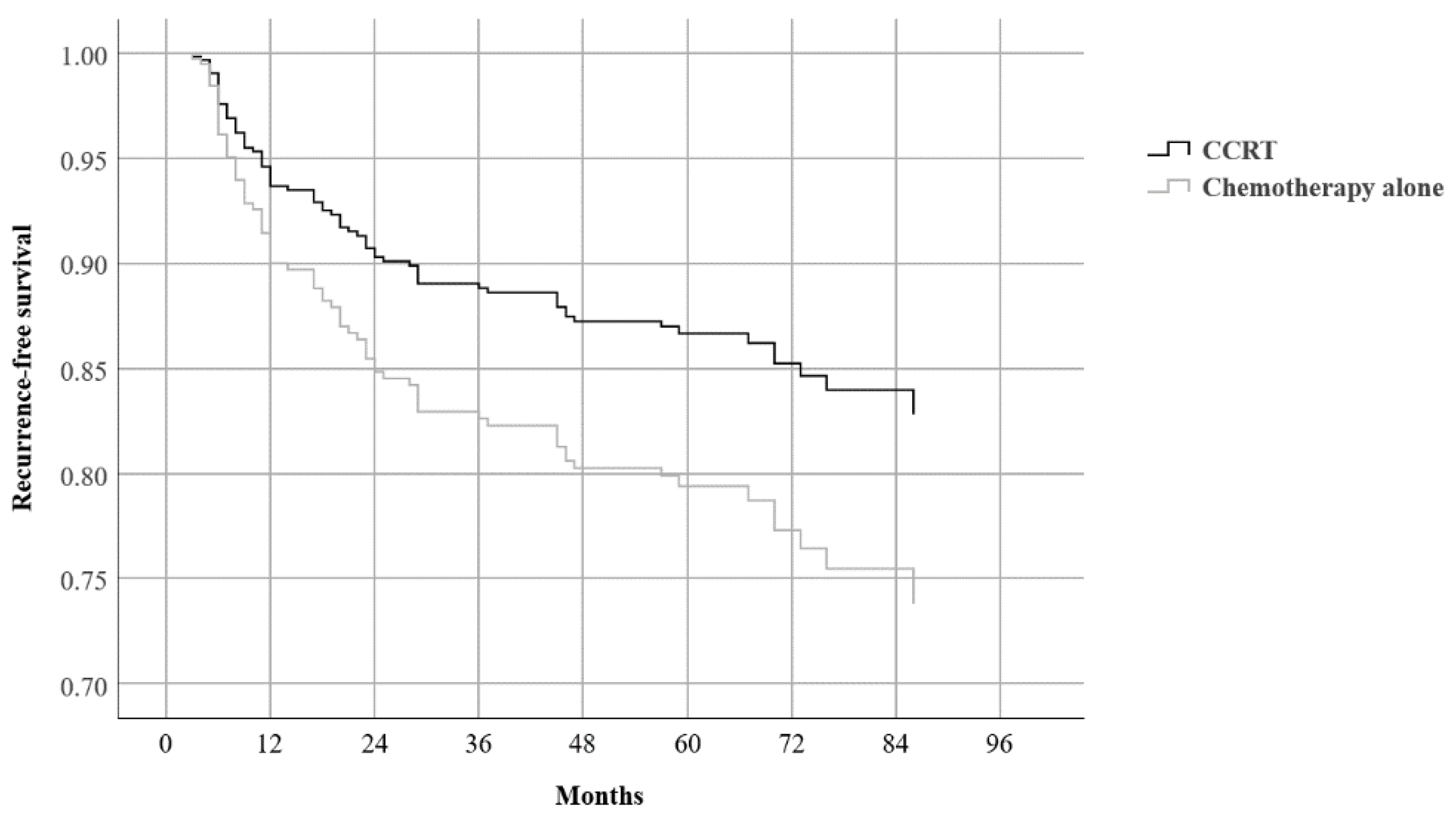
3.4. Subgroup Analysis

Multivariate analysis was repeated for subgroups categorized according to blood cell counts and ratios. As with the results of the analysis of all patients (n = 692), T stage and either N stage or stage group were found to be valid prognostic factors for OS and RFS in all subgroups. The type of adjuvant treatment did not affect the treatment outcomes in any subgroup, except for one: in the subgroup with high ALC (>2074.0/µL), patients who received adjuvant CCRT had a favorable RFS compared to those who received chemotherapy alone (HR 0.620, 95% CI 0.393–0.980, p = 0.040) (Table 5 and Figure 3).

4. Discussion

Our results indicate that preoperative blood cell counts and their ratios are associated with the stage of stomach cancer. ANC, AMC, PC, NLR, and PLR were higher in patients with advanced disease. By contrast, ALC and LMR were lower in patients with advanced disease. Among these factors, AMC was the only independent prognostic factor for both OS and RFS. Subgroup analyses revealed that patients with a high ALC benefited from adjuvant CCRT rather than from chemotherapy alone.
The negative prognostic impact of preoperative AMC on OS and RFS in the present study was similar to that reported in previous studies. Low ALC and high AMC have been reported as prognostic factors for OS and disease-free survival (DFS) in resectable stomach cancer, with cutoff values of 1734/µL and 1720/µL, and 672.4/µL and 510/µL, respectively [17,18]. The prognostic significance of NLR, LMR, and PLR in stomach cancer has been reported in several meta-analyses (Table 6) [13,19,20,21,22]. Although the target population and cutoff values of each study were widely heterogeneous, an increase in NLR and PLR and a decrease in LMR were associated with a worse prognosis. Contrary to the results of those studies, the prognostic significance of NLR and PLR was not found in the present study. While the LMR was closely associated with OS and RFS in univariate analysis, it lost its significance in multivariate analysis owing to AMC. This finding might have been obtained because the subjects of the present study were relatively healthy and patients with early-stage disease were able to receive curative surgical resection and adjuvant treatment. In fact, the mean values of NLR, LMR, and PLR in the present study, which were 1.920 (95% CI 1.824–2.015), 5.184 (95% CI 5.034–5.334), and 128.0 (95% CI 123.7–132.3), respectively, were lower than the reference values suggested by Schiefer et al., which were 4.5, 5.43, and 152, respectively—especially for NLR [23]. Additionally, our values were not significantly different from those of 12,160 healthy Korean populations obtained from the checkup center of a tertiary hospital: NLR of 1.65 (95% CI 0.107–3.193), LMR of 5.31 (95% CI 2.008–8.612), and PLR of 132.40 (95% CI 46.764–218.006) [24].
The underlying mechanism of the prognostic significance of preoperative blood cell counts and their ratios can be explained by the role of each blood cell. The pro-cancer properties of neutrophils, monocytes, and platelets have been reported in many studies. Neutrophils are innate immune cells that also play a role in cancer’s initiation, progression, and metastasis [25]. Neutrophils recruited to inflammatory sites promote cancer initiation by increasing deoxyribonucleic acid (DNA) damage, angiogenesis, and immunosuppression through reactive oxygen species, reactive nitrogen species, and angiogenic factors. Growth factors released by neutrophils and neutrophil extracellular traps (NETs) promote cancer progression. Cytokines and granular proteins released by neutrophils and NETs are also involved in each step of cancer metastasis, including preparation of a pre-metastatic niche, angiogenesis, cancer cell migration, intravasation, extravasation, and survival of cancer cells in the peripheral blood. Cancer cells and stroma release various cytokines that reprogram neutrophils to function as pro-cancer neutrophils. Monocytes are recruited and differentiated into tumor-associated macrophages (TAMs) in response to the tumor microenvironment [26]. TAMs produce various cytokines that promote cancer progression and inhibit cytotoxic T-lymphocyte responses through the programmed death-1/programmed death-ligand-1 signaling pathway and induction of regulatory T cells. Platelet activation by cancer also exerts pro-cancer effects, including stimulating tumor growth, preparing the metastatic niche, and helping the metastatic cells to survive in circulation [27]. Lymphocytes play a central role in the anticancer immune response, and a recent meta-analysis indicated that a low blood lymphocyte count before treatment among patients with solid tumors was associated with worse OS and progression-free survival [28]. The mechanism underlying lymphocytopenia in solid tumors has not been clearly elucidated; however, it is widely believed that lymphocytopenia may occur as a result of cancer-induced immune suppression and increased lymphocyte apoptosis. Based on these immunological mechanisms, increased activation of neutrophils, monocytes, and platelets by tumors and low lymphocyte counts—in other words, high NLR and PLR, and low LMR—could be used as prognostic factors for worse prognosis.
Our finding that there is a specific subgroup that benefits more from CCRT than from chemotherapy alone, even after D2 resection, could represent a significant concept for radiation oncologists. A well-designed phase III RCT was conducted to investigate the role of adjuvant CCRT in patients who received D2 resection, but there were no differences in DFS and OS between the chemotherapy-alone and the CCRT groups [8]. Subgroup analysis indicated that regional recurrence was lower in the CCRT group than in the chemotherapy-alone group (5/230 patients versus 23/228 patients, p < 0.001), and node-positive patients benefited more from CCRT than other subgroups [9]. However, in a subsequent phase III RCT that included only node-positive patients, DFS in CCRT with S-1 and oxaliplatin was not superior compared to that in doublet chemotherapy alone (HR 0.971, p = 0.879) [29]. Although there is insufficient evidence to support adjuvant CCRT after D2 resection, there is a clear benefit in decreasing locoregional recurrence from CCRT. Thus, further studies that discuss not only conventional tumor staging, but also molecular aspects of tumors, are necessary to identify candidates for CCRT.
Recently, microsatellite instability (MSI) status—one of the molecular aspects—has become a spotlight as a prognostic factor and a predictive factor for systemic therapy in resectable stomach cancer. A meta-analysis of four RCTs reported that MSI was associated with longer OS and DFS (HR 1.78, 95% CI 1.17–2.73 and HR 1.88, 95% CI 1.28–2.76, respectively) [30]. It also indicated a potential lack of benefit of perioperative or adjuvant chemotherapy for patients with high-MSI disease (HR 1.50, 95% CI 0.55–4.12 for OS; HR 1.27, 95% CI 0.53–3.04 for DFS), while patients with low-MSI or microsatellite-stable disease benefited from chemotherapy in terms of OS and DFS (HR 0.75, 95% CI 0.60–0.94; and HR 0.65, 95% CI 0.53–0.79, respectively). Although it was a study on advanced endometrial cancer, patients with high-MSI disease had an improved 2-year progression-free survival with adjuvant CCRT compared with that of chemotherapy alone (40.0% versus 29.5%, p = 0.04) [31]. The authors believed that the deficiency in mismatch repair could enhance the response to radiation-induced DNA damage.
We compared patient characteristics between the subgroups categorized based on their mean ALC values to explain the benefit of CCRT in the high-ALC group (Supplementary Table S2). There were no significant differences in the proportions of patients in terms of age > 65 years, T stage, N stage, stage group, Lauren classification, surgical extent, and type of adjuvant treatment. By contrast, the high-ALC group had lower proportions of patients with high NLR (29.6% versus 70.0%, p < 0.001) and high PLR (24.1% versus 75.8%, p < 0.001), and a higher proportion of patients with high LMR (64.6% versus 35.4%, p < 0.001), which represents intact host immunity and low levels of systemic inflammation. Radiation-induced immunogenic cell death can activate adaptive immune response through tumor antigen presentation to cytotoxic T lymphocytes in immunocompetent individuals [32].
This study has several limitations. Our results cannot be applied to node-negative patients, even if they have an advanced T stage, because we included only node-positive patients. Second, there were significant differences in patient characteristics between the different adjuvant treatment groups before PSM, owing to the retrospective study design. We observed tendencies whereby elderly patients preferred chemotherapy alone owing to the convenience of oral chemotherapy, while patients with advanced disease preferred adjuvant CCRT despite the inconvenience of daily visits for radiotherapy. The cutoff values of continuous variables can affect the results—especially those of NLR and LMR, because they are very small compared with those of ALC and PLR.

5. Conclusions

AMC, together with T and N stages, was a prognostic factor for OS and RFS in patients with D2-resected, node-positive stomach cancer. Patients with a high AMC had a worse prognosis than those with a low AMC. There were no differences in treatment outcomes according to adjuvant treatment in most patients. However, in a subgroup with high ALC, patients who received adjuvant CCRT had a favorable RFS compared to those who received adjuvant chemotherapy alone. Thus, preoperative hematological parameters might be useful not only as prognostic factors, but also as predictive factors. Further study is needed to confirm our findings and to develop tailored adjuvant treatments.

Supplementary Materials

The following supporting information can be downloaded at: https://www.mdpi.com/article/10.3390/biomedicines10071565/s1, Table S1: Patient characteristics before the propensity score matching; Table S2: Comparison of patient characteristics according to absolute lymphocyte counts.

Author Contributions

Conceptualization, J.S.P., J.I.Y. and D.H.L.; methodology, J.S.P. and J.I.Y.; software, J.S.P.; formal analysis, J.S.P. and J.I.Y.; investigation, J.S.P. and J.I.Y.; resources J.I.Y.; data curation, J.S.P., J.I.Y., D.H.L., H.N., Y.I.K., J.L., W.K.K., S.H.P., S.T.K., T.S.S., J.H.L., J.Y.A., M.G.C. and J.M.B.; writing—original draft preparation, J.S.P. and J.I.Y.; writing—review and editing, all authors; visualization, J.S.P.; supervision, J.I.Y., D.H.L., J.L., W.K.K. and J.Y.H.; project administration J.S.P. and J.I.Y.; funding acquisition, J.I.Y. All authors have read and agreed to the published version of the manuscript.

Funding

This research was funded by the Basic Science Research Program through the National Research Foundation of Korea (NRF), which is funded by the Ministry of Education (NRF-2020R1F1A1073205) and the Future Medicine 20*30 Project of the Samsung Medical Center [SMX1220101].

Institutional Review Board Statement

This study was conducted in accordance with the Declaration of Helsinki, and approved by the Institutional Review Board of Samsung Medical Center (SMC 2022-03-104-001, the date of approval 29 March 2022).

Informed Consent Statement

Patient consent was waived due to the retrospective nature of this study.

Data Availability Statement

Not applicable.

Conflicts of Interest

The authors declare no conflict of interest. The funders had no role in the design of the study; in the collection, analyses, or interpretation of data; in the writing of the manuscript, or in the decision to publish the results.

References

  1. Sung, H.; Ferlay, J.; Siegel, R.L.; Laversanne, M.; Soerjomataram, I.; Jemal, A.; Bray, F. Global Cancer Statistics 2020: GLOBOCAN Estimates of Incidence and Mortality Worldwide for 36 Cancers in 185 Countries. CA A Cancer J. Clin. 2021, 71, 209–249. [Google Scholar] [CrossRef] [PubMed]
  2. Sakuramoto, S.; Sasako, M.; Yamaguchi, T.; Kinoshita, T.; Fujii, M.; Nashimoto, A.; Furukawa, H.; Nakajima, T.; Ohashi, Y.; Imamura, H.; et al. Adjuvant chemotherapy for gastric cancer with S-1, an oral fluoropyrimidine. N. Engl. J. Med. 2007, 357, 1810–1820. [Google Scholar] [CrossRef] [PubMed]
  3. Bang, Y.J.; Kim, Y.W.; Yang, H.K.; Chung, H.C.; Park, Y.K.; Lee, K.H.; Lee, K.W.; Kim, Y.H.; Noh, S.I.; Cho, J.Y.; et al. Adjuvant capecitabine and oxaliplatin for gastric cancer after D2 gastrectomy (CLASSIC): A phase 3 open-label, randomised controlled trial. Lancet 2012, 379, 315–321. [Google Scholar] [CrossRef]
  4. Sasako, M.; Sakuramoto, S.; Katai, H.; Kinoshita, T.; Furukawa, H.; Yamaguchi, T.; Nashimoto, A.; Fujii, M.; Nakajima, T.; Ohashi, Y. Five-year outcomes of a randomized phase III trial comparing adjuvant chemotherapy with S-1 versus surgery alone in stage II or III gastric cancer. J. Clin. Oncol. 2011, 29, 4387–4393. [Google Scholar] [CrossRef] [Green Version]
  5. Noh, S.H.; Park, S.R.; Yang, H.K.; Chung, H.C.; Chung, I.J.; Kim, S.W.; Kim, H.H.; Choi, J.H.; Kim, H.K.; Yu, W.; et al. Adjuvant capecitabine plus oxaliplatin for gastric cancer after D2 gastrectomy (CLASSIC): 5-year follow-up of an open-label, randomised phase 3 trial. Lancet Oncol. 2014, 15, 1389–1396. [Google Scholar] [CrossRef]
  6. Macdonald, J.S.; Smalley, S.R.; Benedetti, J.; Hundahl, S.A.; Estes, N.C.; Stemmermann, G.N.; Haller, D.G.; Ajani, J.A.; Gunderson, L.L.; Jessup, J.M.; et al. Chemoradiotherapy after surgery compared with surgery alone for adenocarcinoma of the stomach or gastroesophageal junction. N. Engl. J. Med. 2001, 345, 725–730. [Google Scholar] [CrossRef]
  7. Lee, J.; Lim, D.H.; Kim, S.; Park, S.H.; Park, J.O.; Park, Y.S.; Lim, H.Y.; Choi, M.G.; Sohn, T.S.; Noh, J.H.; et al. Phase III trial comparing capecitabine plus cisplatin versus capecitabine plus cisplatin with concurrent capecitabine radiotherapy in completely resected gastric cancer with D2 lymph node dissection: The ARTIST trial. J. Clin. Oncol. 2012, 30, 268–273. [Google Scholar] [CrossRef]
  8. Park, S.H.; Sohn, T.S.; Lee, J.; Lim, D.H.; Hong, M.E.; Kim, K.M.; Sohn, I.; Jung, S.H.; Choi, M.G.; Lee, J.H.; et al. Phase III Trial to Compare Adjuvant Chemotherapy with Capecitabine and Cisplatin Versus Concurrent Chemoradiotherapy in Gastric Cancer: Final Report of the Adjuvant Chemoradiotherapy in Stomach Tumors Trial, Including Survival and Subset Analyses. J. Clin. Oncol. 2015, 33, 3130–3136. [Google Scholar] [CrossRef] [Green Version]
  9. Yu, J.I.; Lim, D.H.; Ahn, Y.C.; Lee, J.; Kang, W.K.; Park, S.H.; Park, J.O.; Park, Y.S.; Lim, H.Y.; Kim, S.T.; et al. Effects of adjuvant radiotherapy on completely resected gastric cancer: A radiation oncologist’s view of the ARTIST randomized phase III trial. Radiother. Oncol. 2015, 117, 171–177. [Google Scholar] [CrossRef]
  10. Cupp, M.A.; Cariolou, M.; Tzoulaki, I.; Aune, D.; Evangelou, E.; Berlanga-Taylor, A.J. Neutrophil to lymphocyte ratio and cancer prognosis: An umbrella review of systematic reviews and meta-analyses of observational studies. BMC Med. 2020, 18, 360. [Google Scholar] [CrossRef]
  11. Nishijima, T.F.; Muss, H.B.; Shachar, S.S.; Tamura, K.; Takamatsu, Y. Prognostic value of lymphocyte-to-monocyte ratio in patients with solid tumors: A systematic review and meta-analysis. Cancer Treat. Rev. 2015, 41, 971–978. [Google Scholar] [CrossRef]
  12. Tan, D.; Fu, Y.; Tong, W.; Li, F. Prognostic significance of lymphocyte to monocyte ratio in colorectal cancer: A meta-analysis. Int. J. Surg. 2018, 55, 128–138. [Google Scholar] [CrossRef]
  13. Cao, W.; Yao, X.; Cen, D.; Zhi, Y.; Zhu, N.; Xu, L. The prognostic role of platelet-to-lymphocyte ratio on overall survival in gastric cancer: A systematic review and meta-analysis. BMC Gastroenterol. 2020, 20, 16. [Google Scholar] [CrossRef] [Green Version]
  14. Sherry, A.D.; von Eyben, R.; Newman, N.B.; Gutkin, P.; Mayer, I.; Horst, K.; Chakravarthy, A.B.; Rafat, M. Systemic Inflammation After Radiation Predicts Locoregional Recurrence, Progression, and Mortality in Stage II-III Triple-Negative Breast Cancer. Int. J. Radiat. Oncol. Biol. Phys. 2020, 108, 268–276. [Google Scholar] [CrossRef]
  15. Liang, Z.G.; Zhang, F.; Li, Y.; Li, L.; Qu, S.; Su, F.; Yu, B.B.; Guan, Y.; Han, L.; Li, K.G.; et al. The Integration of the Pre-Treatment Neutrophil-to-Lymphocyte Ratio in the Eighth Edition of the AJCC Staging System for Nasopharynx Cancer. Front. Oncol. 2021, 11, 724467. [Google Scholar] [CrossRef]
  16. Eastern Rectal Cancer Response Collaborative, Ireland. A multicentre cohort study assessing the utility of routine blood tests as adjuncts to identify complete responders in rectal cancer following neoadjuvant chemoradiotherapy. Int. J. Colorectal. Dis. 2022, 37, 957–965. [Google Scholar] [CrossRef]
  17. Eo, W.K.; Jeong, D.W.; Chang, H.J.; Won, K.Y.; Choi, S.I.; Kim, S.H.; Chun, S.W.; Oh, Y.L.; Lee, T.H.; Kim, Y.O.; et al. Absolute monocyte and lymphocyte count prognostic score for patients with gastric cancer. World J. Gastroenterol. 2015, 21, 2668–2676. [Google Scholar] [CrossRef]
  18. Feng, F.; Zheng, G.; Wang, Q.; Liu, S.; Liu, Z.; Xu, G.; Wang, F.; Guo, M.; Lian, X.; Zhang, H. Low lymphocyte count and high monocyte count predicts poor prognosis of gastric cancer. BMC Gastroenterol. 2018, 18, 148. [Google Scholar] [CrossRef] [Green Version]
  19. Ma, J.Y.; Liu, Q. Clinicopathological and prognostic significance of lymphocyte to monocyte ratio in patients with gastric cancer: A meta-analysis. Int. J. Surg. 2018, 50, 67–71. [Google Scholar] [CrossRef]
  20. Kim, M.R.; Kim, A.S.; Choi, H.I.; Jung, J.H.; Park, J.Y.; Ko, H.J. Inflammatory markers for predicting overall survival in gastric cancer patients: A systematic review and meta-analysis. PLoS ONE 2020, 15, e0236445. [Google Scholar] [CrossRef]
  21. Du, S.; Fang, Z.; Ye, L.; Sun, H.; Deng, G.; Wu, W.; Zeng, F. Pretreatment neutrophil-to-lymphocyte ratio predicts the benefit of gastric cancer patients with systemic therapy. Aging 2021, 13, 17638–17654. [Google Scholar] [CrossRef]
  22. Peng, X.; Zeng, W.; Tang, B.; He, A.; Zhang, M.; Luo, R. Utility of Pretreatment Blood Platelet-To-Lymphocyte Ratio in Prediction of Clinical Outcomes and Chemosensitivity in Patients with Advanced Gastric Cancer: A Meta-Analysis. Med. Sci. Monit. 2022, 28, e933449. [Google Scholar] [CrossRef]
  23. Schiefer, S.; Wirsik, N.M.; Kalkum, E.; Seide, S.E.; Nienhüser, H.; Müller, B.; Billeter, A.; Büchler, M.W.; Schmidt, T.; Probst, P. Systematic Review of Prognostic Role of Blood Cell Ratios in Patients with Gastric Cancer Undergoing Surgery. Diagnostics 2022, 12, 593. [Google Scholar] [CrossRef]
  24. Lee, J.S.; Kim, N.Y.; Na, S.H.; Youn, Y.H.; Shin, C.S. Reference values of neutrophil-lymphocyte ratio, lymphocyte-monocyte ratio, platelet-lymphocyte ratio, and mean platelet volume in healthy adults in South Korea. Medicine 2018, 97, e11138. [Google Scholar] [CrossRef]
  25. Xiong, S.; Dong, L.; Cheng, L. Neutrophils in cancer carcinogenesis and metastasis. J. Hematol. Oncol. 2021, 14, 173. [Google Scholar] [CrossRef]
  26. Yang, L.; Zhang, Y. Tumor-associated macrophages: From basic research to clinical application. J. Hematol. Oncol. 2017, 10, 58. [Google Scholar] [CrossRef] [Green Version]
  27. Palacios-Acedo, A.L.; Mège, D.; Crescence, L.; Dignat-George, F.; Dubois, C.; Panicot-Dubois, L. Platelets, Thrombo-Inflammation, and Cancer: Collaborating with the Enemy. Front. Immunol. 2019, 10, 1805. [Google Scholar] [CrossRef] [Green Version]
  28. Zhao, J.; Huang, W.; Wu, Y.; Luo, Y.; Wu, B.; Cheng, J.; Chen, J.; Liu, D.; Li, C. Prognostic role of pretreatment blood lymphocyte count in patients with solid tumors: A systematic review and meta-analysis. Cancer Cell Int. 2020, 20, 15. [Google Scholar] [CrossRef]
  29. Park, S.H.; Lim, D.H.; Sohn, T.S.; Lee, J.; Zang, D.Y.; Kim, S.T.; Kang, J.H.; Oh, S.Y.; Hwang, I.G.; Ji, J.H.; et al. A randomized phase III trial comparing adjuvant single-agent S1, S-1 with oxaliplatin, and postoperative chemoradiation with S-1 and oxaliplatin in patients with node-positive gastric cancer after D2 resection: The ARTIST 2 trial✩. Ann. Oncol. 2021, 32, 368–374. [Google Scholar] [CrossRef]
  30. Pietrantonio, F.; Miceli, R.; Raimondi, A.; Kim, Y.W.; Kang, W.K.; Langley, R.E.; Choi, Y.Y.; Kim, K.M.; Nankivell, M.G.; Morano, F.; et al. Individual Patient Data Meta-Analysis of the Value of Microsatellite Instability as a Biomarker in Gastric Cancer. J. Clin. Oncol. 2019, 37, 3392–3400. [Google Scholar] [CrossRef]
  31. McEachron, J.; Zhou, N.; Spencer, C.; Chatterton, C.; Shanahan, L.; Katz, J.; Naegele, S.; Singhal, P.K.; Lee, Y.-C. Adjuvant chemoradiation associated with improved outcomes in patients with microsatellite instability-high advanced endometrial carcinoma. Int. J. Gynecol. Cancer 2021, 31, 203–208. [Google Scholar] [CrossRef] [PubMed]
  32. Vaes, R.D.W.; Hendriks, L.E.L.; Vooijs, M.; De Ruysscher, D. Biomarkers of Radiotherapy-Induced Immunogenic Cell Death. Cells 2021, 10, 930. [Google Scholar] [CrossRef] [PubMed]
Figure 1. Error bar plots comparing preoperative blood cell counts and their ratios according to tumor stage: (a) absolute neutrophil count; (b) absolute lymphocyte count; (c) absolute monocyte count; (d) platelet count; (e) neutrophil-to-lymphocyte ratio; (f) lymphocyte-to-monocyte ratio; (g) platelet-to-lymphocyte ratio. Asterisks (*) indicate p < 0.005. The dashed line indicates the mean value from all patients (n = 692). Circles and lines indicate the mean values and 95% confidence intervals in each subgroup, respectively. SD, standard deviation.
Biomedicines 10 01565 g001aBiomedicines 10 01565 g001b
Figure 2. Adjusted survival curves according to AMC in all patients (n = 692): (a) Overall survival curve; (b) recurrence-free survival curve. AMC, absolute monocyte count.
Biomedicines 10 01565 g002
Figure 3. Adjusted recurrence-free survival curve according to the type of adjuvant treatment in patients with an absolute lymphocyte count > 2074.0/µL (n = 345). CCRT, concurrent chemoradiotherapy.
Biomedicines 10 01565 g003
Table 1. Patients’ characteristics.
VariablesAll Patients
(n = 692)
CCRT
(n = 346)
Chemotherapy Alone
(n = 346)
p-Value
Age
 ≤65550 (79.5%)282 (81.5%)268 (77.5%)0.188
 >65142 (20.5%)64 (18.5%)78 (22.5%)
Sex
 Male417 (60.3%)216 (62.4%)201 (58.1%)0.244
 Female275 (39.7%)130 (37.6%)145 (41.9%)
T stage
 T1–2288 (41.6%)147 (42.5%)141 (40.8%)0.820
 T3262 (37.9%)127 (36.7%)135 (39.0%)
 T4142 (20.5%)72 (20.8%)70 (20.2%)
N stage
 N1266 (38.4%)132 (38.2%)134 (38.7%)0.788
 N2248 (35.8%)128 (37.0%)120 (34.7%)
 N3178 (25.7%)86 (24.9%)92 (26.6%)
Stage
 IB–II362 (52.3%)182 (52.6%)180 (52.0%)0.879
 III330 (47.7%)164 (47.4%)166 (48.0%)
Lauren classification
Non-intestinal458 (66.2%)228 (65.9%)230 (66.5%)0.872
Intestinal234 (33.8%)118 (34.1%)116 (33.5%)
Surgical extent
 STG479 (69.2%)242 (69.96%)237 (68.5%)0.681
 TG213 (30.8%)104 (30.1%)109 (31.5%)
ANC (/µL)
 ≤3448.5346 (50.0%)172 (49.7%)174 (50.3%)0.879
 >3448.5346 (50.0%)174 (50.3%)172 (49.7%)
ALC (/µL)
 ≤2074.0347 (50.1%)163 (47.1%)184 (53.2%)0.110
 >2074.0345 (49.9%)183 (52.9%)162 (46.8%)
AMC (/µL)
 ≤422.5346 (50.0%)174 (50.3%)172 (49.7%)0.879
 >422.5346 (50.0%)172 (49.7%)174 (50.3%)
PC (×103/µL)
 ≤240.5346 (50.0%)170 (49.1%)176 (50.9%)0.648
 >240.5346 (50.0%)176 (50.9%)170 (49.1%)
NLR
 ≤1.657347 (50.1%)188 (54.3%)159 (46.7%)0.027 *
 >1.657345 (49.9%)158 (45.7%)187 (54.0%)
LMR
 ≤4.956346 (50.0%)159 (46.0%)187 (54.0%)0.033 *
 >4.956346 (50.0%)187 (54.0%)159 (46.0%)
PLR
 ≤117.6346 (50.0%)180 (52.0%)166 (48.0%)0.287
>117.6346 (50.0%)166 (48.0%)180 (52.0%)
CCRT, concurrent chemoradiotherapy; STG, subtotal gastrectomy; TG, total gastrectomy; ANC, absolute neutrophil count; ALC, absolute lymphocyte count; AMC, absolute monocyte count; PC, platelet count; NLR, neutrophil-to-lymphocyte ratio; LMR, lymphocyte-to-monocyte ration; PLR, platelet-to-lymphocyte ratio. Asterisks (*) indicate p < 0.005.
Table 2. Comparison of OS and RFS according to patient characteristics.
Variables5-Year OSp-Value5-Year RFSp-Value
Age
 ≤6587.5%<0.001 *83.8%<0.001 *
 >6567.5% 67.8%
Sex
 Male81.8%0.25578.9%0.123
 Female85.8% 83.2%
T stage
 T1–294.8% 94.0%
 T381.7%<0.001 *78.7%<0.001 *
 T463.3%<0.001 *56.8%<0.001 *
N stage
 N191.3% 92.9%
 N285.1%0.002 *80.7%<0.001 *
 N369.1%<0.001 *61.4%<0.001 *
Stage
  IB–II93.1% 93.1%
 III72.7%<0.001 *66.6%<0.001 *
Lauren classification
 Non-intestinal84.7% 81.4%
Intestinal80.8%0.33279.2%0.704
Surgical extent
  STG87.0%<0.001 *84.1%0.001 *
  TG75.1% 72.8%
Adjuvant treatment
  CCRT84.4%0.82183.8%0.097
  Chemotherapy alone82.3% 77.5%
ANC (/µL)
 ≤3448.582.7%0.79181.5%0.567
  >3448.584.1% 79.8%
ALC (/µL)
 ≤2074.084.1%0.62282.3%0.344
 >2074.082.6% 79.0%
AMC (/µL)
 ≤422.587.9%0.001 *86.7%<0.001 *
 >422.578.9% 74.6%
PC (×103/µL)
 ≤240.585.3%0.21183.7%0.058
 >240.581.5% 77.5%
NLR
 ≤1.65782.4%0.92180.5%0.992
 >1.65784.3% 80.8%
LMR
 ≤4.95680.0%0.008*76.6%0.013 *
 >4.95686.7% 84.6%
PLR
 ≤117.684.7%0.144982.8%0.153
>117.682.1% 78.5%
OS, overall survival; RFS, recurrence-free survival; STG, subtotal gastrectomy; TG, total gastrectomy; CCRT, concurrent chemoradiotherapy; ANC, absolute neutrophil count; ALC, absolute lymphocyte count; AMC, absolute monocyte count; PC, platelet count; NLR, neutrophil-to-lymphocyte ratio; LMR, lymphocyte-to-monocyte ration; PLR, platelet-to-lymphocyte ratio. Asterisks (*) indicate p < 0.005.
Table 3. Univariate Cox regression analysis for OS and RFS in all patients (n = 692).
OS RFS
VariablesHR95% CIp-ValueHR95% CIp-Value
Age
 ≤651.000 1.000
 >652.8061.981–3.976<0.001 *2.0891.462–2.983<0.001
Sex
 Male1.000 1.000
 Female0.8150.573–1.1600.2570.7640.542–1.0780.764
T stage <0.001 * <0.001 *
 T1–21.000 1.000
T32.8441.730–4.675<0.001 *3.2291.974–5.281<0.001 *
T47.2874.466–11.888<0.001 *7.6584.712–12.447<0.001 *
N stage <0.001 * <0.001 *
N11.000 1.000
N22.1051.293–3.4260.003 *2.9331.769–4.861<0.001 *
N34.5992.895–7.304<0.001 *6.2493.830–10.196<0.001 *
Stage <0.001 * <0.001 *
 IB–II1.000 1.000
 III4.5183.010–6.782<0.001 *5.0813.393–7.607<0.001 *
Lauren classification 0.498
 Intestinal1.000 1.000
Non-intestinal0.8420.595–1.1930.3330.9350.662–1.3210.705
Surgical extent
 STG1.000 1.000
 TG2.2041.572–3.090<0.001 *1.7731.273–2.4700.001 *
Adjuvant treatment
 CCRT1.000 1.000
 Chemotherapy alone1.0400.742–1.4570.8211.3190.949–1.8350.100
ANC (/µL)
 ≤3448.51.000 1.000
 >3448.51.0470.747–1.4670.7921.1000.792–1.5270.568
ALC (/µL)
 ≤2074.00.9190.655–1.2880.6230.8540.615–1.1860.347
 >2074.01.000 1.000
AMC (/µL)
 ≤422.51.000 1.000
 >422.51.8021.270–2.5550.001 *2.0231.434–2.853<0.001 *
PC (× 103/µL)
 ≤240.51.000 1.000
 >240.51.2410.884–1.7410.2131.3740.987–1.9130.060
NLR
 ≤1.6571.000 1.000
 >1.6570.9830.701–1.3780.9210.9980.719–1.3860.992
LMR
 ≤4.9561.5831.122–2.2340.009 *1.5141.085–2.1130.015 *
 >4.9561.000 1.000
PLR
 ≤117.61.000 1.000
>117.61.2820.913–1.8010.1511.2700.913–1.7660.155
OS, overall survival; RFS, recurrence-free survival; HR, hazard ratio; CI, confidence interval; STG, subtotal gastrectomy; TG, total gastrectomy; CCRT, concurrent chemoradiotherapy; ANC, absolute neutrophil count; ALC, absolute lymphocyte count; AMC, absolute monocyte count; PC, platelet count; NLR, neutrophil-to-lymphocyte ratio; LMR, lymphocyte-to-monocyte ration; PLR, platelet-to-lymphocyte ratio. Asterisks (*) indicate p < 0.005.
Table 4. Multivariate Cox regression analysis for OS and RFS in all patients (n = 692).
OS RFS
VariablesHR95% CIp-ValueHR95% CIp-Value
Age
 ≤651.000 1.000
 >652.1901.531–3.132<0.0011.4441.001–2.0840.049
T stage <0.001 <0.001
 T1–21.000 1.000
T32.0991.261–3.4920.0042.5091.521–4.141<0.001
T44.3462.587–7.300<0.0014.7172.829–7.865<0.001
N stage 0.003 <0.001
N11.000 1.000
N21.7701.081–2.8960.0232.4961.500–4.155<0.001
N32.3401.436–3.8140.0013.5912.150–5.999<0.001
Surgical extent
 STG1.000
 TG1.5481.094–2.1910.014
AMC (/µL)
 ≤422.51.000 1.000
 >422.51.6741.180–2.3760.0041.9081.650–2.695<0.001
OS, overall survival; RFS, recurrence-free survival; HR, hazard ratio; CI, confidence interval; STG, subtotal gastrectomy; TG, total gastrectomy; AMC, absolute monocyte count.
Table 5. Multivariate Cox regression analysis for recurrence-free survival in patients with an absolute lymphocyte count > 2074.0/µL (n = 345).
VariablesHazard Ratio95% Confidence Intervalp-Value
T stage <0.001
 T1–21.000
T32.2851.202–4.3440.012
T44.3152.223–8.375<0.001
N stage 0.001
N11.000
N21.9911.046–3.7900.036
N33.3741.751–6.499<0.001
Adjuvant treatment
CCRT0.6200.393–0.9800.040
Chemotherapy alone1.000
CCRT, concurrent chemoradiotherapy.
Table 6. Meta-analyses that investigated the prognostic significance of systemic inflammatory markers in stomach cancer.
AuthorInclusionHazard Ratio
(95% Confidence Interval)
Cut-Off
Kim et al. (2020) [20]41 studies with 18,348 stage I–IV patientsHigh NLR, 1.605 (1.449–1.779) for OS1.44–5.00
Du et al. (2021) [21]36 studies with 8614
patients, inoperable
High NLR, 1.78 (1.59–1.99) for OS; 1.63 (1.39–1.91) for progression-free survival0.4–5.0 (3.0 in 10 studies)
Ma et al. (2018) [19]6 studies with 4908 stage
I–IV patients
High LMR, 0.66 (0.54–0.82) for OS; not for DFS 0.71 (0.38–1.32)3.15–5.15
Cao et al. (2020) [13]28 studies with 15,617 stage I–IV patientsHigh PLR, 1.19 (1.08–1.33) for OS108–305
Peng et al. (2022) [22]17 studies with 3499 stage III–IV patientsHigh PLR, 1.429 (1.246–1.639) for OS; 1.47 (1.14–1.88) for DFS107.7–284
NLR, neutrophil-to-lymphocyte ratio; OS, overall survival; LMR, lymphocyte-to-monocyte ratio; DFS, disease-free survival; PLR, platelet-to-lymphocyte ratio.
Publisher’s Note: MDPI stays neutral with regard to jurisdictional claims in published maps and institutional affiliations.

