Article

Antitumor Properties of a New Macrocyclic Tetranuclear Oxidovanadium(V) Complex with 3-Methoxysalicylidenvaline Ligand

1
“Medical and Pharmaceutical Bionanotechnologies” Laboratory, Institute of Cellular Biology and Pathology “Nicolae Simionescu” of the Romanian Academy, 8 B.P. Hasdeu, 050568 Bucharest, Romania
2
Department of Inorganic Chemistry, Faculty of Chemistry, University of Bucharest, 23 Dumbrava Roşie, 020464 Bucharest, Romania
3
“Liquid and Gas Chromatography” Laboratory, Department of Lipidomics, Institute of Cellular Biology and Pathology “Nicolae Simionescu” of the Romanian Academy, 050568 Bucharest, Romania
4
“C. D. Nenitzescu” Institute of Organic Chemistry of the Romanian Academy, 202B Splaiul Independentei, 060023 Bucharest, Romania
*
Authors to whom correspondence should be addressed.
Biomedicines 2022, 10(6), 1217; https://doi.org/10.3390/biomedicines10061217
Submission received: 15 April 2022 / Revised: 13 May 2022 / Accepted: 20 May 2022 / Published: 24 May 2022

Abstract

:
A wide variety of metal-based compounds have been obtained and studied for their antitumor activity since the intensely used cytostatic drugs (e.g., cisplatin) failed to accomplish their expected pharmacological properties. Thus, we aimed to develop a new vanadium-based drug and assess its antitumor properties using the human hepatocarcinoma (HepG2) cell line. The compound was synthesized from vanadyl sulfate, DL-valine, and o-vanillin and was spectrally and structurally characterized (UV-Vis, IR, CD, and single-crystal/powder-XRD). Compound stability in biological media, cell uptake, and the interaction with albumin were assessed. The mechanisms of its antitumor activity were determined compared to cisplatin by performing cytotoxicity, oxidative and mitochondrial status, DNA fragmentation, β-Tubulin synthesis investigation, and cell cycle studies. Herein, we developed a macrocyclic tetranuclear oxidovanadium(V) compound, [(VVO)(L)(CH3O)]4, having coordinated four Schiff base (H2L) ligands, 3-methoxysalicylidenvaline. We showed that [(VVO)(L)(CH3O)]4: (i) has pH-dependent stability in biological media, (ii) binds to albumin in a dose-dependent manner, (iii) is taken up by cells in a time-dependent way, (iv) has a higher capacity to induce cell death compared to cisplatin (IC50 = 6 μM vs. 10 μM), by altering the oxidative and mitochondrial status in HepG2 cells. Unlike cisplatin, which blocks the cell cycle in the S-phase, the new vanadium-based compound arrests it in S and G2/M-phase, whereas no differences in the induction of DNA fragmentation and reduction of β-Tubulin synthesis between the two were determined. Thus, the [(VVO)(L)(CH3O)]4 antitumor mechanism involved corroboration between the generation of oxidative species, mitochondrial dysfunction, degradation of DNA, cell cycle arrest in the S and G2/M-phase, and β-Tubulin synthesis reduction. Our studies demonstrate the potent antitumor activity of [(VVO)(L)(CH3O)]4 and propose it as an attractive candidate for anticancer therapy.

Graphical Abstract

1. Introduction

Human hepatocarcinoma is the most common form of liver cancer, accounting for approximately 90% of all cancer reported cases worldwide [1]. Numerous drugs have been developed to target and treat liver cancer. Among these, cisplatin, a Pt (II) coordination compound has an important antitumor effect when is used alone or in combination with other antitumor agents (fluorouracil, doxorubicin, oxaliplatin, etc.) [2,3,4].
The pharmacological efficacy of anticancer drugs is dependent on the administration time, however, their long-term use has been shown to lead to severe side effects and cancer cell resistance to the antitumor agent [3,4]. Thus, the development of new chemotherapeutic agents with increased efficiency in the treatment of various types of cancer is necessary and urgent.
Vanadium is an essential trace element in the human body and plays various roles in some important biochemical processes [5].
Many vanadium compounds have been proven to have a wide range of biological and pharmacological effects such as antidiabetic [6], cardioprotective [7], antibacterial [8], and anticancer activities [9], and thus gained notoriety in biomedical research.
It was indicated that various vanadium complexes have higher antitumor activities than simple vanadium compounds (e.g., salts) [10]. Additionally, it was shown that the apoptotic effect of vanadocenes, organometallic compounds of vanadium(IV), is mechanistically different from the cytotoxic action of cisplatin by the fact that it is not triggered by primary DNA damage [11]. On the other hand, some studies associated the antitumor action of these organometallic compounds with the formation of vanadocene-DNA/-biomolecule adducts, still, the involved mechanism has not been clarified [12]. Different organic ligands or some molecules possessing biological properties such as amino acids-derived Schiff bases were used to develop new vanadium (IV or V) complexes with pharmacological properties [6,13,14,15,16]. The anti-tumor activity of vanadium(V) complexes with organic ligands that have the ability to chelate through donor atoms, such as O, N, O’/O, N, N’/O, O’/N, O has been reported [9,14,17,18,19]. Some of these compounds have been shown to induce reactive oxygen species (ROS)-mediated apoptosis [14]. Additionally, DNA degradation appears to be the main mechanism of the anticancer activity of up to now developed vanadium compounds [14,19]. Moreover, the vanadium complexes seem to affect the cell cycle as a result of DNA degradation and ROS generation [14,19,20]. Furthermore, the apoptosis induced in pancreatic cancer cells by the corroboration between ROS and mitochondria dysfunction was recently proposed for an oxidovanadium(IV) complex [13].
It was postulated that essentials for the pharmacological properties of the newly developed vanadium-based drugs and the rapid cell uptake are various aspects such as stability, solubility, and the capacity to bind some plasma proteins [9,14,18]. Based on the structural homology with human serum albumin and its particular intrinsic fluorescence, bovine serum albumin (BSA) is an extensively used protein in drug-protein interaction studies to estimate the drug bioavailability and transport to the specific tissues [21]. The mechanism of interaction between some vanadium compounds and BSA was widely established [6,21,22].
We aimed to develop a new vanadium-based drug with Schiff base ligand obtained from vanadyl sulfate, DL-valine, and 3-methoxysalicylaldehyde (o-vanillin) and then evaluate its stability in biological environments, the interaction with albumin, the cell uptake efficiency, as well as the antitumor properties against human hepatocellular carcinoma (HepG2) cell line, compared to the anticancer drug, cisplatin.
In this paper, we reported the synthesis and physico-chemical characteristics of the novel macrocyclic tetranuclear oxidovanadium(V) complex which has coordinated four Schiff base ligands, namely 3-methoxysalicylidenvaline. The newly developed vanadium-based compound (TetraVV) possesses a higher capacity to induce HepG2 cell death than cisplatin. Moreover, the main pathway of the antitumor action for TetraVV in HepG2 cells comprises apoptosis, cell cycle arrest in the S and G2/M-phase, and β-Tubulin synthesis reduction induced by DNA fragmentation, mitochondrial dysfunction, and generation of ROS and lipid peroxidation products.

2. Materials and Methods

2.1. Chemicals and Kits

All chemicals were purchased from laboratory reagent manufacturers. Acridine orange, ammonium persulfate, bovine serum albumin (BSA), bromophenol blue, citric acid, dimethyl sulfoxide, Dulbecco’s Modified Eagle’s Medium (DMEM), ethanol, ethylenediaminetetraacetic acid (EDTA) tetrasodium salt dihydrate, Live/Dead assay kit, malonaldehyde bis-(dimethyl acetal), Mitochondrial Membrane Potential Kit, nitric acid, Nonidet P40, paraformaldehyde (PFA), perchloric acid, potassium chloride, propidium iodide (PI), RNase A, sodium orthovanadate (Na3VO4), sodium phosphate dibasic (Na2HPO4), thiobarbituric acid, trichloroacetic, Tris-HCl, Triton-X-100, 4-(2-pyridylazo)resorcinol (PAR), D-/L-valine, o-vanillin and vanadyl sulfate trihydrate (VIVOSO4•3H2O) were purchased from SIGMA-Aldrich (Merck KGaA, Darmstadt, Germany). 4′,6-Diamidine-2′-phenylindole (DAPI), fetal bovine serum (FBS), penicillin/streptomycin, 2,3-Bis-(2-Methoxy-4-Nitro-5-Sulfophenyl)-2H-Tetrazolium-5-Carboxanilide (XTT), phenazine methosulfate (PMS) and 2’,7’dichlorofluorescein diacetate (DCFH-DA) were from Thermo Fisher Scientific (Waltham, MA, USA). Cell culture dishes were from TPP® (Trasadingen, Switzerland). The transparent/black 96-well micro test plates, F-bottom were from Ratiolab (Ratiolab GmbH, Dreieich, Germany) or Greiner (Greiner Bio-One GmbH, Kremsmünster, Austria) and UV 96-well micro test plates, F-bottom were from Corning Inc. (New York, NY, USA). Cisplatin was from Tocris Bioscience (Bio-Techne Ltd., Minneapolis, MN, USA). ToxiLightTM BioAssay Kit from Lonza (Lonza Group AG, Basel, Switzerland), ammonium acetate from Carl Roth Gmbh (Karlsruhe, Germany), methanol Chromasolv for HPLC from Riedel (De Haen, France).

2.2. Synthesis of [(VVO)(L)(CH3O)]4 (TetraVV)

The new cyclic tetranuclear oxidovanadium(V) compound, [(VVO)(L)(CH3O)]4 has a molar ratio of 1:1 (VV:L). The new compound was synthesized by reacting VIVOSO4•3H2O (1 mmol, 0.217 g), and the Schiff base formed in a methanol solution starting from o-vanillin and DL-valine [6]. 1.5 mmoles DL-valine (0.176 g), 2 mmoles NaOH (0.08 g) were dissolved in 10 mL warm methanol and mixed with 1 mmol o-vanillin (0.152 g) dissolved in 5 mL methanol. The yellow Schiff base ligand was synthesized by heating the methanol solution of precursors at 60 °C for 1–2 h. The resulting dark-green mixture obtained after the reaction of the Schiff base with vanadyl sulfate hydrate was stirred at 60 °C for 2 h, followed by cooling at room temperature, and filtration. The green-brown solution was allowed at room temperature for 10–15 days and during the slow evaporation, green-brown branched crystals were formed on the glass walls. The solid compound TetraVV was obtained in cca. 40% yield. Elemental analysis was performed, and the data are reported as %C, %H, and %V. Calc: C, 48.38; H, 5.18; N, 4.03. Found: C, 48.41; H, 5.10; N, 4.10. IR (KBr, pellets, cm−1): 595 ν(V – O), 625 ν(V – N), 737–801 νas(V – O – CH3), 873 – 973ν(V = O), 1298 ν(CPh – O), 1470 νs(COO), 1625 νas(COO), 1650 ν(C = N), ≈2958 ν(C – H) methoxy/aromatic. UV-Vis (λmax, ε, M−1 × cm−1 for 1 × 10⁻⁴ M compound) in PBS, pH 7.4: 240 nm (36,184), 280 nm (31,081), 375 nm (7737).

2.3. Physico-Chemical Analyses

  • The EuroEA Elemental Analyser system equipped with Callidus™ software was used for elemental analysis.
  • A Bruker Tensor-V-37 (FT-IR) spectrophotometer was used to record the IR spectra of TetraVV (KBr pellets) in the range of 4000–400 cm−1.
  • A Jasco UV−Vis Spectrophotometer (with quartz cells of 1.0 cm path length) was used to record the electronic spectra of the 100 µM TetraVV in 0.05% DMSO-phosphate buffered solution (PBS, pH = 7.4) in the wavelength range of 600–190 nm.
  • A Jasco J-1500 spectrophotometer was used to study the optical activity of the 500 µM TetraVV in 0.25% DMSO-PBS (pH = 7.4) by recording the circular dichroism (CD) spectra in the 500–200 nm range against DMSO-PBS.
  • The STOE IPDS II diffractometer operating with Mo-Kα (λ = 0.71073 Å) X-ray tube with graphite monochromator (SHELX software) was used for elucidating the molecular structure of TetraVV. The molecular structure was solved by direct methods and refined by full-matrix least-squares techniques based on F2. The non-H atoms were refined with anisotropic displacement parameters. Calculations were performed using the SHELX-2013 crystallographic software package. The structures were solved by direct methods using the SHELXS structure solution program. The H atoms attached to carbon were introduced in idealized positions using the riding model. A summary of the crystallographic data and the structure refinement for crystals of TetraVV are given in Table 1. CCDC reference number: 2166474.
The X-ray powder diffraction measurements were carried out on a Proto AXRD Benchtop using Cu-Kα radiation with a wavelength of 1.54059 Å in the range 5–35° (2θ).

2.4. BSA Binding Assay and the Stability Study in Biological Media

The capacity of TetraVV (1–25 µM) to interact with albumin (2 µM BSA in PBS, pH = 7.4), was assessed by using the fluorescent quenching method described previously [6].
The absorption spectra for 100 µM TetraVV in PBS (pH = 7.4) and colorless Dulbecco Modified Eagle Medium (DMEM) containing 4.5‰ glucose (pH = 7.4) supplemented with 10% fetal bovine serum (FBS) were recorded within 500 – 230 nm using the microplate reader spectrophotometer TECAN Infinite M200Pro.
The pH-dependent stability for 100 µM solution of TetraVV in DMEM 4.5‰ glucose (pH = 7.4 and pH = 4.5, respectively) supplemented with 10% FBS was assessed for 24 h by registering the electronic spectra outright on the prepared solutions (designated as 0 h in the histograms), at 4 h and 24 h, respectively. The solutions read at 4 h and 24 h after preparation were maintained at 37 °C, and the recordings were performed over the entire 500–350 nm range. Data were expressed as the average ± SD of three independent measurements. Additionally, the absorbance of the compound in DMEM 4.5‰ (pH = 7.4 and pH = 4.5, respectively) was read at 405 nm and the concentration was calculated from the calibration curve of TetraVV (10–200 µM).

2.5. Cell Culture and Drug Treatment

The human hepatocarcinoma (HepG2) cell line was purchased from American Type Culture Collection (ATCC, Manassas, VA, USA). The cells were grown in DMEM 4.5‰ glucose, supplemented with 10% FBS, 100 units/mL penicillin, and 100 µg/mL streptomycin (complete medium), as well as at 37 °C in a 5% CO2 incubator.
For all in vitro assays, TetraVV and cisplatin were dissolved in dimethyl sulfoxide (DMSO) to make a stock solution of 100 mM concentration prepared immediately before each experiment. The working concentrations were freshly prepared in the complete culture medium in which DMSO concentration did not exceed 0.064%. HepG2 cells (1.0 × 10⁵ cells/mL) were plated for 48 h and then treated for up to 24 h with various concentrations (0.25–64 µM) of TetraVV and cisplatin or with IC50 concentrations calculated as bellow mentioned. The cells exposed to the medium supplemented with 0.064% or 0.01% DMSO (Control) were taken under consideration.

2.6. Cell Death Evaluation

2.6.1. Viability/Cytotoxicity Assay

For establishing the TetraVV cytotoxicity compared to the antitumor drug cisplatin, three reliable assays were used.
The XTT (3-Bis-(2-Methoxy-4-Nitro-5-Sulfophenyl)-2H-Tetrazolium-5-Carboxanilide) colorimetric test was performed as previously described [23] and involves the quantification of water-soluble orange-colored formazan product released by the viable cells. Cell viability was expressed as % of control cells (100% viability). Additionally, the half-maximal inhibitory (cytotoxic) concentration for TetraVV and cisplatin (IC50) was estimated by fitting the dose (log values)-response curves.
The ToxiLightTM BioAssay Kit involves the bioluminescent quantification of the marker of cellular deterioration, adenylate kinase from the cell medium of cells treated for 4 h and 24 h with IC50 of TetraVV (6 µM) and cisplatin (10 µM). Adenylate kinase measurement was done based on the method described previously [24]. The mean intensities for TetraVV and cisplatin were normalized to DMSO exposed cells (control) and expressed as fold change.
The Live/Dead assay kit which consists of the Calcein-AM/propidium iodide (PI) staining method was also used for studying the cytotoxicity of TetraVV and cisplatin at the corresponding IC50 concentrations. The staining protocol and the image acquisition were performed as we previously described [25]. The samples investigation was done with the 20× objective of the Inverted Microscope Olympus IX81 equipped with TRITC (λexem = 555 nm/580 nm) and FITC (λexem = 494 nm/518 nm) filters (Olympus Corporation, Tokyo, Japan). The data were expressed as dead cells (red intensity)/total cell number (red + green intensity). The final results are the average of at least 20 images/sample (≈3 fields/well). The dead/total cell number for TetraVV and cisplatin was expressed as fold change to control.

2.6.2. Cell Uptake and Cell Morphology Examination

TetraVV internalization by HepG2 cells was investigated for 24 h by measuring the total vanadium(V) content in digested cells. HepG2 cells were treated with the IC50 of TetraVV and 0.006% DMSO (control) for 4 h and 24 h. HepG2 cells were washed three times with PBS, detached from the culture plate, centrifuged at 3000 rpm, 4 °C for 10 min, and counted. The digestion method was adopted from Puckett C.A et al. [26]. Briefly, the cell pellet was resuspended in 65 µL of concentrated nitric acid, sonicated for 10 min in a water sonication bath, and incubated for 1.5 h, at 60 °C with constant shaking. In the end, the resulting homogeneous solution was brought up to 2 mL with water and neutralized with 10 M NaOH to reach a pH value of 6–7 (straw yellow color).
The method for vanadium(V) quantification was adapted from Such-Jen Jane Tsai and Su-Jen Hsu [27] by performing some modifications. Briefly, the digested solutions were supplemented with 1.9 mL of a chelating solution composed of 400 µL of 8 mM acetic acid-ammonium acetate buffer solution (pH = 6.0), 600 µL of 2.5 mM PAR methanolic solution, and 900 µL methanol. The resulting solutions were diluted at a 5 mL volume and incubated for 20 min at room temperature for color-developing.
The concentration of vanadium-PAR chelate in all samples was determined on a UHPLC Agilent Technologies 1290 Infinity instrument equipped with a binary pump, vacuum degasser, column oven, temperature-controlled autosampler, and diode array detector (DAD). Separation was performed on a Zorbax SB-C18 rapid RRHD reversed-phase column (2.1 × 100 mm, particle size 1.8 μm, Agilent Technologies, Santa Clara, CA, USA) maintained at 25 °C, with a flow rate of 0.25 mL/min. The mobile phase comprised the methanol-water (30/70, v/v) containing 8 mM ammonium acetate with a final pH of 6.0. The injected sample volume was 10 μL. Vanadium(V)-PAR (VV-PAR) chelate was detected at 540 nm. The total content of vanadium(V) in the digested cells was calculated by using a calibration curve of vanadium(V) (0.05–7.5 µg) established from a stock standard solution of 3.6 mg/mL Na3VO4 (equivalent to 1 mg/mL vanadium) and was expressed as VV content (ng) contained in 105 HepG2 cells. This method determines the total content of VV either inside the cell or associated with the plasma cell membrane.
Data acquisition and processing were performed using Agilent ChemStation software (B.04.02 Version, Agilent Technologies).
The morphology of HepG2 cells exposed to 0.01% DMSO (Control) and HepG2 cells exposed to IC50 of TetraVV and cisplatin for 4 h and 24 h were examined by using the 10× objective of a Zeiss microscope (Zeiss, Oberkochen, Germany).

2.6.3. Oxidative Stress Evaluation

The intracellular ROS generation was detected using the oxidant-sensitive fluorescent compound, 2′,7′dichlorofluorescin diacetate (DCFH-DA). Cells treated for 4 h and 24 h with 6 µM TetraVV and 10 µM cisplatin were washed twice with PBS and incubated with 20 μM DCFH-DA in PBS for 30 min at 37 °C and in the dark. Cells were washed with PBS and subjected to the membrane disintegration process in lysis buffer (40 mM KCl, 50 mM Tris-HCl, pH = 7.4, 1% Nonidet P40) on ice and to a centrifugation step at 10,000 rpm for 10 min.
The fluorescence intensity was recorded in the collected supernatant against blank cells (unexposed to DCFH-DA). Intracellular levels of ROS were expressed as DCFH-DA fluorescence intensity/µg protein and were normalized to control cells. The total protein concentration (µg/mL) in cell lysate was determined by the BCA method [28].
The extracellular lipid peroxidation products were analyzed by measuring the total malondialdehyde (MDA) content in the conditioned medium of drug-treated HepG2 cells and control cells by the TBARS method adapted from [29]. Briefly, a 500 μL medium was subsequently mixed with 300 μL of 0.1125 N perchloric acid and 300 μL of 40 M thiobarbituric acid. The resulting mixture was boiled at 97 °C for 1 h, cooled quickly at −20 °C for 20 min, and supplemented with 300 μL ethanol and 100 μL of 20% trichloroacetic acid. After short vortexing, the samples were centrifuged at 13,600× g for 6 min and the supernatant was transferred to a black 96-well plate. The fluorescence intensity was recorded at λexem = 525 nm/560 nm using the TECAN infinite M200Pro spectrophotometer. The total content of MDA in the samples was calculated by using a calibration curve of malonaldehyde bis-(dimethyl acetal) (0.16–5 µM) subjected to the same procedures as the samples. The data were expressed as a fold change of control cells.

2.6.4. DNA Fragmentation Study

To study the ability of TetraVV and cisplatin to induce DNA fragmentation into the treated cells, the acridine orange (AO) staining was used [25]. After drug treatment, HepG2 cells were washed three times with PBS, fixed with 4% PFA, permeabilized with 0.2% Triton X-100, and stained with AO (6 µg/mL in 0.2 M Na2HPO4, pH = 2.6/0.1 M citric acid). After 15 min, the cells were washed and stained with DAPI (1 µg/mL). The samples were examined with the 20× and 40× objectives of the Inverted Microscope Olympus IX81 equipped with TRITC, FITC, and Hoechst 33,258 (λexem = 345 nm/478 nm) filters. The percentage of DNA fragmentation was calculated as a ratio of red to (red + green) fluorescence subtracted from the 20× images (at least 24 images/sample, ≈3 fields/well).

2.6.5. Measurement of Mitochondrial Membrane Potential (MtMP)

The MtMP was estimated using the mitochondrial-specific fluorescent dye, JC10 of the Mitochondrial Membrane Potential Kit according to the manufacturers’ instructions. HepG2 cells were plated in a black, clear bottom-96-well cell culture plate (Greiner) as mentioned above. After the treatment, the HepG2 cells were washed three times with warm PBS, and incubated with 50 µL of 1× JC10 in Buffer A for 30 min at 37 °C, in the dark. In the end, a volume of 50 µL of Buffer B was added and the fluorescence of the samples was recorded at λexem = 540 nm/590 nm for red-fluorescent JC-10 aggregates in the mitochondria and λexem = 490 nm/525 nm for green-fluorescent JC-10 monomeric form. The MtMP of drug-treated and control cells was determined by the ratio of JC-10 aggregates/JC-10 monomers and was expressed as a fold change of control cells.

2.6.6. Cell Cycle Analysis

Before the treatment, HepG2 cells were synchronized in a serum-free DMEM culture medium for 12 h. Following the 24 h treatment with IC50 of TetraVV and cisplatin, the cells were washed with PBS, detached with trypsin, and centrifuged at 2000 rpm and 4 °C for 10 min. Cells were fixed and permeabilized with 70% ethanol for 30 min on ice, centrifuged, and resuspended for 1 h in 4 µg/mL PI and 100 µg/mL RNase A in PBS. Cells were then centrifuged, washed twice in PBS, and reconstituted in FACS buffer (0.5% PFA and 1 mM EDTA in PBS). By examining the intensity of PI fluorescence with a CytoFLEX flow cytometer (Beckman Coulter; 488 nm laser, 690/50 nm Bandpass Filter, Brea, CA, USA) and analyzing the data with CytExpert 2.4.0.28 software (Beckman Coulter, Brea, CA, USA), the proportion of apoptotic cells and cell cycle phases distribution were determined. Fluorescence from at least 30,000 cells was collected.

2.6.7. Immunoblotting Detection of β-Tubulin

To assess the effect of TetraVV and cisplatin treatment on the expression of the microtubule’s subunit β-Tubulin, the drug-treated cells, and the control were washed twice with PBS and subjected to the lysis procedure using Radioimmunoprecipitation Assay (RIPA) buffer. All steps, including the sample preparation, electrophoretic separation, and immunological detection were performed as previously mentioned [6]. Primary antibodies: rabbit anti-β-Tubulin (1:1000, Abcam cat no. ab6046, Cambridge, UK) and mouse anti-β-actin (1:2000, Bio-Rad cat. no. MCA5775GA, Hercules, CA, USA) along with secondary antibodies conjugated with horseradish peroxidase (goat anti-mouse IgG and goat anti-rabbit IgG 1:10,000, Thermo Fisher Scientific cat no. 32430 and 32460, Waltham, MA, USA) were used for detection of target proteins. The protein expression of β-Tubulin was normalized to β-actin.

2.7. Statistical Analysis

Results were presented as the mean ± standard deviation (SD) of at least three independent experiments, each performed in triplicate and analyzed for statistical significance using a two-tailed Student t-test and GraphPad Prism 8 software (GraphPad Software, Version 8, San Diego, CA, USA). Statistical significance was considered for p < 0.05. All the fluorescence microscopy images were analyzed using Image J software (National Institutes of Health, Bethesda, MD, USA).

3. Results

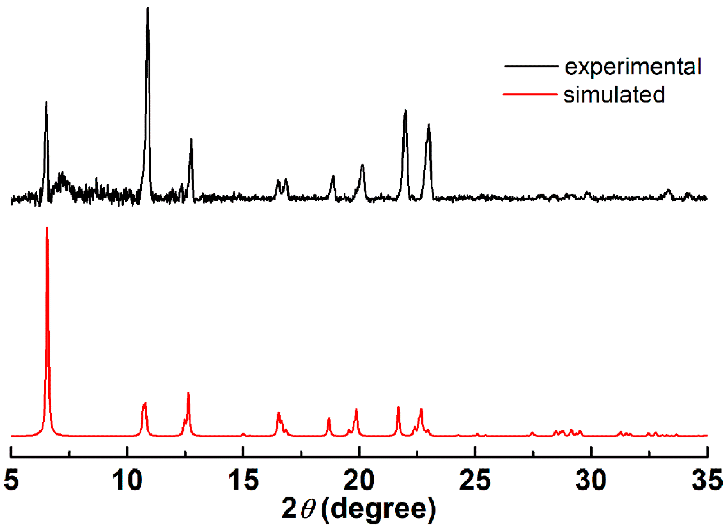
3.1. Structural Studies

The crystallographic data for TetraVV are presented in Table 1.
The crystal structure of TetraVV consists of a neutral macrocyclic tetranuclear species (Figure 1).
The selected bond distances and angles for the TetraVV complex are shown in Table S1. All vanadium atoms are equivalent, generated by symmetry, the bridge between them being assured by one carboxylic oxygen atom O4 (V – O4 = 2.290(4) Å. The vanadium metal atom presents a distorted octahedral geometry, being coordinated in the basal plane by three donor atoms from the Schiff base ligand (V – N1 = 2.120(4) Å, V – O2 = 1.856(4) Å and V – O3 = 1.947(4) Å) and one terminal oxygen atom of the methoxy anion obtain by methanol deprotonation (V – O6 = 1.750(4) Å). The apical positions are occupied by one carboxylic oxygen atom arriving from the Schiff base coordinated to another vanadium center (V – O4 = 2.290(4) Å) and one terminal oxygen atom (V = O5 = 1.567 (5) Å. Moreover, the distance established between two vanadium atoms V1 ‧‧‧ V1 is approximately 5.878 Å and is provided by the bridge formed by the oxygen atom, O4 from the composition of the Schiff base coordinated to the previous vanadium atom by O3, N1, and O2; thus, stabilizing the macrocyclic tetranuclear arrangement.
The axial bond angle (O4 – V1 – O5) and the angles formed between O4 – V1 – O6 and O4 – V1 – N1 of 177.5(3)°, 81.9(2)°, and 83.02(14)°, respectively, slightly deviate from the ideal values of 180° and 90° (Table S1). The relative arrangement of the four groups V = O in the (VVO)4 core and of the Schiff base ligands highlights a perpendicular arrangement, which prevents the ligands from overlapping and promotes the enclosure of the resulting tetranuclear structure.
The bulk sample TetraVV has been analyzed also by X-ray diffraction on powder (PXRD) to ensure crystalline phase purity (Figure 2). The simulated PXRD diffractogram is in good agreement with the simulated one, the differences in peak intensities may be caused by an effect of the preferred orientation of the crystallites.

3.2. Spectral Studies and the Coordination Mode
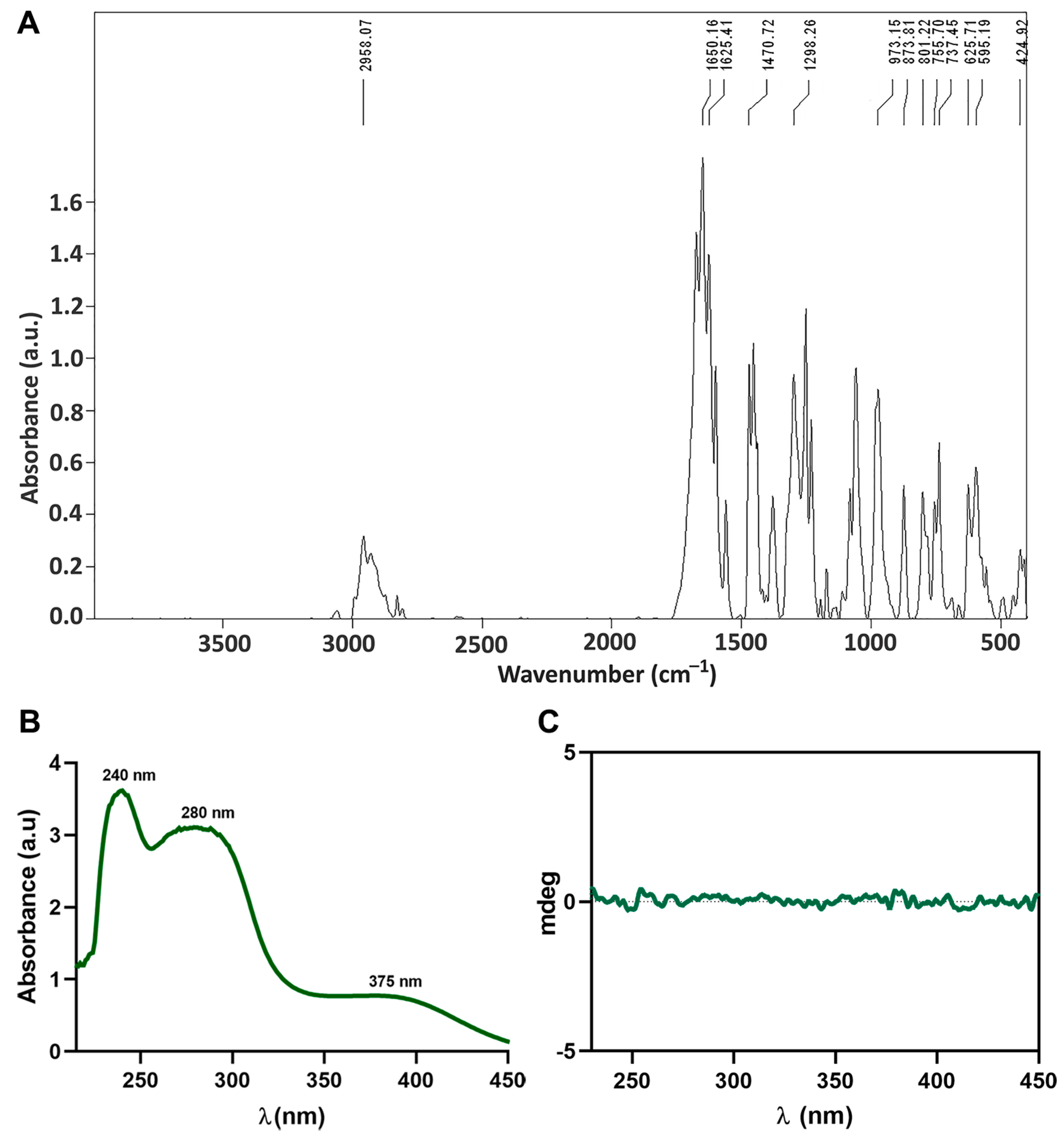
3.2.1. IR Spectrum

The infrared spectrum of the new oxidovanadium(V) compound is shown in Figure 3. The frequency assignments were done based on our previous results [6] and those of other groups [16,30]. The compound is characterized by the appearance of low-intensity absorption bands up to 3000 cm−1 that can be attributed to aromatic and methoxy anion C-H stretch vibration [31,32].
The absence of bands in the range of 2621–2110 cm−1, is associated with the frequency of the NH bond in the amino group of the DL-valine amino acid, and the displacement of the high-intensity band characteristic of the stretching frequency of the aldehyde bond (νC = O) from 1639 cm−1 for o-vanillin [6] to 1650 cm−1 in the case of the compound, is an indicator of the imino group formation in the Schiff base ligand and its coordination to the vanadium centers [6,16].
Additionally, TetraVV shows two bands of medium-high intensity at 1625 cm−1 and 1470 cm−1 assigned to νasCOO and νsCOO with a difference between frequencies up to 200 cm−1 which indicates coordination of the deprotonated carboxyl group in the composition of the Schiff base in a bidentate manner with vanadium atoms [6,16]. The presence of a band of medium intensity at 1298 cm−1 can be associated with the vibration of the phenolic C-O bond (νsCPh – O) [6]. Two bands located at 973 cm−1 and 873 cm−1 were reported to correspond to the symmetric and asymmetric stretching modes of V = O bonds and could be associated with the symmetric and asymmetric stretching modes of the V = O connection [14,33]. The medium-intensity bands between 801–737 cm−1 could be assigned to the vibration stretching of ν(V – OMe) [14]. Moderate to low-intensity bands appearing at 625 cm−1 and 595 cm−1 can be assigned to νsV – N and νsV – O (carboxyl) [6].

3.2.2. Electronic Spectrum

The here developed vanadium-based drug (TetraVV) is a hydrophobic compound that is easily solubilized in DMSO and poorly soluble in water. Thus, to favor the solubilization of the compound in aqueous solutions (PBS or DMEM culture medium) a stock solution of TetraVV in DMSO was initially prepared. The TetraVV absorption spectrum was recorded for 100 µM TetraVV in PBS, pH = 7.4. TetraVV is characterized by 3 absorption bands with λmax = 240 nm, 280 nm, and 375 nm, respectively (Figure 3B). The first two high-intensity bands characterized by λmax = 240 nm and 280 nm can be associated with the π → π* transitions of the benzene ring [34] and to the imino (– CH = N –) group coordination [35]. The third medium intensity band with λmax = 375 nm can be associated with the charge transfer from the oxygen in the double bond V = O to the vanadium atom (O → V) [6].
In addition, the recording of the CD spectrum for the compound TetraVV (solution 500 µM TetraVV in PBS) showed that it is optically inactive (Figure 3C); this is due to the coordination of both R and S enantiomers of the Schiff base ligand at the vanadium centers which makes the molecule symmetrical, a fact proven by elucidating the molecular structure.

3.3. BSA Binding Assay and the Stability Study in Biological Media

The fluorescence spectra of 2 µM BSA-PBS in the presence of DMSO or 1–25 µM TetraVV highlighted the directly proportional relationship between decreasing albumin fluorescence and increasing TetraVV concentration (Figure 4A), which indicates a high ability of the vanadium-based compound to interact with BSA. Using the Stern–Volmer equation and by calculating the Stern–Volmer constant from the regression line: y = ax + b (where: a = slope of the line, y = the ratio of the fluorescence intensity of free BSA solution at 347 nm to the fluorescence intensity of BSA solution at 347 nm in the presence of TetraVV, abbreviated I0/I, x = the concentration of TetraVV) as we previously described [6], we determined the collision extinction constant (Kq) for TetraVV (Figure S1) being equal to 4.45 × e13 M−1s−1, indicating that TetraVV interacts statically with albumin.
The comparison of the absorption spectra of TetraVV (100 µM) in PBS (pH = 7.4) and the DMEM (pH = 7.4) supplemented with 10% FBS, reveals that the electronic spectrum of the TetraVV is shifted to the right (to longer wavelengths) in DMEM with serum, possibly as a result of its interaction with various proteins (such as serum albumin) and growth factors contained by the cell growth medium (Figure 4B).
Furthermore, we assessed the stability of TetraVV in two different biological media: (1) environment with pH = 7.4, which mimics the extracellular medium (plasma) and cytosol of cells, and (2) environment with pH = 4.5, which mimics the weakly acid medium of the lysosomes, the organelles involved in the degradation/hydrolysis of various biomolecules and extracellular components at the subunit level; thus, making them accessible to the cell. For this purpose, solutions of 100 µM TetraVV were prepared in a complete DMEM culture medium with different pHs (pH = 7.4 and pH = 4.5, respectively) and incubated for 4 and 24 h at 37 °C. The absorption spectra of these solutions were recorded in the range 500–350 nm against the corresponding 0.1% DMSO solutions (Figure 4C). The concentration of TetraVV was calculated for each experimental time point (the absorbance was recorded at λmax = 405 nm) (Figure 4D). No changes were observed in the concentration of TetraVV in DMEM at pH = 7.4 (Figure 4C,D). On the other hand, the concentration of TetraVV was reduced by half (p < 0.001) in DMEM at pH = 4.5 compared to pH = 7.4 (Figure 4D) outright after preparation (0 h) and gradually decreased in the weakly acid medium (Figure 4D).

3.4. Cell Death Evaluation

3.4.1. The Cytotoxic Effect of TetraVV on HepG2 Cells

The XTT assay showed the ability of TetraVV and cisplatin to reduce the viability of liver carcinoma cells in a concentration-dependent manner compared to control cells (p < 0.0001) (Figure 5A). However, the concentration that induces death of half of the tested cells (IC50) was much lower (p < 0.001) in the case of TetraVV (~6 µM) vs. cisplatin (~10 µM) (Figure S2A,B).
In addition, we comparatively assessed the cytotoxicity of TetraVV and cisplatin at IC50 concentrations by two reliable tests: the ToxiLight assay which measures the released adenylate kinase (AK) in the cell media, an indicator of cellular damage, and the Live/Dead assay which is a staining method using Calcein-AM for marking viable cells in green and propidium iodide (PI) for marking dead cells in red. Thus, the exposure of the HepG2 cells to IC50 of TetraVV and cisplatin for 24 h showed that TetraVV induced a 2.6-fold increase in AK release (p < 0.0001) in the cell media of liver carcinoma compared to control cells, while a 2-fold increase (p < 0.0001) in AK release was observed for cisplatin (Figure 5B). Additionally, the cell death expressed as dead/total cell no. was increased by about 2-fold (p < 0.0001) in both TetraVV and cisplatin-treated cells vs. control (Figure 5C,D). More than that, the AK release and the dead/total cell no. were increased by 1.9-fold and 1.3-fold, respectively, in TetraVV-treated cells (p < 0.01) vs. cisplatin (Figure 5B–D).

3.4.2. Cell Uptake of TetraVV and the Cell Morphology Examination

The tested drug concentration was chosen from viability/cytotoxicity studies and represents the IC50 of TetraVV (6 µM) and cisplatin (10 µM).
The uptake of TetraVV by the HepG2 cells was investigated at 4 and 24 h by determining the total content of vanadium(V) (ng) in 105 cells. The quantity of VV-PAR chelate in all samples was determined by UHPLC. Representative chromatograms for VV-PAR chelate in control and cells treated for 4 and 24 h with TetraVV are depicted in Figure S3.
The results showed that the vanadium(V) content in HepG2 cells recorded an increase of more than 100% in 24 h compared to 4 h (p < 0.0001), which means that the TetraVV is gradually internalized by HepG2 tumor cells over 24 h (Figure 6A).
Additionally, by performing the ToxiLight assay for HepG2 cells treated with IC50 concentrations of TetraVV and cisplatin, we saw that both drugs had no cytotoxic effects on HepG2 cells at 4 h (Figure 6B), still, as we mentioned above, both TetraVV and cisplatin increased cytotoxicity at 24 h of incubation (Figure 5) and these findings are correlated with the uptake study (Figure 6A).
Optical microscopy images for drug-treated HepG2 cells support the cell uptake and cytotoxicity studies by showing the cell phenotype changes (the cells have a rounded shape, not displayed on the plastic support) at 24 h, similar or even more pronounced to those induced by the antitumor agent, cisplatin (Figure 6C).

3.4.3. The Effect of TetraVV on the Oxidative Status of HepG2 Cells

To investigate the comparative effect of TetraVV and cisplatin treatment on the oxidative status of HepG2 cells, the intracellular ROS and extracellular lipid peroxidation products (as total MDA levels) were determined.
The results revealed a 20% to 50% increase in ROS and MDA levels in HepG2 cells treated with TetraVV for 4 h and 24 h (p < 0.001) compared to control cells, while in HepG2 cells treated with cisplatin an ≈18% increase in both ROS and MDA levels was observed only for the 24 hours’ time incubation (p < 0.05) (Figure 7A–D). More than that, TetraVV induced between 20% and 30% increase in ROS and MDA levels at 4 and 24 h (p < 0.001) compared to cisplatin (Figure 7).

3.4.4. The Effect of TetraVV on DNA Fragmentation and Mitochondrial Status

To assess the TetraVV mode of action compared to cisplatin we analyzed the DNA fragmentation and the apoptotic nuclear morphology by performing acridine orange (AO) and DAPI staining. AO is a fluorescent dye that marks in red the fragmented DNA (single-strand DNA) and in green the double-strand DNA (dsDNA). In addition, we investigated the mitochondria functionality by determining the mitochondrial membrane potential (MtMP) using the mitochondrial-specific fluorescent dye, JC10. AO/DAPI staining of tested HepG2 cells revealed that both TetraVV and cisplatin increased DNA fragmentation and condensation in HepG2 cells compared to control (Figure 8A). Figure 8A reveals the typical apoptotic nuclear morphology as nuclear shrinkage and DNA condensation of cell nuclei (blue fluorescence, indicated by white arrows) and increased DNA fragmentation (red fluorescence).
The percent of DNA fragmentation calculated as a ratio of red to (red + green) fluorescence was increased by about 130% (p < 0.0001) in TetraVV and cisplatin-treated cells vs. control (Figure 8B).
Additionally, our data showed that the cell treatment with TetraVV induced a 34% decrease (p < 0.001) in the MtMP compared to control cells, while only a 17% (p < 0.01) decrease was observed for cisplatin treatment (Figure 8C). More than that, the TetraVV reduced the MtMP of HepG2 cells by about 20% (p < 0.01) compared to cisplatin (Figure 8C).

3.4.5. The Effect of TetraVV on Cell Cycle and β-Tubulin Protein Expression
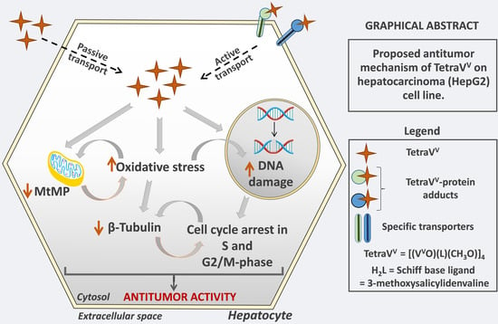
To investigate the comparative effect of TetraVV and cisplatin treatment on the tumor cell growth we analyzed the cell cycle sequential events by flow cytometry using PI staining and the β-Tubulin protein expression.
Our data showed that the percent population of apoptotic cells in the HepG2 cells treated with IC50 of TetraVV (1.55 ± 0.52%, p < 0.0001) and cisplatin (1.88 ± 0.56%, p < 0.0001) was increased when compared to the control (0.69 ± 0.10%) (Figure 9A,B). The proportion of cells in G0/G1-phase decreased in both TetraVV (42 ± 0.8%, p < 0.0001) and cisplatin-treated cells (56 ± 3.4%, p < 0.0001) vs. control (60 ± 1.7%), while the percent population of cells in the S-phase increased in TetraVV (22 ± 2.3%, p < 0.0001) and cisplatin-treated cells (27 ± 2.7%, p < 0.0001) compared to control cells (16 ± 2.00%) (Figure 9A,B). On the other hand, a significant decrease of cells in the apoptotic, G0/G1, and S-phase (p < 0.001) was observed after HepG2 cell treatment with TetraVV compared to cisplatin (Figure 9A,B). More than that, the corresponding percentage population of cells in G2/M-phase increased in TetraVV (34 ± 2.0%, p < 0.0001) compared to both control (22 ± 1.9%, p < 0.0001) and cisplatin (14 ± 3.5%, p < 0.0001), while the treatment with cisplatin decreased significantly the percentage of HepG2 cells in the G2/M-phase vs. control (Figure 9A,B).
The protein expression of β-Tubulin, the subunit of microtubules, was decreased by ≈50% (p < 0.01) in TetraVV and cisplatin-treated cells vs. control, and no significant differences were observed between TetraVV and cisplatin (Figure 9C).

4. Discussion

Hepatocellular carcinoma is a liver cancer most often associated with various chronic liver diseases and cirrhosis, which remains one of the most prevalent cancers all over the world [25]. The use of platinum-based drugs, like cisplatin, carboplatin, or oxaliplatin in the treatment of various cancers, is well documented, however, despite their great antitumor activity, severe side effects associated with drug resistance limit their effectiveness [3,36]. Therefore, many recent studies have been carried out to develop new metal-based drugs with fewer side effects and better chemotherapeutic efficacy than platinum-based compounds [37,38].
To develop a new vanadium-based compound that possesses high antitumor activity, we used a simple method of synthesis described previously [6]. By reacting the Schiff base ligand, 3-methoxysalicylidenvaline formed in the methanol solution from o-vanillin and DL-valine, with vanadyl sulfate, we obtained a new macrocyclic tetranuclear oxidovanadium(V) complex which crystallized in the I41/a space group. The coordination mode of the four 3-methoxysalicylidenvaline ligands to vanadium centers, the formation of the four bridges between the carboxylic oxygen (O4) of each Schiff base coordinated to a vanadium atom and the next vanadium center, and the relative arrangement of the four groups V = O in the (VVO)4 core was indicated by spectral and structural analysis. The coordination of the O, N, O’-chelating ligands at vanadium centers with the formation of chelated rings of 5 and 6 atoms and the establishment of carboxy bridges between neighboring vanadium atoms ensure the enclosure of the tetranuclear structure and explain its stability in biological media.
Reduced drug uptake by cells, as well as other mechanisms of drug resistance, are the main obstacles to successful cancer therapy [39,40]. Essential properties for increasing the bioavailability and improving the efficiency of drug uptake by cells are compound stability in biological environments and the ability to interact with circulating proteins which may promote the drug transport to various tissues [9,14,18].
In agreement with our and other researchers’ previously published data, TetraVV binds to albumin in a static mode, most likely by establishing the hydrogen bonds between the Schiff base ligands coordinated at oxidovanadium(V) centers and OH groups of the tryptophan residues from the BSA structure [6,21,22].
Moreover, our data showed that TetraVV has increased stability in biological environments with physiological pH encountered in plasma and cell cytosol, a feature that ensures its uptake by HepG2 cells. A decreased stability was determined in a weakly acid medium which is most likely a result of cleavage of the interactions (by hydrolysis) established between TetraVV and the transporter proteins (e.g., albumin). This result points out that the acidic pH of the lysosomes will promote the release of the TetraVV after internalization and explains the biological effects observed for the newly developed vanadium-based compound. Recently, the stability of other vanadium compounds in cell culture medium at physiological pH was mentioned and these findings are following our data [14,41].
Up to date, a large number of vanadium compounds have been proposed as antiproliferative agents [9,17,18]. In this work, we analyzed the effect of TetraVV on HepG2 cells viability/cytotoxicity in comparison with the standard antineoplastic agent cisplatin. The new vanadium-based compound was found to display higher cytotoxicity against HepG2 cells compared to cisplatin and this was shown also for other vanadium complexes [14]. As previously reported for another hydrophobic metallic-based compound [37], the TetraVV is time-dependently uptaken by HepG2 cells, probably by both passive and active transport and this can explain the increased cytotoxic activity of TetraVV at 24 h and makes it interesting for possible chemotherapeutic use. Additionally, our uptake results are in line with other studies comprising the well-known metallic antiproliferative compounds such as cisplatin, oxaliplatin [42], or the recently developed vanadium(IV) complex with a trypanostatic effect [43].
Up to now, the main proposed mechanism of action for the recently developed vanadium complexes involves ROS generation and DNA fragmentation [17,19,20]. Thus, to study the mechanism by which TetraVV induces tumor cell death, we firstly assessed the oxidative status and the DNA fragmentation percent in TetraVV -treated HepG2 cells compared to cisplatin. Our data revealed the time-dependent ability of TetraVV to generate ROS and lipid peroxidation products compared to cisplatin which induced oxidative stress to a lower extent only at 24 h. On the other hand, similar to cisplatin, TetraVV induced the damage of dsDNA. In addition, both cisplatin and TetraVV altered the normal nuclear morphology to the apoptotic one, characterized by nuclear shrinkage and DNA condensation, and these findings are in good agreement with previously published studies [17,44].
The alterations of mitochondrial membrane potential (MtMP) initiate cell death in living cells [45]. It was shown that the decrease in the ratio of red-fluorescent JC aggregates in the mitochondria of cells to green-fluorescent JC monomeric form is an indicator of MtMP dysfunction and apoptosis induction [45]. The capacity of a new oxidovanadium(IV) complex to induce apoptosis by simultaneous disruption of MtMP and ROS generation was recently proved [13]. Based on these findings, we further investigated the comparative effect of TetraVV and cisplatin on the mitochondrial status of HepG2 cells. In agreement with the induction of DNA fragmentation, both TetraVV and cisplatin affected the mitochondrial functionality of HepG2 by reducing the MtMP, still, the TetraVV induced a more pronounced mitochondrial disruption than cisplatin as a consequence of oxidative stress generation.
Cisplatin sets off cell death in HepG2 cells by apoptosis and DNA damage and by cell cycle arrest in the DNA replication/synthesis (S)-phase [46]. Our data show that, unlike cisplatin, which induces apoptosis of liver carcinoma cells by arresting the cell cycle in the S-phase, the newly developed vanadium-based compound, TetraVV, exerts this effect by blocking the cell cycle of HepG2 cells in both S and mitosis/cell division (G2/M)-phase. This difference between the cisplatin and TetraVV mechanism of cell death induction in human hepatocellular carcinoma cells may be due to the increased properties of TetraVV to generate oxidative and mitochondrial stress [13,14]. In agreement with our observations, Ni L. et al. evidenced that some multidentate oxidovanadium(IV) complexes arrested the cell cycle in the S and G2/M phases in hepatocellular carcinoma cell lines, but still the authors found a less cytotoxic effect for vanadium complexes than cisplatin [20]. In addition, other oxidovanadium(IV) complexes induced apoptosis in pancreatic cancer cells by simultaneously arresting the cell cycle in the G2/M-phase, ROS generation, and the disruption of mitochondrial membrane potential [13]. Additionally, sodium vanadate [47] and a number of vanadium(V) complexes [48] induced cell cycle arrest in the S-phase in human tumor cells. Contrary to our study, some oxidovanadium complexes were found to cause a G0/G1-phase cell cycle arrest in different types of cancer cell lines [49,50]. Cell cycle arrest is directly involved in reducing cell growth/division and together with the cellular response to DNA damage may be associated with cytoskeletal remodeling and decreased polymerization of the microtubules’ components, α- and β-Tubulin [51]. We have previously shown the involvement of cisplatin in DNA fragmentation, the cytoskeleton remodeling, and β-Tubulin synthesis reduction in human hepatocellular carcinoma cells [25]. Herein, we showed that TetraVV reduced β-Tubulin synthesis after 24 h of treatment probably due to the association between ROS generation, MtMP reduction, DNA fragmentation, and cell cycle arrest in the S and G2/M-phase. However, the mechanism linking the TetraVV effect with β-Tubulin synthesis is only speculative and requires future investigations. The involvement of ROS and MtMP in the destabilization of microtubules has been proposed as a mechanism for the antiproliferative effect of some drugs in breast cancer, still, the mechanisms involved are under debate [52].
Thus, our data demonstrated that apoptosis and cell cycle arrest in S and G2/M-phase, induced by DNA fragmentation, and mitochondrial membrane disruption could be one of the main pathways of the antitumor action of TetraVV compound in HepG2 cells. However, other cell death pathways involving oxidative and inflammatory processes could explain the higher cytotoxic effects of TetraVV compared to cisplatin. The results motivate further studies to investigate in-depth the antitumor mechanisms of the newly developed TetraVV compound.

5. Conclusions

We developed a new macrocyclic tetranuclear oxidovanadium(V) compound with the chemical formula [(VVO)(L)(CH3O)]4 (TetraVV), where L is the deprotonated form of the Schiff base, 3-methoxysalicylidenvaline, obtained from o-vanillin and DL-valine, as a metal-based antitumor agent. The new compound TetraVV binds the albumin in a dose-dependent manner and has pH-dependent stability in biological media, properties which ensure its internalization by the tumor cells and facilitate its release into the cell, where it exerts the biological activities. The cytotoxicity results demonstrate that TetraVV has a higher antitumor effect compared to cisplatin (IC50 = 6 μM vs. 10 μM), the antitumor drug used in medical practice, by inducing approximately 2 times higher cell death in the liver carcinoma and also by altering the oxidative and mitochondrial status in HepG2 cells. Unlike cisplatin which blocks the cell cycle in the S-phase, the new compound arrests it in S and G2/M-phase, whereas no differences in the induction of DNA fragmentation and reduction of β-Tubulin synthesis between the two were determined. Thus, the mechanism involved in the antitumor action of TetraVV comprises the induction of apoptosis, the cell cycle arrest in the S and G2/M-phase, and β-Tubulin synthesis reduction together with DNA fragmentation, mitochondrial dysfunction, and oxidative stress generation.

Supplementary Materials

The following supporting information can be downloaded at: https://www.mdpi.com/article/10.3390/biomedicines10061217/s1, Figure S1: The plot of I0/I vs. [Q] for TetraVV ([(VVO)(L)(CH3O)]4, where L= deprotonated form of the Schiff base ligand 3-methoxysalicylidenvaline; Figure S2: (A) Nonlinear regression for % of cell viability inhibition to Log10 TetraVV/Cisplatin concentration. (B) Calculated IC50. Statistical significance: *** p < 0.001.; Figure S3: Representative chromatograms for VV-PAR chelate in control (A) and cells treated with TetraVV for 4 (B) and 24 h (C); Table S1: Selected bond lengths (Å) and angles (°) for TetraVV. Symmetry operations #1: −y + 5/4, x + 1/4, −z + 1/4, #2: y − 1/4, −x + 5/4, −z + 1/4.

Author Contributions

Conceptualization, M.T., M.C. and D.-L.P.; methodology, M.T.; software, M.T., M.A., M.R. and A.A.P.; validation, M.T., M.A., M.R., M.D. and A.A.P.; formal analysis, M.T.; investigation, M.T., M.A., M.R., M.D., G.V., F.S. and A.A.P.; resources, M.T., M.C. and D.-L.P.; data curation, M.T.; writing—original draft preparation, M.T.; writing—review and editing, M.T., M.C., D.-L.P., A.A.P. and I.M.; visualization, M.T. and M.C.; supervision, M.C., D.-L.P. and I.M.; project administration, M.T. and M.C.; funding acquisition, M.T. and M.C. All authors have read and agreed to the published version of the manuscript.

Funding

This research was funded by two grants from the Romanian Ministry of Education and Research, CNCS—UEFISCDI, within PNCDI III: project number PN-III-P1-1.1-PD-2019-0247, and project number PN-III-P4-ID-PCCF-2016-0050 and by Romanian Academy.

Institutional Review Board Statement

Not applicable.

Informed Consent Statement

This article does not describe a study involving humans.

Data Availability Statement

All data supporting reported results are included in the article.

Conflicts of Interest

The authors of this paper have nothing to disclose.

Abbreviations

AOAcridine orange
BSABovine serum albumin
DAPI4′,6-Diamidine-2′-phenylindole
DMEMDulbecco’s Modified Eagle’s Medium
DMSODimethyl sulfoxide
DCFH-DA2’,7’Dichlorofluorescein diacetate
EDTAEthylenediaminetetraacetic acid
FBSFetal bovine serum
FITCPhalloidin-fluorescein isothiocyanate
HepG2Human hepatocellular carcinoma cell line
H2L3-Methoxysalicylidenvaline
MDAMalondialdehyde
MtMPMitochondrial membrane potential
PAR4-(2-Pyridylazo)resorcinol
PBSPhosphate-buffered solution
PFAParaformaldehyde
PIPropidium iodide
PMSPhenazine methosulfate
ROSReactive oxygen species
Na3VO4Sodium orthovanadate
Na2HPO4Sodium phosphate dibasic
TetraVV[(VVO)(L)(CH3O)]4
LDeprotonated form of the Schiff base ligand
VIVOSO4•3H2OVanadyl sulfate trihydrate
TBARSThiobarbituric acid reactive substances
TRITCTetramethylrhodamine
XTT2,3-Bis-(2-Methoxy-4-Nitro-5-Sulfophenyl)-2H-Tetrazolium-5-Carboxanilide

References

  1. Llovet, J.M.; Kelley, R.K.; Villanueva, A.; Singal, A.G.; Pikarsky, E.; Roayaie, S.; Lencioni, R.; Koike, K.; Zucman-Rossi, J.; Finn, R.S. Hepatocellular carcinoma. Nat. Rev. Dis. Prim. 2021, 7, 6. [Google Scholar] [CrossRef] [PubMed]
  2. Kudo, M.; Ueshima, K.; Yokosuka, O.; Ogasawara, S.; Obi, S.; Izumi, N.; Aikata, H.; Nagano, H.; Hatano, E.; Sasaki, Y.; et al. Sorafenib plus low-dose cisplatin and fluorouracil hepatic arterial infusion chemotherapy versus sorafenib alone in patients with advanced hepatocellular carcinoma (SILIUS): A randomised, open label, phase 3 trial. Lancet Gastroenterol. Hepatol. 2018, 3, 424–432. [Google Scholar] [CrossRef]
  3. Ramadori, G.; Cameron, S. Effects of systemic chemotherapy on the liver. Ann. Hepatol. 2010, 9, 133–143. [Google Scholar] [CrossRef]
  4. Le Grazie, M.; Biagini, M.R.; Tarocchi, M.; Polvani, S.; Galli, A. Chemotherapy for hepatocellular carcinoma: The present and the future. World J. Hepatol. 2017, 9, 907–920. [Google Scholar] [CrossRef] [PubMed]
  5. Crans, D.C.; Smee, J.J.; Gaidamauskas, E.; Yang, L. The Chemistry and Biochemistry of Vanadium and the Biological Activities Exerted by Vanadium Compounds. Chem. Rev. 2004, 104, 849–902. [Google Scholar] [CrossRef]
  6. Turtoi, M.; Anghelache, M.; Patrascu, A.A.; Maxim, C.; Manduteanu, I.; Calin, M.; Popescu, D.L. Synthesis, characterization, and in vitro insulin-mimetic activity evaluation of valine Schiff base coordination compounds of oxidovanadium(v). Biomedicines 2021, 9, 562. [Google Scholar] [CrossRef]
  7. Bhuiyan, M.S.; Fukunaga, K. Cardioprotection by Vanadium Compounds Targeting Akt-Mediated Signaling. J. Pharmacol. Sci. 2009, 110, 1–13. [Google Scholar] [CrossRef] [Green Version]
  8. Zordok, W.A.; Sadeek, S.A. Synthesis, thermal analyses, characterization and biological evaluation of new enrofloxacin vanadium(V) solvates(L) (L = An, DMF, Py, Et3N and o-Tol). J. Mol. Struct. 2016, 1120, 50–61. [Google Scholar] [CrossRef]
  9. Crans, D.C.; Koehn, J.T.; Petry, S.M.; Glover, C.M.; Wijetunga, A.; Kaur, R.; Levina, A.; Lay, P.A. Hydrophobicity may enhance membrane affinity and anti-cancer effects of Schiff base vanadium(v) catecholate complexes. Dalt. Trans. 2019, 48, 6383–6395. [Google Scholar] [CrossRef]
  10. Lampronti, I.; Bianchi, N.; Borgatti, M.; Fabbri, E.; Vizziello, L.; Khan, M.T.H.; Ather, A.; Brezena, D.; Tahir, M.M.; Gambari, R. Effects of vanadium complexes on cell growth of human leukemia cells and protein-DNA interactions. Oncol. Rep. 2005, 14, 9–15. [Google Scholar] [CrossRef]
  11. Aubrecht, J.; Narla, R.K.; Ghosh, P.; Stanek, J.; Uckun, F.M. Molecular genotoxicity profiles of apoptosis-inducing vanadocene complexes. Toxicol. Appl. Pharmacol. 1999, 154, 228–235. [Google Scholar] [CrossRef] [PubMed]
  12. Harding, M.; Mokdsi, G. Antitumour Metallocenes: Structure-Activity Studies and Interactions with Biomolecules. Curr. Med. Chem. 2012, 7, 1289–1303. [Google Scholar] [CrossRef] [PubMed]
  13. Kowalski, S.; Tesmar, A.; Sikorski, A.; Inkielewicz-Stępniak, I. Oxidovanadium(IV) Complex Disrupts Mitochondrial Membrane Potential and Induces Apoptosis in Pancreatic Cancer Cells. Anticancer Agents Med. Chem. 2021, 21, 71–83. [Google Scholar] [CrossRef] [PubMed]
  14. Choroba, K.; Raposo, L.R.; Palion-Gazda, J.; Malicka, E.; Erfurt, K.; Machura, B.; Fernandes, A.R. In vitro antiproliferative effect of vanadium complexes bearing 8-hydroxyquinoline-based ligands-the substituent effect. Dalt. Trans. 2020, 49, 6596–6606. [Google Scholar] [CrossRef] [PubMed]
  15. Cao, Y.; Yi, C.; Liu, H.; Li, H.; Li, Q.; Yuan, Z.; Wei, G. Syntheses, crystal structures and in vitro anticancer activities of oxovanadium(IV) complexes of amino acid Schiff base and 1,10-phenanthroline ligands. Transit. Met. Chem. 2016, 41, 531–538. [Google Scholar] [CrossRef]
  16. Guo, Q.; Li, L.; Dong, J.; Liu, H.; Xu, T.; Li, J. Synthesis, crystal structure and interaction of l-valine Schiff base divanadium(V) complex containing a V2O3 core with DNA and BSA. Spectrochim. Acta Part A Mol. Biomol. Spectrosc. 2013, 106, 155–162. [Google Scholar] [CrossRef]
  17. Parisa, M.; Gholamhossein, M. Anti-cancer properties and catalytic oxidation of sulfides based on vanadium(V) complexes of unprotected sugar-based Schiff-base ligands. Polyhedron 2022, 215, 115655. [Google Scholar] [CrossRef]
  18. Lima, L.M.A.; Murakami, H.; Gaebler, D.J.; Silva, W.E.; Belian, M.F.; Lira, E.C.; Crans, D.C. Acute toxicity evaluation of non-innocent oxidovanadium(V) schiff base complex. Inorganics 2021, 9, 42. [Google Scholar] [CrossRef]
  19. Mirjalili, S.; Dejamfekr, M.; Moshtaghian, A.; Salehi, M.; Behzad, M.; Khaleghian, A. Induction of Cell Cycle Arrest in MKN45 Cells after Schiff Base Oxovanadium Complex Treatment Using Changes in Gene Expression of CdC25 and P53. Drug Res. 2020, 70, 545–551. [Google Scholar] [CrossRef]
  20. Ni, L.; Zhao, H.; Tao, L.; Li, X.; Zhou, Z.; Sun, Y.; Chen, C.; Wei, D.; Liu, Y.; Diao, G. Synthesis, in vitro cytotoxicity, and structure-actIVity relationships (SAR) of multidentate oxidovanadium(IV) complexes as anticancer agents. Dalt. Trans. 2018, 47, 10035–10045. [Google Scholar] [CrossRef]
  21. Jayabharathi, J.; Jayamoorthy, K.; Thanikachalam, V.; Sathishkumar, R. Fluorescence quenching of bovine serum albumin by NNMB. Spectrochim. Acta Part A Mol. Biomol. Spectrosc. 2013, 108, 146–150. [Google Scholar] [CrossRef] [PubMed]
  22. Kazemi, Z.; Rudbari, H.A.; Sahihi, M.; Mirkhani, V.; Moghadam, M.; Tangestaninejad, S.; Mohammadpoor-Baltork, I.; Gharaghani, S. Synthesis, characterization and biological application of four novel metal-Schiff base complexes derived from allylamine and their interactions with human serum albumin: Experimental, molecular docking and ONIOM computational study. J. Photochem. Photobiol. B Biol. 2016, 162, 448–462. [Google Scholar] [CrossRef] [PubMed]
  23. Anghelache, M.; Turtoi, M.; Petrovici, A.R.; Fifere, A.; Pinteala, M.; Calin, M. Development of dextran-coated magnetic nanoparticles loaded with protocatechuic acid for vascular inflammation therapy. Pharmaceutics 2021, 13, 1414. [Google Scholar] [CrossRef] [PubMed]
  24. Bucatariu, S.M.; Constantin, M.; Varganici, C.D.; Rusu, D.; Nicolescu, A.; Prisacaru, I.; Carnuta, M.; Anghelache, M.; Calin, M.; Ascenzi, P.; et al. A new sponge-type hydrogel based on hyaluronic acid and poly(methylvinylether-alt-maleic acid) as a 3D platform for tumor cell growth. Int. J. Biol. Macromol. 2020, 165 Pt B, 2528–2540. [Google Scholar] [CrossRef]
  25. Turtoi, M.; Anghelache, M.; Bucatariu, S.M.; Deleanu, M.; Voicu, G.; Safciuc, F.; Manduteanu, I.; Fundueanu, G.; Simionescu, M.; Calin, M. A novel platform for drug testing: Biomimetic three-dimensional hyaluronic acid-based scaffold seeded with human hepatocarcinoma cells. Int. J. Biol. Macromol. 2021, 185, 604–619. [Google Scholar] [CrossRef]
  26. Puckett, C.A.; Barton, J.K. Mechanism of cellular uptake of a ruthenium polypyridyl complex. Biochemistry 2008, 47, 11711–11716. [Google Scholar] [CrossRef] [Green Version]
  27. Tsai, S.J.J.; Hsu, S.J. Speciation of vanadium(V) and VANADIUM(IV) with 4-(2-pyridylazo) resorcinol by using high-performance liquid chromatography with spectrophotometric detection. Analyst 1994, 119, 403–407. [Google Scholar] [CrossRef]
  28. Smith, P.K.; Krohn, R.I.; Hermanson, G.T.; Mallia, A.K.; Gartner, F.H.; Provenzano, M.D.; Fujimoto, E.K.; Goeke, N.M.; Olson, B.J.; Klenk, D.C. Measurement of protein using bicinchoninic acid. Anal. Biochem. 1985, 150, 76–85. [Google Scholar] [CrossRef]
  29. Carnuta, M.G.; Stancu, C.S.; Toma, L.; Sanda, G.M.; Niculescu, L.S.; Deleanu, M.; Popescu, A.C.; Popescu, M.R.; Vlad, A.; Dimulescu, D.R.; et al. Dysfunctional high-density lipoproteins have distinct composition, diminished anti-inflammatory potential and discriminate acute coronary syndrome from stable coronary artery disease patients. Sci. Rep. 2017, 7, 7295. [Google Scholar] [CrossRef]
  30. Berestova, T.V.; Kuzina, L.G.; Amineva, N.A.; Faizrakhmanov, I.S.; Massalimov, I.A.; Mustafin, A.G. ATR-FTIR spectroscopic investigation of the cis- and trans- bis-(α-amino acids) copper(II) complexes. J. Mol. Struct. 2017, 1137, 260–266. [Google Scholar] [CrossRef]
  31. Mukherjee, G.; Biradha, K. 1D, 2D and 3D coordination polymers of 1,3-phenylene diisonicotinate with Cu(i)/Cu(ii): Cu2I2 building block, anion influence and guest inclusions. CrystEngComm 2014, 16, 4701–4705. [Google Scholar] [CrossRef]
  32. Han, J.X.; Utkin, Y.G.; Chen, H.B.; Burns, L.A.; Curl, R.F. High-resolution infrared spectra of the C-H asymmetric stretch vibration of jet-cooled methoxy radical (CH3). J. Chem. Phys. 2002, 117, 6538–6545. [Google Scholar] [CrossRef]
  33. Xu, Y.; Han, X.; Zheng, L.; Wei, S.; Xie, Y. First investigation on charge-discharge reaction mechanism of aqueous lithium ion batteries: A new anode material of Ag2V4O 11 nanobelts. Dalt. Trans. 2011, 40, 10751–10757. [Google Scholar] [CrossRef] [PubMed]
  34. Ebrahimipour, S.Y.; Sheikhshoaie, I.; Kautz, A.C.; Ameri, M.; Pasban-Aliabadi, H.; Amiri Rudbari, H.; Bruno, G.; Janiak, C. Mono- and dioxido-vanadium(V) complexes of a tridentate ONO Schiff base ligand: Synthesis, spectral characterization, X-ray crystal structure, and anticancer activity. Polyhedron 2015, 93, 99–105. [Google Scholar] [CrossRef]
  35. Majumdar, D. Synthesis of two unprecedented Ni(II)& Oxovanadium Azide bridged complexes derived from compartmental Azo-Linked two different Schiff base H4L & H2L-Characterization by spectroscopic studies (IR, UV-Vis, 1H NMR) and magneto structural co-relationship. Int. J. Chem. Stud. 2016, 4, 46–54. [Google Scholar]
  36. Thayer, A.M. Platinum Drugs Take Their Toll. Chem. Eng. News Arch. 2010, 88, 24–28. [Google Scholar] [CrossRef]
  37. Gul, N.S.; Khan, T.M.; Liu, Y.C.; Choudhary, M.I.; Chen, Z.F.; Liang, H. Pd(II) and Rh(III) complexes with isoquinoline derivatives induced mitochondria-mediated apoptotic and autophagic cell death in HepG2 cells. CCS Chem. 2021, 3, 1626–1641. [Google Scholar] [CrossRef]
  38. Kostova, I. Titanium and Vanadium Complexes as Anticancer Agents. Anticancer. Agents Med. Chem. 2012, 9, 827–842. [Google Scholar] [CrossRef]
  39. Li, Q.; Shu, Y. Pharmacological modulation of cytotoxicity and cellular uptake of anti-cancer drugs by PDE5 inhibitors in lung cancer cells. Pharm. Res. 2014, 31, 86–96. [Google Scholar] [CrossRef] [Green Version]
  40. Mansoori, B.; Mohammadi, A.; Davudian, S.; Shirjang, S.; Baradaran, B. The different mechanisms of cancer drug resistance: A brief review. Adv. Pharm. Bull. 2017, 7, 339–348. [Google Scholar] [CrossRef]
  41. Levina, A.; Crans, D.C.; Lay, P.A. Speciation of metal drugs, supplements and toxins in media and bodily fluids controls in vitro activities. Coord. Chem. Rev. 2017, 352, 473–498. [Google Scholar] [CrossRef]
  42. Corte-Rodríguez, M.; Espina, M.; Sierra, L.M.; Blanco, E.; Ames, T.; Montes-Bayón, M.; Sanz-Medel, A. Quantitative evaluation of cellular uptake, DNA incorporation and adduct formation in cisplatin sensitive and resistant cell lines: Comparison of different Pt-containing drugs. Biochem. Pharmacol. 2015, 98, 69–77. [Google Scholar] [CrossRef] [PubMed]
  43. Mosquillo, M.F.; Smircich, P.; Lima, A.; Gehrke, S.A.; Scalese, G.; Machado, I.; Gambino, D.; Garat, B.; Pérez-Díaz, L. High Throughput Approaches to Unravel the Mechanism of Action of a New Vanadium-Based Compound against Trypanosoma cruzi. Bioinorg. Chem. Appl. 2020, 2020, 1634270. [Google Scholar] [CrossRef] [PubMed] [Green Version]
  44. Lu, L.P.; Suo, F.Z.; Feng, Y.L.; Song, L.L.; Li, Y.; Li, Y.J.; Wang, K.T. Synthesis and biological evaluation of vanadium complexes as novel anti-tumor agents. Eur. J. Med. Chem. 2019, 176, 1–10. [Google Scholar] [CrossRef]
  45. Sivandzade, F.; Bhalerao, A.; Cucullo, L. Analysis of the Mitochondrial Membrane Potential Using the Cationic JC-1 Dye as a Sensitive Fluorescent Probe. Bio-Protocol 2019, 9, e3128. [Google Scholar] [CrossRef]
  46. Wang, P.; Cui, J.; Wen, J.; Guo, Y.; Zhang, L.; Chen, X. Cisplatin induces hepG2 cell cycle arrest through targeting specific long noncoding RNAs and the p53 signaling pathway. Oncol. Lett. 2016, 12, 4605–4612. [Google Scholar] [CrossRef] [Green Version]
  47. Yang, J.; Zhang, Z.; Jiang, S.; Zhang, M.; Lu, J.; Huang, L.; Zhang, T.; Gong, K.; Yan, S.; Yang, Z.; et al. Vanadate-induced antiproliferative and apoptotic response in esophageal squamous carcinoma cell line EC109. J. Toxicol. Environ. Health-Part A Curr. Issues 2016, 79, 864–868. [Google Scholar] [CrossRef]
  48. Reytman, L.; Hochman, J.; Tshuva, E.Y. Anticancer diaminotris(phenolato) vanadium(V) complexes: Ligand-metal interplay. J. Coord. Chem. 2018, 71, 2003–2011. [Google Scholar] [CrossRef]
  49. Ying, P.; Zeng, P.; Lu, J.; Chen, H.; Liao, X.; Yang, N. New Oxidovanadium Complexes Incorporating Thiosemicarbazones and 1,10-Phenanthroline Derivatives as DNA Cleavage, Potential Anticancer Agents, and Hydroxyl Radical Scavenger. Chem. Biol. Drug Des. 2015, 86, 926–937. [Google Scholar] [CrossRef]
  50. Zhang, Y.L.; Wang, X.S.; Fang, W.; Cai, X.Y.; Li, H.Z.; Mao, J.W.; Jin, X.B.; Bai, Y.L.; Lu, J.Z. In vitro study of the cytotoxicities of two mixed-ligand oxovanadium complexes on human hepatoma cells. Pharmazie 2013, 68, 827–834. [Google Scholar] [CrossRef]
  51. Laflamme, G.; Sim, S.; Leary, A.; Pascariu, M.; Vogel, J.; D’Amours, D. Interphase Microtubules Safeguard Mitotic Progression by Suppressing an Aurora B-Dependent Arrest Induced by DNA Replication Stress. Cell Rep. 2019, 26, 2875–2889.e3. [Google Scholar] [CrossRef] [PubMed] [Green Version]
  52. Nambiar, N.; Nagireddy, P.K.R.; Pedapati, R.; Kantevari, S.; Lopus, M. Tubulin- and ROS-dependent antiproliferative mechanism of a potent analogue of noscapine, N-propargyl noscapine. Life Sci. 2020, 258, 118238. [Google Scholar] [CrossRef] [PubMed]
Figure 1. The X-ray crystal structure of TetraVV with the atom labeling scheme of non-carbon atoms. For clarity, hydrogen atoms have been excluded from the diagram.
Biomedicines 10 01217 g001
Figure 2. The experimental and simulated powder X-ray diffractograms for compound TetraVV.
Biomedicines 10 01217 g002
Figure 3. (A) IR spectrum of TetraVV. (B) Electronic spectrum of 100 μM TetraVV in phosphate-buffered solution (PBS), pH = 7.4. (C) CD spectrum of 500 μM TetraVV in PBS, pH = 7.4.
Biomedicines 10 01217 g003
Figure 4. (A) Fluorescence spectra of 2 µM BSA in the presence of 0.1% DMSO or 1–25 µM TetraVV. (B) The electronic spectra of 100 μM TetraVV in PBS, pH = 7.4 vs. DMEM culture medium, pH = 7.4. (C) Time dependent stability of 100 μM TetraVV in DMEM medium at pH = 7.4 and pH = 4.5. (D) The concentration of TetraVV measured at 0, 4 and 24 h in DMEM at pH = 7.4 and pH = 4.5. Data were expressed as mean ± SD: ** p < 0.01, **** p < 0.0001.
Biomedicines 10 01217 g004
Figure 5. (A) The cell viability of HepG2 cells treated with various concentrations (0.25–64 µM) of TetraVV and cisplatin. (B) Adenylate kinase (AK) release expressed as fold change to Control. (C) The number of dead cells (red fluorescence)/total cell no. (red + green fluorescence) calculated from Calcein-AM/propidium iodide (PI) staining and expressed as fold change to Control. (D) Representative images of calcein-AM (green)/PI (red) staining. Scale bar = 100 µm. HepG2 cells were treated with IC50 concentration of TetraVV and cisplatin (6 µM and 10 µM, respectively) or exposed to 0.01% DMSO (vehicle, Control) for 24 h. Data were expressed as mean ± SD: **** p < 0.0001 vs. Control, ## p < 0.01, #### p < 0.0001 vs. Cisplatin.
Biomedicines 10 01217 g005
Figure 6. (A) Vanadium(V) content (ng) per 105 HepG2 cells over 24 h. (B) Adenylate kinase (AK) release expressed as fold change to Control. (C) The cellular morphology of HepG2 cells exposed to TetraVV and cisplatin for up to 24 h. Scale bar = 200 µm. HepG2 cells were treated with IC50 concentration of TetraVV or cisplatin (6 µM or 10 µM, respectively) or exposed to 0.01% DMSO (vehicle, Control) for 4 and 24 h. Data were expressed as mean ± SD: **** p < 0.0001 vs. 4 h.
Biomedicines 10 01217 g006
Figure 7. The intracellular levels of ROS expressed as DCFH-DA fluorescence intensity/µg protein (relative to control cells) in HepG2 cells at 4 h (A) and 24 h (B). The extracellular levels of MDA (relative to control cells) in HepG2 cells at 4 h (C) and 24 h (D). HepG2 cells were treated with 6 µM TetraVV and 10 µM cisplatin or exposed to 0.01% DMSO (Control). Data were expressed as mean ± SD: * p < 0.05, *** p < 0.001 and **** p < 0.0001 vs. Control. ### p < 0.001 and #### p < 0.0001 vs. Cisplatin.
Biomedicines 10 01217 g007
Figure 8. (A) Representative images of acridine orange and the corresponding DAPI images. (B) DNA fragmentation, calculated as percent of red intensity/(red+green) fluorescence. Scale bar = 20 µm. (C) The mitochondrial membrane potential (MtMP), expressed as the ratio of JC10 aggregates (λexem = 540 nm/590 nm) to JC10 monomers (λexem = 490 nm/525 nm). HepG2 cells were treated with IC50 concentrations of TetraVV and cisplatin (6 µM and 10 µM, respectively) or exposed to 0.01% DMSO (Control) over 24 h. Data were expressed as mean ± SD: **** p < 0.0001 vs. Control. ## p < 0.01 vs. Cisplatin. White arrows indicate the nuclear shrinkage and DNA condensation of cell nuclei.
Biomedicines 10 01217 g008
Figure 9. (A) Flow cytometry analysis of the cell cycle progression of DMSO (control), TetraVV, and cisplatin-treated cells. (B) The cell cycle phases (%) as a measure of DNA content, evaluated after propidium iodide (PI) labeling. (C) Determination of β-Tubulin protein expression by WB method. The representative Western blot images are depicted on top of the graph. HepG2 cells were treated with IC50 concentrations of TetraVV and cisplatin (6 µM and 10 µM, respectively) or exposed to 0.01% DMSO (Control) for 24 h. Data were expressed as mean ± SD: ** p < 0.01 and **** p < 0.0001 vs. Control. #### p < 0.001 vs. Cisplatin.
Biomedicines 10 01217 g009
Table 1. The crystallographic data of TetraVV.
FormulaC56H72N4O24V4Z4
MW (g mol−1)1388.93Calculated density (g/cm3)1.458
T/K293(2)Absorption coefficient (cm−1)0.654
λ/Å0.71073F(000)2880
Crystal systemTetragonalCrystal size (mm × mm × mm)0.8 × 0.5 × 0.1
Space groupI41/aθ range/deg2.465 to 24.996
Unit cell Limiting indices−19 < h < 19, −19 < k < 19, −28 < l < 26
a/Å16.377(2)Collected reflections29979
b/Å16.377(2)Sym. Indep. reflections2793
c/Å23.592(5)Rint0.1693
α/deg90Data/restraints/parameters2793/0/199
β/deg90GOF on F21.046
γ/deg90Final R indicesR1 = 0.0627, wR2 = 0.1431
V/Å36328(2)Largest diff peak + hole/eÅ−30.535 and −0.565
Publisher’s Note: MDPI stays neutral with regard to jurisdictional claims in published maps and institutional affiliations.

