Article

Effect of Herbal Formulation on Immune Response Enhancement in RAW 264.7 Macrophages

1
College of Korean Medicine, Gachon University, Seongnam 13120, Korea
2
New Drug Research Team, Kolmar Korea Co. Ltd, Sejong-si 30003, Korea
*
Authors to whom correspondence should be addressed.
Biomolecules 2020, 10(3), 424; https://doi.org/10.3390/biom10030424
Submission received: 29 January 2020 / Revised: 27 February 2020 / Accepted: 2 March 2020 / Published: 9 March 2020

Abstract

:
Immune response is a necessary self-defense mechanism that protects the host from infectious organisms. Many medicinal plants are popularly used in Asian folk medicine to increase body resistance. An herbal formulation named KM1608 was prepared from three medicinal plants: Saussurea lappa, Terminalia chebula, and Zingiber officinale. In this study, we evaluated the immune stimulatory effect of KM1608 on RAW 264.7 murine macrophages. Network pharmacological analyses were used to predict potential immune response pathways of major compounds from KM1608. The cytotoxicity and immuno-stimulating effect of KM1608 were determined using cell viability and nitric oxide assays. The underlying mechanism of immunomodulatory activity was evaluated by quantitative real-time reverse transcription polymerase chain reaction (qRT-PCR) of pro-inflammatory cytokines. The results of network pharmacological analysis suggested that major compounds from KM1608 possess anticancer potential via immune signaling pathways. After treatment with KM1608 at 25–100 µg/mL for 24 h, the level of nitric oxide was increased in the dose-dependent manner. The results of quantitative real-time PCR showed that KM1608 stimulates the expression of immune cytokines (interferon (IFN)-α, -β, IL-1β, -6, IL-10, inducible nitric oxide synthase (iNOS), and cyclooxygenase-2 (COX-2)) in macrophages. KM1608 extract is a potential agent for immune response enhancement.

Graphical Abstract

1. Introduction

Immune response is an important self-defense mechanism that protects the host from numerous pathogenic infections. An immune response includes innate and adaptive immunity. Innate immune response occurs immediately when the infectious agents approach the external barrier, whereas the adaptive response causes the formation of immunological memory that allows a quicker and more effective response upon next encounter with the same pathogen [1]. Abnormality or dysfunction of the immune system may cause an unexpected immune response in the case of hypersensitivity or autoimmunity, as well as immunodeficiency. In addition, malnutrition or immune ageing results in an ineffective immune response. Immunity may be impaired by certain pathogens, such as human immunodeficiency virus (HIV), or in the patients who suffer from immunosuppression at the advanced stage of some chronic diseases [2].
Immunotherapy has long been applied for cancer treatment. The role of the immune system in the interaction with cancer cells is most clearly reflected through anti-tumor immune responses. In the tumor microenvironment, the death cells will release damage-associated molecular patterns, which stimulates tumor-infiltrating antigen-presenting cells to produce type-I interferons (IFNs). IFNs activate CD8α+ dendritic cells to present tumor-associated antigens to CD8+ T-cells. The activated CD8+ T-cells are then recruited to the tumor site to control tumor growth. In cancer treatment, both chemo- and radio-therapy induce tumor cell death via signaling pathways relevant to the immune response [3].
In the present study, we prepared a herbal formulation named KM1608, using three medicinal plants: Saussurea lappa C. B. Clarke (also known as Aucklandia lappa DC.), Terminalia chebula Retz, and Zingiber officinale Roscoe. These herbs are popularly used in Asian folk medicine for the treatment of common diseases such as cold, fever, sore throat, cough, indigestion, vomiting, and diarrhea [4,5,6]. Previous studies have revealed anticancer [7,8,9], anti-inflammatory [10,11], anti-microbial, anti-parasitic, and anti-feedant [4] activities of S. lappa. Terminalia chebula has been reported for its antioxidant [12,13], anti-bacterial [14], anti-carcinogenic, wound healing, and immunomodulatory [5,15,16] activities. The main biological effects of Z. officinale were anti-nausea [17], anti-diabetic [18], anti-neoplastic [19], anti-inflammatory [20,21], anti-oxidant, and immunomodulatory [6,22]. In this study, we investigated the combined immunostimulatory effect of these medicinal plants for cancer treatment.
The aim of our study was to determine the immune response enhancement activity of KM1608 on RAW 264.7 murine macrophage cells. Network pharmacological analyses were performed to predict potential pathways of major compounds from KM1608 that are relevant to the immune response. After KM1608 treatment, the cytotoxicity and immuno-stimulating effect were determined using cell viability and nitric oxide assays. The underlying mechanism of immunomodulatory activity was clarified by evaluating the expression of cytokines, such as IFN-α, IFN-β, tumor necrosis factor-α (TNF-α), IL-1β, IL-6, IL-10, inducible nitric oxide synthase (iNOS), and cyclooxygenase-2 (COX-2).

2. Materials and Methods

2.1. Plant Materials and KM1608 Extraction

All plant materials were purchased from Songrim Muyak (Seoul, Korea). It was authenticated by Professor Donghun Lee, Gachon University College of Korean Medicine, where the voucher specimens (D180107001) were deposited on January 2018. The KM1608 formula of S. lappa, T. chebula, and Z. officinale was created by mixing the respective components according to a ratio of 2:2:1 (w/w/w) based on the results of our previous anti-inflammatory screening on 350 samples included single extracts, mixture, and formulation of medicinal plants [23]. The mixture was refluxed twice with 50% ethanol at 80 °C for 3 h each time and filtered. The filtrate was evaporated under reduced pressure to remove solvent until 35–45% of yield was obtained. After that, it was dispersed in 50% ethanol and filtered using a 0.22 µm membrane filter to acquire 5 mg/mL KM1608 solution for high performance liquid chromatography (HPLC) analysis. In the other hand, the filtrate of KM1608 extract was evaporated and then freeze-dried to acquire the dry powder for biological assays.

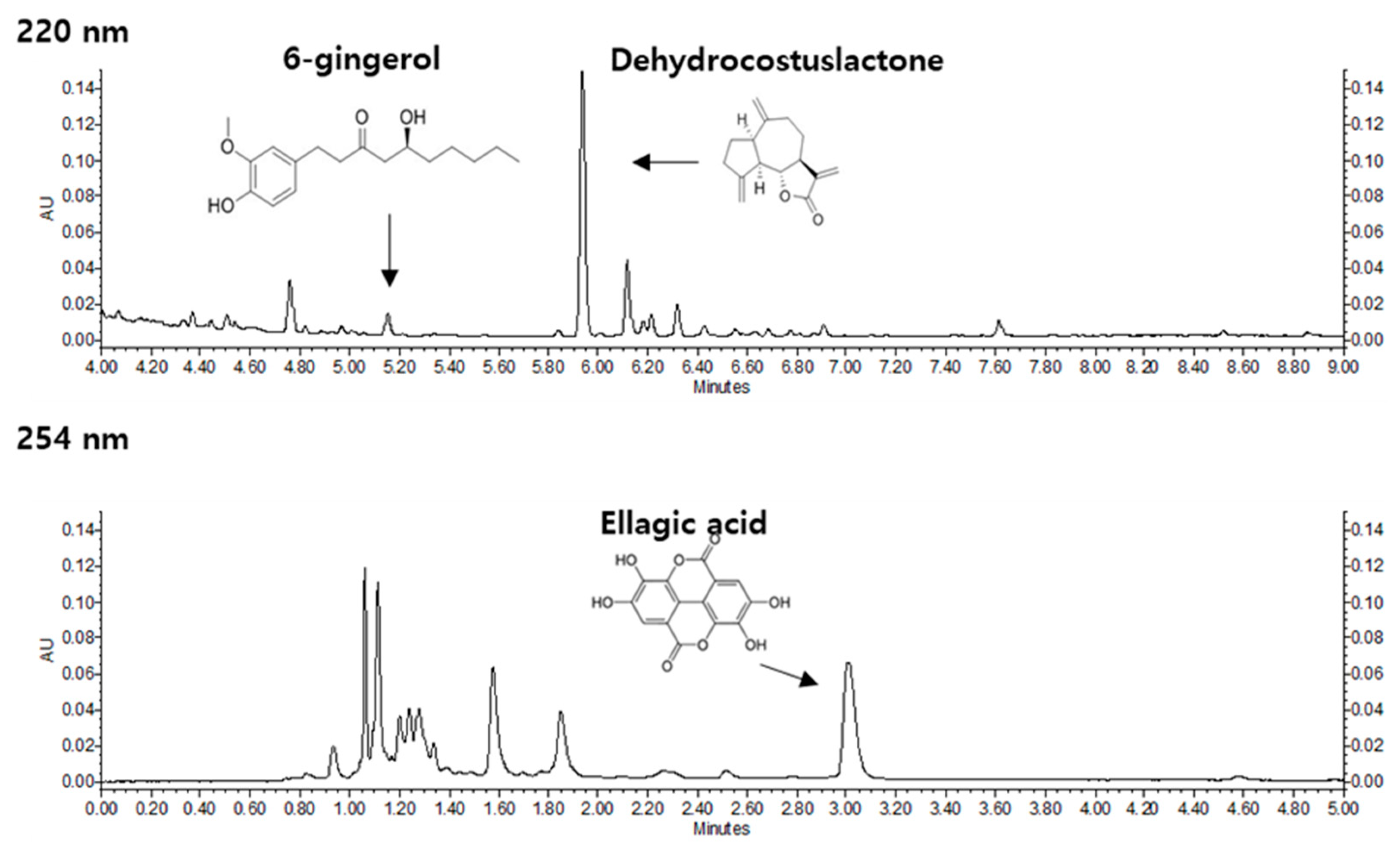
2.2. High performance liquid chromatography-ultraviolet/ diode array detection (HPLC-UV/DAD) Conditions

Quantitative analysis of KM1608 was performed using a Waters ultra performance liquid chromatography (UPLC) system and Waters Acquity UPLC HSS T3 column (100 mm × 2.1 mm, 1.8 μm) (Waters Co., Milford, MA, USA). The mobile phase consisted of water containing 0.1% phosphoric acid (A) and acetonitrile (B). The gradient elution was as follows: 0–4 min of 45% (B), 4–5 min of 45–50% (B), 5–9.9 min of 50–100% (B) for dehydrocostus lactone and 6-gingerol, 0–6 min of 12–14% (B), 6–7 min of 14–100% (B) for ellagic acid. Following gradient elution, the column was washed with 100% (B) for 7 min. The post-running time was 10 min after restoration of the initial condition. The mobile phase flow rate was 0.7 mL/min and the injection volume was 2 μL. Analytical method of HPLC was validated as described previously [23]

2.3. Network Pharmacological Analyses

After verifying active phytochemicals of KM1608 by HPLC analysis, we performed an in silico network pharmacological analysis to identify the targets of compounds and their potential pathways. Target genes of the active compounds were predicted from the Traditional Chinese Medicine Systems Pharmacology (TCMSP) database [24]. We retrieved annotated pathways from the Kyoto Encyclopedia Genes and Genomes (KEGG) database [25] using the target data. Then, the pathways were listed in descending order according to their combined score, leaving only the pathways satisfying the threshold (adjusted p-value ≤ 0.05 and combined score ≥ 30). We visualized a network graph using Cytoscape 3.5.1 [26] by collecting information on active compounds of KM1608, their targets, and related KEGG pathways.

2.4. Cell Culture

The RAW 264.7 murine macrophage cells were obtained from the American Type Culture Collection (ATCC, Rockville, MD, USA). The cells were cultured in Dulbecco’s Modified Eagle medium (DMEM) medium (Cellgro, Manassas, VA, USA) supplemented with 10% fetal bovine serum (Gibco BRL, Carlsbad, MD, USA) and 1% penicillin-streptomycin (Invitrogen, Grand Island, NY, USA). The cells were maintained at 37 °C in a humidified atmosphere of 5% CO2 and sub-cultured every two days.

2.5. Cell Viability Assay

RAW 264.7 cells were seeded in a 96-well plate at the density of 1 × 10⁵ cells/well and grown for 24 h. The next day, cells were treated with 0.5% dimethyl sulfoxide (DMSO) (Sigma-Aldrich, St. Louis, MO, USA), which served as a control, or KM1608 at the concentrations of 25, 50, and 100 µg/mL. After 24 h, 10 µL of EZ-Cytox reagent (DoGen, Seoul, Korea) was added to each well, followed by 30 min incubation. EZ-Cytox reagent contains a highly water-soluble tetrazolium salt, which is reduced by dehydrogenase activities in cells to give a yellow-color formazan dye. The amount of the formazan dye, generated by the activities of dehydrogenases in cells, is directly proportional to the number of living cells. Cell viability was determined through the change of absorbance measured at 450 nm using PowerWave XS microplate reader (Bio-Tek Instruments, Winooski, VT, USA) [27].

2.6. Nitric Oxide Assay

Nitric oxide production was evaluated indirectly by measuring the concentration of accumulated nitrite—the final inert product of nitric oxide metabolism—in the culture medium. RAW264.7 cells (1 × 10⁵ cells/well on 96-well plate) were treated with the phenol red-free medium containing 0.5% DMSO (control) or KM1608 at the indicated concentrations for 24 h. The cell culture supernatant was collected and mixed with Griess reagent (supplemented with 1% sulfanilamide, 5% phosphoric acid, and 0.1% N-(1-naphthyl)-ethylenediamine) at the ratio of 1:1 (v/v) [28]. After incubation at room temperature for 10 min, the absorbance of the mixture was measured at 450 nm with a microplate reader. The nitrite concentration in the supernatant was calculated from a sodium nitrite standard reference curve. Prednisolone (PDS) and 5-aminosalicylic acid (5-ASA) were used as reference drugs.

2.7. Quantitative Real-Time Reverse Transcription polymerase chain reaction (qRT-PCR)

The RAW 264.7 cells were harvested after 12 h of treatment with 0.5% DMSO (control) or KM1608 at the indicated concentrations. Total cellular RNA was isolated with TRIzol reagent (Invitrogen, Carlsbad, CA, USA) as per the manufacturer’s instructions. The isolated RNA was reverse transcribed into complementary DNA (cDNA) using the RevertAid First Strand cDNA Synthesis Kit (Thermo Fisher Scientific, Waltham, MA, USA). The reaction mixture was prepared by mixing cDNA with sense and antisense primers in PowerUp Green Master Mix (Thermo Fisher Scientific). The sense and antisense primers of respective genes which specific for murine are mentioned in Table 1. Polymerase chain reaction (PCR) was performed by QuantStudio 3 Real-Time PCR System (Thermo Fisher Scientific) under these conditions: denaturation at 95 °C for 10 min, followed by 45 amplification cycles at 95 °C for 3 s, and annealing at the appropriate temperature for 30 s. The relative gene expression levels were calculated using the ΔΔCq method with normalization to β-actin as the reference gene [29].

2.8. Western Blotting Analysis

The RAW 264.7 cells were seeded in 6-well plate at the density of 2 × 10⁶ cells/well for 24 h. Cells were treated with the presence or absence of KM1608 at the concentration of 100 µg/mL. After multiple time-points, cells were collected and washed with Dulbecco's phosphate-buffered saline (DPBS), before lysed in radioimmunoprecipitation assay (RIPA) buffer supplemented with 1X protease inhibitor cocktail and 1 mM sodium orthovanadate (Na3VO4) phosphatase inhibitor to get the whole-cell extracts according to the manufacturer’s instructions. Protein concentration of each whole-cell extracts was determined using the Pierce™ BCA Protein Assay Kit (Thermo Scientific). The equal protein amounts of each whole-cell extracts (10 μg/lane) were separated by electrophoresis in a 10% sodium dodecyl sulfate-polyacrylamide gel and blotted onto PVDF transfer membranes. Epitope-specific primary antibodies included phospho-c-Jun NH2-terminal kinase (p-JNK), phospho-extracellular signal–regulated kinase (p-ERK), and β-actin conjugated with secondary antibodies (Cell Signaling, Boston, MA, USA) were used to label the target proteins. The bound antibodies were detected by PierceTM ECL Advance Western Blotting Detection Reagents (Thermo Scientific) and visualized with FUSION Solo Chemiluminescence System (PEQLAB Biotechnologie GmbH, Germany).

2.9. Statistical Analysis

Data are presented as the mean ± standard error of the mean (SEM) from three independent experiments. Statistical analyses were determined using SigmaStat software with one-way analysis of variance (ANOVA), followed by Tukey’s test. A p-value < 0.05 was considered statistically significant.

3. Results

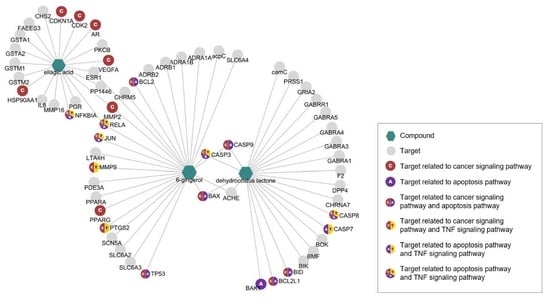
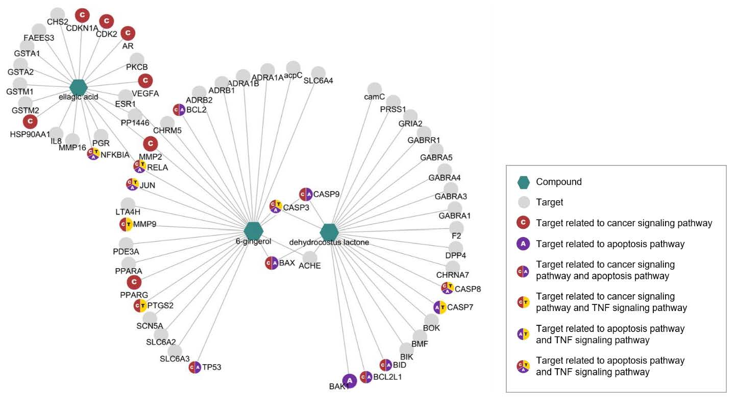
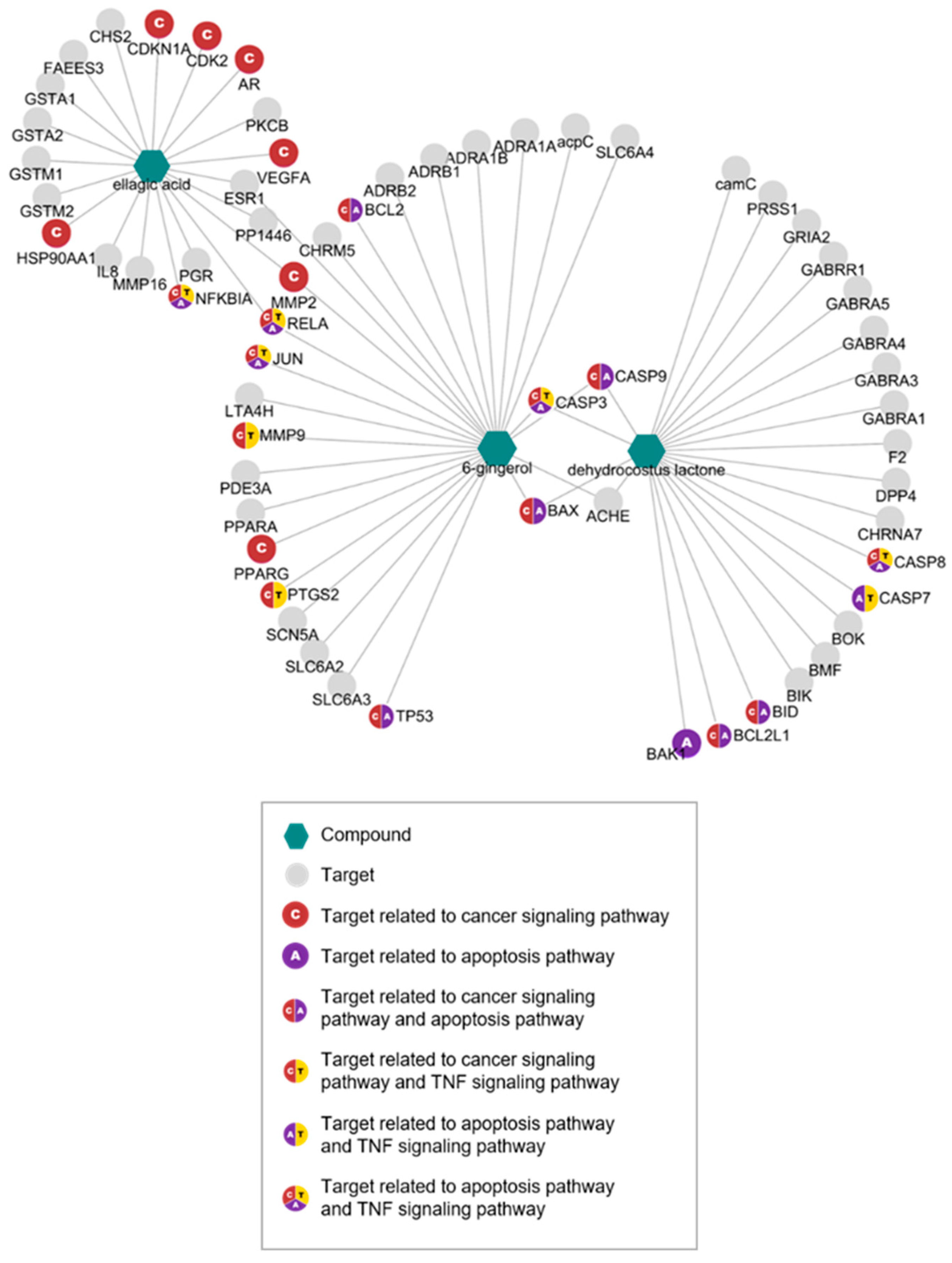
3.1. Network Pharmacological Analyses of KM1608 Compounds

The HPLC analysis showed a wide variety of phytochemicals in KM1608 (Figure 1). Among them, dehydrocostus lactone (DCL), ellagic acid (EA), and 6-gingerol (6G) in S. lappa, T. chebula, and Z. officinale, respectively, are active compounds possessing anti-inflammatory activity [30]. KM1608 contained 0.68–1.2%, 0.94–1.5%, and 0.1–0.2% of DCL, EA, and 6G, respectively.
To determine potential targets and their mechanism of action in the immune system, we performed network pharmacological analyses. We identified 62 targets of the active compounds of KM1608 using an in silico model from TCMSP (Table 2). Overlapped targets between the compounds were investigated: ESR1, PP1446, MMP2, and RELA overlapped in EA and 6G; CASP3, CASP9, BAX, and ACHE overlapped in 6G and DCL; no overlapped targets were found between DCL and EA. KEGG analysis was conducted to determine potential pathways of the targets (Table 3). Among the targets, 22 genes were involved in cancer, apoptosis, and TNF signaling pathways, all of which are related to the immune response (Figure 2).

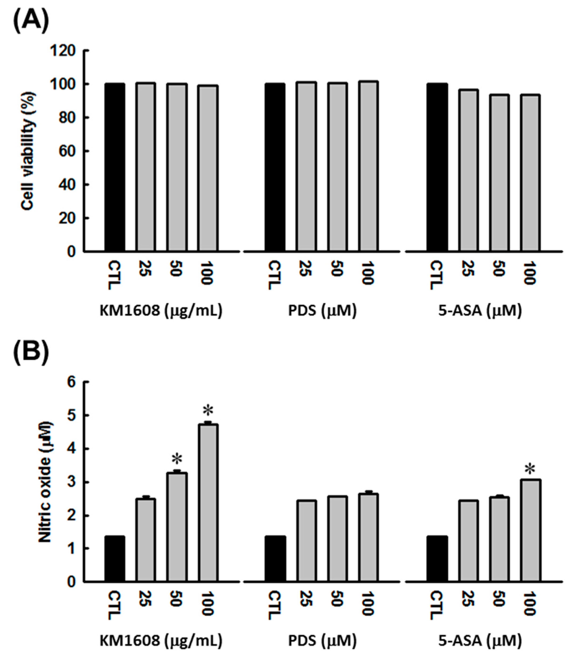
3.2. Stimulatory Effect of KM1608 on Nitric Oxide Production

KM1608 was evaluated for its cytotoxicity on RAW 264.7 macrophages using the cell viability assay. As shown in Figure 3a, treatment with KM1608, up to the concentration of 100 µg/mL, did not suppress cell proliferation. The stimulatory effect of KM1608 on nitric oxide production was investigated by measuring the concentration of accumulated nitrite, the stable product of nitric oxide metabolism, released in cell culture medium. After KM1608 treatment at concentrations of 50 and 100 µg/mL for 24 h, the concentration of accumulated nitrite was increased in a dose-dependent manner (Figure 3b). Prednisolone (PDS) and 5-aminosalicylic acid (5-ASA) were used as reference drugs.

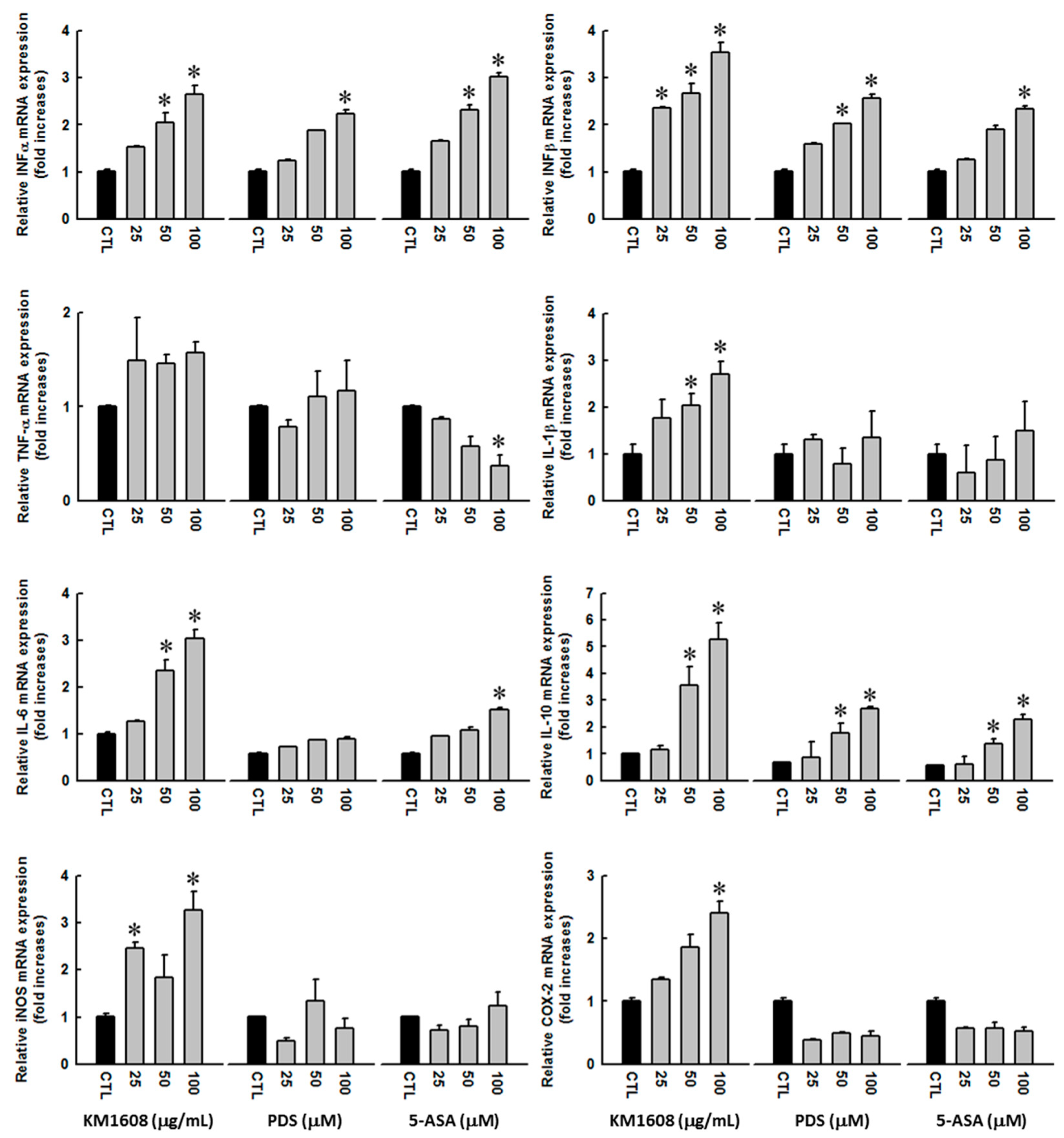
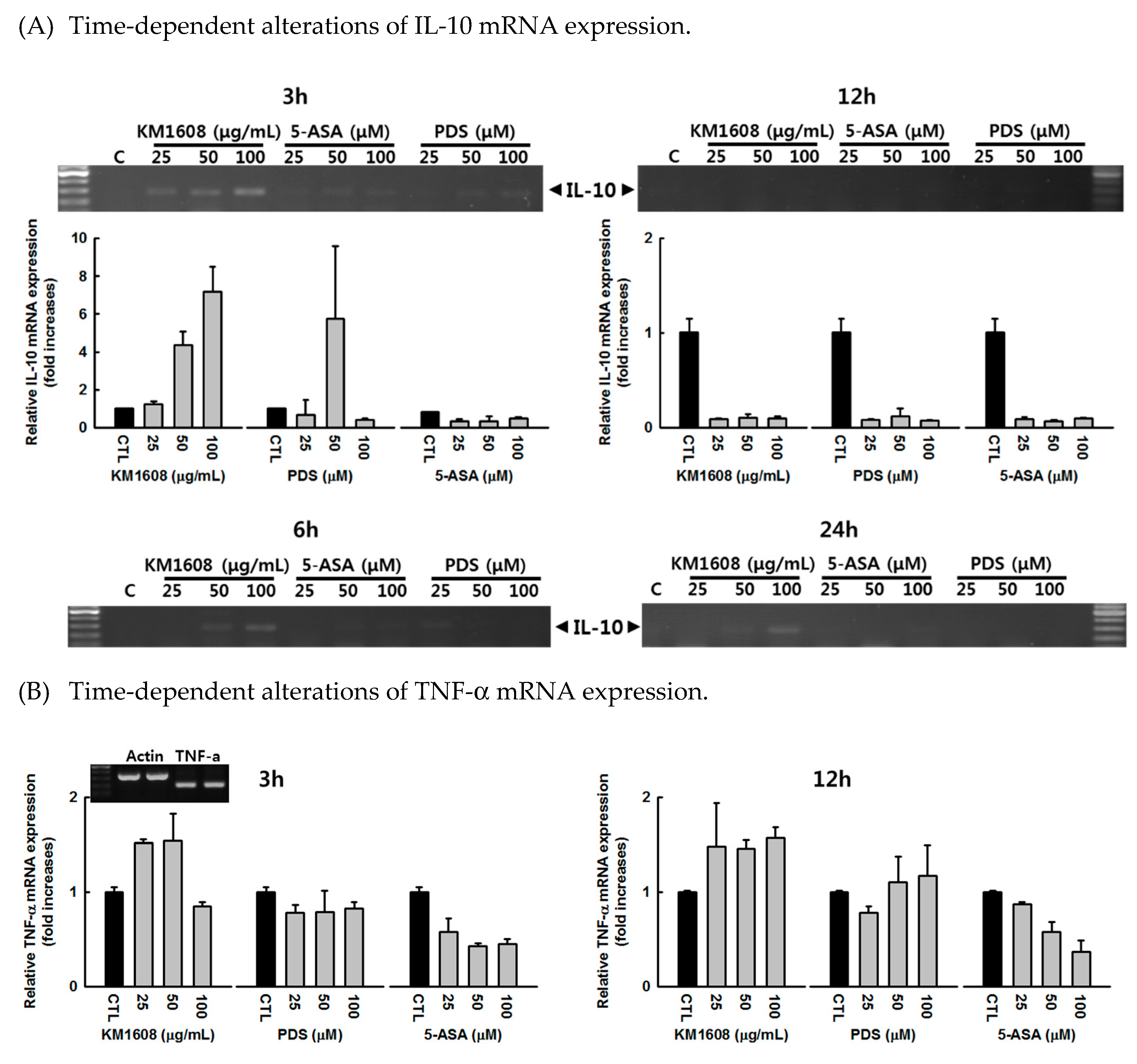
3.3. Effect of KM1608 on Cytokine Expression

An immune response is triggered and regulated by the participation of multiple cytokines at each stage. In this study, we performed qRT-PCR to evaluate the efficacy of KM1608 in stimulating the expression of selected cytokines in macrophage cells. After KM1608 treatment with the concentrations of 25–100 µg/mL for 12 h, the mRNA expression of interferons, interleukins, and pro-inflammatory cytokines in RAW 264.7 cells was significantly increased in a dose-dependent manner (Figure 4). The experimental conditions for gene changes were determined by analyzing various time-dependent alterations of mRNA expression through preliminary studies (Appendix A Figure A1(A)–(C)).

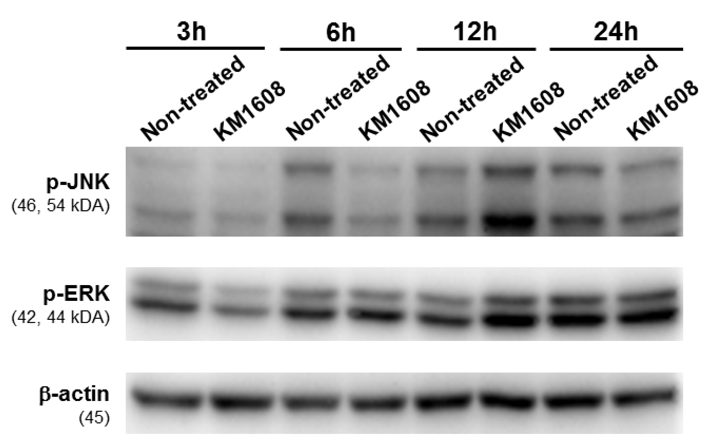
3.4. Effect of KM1608 on MAPKs Signaling Pathway

We evaluated further the protein expression of mitogen-activated protein kinases (MAPKs), which involve in the regulation cytokine production. As a result, 12 h after treatment with KM1608 at the concentration of 100 µg/mL, the protein expression of p-JNK and p-ERK was increased comparing with non-treated group (Figure 5).

4. Discussion

The usage of natural products to support the treatment of chronic diseases is becoming more popular, leading to an increase of studies about the pharmacological effects of medicinal plants as well as their natural compounds for screening the new potential candidates. In this present study, we built up the herbal formulation named KM1608 with the ingredient include the extracts from three medicinal plants S. lappa, T. chebula, and Z. officinale. These herbs have been used for a long time in Asian folk medicine and also separately studied for bioactivities of each one. Thus, we questioned the combination pharmacological effects of the herbal extracts in KM1608.
In order to orientate the in vitro assays, network pharmacological analyses of KM1608 compounds were performed. From the HPLC analysis results, we determined three main compounds in the ingredient of KM1608 were dehydrocostus lactone (DCL), ellagic acid (EA), and 6-gingerol (6G). Results from network pharmacological analysis showed that these compounds target in several signaling pathways such as cancer, apoptosis, hepatitis B, neuroactive ligand-receptor interaction, ALS, nicotine addiction, viral carcinogenesis, p53, and TNF. In comparison with previous studies, dehydrocostus lactone from S. lappa presented the anticancer effects on human leukemia HL-60 cells [4] and human cervical carcinoma HeLa cells [9]. Ellagic acid form T. chebula was reported to suppress the proliferation of human osteosarcoma HOS-1 malignant cells [31]. The cytotoxic effects of 6-gingerol from Z. officinale were also mentioned on cancer cell lines included human hematopoetic leukemia HL-60 cells, murine solid sarcoma S180 cells, and peripheral blood mononuclear PBMC cells [32].
By using an in silico model from TCMSP and KEGG analysis, we identified that these compounds had the potential to take part in the interaction with the immune response signaling pathways. Later experimental results also showed that KM1608 stimulated the nitric oxide production on RAW 264.7 macrophages. Nitric oxide is synthesized during immune and inflammatory responses of the host against infectious agents. Nitric oxide is used as a toxin for the self-defense of the host or acts as an immunoregulatory mediator that participates in both innate and adaptive immune responses [33,34]. Our results also showed that the synthesis of NO was slightly stimulated by treatment with prednisolone (PDS) and 5-aminosalicylic acid (5-ASA), even though they were used as anti-inflammatory drugs. In fact, some previous researches mentioned that NO releasing is capable to enhance the therapeutic effect of anti-inflammatory drugs. The NCX 1022, a derivative of hydrocortisone, prevented the recruitment of granulocytes to the site of inflammation in C57BL6 mice by releasing NO [35]. In another study, treatment with MyoNovin and isosorbide dinitrate, two nitric oxide donor drugs, improve the anti-inflammatory effect of prednisone on dystrophic mdx mice [36]. Prednisolone was also mentioned that suppressed the proliferation of SaOS2, an osteosarcoma cell line, by stimulating the release of NO through the upregulation of iNOS [37].
The KM1608 stimulated the expression of both IFN-α and -β, and exhibited a strong effect on IFN-β. IFN-α and -β belong to type-I interferons (IFN), which induce the innate immune response as well as activate the adaptive immunity against virus infection. Type-I IFNs participate in the immune response by preventing virus replication as well as inducing the antiviral activities of natural killer cells, dendritic cells, and monocytes. They also promote the IL-10 expression in macrophages. Type-I IFNs not only trigger the antiviral immune response, but also downregulate this response to limit the tissue damage [38]. In addition, type-I IFNs participate in the antitumor activity by promoting the adaptive immune response of CD8+ T-cells which controls tumor growth. When delivered to tumor sites via monocytes, IFN-α suppresses the tumor growth and metastasis. IFN-β conjugates with tumor-targeting monoclonal antibodies also induces the antitumor immune response through the activation of dendritic cells [3].
In addition, the mRNA expression of three interleukins including IL-1β, IL-6, and IL-10 was also enhanced upon KM1608 treatment. However, KM1608 did not induce the expression of tumor necrosis factor-α (TNF-α). Different from other cytokines, IL-1β is synthesized from a precursor and requires activation by inflammasome [39]. IL-1β controls the recruitment of neutrophils to the infection site to destroy the damaged tissue [40]. IL-6 plays multiple roles in the immune response, such as recruitment of mononuclear cells, stimulation of B-cells as well as endothelial cells, and inhibition of T-cell apoptosis [41]. In contrast to IL-1β and IL-6, IL-10 acts as an anti-inflammatory cytokine that regulates the immune response to minimize host damage. IL-10 inhibits the activation of T-cells, monocytes, and macrophages to limit and terminate the inflammation [42]. These results seem to be contradictory when KM1608 enhanced the expressions of both pro- and anti-inflammatory cytokines at the same time. However, each cytokine play its own role in the immune response, whether it stimulates the inflammatory process or not. Our results showed that KM1608 enhanced the mRNA expressions of IFN-α, -β, and IL-10, consistent with the previous studies that type-I IFNs stimulate the IL-10 expression in macrophages [38].
In addition, we assessed the mRNA expression of iNOS and COX-2 in RAW 264.7 cells upon KM1608 treatment. KM1608 slightly stimulated the expression of these pro-inflammatory modulators. The role of iNOS in the synthesis of nitric oxide is well known; iNOS catalyzes the conversion of L-arginine to L-citrulline and NO [43]. In this study, we also found that KM1608 induces NO production by macrophages. NO is an important for host defense against infectious organisms. Moreover, NO can induce the cell function as well as regulate the growth and death of many immune cells, such as macrophages, neutrophils, T-cells, and natural killer cells [33]. In contrast to iNOS, COX-2 participates in the immune response by stimulating the cyclooxygenase pathway that leads to the synthesis of PGE2 [44]. PGE2 elicits vasodilatation and increases blood flow that facilitates the recruitment of neutrophils and macrophages to the infection site. PGE2 also promotes the production of a few pro-inflammatory cytokines, such as IL-17 and IL-13 [45].
In the previously published paper of Shin et al. [30], the results showed that the anti-inflammatory activity of KM1608 caused by the significant inhibition of nitric oxide synthesis as well as suppressing the expressions of inflammatory mediators such as IL-6, MCP-1, and TNF-α in lipopolysaccharides (LPS)-treated RAW264.7 cells. With the same material, the extract of herbal formulation KM1608, our recent studies suggested that KM1608 itself stimulated the expression of immune cytokines liked IFN-α, -β, IL-1β, -6, IL-10, iNOS, and COX-2 in RAW264.7 macrophages. We evaluated further the protein expression of MAPKs, which involve in the regulation cytokine production [46]. KM1608 stimulated the time-dependent protein expressions of phospho-c-Jun NH2-terminal kinase (p-JNK) and phospho-extracellular signal–regulated kinase (p-ERK). This result suggested that KM1608 might contribute to the up-regulation of cytokine synthesis through the MAPKs signaling pathway.

5. Conclusions

Network pharmacological analysis results showed that major compounds of KM1608 exhibit anticancer potential via immune signaling pathways. Both innate and adaptive immune responses are controlled by multiple signaling pathways using numerous complement proteins, cytokines, inflammatory mediators, and antibodies. Thus, we chose the most popular cytokines to study the stimulatory effect of KM1608 extract on the immune response. KM1608 induced the mRNA expression of IFN-α, IFN-β, IL-1β, IL-6, IL-10, iNOS, and COX-2 in RAW 264.7 macrophages after treatment at 25–100 μg/mL for 12 h. This effect might be caused by the stimulation of MAPKs signaling pathway through the phosphorylation of JNK and ERK. KM1608 also stimulated the synthesis of nitric oxide, but did not cause cytotoxicity in RAW 264.7 at the same concentration. As a result, KM1608 can be considered a potential candidate for preventive medicine to increase body resistance and support cancer treatment.

Author Contributions

Conceive and design the experiments, T.A.T., B.A.K., and K.S.K.; perform experiments, J.P., J.H.O., D.L., J.S.P., C.E.K., H.-S.C., and S.-B.K.; analyze the data, G.S.H. and K.S.K; contribute for reagents/ materials/ analysis tools, B.A.K. and K.S.K.; write the paper, T.A.T. and K.S.K.; All authors have read and agreed to the published version of the manuscript.

Funding

The Ministry of SMEs and Startups (MSS), Korea, under the “Regional Specialized Industry Development Program (R0006060)” supervised by the Korea Institute for Advancement of Technology (KIAT), supported designing and performing the experiments, analyzing the data and writing the manuscript. The Gachon University research fund of 2018 (GCU-2018-0319) supported performing the experiments and analyzing the data.

Conflicts of Interest

The authors declare that they have no competing interests.

Appendix A

Figure A1. Time-dependent alterations of cytokine expression: (A) IL-10, (B) TNF-α, and (C) COX-2. RAW 264.7 cells were harvested at multiple time points after treatment with 0.5% DMSO (control) or KM1608 at the indicated concentrations. Total cellular RNA was isolated with TRIzol reagent and then reverse transcribed to cDNA. The reaction mixture was prepared by mixing cDNA with sense and antisense primers. PCR was performed using 45 amplification cycles. The relative gene expression levels were calculated by the ΔΔCq method. p < 0.05 compared with the 0.5% DMSO treated group. KM1608: herbal formulation KM1608 extract, PDS: prednisolone, ASA: 5-aminosalicylic acid.
Biomolecules 10 00424 g0a1aBiomolecules 10 00424 g0a1b

References

  1. Mak, T.W.; Saunders, M.E.; Jett, B.D. Innate Immunity. In Primer to the immune response, 2nd ed.; Elsevier Science Publishing Co. Inc.: New York, NY, USA, 2014; pp. 55–83. [Google Scholar]
  2. LeSourd, B. Nutritional factors and immunological ageing. Proc. Nutr. Soc. 2006, 65, 319–325. [Google Scholar] [CrossRef] [PubMed]
  3. Woo, S.-R.; Corrales, L.; Gajewski, T.F. Innate Immune Recognition of Cancer. Annu. Rev. Immunol. 2015, 33, 445–474. [Google Scholar] [CrossRef] [PubMed]
  4. Wang, Y.-F.; Ni, Z.-Y.; Dong, M.; Cong, B.; Shi, Q.-W.; Gu, Y.-C.; Kiyota, H. Secondary Metabolites of Plants from the Genus Saussurea: Chemistry and Biological Activity. Chem. Biodivers. 2010, 7, 2623–2659. [Google Scholar] [CrossRef] [PubMed]
  5. Bag, A.; Bhattacharyya, S.K.; Chattopadhyay, R.R.; Rashid, R.A. The development of Terminalia chebula Retz. (Combretaceae) in clinical research. Asian Pac. J. Trop. Biomed. 2013, 3, 244–252. [Google Scholar] [CrossRef] [Green Version]
  6. Ali, B.H.; Blunden, G.; Tanira, M.O.; Nemmar, A. Some phytochemical, pharmacological and toxicological properties of ginger (Zingiber officinale Roscoe): A review of recent research. Food Chem. Toxicol. 2008, 46, 409–420. [Google Scholar] [CrossRef]
  7. Moon, S.-M.; Yun, S.J.; Kook, J.-K.; Kim, H.-J.; Choi, M.S.; Park, B.R.; Kim, S.-G.; Kim, B.-O.; Lee, S.-Y.; Ahn, H.; et al. Anticancer activity of Saussurea lappa extract by apoptotic pathway in KB human oral cancer cells. Pharm. Boil. 2013, 51, 1372–1377. [Google Scholar] [CrossRef]
  8. Tian, X.; Song, H.S.; Cho, Y.M.; Park, B.; Song, Y.-J.; Jang, S.; Kang, S.C. Anticancer effect of Saussurea lappa extract via dual control of apoptosis and autophagy in prostate cancer cells. Med. 2017, 96, e7606. [Google Scholar] [CrossRef]
  9. Yang, M.; Zhang, J.; Li, Y.; Han, X.; Gao, K.; Fang, J. Bioassay-guided isolation of dehydrocostus lactone from Saussurea lappa: A new targeted cytosolic thioredoxin reductase anticancer agent. Arch. Biochem. Biophys. 2016, 607, 20–26. [Google Scholar] [CrossRef]
  10. Choodej, S.; Pudhom, K.; Mitsunaga, T. Inhibition of TNF-α-Induced Inflammation by Sesquiterpene Lactones from Saussurea lappa and Semi-Synthetic Analogues. Planta Medica 2017, 84, 329–335. [Google Scholar] [CrossRef] [Green Version]
  11. Tag, H.; Khaled, H.E.; Ismail, H.A.; El-Shenawy, N.S. Evaluation of anti-inflammatory potential of the ethanolic extract of the Saussurea lappa root (costus) on adjuvant-induced monoarthritis in rats. J. Basic Clin. Physiol. Pharmacol. 2016, 27. [Google Scholar] [CrossRef]
  12. Kim, M.-S.; Lee, D.Y.; Lee, J.; Kim, H.W.; Sung, S.H.; Han, J.-S.; Jeon, W.K. Terminalia chebula extract prevents scopolamine-induced amnesia via cholinergic modulation and anti-oxidative effects in mice. BMC Complement. Altern. Med. 2018, 18, 136. [Google Scholar] [CrossRef] [PubMed]
  13. Sheng, Z.; Zhao, J.; Muhammad, I.; Zhang, Y. Optimization of total phenolic content from Terminalia chebula Retz. fruits using response surface methodology and evaluation of their antioxidant activities. PLoS ONE 2018, 13, e0202368. [Google Scholar] [CrossRef] [PubMed]
  14. Kher, M.N.; Sheth, N.R.; Bhatt, V.D. In Vitro Antibacterial Evaluation of Terminalia chebula as an Alternative of Antibiotics against Bovine Subclinical Mastitis. Anim. Biotechnol. 2018, 30, 151–158. [Google Scholar] [CrossRef] [PubMed]
  15. Aher, V.D.; Kumar, A.; Wahi. Immunomodulatory effect of alcoholic extract of Terminalia chebula ripe fruits. J. Pharm. Sci. Res. 2010, 2, 539–544. [Google Scholar]
  16. Cock, I.E. The medicinal properties and phytochemistry of plants of the genus Terminalia (Combretaceae). Inflammopharmacology 2015, 23, 203–229. [Google Scholar] [CrossRef]
  17. Palatty, P.L.; Haniadka, R.; Valder, B.; Arora, R.; Baliga, M.S. Ginger in the Prevention of Nausea and Vomiting: A Review. Crit. Rev. Food Sci. Nutr. 2013, 53, 659–669. [Google Scholar] [CrossRef]
  18. Al-Amin, Z.M.; Thomson, M.; Al-Qattan, K.K.; Peltonen-Shalaby, R.; Ali, M. Anti-diabetic and hypolipidaemic properties of ginger (Zingiber officinale) in streptozotocin-induced diabetic rats. Br. J. Nutr. 2006, 96, 660–666. [Google Scholar] [CrossRef] [Green Version]
  19. Baliga, M.S.; Haniadka, R.; Pereira, M.M.; D’Souza, J.J.; Pallaty, P.L.; Bhat, H.P.; Popuri, S. Update on the Chemopreventive Effects of Ginger and its Phytochemicals. Crit. Rev. Food Sci. Nutr. 2011, 51, 499–523. [Google Scholar] [CrossRef]
  20. Kim, M.S.; Kim, J.Y. Ginger attenuates inflammation in a mouse model of dextran sulfate sodium-induced colitis. Food Sci. Biotechnol. 2018, 27, 1493–1501. [Google Scholar] [CrossRef]
  21. Kim, S.; Lee, M.S.; Jung, S.; Son, H.Y.; Park, S.; Kang, B.; Kim, S.Y.; Kim, I.H.; Kim, C.T.; Kim, Y. Ginger extract ameliorates obesity and inflammation via regulating microRNA-21/132 expression and AMPK activation in white adipose tissue. Nutrients 2018, 10, 1–12. [Google Scholar] [CrossRef] [Green Version]
  22. Srinivasan, K. Ginger rhizomes (Zingiber officinale): A spice with multiple health beneficial potentials. PharmaNutrition 2017, 5, 18–28. [Google Scholar] [CrossRef]
  23. Lee, J.; Choi, H.-S.; Lee, J.; Park, J.; Kim, S.-B.; Shin, M.-S.; Lee, S.; Hwang, G.S.; Koo, B.A.; Kang, K.S. Preparation of Herbal Formulation for Inflammatory Bowel Disease Based on In Vitro Screening and In Vivo Evaluation in a Mouse Model of Experimental Colitis. Molecules 2019, 24, 464. [Google Scholar] [CrossRef] [PubMed] [Green Version]
  24. Ru, J.; Li, P.; Wang, J.; Zhou, W.; Li, B.; Huang, C.; Li, P.; Guo, Z.; Tao, W.; Yang, Y.; et al. TCMSP: a database of systems pharmacology for drug discovery from herbal medicines. J. Chemin- 2014, 6, 13. [Google Scholar] [CrossRef] [PubMed] [Green Version]
  25. Kanehisa, M.; Furumichi, M.; Tanabe, M.; Sato, Y.; Morishima, K. KEGG: new perspectives on genomes, pathways, diseases and drugs. Nucleic Acids Res. 2016, 45, D353–D361. [Google Scholar] [CrossRef] [Green Version]
  26. Shannon, P.; Markiel, A.; Ozier, O.; Baliga, N.; Wang, J.T.; Ramage, D.; Amin, N.; Schwikowski, B.; Ideker, T. Cytoscape: A Software Environment for Integrated Models of Biomolecular Interaction Networks. Genome Res. 2003, 13, 2498–2504. [Google Scholar] [CrossRef]
  27. Kim, D.H.; Kim, D.W.; Jung, B.H.; Lee, J.H.; Lee, H.S.; Hwang, G.S.; Kang, K.S.; Lee, J.W. Ginsenoside Rb2 suppresses the glutamate-mediated oxidative stress and neuronal cell death in HT22 cells. J. Ginseng Res. 2019, 43, 326–334. [Google Scholar] [CrossRef]
  28. Lee, D.; Lee, D.S.; Jung, K.; Hwang, G.S.; Lee, H.L.; Yamabe, N.; Lee, H.J.; Eom, D.W.; Kim, K.H.; Kang, K.S. Protective effect of ginsenoside Rb1 against tacrolimus-induced apoptosis in renal proximal tubular LLC-PK1 cells. J. Ginseng Res. 2018, 42, 75–80. [Google Scholar] [CrossRef]
  29. Choi, S.Y.; Kim, J.H.; Quilantang, G.N.; Lee, S.H.; Cho, E.J. Acer okamotoanum inhibit the hydrogen peroxide-induced oxidative stress in C6 glial cells. Nat. Prod. Sci. 2018, 24, 148–154. [Google Scholar] [CrossRef]
  30. Shin, M.-S.; Kim, S.-B.; Lee, J.; Choi, H.-S.; Park, J.; Park, J.Y.; Lee, S.; Hwang, G.S.; Koo, B.A.; Kang, K.S. Beneficial Effect of Herbal Formulation KM1608 on Inflammatory Bowl Diseases: A Preliminary Experimental Study. Mol. 2018, 23, 2068. [Google Scholar] [CrossRef] [PubMed] [Green Version]
  31. Saleem, A.; Husheem, M.; Härkönen, P.; Pihlaja, K. Inhibition of cancer cell growth by crude extract and the phenolics of Terminalia chebula retz. fruit. J. Ethnopharmacol. 2002, 81, 327–336. [Google Scholar] [CrossRef]
  32. De Lima, R.M.T.; Dos Reis, A.C.; Santos, J.V.D.O.; Ferreira, J.R.D.O.; Braga, A.L.; Filho, J.W.G.D.O.; De Menezes, A.-A.P.M.; Da Mata, A.M.O.F.; De Alencar, M.V.O.B.; Rodrigues, D.C.D.N.; et al. Toxic, cytogenetic and antitumor evaluations of [6]-gingerol in non-clinical in vitro studies. Biomed. Pharmacother. 2019, 115, 108873. [Google Scholar] [CrossRef] [PubMed]
  33. Coleman, J.W. Nitric oxide in immunity and inflammation. Int. Immunopharmacol. 2001, 1, 1397–1406. [Google Scholar] [CrossRef]
  34. Roy, A.; Park, H.-J.; Jung, H.A.; Choi, J.S. Estragole exhibits anti-inflammatory activity with the regulation of NF-κB and Nrf-2 signaling pathways in LPS-induced RAW 264.7 cells. Nat. Prod. Sci 2018, 24, 13–20. [Google Scholar] [CrossRef] [Green Version]
  35. Hyun, E.; Bolla, M.; Steinhoff, M.; Wallace, J.L.; Del Soldato, P.; Vergnolle, N. Anti-inflammatory effects of nitric oxide-releasing hydrocortisone NCX 1022, in a murine model of contact dermatitis. Br. J. Pharmacol. 2004, 143, 618–625. [Google Scholar] [CrossRef] [PubMed] [Green Version]
  36. Mizunoya, W.; Upadhaya, R.; Burczynski, F.; Wang, G.; Anderson, J.E. Nitric oxide donors improve prednisone effects on muscular dystrophy in the mdx mouse diaphragm. Am. J. Physiol. Physiol. 2011, 300, C1065–C1077. [Google Scholar] [CrossRef] [PubMed] [Green Version]
  37. Cazzaniga, J.A.M.M.A. Prednisolone inhibits SaOS2 osteosarcoma cell proliferation by activating inducible nitric oxide synthase. World J. Transl. Med. 2016, 5, 53. [Google Scholar] [CrossRef]
  38. Lee, A.J.; Ashkar, A. The Dual Nature of Type I and Type II Interferons. Front. Immunol. 2018, 9. [Google Scholar] [CrossRef] [Green Version]
  39. Palazon-Riquelme, P.; Lopez-Castejon, G. The inflammasomes, immune guardians at defence barriers. Immunol. 2018, 155, 320–330. [Google Scholar] [CrossRef]
  40. Sims, J.; Smith, D.E. The IL-1 family: regulators of immunity. Nat. Rev. Immunol. 2010, 10, 89–102. [Google Scholar] [CrossRef]
  41. Rose-John, S. Interleukin-6 Family Cytokines. Cold Spring Harb. Perspect. Boil. 2017, 10, a028415. [Google Scholar] [CrossRef] [Green Version]
  42. Saraiva, M.; O’Garra, A. The regulation of IL-10 production by immune cells. Nat. Rev. Immunol. 2010, 10, 170–181. [Google Scholar] [CrossRef] [Green Version]
  43. Aktan, F. iNOS-mediated nitric oxide production and its regulation. Life Sci. 2004, 75, 639–653. [Google Scholar] [CrossRef] [PubMed]
  44. Kawahara, K.; Hohjoh, H.; Inazumi, T.; Tsuchiya, S.; Sugimoto, Y. Prostaglandin E2-induced inflammation: Relevance of prostaglandin E receptors. Biochim. Biophys. Acta (BBA) - Mol. Cell Boil. Lipids 2015, 1851, 414–421. [Google Scholar] [CrossRef] [PubMed]
  45. Nakanishi, M.; Rosenberg, D.W. Multifaceted roles of PGE2 in inflammation and cancer. Semin. Immunopathol. 2012, 35, 123–137. [Google Scholar] [CrossRef]
  46. Gadina, M.; Gazaniga, N.; Vian, L.; Furumoto, Y. Small molecules to the rescue: Inhibition of cytokine signaling in immune-mediated diseases. J. Autoimmun. 2017, 85, 20–31. [Google Scholar] [CrossRef] [PubMed]
Figure 1. The HPLC profiles of KM1608. The peaks of 6-gingerol and dehydrocostus lactone at 220 nm, and ellagic acid at 254 nm in KM1608 were compared with those of their respective standard compounds.
Biomolecules 10 00424 g001
Figure 2. Compound-target gene network of active compounds of KM1608. Each node represents compound or target (explained in the box) and the edge represents the interaction between the compound and the target.
Biomolecules 10 00424 g002
Figure 3. Stimulatory effect of KM1608 on nitric oxide production. (A) KM1608 cytotoxicity on RAW 264.7 cells. (B) Stimulatory effect of KM1608 on nitric oxide production in macrophages. RAW 264.7 cells were treated with phenol red-free medium containing 0.5% DMSO (control) or KM1608 at the indicated concentrations for 24 h. The cell culture supernatant was collected and mixed with Griess reagent at a ratio of 1:1 (v/v) and the absorbance was measured at 450 nm with a microplate reader. The concentration of nitrite, the stable product of nitric oxide metabolism, was calculated from a sodium nitrite standard reference curve. The experiments were conducted triplicate. p < 0.05 compared with the 0.5% DMSO treated group. KM1608: herbal formulation KM1608 extract, PDS: prednisolone, ASA: 5-aminosalicylic acid.
Biomolecules 10 00424 g003
Figure 4. Effect of KM1608 on the expression of cytokines. RAW 264.7 cells were harvested 12 h post treatment with 0.5% DMSO (control) or KM1608 at the indicated concentrations. Total cellular RNA was isolated with TRIzol reagent and then reverse transcribed to cDNA. The reaction mixture was prepared by mixing cDNA with sense and antisense primers. PCR was performed using 45 amplification cycles. The relative gene expression levels were calculated by the ΔΔCq method. p < 0.05 compared with the 0.5% DMSO treated group. KM1608: herbal formulation KM1608 extract, PDS: prednisolone, ASA: 5-aminosalicylic acid.
Biomolecules 10 00424 g004
Figure 5. Western blot analysis for the time-dependent expression of mitogen-activated protein kinases (MAPKs) in RAW 264.7 macrophages after treated with KM1608.
Biomolecules 10 00424 g005
Table 1. List of sense and antisense primers for quantitative real-time reverse transcription polymerase chain reaction (qRT-PCR).
GenesSense (5′→3′)Antisense (5′→3′)
IFN-αCCTGTGTGATGCAGGAACCTCACCTCCCAGGCACAGA
IFN-βACTAGAGGAAAAGCAAGAGGACTGGTAAGTCTTCGAATGATG
TNF-αATAGCTCCCAGAAAAGCAAGCCACCCCGAAGTTCAGTAGACA
iNOSCATGCTACTGGAGGTGGGTGCATTGATCTCCGTGACAGCC
COX-2TCTGGAACATTGTGAACAACATCAAGCTCCTTATTTCCCTTCACAC
IL-1βACCTGCTGGTGTGTGACGTTTCGTTGCTTGGTTCTCCTTG
IL-6TGGAGTCACAGAAGGAGTGGCTAAGTCTGACCACAGTGAGGAATGTCCAC
IL-10GTGAAGACTTTCTTTCAAACAAAGCTGCTCCACTGCCTTGCTCTTATT
β-actinTCACCCACACTGTGCCCATCTACGAGGATGCCACAGGATTCCATACCCA
(IFN: interferon, TNF-α: tumor necrosis factor-α, iNOS: inducible nitric oxide synthase, and COX-2: cyclooxygenase-2, IL: interleukin,).
Table 2. Predicted or verified target genes of the active compounds of KM1608.
Targets of 6-gingerol (6G)
Gene NameProtein Name
ACHEAcetylcholinesterase
acpCBeta-lactamase
ADRA1AAlpha-1A adrenergic receptor
ADRA1BAlpha-1B adrenergic receptor
ADRB1Beta-1 adrenergic receptor
ADRB2Beta-2 adrenergic receptor
BAXApoptosis regulator BAX
BCL2Apoptosis regulator Bcl-2
CASP3Caspase-3
CASP9Caspase-9
CHRM5Muscarinic acetylcholine receptor M5
ESR1Estrogen receptor
JUNTranscription factor AP-1
LTA4HLeukotriene A-4 hydrolase
MMP272-kDa type IV collagenase
MMP9Matrix metalloproteinase-9
PDE3ACGMP-inhibited 3’,5’-cyclic phosphodiesterase A
PPARABaculoviral IAP repeat-containing protein 5
PPARGPeroxisome proliferator-activated receptor gamma
PTGS2Prostaglandin G/H synthase 2
RELATranscription factor p65
SCN5ASodium channel protein type 5 subunit alpha
SLC6A2Sodium-dependent noradrenaline transporter
SLC6A3Sodium-dependent dopamine transporter
SLC6A4Sodium-dependent serotonin transporter
TP53Cellular tumor antigen p53
Targets of Dehydrocostus Lactone (DCL)
Gene NameProtein Name
ACHEAcetylcholinesterase
BAK1Bcl-2 homologous antagonist/killer
BAXApoptosis regulator BAX
BCL2L1Bcl-2-like protein 1
BIDBH3-interacting domain death agonist
BIKBcl-2-interacting killer
BMFBcl-2-modifying factor
BOKBcl-2-related ovarian killer protein
camCCytochrome P450-cam
CASP3Caspase-3
CASP7Caspase-7
CASP8Caspase-8
CASP9Caspase-9
CHRNA7Neuronal acetylcholine receptor protein, alpha-7 chain
DPP4Dipeptidyl peptidase IV
F2Thrombin
GABRA1Gamma-aminobutyric acid receptor subunit alpha-1
GABRA3Gamma-aminobutyric-acid receptor subunit alpha-3
GABRA4Gamma-aminobutyric-acid receptor subunit alpha-4
GABRA5Gamma-aminobutyric-acid receptor subunit alpha-5
GABRR1Gamma-aminobutyric-acid receptor subunit rho-1
GRIA2Glutamate receptor 2
PRSS1Trypsin-1
Targets of Ellagic Acid (EA)
Gene NameProtein Name
ARAndrogen receptor
CDK2Cell division protein kinase 2
CDKN1ACyclin-dependent kinase inhibitor 1
CHS2Chitin synthase 2
ESR1Estrogen receptor
FAEES3Glutathione S-transferase P
GSTA1Glutathione S-transferase A1
GSTA2Glutathione S-transferase A2
GSTM1Glutathione S-transferase Mu 1
GSTM2Glutathione S-transferase Mu 2
HSP90AA1Heat shock protein (HSP) 90
IL8Interleukin-8
MMP16Matrix metalloproteinase-16
MMP272-kDa type IV collagenase
NFKBIANuclear factor kappa B inhibitor alpha
PGRProgesterone receptor
PKCBProtein kinase C beta type
PP1446Insulin-like growth factor II
RELATranscription factor p65
VEGFAVascular endothelial growth factor A
Table 3. Result of Kyoto Encyclopedia Genes and Genomes (KEGG) pathway analysis on active compounds of KM1608 (adjusted p-value ≤ 0.05, combined score ≥ 30). indicates the pathway relevant to immune responses.
KEGG Pathway
(Homo sapiens)
Adjusted
p-value
Combine
d Score
Related Genes (Targets)
Cancer 3.22 × 10−1790.44868JUN; CDKN1A; HSP90AA1; MMP2; PTGS2; MMP9; RELA; VEGFA; NFKBIA; CASP9; AR; CASP8; CASP3; CDK2; BCL2; BAX; PPARG; BID; TP53; BCL2L1
Apoptosis 2.28 × 10−1468.93754JUN; RELA; NFKBIA; CASP9; CASP7; CASP8; CASP3; BCL2; BAX; BAK1; BID; TP53; BCL2L1
Hepatitis B1.02 × 10−1259.21799NFKBIA; CASP9; JUN; CDKN1A; CASP8; CASP3; CDK2; BCL2; BAX; TP53; MMP9; RELA
Neuroactive ligand-receptor interaction3.7 × 10−1254.32502GABRA1; GRIA2; PRSS1; GABRA5; GABRA4; CHRNA7; GABRA3; CHRM5; ADRB1; ADRB2; F2; ADRA1B; ADRA1A; GABRR1
Prostate cancer1.69 × 10−1046.99271NFKBIA; CASP9; AR; HSP90AA1; CDKN1A; CDK2; BCL2; TP53; RELA
Amyotrophic lateral sclerosis (ALS)8.21 × 10−1144.6843CASP9; GRIA2; CASP3; BCL2; BAX; BID; TP53; BCL2L1
Nicotine addiction6.39 × 10−1037.8611GABRA1; GRIA2; GABRR1; GABRA5; GABRA4; CHRNA7; GABRA3
Viral carcinogenesis9.31 × 10−936.97465NFKBIA; JUN; CDKN1A; CASP8; CASP3; CDK2; BAX; BAK1; TP53; RELA
p53 signaling pathway6.45 × 10−1036.67846CASP9; CDKN1A; CASP8; CASP3; CDK2; BAX; BID; TP53
Small cell lung cancer3.49 × 10−936.25028NFKBIA; CASP9; CDK2; BCL2; PTGS2; TP53; RELA; BCL2L1
TNF signaling pathway 2.08 × 10−833.21595NFKBIA; JUN; CASP7; CASP8; CASP3; PTGS2; MMP9; RELA

