Article

Cycle-Inhibiting Factor Is Associated with Burkholderia pseudomallei Invasion in Human Neuronal Cells

by
Amporn Rungruengkitkun
1,
Niramol Jitprasutwit
2,3,
Watcharamat Muangkaew
1,
Chantira Suttikornchai
4,
Sarunporn Tandhavanant
1,
Nitaya Indrawattana
1,
Sumate Ampawong
5,
Passanesh Sukphopetch
1,
Narisara Chantratita
1,6 and
Pornpan Pumirat
1,*
1
Department of Microbiology and Immunology, Faculty of Tropical Medicine, Mahidol University, Bangkok 10400, Thailand
2
Center for Vaccine Development, Institute of Molecular Biosciences, Mahidol University, Nakhon Pathom 73170, Thailand
3
Department of Immunology, Faculty of Medicine Siriraj Hospital, Mahidol University, Bangkok 10700, Thailand
4
Department of Protozoology, Faculty of Tropical Medicine, Mahidol University, Bangkok 10400, Thailand
5
Department of Tropical Pathology, Faculty of Tropical Medicine, Mahidol University, Bangkok 10400, Thailand
6
Mahidol-Oxford Tropical Medicine Research Unit, Faculty of Tropical Medicine, Mahidol University, Bangkok 10400, Thailand
*
Author to whom correspondence should be addressed.
Biology 2022, 11(10), 1439; https://doi.org/10.3390/biology11101439
Submission received: 16 August 2022 / Revised: 20 September 2022 / Accepted: 28 September 2022 / Published: 1 October 2022

Abstract

:

Simple Summary

Neurological melioidosis, caused by Burkholderia pseudomallei, can lead to the development of severe symptoms associated with the nervous system. However, the pathogenic mechanism through which this bacterium infects neuronal cells has not been studied. This study showed that B. pseudomallei can infect human neuronal SH-SY5Y cells in vitro. Cycle-inhibiting factor (Cif), a type III secreted effector, is one of the virulence factors produced by B. pseudomallei upon infection. The B. pseudomallei cif-deleted mutant reduced the ability to invade neuronal cells compared with the parental strain. Our finding indicates that Cif is associated with B. pseudomallei invasion of human neuronal cells.

Abstract

Burkholderia pseudomallei is a pathogenic bacterium that causes human melioidosis, which is associated with a high mortality rate. However, the underlying mechanisms of B. pseudomallei pathogenesis are largely unknown. In this study, we examined the infection of human neuronal SH-Sy5y cells by several clinically relevant B. pseudomallei strains. We found that all tested B. pseudomallei strains can invade SH-Sy5y cells, undergo intracellular replication, cause actin-tail formation, and form multinucleated giant cells. Additionally, a deletion mutant of B. pseudomallei cycle-inhibiting factor (cif) was constructed that exhibited reduced invasion in SH-Sy5y cells. Complementation of cif restored invasion of the B. pseudomallei cif-deleted mutant. Our findings enhance understanding of B. pseudomallei pathogenicity in terms of the virulence factor Cif and demonstrate the function of Cif in neurological melioidosis. This may eventually lead to the discovery of novel targets for treatment and a strategy to control the disease.

1. Introduction

Melioidosis is an infectious disease caused by B. pseudomallei, a gram-negative facultative intracellular bacillus. Melioidosis is endemic in Northern Australia and Southeast Asia, including in Myanmar, Malaysia, Singapore, Vietnam, Cambodia, Laos, and Thailand [1,2]. Furthermore, the predicted global distribution of melioidosis indicates that it is probably endemic in countries that have never reported the disease [3]. The worldwide incidence of melioidosis is approximately 165,000 cases per year, whilst the annual death rate is estimated to be approximately 89,000 [3]. Melioidosis is acquired by inhalation, inoculation, or ingestion of B. pseudomallei [4,5,6]. B. pseudomallei is a natural inhabitant of soil, stagnant water, and rice paddies where the disease is endemic. This bacterium is a category B bioterrorism agent due to its infection potential and high virulence [7]. The most common clinical symptoms are pneumonia (51%), genitourinary symptoms (14%), skin lesion (13%), and central nervous system (CNS) or neurological melioidosis (5%) [8,9].
Although neurological melioidosis is a rare condition, it is often fatal, with a mortality rate of approximately 25% [9]. Neurological abnormalities of melioidosis generally present with brain abscesses and encephalitis [10]. Symptoms of meningoencephalitis involving the brainstem, cerebellum, and spinal cord are also observed [9]. Although patients can recover entirely, 13% of patients suffer chronic neurological disability [11]. Most neurological melioidosis cases have been described in Australia [9]. In Thailand, 3% of melioidosis cases were neurological melioidosis [12]. Sporadic cases of neurological melioidosis are also reported in Norway, Taiwan, and Singapore [13,14,15]. These data show that neurological melioidosis and CNS damage caused by B. pseudomallei is a serious concern.
Upon infection, an essential feature of pathogenic B. pseudomallei is its ability to invade several cell types and to stimulate various host-cell responses [16,17]. After internalization, B. pseudomallei can escape the membrane-bound phagosome to enter the cytoplasm [18]. Once inside cytosol, this pathogen has evolved the ability to exploit host actin, harness actin-based motility for intra- and inter-cellular movement, and to induce cell-to-cell fusion, resulting in multinucleated giant cell (MNGC) formation [19]. This phenotype is critical for evading host defense mechanisms, including antimicrobial agents and immune response events [20]. This unique ability is also observed in the tissues of patients with melioidosis [21].
Several virulence factors of B. pseudomallei that contribute to pathogenesis have been identified, including both cell-associated and secreted products. Many Gram-negative pathogens, including B. pseudomallei, also deploy type III secretion systems (T3SSs) [22]. The T3SSs are molecular syringes/needles that inject bacterial virulence proteins directly into host cells. These injected effectors subvert host cell processes and contribute to disease [23]. In this study, we investigated a T3SS translocated effector molecule that inhibits host cell cycle progression, which is called the cycle-inhibiting factor (Cif). Enteropathogenic and enterohaemorrhagic Escherichia coli (EPEC and EHEC) exploit this protein to block cell cycle G2/M transition, induce stress fiber formation, and provoke a delayed cell death [24,25].
In B. pseudomallei, this virulence factor, which is also known as CHBP (Cif homolog in B. pseudomallei), is absent from genomes of closely related B. thailandensis, which usually do not cause human melioidosis [26]. The determination of the crystal structure showed that B. pseudomallei Cif possess a papain-like fold with a Cys-His-Gln catalytic triad similar to EPEC Cif [27,28]. An analysis of the T3SSs revealed that the Burkholderia secretion apparatus (Bsa) T3SS is required for the secretion and delivery of Cif into the host cells [29]. This effector is expressed only in intra-host cell conditions and is detected in B. pseudomallei-infected cells [29]. This is consistent with previous reports that B. pseudomallei Cif is injected into eukaryotic cells by the T3SS of EPEC and EHEC [26,27,28]. In addition, a study of the B. pseudomallei protein microarray probed with melioidosis patient sera identified 170 reactive antigens, including Cif, indicating that Cif is expressed in vivo and might be involved in B. pseudomallei pathogenesis [30].
The modus operandi of Cif is known to deamidate Nedd8, an ubiquitin-like protein, which causes the inhibition of Cullin E3 ubiquitin ligases (CRL), and consequently induces cell cycle arrest [31,32,33]. B. pseudomallei Cif can function as a potent activator of MAPK/ERK signaling and has CRL independent effects to counter the pro-apoptotic effects [34]. However, whether Cif employs additional roles on host cells that are crucial for bacterial pathogenesis is currently poorly understood. One study reported that Cif exerts a bimodal effect on host NF-κB signaling and bacterial replication [35]. The authors demonstrated that HEK293T cells transiently transfected with Cif increase B. thailandensis intracellular bacterial load, compared with the control cells transfected with the empty vector [35]. This could be linked to the consequence of disrupting the B. pseudomallei cif gene, causing a significant reduction in cytotoxicity and plaque formation in HeLa human epithelial cells infected by B. pseudomallei, as demonstrated in our previous study [29]. Therefore, Cif is likely to be important during B. pseudomallei infection.
To better understand the pathogenesis of B. pseudomallei during neurological infection, we used a cell-based model. We used the human neuroblastoma SH-SY5Y cell line to examine the pathogenic ability of three clinical wild-type B. pseudomallei strains, including the reference strain K96243, isolated from patients presenting with melioidosis in northeast Thailand. Furthermore, we postulated that Cif facilitates B. pseudomallei pathogenesis of human neuronal cells; therefore, we compared the parental strain with a cif-deleted mutant in SH-SY5Y cells, focusing on invasion, intracellular replication, actin-tail formation, and MNGC formation. As a result, we showed that Cif contributes to B. pseudomallei invasion of neuronal cells. Our in vitro model can be used to investigate the impact of other bacterial factors of virulence that contribute to the pathogenesis of neurological melioidosis by B. pseudomallei.

2. Materials and Methods

2.1. Ethics Statement

All experiments and methods were performed per relevant guidelines and regulations. This project has been approved by the ethics committee of the Faculty of Tropical Medicine, Mahidol University, Bangkok, Thailand (Reference No: MUTM 2018–057-01).

2.2. Bacterial Strains, Cell Lines, and Growth Conditions

Three B. pseudomallei clinical isolates (576, 1530, and the reference strain K96243), which were obtained from three patients presenting with melioidosis to Sappasithiprasong Hospital, northeastern Thailand, were used as the wild-type strains [36]. Two B. pseudomallei mutant strains (cif-deletion mutant and complemented strains) were constructed in this study as described below. Escherichia coli strain DH5α and RHO3 were used for cloning and generation of B. pseudomallei mutant strains. All B. pseudomallei strains and E. coli DH5α were grown in Luria-Bertani (LB) medium at 37 °C. E. coli RHO3 was grown on LB agar supplemented with 400 μg/mL 2,6-diaminopimelic acid (DAP).
The human neuronal cell line, SH-SY5Y (ATCC® CRL-2266™), was kindly provided by Natthanej Luplertlop [37]. SH-SY5Y cells were maintained at 37 °C in a humidity-controlled incubator with 5% CO2 in Dulbecco’s modified Eagle’s medium (DMEM; Gibco BRL) supplemented with 10% (v/v) heat-inactivated fetal bovine serum (FBS; Gibco BRL) and penicillin-streptomycin solution (Gibco BRL). The cell culture medium was replaced with fresh medium every other day. When the cells reached approximately 90% confluence, a 0.25% (w/v) trypsin-EDTA solution was added after washing with PBS to harvest the cells for passaging.

2.3. Construction of B. pseudomallei Cif-Deleted Mutant and the Complemented Strain

The markerless allele replacement method by pEKM5 suicide vector [38] was used for deletion mutagenesis and complementation of the cif gene. A sequence of B. pseudomallei K96243 cif gene from GenBank (locus_tag = “BPSS1385”) was used in primer design by Primer-BLAST “http://www.ncbi.nlm.nih.gov/tools/primer-blast (accessed on 21 October 2020)”. Primer information is provided in Table 1. To generate the deletion mutant, the 5′ upstream and 3′ downstream fragments of the cif gene were amplified and subjected to overlap extension PCR using BPSS1385-F1 and BPSS1385-R2. The length of PCR amplicon with a deletion in the region on cif was 1053 bp. This overlapped PCR was ligated into pGEM®-T Easy (Promega, Madison, WI, USA) and then transformed into E. coli DH5α. The desired plasmid was validated by PCR. Additionally, the amplified PCR product was gel extracted and the deletion of cif was validated by DNA sequencing. The scarless knockout cassette containing a deletion in the cif gene was sub-cloned into the non-replicative plasmid, pEXKm5 [38], and transformed into E. coli RHO3 and delivered to the host B. pseudomallei K96243 by conjugation. LB agar containing 1000 µg/mL kanamycin and supplemented with 5-bromo-4-chloro-3-indolyl-β-D-glucuronide (X-Gluc) at a final concentration of 50 µg/mL (Promega) were used for selection of the conjugants. The obtained clones were then confirmed by PCR using primers flanking the mutant allele (BPSS1385-F1 and BPSS1385-R2).
After that, the positive conjugants were streaked onto yeast extract tryptone (YT) agar (Yeast Extract & Tryptone, BD; Agar, Oxoid) containing 15% (w/v) sucrose and 50 µg/mL X-Gluc (YT-sucrose-X-Gluc plates), and incubated at 25 °C for 72 hrs. The resultant colonies were purified by re-streaking on YT-sucrose-X-Gluc plates. For complementation, the same pEXKm5-based allele exchange approach was utilized. The PCR amplicon (1639 bp) containing wild-type B. pseudomallei cif sequence was generated by BPSS1385-F1 and BPSS1385-R2 primers. Similarly, the full-length of cif was cloned into pEXKm5 and transformed into E. coli RHO3 for conjugation with the B. pseudomallei cif-deleted mutant. Sucrose selection was employed for merodiploid resolution, resulting in the generation of wild-type sequences and strains that maintained the deletion alleles.
The result of deletion and complementation of cif was validated using PCR and immunoblotting. Amplification was carried out using the mutant deletion allele flanking primers (BPSS1385-F1 and BPSS1385-R2) and the primers that were designed to target the oriT region, ensuring that the oriT on pEXKm5 plasmid backbone sequences were absent [39]. Furthermore, the successful construction of the cif-deleted mutant and the complemented stains were indicated by immunoblotting using antibodies against Cif protein [29]. Whole cell lysates prepared from B. pseudomallei strains were extracted and tested as previously described [29].

2.4. Invasion and Intracellular Replication Assay

Human SH-SY5Y cells were seeded at a density of 5 × 10⁴ cells per well in a 24-well cell culture plate. The next day, the medium was removed and replaced with 200 µL of fresh antibiotic-free DMEM. Overnight cultures of B. pseudomallei strains were adjusted to 1 × 10⁶ cells per ml by OD measurement at 600 nm and used to infect the cells at a multiplicity of infection (MOI) of 1 to 100. After 2-h co-culturing, the infected cells were washed twice with PBS, and then 500 µL of fresh DMEM containing 250 µg/mL kanamycin (Sigma) was added and incubated at 37 °C for 1 h to eliminate any extracellular bacteria. To recover the invading bacteria, B. pseudomallei-infected SH-SY5Y cells were then washed thrice with PBS before cell lysis with 100 µL of 0.1% (w/v) Triton X-100. The number of viable bacteria was determined as colony forming units (CFUs) by performing a serial dilution. Ten microliters of each dilution were dropped onto LB agar plates and incubated at 37 °C for 24–48 h. At 4, 6, 8, and 10 h post-infection, the intracellular bacteria were recovered as described above to assess intracellular replication of B. pseudomallei strains in human SH-SY5Y cells.

2.5. Investigation of Actin-Tail Formation

One day before infection, SH-SY5Y cells were plated on 22 × 22 mm square glass coverslips (Menzel-Glaser) in a 6-well plate (Costar, Corning, NY, USA) and incubated at 37 °C in a humidified 5% CO2 atmosphere. The overnight B. pseudomallei culture was subjected to infection by the SH-SY5Y cells at an MOI of 20. After killing the extracellular bacteria as previously described, actin tail formation was observed at 6 h post-infection. The infected cells were washed with PBS twice and then fixed with 4% (v/v) paraformaldehyde in PBS at room temperature overnight. The fixed cells were washed with PBS before permeabilization with 0.5% (v/v) Triton X-100 in PBS. After 30 min of incubation, 1% (w/v) bovine serum albumin (Sigma-Aldrich, St. Louis, MO, USA) in PBS was added and incubated for 30 min at room temperature. Subsequently, bacteria were stained using a mouse monoclonal anti-B. pseudomallei lipopolysaccharide antibody (Camlab, Cambridge, United Kingdom) followed by Alexa Fluor488-conjugated anti-mouse Immunoglobulin (Molecular Probes, Eugene, OR, USA). Actin filaments and DNA were stained using Alexa Fluor555-conjugated phalloidin (Molecular Probes) and 4′,6′diamidine-2′-phenylindole dihydrochloride (DAPI; Molecular Probes), respectively. Actin-tail formation was examined in 100 fields by confocal laser scanning microscopy (LSM 700; Carl Zeiss, Jena, Germany).

2.6. Determination of MNGC Formation

At 10 h post-infection, SH-SY5Y cells infected with B. pseudomallei strains were stained with Giemsa (Merck, Darmstadt, Germany) as described previously [40]. MNGC formation, which was defined by at least three nuclei in a cell, was evaluated in 100 fields of view using an Olympus BX41 microscope. The percentage of MNGC formation was determined using the following formula: (number of nuclei in a multinucleated giant cell/total number of nuclei counted) × 100. A minimum of 1000 nuclei were counted per experiment.

2.7. Statistical Analysis

All assays were conducted in triplicate, and an unpaired t-test of three independent experiments was performed using the GraphPad Prism 8 program (STATCON). Results were considered significant at a p value ≤ 0.05.

3. Results

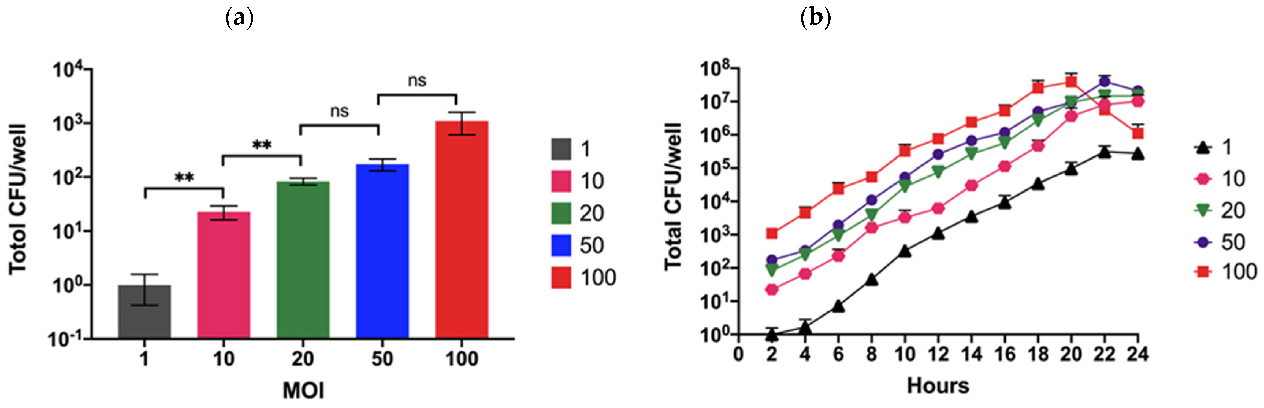
3.1. Optimal B. pseudomallei MOI for Human Neuronal Cell Infection

We observed B. pseudomallei in human neuronal SH-SY5Y cells using three clinical isolates (K96243, 576a, and 1530). We also generated B. pseudomallei K96243 cif-deletion and complemented mutants using a pEXKm5-based allele replacement system (Figure S1). The presence of the cif gene and Cif protein were validated by PCR and immunoblotting, respectively. A cif was confirmed to be absent from the B. pseudomallei K96243 cif-deleted mutant and the presence of cif in the complemented strain was as expected (Figure S2). To characterize and compare the B. pseudomallei strains in human neurons, in vitro growth curves of these strains in culture medium were analyzed. All strains showed the same rate of growth, indicating that the degree of fitness was similar among these strains (Figure S3).
The understanding of B. pseudomallei pathogenesis in neuronal cells is poor, although data from patient cases with neurological symptoms have accumulated. Here, we used the human neuronal SH-SY5Y cell line to investigate the process of B. pseudomallei infection in vitro. The effect of bacterial concentration on cell invasion was determined by co-culturing the reference K96243 strain at a multiplicity of infections (MOI) of 0.1, 0.01, 1, 10, 20, 50, and 100. At a very low MOI of 0.1 and 0.01, the number of bacteria that had invaded cells at 2 h post-infection could not be detected, although internalized bacteria could be recovered 6–8 h after infection (Figure S4). The number of invading bacteria increased with higher MOI, and the number of internalized bacteria significantly increased when using an MOI of 20 (Figure 1a).
However, there was no significant difference between MOI of 20 and 50 and 50 and 100. Moreover, the replication rate of B. pseudomallei in SH-SY5Y cells was similar regardless of the number of bacteria added (Figure 1b). It was notable that the number of intracellular bacteria decreased 20 h post-infection when using an MOI of 100. A possible reason for this was that the cells became damaged and released the intracellular bacteria that were then killed in the cell culture medium containing the antibiotic. Moreover, the infected cells may detach from the cell culture plate, leading to a lower number of bacteria recovered from the infected cells. We chose the MOI that gave the maximum number of recovered intracellular bacteria at 2 h post-infection. As a result, an MOI of 20 was chosen for subsequent investigations.

3.2. Invasion of Human Neuronal Cells by B. pseudomallei Strains

The ability of B. pseudomallei strains to invade human neuronal cells was examined at 3 h post-infection by comparing the percentage of intracellular bacteria relative to the number of bacteria added to the cells. The percentage invasion efficiency of B. pseudomallei reference strain K96243 was 0.014 ± 0.005%, which was significantly lower than those of strains 576a and 1530, at 0.291 ± 0.092% and 0.388±0.082%, respectively (Figure 2a). A contribution of Cif to the invasion of neuronal cells was observed. As shown in Figure 2b, deletion of the cif gene caused a significant reduction in invasion efficiency (0.027 ± 0.003%) compared with the wild-type (0.050 ± 0.006%), and complementation of the cif mutation fully restored the ability of the cif-deleted mutant to invade neuronal cells (0.067 ± 0.017%). These results indicate that Cif is an important factor in the B. pseudomallei invasion of neuronal cells.

3.3. Intracellular Replication of B. pseudomallei in Human Neuronal Cells

After invasion, we tested the ability of B. pseudomallei to multiply in SH-SY5Y cells. Intracellular bacteria were recovered by plating on culture medium plates at 4, 6, 8, and 10 hours post-infection. This clearly showed that the number of intracellular bacteria of all the clinical strains increased continuously over time (Figure 3a). We found that the cif-deleted mutant survived over time but to a lesser extent than the other strains (Figure 3a). This could result from a lower number of cif-deleted mutants that were initially internalized into cells. The growth rates of bacteria were calculated and showed that all B. pseudomallei strains replicated similarly in SH-SY5Y cells, with an average doubling time of 68 ± 6 min (Figure 3b). Although the doubling time of the cif mutant was approximately 97 ± 15 min, there was no significant difference among strains. This indicates that Cif might not be involved in B. pseudomallei intracellular replication.

3.4. B. pseudomallei Can Induce Actin-Tail and MNGC Formation in Human Neuronal Cells

B. pseudomallei is recognized by its ability to induce actin rearrangement that is initiated at one pole of the bacterium tail for intra- and inter-cellular movement. We, therefore, observed actin-tail formation by B. pseudomallei strains at 6 h post-infection by confocal analysis. All strains formed actin tails in the neuronal cells with a typical comet-tail phenotype (Figure 4). B. pseudomallei exploits actin-based motility for intra- and inter-cellular movement, leading to cell fusion for intracellular survival without exposure to antimicrobial agents or antibodies outside the cells. We detected MNGC formation induced by B. pseudomallei at 8 h post-infection (Figure 5a). At 10 h post-infection, we quantified the number of MNGCs and found that the percentages of MNGCs formed by B. pseudomallei 576a and 1530 were significantly greater than that of the reference K96243 strain (Figure 5b).
Compared with the wild-type strain, the cif-deleted mutant caused similar defective MNGC formation in SH-SY5Y cells (Figure 5b). However, this could be the effect of a lower number of bacteria that survived in the cells at this time (Figure S5). It was possible that the number of MNGCs induced by bacteria lacking cif corresponded with the number of intracellular bacteria in SH-SY5Y cells.

3.5. Plaque Formation in Human Neuronal Cells

Following cell fusion, Burkholderia spp. spread to adjacent cells, forming plaques as clear zones representing MNGC death [41]. We have shown that all B. pseudomallei strains in this study induced MNGCs in human neuronal cells to varying degrees. To assess the virulence of the B. pseudomallei strains in SH-SY5Y cells, we determined the number of plaques formed at 22 h post-infection as the last stage of the in vitro infection. Plaque formation induced by K96243, 576a, and 1530 strains was indistinguishable from each other (Figure 6a). Only the cif-deleted mutant of B. pseudomallei K96243 showed a defect in plaque formation (Figure 6a).
The number of plaques formed by each strain was assessed and is shown in Figure 6B. There was no significant difference between the number of plaques formed by the wild-type strains; K96243 was 65 ± 6, 576a was 74 ± 5, and 1530 was 70 ± 8. The number of plaques formed by the cif-deleted mutant was, however, significantly reduced at 20 ± 6. This defective phenotype was recovered when complemented with the cif gene; the number of plaques formed by the complemented strain was 66 ± 7. Similar to the number of MNGCs formed by the cif-deleted mutant, a decreased number of plaques could reflect the lower number of cif-deleted mutant bacteria in cells at 10 h post-infection (Figure S5). This indicated that Cif might not play a role in B. pseudomallei dissemination among human neuronal cells.

4. Discussion

Upon infection of a human host, B. pseudomallei can invade many systems, including the central and peripheral nervous systems [9,42]. Previous studies in mice show a pathway for B. pseudomallei into the CNS, the brain stem, and spinal cord via the olfactory and trigeminal nerves [43,44]. However, the underlying mechanism of B. pseudomallei pathogenesis and neuronal infection remains poorly understood. In the present study, we examined the B. pseudomallei pathogenic process in human neuronal cells. Based on co-culture assays with different B. pseudomallei strains and the immortalized bone marrow-derived SH-SY5Y neuroblastoma cell line, the result showed that all tested B. pseudomallei wild-type strains harbor the bacterial abilities of invasion, intracellular replication, and formation of actin-tails, MNGCs, and plaques.
Few studies have used neuronal cells to investigate the pathogenesis of bacterial pathogens. Borrelia burgdorferi, a bacterium that causes Lyme disease, induces apoptosis of SH-SY5Y cells when surrounded by microglia, the resident macrophage cells of the CNS [45]. However, B. burgdorferi is not toxic to neurons; instead, surrounding inflammation produced by microglia in response to B. burgdorferi causes the SH-SY5Y cells to die [45]. For B. pseudomallei, primary neuron cells have been used in in vitro assays [46], and B. pseudomallei MSHR520, a clinical isolate from an Australian patient, can infect glial cells isolated from the olfactory and trigeminal nerves [46].
Several B. pseudomallei virulence factors facilitate successful infection. Here, we focused on the Bsa T3SS virulence factor, Cif. We used a B. pseudomallei cif-deleted mutant that was defective in the cif gene to investigate B. pseudomallei infection of human neurons. Although insertional mutagenesis is a popular approach to mutate or disrupt a target gene, a cassette or residual scar is left behind that might affect the transcription of downstream genes (polar effect). Therefore, the cif-deleted mutant was constructed by a gene deletion technique.
Based on invasion efficiency, Cif appears to play an important role in B. pseudomallei invasion. The cif-deleted mutant had reduced ability to invade SH-SY5Y cells, consistent with the role of Bsa T3SS in the penetration of B. pseudomallei in other cell types, including epithelial cells, skin fibroblasts, and macrophages [16,40,47]. A bopE mutant of B. pseudomallei (defective in producing the BopE effector of Bsa T3SS) impairs entry into HeLa cells [48]. Moreover, a B. pseudomallei bsaQ mutant (defective in producing a structural component of Bsa T3SS) demonstrates reduced ability in invasion into non-phagocytic cells [49]. In addition to B. pseudomallei, T3SS is required for invasion by many other bacteria, such as Salmonella spp. and Shigella spp. [50]. Although the role of Cif in bacterial invasion remains unclear, our finding could be related to Cif interfering with host cell functions. Like other pathogenic bacteria, B. pseudomallei have developed sophisticated arsenals of virulence factors that hijack the host ubiquitin to their own benefit during infection [51]. Previous studies discovered that Cif, specifically deamidated ubiquitin-like protein Nedd8, resulted in the inhibition of CRL, leading to the induction of cell cycle arrest [31,32,33], but not only host cell cycle arrest. Protein ubiquitination also plays crucial roles in controlling eukaryotic cell homeostasis and pathogenesis of neoplastic, infectious, and neurodegenerative diseases [51]. B. pseudomallei Cif has been shown to modify host central signaling pathways by activating MAPK/ERK signaling to induce the phosphorylation of the pro-apoptotic protein Bim (Bcl-2 interacting mediator of cell death), and potentially leading to a proapototic effect [34]. Additionally, Cif was shown to increase basal NF-κB activity and stimulate the intracellular replication of the B. thailandensis [35]. Thus, it is possible that B. pseudomallei Cif is delivered directly into host cells through a Bsa T3SS where it modulates host cell function that promotes bacterial invasion. On the other hand, Cif expression may function coordinately with other bacterial effectors to promote its invasion. Further investigation is needed to clarify these points.
B. pseudomallei is able to replicate and survive inside mammalian host cells [52]. During survival inside the host cell, B. pseudomallei Bsa T3SS is triggered to deliver effector proteins [53]. B. pseudomallei Bsa T3SS is vital for B. pseudomallei to evade the endocytic vesicle and survive in the cytoplasm of J774.2 murine macrophage-like cells [54]. We previously found that a cif mutation did not affect the capability of B. pseudomallei to survive in skin fibroblasts [40]. Similarly, we found that a B. pseudomallei cif-deleted mutant did not exhibit defective intracellular replication in human neuronal cells. These results contradict a previous study that showed the direct involvement of B. pseudomallei Cif in B. thailandensis intracellular replication in HEK293T cells—although the effect was modest—but not on invasion [35]. A possible explanation for this could be that Cif is not present in B. thailandensis and the level of expression of B. pseudomallei Cif by transfection of Cif plasmid in HEK293T cells might affect the proliferation of B. thailandensis.
This study demonstrated that B. pseudomallei induces MNGC formation in a human neuronal cell line to different degrees depending on the strains. MNGC formation is also dependent on the cell type infected. For example, MNGC formation by B. pseudomallei strain MSHR520 in Schwann cells of trigeminal nerves is greater than that in olfactory ensheathing cells [46]. The B. pseudomallei cif-deleted mutant showed reduced MNGC formation in neuronal cells, despite demonstrating efficient actin tail formation comparable to that of the wild-type strain. This phenotype was consistent with the small number of plaques formed by B. pseudomallei-infected neuron cells. This finding was also consistent with the lower number of intracellular bacteria in this cell type. It is likely that induction of MNGC in SH-SY5Y cells is linked to the ability of intracellular bacterial replication and plaque formation. However, MNGC formation in RAW 264.7 cells, a murine macrophage-like cell line, by B. pseudomallei MSHR520 is not connected to the capacity for intracellular replication [55]. Defective plaque formation of T6SS-1 mutants was observed, though these mutants were able to replicate with a high number of intracellular growth [41]. Thus, the role of B. pseudomallei Cif in MNGC and plaque formation in neuronal cells requires further investigation.
To date, only intracellular motility A (BimA) has been shown to play an essential role in CNS invasion and infection [44]. Additionally, an allele of Burkholderia BimA significantly impacts the clinical presentation and outcome of patients with CNS melioidosis [56]. B. pseudomallei MSHR668 is a clinical isolate with documented central nervous system infection and harbors the B. mallei-like bimA allele that is believed to be associated with the development and severity of CNS melioidosis [57,58]. However, other virulence factors that contribute to the complications of CNS melioidosis need to be explored. For example, lipopolysaccharide is likely to be associated with pathogenicity and clinical presentation in humans [59]. B. pseudomallei K96243 displays type A lipopolysaccharide, while B. pseudomallei 576a displays type B [60].

5. Conclusions

We showed that B. pseudomallei can invade human neuronal cells, multiply, form actin tails, and induce MNGCs. Cif is a virulence factor of B. pseudomallei during the process of neuronal invasion. These findings provide new insight into B. pseudomallei infection of neuronal cells that will inform the prevention and treatment of neurological melioidosis.

Supplementary Materials

The following supporting information can be downloaded at: https://www.mdpi.com/article/10.3390/biology11101439/s1, Figure S1: Construction of B. pseudomallei cif-deleted mutant and a complemented strain; Figure S2: PCR -Western Blot; Figure S3: The growth curve in LB medium culture of B. pseudomallei strains used in this study; Figure S4: Intracellular replication of B. pseudomallei K96243 in SH-SY5Y cells at MOI of 0.1 and 0.01; Figure S5: The number of intracellular B. pseudomallei at 10 h post-infection in the SH-SY5Y cells.

Author Contributions

Conceptualization, A.R., N.J., N.I. and P.P.; funding acquisition, A.R. and P.P.; methodology, A.R., N.J., W.M., C.S., S.T. and P.P.; software, S.A., P.S., N.C. and P.P.; formal analysis, A.R., N.J., N.I. and P.P.; investigation, A.R., N.J., W.M., C.S. and P.P.; resources, S.A., P.S., N.C. and P.P.; data curation, A.R. and P.P.; writing—original draft preparation, A.R., N.J. and P.P.; writing—review and editing, N.J. and P.P. visualization, N.J. and P.P.; supervision, P.P.; project administration, A.R. and P.P. All authors have read and agreed to the published version of the manuscript.

Funding

This research was funded by the National Research Council of Thailand, grant number NRCT5-RSA63015-14; and the research grant by the Faculty of Tropical Medicine, Mahidol University, the Fiscal year 2018.

Institutional Review Board Statement

The study was conducted in accordance with the Declaration of Helsinki, and approved by the Ethics Committee of the Faculty of Tropical Medicine, Mahidol University, Bangkok, Thailand (Reference No: MUTM 2018–057-01).

Informed Consent Statement

Not applicable.

Data Availability Statement

The data presented in this study are available on request from the corresponding author.

Acknowledgments

Authors are thankful to Department of Microbiology and Immunology, Central laboratory and the Office of Research Services, Faculty of Tropical Medicine for supporting the publication of this article.

Conflicts of Interest

The authors declare no conflict of interest. The funders had no role in the design of the study; in the collection, analyses, or interpretation of data; in the writing of the manuscript; or in the decision to publish the results.

References

  1. Currie, B.J.; Fisher, D.A.; Howard, D.M.; Burrow, J.N.; Selvanayagam, S.; Snelling, P.L.; Anstey, N.M.; Mayo, M.J. The Epidemiology of Melioidosis in Australia and Papua New Guinea. Acta Trop. 2000, 74, 121–127. [Google Scholar] [CrossRef]
  2. Leelarasamee, A. Melioidosis in Southeast Asia. Acta Trop. 2000, 74, 129–132. [Google Scholar] [CrossRef]
  3. Limmathurotsakul, D.; Golding, N.; Dance, D.A.; Messina, J.P.; Pigott, D.M.; Moyes, C.L.; Rolim, D.B.; Bertherat, E.; Day, N.P.; Peacock, S.J.; et al. Predicted Global Distribution of Burkholderia pseudomallei and Burden of Melioidosis. Nat. Microbiol. 2016, 1, 15008. [Google Scholar] [CrossRef] [PubMed] [Green Version]
  4. Chen, P.S.; Chen, Y.S.; Lin, H.H.; Liu, P.J.; Ni, W.F.; Hsueh, P.T.; Liang, S.H.; Chen, C.; Chen, Y.L. Airborne Transmission of Melioidosis to Humans from Environmental Aerosols Contaminated with B. pseudomallei. PLoS Negl. Trop. Dis. 2015, 9, e0003834. [Google Scholar] [CrossRef]
  5. Limmathurotsakul, D.; Wongsuvan, G.; Aanensen, D.; Ngamwilai, S.; Saiprom, N.; Rongkard, P.; Thaipadungpanit, J.; Kanoksil, M.; Chantratita, N.; Day, N.P.; et al. Melioidosis Caused by Burkholderia pseudomallei in Drinking Water, Thailand, 2012. Emerg. Infect. Dis. 2014, 20, 265–268. [Google Scholar] [CrossRef]
  6. Limmathurotsakul, D.; Kanoksil, M.; Wuthiekanun, V.; Kitphati, R.; deStavola, B.; Day, N.P.; Peacock, S.J. Activities of Daily Living Associated with Acquisition of Melioidosis in Northeast Thailand: A Matched Case-Control Study. PLoS Negl. Trop. Dis. 2013, 7, e2072. [Google Scholar] [CrossRef] [Green Version]
  7. Larsen, J.C.; Johnson, N.H. Pathogenesis of Burkholderia pseudomallei and Burkholderia mallei. Mil. Med. 2009, 174, 647–651. [Google Scholar] [CrossRef] [Green Version]
  8. Currie, B.J.; Ward, L.; Cheng, A.C. The Epidemiology and Clinical Spectrum of Melioidosis: 540 Cases from the 20 year Darwin Prospective Study. PLoS Negl. Trop. Dis. 2010, 4, e900. [Google Scholar] [CrossRef] [Green Version]
  9. Currie, B.J.; Fisher, D.A.; Howard, D.M.; Burrow, J.N. Neurological Melioidosis. Acta Trop. 2000, 74, 145–151. [Google Scholar] [CrossRef]
  10. Muthusamy, K.A.; Waran, V.; Puthucheary, S.D. Spectra of Central Nervous System Melioidosis. J. Clin. Neurosci. 2007, 14, 1213–1215. [Google Scholar] [CrossRef]
  11. Wongwandee, M.; Linasmita, P. Central Nervous System Melioidosis: A Systematic Review of Individual Participant Data of Case Reports and Case Series. PLoS Negl. Trop. Dis. 2019, 13, e0007320. [Google Scholar] [CrossRef] [PubMed] [Green Version]
  12. Punyagupta, S. Review of 686 Cases and Presentation of a New Clinical Classification. In Melioidosis; Punyagupta, S., Sirisanthana, T., Stapatayavong, B., Eds.; Bangkok Medical Publisher: Bangkok, Thailand, 1989. [Google Scholar]
  13. Hesstvedt, L.; Reikvam, D.H.; Dunlop, O. Neurological Melioidosis in Norway Presenting with a Cerebral Abscess. IDCases 2015, 2, 16–18. [Google Scholar] [CrossRef] [PubMed] [Green Version]
  14. Liang, C.C.; Chen, S.Y.; Chen, T.Y.; Chen, S.T. Central Nervous System Melioidosis Mimics Malignancy: A Case Report and Literature Review. World Neurosurg. 2016, 89, 732.e19–732.e23. [Google Scholar] [CrossRef] [PubMed]
  15. Chadwick, D.R.; Ang, B.; Sitoh, Y.Y.; Lee, C.C. Cerebral Melioidosis in Singapore: A Review of Five Cases. Trans. R. Soc. Trop. Med. Hyg. 2002, 96, 72–76. [Google Scholar] [CrossRef]
  16. Jones, A.L.; Beveridge, T.J.; Woods, D.E. Intracellular Survival of Burkholderia pseudomallei. Infect. Immun. 1996, 64, 782–790. [Google Scholar] [CrossRef] [Green Version]
  17. Duangurai, T.; Indrawattana, N.; Pumirat, P. Burkholderia pseudomallei Adaptation for Survival in Stressful Conditions. BioMed Res. Int. 2018, 2018, 3039106. [Google Scholar] [CrossRef] [Green Version]
  18. Wiersinga, W.J.; van der Poll, T.; White, N.J.; Day, N.P.; Peacock, S.J. Melioidosis: Insights into the Pathogenicity of Burkholderia pseudomallei. Nat. Rev. Microbiol. 2006, 4, 272–282. [Google Scholar] [CrossRef]
  19. Kespichayawattana, W.; Rattanachetkul, S.; Wanun, T.; Utaisincharoen, P.; Sirisinha, S. Burkholderia pseudomallei Induces Cell Fusion and Actin-Associated Membrane Protrusion: A Possible Mechanism for Cell-to-Cell Spreading. Infect. Immun. 2000, 68, 5377–5384. [Google Scholar] [CrossRef] [Green Version]
  20. Gan, Y.H. Interaction between Burkholderia pseudomallei and the Host Immune Response: Sleeping with the Enemy? J. Infect. Dis. 2005, 192, 1845–1850. [Google Scholar] [CrossRef] [Green Version]
  21. Wong, K.T.; Puthucheary, S.D.; Vadivelu, J. The Histopathology of Human Melioidosis. Histopathology 1995, 26, 51–55. [Google Scholar] [CrossRef]
  22. Hueck, C.J. Type III Protein Secretion Systems in Bacterial Pathogens of Animals and Plants. Microbiol. Mol. Biol. Rev. 1998, 62, 379–433. [Google Scholar] [CrossRef] [PubMed] [Green Version]
  23. Vander Broek, C.W.; Stevens, J.M. Type III Secretion in the Melioidosis Pathogen Burkholderia pseudomallei. Front. Cell. Infect. Microbiol. 2017, 7, 255. [Google Scholar] [CrossRef] [PubMed] [Green Version]
  24. Marches, O.; Ledger, T.N.; Boury, M.; Ohara, M.; Tu, X.; Goffaux, F.; Mainil, J.; Rosenshine, I.; Sugai, M.; De Rycke, J.; et al. Enteropathogenic and Enterohaemorrhagic Escherichia coli Deliver a Novel Effector Called Cif, Which Blocks Cell Cycle G2/M Transition. Mol. Microbiol. 2003, 50, 1553–1567. [Google Scholar] [CrossRef] [PubMed]
  25. Samba-Louaka, A.; Nougayrede, J.P.; Watrin, C.; Oswald, E.; Taieb, F. The Enteropathogenic Escherichia coli Effector Cif Induces Delayed Apoptosis in Epithelial Cells. Infect. Immun. 2009, 77, 5471–5477. [Google Scholar] [CrossRef] [PubMed] [Green Version]
  26. Yao, Q.; Cui, J.; Zhu, Y.; Wang, G.; Hu, L.; Long, C.; Cao, R.; Liu, X.; Huang, N.; Chen, S.; et al. A Bacterial Type III Effector Family Uses the Papain-Like Hydrolytic Activity to Arrest the Host Cell Cycle. Proc. Natl. Acad. Sci. USA 2009, 106, 3716–3721. [Google Scholar] [CrossRef] [Green Version]
  27. Jubelin, G.; Chavez, C.V.; Taieb, F.; Banfield, M.J.; Samba-Louaka, A.; Nobe, R.; Nougayrede, J.P.; Zumbihl, R.; Givaudan, A.; Escoubas, J.M.; et al. Cycle Inhibiting Factors (Cifs) are a Growing Family of Functional Cyclomodulins Present in Invertebrate and Mammal Bacterial Pathogens. PLoS ONE 2009, 4, e4855. [Google Scholar] [CrossRef] [Green Version]
  28. Crow, A.; Race, P.R.; Jubelin, G.; Varela Chavez, C.; Escoubas, J.M.; Oswald, E.; Banfield, M.J. Crystal Structures of Cif from Bacterial Pathogens Photorhabdus luminescens and Burkholderia pseudomallei. PLoS ONE 2009, 4, e5582. [Google Scholar] [CrossRef] [Green Version]
  29. Pumirat, P.; Broek, C.V.; Juntawieng, N.; Muangsombut, V.; Kiratisin, P.; Pattanapanyasat, K.; Stevens, J.M.; Stevens, M.P.; Korbsrisate, S. Analysis of the Prevalence, Secretion and Function of a Cell Cycle-Inhibiting Factor in the Melioidosis Pathogen Burkholderia pseudomallei. PLoS ONE 2014, 9, e96298. [Google Scholar] [CrossRef] [Green Version]
  30. Felgner, P.L.; Kayala, M.A.; Vigil, A.; Burk, C.; Nakajima-Sasaki, R.; Pablo, J.; Molina, D.M.; Hirst, S.; Chew, J.S.; Wang, D.; et al. A Burkholderia pseudomallei Protein Microarray Reveals Serodiagnostic and Cross-Reactive Antigens. Proc. Natl. Acad. Sci. USA 2009, 106, 13499–13504. [Google Scholar] [CrossRef] [Green Version]
  31. Cui, J.; Yao, Q.; Li, S.; Ding, X.; Lu, Q.; Mao, H.; Liu, L.; Zheng, N.; Chen, S.; Shao, F. Glutamine Deamidation and Dysfunction of Ubiquitin/Nedd8 Induced by a Bacterial Effector Family. Science 2010, 329, 1215–1218. [Google Scholar] [CrossRef] [Green Version]
  32. Morikawa, H.; Kim, M.; Mimuro, H.; Punginelli, C.; Koyama, T.; Nagai, S.; Miyawaki, A.; Iwai, K.; Sasakawa, C. The Bacterial Effector Cif Interferes with SCF Ubiquitin Ligase Function by Inhibiting Deneddylation of Cullin1. Biochem. Biophys. Res. Commun. 2010, 401, 268–274. [Google Scholar] [CrossRef] [PubMed]
  33. Boh, B.K.; Ng, M.Y.; Leck, Y.C.; Shaw, B.; Long, J.; Sun, G.W.; Gan, Y.H.; Searle, M.S.; Layfield, R.; Hagen, T. Inhibition of Cullin Ring Ligases by Cycle Inhibiting Factor: Evidence for Interference with Nedd8-Induced Conformational Control. J. Mol. Biol. 2011, 413, 430–437. [Google Scholar] [CrossRef]
  34. Ng, M.Y.; Wang, M.; Casey, P.J.; Gan, Y.H.; Hagen, T. Activation of MAPK/ERK Signaling by Burkholderia pseudomallei Cycle Inhibiting Factor (Cif). PLoS ONE 2017, 12, e0171464. [Google Scholar] [CrossRef] [PubMed]
  35. Ng, M.Y.; Gan, Y.H.; Hagen, T. Characterisation of Cellular Effects of Burkholderia pseudomallei Cycle Inhibiting Factor (Cif). Biol. Open 2018, 7, bio028225. [Google Scholar] [CrossRef] [PubMed] [Green Version]
  36. Pumirat, P.; Vanaporn, M.; Boonyuen, U.; Indrawattana, N.; Rungruengkitkun, A.; Chantratita, N. Effects of Sodium Chloride on Heat Resistance, Oxidative Susceptibility, Motility, Biofilm and Plaque Formation of Burkholderia pseudomallei. Microbiologyopen 2017, 6, e00493. [Google Scholar] [CrossRef] [PubMed] [Green Version]
  37. Luplertlop, N.; Suwanmanee, S.; Muangkaew, W.; Ampawong, S.; Kitisin, T.; Poovorawan, Y. The Impact of Zika Virus Infection on Human Neuroblastoma (SH-SY5Y) Cell Line. J. Vector Borne Dis. 2017, 54, 207–214. [Google Scholar] [CrossRef] [PubMed]
  38. Lopez, C.M.; Rholl, D.A.; Trunck, L.A.; Schweizer, H.P. Versatile Dual-Technology System for Markerless Allele Replacement in Burkholderia pseudomallei. Appl. Environ. Microbiol. 2009, 75, 6496–6503. [Google Scholar] [CrossRef] [Green Version]
  39. Chantratita, N.; Tandhavanant, S.; Wikraiphat, C.; Trunck, L.A.; Rholl, D.A.; Thanwisai, A.; Saiprom, N.; Limmathurotsakul, D.; Korbsrisate, S.; Day, N.P.; et al. Proteomic Analysis of Colony Morphology Variants of Burkholderia pseudomallei Defines a Role for the Arginine Deiminase System in Bacterial Survival. J. Proteom. 2012, 75, 1031–1042. [Google Scholar] [CrossRef] [Green Version]
  40. Kaewpan, A.; Duangurai, T.; Rungruengkitkun, A.; Muangkaew, W.; Kanjanapruthipong, T.; Jitprasutwit, N.; Ampawong, S.; Sukphopetch, P.; Chantratita, N.; Pumirat, P. Burkholderia pseudomallei Pathogenesis in Human Skin Fibroblasts: A Bsa Type III Secretion System Is Involved in the Invasion, Multinucleated Giant Cell Formation, and Cellular Damage. PLoS ONE 2022, 17, e0261961. [Google Scholar] [CrossRef]
  41. French, C.T.; Toesca, I.J.; Wu, T.H.; Teslaa, T.; Beaty, S.M.; Wong, W.; Liu, M.; Schroder, I.; Chiou, P.Y.; Teitell, M.A.; et al. Dissection of the Burkholderia Intracellular Life Cycle Using a Photothermal Nanoblade. Proc. Natl. Acad. Sci. USA 2011, 108, 12095–12100. [Google Scholar] [CrossRef] [Green Version]
  42. Currie, B.J.; Fisher, D.A.; Howard, D.M.; Burrow, J.N.; Lo, D.; Selva-Nayagam, S.; Anstey, N.M.; Huffam, S.E.; Snelling, P.L.; Marks, P.J.; et al. Endemic Melioidosis in Tropical Northern Australia: A 10-Year Prospective Study and Review of the Literature. Clin. Infect. Dis. 2000, 31, 981–986. [Google Scholar] [CrossRef] [PubMed] [Green Version]
  43. St John, J.A.; Ekberg, J.A.; Dando, S.J.; Meedeniya, A.C.; Horton, R.E.; Batzloff, M.; Owen, S.J.; Holt, S.; Peak, I.R.; Ulett, G.C.; et al. Burkholderia pseudomallei Penetrates the Brain Via Destruction of the Olfactory and Trigeminal Nerves: Implications for the Pathogenesis of Neurological Melioidosis. mBio 2014, 5, e00025. [Google Scholar] [CrossRef] [PubMed] [Green Version]
  44. St John, J.A.; Walkden, H.; Nazareth, L.; Beagley, K.W.; Ulett, G.C.; Batzloff, M.R.; Beacham, I.R.; Ekberg, J.A. Burkholderia pseudomallei Rapidly Infects the Brain Stem and Spinal Cord Via the Trigeminal Nerve after Intranasal Inoculation. Infect. Immun. 2016, 84, 2681–2688. [Google Scholar] [CrossRef] [PubMed] [Green Version]
  45. Myers, T.A.; Kaushal, D.; Philipp, M.T. Microglia are Mediators of Borrelia burgdorferi-Induced Apoptosis in SH-SY5Y Neuronal Cells. PLoS Pathog. 2009, 5, e1000659. [Google Scholar] [CrossRef] [Green Version]
  46. Walkden, H.; Delbaz, A.; Nazareth, L.; Batzloff, M.; Shelper, T.; Beacham, I.R.; Chacko, A.; Shah, M.; Beagley, K.W.; Tello Velasquez, J.; et al. Burkholderia pseudomallei Invades the Olfactory Nerve and Bulb after Epithelial Injury in Mice and Causes the Formation of Multinucleated Giant Glial Cells in Vitro. PLoS Negl. Trop. Dis. 2020, 14, e0008017. [Google Scholar] [CrossRef] [Green Version]
  47. Phewkliang, A.; Wongratanacheewin, S.; Chareonsudjai, S. Role of Burkholderia pseudomallei in the Invasion, Replication and Induction of Apoptosis in Human Epithelial Cell Lines. Southeast Asian J. Trop. Med. Public Health 2010, 41, 1164–1176. [Google Scholar]
  48. Stevens, M.P.; Friebel, A.; Taylor, L.A.; Wood, M.W.; Brown, P.J.; Hardt, W.D.; Galyov, E.E. A Burkholderia pseudomallei Type III Secreted Protein, BopE, Facilitates Bacterial Invasion of Epithelial Cells and Exhibits Guanine Nucleotide Exchange Factor Activity. J. Bacteriol. 2003, 185, 4992–4996. [Google Scholar] [CrossRef] [Green Version]
  49. Muangsombut, V.; Suparak, S.; Pumirat, P.; Damnin, S.; Vattanaviboon, P.; Thongboonkerd, V.; Korbsrisate, S. Inactivation of Burkholderia pseudomallei bsaQ Results in Decreased Invasion Efficiency and Delayed Escape of Bacteria from Endocytic Vesicles. Arch. Microbiol. 2008, 190, 623–631. [Google Scholar] [CrossRef]
  50. Du, J.; Reeves, A.Z.; Klein, J.A.; Twedt, D.J.; Knodler, L.A.; Lesser, C.F. The Type III Secretion System Apparatus Determines the Intracellular Niche of Bacterial Pathogens. Proc. Natl. Acad. Sci. USA 2016, 113, 4794–4799. [Google Scholar] [CrossRef] [Green Version]
  51. Kim, M.; Otsubo, R.; Morikawa, H.; Nishide, A.; Takagi, K.; Sasakawa, C.; Mizushima, T. Bacterial Effectors and Their Functions in the Ubiquitin-Proteasome System: Insight from the Modes of Substrate Recognition. Cells 2014, 3, 848–864. [Google Scholar] [CrossRef] [Green Version]
  52. Harley, V.S.; Dance, D.A.; Drasar, B.S.; Tovey, G. Effects of Burkholderia pseudomallei and Other Burkholderia Species on Eukaryotic Cells in Tissue Culture. Microbios 1998, 96, 71–93. [Google Scholar] [PubMed]
  53. Allwood, E.M.; Devenish, R.J.; Prescott, M.; Adler, B.; Boyce, J.D. Strategies for Intracellular Survival of Burkholderia pseudomallei. Front. Microbiol. 2011, 2, 170. [Google Scholar] [CrossRef] [PubMed] [Green Version]
  54. Stevens, M.P.; Wood, M.W.; Taylor, L.A.; Monaghan, P.; Hawes, P.; Jones, P.W.; Wallis, T.S.; Galyov, E.E. An Inv/Mxi-Spa-like Type III Protein Secretion System in Burkholderia pseudomallei Modulates Intracellular Behaviour of the Pathogen. Mol. Microbiol. 2002, 46, 649–659. [Google Scholar] [CrossRef] [PubMed]
  55. Horton, R.E.; Grant, G.D.; Matthews, B.; Batzloff, M.; Owen, S.J.; Kyan, S.; Flegg, C.P.; Clark, A.M.; Ulett, G.C.; Morrison, N.; et al. Quorum Sensing Negatively Regulates Multinucleate cell Formation During Intracellular Growth of Burkholderia pseudomallei in Macrophage-Like Cells. PLoS ONE 2013, 8, e63394. [Google Scholar] [CrossRef] [Green Version]
  56. Gora, H.; Hasan, T.; Smith, S.; Wilson, I.; Mayo, M.; Woerle, C.; Webb, J.R.; Currie, B.J.; Hanson, J.; Meumann, E.M. Melioidosis of the Central Nervous System; Impact of the BimAbm Allele on Patient Presentation and Outcome. Clin. Infect. Dis. 2022, ciac111. [Google Scholar] [CrossRef]
  57. Sarovich, D.S.; Price, E.P.; Webb, J.R.; Ward, L.M.; Voutsinos, M.Y.; Tuanyok, A.; Mayo, M.; Kaestli, M.; Currie, B.J. Variable Virulence Factors in Burkholderia pseudomallei (Melioidosis) Associated with Human Disease. PLoS ONE 2014, 9, e91682. [Google Scholar] [CrossRef] [PubMed] [Green Version]
  58. Morris, J.L.; Fane, A.; Sarovich, D.S.; Price, E.P.; Rush, C.M.; Govan, B.L.; Parker, E.; Mayo, M.; Currie, B.J.; Ketheesan, N. Increased Neurotropic Threat from Burkholderia pseudomallei Strains with a B. mallei-like Variation in the BimA Motility Gene, Australia. Emerg. Infect. Dis. 2017, 23, 740–749. [Google Scholar] [CrossRef] [PubMed]
  59. Tuanyok, A.; Stone, J.K.; Mayo, M.; Kaestli, M.; Gruendike, J.; Georgia, S.; Warrington, S.; Mullins, T.; Allender, C.J.; Wagner, D.M.; et al. The Genetic and Molecular Basis of O-Antigenic Diversity in Burkholderia pseudomallei Lipopolysaccharide. PLoS Negl. Trop. Dis. 2012, 6, e1453. [Google Scholar] [CrossRef] [Green Version]
  60. Welkos, S.L.; Klimko, C.P.; Kern, S.J.; Bearss, J.J.; Bozue, J.A.; Bernhards, R.C.; Trevino, S.R.; Waag, D.M.; Amemiya, K.; Worsham, P.L.; et al. Characterization of Burkholderia pseudomallei Strains Using a Murine Intraperitoneal Infection Model and in Vitro Macrophage Assays. PLoS ONE 2015, 10, e0124667. [Google Scholar] [CrossRef] [Green Version]
Figure 1. Effects of MOI on invasion and replication of B. pseudomallei K96243 in SH-SY5Y cells. SH-SY5Y cells were infected with B. pseudomallei K96243 at an MOI of 1, 10, 20, 50, and 100. The numbers of intracellular bacteria were determined by lysing the cells and counting viable bacteria on culture plates. Values are shown as the mean ± SEM of three independent experiments. ns: not significant, ** p < 0.01. (a) Total numbers of bacteria recovered from SH-SY5Y cells infected with B. pseudomallei K96243 at 2 h post-infection. (b) Intracellular replication of B. pseudomallei K96243 in SH-SY5Y cells at different time points.
Biology 11 01439 g001
Figure 2. Invasion efficiency of B. pseudomallei strains into SH-SY5Y cells. B. pseudomallei strains infected SH-SY5Y cells at an MOI of 20. At 2 h post-infection, the invading bacteria were harvested and plated on culture plates for enumeration. Values are shown as the mean ± SEM of three independent experiments. ns: not significant, * p < 0.05, ** p < 0.01. (a) Invasion of B. pseudomallei K96243, 576a, and 1530. (b) Invasion of B. pseudomallei K96243, cif-deleted mutant (Δcif), and complemented cif mutant strain (Δcif::cif).
Biology 11 01439 g002
Figure 3. Intracellular survival and replication rate of B. pseudomallei strains in SH-SY5Y cells. (a) Intracellular replication and (b) doubling time were determined for each tested bacterial strain. Data are the mean ± SEM.
Biology 11 01439 g003
Figure 4. Actin-tails of B. pseudomallei strains in SH-SY5Y cells. SH-SY5Y cells infected with B. pseudomallei K96243, 576a, 1530, cif-deletion, and cif complemented strains. At 6 h post-infection, the infected cells were stained to detect actin-tails. Actin-tails in SH-SY5Y cells were examined by direct immunofluorescence staining with Alexa Fluor555-conjugated phalloidin (red) and DNA was stained using Hoechst 33258 (blue). Bacteria were stained using mouse monoclonal anti-B. pseudomallei lipopolysaccharide antibody and detected with Alexa Fluor488-conjugated phalloidin (green). Scale bar = 10 µm.
Biology 11 01439 g004
Figure 5. MNGC formation in SH-SY5Y cells. (a) MNGC formation induced by B. pseudomallei K96243, 576a, 1530, the cif-deleted mutant (Δcif), and the complemented cif mutant strain (Δcif::cif). Cells were stained with Giemsa. Images were captured by standard light microscopy with a 40× objective lens. (b) Percentage of MNGC formation induced by B. pseudomallei K96243, 576a, 1530, Δcif, and Δcif::cif strains. Values are shown as the mean ± SEM of three independent experiments. * p < 0.05, ** p < 0.01.
Biology 11 01439 g005
Figure 6. Plaque formation in SH-SY5Y cells. SH-SY5Y monolayer cells were infected with different B. pseudomallei strains using an MOI of 20 and incubated for 22 h. (a) Representative image of plaque formation in SH-SY5Y cells infected by different B. pseudomallei strains (b) Plaque-forming efficiency in SH-SY5Y monolayers 22 h after infection. Values are shown as the mean ± SEM of three independent experiments. ** p < 0.01.
Biology 11 01439 g006
Table 1. Primers used in this study.
Primer NameSequence (5′-3′)PurposeSize (bp)Source
BPSS1385 F1CATGTGCGATCATGCAATTTUpstream BPSS1385304This study
BPSS1385 R1GCGGGCTACTTGGGAGTTUpstream BPSS1385
BPSS1385 F2AACTCCCAAGTAGCCCGCTAGCGAAACCACGAAGAGGTDownstream BPSS1385 283
BPSS1385 R2CTACGGCCACGACCAAGAT Downstream BPSS1385
BPSS1385 FAGAGGCTGCTAATCCACCCFull length BPSS13851053
BPSS1385 RACATCTGCTGCGGTCTCACFull length BPSS1385
OriT-FTCCGCTGCATAACCCTGCTTCValidation of the presence of pEXKm5 plasmid backbone236[39]
OriT-RCAGCCTCGCAGAGCAGGATTC
Publisher’s Note: MDPI stays neutral with regard to jurisdictional claims in published maps and institutional affiliations.

