Article

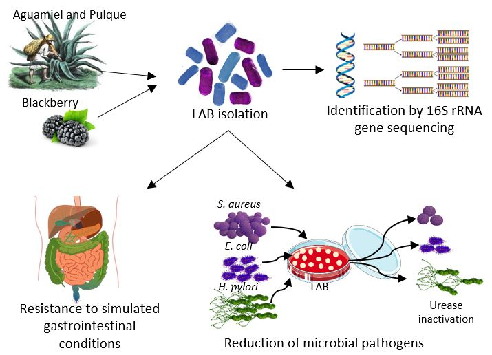
In Vitro Probiotic Potential of Lactic Acid Bacteria Isolated from Aguamiel and Pulque and Antibacterial Activity Against Pathogens

by
Alicia Cervantes-Elizarrarás
1,
Nelly del Socorro Cruz-Cansino
1,
Esther Ramírez-Moreno
1,
Vicente Vega-Sánchez
2,
Norma Velázquez-Guadarrama
3,
Quinatzin Yadira Zafra-Rojas
1 and
Javier Piloni-Martini
2,*
1
Instituto de Ciencias de la Salud, Universidad Autónoma del Estado de Hidalgo, Circuito Ex Hacienda La Concepción S/N, Carretera Pachuca-Actopan, San Agustín Tlaxiaca 42160, Hidalgo, Mexico
2
Instituto de Ciencias Agropecuarias, Universidad Autónoma del Estado de Hidalgo. Av. Universidad Km 1, Rancho Universitario, Tulancingo 43600, Hidalgo, Mexico
3
Laboratorio de Infectología, Departamento de Infectología, Hospital Infantil de México Federico Gómez, Col. Doctores, Delegación: Cuauhtémoc, Ciudad de México 06720, Mexico
*
Author to whom correspondence should be addressed.
Appl. Sci. 2019, 9(3), 601; https://doi.org/10.3390/app9030601
Submission received: 18 January 2019 / Revised: 1 February 2019 / Accepted: 8 February 2019 / Published: 12 February 2019
(This article belongs to the Section Applied Biosciences and Bioengineering)

Abstract

:
Probiotics can act as a natural barrier against several pathogens, such Helicobacter pylori, a bacterium linked to stomach cancer. The aim of the present study was to isolate and identify lactic acid bacteria (LAB) from pulque and aguamiel, and evaluate their probiotic potential and antimicrobial effect on Escherichia coli, Staphylococcus aureus, and Helicobacter pylori. Ten isolates were selected and evaluated for in vitro resistance to antibiotics and gastrointestinal conditions, and antimicrobial activity against E. coli and S. aureus and the effect on H. pylori strains. 16S rRNA identification was performed. Ten potential probiotic isolates were confirmed as belonging to the genera Lactobacillus and Pediococcus. All the strains were susceptible to clinical antibiotics, except to vancomycin. Sixty percent of the isolates exhibited antimicrobial activity against E. coli and S. aureus. The growth of H. pylori ATCC 43504 was suppressed by all the LAB, and the urease activity from all the H. pylori strains was inhibited, which may decrease its chances for survival in the stomach. The results suggest that LAB isolated from pulque and aguamiel could be an option to establish a harmless relationship between the host and H. pylori, helping in their eradication therapy.

Graphical Abstract

1. Introduction

Pulque is a viscous, non-distilled alcoholic beverage produced and consumed in Mexico, obtained by the fermentation of maguey sap (aguamiel) from the species Agave atrovensis and A. americana. Typically, fermentation occurs under non-aseptic conditions, which promotes the presence of a great variety of microorganisms including those naturally present in the aguamiel and those incorporated during collection, transport, inoculation, and manipulation [1,2]. Several studies describe the presence of Saccharomyces cerevisiae, species of Kluyveromyces, Zymomona, Leuconostoc, and Lactobacillus [1,3,4]. These microorganisms in fermented products are associated with human health benefits and probiotic potential. Research suggests that probiotics improve immune modulatory properties and lactose tolerance, decrease serum cholesterol, and increase utilization of nutrients [5,6,7]. Furthermore, they can inactivate or act as natural barrier against pathogens such as Listeria monocytogenes, Escherichia coli, Bacillus cereus, Staphylococcus aureus, Salmonella Typhimurium, Salmonella Enteritidis, Pseudomonas aeruginosa, Aeromonas hydrophila, Corynebacterium fimi [7,8,9,10], or Helicobacter pylori [11,12,13]. Approximately 50% of the world’s population is infected with H. pylori, a bacteria linked to gastric ulcers and stomach cancer [14]. Consumption of supplementary probiotic preparations during standard triple therapy may improve the eradication rate of H. pylori and decrease the incidence of adverse effects, particularly diarrhea, in patients [15].
Probiotic functionality requires bacteria to survive the acidic conditions of the stomach and resist the bile acids at the beginning of the duodenum [16,17]. Additionally, probiotic bacteria must not transfer antibiotic resistance genes because after ingestion they may interact with indigenous microbiota of the human gastrointestinal tract [18,19,20]. The aim of this study was to isolate and identify lactic acid bacteria from aguamiel and pulque and evaluate their probiotic potential and effect against H. pylori.

2. Materials and Methods

2.1. Isolation of Lactic Acid Bacteria

Pulque and aguamiel samples were collected in September 2015 from artisan producers in the town of El Venado, Mineral de la Reforma, Hidalgo, Mexico. Samples were collected twice in that month, placed in sterile bottles, and transported to the laboratory and held at 4 °C until analysis the next day. Each sample (1 mL) was suspended in 50 mL of Man Rogosa Sharpe (MRS) broth (BD, Sparks, MD, USA) and incubated at 37 °C for 24 h. To isolate presumptive lactic acid bacteria (LAB), serial dilutions were made in peptone water and plated on MRS agar (BD, Sparks, MD, USA) at 37 °C for 24 h. Gram-positive and catalase-negative isolates were cultivated in MRS broth at 37 °C for 24 h, and purified by three successive transfers in MRS agar. Pure cultures were stored at −70 °C in sterile glycerol/MRS broth mixture (50:50%; v/v).

2.2. Characterization of Isolates as Potential Probiotics

2.2.1. Resistance to Antibiotics of Isolated Strains

The minimum inhibitory concentration (MIC) was determined by the broth microdilution assay following the Clinical and Laboratory Standards Institute (CLSI) M100-S25 guidelines. The strains were tested for their susceptibilities against amoxicillin, amikacin, chloramphenicol, gentamicin, levofloxacin, spectinomycin, tetracycline, and vancomycin. Different individual concentrations (500–0.5 µg/mL) of antibiotics were prepared in Mueller-Hinton broth (BD, Sparks, MD, USA) and then were placed in the microplate wells. The inoculum was adjusted to a turbidity equivalent to 0.5 McFarland standard (≈1.5 × 10⁸ cfu/mL). For the assay, 10 μL of the inoculum were inoculated in each well with 90 μL of the corresponding antibiotic concentration (final volume 100 μL). The plates were covered and incubated at 37 °C for 24 h. The MIC was defined as the lowest concentration of antibiotic giving a complete inhibition of visible growth in comparison to an antibiotic-free control well.

2.2.2. Survival Assessment of LAB Strains under Simulated Gastrointestinal Tract (GIT) Conditions

The resistance of strains to a low pH environment was tested by the method of Lee et al. [9] with some modifications. LAB from the 18 hours of overnight culture were harvested by centrifugation (10,000× g, 5 min, 4 °C), and washed three times with PBS at pH 7.2. The pellet was resuspended in MRS broth containing 1% pepsin and at pH 2 adjusted using HCl (1 N). Resistance was assessed by simulating the time spent by food in the stomach and viable colony count after incubation at 37 °C for 2 h.
The tolerance of strains to bile salt was assessed using MRS broth containing 1% pancreatin enriched with 0.3% (w/v) cholic acid and sodium taurocholate (Sigma-Aldrich, St. Louis, MO, USA) (50:50). The viable colony counts were carried out using the pour plate method with the appropriate dilutions after incubation for 24 h. The MRS broth with 18 h of overnight culture was used as control. The survival rate was calculated as log10 values of colony-forming units per mL (CFU mL−1).

2.2.3. Antimicrobial Activity

Antimicrobial activity was evaluated using the double-layer plaque assay [21]. The double-layer assay involved first growing the competitive–exclusion isolate and then applying a second layer of medium containing the pathogen strains. Lactobacillus casei Shirota was used as the reference probiotic.
LAB were inoculated in an area of 1 cm in diameter on the first layer of MRS agar in the petri dish. The cultures were then incubated for 24 h at 37 °C under anaerobic conditions. Tryptic soy agar was poured onto the surface of each inoculated plate as a second layer and allowed to cool down to room temperature. Pathogen strains were inoculated by striking the surface using a swab. Staphylococcus aureus ATCC 29213 and Escherichia coli ATCC 25922 were used as reference strains.

2.3. Effect of LAB on the Growth of Helicobacter pylori Strains

To evaluate the effect of LAB on H. pylori growth, standard strains ATCC 43504 and ATCC 700392, and three babA2, vacA, and cagA positive clinical strains—named 1L, 1B, and 2—isolated from gastric biopsies of pediatric patients, were used. These strains were selected since these genes are associated with higher risk of peptic ulcer disease and intestinal metaplasia [14]. All the strains were obtained from the collection of the Hospital Infantil de México Federico Gómez. H. pylori strains were grown on Casman agar base (BBL) plates supplemented with 5% defibrinated sheep blood at 37 °C under microaerophilic conditions (5% CO2). The strains were identified using Gram stain, morphology, urease, catalase, and oxidase tests.
LAB were inoculated onto the surface of the first layer of MRS agar in Petri dishes and were incubated for 24 h at 37 °C. A second layer of Mueller–Hinton II agar medium (Becton Dickinson, Franklin Lakes, NJ, USA) supplemented with 5% defibrinated sheep blood was used as culture medium for H. pylori. Strains were inoculated in a concentration equivalent to the 2.0 McFarland standard. The plates were incubated at 37 °C under microaerophilic conditions for 72 h. The minimal inhibitory concentration for clarithromycin was used as positive control and was determined by agar dilution method following the Clinical and Laboratory Standards Institute (CLSI M45-A) guidelines. Results were expressed as “total inhibition”, “partial inhibition”, or “no inhibition”. Strains were tested for urease activity using the urease rapid test (Urea/Christensen, BBL, MD) before and after being in contact with LAB.

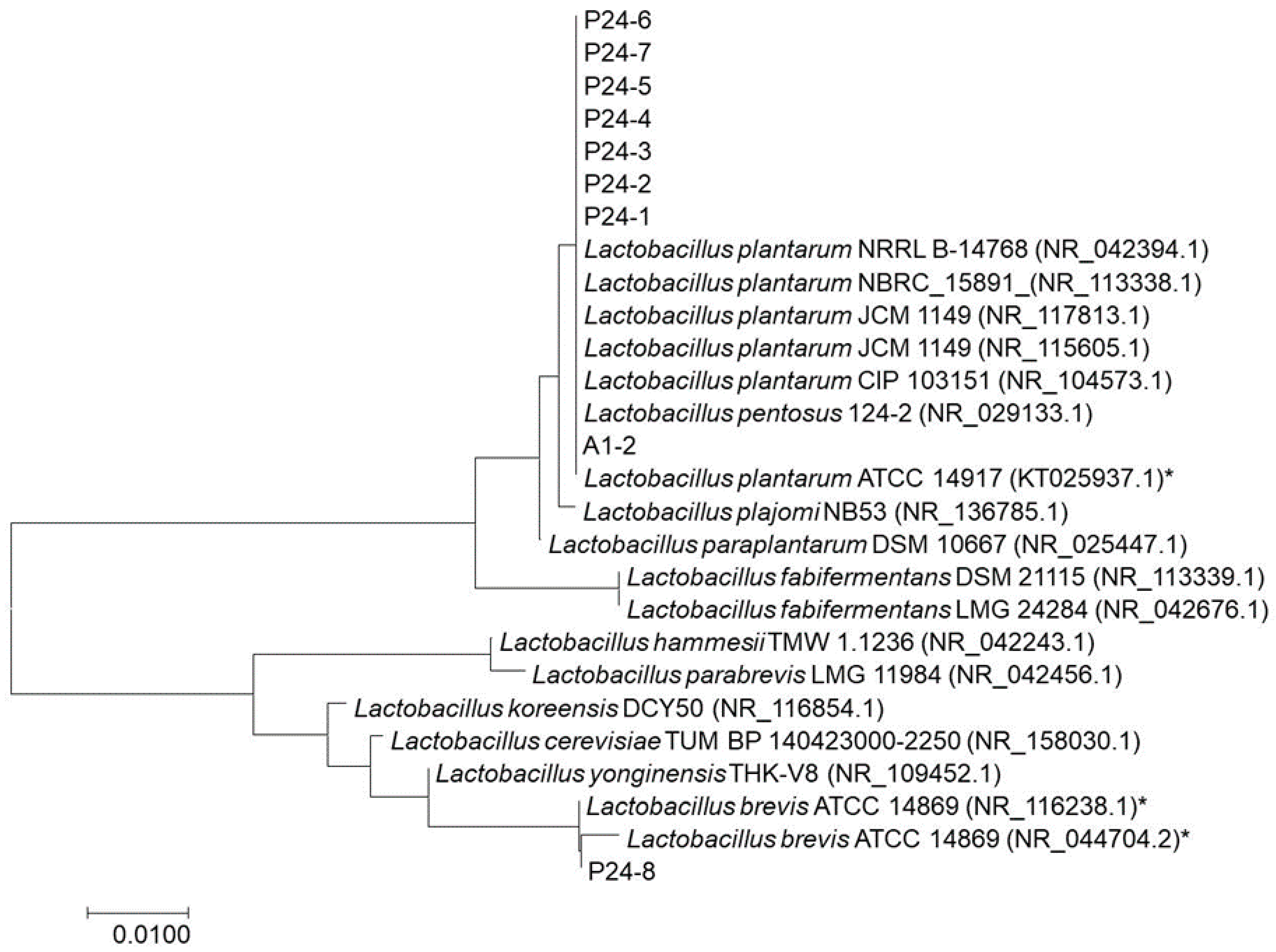
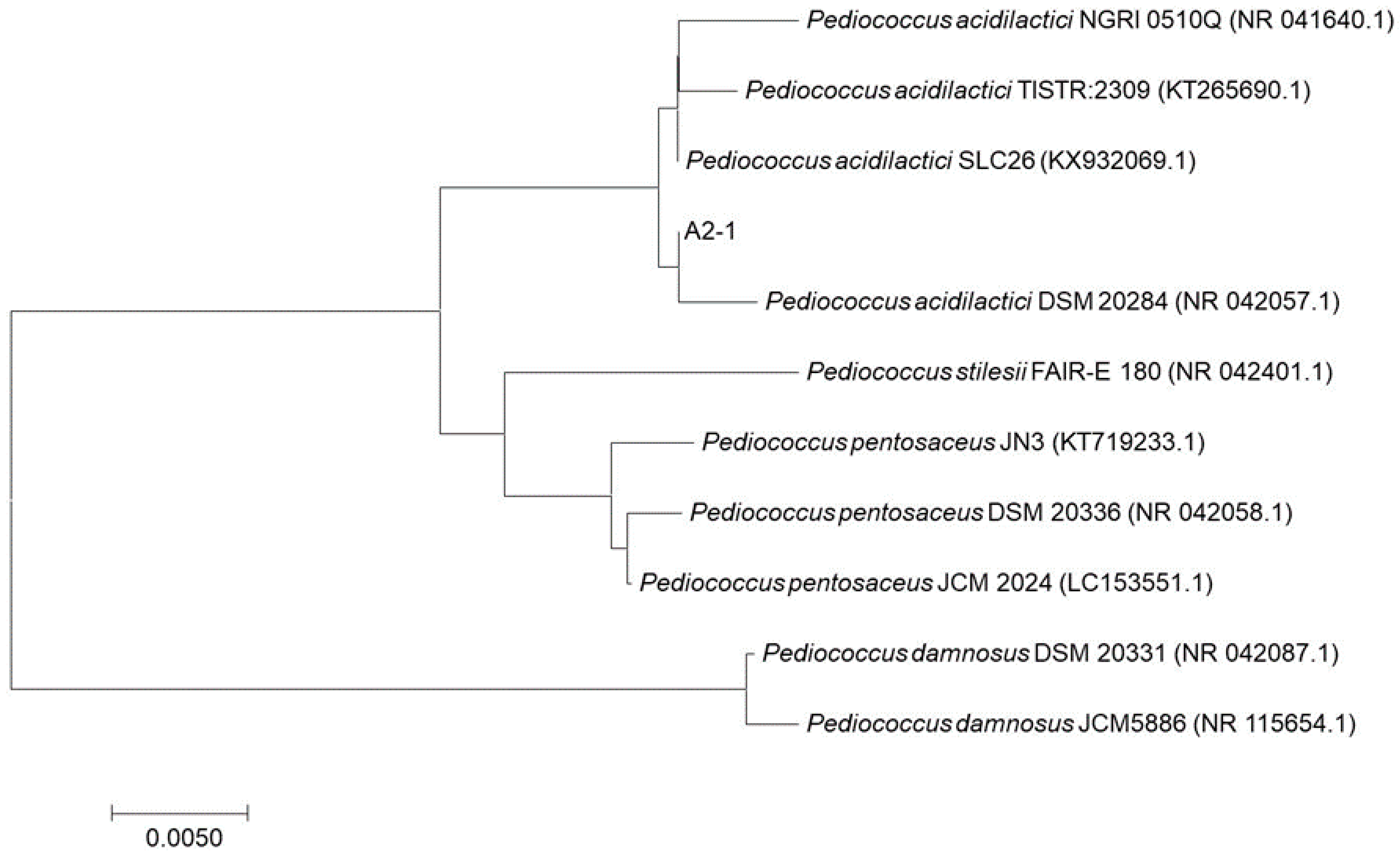
2.4. Identification of Genus and Species using PCR

DNA was extracted using a DNeasy blood and tissue kit (Qiagen, Valencia, CA, USA) for Gram-positive bacteria. The isolates were identified to genus level using PCR (polymerase chain reaction) using 16S rRNA gene sequencing. PCR parameters and universal primers 27f (5′-AGAGTTTGATCCTGGCTCAG-3′) and 1492r (5′-TACGGYTACCTTGTTACGACTT- 3′) were used. DNA sequencing of amplified fragments was carried out by the sequencing service of Langebio, Mexico. The amplified products were sequenced and aligned to 16S rRNA gene sequences in the GenBank data base using the BLAST algorithm. A phylogenetic tree was also constructed to determine the closest bacterial species by the neighbor joining method [22].

2.5. Statistical Analysis

All analyses were conducted in triplicate and the results were statistically analyzed by computing means and standard deviations of the mean, except for the antimicrobial activity against H. pylori. Mean differences among data were assessed by Tukey’s HSD (Honestly-significant-difference) test, with significance level at p < 0.05.

3. Results

3.1. Isolation of LAB

Typical, round, convex, and creamy colonies were obtained from each sample. A total of 10 strains Gram-positive and catalase-negative were obtained from the aguamiel (2) and pulque (8) samples.

3.2. Characterization of Isolates as Potential Probiotics

3.2.1. Resistance of LAB Isolated Strains to Antibiotics

The bacteria were considered “sensitive” if minimum inhibitory concentrations (MICs) values were ˂8 μg/mL, “moderately resistant” if MICs ≥ 8 μg/mL, and “resistant” if MICs > 328 μg/mL [23]. LAB isolates were sensitive to the majority of antibiotics evaluated, except for isolates P24-6, P24-7, and P24-8, which were moderately resistant to levofloxacin and to tetracycline, respectively. Also, all the strains were resistant to vancomycin at its highest concentration (Table 1).

3.2.2. Survival of LAB Strains under Simulated Human Gastrointestinal Tract (GIT) Conditions

The pH of gastric juice was kept at pH 2.0 for 2 h to simulate the conditions in the stomach. The results showed that low pH and pepsin suppressed LAB isolates, although they exhibited more resistance to gastric juice than the reference strain. Their viability was between 63.2–96.3%, with strains P24-7 and P24-8 being the most resistant (95 and 96% survival rate, respectively) (Table 2).
After being exposed to the gastric juice conditions, the strains were subjected to the presence of bile salts and pancreatin. The survival percentage was between 47.8 and 89.2%, with P24-8 being the most resistant to the simulated gastrointestinal conditions, while strain A2-1 was the most sensitive (Table 2).

3.2.3. Antimicrobial Activity

Production of antimicrobial substances is one of the main probiotic properties for strain selection [7]. In this study, 60% of the isolates (6 of 10) had inhibitory activity against E. coli and S. aureus. P24-4 had the lowest inhibitory effect on E. coli but the highest against S. aureus. P24-2 showed higher antimicrobial activity against both pathogens, while the reference strain inhibited E. coli but not S. aureus growth (Table 2).

3.3. Effect of LAB on Helicobacter pylori Growth

Colonization by H. pylori can be followed by inflammation of the gastric mucus layer, and is a risk factor in the development of atrophic gastritis, peptic ulcers, and gastric cancer [24]. As shown in Table 3, all the isolated strains inhibited H. pylori ATCC 43504, with P24-2, P24-6, and P24-8 being those that had a total inhibition. Most of the strains could not inhibit H. pylori ATCC 700392 and the clinical strains, except P24-6 and A1-2, which had an effect on strain 1L. However, all LAB inhibited the activity of the enzyme urease (data not shown).

3.4. Genus and Species Identifications using PCR

Molecular methods are important for bacterial identification and perhaps more accurate for LAB than the conventional phenotypic methods [25]. Through the identification of the bacterial species isolated in this study, we found that the lactobacilli were mostly Lactobacillus plantarum, except P24-8, which had more similarity with Lactobacillus brevis, and strain A2-1 with Pediococcus acidilactici, as shown in their phylogenetic trees (Figure 1 and Figure 2, respectively). Phylogenetic analysis of 16S rRNA sequences demonstrated a high similarity (up to 98%) to isolates or environmental clones previously obtained from diverse sources and deposited in the GenBank database (Table 4).

4. Discussion

Probiotic bacteria must be effective in keeping the favorable balance of microflora and in benefiting the host’s health. They should lack the capacity to transfer antibiotic resistance, which is one of the greatest threats to human health [7,18]. Bacteria such as S. aureus, E. coli, and H. pylori can be intrinsically resistant to certain antibiotics, and also acquire resistance through genes mutations and horizontal gene transfer [26]. In this study, all the strains showed resistance to vancomycin at its highest concentration. Some LAB, such as Leuconostoc mesenteroides C4, Leu. mesenteroides C10, Leu. mesenteroides F27, L. plantarum C182 and L. casei, have shown resistance to vancomycin [16,18]. The vancomycin-resistant phenotype is perhaps the best-characterized intrinsic resistance among LAB, and is explained by the relatively poor binding of vancomycin to the peptidoglycan with D-alanyl-D-lactate termini, which some lactic acid bacteria have [27].
Acid and bile tolerance were evaluated to predict the survival of probiotics after oral administration. The gastric juice secreted in the stomach reduces the presence of most microorganisms due to its low pH (about 2.0) [28]; meanwhile, bile salts disorganize the structure of the cell membrane [29]. Therefore, resistance to these conditions is ideal for the selection of probiotic strains. Several LAB have exhibited high survivability in simulated gastric juice with pepsin at pH 3.0; however, cell viability decreased below the detection limit when exposed to pH 2 [5,7,8,10,30]. In our study, isolated LAB showed high viability (between 63.2–96.3%) in the presence of 1% pepsin and pH 2.
In acidic conditions, bacteria maintain the pH homeostasis by discharging H+ from the cell via the membrane H+-ATPase [31,32], with this enzymatic activity being higher in acid-tolerant bacteria [33]. Some lactobacilli utilize other responses to resist hostile stomach conditions, such as repair proteins for DNA damage and changes in cellular envelope and altered metabolism [34,35].
Other studies demonstrated that some LAB can have levels of resistance against bile salt concentrations of 0.3 to 1.5% [10,16,36]. Exopolysaccharide (EPS) production may provide certain tolerance to simulated gastrointestinal conditions since it is lightly attached to the cell wall [6]. Furthermore, pre-exposure of lactobacilli strains to acidic environments, such as the stomach or food matrices, could enhance their performance as probiotic strains by improving their adhesion to mucosal cells and thus increasing bacteria colonization in the gut [34,37].
Bile salts resistance may also be attributed to the enzyme responsible for bile salt deconjugation, a hydrolase present in some LAB. This enzyme is also associated with the reduction of serum cholesterol, and the toxicity and side effects of bile salts [8,25,29]. The results of our study indicate that 70% of the evaluated strains showed equal or greater resistance than L. casei Shirota to the gastrointestinal conditions.
The antibacterial activity of LAB could result from the production of a great variety of compounds, including sugar catabolites (e.g., lactic acid and acetic acid); oxygen catabolites (such as hydrogen peroxide); proteinaceous compounds (e.g., bacteriocins, other low-molecular-mass peptides, and antifungal peptides/proteins); fat and amino acid metabolites (e.g., fatty acids, phenyllactic acid, and OH-phenyllactic acid); and others such as reuterin and reutericyclin [38]. Antibacterial capacity can also differ under the same pH conditions (e.g., the stronger effect of acetic acid compared to lactic acid under similar pH values) [7]. Guo et al. [7] found that 50% of the isolates from swine (251 strains) inhibited the growth of E. coli O55. Meanwhile, Bao et al. [8] described that 45% of L. fermentum strains inhibited E. coli O157 882364, and all the strains had antimicrobial activity against S. aureus, revealing that even if the strains belong to the same species, their antibacterial effect can be different. Inhibition of Gram-negative bacteria could result from the production of lactic acid and/or other organic acids that can penetrate the cell membrane affecting its function, acidifying the cytoplasm, and inhibiting acid-sensitive enzymes [39]. The production of bacteriocins also contributes to the antibacterial activity against several Gram-positive and -negative pathogens, as well as the competition for nutrients, and hydrogen peroxide production [40,41,42]. In this study, 60% of the isolates showed antimicrobial activity against E. coli and S. aureus.
There is increasing evidence that highlights the efficacy of probiotics in the management of Helicobacter pylori infection. Inhibition is associated with the production of antimicrobial compounds, such as lactic acid, and by competing for substrate and binding sites [43,44]. Although ingestion of probiotics may reduce the density of infection, they are unable to completely eradicate H. pylori from the stomach [15,44]. In this study, the isolated strains inhibited H. pylori ATCC 43504 growth and the urease activity.
H. pylori are capable of surviving the hostile acidic condition in the stomach by regulating urease activity [14], and when this activity is lost, the bacterial survival in an acidic environment is diminished. Other LAB can prevent H. pylori infection by decreasing gastric mucosal inflammation and gastric microbiota alteration as had been described for L. plantarum ZDY 2013 [45]. Similar studies revealed that supplementation with L. plantarum Lp91 regulated the anti-inflammatory activity in mouse colitis [46]. Therefore, determining the activity of the isolated strains in this study is important to evaluate them as potential probiotics against H. pylori infection.
Escalante et al. [1] previously found Lactobacillus spp AC07, Lactobacillus spp. ASF360, Lactobacillus spp. Y10., Lactobacillus acidophilus, Lactobacillus hilgardii, Lactobacillus paracollinoides, Lactobacillus sanfranciscencis, Lactococcus spp., Lactococcus lactis, Lactococcus lactis subsp. cremoris, Leuconostoc kimchi, Leuconostoc citreum, Leuconostoc gasicomitatum, Leuconostoc mesenteroides, Leuconostoc pseudomesenteroides, Pediococcus urinaeequi, and Streptococcus devriesei in aguamiel and during pulque fermentation. Nevertheless, they found some clones with less than 95% relatedness to NCBI database sequences, which may indicate the presence of new species.

5. Conclusions

This study demonstrated that pulque and aguamiel are a source of potential probiotic lactic acid bacteria from the Lactobacillus and Pediococcus genera.
Strain P24-8 showed a high tolerance to simulated gastrointestinal tract conditions, and pepsin, pancreatin, and bile salts. It also showed a significant antibacterial activity against H. pylori ATCC 43504. Strain P24-2 presented great tolerance to simulated gastrointestinal tract conditions and had an important broad antibacterial activity against E. coli ATCC 25922, S. aureus ATCC 29213, and H. pylori ATCC 43504. These findings are also important since the use of probiotics could prevent multidrug-resistant bacterial infections caused by H. pylori, E. coli, and S. aureus.
All the evaluated strains inhibited the urease activity from H. pylori strains, which is the enzyme that reduces the pH of its close environment, and thus the chance of H. pylori survival in the harsh acidic environment of the stomach.
Our results suggest that probiotic lactic acid bacteria isolated from pulque and aguamiel could be an option to establish and manage a harmless relationship between the host and H. pylori, helping in their eradication therapy. This is a cheap alternative and does not carry the risk of extensive antibiotic resistance. However, further in vitro and in vivo research is needed to verify the activities of these strains against H. pylori and other potential beneficial effects.

Author Contributions

Investigation and writing—original draft preparation, A.C.-E.; design, validation and supervision, N.d.S.C.-C. and J.P.-M.; resources and supervision N.V.-G and V.V.-S.; data analysis, E.R.-M. and Q.Y.Z.-R. All the authors checked the final manuscript.

Funding

This study was possible thanks to the financial support from the Programa de Fortalecimiento a la Calidad Educativa (PFCE 2017) and Hospital Infantil de Mexico Federico Gomez. The first author was supported by a fellowship from the Consejo Nacional de Ciencia y Tecnología (CONACYT) (No. 555686).

Conflicts of Interest

The authors declare no conflict of inerest.

References

  1. Escalante, A.; Giles-Gómez, M.; Hernández, G.; Córdova-Aguilar, M.S.; López-Munguía, A.; Gosset, G.; Bolívar, F. Analysis of bacterial community during the fermentation of pulque, a traditional Mexican alcoholic beverage, using a polyphasic approach. Int. J. Food Microbiol. 2008, 124, 126–134. [Google Scholar] [CrossRef]
  2. Valadez-Blanco, R.; Bravo-Villa, G.; Santos-Sánchez, N.F.; Velasco-Almendarez, S.I.; Montville, T.J. The artisanal production of pulque, a traditional beverage of the Mexican highlands. Probiotics Antimicro. Prot. 2012, 4, 140–144. [Google Scholar]
  3. Cruz-Guerrero, A.E.; Olvera, J.L.; García-Garibay, M.; Gómez-Ruiz, L. Inulinase-hyperproducing strains of Kluyveromyces sp. isolated from aguamiel (Agave sap) and pulque. World J. Microbiol. Biotechnol. 2006, 22, 115. [Google Scholar] [CrossRef]
  4. Escalante, A.; Flores, M.E.; Martínez, A.; López-Munguía, A.; Bolívar, F.; Gosset, G. Characterization of bacterial diversity in pulque, a traditional Mexican alcoholic fermented beverage, as determined by 16S rDNA analysis. FEMS Microbiol. Lett. 2004, 235, 273–279. [Google Scholar] [CrossRef]
  5. Angmo, K.; Kumari, A.; Bhalla, T.C. Probiotic characterization of lactic acid bacteria isolated from fermented foods and beverage of Ladakh. LWT-Food Sci. Technol. 2016, 66, 428–435. [Google Scholar] [CrossRef]
  6. Bengoa, A.A.; Llamas, M.G.; Iraporda, C.; Dueñas, M.T.; Abraham, A.G.; Garrote, G.L. Impact of growth temperature on exopolysaccharide production and probiotic properties of Lactobacillus paracasei strains isolated from kefir grains. Food Microbiol. 2018, 69, 212–218. [Google Scholar] [CrossRef]
  7. Guo, X.H.; Kim, J.M.; Nam, H.M.; Park, S.Y.; Kim, J.M. Screening lactic acid bacteria from swine origins for multistrain probiotics based on in vitro functional properties. Anaerobe 2010, 16, 321–326. [Google Scholar] [CrossRef]
  8. Bao, Y.; Zhang, Y.; Zhang, Y.; Liu, Y.; Wang, S.; Dong, X.; Wang, Y.; Zhang, H. Screening of potential probiotic properties of Lactobacillus fermentum isolated from traditional dairy products. Food Control 2010, 21, 695–701. [Google Scholar] [CrossRef]
  9. Lee, N.K.; Lee, E.K.; Paik, H.D. Potential probiotic properties of phytase-producing Lactobacillus salivarius FC113. Ann. Microbiol. 2013, 63, 555–560. [Google Scholar] [CrossRef]
  10. Pieniz, S.; Andreazza, R.; Anghinoni, T.; Camargo, F.; Brandelli, A. Probiotic potential, antimicrobial and antioxidant activities of Enterococcus durans strain LAB18s. Food Control 2014, 37, 251–256. [Google Scholar] [CrossRef]
  11. Cats, A.; Kuipers, E.J.; Bosschaert, M.A.R.; Pot, R.G.J.; Vandenbroucke-Grauls, C.M.J.E.; Kusters, J.G. Effect of frequent consumption of a Lactobacillus casei-containing milk drink in Helicobacter pylori-colonized subjects. Aliment. Pharm. Ther. 2003, 17, 429–435. [Google Scholar] [CrossRef]
  12. Chen, X.; Tian, F.; Liu, X.; Zhao, J.; Zhang, H.P.; Zhang, H.; Chen, W. In vitro screening of lactobacilli with antagonistic activity against Helicobacter pylori from traditionally fermented foods. J. Dairy Sci. 2010, 93, 5627–5634. [Google Scholar] [CrossRef] [PubMed]
  13. Du, Y.Q.; Su, T.; Fan, J.G.; Lu, Y.X.; Zheng, P.; Li, X.H.; Guo, C.Y.; Xu, P.; Gong, Y.F.; Li, Z.S. Adjuvant probiotics improve the eradication effect of triple therapy for Helicobacter pylori infection. World J. Gastroenterol. 2012, 18, 6302. [Google Scholar] [CrossRef] [PubMed]
  14. Kao, C.Y.; Sheu, B.S.; Wu, J.J. Helicobacter pylori infection: An overview of bacterial virulence factors and pathogenesis. Biomedical J. 2016, 39, 14–23. [Google Scholar] [CrossRef] [PubMed]
  15. Emara, M.H.; Elhawari, S.A.; Yousef, S.; Radwan, M.I.; Abdel-Aziz, H.R. Emerging role of probiotics in the management of Helicobacter pylori infection: Histopathologic Perspectives. Helicobacter 2016, 21, 3–10. [Google Scholar] [CrossRef] [PubMed]
  16. Lee, K.W.; Shim, J.M.; Park, S.K.; Heo, H.J.; Kim, H.J.; Ham, K.S.; Kim, J.H. Isolation of lactic acid bacteria with probiotic potentials from kimchi, traditional Korean fermented vegetable. LWT-Food Sci. Technol. 2016, 71, 130–137. [Google Scholar] [CrossRef]
  17. Mojgani, N.; Hussaini, F.; Vaseji, N. Characterization of indigenous Lactobacillus strains for probiotic properties. Jundishapur J. Microb. 2015, 8, e17523. [Google Scholar] [CrossRef] [PubMed]
  18. D’Aimmo, M.R.; Modesto, M.; Biavati, B. Antibiotic resistance of lactic acid bacteria and Bifidobacterium spp. isolated from dairy and pharmaceutical products. Int. J. Food Microbiol. 2007, 115, 35–42. [Google Scholar] [CrossRef]
  19. Fraqueza, M.J. Antibiotic resistance of lactic acid bacteria isolated from dry-fermented sausages. Int. J. Food Microbiol. 2015, 212, 76–88. [Google Scholar] [CrossRef]
  20. Toomey, N.; Bolton, D.; Fanning, S. Characterisation and transferability of antibiotic resistance genes from lactic acid bacteria isolated from Irish pork and beef abattoirs. Res. Microbiol. 2010, 161, 127–135. [Google Scholar] [CrossRef]
  21. Zhao, T.; Doyle, M.P.; Zhao, P. Control of Listeria monocytogenes in a biofilm by competitive-exclusion microorganisms. Appl. Environ. Microb. 2004, 70, 3996–4003. [Google Scholar] [CrossRef] [PubMed]
  22. Saitou, N.; Nei, M. The neighbor-joining method: A new method for reconstructing phylogenetic trees. Mol. Biol. Evol. 1987, 4, 406–425. [Google Scholar] [PubMed]
  23. Walsh, C. Antibiotics: Actions, Origins, Resistance; ASM Press: Washington, DC, USA, 2003; pp. 91–106. [Google Scholar]
  24. Amieva, M.; Peek, R.M., Jr. Pathobiology of Helicobacter pylori–induced gastric cancer. Gastroenterology 2016, 150, 64–78. [Google Scholar] [CrossRef] [PubMed]
  25. Shehata, M.G.; El Sohaimy, S.A.; El-Sahn, M.A.; Youssef, M.M. Screening of isolated potential probiotic lactic acid bacteria for cholesterol lowering property and bile salt hydrolase activity. Ann. Agric. Sci. 2016, 61, 65–75. [Google Scholar] [CrossRef] [Green Version]
  26. Blair, J.M.; Webber, M.A.; Baylay, A.J.; Ogbolu, D.O.; Piddock, L.J. Molecular mechanisms of antibiotic resistance. Nat. Rev. Microbiol. 2015, 13, 42. [Google Scholar] [CrossRef] [PubMed]
  27. Zhang, S.; Oh, J.H.; Alexander, L.M.; Özçam, M.; Van Pijkeren, J.P. D-Ala-D-Ala ligase as a broad-host-range counterselection marker in vancomycin-resistant lactic acid bacteria. J. Bacteriol. 2018, 200, e00607-17. [Google Scholar] [CrossRef] [PubMed]
  28. Vinderola, C.G.; Reinheimer, J.A. Lactic acid starter and probiotic bacteria: A comparative “in vitro” study of probiotic characteristics and biological barrier resistance. Food Res. Int. 2003, 36, 895–904. [Google Scholar] [CrossRef]
  29. Margolles, A.; García, L.; Sánchez, B.; Gueimonde, M.; de los Reyes-Gavilán, C.G. Characterisation of a Bifidobacterium strain with acquired resistance to cholate-a preliminary study. Int. J. Food Microbiol. 2003, 82, 191–198. [Google Scholar] [CrossRef]
  30. Jacobsen, C.N.; Nielsen, V.R.; Hayford, A.E.; Møller, P.L.; Michaelsen, K.F.; Paerregaard, A.; Jakobsen, M. Screening of probiotic activities of forty-seven strains of Lactobacillus spp. by in vitro techniques and evaluation of the colonization ability of five selected strains in humans. Appl. Environ. Microb. 1999, 65, 4949–4956. [Google Scholar]
  31. Booth, I.R. Regulation of cytoplasmic pH in bacteria. Microbiol. Rev. 1985, 49, 359. [Google Scholar]
  32. Matsumoto, M.; Ohishi, H.; Benno, Y. H+-ATPase activity in Bifidobacterium with special reference to acid tolerance. Int. J. Food Microbiol. 2004, 93, 109–113. [Google Scholar] [CrossRef] [PubMed]
  33. Miwa, T.; Esaki, H.; Umemori, J.; Hino, T. Activity of H (+)-ATPase in ruminal bacteria with special reference to acid tolerance. Appl. Environ. Microb. 1997, 63, 2155–2158. [Google Scholar]
  34. Casado Muñoz, M.C.; Benomar, N.; Ennahar, S.; Horvatovich, P.; Lerma, L.L.; Knapp, C.W.; Galvez, A.; Abriouel, H. Comparative proteomic analysis of a potentially probiotic Lactobacillus pentosus MP-10 for the identification of key proteins involved in antibiotic resistance and biocide tolerance. Int. J. Food Microbiol. 2016, 222, 8–15. [Google Scholar] [CrossRef] [PubMed]
  35. Pérez Montoro, B.; Benomar, N.; Gómez, N.C.; Ennahar, S.; Horvatovich, P.; Knapp, C.W.; Gálvez, A.; Abriouel, H. Proteomic analysis of Lactobacillus pentosus for the identification of potential markers involved in acid resistance and their influence on other probiotic features. Food Microbiol. 2018, 72, 31–38. [Google Scholar] [CrossRef] [PubMed]
  36. Lin, W.H.; Yu, B.; Jang, S.H.; Tsen, H.Y. Different probiotic properties for Lactobacillus fermentum strains isolated from swine and poultry. Anaerobe 2007, 13, 107–113. [Google Scholar] [CrossRef] [PubMed]
  37. Bengoa, A.A.; Zavala, L.; Carasi, P.; Trejo, S.A.; Bronsoms, S.; de los Ángeles Serradell, M.; Garrote, G.L.; Abraham, A.G. Simulated gastrointestinal conditions increase adhesion ability of Lactobacillus paracasei strains isolated from kefir to Caco-2 cells and mucin. Food Res. Int. 2017, 103, 462–467. [Google Scholar] [CrossRef] [PubMed]
  38. Makras, L.; Triantafyllou, V.; Fayol-Messaoudi, D.; Adriany, T.; Zoumpopoulou, G.; Tsakalidou, E.; Servin, A.; De Vuyst, L. Kinetic analysis of the antibacterial activity of probiotic lactobacilli towards Salmonella enterica serovar Typhimurium reveals a role for lactic acid and other inhibitory compounds. Res. Microbiol. 2006, 157, 241–247. [Google Scholar] [CrossRef] [PubMed]
  39. Hirshfield, I.N.; Terzulli, S.; O’Byrne, C. Weak organic acids: a panoply of effects on bacteria. Sci. Prog. 2003, 86, 245–269. [Google Scholar] [CrossRef]
  40. Zhang, Z.; Tao, X.; Shah, N.P.; Wei, H. Antagonistics against pathogenic Bacillus cereus in milk fermentation by Lactobacillus plantarum ZDY2013 and its anti-adhesion effect on Caco-2 cells against pathogens. J. Dairy Sci. 2016, 99, 2666–2674. [Google Scholar] [CrossRef]
  41. Biswas, K.; Upadhayay, S.; Rapsang, G.F.; Joshi, S.R. Antibacterial and Synergistic Activity Against b-Lactamase-Producing Nosocomial Bacteria by Bacteriocin of LAB Isolated From Lesser Known Traditionally Fermented Products of India. HAYATI J. Biosci. 2018, 24, 87–95. [Google Scholar] [CrossRef]
  42. Magnusson, J.; Ström, K.; Roos, S.; Sjögren, J.; Schnürer, J. Broad and complex antifungal activity among environmental isolates of lactic acid bacteria. FEMS Microbiol. Lett. 2003, 219, 129–135. [Google Scholar] [CrossRef] [Green Version]
  43. De Vrese, M.; Kristen, H.; Rautenberg, P.; Laue, C.; Schrezenmeir, J. Probiotic lactobacilli and bifidobacteria in a fermented milk product with added fruit preparation reduce antibiotic associated diarrhea and Helicobacter pylori activity. J. Dairy Res. 2011, 78, 396–403. [Google Scholar] [CrossRef] [PubMed]
  44. Westerik, N.; Reid, G.; Sybesma, W.; Kort, R. The probiotic Lactobacillus rhamnosus for alleviation of Helicobacter pylori-associated gastric pathology in East Africa. Front. Microbiol. 2018, 9, 1873. [Google Scholar] [CrossRef] [PubMed]
  45. Pan, M.; Wan, C.; Xie, Q.; Huang, R.; Tao, X.; Shah, N.P.; Wei, H. Changes in gastric microbiota induced by Helicobacter pylori infection and preventive effects of Lactobacillus plantarum ZDY 2013 against such infection. J. Dairy Sci. 2016, 99, 970–981. [Google Scholar] [CrossRef] [PubMed]
  46. Duary, R.K.; Bhausaheb, M.A.; Batish, V.K.; Grover, S. Anti-inflammatory and immunomodulatory efficacy of indigenous probiotic Lactobacillus plantarum Lp91 in colitis mouse model. Mol. Biol. Rep. 2012, 39, 4765–4775. [Google Scholar] [CrossRef] [PubMed]
Figure 1. Phylogenetic relationship of Lactobacillus isolated from aguamiel and pulque. Species are inferred by neighbor-joining analysis of the 16S rRNA gene, based on evolutionary distances calculated using the Kimura two-parameter model. Numbers in parentheses are accession numbers of published sequences. * Type strain.
Applsci 09 00601 g001
Figure 2. Phylogenetic relationship of lactic acid bacteria A1-2 isolated from aguamiel. Species are inferred by neighbor-joining analysis of the 16S rRNA gene based on evolutionary distances calculated using the Kimura two-parameter model. Numbers in parentheses are accession numbers of published sequences.
Applsci 09 00601 g002
Table 1. MIC (minimum inhibitory concentration) values for the isolated strains.
MIC (µg/mL)
StrainAmkAmxChlGntLvfSpcTtrVnc
P24-1≤1.0≤1.0≤1.0≤1.0≤1.0≤1.0≤1.0≤100
P24-2≤1.0≤1.0≤1.0≤1.0≤1.0≤1.0≤1.0≤100
P24-3≤1.0≤1.0≤1.0≤1.0≤1.0≤1.0≤1.0≤100
P24-4≤1.0≤1.0≤1.0≤1.0≤1.0≤1.0≤1.0≤100
P24-5≤1.0≤1.0≤1.0≤1.0≤1.0≤1.0≤1.0≤100
P24-6≤1.0≤1.0≤1.0≤1.0≤10≤1.0≤1.0≤100
P24-7≤1.0≤1.0≤1.0≤1.0≤10≤1.0≤1.0≤100
P24-8≤1.0≤1.0≤1.0≤1.0≤10≤1.0≤10≤100
A1-2≤1.0≤1.0≤1.0≤1.0≤10≤1.0≤1.0≤100
A2-1≤1.0≤1.0≤1.0≤1.0≤10≤1.0≤1.0≤100
L. casei≤1.0≤1.0≤1.0≤1.0≤10≤10≤1.0≥250
Amk: amikacin; Amx: amoxicillin; Chl: chloramphenicol; Gnt: gentamicin; Lvf: levofloxacin; Spc: spectinomycin; Ttr: tetracycline; Vnc: vancomycin.
Table 2. Probiotic properties of isolated LAB.
StrainResistance to Gastric Juice
(Log CFU/mL)
Resistance to Bile Salts and Pancreatin
(Log CFU/mL)
Antibacterial Activity
(mm)
Control *pH 2 and PepsinSurvival Rate (%)Control *Bile Salts and Pancreatin **Survival Rate (%) **E. coliS. aureus
P24-19.2 ± 0.065.9 ± 0.0164.412.5 ± 0.068.4 ± 0.0166.929.0 ± 2.1 ab26.0 ± 2.0 ab
P24-29.3 ± 0.025.9 ± 0.0363.211.8 ± 0.027.5 ± 0.0358.833.3 ± 3.5 a28.3 ± 2.5 a
P24-39.4 ± 0.107.6 ± 0.0280.711.6 ± 0.107.1 ± 0.0264.528.3 ± 2.8 ab24.3 ± 1.5 ab
P24-49.0± 0.035.5 ± 0.0361.511.3 ± 0.036.2 ± 0.0354.319.0 ± 6.8 c28.7 ± 1.5 a
P24-59.6 ± 0.055.9 ± 0.0261.512.4 ± 0.0510.2 ± 0.0266.0--
P24-69.1 ± 0.037.0 ± 0.1476.512.5 ± 0.0310.1 ± 0.1465.0--
P24-79.6 ± 0.029.1 ± 0.095.012.6 ± 0.029.6 ± 0.0152.5--
P24-89.9 ± 0.049.5 ± 0.096.312.6 ± 0.0411.2 ± 0.0165.3--
A1-28.9 ± 0.027.5 ± 0.1884.212.6 ± 0.027.6 ± 0.1860.830.0 ± 5.4 ab22.3 ± 4.0 b
A2-18.8 ± 0.026.4 ± 0.0273.312.7 ± 0.026.1 ± 0.0255.724.0 ± 1.6 bc21.7 ± 3.2 b
L. casei Shirota8.6 ± 0.044.4 ± 0.0985.012.3 ± 0.047.8 ± 0.0962.522.3 ± 1.5 bc-
* Control: cells under no stress. ** Cells exposed to pH 2.0 and pepsin for 2 h and then exposed to 0.3% bile salts and pancreatin for 24 h. - No inhibition. Values are means of triplicate determinations. ±: mean-standard deviation. a,b,c different subscripts lowercase letters indicate significantly different at p < 0.05 as measured by Tukey’s HSD between different strains.
Table 3. Antibacterial activity of isolated LAB against Helicobacter pylori strains.
Helicobacter pylori
StrainATCC 43504ATCC 7003921L1B2
P24-1±----
P24-2++----
P24-3±----
P24-4±----
P24-5±----
P24-6++-++--
P24-7±----
P24-8++----
A1-2±-±--
A2-1±----
L. casei±----
- no inhibition; ± partial inhibition; ++ total inhibition.
Table 4. Identification of lactic acid bacteria isolates by 16S rRNA sequences.
Strain Species Identity% Identity *Accession Number
P24-1Lactobacillus plantarum ATCC 14917100KT025937.1
Lactobacillus plantarum JCM 1149100NR_115605.1
P24-2Lactobacillus plantarum JCM 114999.9NR_117813.1
Lactobacillus pentosus 124-299.9NR_029133.1
P24-3Lactobacillus plantarum JCM 114999.8NR_115605.1
Lactobacillus plantarum NBRC 1589199.8NR_113338.1
P24-4Lactobacillus plantarum JCM 1149100NR_115605.1
Lactobacillus plantarum NBRC 1589199.8NR_113338.1
P24-5Lactobacillus plantarum ATCC 14917100KT025937.1
Lactobacillus plantarum JCM 1149100NR_115605.1
P24-6Lactobacillus plantarum ATCC 14917100KT025937.1
Lactobacillus plantarum JCM 1149100NR_115605.1
P24-7Lactobacillus plantarum CIP 103151100NR_104573.1
Lactobacillus plantarum JCM 1149100NR_117813.1
P24-8Lactobacillus brevis ATCC 14869100NR_116238.1
Lactobacillus yonginensis THK-V8 97.8NR_109452.1
A1-2Lactobacillus plantarum CIP 103151100NR_104573.1
Lactobacillus plantarum JCM 1149100NR_117813.1
A2-1Pediococcus acidilactici DSM 2028499.6NR_042057.1
Pediococcus acidilactici NGRI 0510Q98.3NR_041640.1
* Identity were determined through pairwise alignments using the European Molecular Biology Open Software Suite (EMBOSS) Water algorithms.

