Article

Apigenin Promotes Proliferation and Mineralization of Human Osteoblasts and Up-Regulates Osteogenic Markers

by
Emira D’Amico
1,†,
Tania Vanessa Pierfelice
1,†,
Giovanna Iezzi
1,
Natalia Di Pietro
1,2,
Stefania Lepore
1,
Felice Lorusso
3,
Antonio Scarano
3,
Assunta Pandolfi
1,2,
Adriano Piattelli
4,5,6,7 and
Morena Petrini
1,*
1
Department of Medical, Oral and Biotechnological Sciences, University G. d’Annunzio of Chieti-Pescara, 66100 Chieti, Italy
2
Center for Advanced Studies and Technology-CAST, University G. d’Annunzio of Chieti-Pescara, 66100 Chieti, Italy
3
Department of Innovative Technologies in Medicine & Dentistry, University G. d’Annunzio of Chieti-Pescara, 66100 Chieti, Italy
4
School of Dentistry, Saint Camillus International University of Health and Medical Sciences, Via di Sant’Alessandro 8, 00131 Rome, Italy
5
Dental School, University of Belgrade, 11000 Belgrade, Serbia
6
Fondazione Villa Serena per la Ricerca, 65013 Città Sant’Angelo, Italy
7
Casa di Cura Villa Serena del Dott. L. Petruzzi, 65013 Città Sant’Angelo, Italy
*
Author to whom correspondence should be addressed.
These authors contributed equally to this work.
Appl. Sci. 2022, 12(17), 8510; https://doi.org/10.3390/app12178510
Submission received: 2 August 2022 / Revised: 19 August 2022 / Accepted: 23 August 2022 / Published: 25 August 2022
(This article belongs to the Special Issue Materials and Technologies in Oral Research 2nd Edition)

Abstract

:
Apigenin (APG), a natural flavonoid compound with anti-inflammatory and antioxidative properties, was found to promote in vitro osteogenic differentiation and to accelerate in vivo bone formation, indicating APG as a promising molecule in bone repair, with potential clinical application in bone-deficient conditions. In particular, in dentistry, it is fundamental to increase the available bone volume for implant placement in the maxilla. Therefore, this study aims to investigate the APG effects on osteoblasts (hOBs) obtained from a human jaw. hOBs were incubated with increasing concentrations of APG (5, 10, 20 µM) to assess cell viability and morphology at 24 h and proliferation at 48 and 72 h. Upon establishing the absence of cytotoxicity and any morphological changes, APG showed a stimulating effect on cell growth, with significative results using 5 µM (5-APG) at 48 h. Thus, 5-APG was chosen for further investigations in order to assess alkaline phosphate (ALP) at 7 days, mineralization at 14 days and expression of ALP, Osteocalcin (OCN) and Collagen 1 (COL1) genes at 7 days. Our results showed that 5-APG accelerated osteoblast mineralization activities and significantly upregulated ALP and COL1 gene expression. Hence, this study demonstrated that APG is able to promote human oral osteoblasts proliferation and mineralization, suggesting its potential usefulness in dentistry.

1. Introduction

Flavonoids are a group of natural polyphenol substances abundant in vegetables and fruits. Flavonoids are common in human diets and have bioactive effects, such as antimicrobial, anti-inflammatory and antioxidant properties, which reduce the risk of disease. Apigenin (APG), a natural bioactive molecule (4′,5,7-trihydroxyflavone), is a 270 Da flavonoid widely distributed in many fruits and vegetables, such as oranges, parsley, celery and garlic, and mostly in aromatic plants, such as chamomile, tea, mint and lotus [1]. APG is a substance contained in vegetables and plants in water-soluble glycosylated form, but after a purification process, it appears as a chemically unstable yellow powder, insoluble in water, moderately soluble in hot alcohol and soluble in dimethyl sulfoxide (DMSO) [2,3]. For this reason, APG needs to be stored in the dark at −20 °C. Apigenin has different biological effects, including improvement in the cancer cell response to chemotherapy, anti-cancer effects, anti-platelet activity and modulation of the immune cell function [4,5,6,7]. Studies showed that APG decreases inflammation and pain by inhibiting different cellular processes and pro-inflammatory pathways [8,9,10]. Significant evidence has shown the protective role of apigenin against metabolic disease and diabetes [11]. In particular, apigenin significantly decreases lipid accumulation, total intracellular cholesterol (TC) and intracellular triglyceride (TG) levels, through different mechanisms.
Firstly, APG is able to contrast obesity by up-regulating the expression of anorexigenic neuropeptides pro-opiomelanocortin (POMC) and cocaine- and amphetamine-related transcript (CART), which inhibit the food consumption [11,12]. APG is able to activate lipolysis-related genes and to induce fatty acid oxidation [11]. Moreover, in vitro and in vivo studies have shown that APG regulates lipid metabolism by stimulation of PPARγ signaling that inhibits adipocyte differentiation and the subcutaneous injection of this molecule in obese mouse models reduced visceral fat mass [12,13,14].
The positive effects of APG against metabolic disease are also connected to beneficial effects of this molecule in type 2 diabetes mellitus (T2DM), obesity and inflammation [15]. In particular, APG is able to decrease insulin resistance, to reduce abnormal glycolipid metabolism, and to alleviate oxidative stress [11,16]. Moreover, APG blocks the progression of T2DM by promoting α-amylase inhibition, with a consequent reduction in dietary carbohydrate digestion and glucose absorption delay [17].
The anti-oxidative effects of APG are exerted by the induction of catalase, the inhibition of advanced glycation end products (AGEs), and the Regulation of Keap1-Nrf2 signaling [18]. Catalase is an enzyme that catalyzes the decomposition of hydrogen peroxide into water and molecular oxygen. Other effects of APG are beneficial for hypertension, such as the reduction in oxidative stress, the inhibition of NF-κB activation, the improvement in NO bioactivity, the regulation of apoptosis-related mitochondrial genes and the promotion of vasodilation in vascular endothelium [11,19].
In the oral cavity, APG accelerated the healing of oral mucositis induced in hamsters through the application of 5-fluorouracil [20].
Another important action of APG is the inhibitory effect on cell survival and apoptotic effect in oral cancer cells. The apoptosis in squamous cell carcinoma of the oral tongue (SCC-25) cells by APG, also determined the cell cycle arrest acting as a CDK1 inhibitor and inducing a decrease in cyclin D1 expression [21]. Further, Silvan et al. showed the inhibitory action of APG against induced oral carcinogenesis using topical application of 0.5% DMBA on hamsters [22]. The effects of APG were improvements in antioxidant defense mechanism and the modulation of the activities of phase I and phase II detoxification cascade toward increased excretion of active metabolites of DMBA [22].
Concerning the bone physiology, apigenin seems to exhibit various functions [23]. Interestingly, in an animal study, APG had a protective effect counteracting osteoporosis progress, indicating its role in the skeletal system [24]. Among natural compounds that showed positive effects on bone metabolism, APG was also found to improve new bone formation and accelerate fracture healing in vivo [25,26]. Previously, it was reported that APG increased the growth and differentiation of osteoblastic MC3T3-E1 cells and decreased the TNF-α-induced production of IL-6 and nitric oxide (NO) in osteoblasts [9]. It appears that APG affects all stages of osteoclastogenesis, including inhibition of osteoclast differentiation, osteoclast survival and bone resorption [27]. In vitro results suggested that this molecule attenuated oxidative-induced cell damage in osteoblastic cells, showing a protective effect [28]. Moreover, a recent study reported that APG facilitated reparative dentin formation through the modulation of inflammation and the activation of signaling regulations, revealing its potential therapeutic aspect for regenerating dentin in exposed pulp caused by dental caries and traumatic injury [29].
Thus, apigenin is a bioactive flavonoid with multiple functions toward bone metabolism that could be a potential therapeutic candidate for clinical challenges where the availability of an adequate quantity of bone is a prerequisite. Inadequate alveolar bone height is a common problem for dental implant placement in posterior maxilla [30]. A variety of surgical techniques has been applied and various bone substitutes have been developed to reconstruct the posterior maxilla when bone volume is insufficient [31]. Considering the multiple effects previously described on bone metabolism, APG might represent a promising active agent to enhance the formation of new bone in maxillary surgery and might offer a new strategy for clinical bone regeneration. Nevertheless, having different crucial implications on bone health, there are a limited number of in vitro studies on apigenin and even less on the effects of apigenin in oral apparatus. Considering the implication of bone-related diseases in dentistry fields and that the bone regeneration as well as good osseintegration represent a challenge, a study on the effects of molecules, such as apigenin, on oral osteoblasts might be a good starting point for developing new tools, considering that apigenin is a natural safe molecule. Thus, the aim of this work was to evaluate the effects of different concentrations of APG on human oral osteoblasts (hOBs).
In the present study, hOBs were isolated from human biopsies, cultured in vitro and incubated with APG to assess the viability, the proliferation, the morphology, calcium deposition and the ALP activity in cells. Then, to confirm the mineralization activity of hOBs, the gene expression of key osteoblastic genes, such as osteocalcin (OCN), collagen 1 (COL1) and alkaline phosphate (ALP), was further investigated.

2. Materials and Methods

2.1. Experimental Design

Human oral osteoblasts were extracted from bone biopsies, obtained from no 12 volunteers managed at the dental clinic of the G. D’Annunzio University according to the protocol approved by Ethics Committee of University of Chieti-Pescara (reference numbers: BONEISTO N. 22-10.07.2021). Inclusion criteria were as follows: patients aged between 18 and 60 with controlled oral hygiene subjected to routine dental practice, while exclusion criteria were patients with systemic diseases, with an history of osteoporosis and subjected to a therapy that interferes with bone metabolism and heavy smokers (more than 10 cigarettes/day). The cells were incubated with APG at different concentrations (5, 10, 20 µM). The effects of APG were assessed on lactate dehydrogenase (LDH) release, morphology, cell proliferation, ALP levels, mineralization activity and gene expression of ALP, OCN and COL-1.

2.2. Cell Culture and Treatments

The protocol used to extract primary oral osteoblasts (hOBs) from bone biopsies was based on the study of Pierfelice TV et al., 2022 [32]. Briefly, after sampling, bone fragments were subjected to three enzymatic digestions at 37 °C for 20, 30 and 60 min using a solution consisting of collagenase type 1A (Sigma-Aldrich, St. Louis, MO, USA) and trypsin-EDTA 0.25% (Sigma-Aldrich, St. Louis, MO, USA) dissolved in Dulbecco’s Modified Eagle’s medium (DMEM, Corning, New York, NY, USA) at 10% fetal bovine serum (FBS, Gibco-Life Technologies, Monza, Italy). This solution was centrifuged at 1200 rpm for 10 min and the pellet obtained was transferred into a T25 culture flask with low-glucose (1 g/L) DMEM supplemented with 10% FBS, 1% antibiotics (100 μg/mL−1 streptomycin and 100 IU/mL−1 penicillin) and 1% L-glutamine (Corning, New York, NY, USA) at 5% CO2 and 37 °C. Cells were used between the 3rd and the 5th passage upon the characterization by cytometric analysis. After the seeding, cells were treated with APG (apigenin ≥ 95.0% (HPLC) Sigma-Aldrich, St. Louis, MO, USA) at 5 µM (5-APG), 10 µM, 20 µM concentrations. Because of poor aqueous solubility, APG was previously dissolved in DMSO (Sigma-Aldrich, St. Louis, MO, USA) to achieve these concentrations. Treated cells with dimethyl sulfoxide (DMSO) were considered as control (CTRL). DMSO final concentration was established at 0.1% in all tested samples.

2.3. Cytotoxicity Assay

Cells were seeded into 96-well plates at a density of 1 × 10⁴ cells/well and treated with 5, 10, 20 μM of APG for 24 h. The supernatant was collected from each well and the activity of released LDH was determined using cytotoxicity detection kit LDH (Roche, Basilea, Swiss) according to manufacturer’s protocol. Cytotoxicity was calculated as follows: cytotoxicity (%) = (test substance − low control) / (high control − low control) × 100.

2.4. Toluidine Blue Staining

Next, 2 × 10⁴ cells/well were seeded and incubated with 5 µM of APG for 24 h. Before staining, cells were fixed with ice-cold 70% ethanol for 10 min and after rinsing three times with distilled water, cells were stained with toluidine blue (1 mg/mL) (Sigma-Aldrich, St. Louis, MO, USA) and 1% borax ((Sigma-Aldrich, St. Louis, MO, USA) for 7 min. Then, the cells were washed with distilled water several times and were examined under microscope. Images were taken using a stereo microscopy connected with a camera at 6× and 40× (Leica, Wild Heer-brugg, Wetzlar, Germany).

2.5. Proliferation Assay

The proliferation of hOB cells was determined by CellTiter96-assay (MTS, Promega, Madison, WI, USA). In brief, cells were plated at a density of 1 × 10⁴ cells/well in 96-well plates and incubated overnight. Next day, cells were treated with APG at previously indicated concentrations for 48 h and 72 h. To examine cell proliferation, 10 μL of MTS solution was added to each well. The absorbance was recorded at 490 nm with a spectrometer (Synergy H1 Hybrid BioTek Instruments, Winooski, VT, USA) and the cell proliferation rate was calculated as percentages with respect to control.

2.6. ALP Assay

ALP assay kit colorimetric AB83369 (Abcam Inc, Cambridge, UK) was used to evaluate ALP activity. Thus, 5 × 10⁴ cells/well were seeded in 24-well culture plates and treated with 5 µM of APG. Cell lysate was obtained after 7 days of culture washing three times with PBS, resuspending in assay buffer and finally homogenizing cell suspension through Tissue Rupture device (QIAGEN, Hilden, Germany). This solution was centrifugated at 10,000× g for 15 min and the supernatant was collected. Then, the relative ALP activity was measured according to manufacturer instructions. The absorbance was measured at 450 nm by a microplate reader (Synergy H1 Hybrid BioTek Instruments, Winooski, VT, USA).

2.7. Alizarin Red Staining and Quantification of Calcium Deposition

Next, 5 × 10⁴ cells/well were seeded into 24-well culture plate and after 14 days of culture, hOBs were fixed with glutaraldehyde solution (2.5%) for 2 h. Calcium deposition was evaluated in adherent cells, by adding to the culture, the Alizarin Red staining solution (Sigma-Aldrich, St. Louis, MO, USA) for 1 h at room temperature. The excess dye was removed using deionized water. The intensity of the red color pointed to the presence of calcium deposits. Images were taken by a stereomicroscope connected with a camera at a magnification of 3× (Leica, Wetzlar, Germany). Furthermore, Cetylpyridinium Chloride (CPC) was used to quantify calcium nodules. Then, 1 mL of 10% CPC solution was added to the cells (Sigma-Aldrich, St. Louis, MO, USA) for 1 h to chelate calcium ions. After incubation, the absorbance was read at 540 nm by a microplate reader (Synergy H1 Hybrid BioTek Instruments).

2.8. Gene Expression

Total RNA was isolated in cells (1 × 10⁶ cells) treated with 5 µM of APG for 7 days using Trifast reagent (EuroClone, Pero (MI), Italy) according to the manufacturer. GoTaq® 2 Step RT-qPCR Kit (Promega, Madison, WI, USA) was used to obtain complementary DNA (cDNA) in accordance with the manufacturer’s instructions. RT-qPCR was carried out using SYBR Green (GoTaq® 2 Step RT-qPCR Kit, Promega). The mixes were prepared by combining each cDNA sample with 1 μL cDNA, 0.2 μL of primers mixture and 5 μL of master mix in a final volume of 10 µL. Then, mixes were plated in 96-well plate and gene expression was determined using Quant Studio 7 Pro Real-Time PCR System (ThermoFisher, Waltham, MA, USA). The results were normalized to β-actin (βACT) using the 2−ΔΔCt method. Primer sequences are reported in Table 1.

2.9. Statistical Analysis

GraphPad Prism8 (GraphPad Software, San Diego, CA, USA) was used for statistical analysis of the results. Differences between groups were assessed with one-way analysis of variance (ANOVA) for cytotoxicity and proliferation assays. For the further experiments t-test was performed to compare the statistical differences between test group and control group. A p < 0.05 was considered statistically significant. Data are presented as mean ± standard deviation (SD) from at least three biologically repeated experiments.

3. Results

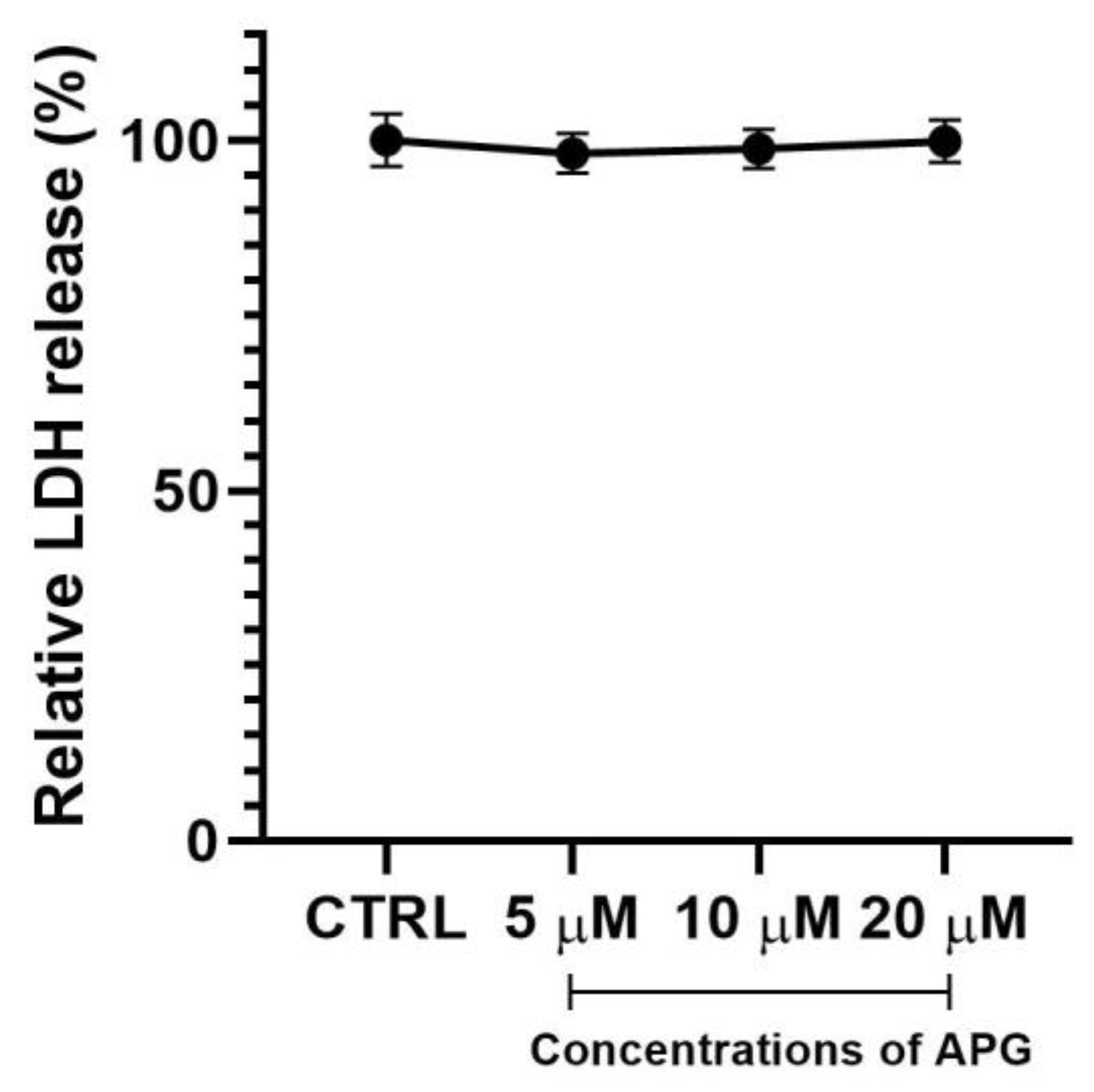
3.1. Apigenin Showed No Significant Effects on Cell Viability and Morphology of hOBs

The exposition to increasing concentrations of APG (5, 10 and 20 µM) for 24 h resulted in no cytotoxicity for osteoblasts (Figure 1).
APG at 5 µM produced a lower level of LDH release compared to untreated cells (CTRL) and compared to the other concentrations. However, the result for 5 µM was not statistically significant. The results of LDH assay were confirmed from the analysis of morphology, which was investigated only for the concentration of 5 µM (5-APG) at 24 h, because LDH release showed no differences at higher concentrations 10 and 20 µM. Microscopically no morphological differences (Figure 2B) were seen in comparison to the control cells (Figure 2A). As illustrated in Figure 2, cells appeared spindle shaped and well spread, better seen at higher magnification 40× (Figure 2C,D).

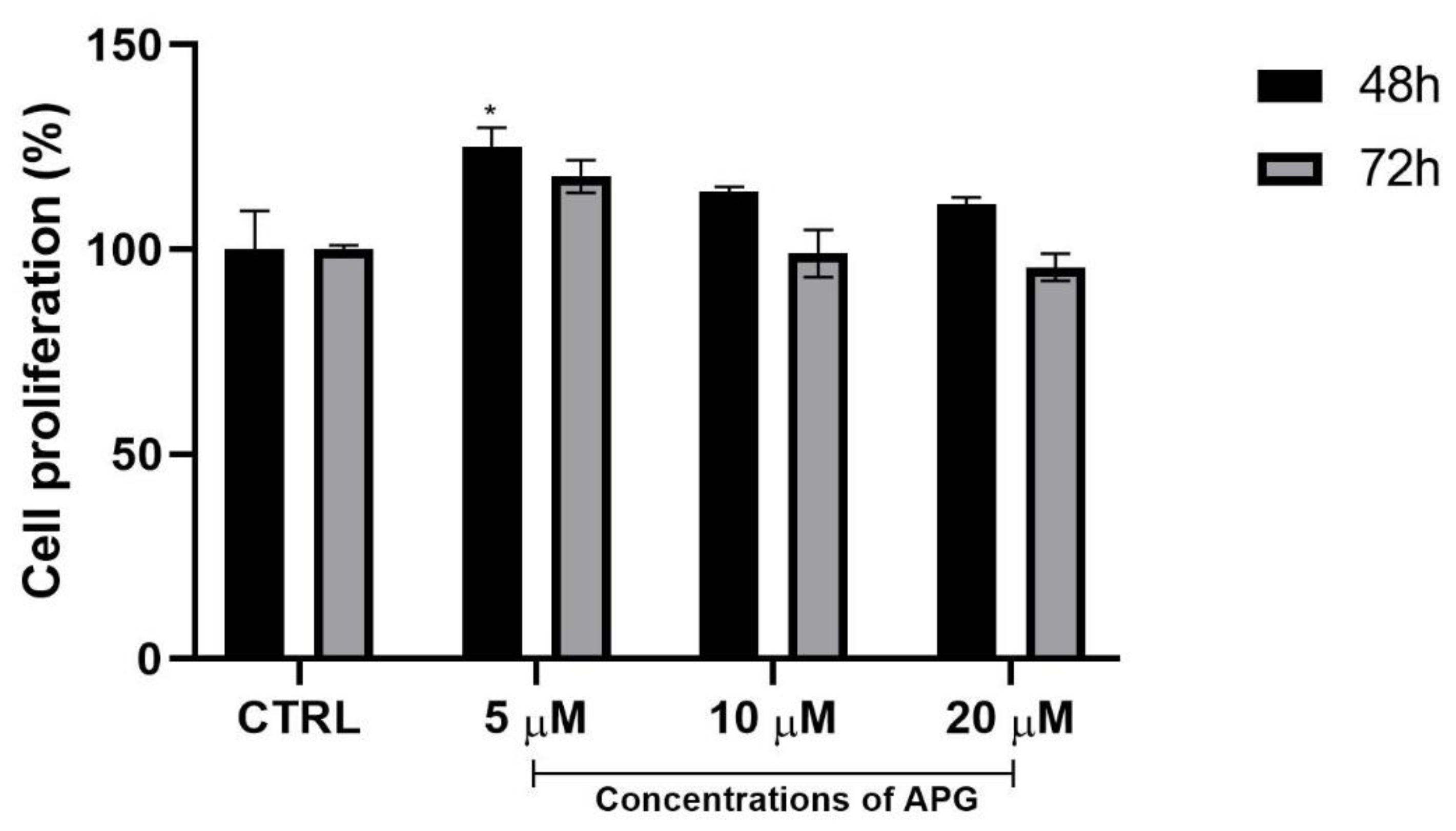
3.2. Apigenin Promoted Proliferation of hOBs

Based on the results of LDH assay and morphology observations, proliferative rate of cells treated with APG was evaluated at 48 h and 72 h. At 48 h, APG stimulated cell growth at all concentrations. In addition, the response of the cells was inversely proportional to the concentrations. In particular, the highest and most statistically significant rate was found when hOBs were treated with 5 µM compared to CTRL. A similar trend was observed when cells were incubated with APG for 72 h. However, just at 5 µM, cell growth resulted were enhanced, while at the higher 10 and 20 µM concentrations, cell growth appeared like the control; thus, any increment in the proliferation was observed at these concentrations (Figure 3).

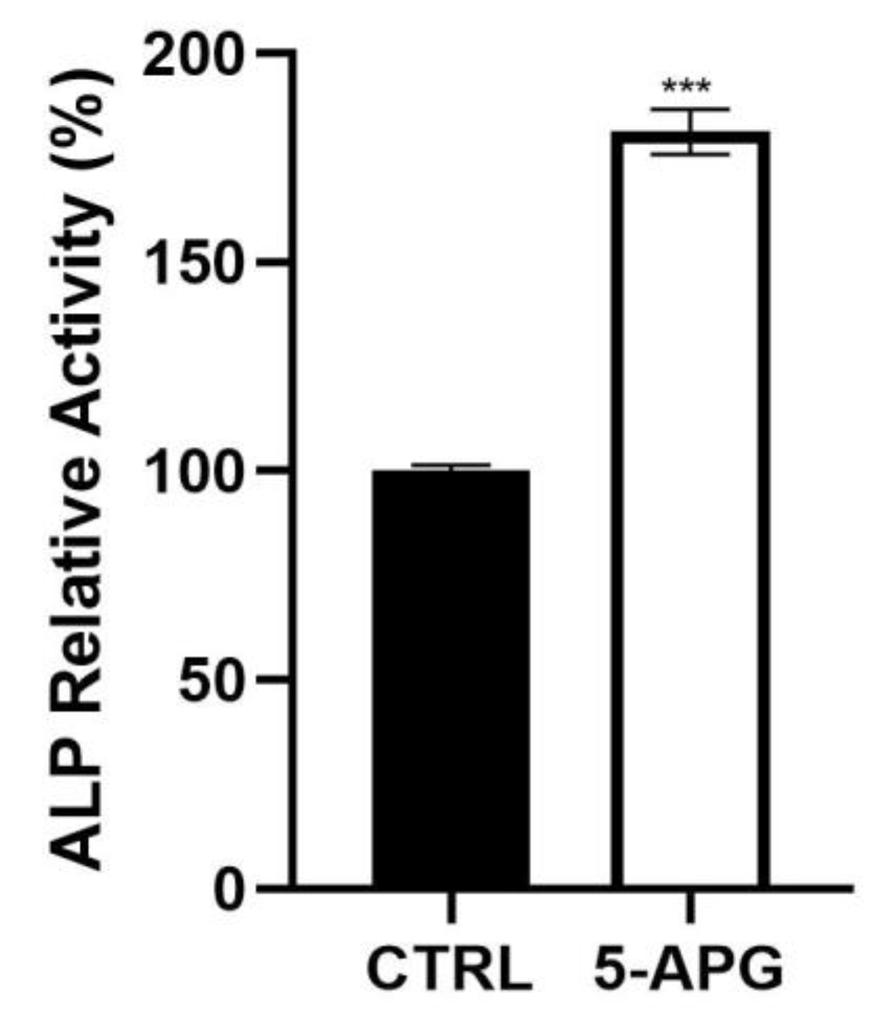
3.3. Apigenin Increased ALP Activity

Considering the results of LDH, morphology and cellular proliferation, ALP activity was evaluated only for APG at 5 µM (5-APG). The effects of 5-APG on hOBs were analyzed by cellular ALP activity. As shown in Figure 4, when the enzymatic activity of the cellular lysates was estimated at 7 days, a statistically significant increment was observed in hOBs treated with 5-APG with respect of CTRL.

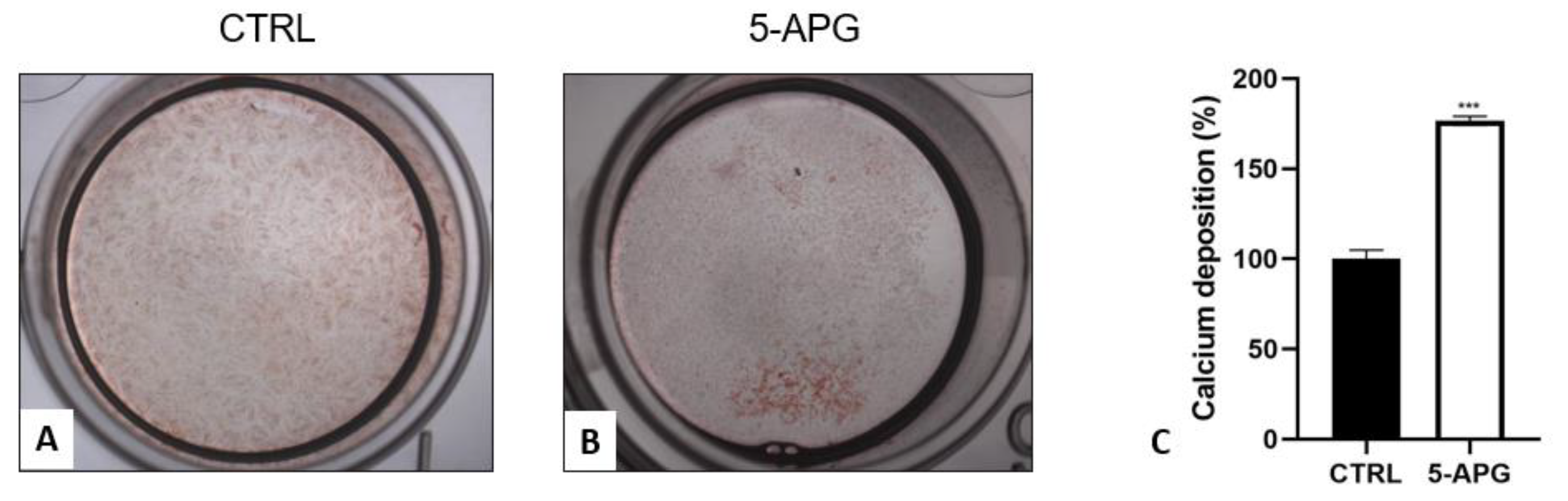
3.4. Apigenin Stimulated Mineral Deposition

When hOBs were incubated with 5 µM of APG, mineralization appeared stimulated with more mineralized nodules that can be well observed. These results were confirmed by the quantification of the calcium deposition. The images of the Alizarin Red staining (ARS) showed that a significant higher calcium nodule formation was found with 5 µM-APG. At 14 days, the calcified nodules appeared denser in wells where osteoblasts were incubated with 5-APG (Figure 5B) than in CTRL (Figure 5A). Moreover, quantitative analysis showed that treatment with APG significantly increased calcium deposition compared to CTRL (Figure 5C).

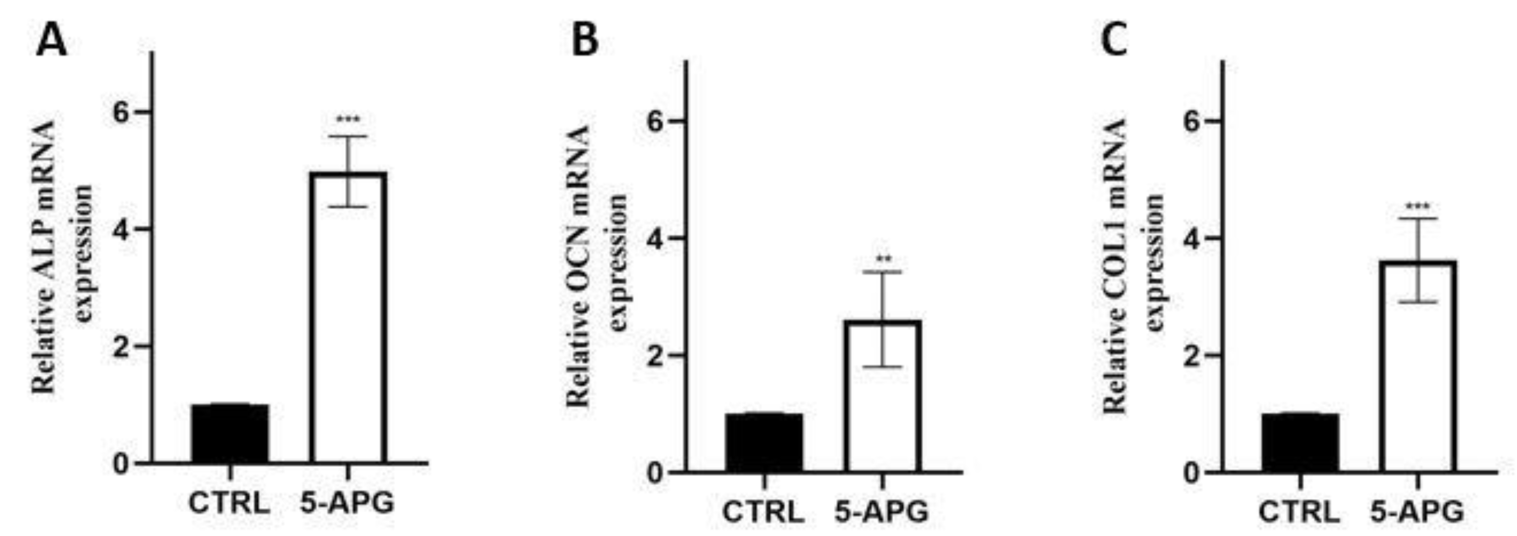
3.5. Apigenin Up-Regulated Mineralization-Related Markers

The expression of osteoblast marker genes ALP, OCN and COL1, evaluated by RT-qPCR, resulted in significantly higher hOBs treated with APG (5 µM) than in CTRL, as shown in Figure 6. The ALP expression resulted in the most up-regulated (Figure 6A) followed by COL1 (Figure 6C) and then by OCN (Figure 6B).

4. Discussion

In the present study, the biological effects of APG, a natural flavone with antioxidant and anti-inflammatory properties [9,10,28], were explored on oral osteoblasts cultured from human biopsies.
APG, thereby, might be considered as a potential active phytochemical compound to develop a therapeutic strategy for clinical practice. The bioactivity and biological effects of APG are dependent on its chemical structure: the inhibition of α-glucosidase and α-amylase is induced by the double bonds in the two aromatic rings and hydroxyl groups on C-7 and C-4′ [33]; the immunomodulatory properties are exerted by the C-4′ hydroxyl group in ring B [34]; the Liver X receptor activation is induced by the hydroxyl radicals at position 5, 7 and 4′ [35]. The aim of this study was to evaluate whether APG could be further considered as a bioactive agent capable of promoting bone regeneration for clinical applications in dentistry. In particular, APG could be beneficial to increase the osteoinductive properties in a bone graft mixed with other heterologous biomaterials. Treatment with APG (5, 10, 20 µM) did not affect the viability of hOBs, indicating that apigenin has no cytotoxicity for these cells. This result was confirmed by the images of the morphology study at 24 h, in which any morphological changes were detected in osteoblasts, which appeared typically shaped. Moreover, 5 µM APG slightly enhanced the cell metabolic activity, indicating a pro-proliferative effect at this concentration. Therefore, a proliferation study was performed at 48 h and 72 h after the hOBs were incubated with APG at concentrations of 5, 10 and 20 µM. At 48 h, APG stimulated cell proliferation at all concentrations, in a dose-dependent manner, where the response resulted inversely to the concentration and the highest and most statistically significant growth was observed in hOBs incubated with 5 µM APG compared to control cells. These results are in accordance with previous studies; indeed, APG was found to promote osteogenic differentiation in vitro and accelerate bone formation in vivo, indicating that APG may be a promising activator for bone repair [25]. APG has been reported to improve bone loss in ovariectomized rats [24]. In addition, 5 µM APG was able to stimulate cell proliferation also at 72 h, but the result was not significative. Thus, the lower concentration, 5 µM apigenin, seems to be the suitable concentration and this represents a strength to prevent potential adverse side effects related to higher-dose administration [36]. In a study with a murine model, higher doses of apigenin (100 or 200 mg/kg) caused damage to the the liver histoarchitecture, increased ALT, AST, ALP, ROS, ratio of oxidized to reduced glutathione (GSSG/GSH) and LPO [37]. A previous in vitro study reported that higher concentrations (>50 µM) of apigenin induced intracellular ROS accumulation with consequent cell death [38].
Based upon the results of cytotoxicity, morphology and proliferation study, APG at a concentration of 5 µM (5-APG) was selected for further investigations concerning the formation of the mineralized extracellular matrix. Thus, the activity of the ALP was assessed as the early phenotypic marker for mature osteoblasts, while the mineralized nodule formation as a phenotypic marker for the later stage. The results indicated that APG (5 µM) significantly increased activity of ALP at 7 days and the mineralized nodule formation at 14 days, suggesting that apigenin accelerates mineralization in osteoblasts. Based on the results of ALP activity and mineralized nodule formation, APG was verified to promote marker genes that are involved in the bone mineralization, OCN, COL1 and ALP. It was found that other phenolic compounds favored bone preservation and repair by increasing the expression of genes related to osteoblastogenesis, including RUNX-2, OSX, COL1, OSC and ALP [39]. In the present study, the expression of examined genes, mainly ALP and COL1, resulted in up-regulation after treatment with 5 µM APG. Considering that other phenolic compounds were found to stimulate bone tissue regeneration by acting on the bone morphogenetic protein (BMP) signaling pathway and that BMPs, in turn, act on the expression of bone-related genes, such as ALP [40,41,42], this pathway might be a possible molecular mechanism underlying the activity of apigenin in bone. Recently, the correlation between nutrients and bone health focused the attention of researchers to highlight the flavonoids from diet as important factors able to affect bone physiology [26,43]. Taken together, the data in the present study suggest that apigenin, a flavonoid compound, may have a beneficial effect on oral bone physiology, favoring osteoblast activities. These findings suggest that apigenin might provide insights into the development of tools useful for bone formation during clinical procedures, such as maxillary sinus floor elevation. In dentistry fields, apigenin might be taken into account for different aspects considering that the ideal periodontal biomaterials should fight against pathogenic bacteria and promote bone regeneration in bone reconstruction lesions or in bone defects. The antibacterial activities of apigenin against various Gram-negative and -positive bacterial species were reported [44]. Studies strongly recommend apigenin because of its antibacterial activities for the treatment of oral bacteria. These studies also showed the better effectiveness of antibiotics in combination with apigenin against these bacteria as compared to antibiotic treatment alone, indicating a synergic effect between apigenin and antibiotics [44,45]. Antiviral activities of apigenin were reported against different viruses [46,47]. Apigenin, due to its multiple properties, might be taken into account also for tissue engineering scaffolds in maintaining space, in supporting cell attachment and proliferation after being grafted to a bone defect. Indeed, tissue engineering scaffolds are developed in combination with growth factors or stem cells, as well as antibacterial components to improve osteoinduction, osteogenesis and antibacterial activity of bone grafting [48].
The beneficial effects of apigenin, on oral osteoblasts, here demonstrated, in addition to the already known other biological properties of apigenin [1,7], might potentially make apigenin a multifunctional bioactive agent, helping different situations in the dentistry field, considering that apigenin is a natural molecule, safe, with a very low risk of toxicity. Current treatment options for bone-related diseases, consisting of antiresorptive agents (such as bisphosphonates, hormone-replacement therapy, selective estrogen-receptor modulators and anti-RANKL antibodies) and/or anabolic agents (such as intermittent low doses of teriparatide and antisclerostin antibodies), are not free from adverse effects that limit their use [49]. Thus, the beneficial properties of apigenin on oral osteoblasts include that it is a natural molecule, safe, representing its strength for future use in the dentistry field, in which bone regeneration and new bone formation represent a challenge. The limit of APG clinical application is its poor solubility. At present, to overcome this limit, some approaches to nanoparticle drug carriers have been formulated to improve the solubility, dissolution and the oral bioavailability of APG [50]. In the dentistry field, this limit of apigenin might not represent an obstacle, in the development of tools useful to improve osteoconductivity and osseointegration, for example, as coatings on dental implant surfaces or to favor dental restorations by incorporating apigenin into dental composites. A strength of our study is that the lowest concentration here used, 5 µM, resulted in a suitable concentration to improve the biological functions of oral osteoblasts and this represents a strength to prevent potential adverse side effects related to higher doses [36,37,38]. Further in vitro investigation is required to clarify the detailed molecular mechanisms underlying the activity of apigenin in the bone of the oral cavity and further in vivo investigations are needed to verify a possible clinical application.

5. Conclusions

In conclusion, this study showed that apigenin promotes bone-forming osteoblasts through stimulating proliferative and mineralization activities and up-regulating osteogenic markers. Thus, the next step in our research will be to test apigenin as a dental implant coating to favor osseointegration, which represents a challenge in the dentistry field.

Author Contributions

Conceptualization, M.P., G.I.; methodology, T.V.P., E.D.; validation, M.P., G.I., A.P. (Adriano Piattelli); formal analysis, N.D.P., F.L., A.S.; investigation, T.V.P., E.D., S.L.; resources, G.I., M.P., A.P. (Assunta Pandolfi); data curation, T.V.P., E.D.; writing—original draft preparation, T.V.P., E.D., M.P.; writing—review and editing, G.I., A.P. (Adriano Piattelli), A.P. (Assunta Pandolfi), A.S., N.D.P., F.L.; supervision, M.P., G.I., A.P. (Adriano Piattelli). All authors have read and agreed to the published version of the manuscript.

Funding

This work was supported by G.I., M.P. FAR GRANT University of Chieti–Pescara Fund; by Assunta Pandolfi PON-MISE Sustainable Growth Funding-DD 27 September 2018 (Grant Number: 21, ex 230).

Institutional Review Board Statement

The study was conducted in accordance with the Declaration of Helsinki and approved by Ethics Committee of University G. d’Annunzio of Chieti-Pescara (reference numbers: BONEISTO N. 22-10.07.2021).

Informed Consent Statement

Informed consent was obtained from all subjects involved in the study.

Data Availability Statement

Not applicable.

Conflicts of Interest

The authors declare no conflict of interest.

References

  1. Abid, R.; Ghazanfar, S.; Farid, A.; Sulaman, S.M.; Idrees, M.; Amen, R.A.; Muzammal, M.; Shahzad, M.K.; Mohamed, M.O.; Khaled, A.A.; et al. Pharmacological Properties of 4′,5,7-Trihydroxyflavone (Apigenin) and Its Impact on Cell Signaling Pathways. Molecules 2022, 27, 4304. [Google Scholar] [CrossRef]
  2. Saldanha, E.; Pai, R.J.; George, T.; D’Souza, S.; Adnan, M.; Pais, M.; Naik, T.; D’Souza, R.C.C.; D’Cunha, R.; Shrinath Baliga, M. Health Effects of Various Dietary Agents and Phytochemicals (Therapy of Acute Pancreatitis). Ther. Probiotic Unconv. Foods 2018, 303–314. [Google Scholar] [CrossRef]
  3. Švehlíková, V.; Bennett, R.N.; Mellon, F.A.; Needs, P.W.; Piacente, S.; Kroon, P.A.; Bao, Y. Isolation, Identification and Stability of Acylated Derivatives of Apigenin 7-O-Glucoside from Chamomile (Chamomilla recutita [L.] Rauschert). Phytochemistry 2004, 65, 2323–2332. [Google Scholar] [CrossRef]
  4. Yan, X.; Qi, M.; Li, P.; Zhan, Y.; Shao, H. Apigenin in Cancer Therapy: Anti-Cancer Effects and Mechanisms of Action. Cell Biosci. 2017, 7, 50. [Google Scholar] [CrossRef]
  5. Jang, J.Y.; Sung, B.; Kim, N.D. Role of Induced Programmed Cell Death in the Chemopreventive Potential of Apigenin. Int. J. Mol. Sci. 2022, 23, 3757. [Google Scholar] [CrossRef]
  6. Navarro-Núñez, L.; Lozano, M.L.; Palomo, M.; Martínez, C.; Vicente, V.; Castillo, J.; Benavente-García, O.; Diaz-Ricart, M.; Escolar, G.; Rivera, J. Apigenin Inhibits Platelet Adhesion and Thrombus Formation and Synergizes with Aspirin in the Suppression of the Arachidonic Acid Pathway. J. Agric. Food Chem. 2008, 56, 2970–2976. [Google Scholar] [CrossRef]
  7. Salehi, B.; Venditti, A.; Sharifi-Rad, M.; Kręgiel, D.; Sharifi-Rad, J.; Durazzo, A.; Lucarini, M.; Santini, A.; Souto, E.B.; Novellino, E.; et al. The Therapeutic Potential of Apigenin. Int. J. Mol. Sci. 2019, 20, 1305. [Google Scholar] [CrossRef]
  8. Rezai-Zadeh, K.; Ehrhart, J.; Bai, Y.; Sanberg, P.R.; Bickford, P.; Tan, J.; Douglas, R.D. Apigenin and Luteolin Modulate Microglial Activation via Inhibition of STAT1-Induced CD40 Expression. J. Neuroinflamm. 2008, 5, 41. [Google Scholar] [CrossRef]
  9. Hu, W.; Wang, X.; Wu, L.; Shen, T.; Ji, L.; Zhao, X.; Si, C.L.; Jiang, Y.; Wang, G. Apigenin-7-O-β-D-Glucuronide Inhibits LPS-Induced Inflammation through the Inactivation of AP-1 and MAPK Signaling Pathways in RAW 264.7 Macrophages and Protects Mice against Endotoxin Shock. Food Funct. 2016, 7, 1002–1013. [Google Scholar] [CrossRef]
  10. Yano, S.; Umeda, D.; Maeda, N.; Fujimura, Y.; Yamada, K.; Tachibana, H. Dietary Apigenin Suppresses IgE and Inflammatory Cytokines Production in C57BL/6N Mice. J. Agric. Food Chem. 2006, 54, 5203–5207. [Google Scholar] [CrossRef]
  11. Xu, Y.; Li, X.; Wang, H. Protective Roles of Apigenin Against Cardiometabolic Diseases: A Systematic Review. Front. Nutr. 2022, 9, 875826. [Google Scholar] [CrossRef]
  12. Su, T.; Huang, C.; Yang, C.; Jiang, T.; Su, J.; Chen, M.; Fatima, S.; Gong, R.; Hu, X.; Bian, Z.; et al. Apigenin Inhibits STAT3/CD36 Signaling Axis and Reduces Visceral Obesity. Pharmacol. Res. 2020, 152, 104586. [Google Scholar] [CrossRef]
  13. Ono, M.; Fujimori, K. Antiadipogenic Effect of Dietary Apigenin through Activation of AMPK in 3T3-L1 Cells. J. Agric. Food Chem. 2011, 59, 13346–13352. [Google Scholar] [CrossRef]
  14. Guo, X.; Liu, J.; Cai, S.; Wang, O.; Ji, B. Synergistic Interactions of Apigenin, Naringin, Quercetin and Emodin on Inhibition of 3T3-L1 Preadipocyte Differentiation and Pancreas Lipase Activity. Obes. Res. Clin. Pract. 2016, 10, 327–339. [Google Scholar] [CrossRef]
  15. Solini, A.; Suvan, J.; Santini, E.; Gennai, S.; Seghieri, M.; Masi, S.; Petrini, M.; D’Aiuto, F.; Graziani, F. Periodontitis Affects Glucoregulatory Hormones in Severely Obese Individuals. Int. J. Obes. 2019, 43, 1125–1129. [Google Scholar] [CrossRef]
  16. Taylor, R. Type 2 Diabetes. Diabetes Care 2013, 36, 1047–1055. [Google Scholar] [CrossRef]
  17. Zhang, B.; Li, X.; Sun, W.; Xing, Y.; Xiu, Z.; Zhuang, C.; Dong, Y. Dietary Flavonoids and Acarbose Synergistically Inhibit α-Glucosidase and Lower Postprandial Blood Glucose. J. Agric. Food Chem. 2017, 65, 8319–8330. [Google Scholar] [CrossRef]
  18. Lee, J.N.; Dutta, R.K.; Maharjan, Y.; Liu, Z.; Lim, J.-Y.; Kim, S.-J.; Cho, D.-H.; So, H.-S.; Choe, S.-K.; Park, R. Catalase Inhibition Induces Pexophagy through ROS Accumulation. Biochem. Biophys. Res. Commun. 2018, 501, 696–702. [Google Scholar] [CrossRef]
  19. Ren, B.; Qin, W.; Wu, F.; Wang, S.; Pan, C.; Wang, L.; Zeng, B.; Ma, S.; Liang, J. Apigenin and Naringenin Regulate Glucose and Lipid Metabolism, and Ameliorate Vascular Dysfunction in Type 2 Diabetic Rats. Eur. J. Pharmacol. 2016, 773, 13–23. [Google Scholar] [CrossRef]
  20. Molina Prats, P.; Gómez Garcia, F.; Martinez Diaz, F.; Amaral Mendes, R.; Lopez-Jornet, P. The Therapeutic Effects of Apigenin and Dexamethasone on 5-Fluorouracil-Induced Oral Mucositis—A Pilot Study Using a Syrian Hamster Model. J. Oral Pathol. Med. 2017, 46, 142–147. [Google Scholar] [CrossRef]
  21. Maggioni, D.; Garavello, W.; Rigolio, R.; Pignataro, L.; Gaini, R.; Nicolini, G. Apigenin Impairs Oral Squamous Cell Carcinoma Growth in Vitro Inducing Cell Cycle Arrest and Apoptosis. Int. J. Oncol. 2013, 43, 1675–1682. [Google Scholar] [CrossRef]
  22. Silvan, S.; Manoharan, S.; Baskaran, N.; Anusuya, C.; Karthikeyan, S.; Prabhakar, M.M. Chemopreventive Potential of Apigenin in 7,12-Dimethylbenz(a)Anthracene Induced Experimental Oral Carcinogenesis. Eur. J. Pharmacol. 2011, 670, 571–577. [Google Scholar] [CrossRef]
  23. Ramesh, P.; Jagadeesan, R.; Sekaran, S.; Dhanasekaran, A.; Vimalraj, S. Flavonoids: Classification, Function, and Molecular Mechanisms Involved in Bone Remodelling. Front. Endocrinol. 2021, 12, 1564. [Google Scholar] [CrossRef]
  24. Park, J.A.; Ha, S.K.; Kang, T.H.; Oh, M.S.; Cho, M.H.; Lee, S.Y.; Park, J.H.; Kim, S.Y. Protective Effect of Apigenin on Ovariectomy-Induced Bone Loss in Rats. Life Sci. 2008, 82, 1217–1223. [Google Scholar] [CrossRef]
  25. Pan, F.F.; Shao, J.; Shi, C.J.; Li, Z.P.; Fu, W.M.; Zhang, J.F. Apigenin Promotes Osteogenic Differentiation of Mesenchymal Stem Cells and Accelerates Bone Fracture Healing via Activating Wnt/β-Catenin Signaling. Am. J. Physiol. Endocrinol. Metab. 2021, 320, E760–E771. [Google Scholar] [CrossRef]
  26. Mandatori, D.; Penolazzi, L.; Pelusi, L.; Lambertini, E.; Michelucci, F.; Porreca, A.; Cerritelli, P.; Pipino, C.; di Iorio, A.; Bruni, D.; et al. Three-Dimensional Co-Culture System of Human Osteoblasts and Osteoclast Precursors from Osteoporotic Patients as an Innovative Model to Study the Role of Nutrients: Focus on Vitamin K2. Nutrients 2021, 13, 2823. [Google Scholar] [CrossRef]
  27. Bandyopadhyay, S.; Lion, J.M.; Mentaverri, R.; Ricupero, D.A.; Kamel, S.; Romero, J.R.; Chattopadhyay, N. Attenuation of Osteoclastogenesis and Osteoclast Function by Apigenin. Biochem. Pharmacol. 2006, 72, 184–197. [Google Scholar] [CrossRef]
  28. Jung, W.W. Protective Effect of Apigenin against Oxidative Stress-Induced Damage in Osteoblastic Cells. Int. J. Mol. Med. 2014, 33, 1327–1334. [Google Scholar] [CrossRef]
  29. Aryal, Y.P.; Yeon, C.Y.; Kim, T.Y.; Lee, E.S.; Sung, S.; Pokharel, E.; Kim, J.Y.; Choi, S.Y.; Yamamoto, H.; Sohn, W.J.; et al. Facilitating Reparative Dentin Formation Using Apigenin Local Delivery in the Exposed Pulp Cavity. Front. Physiol. 2021, 12, 2157. [Google Scholar] [CrossRef]
  30. Avila-Ortiz, G.; Neiva, R.; Galindo-Moreno, P.; Rudek, I.; Benavides, E.; Wang, H.L. Analysis of the Influence of Residual Alveolar Bone Height on Sinus Augmentation Outcomes. Clin. Oral Implant. Res. 2012, 23, 1082–1088. [Google Scholar] [CrossRef] [Green Version]
  31. Bathla, S.C.; Fry, R.R.; Majumdar, K. Maxillary Sinus Augmentation. J. Indian Soc. Periodontol. 2018, 22, 468–473. [Google Scholar] [CrossRef] [PubMed]
  32. Pierfelice, T.V.; D’amico, E.; Iezzi, G.; Piattelli, A.; di Pietro, N.; D’arcangelo, C.; Comuzzi, L.; Petrini, M. Nanoporous Titanium Enriched with Calcium and Phosphorus Promotes Human Oral Osteoblast Bioactivity. Int. J. Environ. Res. Public Health 2022, 19, 6212. [Google Scholar] [CrossRef] [PubMed]
  33. Li, K.; Yao, F.; Xue, Q.; Fan, H.; Yang, L.; Li, X.; Sun, L.; Liu, Y. Inhibitory Effects against α-Glucosidase and α-Amylase of the Flavonoids-Rich Extract from Scutellaria Baicalensis Shoots and Interpretation of Structure–Activity Relationship of Its Eight Flavonoids by a Refined Assign-Score Method. Chem. Cent. J. 2018, 12, 82. [Google Scholar] [CrossRef] [PubMed]
  34. Kilani-Jaziri, S.; Mustapha, N.; Mokdad-Bzeouich, I.; el Gueder, D.; Ghedira, K.; Ghedira-Chekir, L. Flavones Induce Immunomodulatory and Anti-Inflammatory Effects by Activating Cellular Anti-Oxidant Activity: A Structure-Activity Relationship Study. Tumor Biol. 2016, 37, 6571–6579. [Google Scholar] [CrossRef]
  35. Fouache, A.; Zabaiou, N.; de Joussineau, C.; Morel, L.; Silvente-Poirot, S.; Namsi, A.; Lizard, G.; Poirot, M.; Makishima, M.; Baron, S.; et al. Flavonoids Differentially Modulate Liver X Receptors Activity—Structure-Function Relationship Analysis. J. Steroid Biochem. Mol. Biol. 2019, 190, 173–182. [Google Scholar] [CrossRef]
  36. Birkett, D.J. Pharmacokinetics Made Easy 10 Pharmacodynamics—The Concentration-Effect Relationship. Aust. Prescr. 1995, 18, 102–104. [Google Scholar] [CrossRef]
  37. Singh, P.; Mishra, S.K.; Noel, S.; Sharma, S.; Rath, S.K. Acute Exposure of Apigenin Induces Hepatotoxicity in Swiss Mice. PLoS ONE 2012, 7, e31964. [Google Scholar] [CrossRef]
  38. Miyoshi, N.; Naniwa, K.; Yamada, T.; Osawa, T.; Nakamura, Y. Dietary Flavonoid Apigenin Is a Potential Inducer of Intracellular Oxidative Stress: The Role in the Interruptive Apoptotic Signal. Arch. Biochem. Biophys. 2007, 466, 274–282. [Google Scholar] [CrossRef]
  39. Melguizo-Rodríguez, L.; Manzano-Moreno, F.J.; Illescas-Montes, R.; Ramos-Torrecillas, J.; de Luna-Bertos, E.; Ruiz, C.; García-Martínez, O. Bone Protective Effect of Extra-Virgin Olive Oil Phenolic Compounds by Modulating Osteoblast Gene Expression. Nutrients 2019, 11, 1722. [Google Scholar] [CrossRef]
  40. Liang, W.; Lin, M.; Li, X.; Li, C.; Gao, B.; Gan, H.; Yang, Z.; Lin, X.; Liao, L.; Yang, M. Icariin Promotes Bone Formation via the BMP-2/Smad4 Signal Transduction Pathway in the HFOB 1.19 Human Osteoblastic Cell Line. Int. J. Mol. Med. 2012, 30, 889–895. [Google Scholar] [CrossRef] [Green Version]
  41. Xu, B.; Wang, X.; Wu, C.; Zhu, L.; Chen, O.; Wang, X. Flavonoid Compound Icariin Enhances BMP-2 Induced Differentiation and Signalling by Targeting to Connective Tissue Growth Factor (CTGF) in SAMP6 Osteoblasts. PLoS ONE 2018, 13, e0200367. [Google Scholar] [CrossRef]
  42. Rawadi, G.; Vayssière, B.; Dunn, F.; Baron, R.; Roman-Roman, S. BMP-2 Controls Alkaline Phosphatase Expression and Osteoblast Mineralization by a Wnt Autocrine Loop. J. Bone Miner. Res. 2003, 18, 1842–1853. [Google Scholar] [CrossRef]
  43. Movassagh, E.Z.; Vatanparast, H. Current Evidence on the Association of Dietary Patterns and Bone Health: A Scoping Review. Adv. Nutr. 2017, 8, 1–16. [Google Scholar] [CrossRef]
  44. Nayaka, H.B.; Londonkar, R.L.; Umesh, M.K.; Tukappa, A. Antibacterial Attributes of Apigenin, Isolated from Portulaca Oleracea L. Int. J. Bacteriol. 2014, 2014, 175851. [Google Scholar] [CrossRef]
  45. Adamczak, A.; Ożarowski, M.; Karpiński, T.M. Antibacterial Activity of Some Flavonoids and Organic Acids Widely Distributed in Plants. J. Clin. Med. 2019, 9, 109. [Google Scholar] [CrossRef]
  46. Zhang, W.; Qiao, H.; Lv, Y.; Wang, J.; Chen, X.; Hou, Y.; Tan, R.; Li, E. Apigenin Inhibits Enterovirus-71 Infection by Disrupting Viral RNA Association with Trans-Acting Factors. PLoS ONE 2014, 9, e110429. [Google Scholar] [CrossRef]
  47. Shibata, C.; Ohno, M.; Otsuka, M.; Kishikawa, T.; Goto, K.; Muroyama, R.; Kato, N.; Yoshikawa, T.; Takata, A.; Koike, K. The Flavonoid Apigenin Inhibits Hepatitis C Virus Replication by Decreasing Mature MicroRNA122 Levels. Virology 2014, 462–463, 42–48. [Google Scholar] [CrossRef]
  48. Liao, Y.; Li, H.; Shu, R.; Chen, H.; Zhao, L.; Song, Z.; Zhou, W. Mesoporous Hydroxyapatite/Chitosan Loaded with Recombinant-Human Amelogenin Could Enhance Antibacterial Effect and Promote Periodontal Regeneration. Front. Cell. Infect. Microbiol. 2020, 10, 180. [Google Scholar] [CrossRef]
  49. Wong, S.K.; Chin, K.Y.; Ima-Nirwana, S. Quercetin as an Agent for Protecting the Bone: A Review of the Current Evidence. Int. J. Mol. Sci. 2020, 21, 6448. [Google Scholar] [CrossRef]
  50. Nozhat, Z.; Heydarzadeh, S.; Memariani, Z.; Ahmadi, A. Chemoprotective and Chemosensitizing Effects of Apigenin on Cancer Therapy. Cancer Cell Int. 2021, 21, 574. [Google Scholar] [CrossRef]
Figure 1. Effects of APG treatment (24 h) on LDH release. Values represent mean ± SD expressed in percentages compared to control (CTRL).
Applsci 12 08510 g001
Figure 2. Effects of APG at 5 µM (5-APG) for 24 h on cell morphology, evaluated by toluidine blue staining. Cells were observed using a stereomicroscope at the magnification of 6× (A,B) and 40× (C,D).
Applsci 12 08510 g002
Figure 3. Effects of APG, used at increasing concentration for 48 h and 72 h, on hOBs proliferation determined using MTS assay. Data are expressed as percentages with respect to the control. Statistical analysis was performed by ANOVA test (* p < 0.05 compared to CTRL).
Applsci 12 08510 g003
Figure 4. Effects of apigenin at 5 µM (5-APG) for 7 days on the alkaline phosphatase activity (ALP). Data are presented as the mean ± SD and are expressed as percentages over the control (cells not treated with APG) (*** p < 0.0001).
Applsci 12 08510 g004
Figure 5. Effects of APG at 5 µM (5-APG) for 14 days on mineralization (A,B). Calcium nodules appeared bright red in color by Alizarin Red staining. (C) Calcium deposition was quantified by CPC. The comparison of data between controls and test group showed significant difference (*** p < 0.0001).
Applsci 12 08510 g005
Figure 6. Effects of 5 µM of APG (5-APG) on gene expression. ALP (alkaline phosphate) (A), OCN (osteocalcin) (B), COL1 (collagen 1) (C) gene expression was evaluated after 7 days of treatment. Results are reported as mean expression values normalized on the housekeeping β-actin in triplicate with error bars representing the SD (** p < 0.001; *** p < 0.0001).
Applsci 12 08510 g006
Table 1. Primer sequences used in RT-qPCR.
GeneForward Primer (5′-3′)Reverse Primer (5′-3′)
ALP
OCN
COL1
AATGAGTGAGTGACCATCCTGG
TCAGCCAACTCGTCACAGTC AGTCAGAGTGAGGACAGTGAATTG
GCACCCCAAGACCTGCTTTAT
GGCGCTACCTGTATCAATGG CACATCACACCAGGAAGTGC
βACTCCAGAGGCGTACAGGGATAGGAGAAGATGACCCAGGACTCTC
Publisher’s Note: MDPI stays neutral with regard to jurisdictional claims in published maps and institutional affiliations.

