Article

Orthodontic Loads in Teeth after Regenerative Endodontics: A Finite Element Analysis of the Biomechanical Performance of the Periodontal Ligament

by
Cristina Bucchi
1,*,
Massimo Del Fabbro
2,3 and
Jordi Marcé-Nogué
4,5
1
Department of Integral Adult Dentistry, Oral Biology Research Centre, Universidad de La Frontera, Av. Francisco Salazar 01145, Temuco 4811230, Chile
2
Dipartimento di Scienze Biomediche, Chirurgiche ed Odontoiatriche, Università degli Studi di Milano, Via della Commenda 10, 20122 Milan, Italy
3
IRCCS Istituto Ortopedico Galeazzi, Via Riccardo Galeazzi, 4, 20161 Milan, Italy
4
Department of Mechanical Engineering, Universitat Rovira i Virgili, Carrer de l’Escorxador, s/n, 43003 Tarragona, Spain
5
Institut Català de Paleontologia Miquel Crusafont, Universitat Autònoma de Barcelona, Carrer de les Columnes, s/n, 08193 Cerdanyola del Valles, Spain
*
Author to whom correspondence should be addressed.
Appl. Sci. 2022, 12(14), 7063; https://doi.org/10.3390/app12147063
Submission received: 25 April 2022 / Revised: 27 May 2022 / Accepted: 29 May 2022 / Published: 13 July 2022
(This article belongs to the Topic State-of-the-Art Dentistry and Oral Health)

Abstract

:
The objective of this study was to analyse the stress distribution in the periodontal ligament and tooth structure of a cementum-reinforced tooth, a dentine-reinforced tooth and an immature tooth during orthodontic loads using a finite element analysis. A finite element model of a maxillary incisor and its supporting tissues was developed. The root was segmented into two parts: a part that represented a root in an immature state and an apical part that represented the tissue formed after regenerative endodontics. The apical part was given the mechanical properties of dentine or cementum. The three models underwent simulation of mesial load, palatal inclination and rotation. The mean stress values and stress distribution patterns of the periodontal ligament of the dentine- and cementum-reinforced teeth were similar in all scenarios. The maturation of the root, with either dentine or cementum, was beneficial for all scenarios, since the periodontal ligament of the immature tooth showed the highest mean stress values. Under the condition of this computational study, orthodontic loads can be applied in teeth previously treated with regenerative endodontics, since the distribution of stress is similar to those of physiologically mature teeth. In vivo studies should be performed to validate these results.

1. Introduction

Regenerative endodontics is a relatively new treatment aimed at regenerating the pulp-dentine complex after pulp necrosis in immature teeth [1]. Until very recently the standard treatment for such teeth was apexification. However, the latter did not allow further root development or revascularisation of the tooth, which remained with thin and short dentine walls [2]. Regenerative endodontics consists of a new cell-based approach and utilises the stem cells located at the periapical tissues that invade the root canal after over-instrumentation of the apex [3], and the growth factors present in the dentine are able to direct stem cell proliferation, migration and differentiation [4], with the final aim of revitalising and inducing further root development of the necrotic tooth. Regenerative endodontics is considered a successful clinical treatment since most teeth show further root development, healing of periapical lesions and resolution of symptomatology [5]. However, with the exception of occasional studies [6], regeneration of the pulp-dentine complex has not been observed in vivo. Thus, the posttreatment development of the root is due to the apposition of cementum-like tissue or osteodentine instead of tubular dentine [7,8,9].
Dentine is an organised and mineralised tissue with a tubuli-like structure [10] that is able to endure compressive forces, while cementum and other reparative tissues formed after regenerative endodontics have collagen fibres not packed in an orderly fashion [11] and a higher organic composition [12]. As a result, cementum has poorer mechanical properties than dentine [13]. A computational study showed that the mechanical performance of roots reinforced with cementum after regenerative endodontics have been proven to distribute the mechanical stress disadvantageously and unevenly compared to a root reinforced with dentine [14]. In teeth after regenerative endodontics, cementum barely endures stress, which is mainly concentrated in the dentine part of the root [14]. However, it is unknown whether this disadvantageous distribution of stress is transferred and maintained in the periodontal ligament and alveolar bone, producing high peaks of mechanical stress in specific parts of these tissues (i.e., in the neighbouring areas of the dentine with peak stress), or in contrast, whether the periodontal ligament is able to re-distribute the mechanical stress more evenly due to its unique biomechanical and cellular nature [15]. The performance of the periodontal ligament of teeth after regenerative endodontics has not been studied.
Periodontal ligament stem cells are sensitive to mechanical loading and play a critical role in periodontal and osseous remodelling during orthodontic movement by regulating the balance between osteoblastic and osteoclastic activity [16]. Orthodontic tooth movement is produced by mechanical forces and promoted by the remodelling of the periodontal ligament and alveolar bone. Under appropriate stimulation, on the compression side, the periodontal ligament becomes compressed, resulting in a focal necrosis [17]. These necrotic sites release chemokines that induce the migration of multinucleated cells that resorb the necrotic periodontal ligament as well as the underlying bone and cementum [17], and this process is stopped only when mechanical stimulation ceases. Thus, appropriate orthodontic forces produce minor reversible injury to the tooth-supporting tissues [17]. However, when these forces are inappropriate, such as in the case of excessive force magnitude or adequate magnitude but with high concentration of the stress in specific areas, they lead to a sustained inflammation process that reabsorbs the root [18].
This has raised the question of whether cementum-reinforced teeth (i.e., teeth that have been previously treated with regenerative endodontics), which seem to distribute mechanical stress disadvantageously compared to physiologically developed roots [14], can endure orthodontic movement or if they are at risk of inflammatory root resorption, as suggested by the European Society of Endodontology Position Statement for Revitalisation [19]. As for today, there is little clinical evidence, limited to case reports, of revitalised teeth treated with orthodontics [20,21].
The objective of this study was to analyse the stress distribution in the periodontal ligament of a cementum-reinforced tooth, a dentine-reinforced tooth and an immature tooth during simulation of orthodontic loads in three different directions using a finite element analysis (FEA) of a maxillary central incisor.

2. Materials and Methods

This study was approved by the Ethics Committee of the University of Barcelona and conducted in accordance with the Code of Ethics of the World Medical Association (Declaration of Helsinki) for experiments involving humans. The FEA model was constructed based on a μCT scan of the maxilla of a male 59-year-old donor (Quantum FX microCT, PerkinElmer, Waltham, MA, USA).
The μCT scan was conducted with 90 kV and 160 mA, FOV 60 mm, exposure time of 4.5 min and voxel size of 118 × 118 × 118 μm. The right maxillary central incisor was selected as the treated tooth and the dental and supporting tissues, including enamel, dentine, cementum, periodontal ligament and alveolar bone, were segmented with Seg3D software (v. 2.4.3, Center for Integrative Biomedical Computing, Salt Lake City, UT, USA). The periodontal ligament was segmented according to its natural shape. It had a maximum thickness of 0.38 mm in the coronal part and a minimum thickness of 0.11 mm in the middle third, having an hourglass distribution. As reported previously [14], the root was segmented into two parts: the coronal part, representing the immature root (stage 4 based on Cvek’s classification [22], and an apical part (the core part and the tip of the root), representing the tissue formed after regenerative endodontics (Figure 1a).

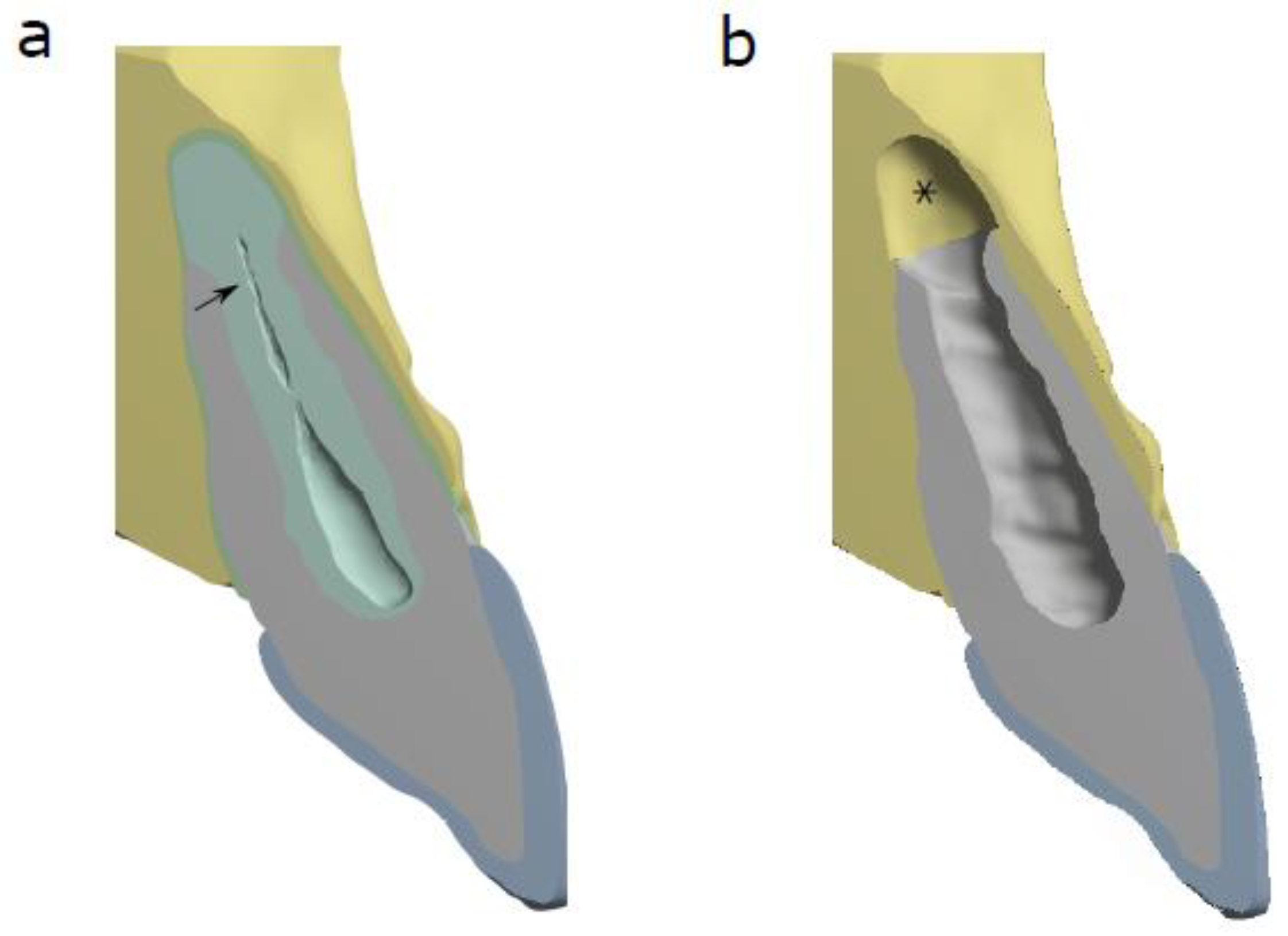
2.1. 3D FEA Models and Tissue Properties

Three different non-linear models representing three different scenarios were solved using ANSYS Mechanical 19.1. Each model was meshed using tetrahedral elements with a quadratic interpolation, except the elements of the mesh of the PDL that were created using a linear interpolation.
Three models were designed. Model 1 represented the tooth in an immature state (IT) (i.e., coronal part without the apical part) (Figure 1b), Model 2 represented a physiologically mature tooth (DR) (the coronal and apical part were given the mechanical properties of dentine) and Model 3 represented a tooth after regenerative endodontics (CR) (coronal part was given the mechanical properties of dentine and the apical part the mechanical properties of cementum) (Figure 1a). The segmented alveolar bone was equal in the three models, and it was not modified in the IT since the space from the tip of the root to the bottom of the alveolar socket represented the space occupied by the apical papilla (Figure 1b). This made it possible to perform the simulations in standardised models and compare the outcomes among the models.
The material properties (Young’s Modulus and the Poisson coefficient) for enamel, dentine, cementum and bone are described in Table 1. Elastic, linear and homogeneous material properties were assumed for enamel, dentine, cementum and bone, while a non-linear hyperelastic material was assumed for the periodontal ligament using a nine-parameter Mooney–Rivlin constitutive equation [23], as previously described [14] (Table 2).

2.2. Loading Scenario

Three typical orthodontic loads (load applied in a mesial direction (mesial load), palatal inclination and rotation) were simulated for Models 1, 2 and 3. Briefly, for the mesial load scenario, a force of 0.6 N was simulated in a mesial direction; for the inclination scenario, a load of 0.6 N was simulated in a palatal direction (the crown moves from the vestibular side to the palatal side); and for the rotation scenario, a load of 0.6 N was applied at the mesiovestibular aspect [24].

2.3. Analysis of Stress

Different metrics of stress distributions were evaluated for the three models (immature tooth, dentine-reinforced tooth and cementum-reinforced tooth) in the different loading scenarios. Stress values of the tooth and periodontal ligament of each model were analysed separately to evaluate stress distribution throughout the model. For the tooth tissues and the periodontal ligament, we computed the Rankine stress (or maximum stress theory) [25] to convert the multiaxial stress state to an equivalent stress state in function of the principal stresses. The plot of maximum principal stresses shows the positive values of stress pointing the maximum values in tension.
Results of the mean stress were summarised. The mean stress for each scenario was reported for the periodontal ligament and teeth in each model situation [26].

3. Results

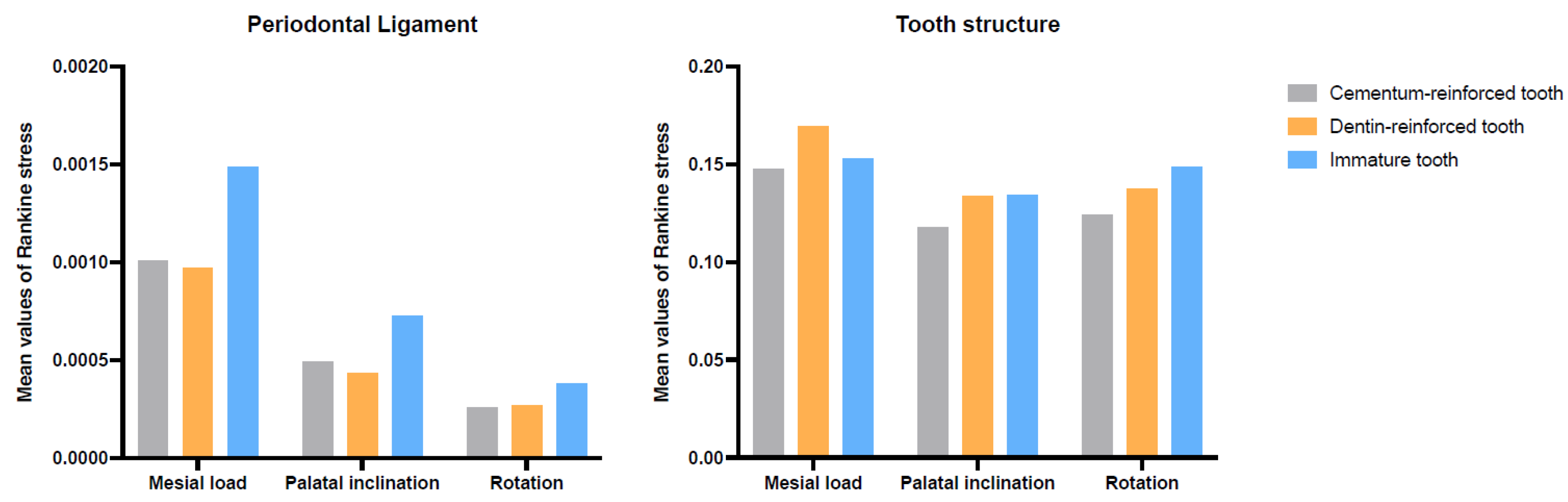
3.1. Mean Stress Values of the Periodontal Ligament and Tooth for Each Scenario

The highest mean stress values of the periodontal ligament were observed for mesial load followed by palatal inclination and rotation (Figure 2).
In all scenarios, the mean stress values of the periodontal ligament of the immature tooth were higher than the mean stress values of the periodontal ligament of the dentine-reinforced tooth and the cementum-reinforced tooth (Figure 2). In the mesial load and palatal inclination scenarios, the mean stress values were slightly higher in the cementum-reinforced tooth compared to the dentine-reinforced tooth (Figure 2).
In the mesial load scenario, the mean stress value of the dentine-reinforced tooth was higher than the mean stress values of both immature and cementum-reinforced teeth (Figure 2). In the palatal inclination scenario, the mean stress of the immature tooth was similar to the dentine-reinforced tooth. The mean stress values of the immature tooth were higher in the rotation scenario compared to the mature teeth (with either dentine or cementum) (Figure 2).
Mean stress values were considerably lower in the periodontal ligament compared to the tooth in all models and simulations (Figure 2).

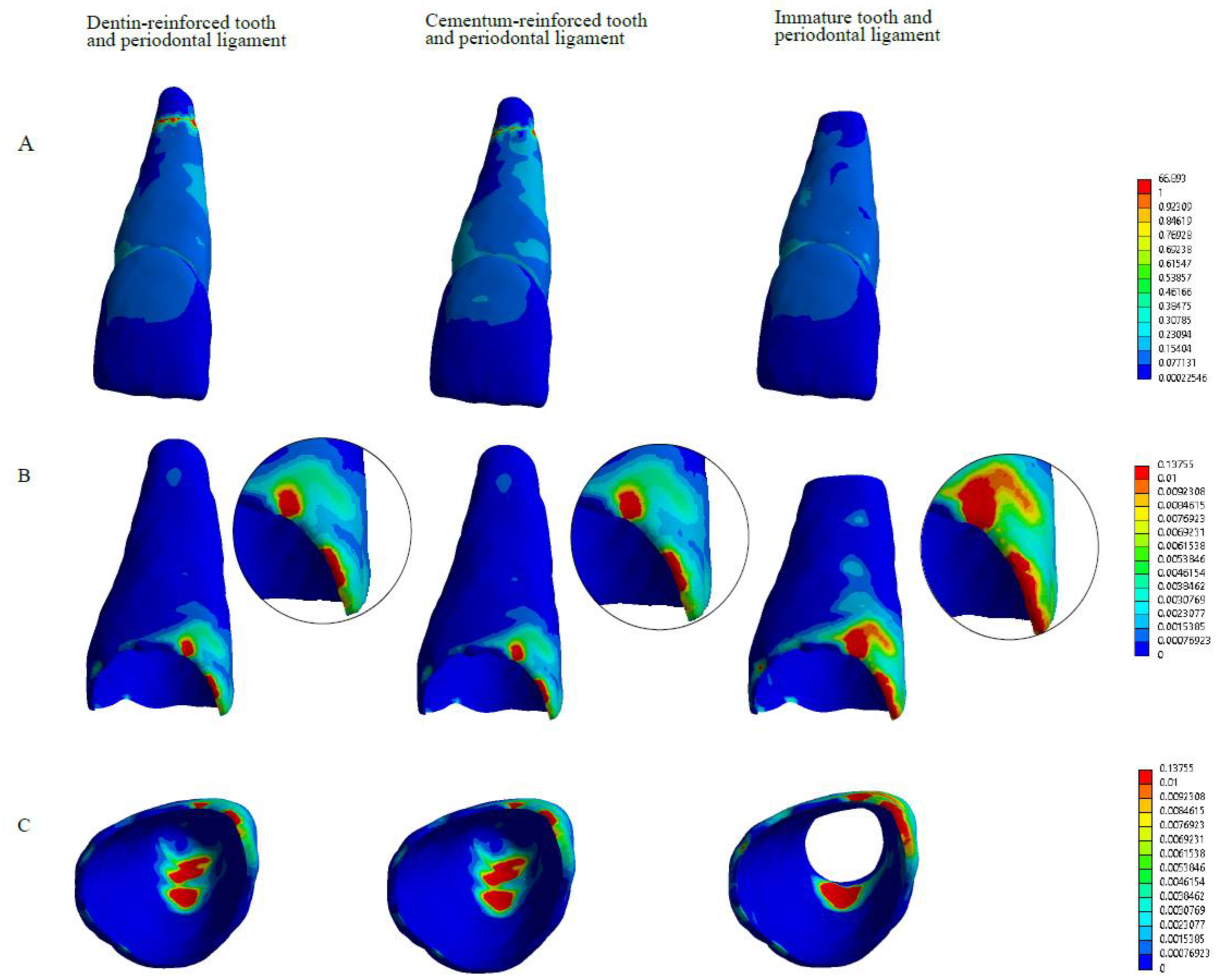
3.2. Distribution of the Stress of the Tooth Structure for Mesial Load

Figure 3 depicts the distribution of the stress in the mesial load scenario for the three models. The distribution of the stress in the tooth structure and periodontal ligament of the dentine-reinforced tooth and cementum-reinforced tooth were similar in this scenario (Figure 3A–C).
The highest values of tensile stress compromises a greater area of the ligament in the immature tooth compared to the periodontal ligament of the dentine-reinforced tooth and cementum-reinforced tooth in the mesial load scenario (see amplified areas of Figure 3B). In the dentine-reinforced tooth, cementum-reinforced tooth and immature tooth, the highest values of tensile stress values in the periodontal ligament were located at the mesial aspect and at the apical third (Figure 3B,C).

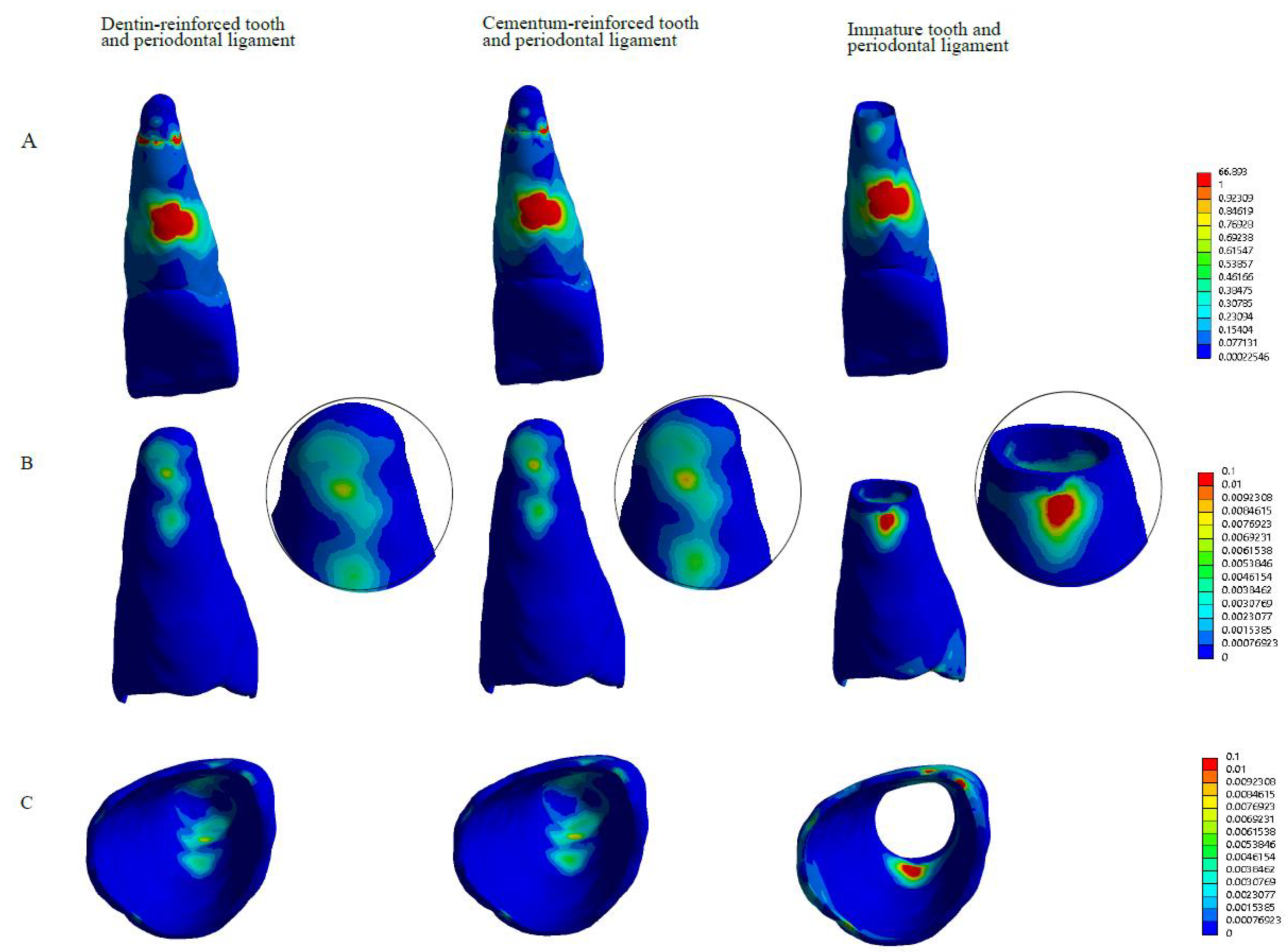
3.3. Distribution of the Stress of the Tooth Structure for Palatal Inclination

Figure 4 depicts the distribution of the stress in palatal inclination scenario and for the three models. The distribution of the stress in the tooth structure and periodontal ligament of the dentine-reinforced tooth and cementum-reinforced tooth were similar in this scenario (Figure 4).
In the dentine-reinforced tooth and cementum-reinforced tooth, the highest values of tensile stress in the periodontal ligament were located at the apical third, while for the immature tooth, it was located at the apical and coronal third in the palatal inclination scenario (Figure 4B,C).

3.4. Distribution of the Stress of the Tooth Structure for Rotation Scenario

Figure 5 depicts the distribution of the stress in the rotation scenario for the three models. The distribution of the stress in the tooth structure and periodontal ligament of the dentine-reinforced tooth and cementum-reinforced tooth were similar in this scenario (Figure 3A–C).
In the dentine-reinforced tooth and cementum-reinforced tooth, the highest values of tensile stress in the periodontal ligament were located at the apical third, while for the immature tooth, it was located at the apical and coronal third in the rotation scenario (Figure 5B,C).

4. Discussion

Regenerative endodontics has several advantages over apexification and root canal treatment, such as revitalisation and further root development of the immature necrotic teeth [2]. However, the newly formed tissue lacks the presence of odontoblast-like cells [7] and, as a result, the root maturation is based on reparative tissues [27], such as cementum-like tissue or osteodentine. Our objective was to determine the biomechanical implications over supporting tissues of the maturation of the root with cementum instead of dentine, when the tooth is subjected to orthodontic loads. The results of this FEA study showed that the biomechanical performance of the periodontal ligament and tooth structure of the dentine- and cementum-reinforced teeth models was similar in all scenarios, and that the periodontal ligament acted by reducing the mean stress values in all models.
The periodontal ligament acts as a mechanical connection between the tooth and the alveolar bone; therefore, the delivery of orthodontic loads to the tooth causes stress in the periodontal ligament, and it is related to resorption of the periodontal tissues [28]. On the pressure side, the external mechanical force induces collagenous extracellular matrix resorption of the periodontal ligament and bone [29] due to clastic activity. The recruited osteoclasts resorb and remodel the alveolar bone during orthodontic movement [29]. A disadvantageous stress distribution with peak stress values concentrated in some areas of the periodontal ligament could lead to a pathological process where resorption is not controlled and affects the tooth’s hard tissues. Root resorption is prone to occur in areas of stress concentration at the compression site during the process of orthodontic tooth movement [30]. Consequently, the question arises as to whether teeth previously treated with regenerative endodontics and that have completed their root development with cementum can endure orthodontic treatment or if they are at risk of root resorption [19]. In our simulation, the mean stress values of the periodontal ligament were similar among the dentine and cementum-reinforced teeth. Moreover, the stress distribution patterns in the periodontal ligament were similar among the dentine- and cementum-reinforced teeth as well. These results suggest that the performance of previously revitalised teeth subjected to orthodontic loads might be comparable to that of physiologically developed teeth, although this needs to be confirmed in in vivo situations. Thus, controlled clinical trials and/or experimental studies are needed.
The results of this study also showed that the periodontal ligament acted by dissipating the mechanical loads incurred during simulation of orthodontic loads. Moreover, stress distribution patterns demonstrated that peak stress values were reduced from the teeth to the ligament.
Cementum has poorer mechanical properties than dentine [13]. The amount of cementum surrounding the root of physiologically developed teeth (i.e., with dentine) has implications in the mechanical performance of teeth, since mechanical stresses increase at the root apex with increasing thickness of the apical cementum [31]. A previous computational study showed a disadvantageous stress distribution along the root in teeth that have completed their root development with cementum compared to physiologically developed teeth [14]. However, it must be noted that these differences in peak stress values are remarkable in high magnitude forces, such as those applied during biting or traumatic dental injuries and are mild with low magnitude forces, such as those applied in orthodontics [14]. Regarding stress distribution in the tooth, the present study shows similar mean stress values and stress distribution patterns for cementum- and dentine-reinforced teeth in mesial load, palatal inclination and rotation. In addition, the maturation of the root, with either dentine or cementum, was shown to be beneficial for all scenarios, where the immature tooth and its periodontal ligament showed higher mean stress values. The disadvantageous stress distribution of immature teeth compared to mature teeth has been observed in FEA studies [14,32] and poorer fracture resistance [33] and prognosis [22] have been shown in clinical and animal studies as well [34], and therefore, it is necessary to limit the orthodontic force to prevent root resorption.
The finite element model can simulate tooth-periodontal ligament complex under orthodontic loads. It allows the construction of standardised models to simulate the biomechanical performance of teeth and supporting tissues. Remodelling of the periodontal ligament for FEA analyses has been studies extensively, and the large inconsistency in results can be explained by the modelling assumptions (i.e., linear or non-linear, elastic or viscoelastic) given to the models [35]. The periodontal ligament is known to be a non-linear and time-dependent material, as proven by several experimental studies [34,35,36,37]. However, finite element model studies simulating orthodontic loads often incorporate homogeneous, linear elastic, isotropic and continuous periodontal ligament properties [17]. Non-linear simulations of the periodontal ligament provide more accurate and reliable calculation for the stresses and strains over several tooth movements than the ones provided by linear simulations [37]. Similarly, viscoelastic time-dependent properties of the ligament are the reason of strain energy dissipation, without which excessive load would cause tooth fracture [38,39]. In this study, we assumed non-linear periodontal ligament properties [17] to capture the elastic nature and deformation capacity of the periodontal ligament. The inclusion of the time-dependent viscoelastic properties was discarded in this study because of the comparative nature of the work. When comparing the same scenario under the three different models we expected the same qualitative results before the relaxation than after the relaxation. Thus, it must be considered that ‘time’ was not a variable included in our analysis, and in consequence this study shows the stress distribution in a specific moment and not the fatigue caused by the orthodontic treatment. The results of this research must be validated in clinical studies, since no computational study can represent the complex response of the teeth and surrounding tissues under orthodontic loads.
To the best of our knowledge, the mechanical properties of the newly formed tissues after regenerative endodontics (periodontal ligament and cementum-like tissue) have not been studied so far. The histological evidence of human teeth treated with regenerative endodontics, limited to case reports and mainly focused on the analysis of the intracanal tissues [6,8,40], do not report differences regarding the shape, thickness nor distribution of the collagen fibres of the newly formed periodontal ligament (the ligament formed between the newly formed hard tissue and the alveolar bone) and the pre-existing one (the periodontal ligament between the immature root and the alveolar bone). Similarly, the tissue responsible for root maturation is histologically similar to the cellular cementum [6,8,40], tissue present in the apical third of mature teeth, or osteocementum. Considering this data, in the present study the shape and mechanical properties given to the periodontal ligament and cementum of the cementum-reinforced tooth were assumed equal to that of a physiologically developed tooth. Thus, the basic data used in this study (i.e., Young’s modulus and Poisson coefficient of the cementum and hyperelastic properties of the periodontal ligament) are not the ones of an immature tooth after regenerative endodontic, but an extrapolation of the mechanical properties of a physiologically developed tooth. Future studies should assess whether the mechanical properties of the immature teeth and of the newly formed tissues after regenerative endodontics are indeed equivalent to those of physiologically developed teeth.

5. Conclusions

As far as we know, this is the first study analysing the performance of the periodontal ligament in teeth previously treated with regenerative endodontics. Under the conditions of this finite element analysis study, stress distribution in the tooth and periodontal ligament in a physiologically developed tooth and in a tooth after regenerative endodontics is similar in all three simulations of orthodontic loads. The periodontal ligament acted by dissipating the mechanical loads incurred during orthodontic forces. Maturation of the root with either dentine or cementum is beneficial to the stress distribution in all simulated scenarios. Among the limitations of this computational study, it can be concluded that orthodontic loads can be applied in teeth that have been treated with regenerative endodontics in the past; however, in vivo studies should be performed to validate these results.
Future studies should verify the mechanical characteristics of the newly formed tissues after regenerative endodontics to validate these results.
Finite element analysis opens new possibilities of research in the field of regenerative endodontics, since the biomechanical performance of teeth and supporting tissues can be simulated in standardised conditions. Future studies can be directed to analyse the mechanical performance of teeth after regenerative endodontics restored with different materials, and its comparison with teeth undergoing other endodontic treatments such as apexification, among others.

Author Contributions

Conceptualisation, C.B. and J.M.-N.; methodology, C.B. and J.M.-N.; software, J.M.-N.; validation, J.M.-N.; formal analysis, C.B., J.M.-N. and M.D.F.; investigation, C.B.; resources, C.B. and J.M.-N.; data curation, C.B. and J.M.-N.; writing—original draft preparation, C.B.; writing—review and editing, C.B., J.M.-N. and M.D.F.; visualisation, C.B.; supervision, M.D.F.; project administration, C.B. All authors have read and agreed to the published version of the manuscript.

Funding

This research received no external funding.

Institutional Review Board Statement

The study was conducted in accordance with the Declaration of Helsinki and approved by the Ethics Committee of Universitat de Barcelona.

Informed Consent Statement

Informed consent was obtained from all subjects involved in the study.

Data Availability Statement

The data are included within the article.

Acknowledgments

This work was supported by the Serra-Hunter (URV) and the CERCA programme (ICP) from the Generalitat de Catalunya.

Conflicts of Interest

The authors declare no conflict of interest.

References

  1. Murray, P.E.; Garcia-Godoy, F.; Hargreaves, K.M. Regenerative Endodontics: A Review of Current Status and a Call for Action. J. Endod. 2007, 33, 377–390. [Google Scholar] [CrossRef] [PubMed]
  2. Lin, J.; Zeng, Q.; Wei, X.; Zhao, W.; Cui, M.; Gu, J.; Lu, J.; Yang, M.; Ling, J. Regenerative Endodontics Versus Apexification in Immature Permanent Teeth with Apical Periodontitis: A Prospective Randomized Controlled Study. J. Endod. 2017, 43, 1821–1827. [Google Scholar] [CrossRef]
  3. Lovelace, T.W.; Henry, M.A.; Hargreaves, K.M.; Diogenes, A. Evaluation of the delivery of mesenchymal stem cells into the root canal space of necrotic immature teeth after clinical regenerative endodontic procedure. J. Endod. 2011, 37, 133–138. [Google Scholar] [CrossRef] [PubMed]
  4. Galler, K.M.; D'Souza, R.N.; Federlin, M.; Cavender, A.C.; Hartgerink, J.D.; Hecker, S.; Schmalz, G. Dentin conditioning codetermines cell fate in regenerative endodontics. J. Endod. 2011, 237, 1536–1541. [Google Scholar] [CrossRef]
  5. Lolato, A.; Bucchi, C.; Taschieri, S.; Kabbaney, A.E.; Fabbro, M.D. Platelet concentrates for revitalization of immature necrotic teeth: A systematic review of the clinical studies. Platelets 2016, 27, 383–392. [Google Scholar] [CrossRef] [PubMed]
  6. Austah, O.; Joon, R.; Fath, W.M.; Chrepa, V.; Diogenes, A.; Ezeldeen, M.; Couve, E.; Ruparel, N.B. Comprehensive Characterization of 2 Immature Teeth Treated with Regenerative Endodontic Procedures. J. Endod. 2018, 44, 1802–1811. [Google Scholar] [CrossRef]
  7. Lei, L.; Chen, Y.; Zhou, R.; Huang, X.; Cai, Z. Histologic and Immunohistochemical Findings of a Human Immature Permanent Tooth with Apical Periodontitis after Regenerative Endodontic Treatment. J. Endod. 2015, 41, 1172–1179. [Google Scholar] [CrossRef]
  8. Nosrat, A.; Kolahdouzan, A.; Hosseini, F.; Mehrizi, E.A.; Verma, P.; Torabinejad, M. Histologic outcomes of uninfected human immature teeth treated with regenerative endodontics: 2 case reports. J. Endod. 2015, 41, 1725–1729. [Google Scholar] [CrossRef]
  9. Del Fabbro Lolato, A.; Bucchi, C.; Taschieri, S.; Weinstein, R.L. Autologous Platelet Concentrates for Pulp and Dentin Regeneration: A Literature Review of Animal Studies. J. Endod. 2016, 42, 250–257. [Google Scholar] [CrossRef]
  10. Goldberg, M.; Kulkarni, A.B.; Young, M.; Boskey, A. Dentin: Structure, Composition and Mineralization. Front. Biosci. 2011, 3, 711–735. [Google Scholar] [CrossRef]
  11. Yamauchi, N.; Nagaoka, H.; Yamauchi, S.; Teixeira, F.B.; Miguez, P.; Yamauchi, M. Immunohistological characterization of newly formed tissues after regenerative procedure in immature dog teeth. J. Endod. 2011, 37, 1636–1641. [Google Scholar] [CrossRef] [PubMed]
  12. Salmon, C.R.; Tomazela, D.M.; Ruiz, K.G.; Foster, B.L.; Paes Leme, A.F.; Sallum, E.A. Proteomic analysis of human dental cementum and alveolar bone. J. Proteom. 2013, 91, 544–555. [Google Scholar] [CrossRef] [PubMed] [Green Version]
  13. Ho, S.P.; Yu, B.; Yun, W.; Marshall, G.W.; Ryder, M.I.; Marshall, S.J. Structure, chemical composition and mechanical properties of human and rat cementum and its interface with root dentin. Acta Biomater. 2010, 5, 707–718. [Google Scholar] [CrossRef] [PubMed] [Green Version]
  14. Bucchi, C.; Marcé-Nogué, J.; Galler, K.M.; Widbiller, M. Biomechanical performance of an immature maxillary central incisor after revitalization: A finite element analysis. Int. Endod. J. 2019, 52, 1508–1518. [Google Scholar] [CrossRef]
  15. Jonsdottir, S.H.; Giesen, E.B.W.; Maltha, J.C. Biomechanical behaviour of the periodontal ligament of the beagle dog during the first 5 hours of orthodontic force application. Eur. J. Orthod. 2006, 28, 547–552. [Google Scholar] [CrossRef] [PubMed] [Green Version]
  16. Zhang, L.; Liu, W.; Zhao, J.; Zhang, L.; Liu, W.; Zhao, J.; Jin, F.; Jin, Y. Mechanical stress regulates osteogenic differentiation and RANKL/OPG ratio in periodontal ligament stem cells by the Wnt/beta-catenin pathway. Biochim. Biophys. Acta 2016, 1860, 2211–2219. [Google Scholar] [CrossRef]
  17. Wise, G.E.; King, G.J. Mechanisms of tooth eruption and orthodontic tooth movement. J. Dent. Res. 2008, 87, 414–434. [Google Scholar] [CrossRef] [Green Version]
  18. Weltman, B.; Vig, K.W.; Fields, H.W.; Shanker, S.; Kaizar, E.E. Root resorption associated with orthodontic tooth movement: A systematic review. Am. J. Orthod. Dentofac. Orthop. 2010, 137, 462–476. [Google Scholar] [CrossRef] [Green Version]
  19. Galler, K.M.; Krastl, G.; Simon, S.; Van Gorp, G.; Meschi, N.; Vahedi, B.; Lambrechts, P. European Society of Endodontology position statement: Revitalization procedures. Int. Endod. J. 2016, 49, 717–723. [Google Scholar] [CrossRef]
  20. Natera, M.; Mukherjee, P.M. Regenerative Endodontic Treatment with Orthodontic Treatment in a Tooth with Dens Evaginatus: A Case Report with a 4-year Follow-up. J. Endod. 2018, 44, 952–955. [Google Scholar] [CrossRef]
  21. Chaniotis, A. Orthodontic Movement after Regenerative Endodontic Procedure: Case Report and Long-term Observations. J. Endod. 2018, 44, 432–437. [Google Scholar] [CrossRef]
  22. Cvek, M. Prognosis of luxated non-vital maxillary incisors treated with calcium hydroxide and filled with gutta-percha. A retrospective clinical study. Dent. Traumatol. 1992, 8, 45–55. [Google Scholar] [CrossRef]
  23. Qian, L.; Todo, M.; Morita, Y.; Matsushita, Y.; Koyano, K. Deformation analysis of the periodontium considering the viscoelasticity of the periodontal ligament. Dent Mater 2009, 25, 1285–1292. [Google Scholar] [CrossRef]
  24. Doblaré, M.; García, J.M.; Gómez, M.J. Modelling bone tissue fracture and healing: A review. Eng. Fract. Mech. 2004, 71, 1809–1840. [Google Scholar] [CrossRef]
  25. Pérez-González, A.; Iserte-Vilar, J.L.; González-Lluch, C. Interpreting finite element results for brittle materials in endodontic restorations. Biomed. Eng. Online 2011, 10, 44. [Google Scholar] [CrossRef] [Green Version]
  26. Walmsley, C.W.; Smits, P.D.; Quayle, M.R.; McCurry, M.R.; Richards, H.S.; Oldfield, C.C.; Wroe, S.; Clausen, P.D.; McHenry, C.R. Why the Long Face? The Mechanics of Mandibular Symphysis Proportions in Crocodiles. PLoS ONE 2013, 8, e53873. [Google Scholar] [CrossRef] [Green Version]
  27. Alexander, A.; Torabinejad, M.; Vahdati, S.A.; Nosrat, A.; Verma, P.; Grandhi, A.; Shabahang, S. Regenerative Endodontic Treatment in Immature Noninfected Ferret Teeth Using Blood Clot or SynOss Putty as Scaffolds. J. Endod. 2020, 6, 209–215. [Google Scholar] [CrossRef]
  28. Ciavarella, D.; Tepedino, M.; Gallo, C.; Montaruli, G.; Zhurakivska, K.; Coppola, L.; Troiano, G.; Chimenti, C.; Laurenziello, M.; Lo Russo, L. Post-orthodontic position of lower incisors and gingival recession: A retrospective study. J. Clin. Exp. Dent. 2017, 9, e1425–e1430. [Google Scholar] [CrossRef]
  29. Rangiani, A.; Jing, Y.; Ren, Y.; Yadav, S.; Taylor, R.; Feng, J.Q. Critical roles of periostin in the process of orthodontic tooth movement. Eur. J. Orthod. 2016, 38, 373–378. [Google Scholar] [CrossRef] [Green Version]
  30. Brudvik, P.; Rygh, P. Multi-nucleated cells remove the main hyalinized tissue and start resorption of adjacent root surfaces. Eur. J. Orthod. 1994, 16, 265–273. [Google Scholar] [CrossRef]
  31. Li, Z.; Yu, M.; Jin, S.; Wang, Y.; Luo, R.; Huo, B.; Liu, D.; He, D.; Zhou, Y.; Liu, Y. Stress distribution and collagen remodeling of periodontal ligament during orthodontic tooth movement. Front. Pharmacol. 2019, 10, 1263. [Google Scholar] [CrossRef] [PubMed]
  32. Shaw, A.M.; Sameshima, G.T.; Vu, H.V. Mechanical stress generated by orthodontic forces on apical root cementum: A finite element model. Orthod. Craniofac. Res. 2004, 7, 98–107. [Google Scholar] [CrossRef] [PubMed]
  33. Anthrayose, P.; Nawal, R.R.; Yadav, S.; Talwar, S.; Yadav, S. Effect of revascularization and apexification procedures on biomechanical behaviour of immature maxillary central incisor teeth: A three-dimensional finite element analysis study. Clin. Oral Investig. 2021, 25, 6671–6679. [Google Scholar] [CrossRef] [PubMed]
  34. Zhou, R.; Wang, Y.; Chen, Y.; Chen, S.; Lyu, H.; Cai, Z.; Huang, X. Radiographic, Histologic, and Biomechanical Evaluation of Combined Application of Platelet-rich Fibrin with Blood Clot in Regenerative Endodontics. J. Endod. 2017, 43, 2034–2040. [Google Scholar] [CrossRef] [PubMed]
  35. Papadopoulou, K.; Hasan, I.; Keilig, L.; Reimann, S.; Eliades, T.; Jäger, A.; Deschner, J.; Bourauel, C. Biomechanical time dependency of the periodontal ligament: A combined experimental and numerical approach. Eur. J. Orthod. 2013, 35, 811–818. [Google Scholar] [CrossRef] [PubMed] [Green Version]
  36. Toms, S.R.; Eberhardt, A.W. A nonlinear finite element analysis of the periodontal ligament under orthodontic tooth loading. Am. J. Orthod. Dentofac. Orthop. 2003, 123, 657–665. [Google Scholar] [CrossRef]
  37. Keilig, L.; Drolshagen, M.; Tran, K.L.; Hasan, I.; Reimann, S.; Deschner, J.; Brinkmann, K.; Krause, R.; Favino, M.; Bourauel, C. In vivo measurements and numerical analysis of the biomechanical characteristics of the human periodontal ligament. Ann. Anat. 2016, 206, 80–88. [Google Scholar] [CrossRef]
  38. Pini, M.; Zysset, P.; Botsis, J.; Contro, R. Tensile and compressive behaviour of the bovine periodontal ligament. J. Biomech. 2004, 37, 111–119. [Google Scholar] [CrossRef]
  39. Shibata, T.; Botsis, J.; Bergomi, M.; Mellal, A.; Komatsu, K. Mechanical behavior of bovine periodontal ligament under tension-compression cyclic displacements. Eur. J. Oral Sci. 2006, 114, 74–82. [Google Scholar] [CrossRef] [PubMed]
  40. Martin, G.; Ricucci, D.; Gibbs, J.L.; Lin, L.M. Histological findings of revascularized/revitalized immature permanent molar with apical periodontitis using platelet-rich plasma. J. Endod. 2013, 39, 138–144. [Google Scholar] [CrossRef]
Figure 1. Segmented tissues and model development. Segmentation of a mature tooth in the alveolar bone (a). The apical part (black arrow) of the mature tooth was given the mechanical properties of dentine or cementum for Models 2 and 3, respectively. Segmentation of an immature tooth in the alveolar bone (b). Asterisk (*): space occupied by the apical papilla. Image edited from Bucchi et al., 2019 [14].
Applsci 12 07063 g001
Figure 2. Mean Rankine stress of the teeth and periodontal ligament of Models 1, 2 and 3 for mesial load, palatal inclination and rotation scenarios.
Applsci 12 07063 g002
Figure 3. Stress distribution in the tooth (A) and in the periodontal ligament (B,C) of Models 1, 2 and 3 in mesial load scenario. Similar stress patterns are observed between all tissues of Models 2 and 3. Maximum tensile stress of the teeth (A) and ligaments (B,C) are coloured in red.
Applsci 12 07063 g003
Figure 4. Stress distribution in the tooth (A) and in the periodontal ligament (B,C) of Models 1, 2 and 3 in palatal inclination scenario. Similar stress patterns are observed between all tissues of Models 2 and 3. Maximum tensile stress of the teeth (A) and ligaments (B,C) are coloured in red.
Applsci 12 07063 g004
Figure 5. Stress distribution in the tooth (A) and in the periodontal ligament (B,C) of Models 1, 2 and 3 in the rotation scenario. Similar stress patterns are observed between all tissues of Models 2 and 3. Maximum tensile stress of the teeth (A) and ligaments (B,C) are coloured in red.
Applsci 12 07063 g005
Table 1. Young’s modulus and Poisson coefficient of dental and supporting tissues.
TissueYoung’s ModulusPoisson Coefficient
Enamel84,1000.31
Dentine18,6000.31
Cementum82000.3
Bone14,7000.31
Table 2. Hyperelastic properties of the periodontal ligament. The Mooney–Rivlin constitutive equation was used with the listed constants.
C10C01C20C11C02C30C21C12C03
−0.00480.005050.0080.001200.004000
Publisher’s Note: MDPI stays neutral with regard to jurisdictional claims in published maps and institutional affiliations.

