Article

Soluble Receptor of Advanced Glycation End-Products (sRAGE) in Pediatric Asthma: A Prospective Study in 68 Children Aged 7 Years

1
Department of Pediatrics, CHU Clermont-Ferrand, 63000 Clermont-Ferrand, France
2
Department of Sport Medicine and Functional Explorations, CHU Clermont-Ferrand, 63000 Clermont-Ferrand, France
3
Department of Medical Biochemistry and Molecular Genetics, CHU Clermont-Ferrand, 63000 Clermont-Ferrand, France
4
Team “Translational Approach to Epithelial Injury and Repair”, iGReD, Université Clermont Auvergne, Clermont-Ferrand, 63000 Clermont-Ferrand, France
5
Department of Clinical Research and Innovation, CHU Clermont-Ferrand, 63000 Clermont-Ferrand, France
6
Department of Clinical Research for Children (CRECHE), Centre Hospitalier Universitaire Clermont-Ferrand, 63000 Clermont-Ferrand, France
7
Department of Immunology, CHU Clermont-Ferrand, 63000 Clermont-Ferrand, France
8
INRA, UMR 1019, Université Clermont Auvergne, 63000 Clermont-Ferrand, France
*
Author to whom correspondence should be addressed.
Appl. Sci. 2022, 12(12), 5926; https://doi.org/10.3390/app12125926
Submission received: 1 May 2022 / Revised: 7 June 2022 / Accepted: 8 June 2022 / Published: 10 June 2022
(This article belongs to the Special Issue Asthma and Respiratory Disease: Prediction, Diagnosis and Treatment)

Abstract

:

Featured Application

Plasma sRAGE may be a biomarker of well-treated children with asthma. It could be useful in the follow-up of these children.

Abstract

Background: Asthma is a chronic inflammatory disease of the airways common in children. Soluble advanced glycation end-product receptor (sRAGE) is a blood biomarker of lung damage and inflammation. We sought to determine whether it could also be a biomarker in childhood asthma. Methods: We conducted a prospective, observational, analytical study at Clermont-Ferrand University Hospital. We measured plasma sRAGE levels in asthmatic and healthy children aged 7 years. Results: Of the 68 children assessed, 15 (22.05%) presented asthma. All presented normal respiratory function. The mean plasma sRAGE level was 1875 pg/mL in the children with asthma and 1794 pg/mL in the healthy children (p = 0.525). The mean plasma sRAGE level was significantly decreased with tobacco exposure during pregnancy: 1478 pg/mL versus 1870 pg/mL without (p = 0.007). Lower levels were observed in children living in apartments (1557 pg/mL) than in those living in houses (1863 pg/mL) (p = 0.031). Conclusions: No difference was observed in plasma sRAGE levels in children with asthma in our well-treated and controlled population. Environmental exposure may affect these levels. Further studies are required to better characterize the role of sRAGE.

1. Introduction

Asthma is caused by chronic inflammation of the airways associated with airway wall remodeling. It is important to evaluate inflammatory status, especially in severe cases, to adapt therapeutics. Currently, the main methods to evaluate airway inflammation are sputum analysis, exhaled breath condensate, and fraction of exhaled nitric oxide (FeNO) [1].
The receptor for advanced glycation end-products (RAGE) is a trans-membranous receptor present in large quantities in the lungs, particularly in alveolar epithelial type I cells [2,3]. It is an important mediator in allergic airway inflammation (AAI) and asthma [4]. RAGE belongs to the immunoglobulin superfamily [5]. It is found in two major forms: membrane-bound RAGE (mRAGE) and soluble RAGE (sRAGE) [6]. Three domains compose mRAGE: an extracellular domain for the recognition and linking of RAGE ligands, a hydrophobic transmembrane domain, and an intracellular domain for activating intracellular pathways. The best-known ligands are advanced glycation end-products (AGEs), and damage-associated molecular patterns (DAMPs) also called alarmins, high mobility group box 1 protein (HMGB1), proteins from the S100 family, and beta-amyloid peptides [7]. sRAGE comprises only the extracellular domain of mRAGE. It is synthesized by alternative splicing events or proteolytic cleavage of mRAGE [6,8,9]. As a decoy receptor, it binds to mRAGE ligands without activating intracellular inflammatory signaling (Figure 1) [10].
sRAGE seems to reflect pulmonary damage processes and has been studied as a biomarker of lung epithelial injury and alveolar fluid clearance [11]. Blood level of sRAGE is correlated with the severity and prognosis of adult and pediatric acute respiratory distress syndrome (ARDS) [12,13]. Studies show its utility for evaluating ARDS in adults and children, with elevated blood sRAGE levels. In adult ARDS, Jabaudon et al. report evidence that the initial alveolar epithelial injury with high plasma sRAGE level is an independent variable associated with 90-day mortality [14]. In pediatric ARDS, sRAGE levels were higher in a non-survivor group, associated with non-pulmonary organ failure and with mortality directly due to lung injury [15]. In this clinical context, median levels observed in these cases were strongly elevated (5000 pg/mL [2600–7200], p = 0.005). Because sRAGE is more highly expressed in BAL fluid than in plasma (ratio of 4), its plasma elevation in ARDS could reflect alveolo-capillary membrane damage with marked leakage of sRAGE from BAL to plasma [16].
Asthma being an inflammatory disease, RAGE and its soluble form might be expected to play a role in its pathogenesis. The RAGE pathway is involved and can promote allergic airway disease [4]. Mouse RAGE knockouts showed that the absence of RAGE abolished airway hypersensitivity, eosinophilic inflammation, and airway remodeling present in asthma, strong evidence that RAGE might be one of the principal mediators of asthma pathogenesis [17]. Few studies are available, and the correlation between blood sRAGE levels in young children with recurrent wheezing and its severity is still unclear [1,18]. In this study, we assessed plasma sRAGE levels in children aged 7 years and evaluated its correlation with asthma. We went on to examine the association between the environment and sRAGE levels.

2. Materials and Methods

2.1. Study Design

We assessed healthy and asthmatic children from a cohort gathered during the 2011–2012 winter epidemic season at the University Hospital of Clermont-Ferrand (Auvergne Rhône-Alpes, France). For this cohort, inclusion criteria were as follows: presentation to the Pediatric Emergency Unit of Clermont Ferrand, age less than 1 year, and a first episode of bronchiolitis. Exclusion criteria were as follows: bronchopulmonary dysplasia, history of prematurity under 34 weeks, cystic fibrosis, known immune deficiencies, suspected primary ciliary dyskinesia, congenital heart disease, and acute renal failure.

2.2. Study Population

All children aged 7 years were included prospectively in the cohort described above.

2.3. Collected Data

2.3.1. Clinical Data

After the initial inclusion, the parents of each child were contacted by telephone every 3 months until age 3. A standardized questionnaire on respiratory symptoms was used for every interview. A clinical examination was performed and an immunological profile was determined at age 3 at the Outpatients Department of CHU-Estaing Hospital [19].
A questionnaire was given asking about respiratory symptoms (day or night cough, discomfort during exercise, use of salbutamol), respiratory treatments (corticosteroid per os or inhaled, long-acting bronchodilator), hospitalization for asthma, wheezing outside viral episodes, personal atopy (presence of atopic dermatitis, eczema, rhino-conjunctivitis, food anaphylaxis from birth), environmental exposure to tobacco smoke (currently or during pregnancy), pets, type of housing, daycare attendance, and number of siblings.
Data also included were sex, weight, height, and precariousness assessed using the Assessment of Precariousness and Health Inequalities in Health Examination Centers (EPICES) questionnaire [20]. Precariousness was recorded if the score was equal to or greater than 30. Data from the medical examination comprised asthma symptoms, eczema, rhino-conjunctivitis, and chest deformity.
Asthma was recorded if the child presented three or more respiratory symptoms at least twice or an episode lasting four weeks or more. We considered the following as respiratory symptoms of asthma: tachypnea, wheezing, expiratory stridor, respiratory chest retraction, doctor-diagnosed wheezes, or the use of anti-asthmatic medication with medical prescription [21,22].

2.3.2. Pulmonary Function Evaluation

Spirometry measurements were made with a Jaeger MasterScreen spirometer, calibrated daily. A forced vital capacity (FVC) test was performed in accordance with American Thoracic Society/ European Thoracic Society 2005 spirometry standards [23]. Three acceptable FVC maneuvers were performed. Maximum expiratory flow at 25%, 50%, and 75% expiratory volume (MEF 25, MEF 50, MEF 75), forced expiratory volume in 1 s (FEV1), forced vital capacity (FVC), peak expiratory flow (PEF), and maximum mid-expiratory flow (MMEF) were recorded for all the children. Interrupter resistance (Rint) was measured using a SpiroDyn apparatus (Dyn’R, Toulouse) calibrated daily for flow, collected during a 100 ms occlusion during expiration. The child was breathing quietly, seated, head in the neutral position connected to the flowmeter by a mouthpiece. Measures were performed without bronchodilator and 15 min after inhaled bronchodilator (400 µg of salbutamol). Normal respiratory function was defined by normal FVC and normal FEV1/FVC ratio, and a significant obstructive syndrome by FEV1/FVC < 0.8. Significant reversibility was determined by a reduction of 35% in Rint or a rise of 12% in FEV1 [24].

2.3.3. Biological Assessment

After venipuncture, blood samples were immediately centrifuged and aliquoted. The aliquots were stored at −80 °C until analysis. The stability of the collection was tested annually. sRAGE levels were assayed in duplicate using commercial ELISA kits (RAGE Quantikine, R & D Systems, Minneapolis, MN, USA). The detection threshold was 78 pg/mL. The intra- or inter-assay CV was under 5%. The personnel responsible for carrying out the sRAGE tests were blind to the patient’s situation. Complete blood count was measured on a Sysmex analyzer (Sysmex France, Villepinte, France). Eosinophilia was defined as levels above 0.300 G/L [25].
Skin prick tests were performed with histamine and physiological serum as positive and negative controls, respectively. Specific prick tests with the main aeroallergens (Dermatophagoides pteronyssinus, Dermatophagoides farinae, Alternaria, timothy grass, birch, cats, and dogs) and the main trophallergens (egg, peanut, walnut, almond, hazelnut, soy) were carried out concurrently. Prick tests were analyzed at 20 min and were considered positive with a papule diameter equal to or greater than 3 mm, compared to the control. Sensitization to aeroallergens and trophallergens was confirmed by unitary assay of specific serum IgE by ImmunoCAP (Phadia 250®, Phadia AB, Uppsala, Sweden). A result was considered positive when the specific IgE level exceeded 0.35 kUA/L. Multi-sensitization was recorded if tests (SPT or IgE assay) were positive for at least two allergens.

2.4. Statistical Analysis

Statistical analysis was carried out with the Stata v15 software (StataCorp, College Station, TX, USA). The study sample is described by frequency and percentage for categorical data and by means ± standard deviation for continuous data or by median and interquartile range when data were not normal. Comparisons of sRAGE levels were made using Student’s t-test for two groups (or ANOVA for three or more groups), or Mann and Whitney’s test (or Kruskal–Wallis for three or more groups) when data were not normal. Relationships of sRAGE with continuous data were sought using Pearson’s correlation coefficient (or Spearman’s in the case of non-normal distribution). All tests were two-sided and a p-value < 5% was considered significant.

3. Results

3.1. Population

In all, 68 children were evaluated at age 7 years. Our sample was composed mainly of male children (63.2%), aged 7.68 ± 0.30 years. Mean body mass index was 15.9 kg/m² (±2.1 SD). Fifty-six children (82.3%) lived in a house. Forty-eight (70.6%) had pets at home, twenty-five (36.8%) had cats. Twenty-six children (38.2%) were exposed to tobacco smoking, ten (14.7%) during pregnancy. Precariousness was suspected for seven children (10.3%). Asthma rate was 22.1% in the population (15 children). Among these, twelve (80%) had persistent asthma since age 3 years and three (20%) developed symptoms at school age. At clinical examination, only one child presented wheezing. None had any chest deformity. All except one used salbutamol (93%) and 11 (73%) followed a background treatment. An atopic family history was noted for 47 patients (69.1%), 17 patients (25%) presented an atopic dermatitis history, 20 (29.4%) presented respiratory allergic symptoms, and no children had a food allergy. No children had any obstructive ventilation disorder (FEV1 104% ± 9.3%, FEV1/FVC 107 ± 6%). Reversibility was present in 16 children (23.5% of our population): 5 in the asthma category (35.7%) and 11 in the non-asthma category (21.0%). We found aero-sensitization in 16 (20%) children and eosinophilia in 30 children (44.8%).

3.2. sRAGE Levels

3.2.1. Mean Levels

The mean plasma sRAGE level was 1812 (±428 SD) pg/mL at 7 years. There was no significant difference between plasma sRAGE level according to sex in our population (1842 ± 430 SD) pg/mL in females and 1795 (±432 SD) pg/mL in males (p = 0.663). We found no correlation between sRAGE and BMI (r = 0.17, p = 0.17) or with birth term (r = −0.08, p = 0.53).

3.2.2. sRAGE in Asthma and Atopic Status
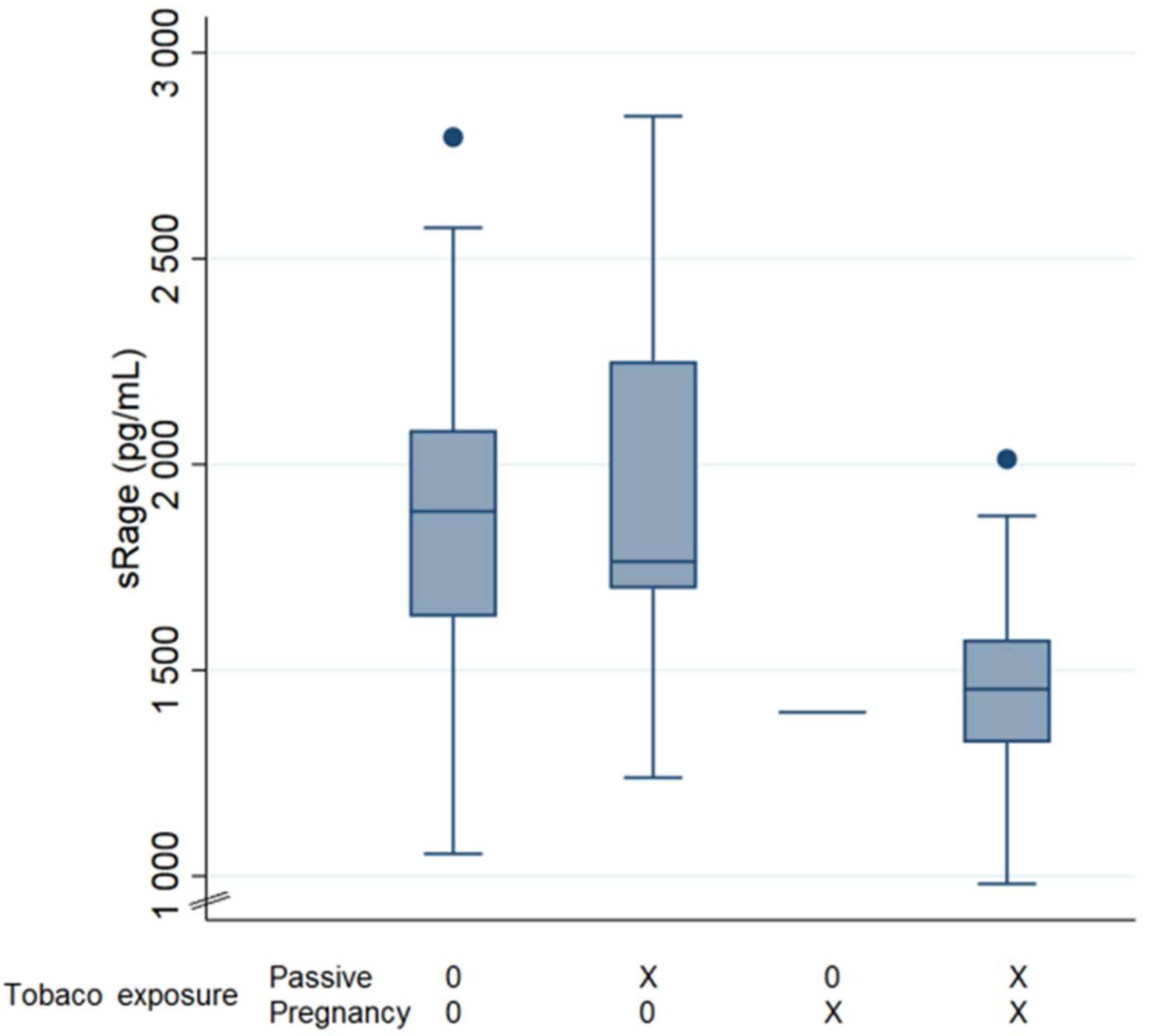
Plasma sRAGE levels did not differ between asthmatic children and healthy children: 1875 (±472) pg/mL versus 1794 (±418) pg/mL, respectively (p = 0.525) (Table 1). sRAGE levels did not differ in children with significant reversibility (1690 ± 683 pg/mL versus 1787 ± 381 pg/mL, respectively). sRAGE levels were not correlated to eosinophil level (r = −0.10, p = 0.42), and no difference was observed in eosinophilic (1821 ± 380 pg/mL) versus non-eosinophilic children (1819 ± 465 pg/mL, p = 0.981) (Table 1). Likewise, sRAGE levels did not differ in sensitized (1908 ± 494 pg/mL) versus non-sensitized children (1783 ± 406 pg/mL, p = 0.309) (Table 1). The same results were observed in the asthmatic population (for aeroallergen sensitization (p = 0.418) and eosinophilic status (p = 1)).

3.2.3. Environment

Mean plasma sRAGE levels were significantly decreased in children exposed to tobacco smoking during pregnancy: 1478 ± 296 pg/mL versus 1870 ± 422 pg/mL, p = 0.007 (Table 1). In contrast, current passive tobacco exposure had no impact on sRAGE levels (1772 ± 479 pg/mL versus 1837 ± 397 pg/mL, p = 0.547) (Table 1). Association of antenatal and current exposure was associated with decreased levels of sRAGE, but not significantly (p = 0.053) (Figure 2). In children with asthma, sRAGE levels were lowered in those exposed to tobacco during pregnancy (1257 ± 96 pg/mL versus 2099 ± 303 pg/mL in non-exposed, p = 0.004) unlike those currently exposed to tobacco, with 1686 (±271 SD) pg/mL versus 1805 (± 388 SD) pg/mL in non-exposed, p = 0.550. Lower levels were observed in children living in apartments (1557 ± 372 pg/mL) than in those living in houses (1863 ± 427 pg/mL) (p = 0.031) (Table 1). The presence of animals did not modify sRAGE levels at age 7 (p = 0.322), specifically not cats (p = 0.579).

4. Discussion

This study investigated plasma sRAGE levels in asthma in children aged 7 years. The study enrolled 68 children, healthy or with asthma.
The role of sRAGE in the pathogenesis of asthma in children is poorly understood. sRAGE levels have been scantly studied in children, and we currently have no reference values in the literature that are significantly different from adults. The processes of lung maturation and alveolation are thought to explain higher sRAGE levels in infants, and a decrement was observed in its levels as alveolation phenomena gradually cease [3]. In a previous study, conducted on infants aged under 1 year, we found a mean serum sRAGE level of 2203 (±905 SD) pg/mL in the healthy group [26]. At age 7 years, we observed a mean plasma sRAGE level of 1794 (±418 SD) pg/mL in healthy children. In an adult population, the mean plasma sRAGE level significantly decreased compared with 550 pg/mL in controls [27]. However, these observations need to be confirmed given the small number of studies available and the variability in the levels observed [28,29].
Unlike ARDS, bronchial inflammation seems to be responsible for decreasing sRAGE levels in adult and pediatric health disorders. Decreased sRAGE plasma levels are associated with worsening COPD [30]. In pediatric patients, significantly lower sRAGE levels were observed in patients with asthma and a cut-off value of serum sRAGE levels for asthma diagnosis of 1080 pg/mL has been proposed. sRAGE levels could be correlated and used as a severity biomarker, with lowered levels in uncontrolled and severe asthma subgroups compared to others [18]. In these conditions, sRAGE could act as a decoy and serve as an anti-inflammatory factor to limit the activation of the RAGE cascade. If sRAGE is considered to “block” the inflammatory response by trapping circulating RAGE ligands, it could be “consumed” and so show low blood levels. Unlike the lesions induced by ARDS, asthma does not reach the alveolo-capillary membrane and so we might expect the blood levels to approach the levels found in the BAL fluid with consumption proportional to that of the inflammation of the airways. Our present study showed sRAGE levels to be conserved in patients with asthma. This may be because almost all our patients presented controlled asthma, with normal respiratory function, and daily treatment. A well-taken inhaled corticosteroid treatment controls lung inflammation and could therefore normalize serum level of sRAGE. The study of Sukkar et al. in clinically stable adult asthma patients demonstrated that the levels could remain relatively stable regardless of the medical therapy used [31]. In addition, the type of inflammation could modify the expected values of sRAGE. Eosinophilic or neutrophilic inflammation may influence sRAGE level [29]. In our study, eosinophilia and sensitization did not impact sRAGE levels, although our asthmatic population was too small to draw any firm conclusion.
Our study shows no increase in the plasma level of sRAGE in pediatric asthma. sRAGE does not appear to be a diagnostic marker for childhood asthma. However, in our study, all the children with asthma were taking inhaled corticosteroids and had normal lung function. If an effective treatment reduces bronchial inflammation, it can normalize the level of sRAGE. We therefore hypothesize that serum sRAGE assay could be a good biomarker of well-controlled asthma. It would then be useful to measure the plasma level of sRAGE in the follow-up of children with asthma: a normal level could be the indicator of controlled asthma and could help to adapt therapy. This is innovative because currently no blood biomarker is routinely used to monitor children with asthma.
Environmental exposure to factors such as atmospheric pollution and tobacco smoke can cause bronchial inflammation. Decreased sRAGE levels were observed in children living in apartments. We can reasonably assume that this type of housing corresponds to urban living. Being exposed to urban pollution, these children may thus have underlying inflammation of the lower airways responsible for sRAGE consumption. The same results were observed with tobacco exposure. One study found that formation of AGEs and RAGE expression were increased by exposure to cigarette smoke [32]. However, the effect of cigarette smoking on sRAGE is inconsistent across the literature. Decreased, elevated, and unchanged levels of sRAGE have all been found in different studies, as reviewed by Prasad et al. [33]. Most confirmed our results, with reduced sRAGE levels. Duration and time of the exposure may be the determining factor. Recently, Pouwels et al. showed that smoking immediately and strongly decreased serum sRAGE levels [34]. The time lag between tobacco exposure and blood testing should be taken into account. We did not analyze the time lag and intensity of prior smoking exposure in our study, which could be a source of bias. However, in utero exposure appeared to impact sRAGE levels more strongly than postnatal exposure. Children with high early-life exposure were more likely than unexposed children to have early transient and persistent asthma and a reduction in FEV1/FVC in adulthood [35,36]. As sRAGE is correlated to alveolation processes, we can expect persistent consequences of tobacco smoking during pregnancy on sRAGE levels in children to be exposed. sRAGE anomalies may well reflect an underlying abnormal alveolization process. Further studies are needed and concomitant assessment in BAL fluid and serum could help to better understand the role of RAGE in pediatric asthma.

5. Conclusions

Our study shows that plasma sRAGE is not a marker for the diagnosis of asthma in children and that it does not vary according to the patient’s atopic status. The normal level found in well-treated patients with asthma suggests that it would be a good biomarker of controlled asthma and could be useful in monitoring, but further studies are needed to explore plasma sRAGE in uncontrolled asthma.

Author Contributions

Conceptualization, C.E., A.L. and K.C. Methodology, C.E., E.R. and A.U. Formal Analysis, A.M. Investigation, J.M. Resources, V.S., J.M., V.J. and B.E. Data Curation, A.M. and J.M. Writing—Original Draft Preparation, J.M. Writing—Review and Editing, K.C. Visualization, J.M. and K.C. Supervision, C.E. and V.S. Project Administration, C.E. All authors have read and agreed to the published version of the manuscript.

Funding

This research received no external funding.

Institutional Review Board Statement

The study was conducted in accordance with the Declaration of Helsinki and approved by our institutional review board (Comité de Protection des Personnes Nord Ouest IV, France, approval number 2018-A02659-46).

Informed Consent Statement

Informed consent was obtained from all the children and their parents involved in the study.

Data Availability Statement

Data availability is offered on demand.

Conflicts of Interest

The authors declare they have no conflict of interest.

References

  1. Li, Y.; Wu, R.; Tian, Y.; Bao, T.; Tian, Z. Fraction of Exhaled Nitric Oxide and Soluble Receptors for Advanced Glycation End Products Are Negatively Correlated in Children with Recurrent Wheezing. Asian Pac. J. Allergy Immunol. 2017, 35, 33–37. [Google Scholar] [CrossRef] [PubMed]
  2. Ahmad, S.; Khan, M.Y.; Rafi, Z.; Khan, H.; Siddiqui, Z.; Rehman, S.; Shahab, U.; Khan, M.S.; Saeed, M.; Alouffi, S.; et al. Oxidation, glycation and glycoxidation-The vicious cycle and lung cancer. Semin. Cancer Biol. 2018, 49, 29–36. [Google Scholar] [CrossRef] [PubMed]
  3. Ahmad, S.; Khan, H.; Siddiqui, Z.; Khan, M.Y.; Rehman, S.; Shahab, U.; Godovikova, T.; Silnikov, V.; Moinuddin. AGEs, RAGEs and s-RAGE; friend or foe for cancer. Semin. Cancer Biol. 2018, 4, 44–55. [Google Scholar] [CrossRef]
  4. Brandt, E.B.; Lewkowich, I.P. RAGE-Induced Asthma: A Role for the Receptor for Advanced Glycation End-Products in Promoting Allergic Airway Disease. J. Allergy Clin. Immunol. 2019, 144, 651–653. [Google Scholar] [CrossRef] [PubMed] [Green Version]
  5. Hurtado Del Pozo, C.; Ruiz, H.H.; Arivazhagan, L.; Aranda, J.F.; Shim, C.; Daya, P.; Derk, J.; MacLean, M.; He, M.; Frye, L.; et al. A Receptor of the Immunoglobulin Superfamily Regulates Adaptive Thermogenesis. Cell Rep. 2019, 28, 773–791. [Google Scholar] [CrossRef]
  6. Yonekura, H.; Yamamoto, Y.; Sakurai, S.; Petrova, R.G.; Abedin, M.J.; Li, H.; Yasui, K.; Takeuchi, M.; Makita, Z.; Takasawa, S.; et al. Novel Splice Variants of the Receptor for Advanced Glycation End-Products Expressed in Human Vascular Endothelial Cells and Pericytes, and Their Putative Roles in Diabetes-Induced Vascular Injury. Biochem. J. 2003, 370, 1097–1109. [Google Scholar] [CrossRef]
  7. Shahab, U.; Ahmad, M.K.; Mahdi, A.A.; Waseem, M.; Arif, B.; Moinuddi; Ahmad, S. The receptor for advanced glycation end products: A fuel to pancreatic cancer. Semin. Cancer Biol. 2018, 49, 37–43. [Google Scholar] [CrossRef]
  8. Harashima, A.; Yamamoto, Y.; Cheng, C.; Tsuneyama, K.; Myint, K.M.; Takeuchi, A.; Yoshimura, K.; Li, H.; Watanabe, T.; Takasawa, S.; et al. Identification of Mouse Orthologue of Endogenous Secretory Receptor for Advanced Glycation End-Products: Structure, Function and Expression. Biochem. J. 2006, 396, 109–115. [Google Scholar] [CrossRef] [Green Version]
  9. Hanford, L.E.; Enghild, J.J.; Valnickova, Z.; Petersen, S.V.; Schaefer, L.M.; Schaefer, T.M.; Reinhart, T.A.; Oury, T.D. Purification and Characterization of Mouse Soluble Receptor for Advanced Glycation End Products (SRAGE). J. Biol. Chem. 2004, 279, 50019–50024. [Google Scholar] [CrossRef] [Green Version]
  10. Oczypok, E.A.; Perkins, T.N.; Oury, T.D. All the “RAGE” in Lung Disease: The Receptor for Advanced Glycation Endproducts (RAGE) Is a Major Mediator of Pulmonary Inflammatory Responses. Paediatr. Respir. Rev. 2017, 23, 40–49. [Google Scholar] [CrossRef]
  11. van der Zee, P.; Rietdijk, W.; Somhorst, P.; Endeman, H.; Gommers, D. A Systematic Review of Biomarkers Multivariately Associated with Acute Respiratory Distress Syndrome Development and Mortality. Crit. Care 2020, 24, 243. [Google Scholar] [CrossRef] [PubMed]
  12. Jabaudon, M.; Futier, E.; Roszyk, L.; Chalus, E.; Guerin, R.; Petit, A.; Mrozek, S.; Perbet, S.; Cayot-Constantin, S.; Chartier, C.; et al. Soluble Form of the Receptor for Advanced Glycation End Products Is a Marker of Acute Lung Injury but Not of Severe Sepsis in Critically Ill Patients. Crit. Care Med. 2011, 39, 480–488. [Google Scholar] [CrossRef] [PubMed]
  13. Mrozek, S.; Jabaudon, M.; Jaber, S.; Paugam-Burtz, C.; Lefrant, J.-Y.; Rouby, J.-J.; Asehnoune, K.; Allaouchiche, B.; Baldesi, O.; Leone, M.; et al. Elevated Plasma Levels of SRAGE Are Associated with Nonfocal CT-Based Lung Imaging in Patients with ARDS. Chest 2016, 150, 998–1007. [Google Scholar] [CrossRef] [PubMed]
  14. Jabaudon, M.; Blondonnet, R.; Pereira, B.; Cartin-Ceba, R.; Lichtenstern, C.; Mauri, T.; Determann, R.M.; Drabek, T.; Hubmayr, R.D.; Gajic, O.; et al. Plasma SRAGE Is Independently Associated with Increased Mortality in ARDS: A Meta-Analysis of Individual Patient Data. Intensive Care Med. 2018, 44, 1388–1399. [Google Scholar] [CrossRef] [PubMed] [Green Version]
  15. Yehya, N.; Thomas, N.J.; Meyer, N.J.; Christie, J.D.; Berg, R.A.; Margulies, S.S. Circulating Markers of Endothelial and Alveolar Epithelial Dysfunction Are Associated with Mortality in Pediatric Acute Respiratory Distress Syndrome. Intensive Care Med. 2016, 42, 1137–1145. [Google Scholar] [CrossRef] [PubMed]
  16. Yerkovich, S.T.; Chang, A.B.; Carroll, M.L.; Petsky, H.L.; Scrivener, G.; Upham, J.W. Soluble Receptor for Advanced Glycation End Products (SRAGE) Is Present at High Concentrations in the Lungs of Children and Varies with Age and the Pattern of Lung Inflammation. Respirology 2012, 17, 841–846. [Google Scholar] [CrossRef] [PubMed]
  17. Milutinovic, P.S.; Alcorn, J.F.; Englert, J.M.; Crum, L.T.; Oury, T.D. The Receptor for Advanced Glycation End Products Is a Central Mediator of Asthma Pathogenesis. Am. J. Pathol. 2012, 181, 1215–1225. [Google Scholar] [CrossRef] [Green Version]
  18. El-Seify, M.Y.H.; Fouda, E.M.; Nabih, E.S. Serum Level of Soluble Receptor for Advanced Glycation End Products in Asthmatic Children and Its Correlation to Severity and Pulmonary Functions. Clin. Lab. 2014, 60, 957–962. [Google Scholar] [CrossRef]
  19. Amat, F.; Plantard, C.; Mulliez, A.; Petit, I.; Rochette, E.; Verdan, M.; Henquell, C.; Labbé, G.; Heraud, M.C.; Evrard, B.; et al. RSV-HRV Co-Infection Is a Risk Factor for Recurrent Bronchial Obstruction and Early Sensitization 3 Years after Bronchiolitis. J. Med. Virol. 2018, 90, 867–872. [Google Scholar] [CrossRef]
  20. Sass, C.; Guéguen, R.; Moulin, J.-J.; Abric, L.; Dauphinot, V.; Dupré, C.; Giordanella, J.-P.; Girard, F.; Guenot, C.; Labbé, É.; et al. Comparaison du score individuel de précarité des Centres d’examens de santé, EPICES, à la définition socio-administrative de la précarité. Sante Publique 2006, 18, 513–522. [Google Scholar] [CrossRef]
  21. Hovland, V.; Riiser, A.; Mowinckel, P.; Carlsen, K.-H.; Carlsen, K.C.L. The Significance of Early Recurrent Wheeze for Asthma Outcomes in Late Childhood. Eur. Respir. J. 2013, 41, 838–845. [Google Scholar] [CrossRef] [PubMed] [Green Version]
  22. Moral, L.; Vizmanos, G.; Torres-Borrego, J.; Praena-Crespo, M.; Tortajada-Girbés, M.; Pellegrini, F.J.; Asensio, Ó. Asthma Diagnosis in Infants and Preschool Children: A Systematic Review of Clinical Guidelines. Allergol. Immunopathol. 2019, 47, 107–121. [Google Scholar] [CrossRef] [PubMed]
  23. Standardisation of Spirometry|European Respiratory Society. Available online: https://erj.ersjournals.com/content/26/2/319.long (accessed on 26 April 2022).
  24. Beydon, N.; Matecki, S. L’exploration fonctionnelle respiratoire du jeune enfant. MISE AU POINT 5. Available online: https://www.edimark.fr/lettre-pneumologue/exploration-fonctionnelle-respiratoire-jeune-enfant (accessed on 26 April 2022).
  25. Ortega, H.G.; Liu, M.C.; Pavord, I.D.; Brusselle, G.G.; FitzGerald, J.M.; Chetta, A.; Humbert, M.; Katz, L.E.; Keene, O.N.; Yancey, S.W.; et al. Mepolizumab Treatment in Patients with Severe Eosinophilic Asthma. N. Engl. J. Med. 2014, 371, 1198–1207. [Google Scholar] [CrossRef] [PubMed] [Green Version]
  26. Egron, C.; Roszyk, L.; Rochette, E.; Jabaudon, M.; Sapin, V.; Mulliez, A.; Labbé, A.; Coste, K. Serum Soluble Receptor for Advanced Glycation End-Products during Acute Bronchiolitis in Infant: Prospective Study in 93 Cases. Pediatr. Pulmonol. 2018, 53, 1429–1435. [Google Scholar] [CrossRef] [PubMed] [Green Version]
  27. Lyu, Y.; Zhao, H.; Ye, Y.; Liu, L.; Zhu, S.; Xia, Y.; Zou, F.; Cai, S. Decreased Soluble RAGE in Neutrophilic Asthma Is Correlated with Disease Severity and RAGE G82S Variants. Mol. Med. Rep. 2018, 17, 4131–4137. [Google Scholar] [CrossRef] [PubMed] [Green Version]
  28. García-Salido, A.; Oñoro, G.; Melen, G.J.; Gómez-Piña, V.; Serrano-González, A.; Ramírez-Orellana, M.; Casado-Flores, J. Serum SRAGE as a Potential Biomarker for Pediatric Bronchiolitis: A Pilot Study. Lung 2015, 193, 19–23. [Google Scholar] [CrossRef] [PubMed]
  29. Patregnani, J.T.; Brooks, B.A.; Chorvinsky, E.; Pillai, D.K. High BAL SRAGE Is Associated with Low Serum Eosinophils and IgE in Children with Asthma. Children 2020, 7, 110. [Google Scholar] [CrossRef]
  30. Iwamoto, H.; Gao, J.; Pulkkinen, V.; Toljamo, T.; Nieminen, P.; Mazur, W. Soluble Receptor for Advanced Glycation End-Products and Progression of Airway Disease. BMC Pulm. Med. 2014, 14, 68. [Google Scholar] [CrossRef] [Green Version]
  31. Sukkar, M.B.; Wood, L.G.; Tooze, M.; Simpson, J.L.; McDonald, V.M.; Gibson, P.G.; Wark, P.A.B. Soluble RAGE Is Deficient in Neutrophilic Asthma and COPD. Eur. Respir. J. 2012, 39, 721–729. [Google Scholar] [CrossRef]
  32. Reynolds, P.R.; Kasteler, S.D.; Cosio, M.G.; Sturrock, A.; Huecksteadt, T.; Hoidal, J.R. RAGE: Developmental Expression and Positive Feedback Regulation by Egr-1 during Cigarette Smoke Exposure in Pulmonary Epithelial Cells. Am J. Physiol. Lung Cell. Mol. Physiol. 2008, 294, L1094–L1101. [Google Scholar] [CrossRef] [Green Version]
  33. Prasad, K.; Dhar, I.; Caspar-Bell, G. Role of Advanced Glycation End Products and Its Receptors in the Pathogenesis of Cigarette Smoke-Induced Cardiovascular Disease. Int. J. Angiol. 2015, 24, 75–80. [Google Scholar] [CrossRef] [Green Version]
  34. Pouwels, S.D.; Klont, F.; Kwiatkowski, M.; Wiersma, V.R.; Faiz, A.; van den Berge, M.; Horvatovich, P.; Bischoff, R.; ten Hacken, N.H.T. Cigarette Smoking Acutely Decreases Serum Levels of the Chronic Obstructive Pulmonary Disease Biomarker SRAGE. Am. J. Respir. Crit. Care Med. 2018, 198, 1456–1458. [Google Scholar] [CrossRef] [PubMed]
  35. Thacher, J.D.; Gehring, U.; Gruzieva, O.; Standl, M.; Pershagen, G.; Bauer, C.-P.; Berdel, D.; Keller, T.; Koletzko, S.; Koppelman, G.H.; et al. Maternal Smoking during Pregnancy and Early Childhood and Development of Asthma and Rhinoconjunctivitis—A MeDALL Project. Environ. Health Perspect. 2018, 126, 047005. [Google Scholar] [CrossRef] [PubMed] [Green Version]
  36. Toppila-Salmi, S.; Luukkainen, A.T.; Xu, B.; Lampi, J.; Auvinen, J.; Dhaygude, K.; Järvelin, M.-R.; Pekkanen, J. Maternal Smoking during Pregnancy Affects Adult Onset of Asthma in Offspring: A Follow up from Birth to Age 46 Years. Eur. Respir. J. 2020, 55, 1901857. [Google Scholar] [CrossRef] [PubMed]
Figure 1. sRAGE synthesis and action. sRAGE is produced by alternative splicing events or by proteolytic cleavage. sRAGE includes the extracellular domain of mRAGE. On release, it acts as a decoy, trapping the mRAGE receptor ligands and blocking signal transduction.
Applsci 12 05926 g001
Figure 2. sRAGE level according to tobacco exposure. sRAGE level in pg/mL. 0: no tobacco, x: tobacco.
Applsci 12 05926 g002
Table 1. Mean serum sRAGE levels.
Mean Plasma sRAGE (pg/mL) ± SD
YesNNoNp
Asthma 7 years1875 ± 472151794 ± 418530.525
Patient with inhaled treatment1893 ± 453231771 ± 414450.271
Personal atopic status
Aeroallergen sensitization1908 ± 494161783 ± 406520.309
Eosinophilia1821 ± 380301819 ± 465370.981
Environmental exposures
Tobacco smoking in utero1478 ± 296101870 ± 422580.007 *
Passive tobacco smoking1772 ± 479261837 ± 397420.547
Apartment1557 ± 372111863 ± 427560.031 *
Cat1774 ± 311251834 ± 485430.579
Data are presented as N and mean ± standard deviation (SD). * means p ≤ 0.05.
Publisher’s Note: MDPI stays neutral with regard to jurisdictional claims in published maps and institutional affiliations.

