Review

A Molecular View on Biomaterials and Dental Stem Cells Interactions: Literature Review

by
Fernando J. Aguilar-Ayala
1,†,
Fernando J. Aguilar-Pérez
1,†,
Geovanny I. Nic-Can
2,
Rafael Rojas-Herrera
3,
Gabriela Chuc-Gamboa
1,
David Aguilar-Pérez
1 and
Beatriz A. Rodas-Junco
2,*
1
Facultad de Odontología, Universidad Autónoma de Yucatán, Mérida 97000, Mexico
2
CONACYT—Facultad de Ingeniería Química, Universidad Autónoma de Yucatán, Mérida 97000, Mexico
3
Facultad de Ingeniería Química, Universidad Autónoma de Yucatán, Mérida 97000, Mexico
*
Author to whom correspondence should be addressed.
These authors contributed equally to this work.
Appl. Sci. 2022, 12(12), 5815; https://doi.org/10.3390/app12125815
Submission received: 11 May 2022 / Revised: 29 May 2022 / Accepted: 31 May 2022 / Published: 8 June 2022
(This article belongs to the Special Issue Dental Materials: Latest Advances and Prospects - Volume II)

Abstract

:
Biomaterials and stem cells are essential components in the field of regenerative medicine. Various biomaterials have been designed that have appropriate biochemical and biophysical characteristics to mimic the microenvironment of an extracellular matrix. Dental stem cells (DT-MSCs) represent a novel source for the development of autologous therapies due to their easy availability. Although research on biomaterials and DT-MSCs has progressed, there are still challenges in the characteristics of biomaterials and the molecular mechanisms involved in regulating the behavior of DT-MSCs. In this review, the characteristics of biomaterials are summarized, and their classification according to their source, bioactivity, and different biological effects on the expansion and differentiation of DT-MSCs is summarized. Finally, advances in research on the interaction of biomaterials and the molecular components involved (mechanosensors and mechanotransduction) in DT-MSCs during their proliferation and differentiation are analyzed. Understanding the molecular dynamics of DT-MSCs and biomaterials can contribute to research in regenerative medicine and the development of autologous stem cell therapies.

1. Introduction

The oral cavity has aroused particular interest as a source for obtaining mesenchymal stem cells (MSCs) because there are different oral tissues from which MSCs can be isolated. Dental tissue-derived mesenchymal stem cells (DT-MSCs) are easy to culture, as they can be obtained from a wide range of primary and permanent teeth without ethical controversy. This makes them a valuable and accessible source of autologous stem cells [1]. Most DT-MSCs are derived from the neural crest and can differentiate into multiple cell types, including epithelial cells, odontoblasts, osteoblasts, chondroblasts, adipocytes, neuronal cells, glial cells, and muscle cells. Thus, they are currently considered a promising resource for their therapeutic application in regenerative medicine. However, as in other types of stem cells, for its application to be successful, its proliferation and differentiation must be controlled in an environment that mimics in vivo conditions. Therefore, an artificial niche, such as biomaterials, is a fundamental strategy to exploit the therapeutic potential of DT-MSCs. Currently, in the design of biomaterials, characteristics, such as the physical chemistry of the material, its biological interaction, mechanical properties, specific biological functionalities, and shape or geometry at different scales, such as on the macro, micro, and nano levels, are considered during their cell–biomaterial interactions [2]. However, the biomaterial’s chemical composition (natural or synthetic) also plays an important role in improving stem cells’ in vitro biological response. In this sense, natural biomaterials offer the advantage of being biocompatible and biodegradable, while synthetic ones also allow the possibility of improving their chemical and physical properties for a specific application. In summary, research on biomaterials and DT-MSCs has progressed; however, challenges remain regarding the ideal characteristics of biomaterials and the molecular mechanisms involved in regulating behavior in DT-MSCs. Therefore, the aim of this literature review is to provide a molecular view of biomaterial–cell interaction to understand how signals from biomaterials act as potent regulators of the microenvironment in DT-MSCs. In the first part, the background on DT-MSCs is presented, with a specific focus on their biological characteristics. Next, the state of the art is analyzed with respect to the different chemical and physical characteristics of biomaterials and the molecular mechanisms involved during their interaction with DT-MSCs. In this sense, various in vitro and in vivo experimental studies are considered that report valuable information to understand the behavior and differentiation of these cells in biomaterials.

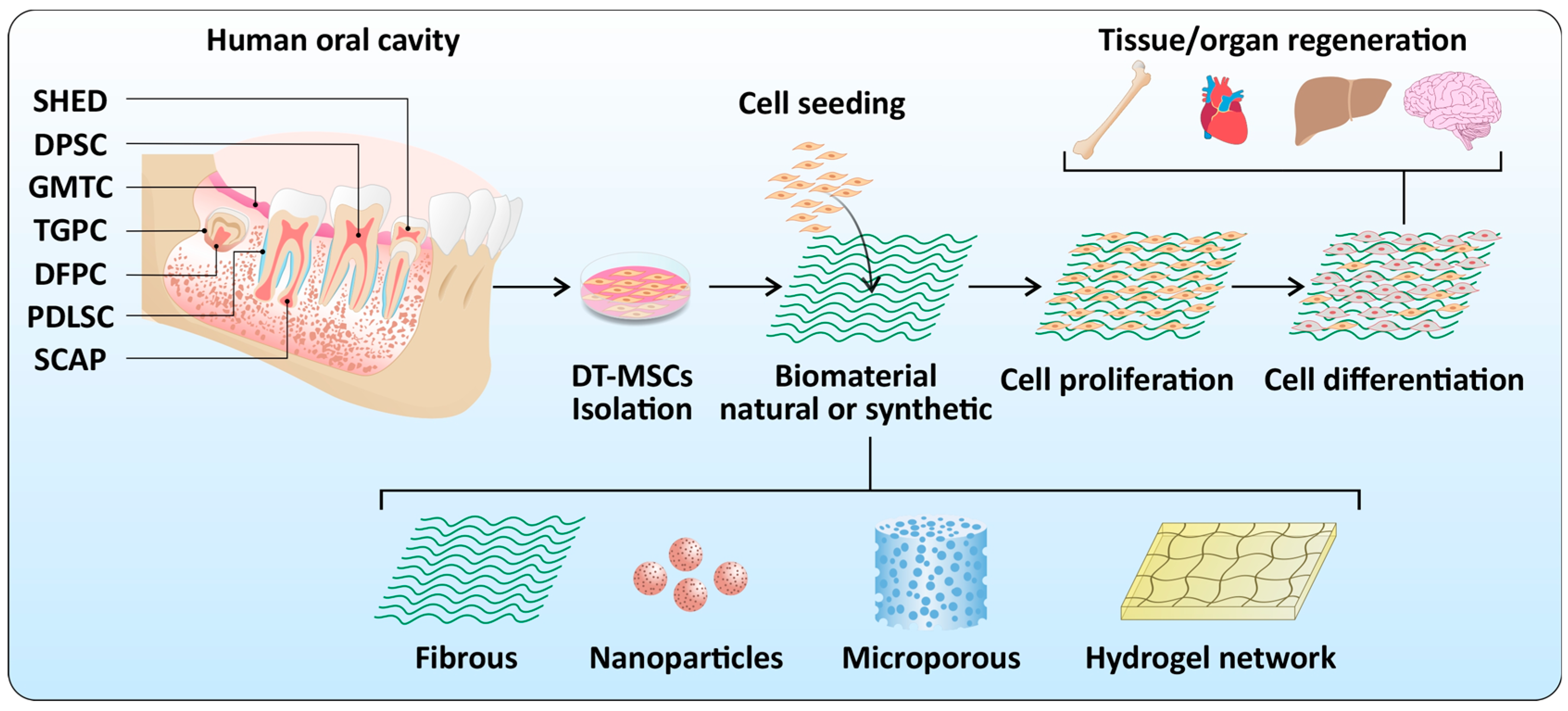
2. Dental Stem Cells
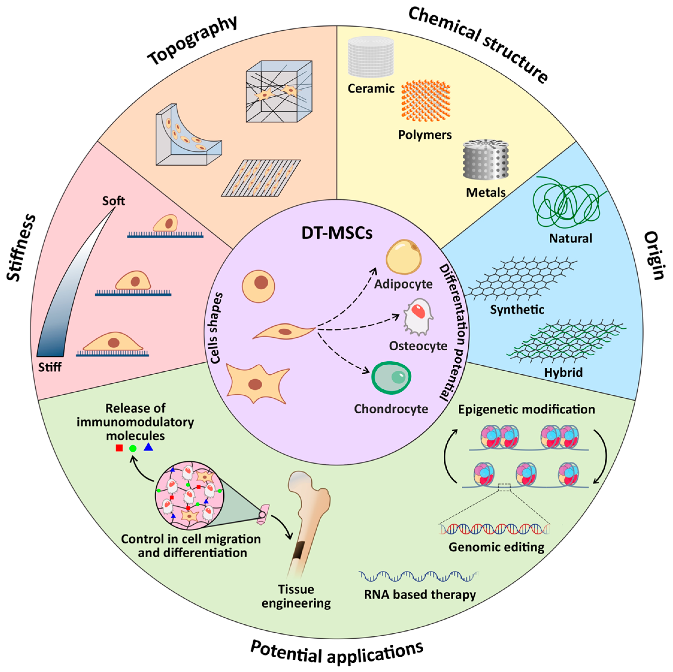
Mesenchymal stem cells (MSCs) are a group of cells capable of self-renewal and function as a repair system for damaged tissues. When a stem cell divides, each cell has the potential to remain a stem cell or to become another type of cell with a more specialized function. Thus, stem cells have two characteristics: the ability to self-renew and differentiate in any cell lineage (e.g., osteogenic, chondrogenic, adipogenic, myogenic, and neurogenic) comparable to those established for bone marrow-derived MSCs. They can be classified according to their origin in embryonic stem cells, adult stem cells (tissue-specific), and induced pluripotent cells (iPS) [3]. All of them are attractive for their use in the regeneration of damaged tissues; however, their clinical application is still limited due to the transplanted cells’ low survival and differentiation potential. For a thorough understanding of the origin and biology of different stem cells, the reader is referred to excellent reviews on this subject [4,5].
Oral cavity tissues are rich sources of adult stem cells. These cells are called dental tissue-derived mesenchymal stem cells (DT-MSCs), and the method of obtaining them is relatively easy in dental tissues and, even better, they are a source of autologous stem cells [1,6]. Likewise, they present a high proliferation, ability to differentiate into multiple cell types, and the expression of positive and embryonic markers of MSCs (i.e., OCT4, Nanog, SOX2, and KLF4), which makes them more attractive for their application in regenerative medicine [1,7,8,9]. Different types of DT-MSCs have been identified, such as dental pulp stem cells (DPSCs), exfoliated deciduous tooth stem cells (SHEDs), periodontal ligament stem cells (PDLSCs), dental follicle stem cells (DFSCs), and stem cells from the apical papilla (SCAP) (Figure 1) [1]. Although these types of cells have been shown to have stem, clonogenicity, and self-renewal characteristics, their differentiation capacity varies according to the origin of the cells. Some authors have pointed out that this behavior could be due to: (1) the heterogeneity of DT-MSCs and (2) the microenvironment of stem cells in dental tissues, for example, teeth in pre-eruptive formation (dental follicle and apical papilla) versus erupted teeth (dental pulp or periodontal ligament) [3,10,11]. Thus, one of the challenges for the clinical application of DT-MSCs is to mimic the appropriate microenvironment so that cells can proliferate or differentiate properly [3]; to achieve this, cells need extracellular matrix (ECM) components (Figure 1). Numerous studies have focused on developing artificial ECM microenvironments that are favorable for DT-MSC regeneration activities. The combination of DT-MSCs with biomaterials is one of the key procedures for developing autologous therapeutics in the regeneration of dental and nondental tissues. Next, the relevant aspects of biomaterials will be reviewed, as well as the most important cellular and molecular mechanisms involved in the regenerative process of stem cells of dental origin.

3. Biomaterials

Biomaterials are natural, synthetic, or semisynthetic substances designed to be implanted into biological environments [12]. In regenerative medicine, biomaterials are a vehicle for cells, as they provide a structure for their proliferation and secretion activities [13]. Several authors point out specific characteristics that a biomaterial must have for its application in regenerative medicine (Figure 2). Among them are nontoxicity, biocompatibility (ability to interact with the tissues of living beings), and chemical composition (it must mimic the components of the extracellular matrix) to promote a suitable environment for cell growth and differentiation [13,14,15,16].
The interaction between stem cells and the chemical composition of biomaterials are critical factors that influence cell behavior. Biomaterials must be a bioactive matrix that provides the cells with sites of adhesion, growth, and three-dimensional spatial organization [17,18]. There is a great variety of biomaterials reported in the literature, among which are natural or synthetic polymers, extracellular matrix, self-assembly systems, hydrogels, and bioceramics that have been used with DT-MSCs (Table 1) [15,18].
Table 1. Relevant findings of the biomaterial/oral cavity SCs interaction reported in the literature.

3.1. Natural Biomaterials

Research carried out in the field of biomaterials using cells of dental origin has increased in recent years. Most studies have focused on explaining the cellular mechanisms that lead to the formation of dental structures (for example, dentin, the periodontal ligament, dental pulp, or enamel), which has contributed to this knowledge, leading to the design of novel biomaterials destined to stimulate regeneration in dental and nondental tissues.
Natural biomaterials are constituents of the ECM or represent macromolecular properties that are similar to the ECM. These can be classified into two main categories, protein-based and natural biomaterials based on polysaccharides. Collagen, fibrin, and elastin are the most explored natural protein-based biomaterials [13,37]. Polysaccharide-based biomaterials are natural polymers consisting of sugar monomers. Chitosan, alginate, glycosaminoglycans (GAGs), and hyaluronic acid are examples [38]. Most natural biomaterials present favorable biocompatibility and immunogenicity and low cost [39], making them attractive for their application in tissue engineering in the field of endodontics. One of the most widely studied biomaterials in the dental field is collagen due to its role in ECM in dental pulp and dentin, its function as a natural hemostatic agent [40], its hydrophilicity, biocompatibility, biodegradability, and its low immunogenicity and cytotoxicity [41]. In this sense, biomaterials based on type I collagen (Col-I) have been developed to generate tissues, such as dental pulp [42], dentin [43], and guided bone regeneration treatments [44]. This shows that collagen-based biomaterials are an important element in dental research; however, the difficulties encountered in regenerative endodontics of root canals indicate irregular biodegradation and the generation of connective tissue instead of dentin in vivo. Another widely investigated polysaccharide is chitosan (CHS), which is purified mainly from chitin. Chemically, CHS is a polymeric material comprised of N-acetylglucosamine and glucosamine copolymer units [45]. CHS has also been reported to be a direct pulp cap with the purpose of initiating the formation of reparative dentin to help protect the pulp, favoring the differentiation of DPSCs into odontoblast-like cells [46]. The use of CHS in conjunction with DPSCs has been studied to induce bone regeneration and for the treatment of chronic periodontitis. [47]. For example, Kamal and Khalil (2018) [48] used CHS with DPSCs to evaluate the potential for bone formation around dental implants. They noted that this method helped bone maturation around the implant, suggesting its potential use in bone regeneration [48].
Fibrin is a fibrillar biopolymer and the main component of the blood coagulation matrix. This protein has been reported as a vehicle for the release of dental stem cells since it facilitates their union, growth, and differentiation [49,50]. Its advantages include excellent biocompatibility, bioresorbability, hemostatic properties, nontoxicity of degradation products, and the short-term generation of an ECM produced by stem cells incorporated into the biomaterial. Although there is increasing development of biomaterials with natural polymers due to their similarity with ECM, the biochemical characteristics of fibrin make it suitable as a platform for autologous cell release. For a better understanding of the composition, structure, biochemical characteristics and mechanical properties of fibrin, the reader is referred to excellent reviews about this biomaterial [51,52,53]. One of the interesting features of fibrin is that it can form a three-dimensional network of elastic fibers which can promote biological interactions during the regeneration of target tissues. [49]. To date, two types of fibrin-based products have been used: glue and hydrogels. The first type is obtained from human plasma (homologous or autologous) as a source of fibrinogen and functions as a bioadhesive for hemostasis in surgical procedures (i.e., allogeneic plasma commercial Tissucol/Tisseel, Beriplast, and Quixil), while hydrogels are made from allogeneic fibrinogen and purified thrombin. One advantage of hydrogels is that they can be biofunctionalized to increase the cellular response through the incorporation of cell binding sequences, such as arginine–glycine–aspartic acid (RGD), for a better function as a cell vehicle [50,51,52,53,54]. The use of fibrin gel as a vehicle for carrying cells in a three-dimensional scaffold has been extensively investigated for the regeneration of bone tissue. In dental tissues, numerous approaches have been developed to achieve regeneration, for example, fibrin hydrogels or modified fibrin hydrogels (polyethylene glycol, chitosan, collagen) for dental pulp regeneration [18,50,55]. However, these approaches are limited because a specific spatial geometry is not achieved for each patient, nor is there control of the union, proliferation, and migration of cells within the structure. Three-dimensional (3D) bioprinting is now the most attractive approach for its application in regenerative medicine in dental and nondental tissues. This is a manufacturing technique that allows stem cells to be precisely placed within the biomaterial that acts as a temporary ECM [56,57]. With this technology, it has been possible to design a fibrin-based bio-ink with DPSCs to form an autologous dentin-pulp complex [58] or in bone regeneration [59].

3.2. Synthetic Biomaterials

Biomaterials of synthetic origin are used due to the growing necessity of specific characteristics of scaffolds for regenerative medicine due to the limitations of the natural biomaterials that need to be modified, the limited mechanical strength, and the difficulty of obtaining 3D scaffolds required for tissue engineering, providing cells with the necessary environment to proliferate and differentiate into a lineage-specific manner [60]. Biomaterials of synthetic origin have been widely used for the last 100 years, considering that inert materials (i.e., metal alloys) were the first to be implanted in the human body for the reconstruction of affected tissues and/or organs [61]. Currently, a range of materials are used for regenerative medicine, primarily for their characteristics, such as biocompatibility, physicochemistry, mechanical behavior, biodegradability, and modulation of cell response, among others. A variety of engineered synthetic biomaterials that are chemically and physically designed to fulfill the cellular specific needs of these critical parameters have been reported in the literature [62]. Some synthetic biomaterials, such as metallic alloys, ceramics, polymers, and hydrogels, are reported to be promotors of stem cell differentiation [63].
Regarding synthetic metal biomaterials, inert titanium alloys are commonly used in dentistry for bone tissue engineering due to their ability to induce osteogenic differentiation of dental stem cells; as reported in the literature, composite biomaterials with a polymeric matrix of polycaprolactone diol-based segmented polyurethanes and titanium particles enhance the viability of pulp stem cells and osteoblasts, as it increases with the amount of titanium in composites [64]. According to Hanafy, mineral trioxide aggregates and nanohydroxyapatite could enhance the odontogenic differentiation of human dental pulp stem cells, as assessed by tracing genes characteristic of different stages of odontoblasts via qRT–PCR and calcific nodule formation evaluated by Alizarin red staining [65].
Ceramics, as bioactive glass, promote odontogenic differentiation of DPSCs; as reported by Ahn et al. (2020) [66] in their study, composites of mesoporous bioactive glass nanoparticles (MBNs) and graphene oxide (GO) were prepared and analyzed, and they concluded that MBN/GO promoted the proliferation and odontogenic differentiation of DPSCs [66]. In another study, composites of nano bioactive glass synthesized by the sol-gel method (58SiO2:40CaO:5P2O5) and Biodentine (Septodont, Saint Maur des Fosses, France) were prepared and tested, and they found cell adhesion and proliferation on nBG/BD nanocomposites and increased odontogenic differentiation of DPSCs, as measured by alkaline phosphatase activity after 7 and 14 days of exposure [67].
Polymers and composites of polymeric matrices are biomaterials that have been the focus of increased interest in recent years due to their capability for being designed, modified, and reinforced according to the requirements for an extensive range of therapeutic and regenerative purposes. Polymers for regenerative medicine include polyethylene glycol (PEG), polylactic acid (PLA), poly(lactic-co-glycolic acid) (PLGA), polyvinyl alcohol (PVA), and polycaprolactone (PCL) [13]. A study by Wang concluded that poly(L-lactic acid) (PLLA), with recombinant human bone morphogenetic protein 2 (BMP-2), produced odontogenic differentiation of SCAP, leading to in vivo dentin regeneration [68]. Another study with PCL scaffolds reported attachment, growth, and proliferation of human SCAPs with calcification nodules detected [69]. Alipour et al. (2019) [70] reported that PCL-PEG-PCL/zeolite nanofibrous scaffolds produced adhesion and proliferation of DPSCs and their osteo/odontogenic differentiation and concluded that zeolite nanoparticles on PCL-PEG-PCL scaffolds could have a crucial role in osteoblastic physiology [70]. One of the essential aspects of polymers is their degradability/biodegradability, which can be modulated according to the needs of therapeutics, and even synthetic polymers can be designed to carry and deliver drugs to a specific target by their degradation process [63].
Hydrogels are derived from polymers, and they are a three-dimensional, hydrophilic polymer or copolymer network that can soak up large amounts of water or biological fluids due to their affinity to absorb water, which is attributed to the presence of hydrophilic groups; they are commonly temperature-sensitive biomaterials [62]. Luo et al. (2021) [71] reported that a methacryloyl gelatin (GelMA) hydrogel and human basic fibroblast growth factor seeded with DPSCs wrapped by a cellulose/soy protein isolate composite membrane was proven to be a promising tissue engineering approach to treat significant gap defects in peripheral nerve injuries [71]. Synthetic hydrogels appear to be promising for tissue engineering since in vivo-forming hydrogels can be prepared as a solution and can easily incorporate cells and growth factors to be injected to fill the shape of the in vivo cavity via minimally invasive procedures, as described by Jang et al. (2016) [72] who obtained an in vivo forming solution of methoxy polyethylene glycol-b-poly(ε-caprolactone), DPSCs, and osteogenic factors. They concluded that DPSCs embedded in an in vivo-forming hydrogel may provide benefits as a noninvasive formulation for bone tissue engineering applications [72].

4. Dental Stem Cells and Biomaterial Interactions

Biomaterials (natural or synthetic) are essential components in the construction of scaffolds, providing an artificial three-dimensional environment to regulate the behavior of stem cells. In addition, they allow for evaluating the effect of the physical environment in the cells [73]. An important aspect to consider is the biomaterial–cell interaction since they affect the adhesion, viability, proliferation, matrix production, and differentiation of stem cells [74]. Knowledge of how the interaction between cells and biomaterials is carried out is essential for achieving effective repair in regenerative medicine. Mechanobiology is an interdisciplinary field that investigates the mechanisms by which stem cells can sense (mechanosensing) and respond (mechanotransduction) to changes in their environment [75]. A comprehensive review of the mechanobiology in stem cells in response to mechanical signals can be reviewed in Argentati et al. (2019) [76]. In this section, we describe the mechano-molecular players recruited and interconnected with each other during the biomaterial–DT-MSCs interaction and their impact on cell proliferation and differentiation.

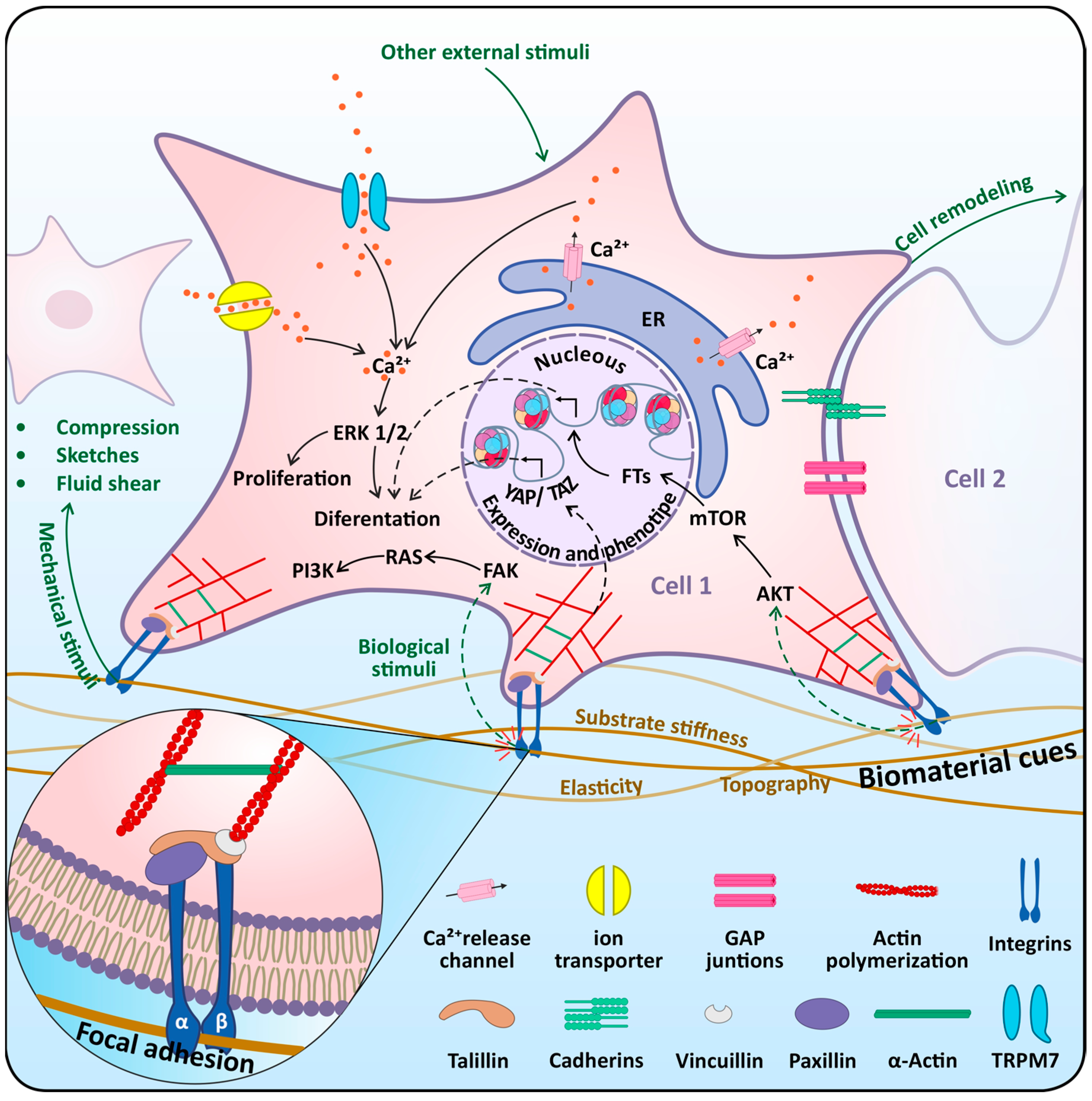
4.1. Mechasensors in Dental Stem Cells

Currently, the design of biomaterials with specific characteristics is a novel approach for evaluating the effect of chemical, physical or topographic changes in stem cells during their proliferation and differentiation under in vitro conditions. The reason for this is that stem cells are very sensitive to forces and can convert mechanical stimuli into a chemical response [77]. There is much evidence that DPSCs are mechanosensitive cells and capable of recognizing physical and mechanical signals during their differentiation process. In this context, the participation of mechanosensors, such as mechanosensitive ion channels, cytoskeleton proteins, and assembly proteins, has been recognized in DPSCs.
Mechanosensitive ion channels (MICs) are receptors that convert extracellular mechanical force into intracellular biochemical information. However, how do the channels mediate these sensations and turn them into a stimulus? Does this occur through direct or indirect activation of the channel? To answer this question, Xiao et al. (2016) [78] proposed two scenarios. The first is that mechanical force is released into the channel by lipid bilayer tension, generating a hydrophobic mismatch that helps the channel open. In the second, the participation of accessory proteins (for example, those of the cytoskeleton or the components of the ECM) has been proposed, and mechanical stress transmitted by the cytoskeleton causes Ca2+ release from the endoplasmic reticulum via the inositol trisphosphate receptor (IP3R) [78]. There are reports of MICs that play an important role in the transduction of mechanical forces in the activation of signaling pathways involved in cell proliferation and differentiation in stem cells. In this context, two MICs have been recognized in DPSCs: Piezo proteins and members of the transient receptor potential (TRP) channel family.
The Piezo channel is a three-bladed helix-shaped trimeric complex that includes two subtypes: Piezo1 and Piezo2 [79]. In stem cells, these proteins play an important role in shear stress and traction signals during their proliferation [80,81,82]. Several researchers agree that when the Piezo protein is activated, there is an influx of Ca2+ [65]. However, there is still controversy about whether the integrity of the cytoskeleton is affected by the activity of the channel in response to mechanical stimuli. The role of Piezo proteins in DPSCs has been studied through adjuvant therapies for the repair of dental tissues. An example of this is the use of low-intensity pulsed ultrasound (LIPUS) recognition therapy employed in dental tissue repair [64]. In cells, LIPUS acts as a mechanobiological stimulus that activates various signaling pathways that regulate cell proliferation and differentiation. For example, Jin et al. (2015) [83] investigated the presence of Piezo1 and Piezo2 in DPSCs and PDLSCs and their involvement in LIPUS-associated proliferation and MAPK signaling. The authors demonstrated that DPSC proliferation was associated with LIPUS stimulation by activating MAPK signaling, while in PLSCs, it could occur through another mechanism [83]. Another study by Mousawi et al. (2020) [84] examined the role of Piezo1 in ATP release in DPSCs migration in vitro. The authors reported that the activation of Piezo1 induces the release of ATP, modulating the activity of P2 receptors and inducing the activation of proline-rich tyrosine kinase 2 (PYK2) and MEK/ERK. Another ionic channel broadly expressed in DPSCs is the transient receptor potential (TRP) channel, including transient receptor potential melastatin types 4 and 7 (TRPM4 and TRPM7, respectively) [85,86,87].
TRPM controls Ca2+ signals and is sensitive to mechanical stimuli, such as patch-clamp pipette suction and patch-clamp pipette stretching. Xiao et al. (2015) [88] reported that, in human bone marrow MSCs subjected to stretching or suction, TRPM7 activation appears to be independent of actin polymerization disruption since suction-induced TRPM7 activation was not abolished [88]. In DPSCs, research has also focused on TRPM7 and osteogenesis. For example, Cui et al. (2013) [87] reported that TRPM7 participates in the pulp-repair process through the regulation of proliferation, migration, and osteogenic differentiation of DPSCs, while TRPM4 is necessary for adipogenesis in stem cells of DFSCs in rats [87]. The authors agree that the activation of TRPM gives rise to the release of Ca2+ mediated by IP3 type 2 (IP3R2) from the endoplasmic reticulum, amplifying Ca2+ signaling and inducing osteogenesis through the activation of the transcription factor NFATc1 [88,89,90]. Taking these results together, the data suggest that MICs are important molecular sensors. However, studies have only focused on DPSCs and osteogenesis. It would be interesting to see if a similar regulatory pathway is conserved in other types of DT-MSCs.
Other important mechanosensors are macromolecular complexes that include cytoskeletal proteins (microtubules, f-actin microfilaments, intermediate filaments, and actin-linking proteins), nucleoskeletal-related proteins (SUN1, SUN2, lamins), adherens junctions (cadherins, α-catenin, β-catenin), focal adhesion proteins (vinculin), integrins, and ECM-related proteins (fibronectin) [57,58]. These mechanosensors turn on the rearrangement of molecular components by activating several intracellular signaling pathways that are involved in growth, cell proliferation, and the regulation of gene expression (Figure 3).
The cytoskeleton plays important roles in cell morphology, adhesion, growth, and signaling [91]. In this context, stem cells alter their cytoskeleton in response to mechanical forces that cause cell reorganization through actin polymerization and microtubule assembly or by disassembling cytoskeletal components and their ECM junctions [75]. Topography of biomaterials is a promising approach to guide cytoskeleton behavior and differentiation in stem cells. Several works have focused on analyzing how the topographic signals (macro, micro, or nanoscale) of biomaterials affect the behavior of components of the cytoskeleton in DT-MSCs. For example, Du et al. (2019) [92] determined that the topographic signals of a poly(lactic-co-glycolic acid) bilayer (PLGA) change F-actin alignment and DPSCs morphology by modulating Yes-associated protein (YAP) signaling to control osteogenic differentiation/odontogenicity [92]. Collart-Dutilleul et al. (2014) [93] studied the influence of the micropore size of a nanostructured silicon biomaterial on DPSCs adhesion through the formation of lamellipodia (dense network of actin filaments) and filopodia. They concluded that the porosity of the biomaterial promotes the formation of filopodia and increases cell migration [93]. In another study, Conserva et al. (2018) [94] studied the growth of DPSCs by laser ablation laser-microgrooved (8-µm-sized microgrooves or microchannels) surfaces. Their results showed that DPSCs proliferated following the direction of the microgrooves, which could have significant implications for bone regeneration [94]. Additionally, Bachhuka et al. (2017) [95] reported that DPSCs proliferated faster where there was a greater density of nanotopography favoring osteogenic differentiation [95]. Marconi et al. (2021) [96] investigated the in vitro effects of titanium implants on PDLSC culture. The authors reported that the topography of the titanium implant surface enhances the release of ECM components in PDLSCs, which has an impact on the process of implant osseointegration [96]. Other authors, such as Hasturk et al. (2019) [97], proposed a novel biomaterial with 4- and 8-μm square prism micropillars on a poly(methyl methacrylate) surface as an alternative to enhance the osteogenesis of DPSCs. Their results showed that the interpillar spaces generated a high degree of tension in the cytoskeleton and induced differentiation toward the bone lineage [97]. These studies showed that the interaction between biomaterials and the cytoskeleton is important during the osteogenic differentiation response in DT-MSCs; however, there is still a long way to go to understand the mechanisms involved in biomaterial–DT-MSC interactions in other differentiation processes.
The ECM surrounding cells exerts a mechanical influence that determines phenotype, motility, and matrix production. This allows tissues to function correctly by modulating stem cell adhesion, proliferation, migration, and differentiation [73,75]. During their growth, stem cells secrete structural components of the extracellular matrix (collagen, elastin, laminin, fibronectin, hyaluronic acid, chondroitin sulfate, and syndecans) that function as mediators between the cells and the ECM. One of the strategies for the study of ECM mechanosensors in DPSCs and their physical environment is the development of scaffolds with components of the ECM. For example, Ravindran et al. (2014) [98] generated a scaffold ECM from dental pulp to induce odontogenic differentiation in DPSCs, PDLSCs, and HMSCs as a strategy for the treatment of dental caries [98]. Another example is the work of Paduado et al. (2016) [99], who demonstrated that a hydrogel scaffold derived from decellularized and demineralized bovine bone (bECM) favors the odontogenic differentiation of DPSCs and could be applied for the regeneration treatment of dentin and pulp. Currently, research groups focus on the search for alternative sources of ECM substrates of human origin, such as for human cell culture, due to the limitations represented using commercial biomaterials that are derived from animal sources or cancer lines (for example, Matrigel, which is derived from mouse sarcoma) for clinical application [99]. In this context, a novel study was reported by Heng et al. (2016) [100], who used decellularized matrix (DECM) from SHED stem cells and PLSCs as a substrate for ex vivo culture of DPSCs. Their results showed that DECM of dental cells enhanced DPSCs adhesion, which correlated with increased expression of vinculin, a key focal adhesion protein [100].
Another approach that has been addressed is the analysis of adhesion proteins, such as integrins. Integrins are heterodimeric receptors composed of alpha and beta subunits linked by noncovalent bonds. The combination of these subunits results in various receptors that exhibit preferential affinity for specific ECM molecules. For example, α2β1 integrin recognizes the Asp-Gly-Glu-Ala amino acid sequence in collagen, while α5β1 recognizes the RGD sequence in fibronectin and αvβ3 in vitronectin [101]. The adhesion process in animal cells is mediated by integrins, which generate supramolecular protein complexes with cytoskeletal proteins called focal contacts (FAs) (Figure 3). FAs are networks of proteins that provide structural integrity to cells and constitute a dynamic bridge between the ECM and actin of the cytoskeleton (Figure 3). In the case of stem cells, this process is essential for tissue integration of the biomaterial. In this context, Lee et al. (2014) [102] studied the interaction between PDLSCs and a mussel-inspired polydopamine (PDA) adhesive biomaterial through the expression of integrins α5 and β1. The authors demonstrated that integrin–PI3K linkages mediate cell adhesion and regulate osteogenic differentiation of PDLSCs [102]. Likewise, using silica-based materials, Hung and colleagues [103] investigated the role of αv integrin in the odontogenic differentiation of DPSCs. They reported that the silicon-containing biomaterial favored cell adhesion through increased fibronectin adsorption and integrin expression [103]. Liu et al. (2013) [104] analyzed the effect of crystal alignment (ordered/disorganized) of apatite (an enamel-like substrate) on the expression of adhesion-related genes to produce an enamel/dentin superstructure in vitro. The authors reported that the ordered alignment of apatite provided a favorable environment for DPSCs adhesion, which was linked to the upregulated expression of integrins alpha 7 and 8 (ITGA 7 and 8), integrin beta 3 and 4 (ITGB3 and 4), vitronectin receptor-integrin alpha V (ITGAV), and the key adhesion protein fibronectin 1 (FN1) [104].

4.2. Mechanotransduction Pathways in Dental Stem Cells

As mentioned in the previous section, the attachment of cells to biomaterial substrates is essential to establishing communication between cells and the ECM microenvironment. In this way, the activated biochemical signals can modulate specific signaling pathways to regulate cell activity in processes, such as adhesion, proliferation, and differentiation. This section will address current studies on the signaling pathways involved in the differentiation process in biomaterials with DPSCs.
Biomaterials act as adhesion substrates that send mechanical signals to cells, influencing the differentiation of DT-MSCs. Stem cell differentiation during biomaterial interactions involves many signaling pathways (Figure 3). For example, Yun et al. (2015) [105] examined with DPSCs a scaffold of magnetite and polycaprolactone nanoparticles and the signaling pathways involved in the mechanisms of adhesion, migration, and odontogenic differentiation. Their results demonstrated that integrin (subunits α1, α2, β1, and β3) signaling with activation of FAK/MAPK and NF-κB by the scaffold is involved in cellular events in DPSCs [105]. Another study by Lee et al. (2014) [102] evaluated the osteoinductive effect of a bioadhesive on PDLSCs for use as a dental implant. They reported that the biomaterial stimulated osteogenic differentiation of PDLSCs through activation of α5/β1 integrin-PI3K signaling [102]. Additionally, Zhang et al. (2012) [106] investigated whether MAPK signaling pathways are a mediator in odontogenic differentiation of DPSCs cultured on five different biomaterials. Their results showed enhanced odontogenic differentiation and dentin-like tissue formation in natural biomaterials derived from mineralized tissue (dentin matrix and bovine bone ceramic) through the phosphorylation of ERK1/2 and p38 [106]. Another study by Guo et al. in 2018 [107] employed a pharmacological perturbation approach to identify which signaling pathways were involved in the differentiation and mineralization of DPSCs cultured on a fluorapatite-modified polycaprolactone nanofiber biomaterial. The authors reported that the Hedgehog, insulin, and Wnt signaling pathways are involved in biomaterial-induced DPSCs differentiation and can activate the osteogenesis process through autophagic modulation [107].
There are currently novel biomaterial designs in the literature that have been used to investigate the signaling pathways involved during DPSCs differentiation. The design of nanoarrays as therapeutic platforms has aroused particular interest because dental stem cells can recognize nanofibrous topology and respond to biochemical signals from their environment. In this context, Lim et al. (2016) [108] designed in vitro nanofiber arrays containing bioactive glass nanoparticles that release dexamethasone, a signaling molecule of odontogenesis. The authors reported that the integrin pathway (α2, α5, β1), bone morphogenetic protein, and mTOR signaling pathways are possible mechanisms involved in the stimulation of odontogenesis. Other signaling pathways have also been reported in cell differentiation events in three-dimensional nanomaterials [108]. For example, Zhou et al. (2018) [109] designed a biomaterial containing a tetrahedral DNA nanostructure (TDN) self-assembled by four specific single-stranded DNAs (ssDNAs) by complementary base pairing. The authors reported that TDN stimulated the osteogenic differentiation of PDLSCs by activating the Wnt/β-catenin pathway, while in DPSCs, TDN increased the expression of HES1, HEY1, and NOTCH1, which are crucial factors of the Notch pathway [109]. These results indicate that although the cells have the same dental origin, their biochemical response can be different for the same biomaterial, which will be interesting to study in the future to develop methodologies for the regeneration of dental and nondental tissues.
One approach that has been addressed is to study signaling events in the epigenome, for which signals from biomaterials, such as topography, elasticity, material chemistry, and mechanical forces/stimulus, influence the state of the epigenome [110]. The term epigenetics refers to heritable changes in gene expression patterns that do not involve alterations in the DNA sequence [111]. It will be interesting to determine how biomaterial signals act by modulating epigenome mechanisms, including chromatin remodeling, DNA methylation, and posttranslational modifications at the amino-terminal tails of nucleosome histones. To date, there are few reports that have investigated how biomaterials with different physical and chemical characteristics alter the epigenome in stem cells [63,111]. However, we have not been able to find reports focused on DT-MSCs.

5. Conclusions

Although current results are promising, there are still many unsolved questions regarding the mechanical regulation of stem cell activities, and the study of MSCs during these processes is just beginning. In the coming years, the development and application of new techniques in live imaging, tissue culture, and real-time mechanical stimulation delivery will significantly increase our current knowledge about mechanobiology, as well as stem cell biology. If well the studies discussed here highlight the players involved in the response and processes of DT-MSCs during their interaction with biomaterials, much remains to be investigated. In this scenario, we consider it essential to tackle the following aspects:
  • Design and develop of smart biomaterials that favor the proliferation and differentiation of DT-MSCs on a large scale.
  • Integrate multi-omic tools would allow a global perspective of the interactions between cells and biomaterials at the genomic, proteomic, and metabolomic levels.
  • Delivery into the mechanogenomic field to facilitate the design of highly functionalized biomaterials and the epigenetic manipulation that can be performed to control the fate of DT-MSCs for their application in regenerative medicine.

Author Contributions

Conceptualization, B.A.R.-J.; writing—original draft preparation, F.J.A.-A. and F.J.A.-P., writing—reviewed editing, B.A.R.-J., G.C.-G. and D.A.-P., technical assistance R.R.-H. and G.I.N.-C. All authors have read and agreed to the published version of the manuscript.

Funding

This research received no external funding.

Institutional Review Board Statement

Not applicable.

Informed Consent Statement

Not applicable.

Data Availability Statement

Not applicable.

Acknowledgments

This work was supported by Facultad de Ingeniería Química and Facultad de Odontología from Universidad Autónoma de Yucatán. The authors also acknowledge the support of the Seeding Labs program. The authors would like to thank Julio Montero del Toro for the design of the images.

Conflicts of Interest

The authors declare no conflict of interest.

References

  1. Rodas-Junco, B.A.; Canul-Chan, M.; Rojas-Herrera, R.A.; De-la-Peña, C.; Nic-Can, G.I. Stem cells from dental pulp: What epigenetics can do with your tooth. Front. Physiol. 2017, 8, 999. [Google Scholar] [CrossRef] [PubMed] [Green Version]
  2. Zadpoor, A.A. Meta-biomaterials. Biomater. Sci. 2020, 8, 18–38. [Google Scholar] [CrossRef] [PubMed] [Green Version]
  3. Granz, C.L.; Gorji, A. Dental stem cells: The role of biomaterials and scaffolds in developing novel therapeutic strategies. World J. Stem Cells 2020, 12, 897. [Google Scholar] [CrossRef] [PubMed]
  4. Zakrzewski, W.; Dobrzyński, M.; Szymonowicz, M.; Rybak, Z. Stem cells: Past, present, and future. Stem Cell Res. Ther. 2019, 10, 68. [Google Scholar] [CrossRef] [PubMed]
  5. Worku, M.G. Pluripotent and multipotent stem cells and current therapeutic applications. Stem Cells Cloning Adv. Appl. 2021, 14, 3. [Google Scholar] [CrossRef]
  6. Sharpe, P.T. Dental mesenchymal stem cells. Development 2016, 143, 2273–2280. [Google Scholar] [CrossRef] [Green Version]
  7. Hussein, A.M.; Darwish, Z.E.; Raslan, H.S.; Attia, M.A.; Abdel-Hamid, H.M. Dental stem cells (concepts and applications). Alex. Dent. J. 2021, 46, 66–71. [Google Scholar] [CrossRef]
  8. Rodríguez-Lozano, F.J.; Bueno, C.; Insausti, C.L.; Meseguer, L.; Ramirez, M.C.; Blanquer, M.; Marin, N.; Martínez, S.; Moraleda, J.M. Mesenchymal stem cells derived from dental tissues. Int. Endod. J. 2011, 44, 800–806. [Google Scholar] [CrossRef] [Green Version]
  9. Lan, X.; Sun, Z.; Chu, C.; Boltze, J.; Li, S. Dental pulp stem cells: An attractive alternative for cell therapy in ischemic stroke. Front. Neurol. 2019, 10, 824. [Google Scholar] [CrossRef]
  10. Egusa, H.; Sonoyama, W.; Nishimura, M.; Atsuta, I.; Akiyama, K. Stem cells in dentistry–part I: Stem cell sources. J. Prosthodont. Res. 2012, 56, 151–165. [Google Scholar] [CrossRef] [Green Version]
  11. Mercado-Rubio, M.D.; Pérez-Argueta, E.; Zepeda-Pedreguera, A.; Aguilar-Ayala, F.J.; Peñaloza-Cuevas, R.; Kú-González, A.; Rojas-Herrera, R.A.; Rodas-Junco, B.A.; Nic-Can, G.I. Similar Features, Different Behaviors: A Comparative In Vitro Study of the Adipogenic Potential of Stem Cells from Human Follicle, Dental Pulp, and Periodontal Ligament. J. Pers. Med. 2021, 11, 738. [Google Scholar] [CrossRef] [PubMed]
  12. Chan, B.P.; Leong, K.W. Scaffolding in tissue engineering: General approaches and tissue-specific considerations. Eur. Spine J. 2008, 17, 467–479. [Google Scholar] [CrossRef] [PubMed] [Green Version]
  13. Xu, Y.; Chen, C.; Hellwarth, P.B.; Bao, X. Biomaterials for stem cell engineering and biomanufacturing. Bioact. Mater. 2019, 4, 366–379. [Google Scholar] [CrossRef] [PubMed]
  14. Baranwal, A.; Kumar, A.; Priyadharshini, A.; Oggu, G.S.; Bhatnagar, I.; Srivastava, A.; Chandra, P. Chitosan: An undisputed bio-fabrication material for tissue engineering and bio-sensing applications. Int. J. Biol. Macromol. 2018, 110, 110–123. [Google Scholar] [CrossRef] [PubMed]
  15. Vinatier, C.; Guicheux, J. Cartilage tissue engineering: From biomaterials and stem cells to osteoarthritis treatments. Ann. Phys. Rehabil. Med. 2016, 59, 139–144. [Google Scholar] [CrossRef]
  16. Anitua, E.; Troya, M.; Zalduendo, M. Progress in the use of dental pulp stem cells in regenerative medicine. Cytotherapy 2018, 20, 479–498. [Google Scholar] [CrossRef]
  17. Martino, S.; D’Angelo, F.; Armentano, I.; Kenny, J.M.; Orlacchio, A. Stem cell-biomaterial interactions for regenerative medicine. Biotechnol. Adv. 2012, 30, 338–351. [Google Scholar] [CrossRef]
  18. Galler, K.M.; Cavender, A.C.; Koeklue, U.; Suggs, L.J.; Schmalz, G.; D’Souza, R.N. Bioengineering of dental stem cells in a PEGylated fibrin gel. Regen. Med. 2011, 6, 191–200. [Google Scholar] [CrossRef]
  19. Lambrichts, I.; Driesen, R.B.; Dillen, Y.; Gervois, P.; Ratajczak, J.; Vangansewinkel, T.; Wolfs, E.; Bronckaers, A.; Hilkens, P. Dental Pulp Stem Cells: Their Potential in Reinnervation and Angiogenesis by Using Scaffolds. J. Endod. 2017, 43, S12–S16. [Google Scholar] [CrossRef]
  20. Asghari Sana, F.; Çapkın Yurtsever, M.; Kaynak Bayrak, G.; Tunçay, E.Ö.; Kiremitçi, A.S.; Gümüşderelioğlu, M. Spreading, proliferation and differentiation of human dental pulp stem cells on chitosan scaffolds immobilized with RGD or fibronectin. Cytotechnology 2017, 69, 617–630. [Google Scholar] [CrossRef]
  21. Pecci-Lloret, M.P.; Vera-Sánchez, M.; Aznar-Cervantes, S.; García-Bernal, D.; Sánchez, R.O.; Pecci-Lloret, M.R.; Moraleda, J.M.; Cenis, J.L.; Rodríguez-Lozano, F.J. Analysis of the Adherence of Dental Pulp Stem Cells on Two-Dimensional and Three-Dimensional Silk Fibroin-Based Biomaterials. J. Craniofac. Surg. 2017, 28, 939–943. [Google Scholar] [CrossRef] [PubMed]
  22. Núñez-Toldrà, R.; Martínez-Sarrà, E.; Gil-Recio, C.; Carrasco, M.Á.; Al Madhoun, A.; Montori, S.; Atari, M. Dental pulp pluripotent-like stem cells (DPPSC), a new stem cell population with chromosomal stability and osteogenic capacity for biomaterials evaluation. BMC Cell Biol. 2017, 18, 21. [Google Scholar] [CrossRef] [PubMed] [Green Version]
  23. Yao, L.; Flynn, N. Dental pulp stem cell-derived chondrogenic cells demonstrate differential cell motility in type I and type II collagen hydrogels. Spine J. 2018, 18, 1070–1080. [Google Scholar] [CrossRef] [PubMed]
  24. Louvrier, A.; Euvrard, E.; Nicod, L.; Rolin, G.; Gindraux, F.; Pazart, L.; Houdayer, C.; Risold, P.Y.; Meyer, F.; Meyer, C. Odontoblastic differentiation of dental pulp stem cells from healthy and carious teeth on an original PCL-based 3D scaffold. Int. Endod. J. 2018, 51 (Suppl. S4), e252–e263. [Google Scholar] [CrossRef] [Green Version]
  25. Soancă, A.; Lupse, M.; Moldovan, M.; Pall, E.; Cenariu, M.; Roman, A.; Tudoran, O.; Surlin, P.; Șorițău, O. Applications of inflammation-derived gingival stem cells for testing the biocompatibility of dental restorative biomaterials. Ann. Anat. Anat. Anz. Off. Organ Anat. Ges. 2018, 218, 28–39. [Google Scholar] [CrossRef]
  26. Saberi, E.; Farhad-Mollashahi, N.; Sargolzaei Aval, F.; Saberi, M. Proliferation, odontogenic/osteogenic differentiation, and cytokine production by human stem cells of the apical papilla induced by biomaterials: A comparative study. Clin. Cosmet. Investig. Dent. 2019, 11, 181–193. [Google Scholar] [CrossRef] [Green Version]
  27. Parthiban, S.P.; He, W.; Monteiro, N.; Athirasala, A.; França, C.M.; Bertassoni, L.E. Engineering pericyte-supported microvascular capillaries in cell-laden hydrogels using stem cells from the bone marrow, dental pulp and dental apical papilla. Sci. Rep. 2020, 10, 21579. [Google Scholar] [CrossRef]
  28. Mansouri, N.; Al-Sarawi, S.; Losic, D.; Mazumdar, J.; Clark, J.; Gronthos, S.; O’Hare Doig, R. Biodegradable and biocompatible graphene-based scaffolds for functional neural tissue engineering: A strategy approach using dental pulp stem cells and biomaterials. Biotechnol. Bioeng. 2021, 118, 4217–4230. [Google Scholar] [CrossRef]
  29. Birant, S.; Gokalp, M.; Duran, Y.; Koruyucu, M.; Akkoc, T.; Seymen, F. Cytotoxicity of NeoMTA Plus, ProRoot MTA and Biodentine on human dental pulp stem cells. J. Dent. Sci. 2021, 16, 971–979. [Google Scholar] [CrossRef]
  30. Gu, Y.; Xie, X.; Zhuang, R.; Weir, M.D.; Oates, T.W.; Bai, Y.; Zhao, L.; Xu, H.H.K. A Biphasic Calcium Phosphate Cement Enhances Dentin Regeneration by Dental Pulp Stem Cells and Promotes Macrophages M2 Phenotype In Vitro. Tissue Eng. Part A 2021, 27, 1113–1127. [Google Scholar] [CrossRef]
  31. Vagropoulou, G.; Trentsiou, M.; Georgopoulou, A.; Papachristou, E.; Prymak, O.; Kritis, A.; Epple, M.; Chatzinikolaidou, M.; Bakopoulou, A.; Koidis, P. Hybrid chitosan/gelatin/nanohydroxyapatite scaffolds promote odontogenic differentiation of dental pulp stem cells and in vitro biomineralization. Dent. Mater. 2021, 37, e23–e36. [Google Scholar] [CrossRef] [PubMed]
  32. Hagar, M.N.; Yazid, F.; Luchman, N.A.; Ariffin, S.H.Z.; Wahab, R.M.A. Comparative evaluation of osteogenic differentiation potential of stem cells derived from dental pulp and exfoliated deciduous teeth cultured over granular hydroxyapatite based scaffold. BMC Oral Health 2021, 21, 263. [Google Scholar] [CrossRef] [PubMed]
  33. Gendviliene, I.; Simoliunas, E.; Alksne, M.; Dibart, S.; Jasiuniene, E.; Cicenas, V.; Jacobs, R.; Bukelskiene, V.; Rutkunas, V. Effect of extracellular matrix and dental pulp stem cells on bone regeneration with 3D printed PLA/HA composite scaffolds. Eur. Cell. Mater. 2021, 41, 204–215. [Google Scholar] [CrossRef] [PubMed]
  34. Zhang, R.; Li, C.-J.; Zhang, S.; Ma, X.-C.; Xiao, R.; Liu, H.-C. Effects of rhBMP-2 on Bone Formation Capacity of Rat Dental Stem/Progenitor Cells from Dental Follicle and Alveolar Bone Marrow. Stem Cells Dev. 2021, 30, 441–457. [Google Scholar] [CrossRef]
  35. Atila, D.; Hasirci, V.; Tezcaner, A. Coaxial electrospinning of composite mats comprised of core/shell poly(methyl methacrylate)/silk fibroin fibers for tissue engineering applications. J. Mech. Behav. Biomed. Mater. 2022, 128, 105105. [Google Scholar] [CrossRef]
  36. Zhang, Y.; Dou, X.; Zhang, L.; Wang, H.; Zhang, T.; Bai, R.; Sun, Q.; Wang, X.; Yu, T.; Wu, D.; et al. Facile fabrication of a biocompatible composite gel with sustained release of aspirin for bone regeneration. Bioact. Mater. 2022, 11, 130–139. [Google Scholar] [CrossRef]
  37. Matichescu, A.; Ardelean, L.C.; Rusu, L.-C.; Craciun, D.; Bratu, E.A.; Babucea, M.; Leretter, M. Advanced biomaterials and techniques for oral tissue engineering and regeneration—A review. Materials 2020, 13, 5303. [Google Scholar] [CrossRef]
  38. Tahmasebi, E.; Alam, M.; Yazdanian, M.; Tebyanian, H.; Yazdanian, A.; Seifalian, A.; Mosaddad, S.A. Current biocompatible materials in oral regeneration: A comprehensive overview of composite materials. J. Mater. Res. Technol. 2020, 9, 11731–11755. [Google Scholar] [CrossRef]
  39. Zhang, S.; Yang, X.; Fan, M. BioAggregate and iR oot BP Plus optimize the proliferation and mineralization ability of human dental pulp cells. Int. Endod. J. 2013, 46, 923–929. [Google Scholar] [CrossRef]
  40. Khan, R.; Khan, M.H. Use of collagen as a biomaterial: An update. J. Indian Soc. Periodontol. 2013, 17, 539–542. [Google Scholar] [CrossRef]
  41. Lin, K.; Zhang, D.; Macedo, M.H.; Cui, W.; Sarmento, B.; Shen, G. Advanced collagen-based biomaterials for regenerative biomedicine. Adv. Funct. Mater. 2019, 29, 1804943. [Google Scholar] [CrossRef]
  42. Prescott, R.S.; Alsanea, R.; Fayad, M.I.; Johnson, B.R.; Wenckus, C.S.; Hao, J.; John, A.S.; George, A. In vivo generation of dental pulp-like tissue by using dental pulp stem cells, a collagen scaffold, and dentin matrix protein 1 after subcutaneous transplantation in mice. J. Endod. 2008, 34, 421–426. [Google Scholar] [CrossRef] [PubMed] [Green Version]
  43. Almushayt, A.; Narayanan, K.; Zaki, A.E.; George, A. Dentin matrix protein 1 induces cytodifferentiation of dental pulp stem cells into odontoblasts. Gene Ther. 2006, 13, 611–620. [Google Scholar] [CrossRef] [PubMed] [Green Version]
  44. Rico-Llanos, G.A.; Borrego-González, S.; Moncayo-Donoso, M.; Becerra, J.; Visser, R. Collagen type I biomaterials as scaffolds for bone tissue engineering. Polymers 2021, 13, 599. [Google Scholar] [CrossRef] [PubMed]
  45. Younes, I.; Rinaudo, M. Chitin and chitosan preparation from marine sources. Structure, properties and applications. Mar. Drugs 2015, 13, 1133–1174. [Google Scholar] [CrossRef] [PubMed] [Green Version]
  46. Zhang, D.; Chen, Q.; Shi, C.; Chen, M.; Ma, K.; Wan, J.; Liu, R. Dealing with the Foreign-Body Response to Implanted Biomaterials: Strategies and Applications of New Materials. Adv. Funct. Mater. 2021, 31, 2007226. [Google Scholar] [CrossRef]
  47. Husain, S.; Al-Samadani, K.H.; Najeeb, S.; Zafar, M.S.; Khurshid, Z.; Zohaib, S.; Qasim, S.B. Chitosan biomaterials for current and potential dental applications. Materials 2017, 10, 602. [Google Scholar] [CrossRef] [Green Version]
  48. Kamal, A.; Khalil, E. Assessment of Human Dental Pulp Stem Cells with Chitosan Scaffold Versus Xenografts in Implant Osseointegration. An Experimental Study in a Rabbit Model. Egypt. Dent. J. 2018, 64, 3499–3509. [Google Scholar] [CrossRef]
  49. Park, C.H.; Woo, K.M. Fibrin-Based Biomaterial Applications in Tissue Engineering and Regenerative Medicine. In Biomimetic Medical Materials: From Nanotechnology to 3D Bioprinting; Noh, I., Ed.; Springer: Singapore, 2018; Volume 1064, pp. 253–261. ISBN 978-981-13-0445-3. [Google Scholar]
  50. Ducret, M.; Costantini, A.; Gobert, S.; Farges, J.C.; Bekhouche, M. Fibrin-based scaffolds for dental pulp regeneration: From biology to nanotherapeutics. Eur. Cells Mater. 2021, 41, 1–14. [Google Scholar] [CrossRef]
  51. Bujoli, B.; Scimeca, J.C.; Verron, E. Fibrin as a multipurpose physiological platform for bone tissue engineering and targeted delivery of bioactive compounds. Pharmaceutics 2019, 11, 556. [Google Scholar] [CrossRef] [Green Version]
  52. Noori, A.; Ashrafi, S.J.; Vaez-Ghaemi, R.; Hatamian-Zaremi, A.; Webster, T.J. A review of fibrin and fibrin composites for bone tissue engineering. Int. J. Nanomed. 2017, 12, 4937–4961. [Google Scholar] [CrossRef] [PubMed] [Green Version]
  53. Bolhari, B.; Meraji, N.; Ghorbanzadeh, A.; Sarraf, P.; Moayeri, R. Applications of fibrin-based products in endodontics: A literature review. Dent. Hypotheses 2019, 10, 85. [Google Scholar]
  54. Ahmed, T.A.E.; Dare, E.V.; Hincke, M. Fibrin: A versatile scaffold for tissue engineering applications. Tissue Eng. Part B Rev. 2008, 14, 199–215. [Google Scholar] [CrossRef] [PubMed]
  55. Abbass, M.M.S.; El-Rashidy, A.A.; Sadek, K.M.; El Moshy, S.; Radwan, I.A.; Rady, D.; Dörfer, C.E.; Fawzy El-Sayed, K.M. Hydrogels and dentin–pulp complex regeneration: From the benchtop to clinical translation. Polymers 2020, 12, 2935. [Google Scholar] [CrossRef] [PubMed]
  56. Moore, C.A.; Shah, N.N.; Smith, C.P.; Rameshwar, P. 3D bioprinting and stem cells. In Somatic Stem Cells; Springer: Berlin/Heidelberg, Germany, 2018; pp. 93–103. [Google Scholar]
  57. Skeldon, G.; Lucendo-Villarin, B.; Shu, W. Three-dimensional bioprinting of stem-cell derived tissues for human regenerative medicine. Philos. Trans. R. Soc. B Biol. Sci. 2018, 373, 20170224. [Google Scholar] [CrossRef] [Green Version]
  58. Han, J.; Kim, D.S.; Jang, H.; Kim, H.R.; Kang, H.W. Bioprinting of three-dimensional dentin–pulp complex with local differentiation of human dental pulp stem cells. J. Tissue Eng. 2019, 10, 2041731419845849. [Google Scholar] [CrossRef]
  59. Enukashvily, N.I.; Dombrovskaya, J.A.; Kotova, A.V.; Semenova, N.; Karabak, I.; Banashkov, R.E.; Baram, D.; Paderina, T.; Bilyk, S.S.; Grimm, W.-D. Fibrin Glue Implants Seeded with Dental Pulp and Periodontal Ligament Stem Cells for the Repair of Periodontal Bone Defects: A Preclinical Study. Bioengineering 2021, 8, 75. [Google Scholar] [CrossRef]
  60. Singh, A.; Elisseeff, J. Biomaterials for stem cell differentiation. J. Mater. Chem. 2010, 20, 8832–8847. [Google Scholar] [CrossRef]
  61. Samavedi, S.; Poindexter, L.K.; Van Dyke, M.; Goldstein, A.S. Synthetic Biomaterials for Regenerative Medicine Applications; Elsevier Inc.: Amsterdam, The Netherlands, 2014; ISBN 9780123985231. [Google Scholar]
  62. Kim, J.K.; Kim, H.J.; Chung, J.Y.; Lee, J.H.; Young, S.B.; Kim, Y.H. Natural and synthetic biomaterials for controlled drug delivery. Arch. Pharm. Res. 2014, 37, 60–68. [Google Scholar] [CrossRef]
  63. Gaharwar, A.K.; Singh, I.; Khademhosseini, A. Engineered biomaterials for in situ tissue regeneration. Nat. Rev. Mater. 2020, 5, 686–705. [Google Scholar] [CrossRef]
  64. Aguilar-Pérez, F.J.; Vargas-Coronado, R.F.; Cervantes-Uc, J.M.; Cauich-Rodríguez, J.V.; Rosales-Ibañez, R.; Rodríguez-Ortiz, J.A.; Torres-Hernández, Y. Titanium—Castor oil based polyurethane composite foams for bone tissue engineering. J. Biomater. Sci. Polym. Ed. 2019, 30, 1415–1432. [Google Scholar] [CrossRef] [PubMed]
  65. Hanafy, A.K.; Shinaishin, S.F.; Eldeen, G.N.; Aly, R.M. Nano hydroxyapatite & mineral trioxide aggregate efficiently promote odontogenic differentiation of dental pulp stem cells. Open Access Maced. J. Med. Sci. 2018, 6, 1727–1731. [Google Scholar] [CrossRef] [PubMed] [Green Version]
  66. Ahn, J.H.; Kim, I.R.; Kim, Y.; Kim, D.H.; Park, S.B.; Park, B.S.; Bae, M.K.; Kim, Y. Il The effect of mesoporous bioactive glass nanoparticles/graphene oxide composites on the differentiation and mineralization of human dental pulp stem cells. Nanomaterials 2020, 10, 620. [Google Scholar] [CrossRef] [Green Version]
  67. Corral Nunez, C.; Altamirano Gaete, D.; Maureira, M.; Martin, J.; Covarrubias, C. Nanoparticles of bioactive glass enhance biodentine bioactivity on dental pulp stem cells. Materials 2021, 14, 2684. [Google Scholar] [CrossRef] [PubMed]
  68. Wang, W.; Dang, M.; Zhang, Z.; Hu, J.; Eyster, T.W.; Ni, L.; Ma, P.X. Dentin regeneration by stem cells of apical papilla on injectable nanofibrous microspheres and stimulated by controlled BMP-2 release. Acta Biomater. 2016, 36, 63–72. [Google Scholar] [CrossRef] [Green Version]
  69. Jamal, M.; Greish, Y.; Chogle, S.; Goodis, H.; Karam, S.M. Growth and Differentiation of Dental Stem Cells of Apical Papilla on Polycaprolactone Scaffolds. Adv. Exp. Med. Biol. 2018, 1077, 31–40. [Google Scholar] [CrossRef] [PubMed]
  70. Alipour, M.; Aghazadeh, M.; Akbarzadeh, A.; Vafajoo, Z.; Aghazadeh, Z.; Raeisdasteh Hokmabad, V. Towards osteogenic differentiation of human dental pulp stem cells on PCL-PEG-PCL/zeolite nanofibrous scaffolds. Artif. Cells Nanomed. Biotechnol. 2019, 47, 3431–3437. [Google Scholar] [CrossRef] [Green Version]
  71. Luo, L.; He, Y.; Jin, L.; Zhang, Y.; Guastaldi, F.P.; Albashari, A.A.; Hu, F.; Wang, X.; Wang, L.; Xiao, J.; et al. Application of bioactive hydrogels combined with dental pulp stem cells for the repair of large gap peripheral nerve injuries. Bioact. Mater. 2021, 6, 638–654. [Google Scholar] [CrossRef]
  72. Jang, J.Y.; Park, S.H.; Park, J.H.; Lee, B.K.; Yun, J.H.; Lee, B.; Kim, J.H.; Min, B.H.; Kim, M.S. In Vivo Osteogenic Differentiation of Human Dental Pulp Stem Cells Embedded in an Injectable In Vivo-Forming Hydrogel. Macromol. Biosci. 2016, 16, 1158–1169. [Google Scholar] [CrossRef]
  73. McMurray, R.J.; Dalby, M.J.; Tsimbouri, P.M. Using biomaterials to study stem cell mechanotransduction, growth and differentiation. J. Tissue Eng. Regen. Med. 2015, 9, 528–539. [Google Scholar] [CrossRef]
  74. Ceccarelli, G.; Presta, R.; Benedetti, L.; Cusella De Angelis, M.G.; Lupi, S.M.; Rodriguez, Y.; Baena, R. Emerging Perspectives in Scaffold for Tissue Engineering in Oral Surgery. Stem Cells Int. 2017, 2017, 4585401. [Google Scholar] [CrossRef] [PubMed]
  75. Naqvi, S.M.; McNamara, L.M. Stem cell mechanobiology and the role of biomaterials in governing mechanotransduction and matrix production for tissue regeneration. Front. Bioeng. Biotechnol. 2020, 8, 597661. [Google Scholar] [CrossRef] [PubMed]
  76. Argentati, C.; Morena, F.; Tortorella, I.; Bazzucchi, M.; Porcellati, S.; Emiliani, C.; Martino, S. Insight into mechanobiology: How stem cells feel mechanical forces and orchestrate biological functions. Int. J. Mol. Sci. 2019, 20, 5337. [Google Scholar] [CrossRef] [PubMed] [Green Version]
  77. D’Angelo, F.; Tiribuzi, R.; Armentano, I.; Kenny, J.M.; Martino, S.; Orlacchio, A. Mechanotransduction: Tuning Stem Cells Fate. J. Funct. Biomater. 2011, 2, 67–87. [Google Scholar] [CrossRef] [Green Version]
  78. Xiao, E.; Chen, C.; Zhang, Y. The mechanosensor of mesenchymal stem cells: Mechanosensitive channel or cytoskeleton? Stem Cell Res. Ther. 2016, 7, 7–10. [Google Scholar] [CrossRef] [Green Version]
  79. Wang, Y.; Xiao, B. The mechanosensitive Piezo1 channel: Structural features and molecular bases underlying its ion permeation and mechanotransduction. J. Physiol. 2018, 596, 969–978. [Google Scholar] [CrossRef] [Green Version]
  80. Coste, B.; Mathur, J.; Schmidt, M.; Earley, T.J.; Ranade, S.; Petrus, M.J.; Dubin, A.E.; Patapoutian, A. Piezo1 and Piezo2 are essential components of distinct mechanically activated cation channels. Science 2010, 330, 55–60. [Google Scholar] [CrossRef] [Green Version]
  81. He, L.; Ahmad, M.; Perrimon, N. Mechanosensitive channels and their functions in stem cell differentiation. Exp. Cell Res. 2019, 374, 259–265. [Google Scholar] [CrossRef]
  82. Marrelli, M.; Codispoti, B.; Shelton, R.M.; Scheven, B.A.; Cooper, P.R.; Tatullo, M.; Paduano, F. Dental pulp stem cell mechanoresponsiveness: Effects of mechanical stimuli on dental pulp stem cell behavior. Front. Physiol. 2018, 9, 1685. [Google Scholar] [CrossRef]
  83. Jin, Y.; Li, J.; Wang, Y.; Ye, R.; Feng, X.; Jing, Z.; Zhao, Z. Functional role of mechanosensitive ion channel Piezo1 in human periodontal ligament cells. Angle Orthod. 2015, 85, 87–94. [Google Scholar] [CrossRef] [Green Version]
  84. Mousawi, F.; Peng, H.; Li, J.; Ponnambalam, S.; Roger, S.; Zhao, H.; Yang, X.; Jiang, L.H. Chemical activation of the Piezo1 channel drives mesenchymal stem cell migration via inducing ATP release and activation of P2 receptor purinergic signaling. Stem Cells 2020, 38, 410–421. [Google Scholar] [CrossRef] [PubMed] [Green Version]
  85. Fels, B.; Bulk, E.; Pethő, Z.; Schwab, A. The role of TRP channels in the metastatic cascade. Pharmaceuticals 2018, 11, 48. [Google Scholar] [CrossRef] [PubMed] [Green Version]
  86. Nelson, P.; Tran, T.D.N.; Zhang, H.; Zolochevska, O.; Figueiredo, M.; Feng, J.M.; Gutierrez, D.L.; Xiao, R.; Yao, S.; Penn, A.; et al. Transient receptor potential melastatin 4 channel controls calcium signals and dental follicle stem cell differentiation. Stem Cells 2013, 31, 167–177. [Google Scholar] [CrossRef] [PubMed] [Green Version]
  87. Cui, L.; Xu, S.M.; Ma, D.D.; Wu, B.L. The effect of TRPM7 suppression on the proliferation, migration and osteogenic differentiation of human dental pulp stem cells. Int. Endod. J. 2014, 47, 583–593. [Google Scholar] [CrossRef] [PubMed]
  88. Xiao, E.; Yang, H.Q.; Gan, Y.-H.H.; Duan, D.-H.H.; He, L.-H.H.; Guo, Y.; Wang, S.Q.; Zhang, Y. TRPM7 senses mechanical stimulation inducing osteogenesis in human bone marrow mesenchymal stem cells. Stem Cells 2015, 33, 615–621. [Google Scholar] [CrossRef]
  89. Liu, Y.-S.; Liu, Y.-A.; Huang, C.-J.; Yen, M.-H.; Tseng, C.-T.; Chien, S.; Lee, O.K. Mechanosensitive TRPM7 mediates shear stress and modulates osteogenic differentiation of mesenchymal stromal cells through Osterix pathway. Sci. Rep. 2015, 5, 1–13. [Google Scholar] [CrossRef] [Green Version]
  90. Kim, G.Y.; Lee, C.H. Antimicrobial susceptibility and pathogenic genes of Staphylococcus aureus isolated from the oral cavity of patients with periodontitis. J. Periodontal Implant. Sci. 2015, 45, 223–228. [Google Scholar] [CrossRef] [Green Version]
  91. Yourek, G.; Hussain, M.A.; Mao, J.J. Cytoskeletal changes of mesenchymal stem cells during differentiation. ASAIO J. 2007, 53, 219–228. [Google Scholar] [CrossRef] [Green Version]
  92. Du, Y.; Montoya, C.; Orrego, S.; Wei, X.; Ling, J.; Lelkes, P.I.; Yang, M. Topographic cues of a novel bilayered scaffold modulate dental pulp stem cells differentiation by regulating YAP signalling through cytoskeleton adjustments. Cell Prolif. 2019, 52, e12676. [Google Scholar] [CrossRef]
  93. Collart-Dutilleul, P.Y.; Panayotov, I.; Secret, E.; Cunin, F.; Gergely, C.; Cuisinier, F.; Martin, M. Initial stem cell adhesion on porous silicon surface: Molecular architecture of actin cytoskeleton and filopodial growth. Nanoscale Res. Lett. 2014, 9, 1–10. [Google Scholar] [CrossRef] [Green Version]
  94. Conserva, E.; Consolo, U.; Bellini, P. Adhesion and proliferation of human dental pulp stem cells on a laser microtextured implant surface: An in vitro study. Oral Health Care 2018, 3, 1–6. [Google Scholar] [CrossRef]
  95. Bachhuka, A.; Delalat, B.; Ghaemi, S.R.; Gronthos, S.; Voelcker, N.H.; Vasilev, K. Nanotopography mediated osteogenic differentiation of human dental pulp derived stem cells. Nanoscale 2017, 9, 14248–14258. [Google Scholar] [CrossRef] [PubMed] [Green Version]
  96. Marconi, G.D.; Fonticoli, L.; Della Rocca, Y.; Rajan, T.S.; Piattelli, A.; Trubiani, O.; Pizzicannella, J.; Diomede, F. Human periodontal ligament stem cells response to titanium implant surface: Extracellular matrix deposition. Biology 2021, 10, 931. [Google Scholar] [CrossRef] [PubMed]
  97. Hasturk, O.; Ermis, M.; Demirci, U.; Hasirci, N.; Hasirci, V. Square prism micropillars on poly (methyl methacrylate) surfaces modulate the morphology and differentiation of human dental pulp mesenchymal stem cells. Colloids Surf. B Biointerfaces 2019, 178, 44–55. [Google Scholar] [CrossRef]
  98. Ravindran, S.; Huang, C.C.; George, A. Extracellular matrix of dental pulp stem cells: Applications in pulp tissue engineering using somatic MSCs. Front. Physiol. 2014, 4, 395. [Google Scholar] [CrossRef] [Green Version]
  99. Paduano, F.; Marrelli, M.; Alom, N.; Amer, M.; White, L.J.; Shakesheff, K.M.; Tatullo, M. Decellularized bone extracellular matrix and human dental pulp stem cells as a construct for bone regeneration. J. Biomater. Sci. Polym. Ed. 2017, 28, 730–748. [Google Scholar] [CrossRef] [Green Version]
  100. Heng, B.C.; Zhu, S.; Xu, J.; Yuan, C.; Gong, T.; Zhang, C. Effects of decellularized matrices derived from periodontal ligament stem cells and SHED on the adhesion, proliferation and osteogenic differentiation of human dental pulp stem cells in vitro. Tissue Cell 2016, 48, 133–143. [Google Scholar] [CrossRef]
  101. Bačáková, L.; Filová, E.; Rypáček, F.; Švorčík, V.; Starý, V. Cell Adhesion on Artificial Materials for Tissue Engineering. Physiol. Res. 2004, 53, S35–S45. [Google Scholar]
  102. Lee, J.S.; Yi, J.K.; An, S.Y.; Heo, J.S. Increased osteogenic differentiation of periodontal ligament stem cells on polydopamine film occurs via activation of integrin and PI3K signaling pathways. Cell. Physiol. Biochem. 2014, 34, 1824–1834. [Google Scholar] [CrossRef]
  103. Hung, C.J.; Hsu, H.I.; Lin, C.C.; Huang, T.H.; Wu, B.C.; Kao, C.T.; Shie, M.Y. The role of integrin αv in proliferation and differentiation of human dental pulp cell response to calcium silicate cement. J. Endod. 2014, 40, 1802–1809. [Google Scholar] [CrossRef]
  104. Liu, J.; Jin, T.C.; Chang, S.; Czajka-Jakubowska, A.; Clarkson, B.H. Adhesion and growth of dental pulp stem cells (DPSCs) on enamel-like fluorapatite (FA) surfaces. J. Biomed. Mater. Res. A 2011, 96, 528–534. [Google Scholar] [CrossRef] [PubMed] [Green Version]
  105. Yun, H.M.; Lee, E.S.; Kim, M.J.; Kim, J.J.; Lee, J.H.; Lee, H.H.; Park, K.R.; Yi, J.K.; Kim, H.W.; Kim, E.C. Magnetic nanocomposite scaffold-induced stimulation of migration and odontogenesis of human dental pulp cells through integrin signaling pathways. PLoS ONE 2015, 10, e0138614. [Google Scholar] [CrossRef] [PubMed] [Green Version]
  106. Zhang, H.; Liu, S.; Zhou, Y.; Tan, J.; Che, H.; Ning, F.; Zhang, X.; Xun, W.; Huo, N.; Tang, L.; et al. Natural mineralized scaffolds promote the dentinogenic potential of dental pulp stem cells via the mitogen-activated protein kinase signaling pathway. Tissue Eng.—Part A 2012, 18, 677–691. [Google Scholar] [CrossRef] [PubMed]
  107. Guo, T.; Cao, G.; Li, Y.; Zhang, Z.; Nör, J.E.; Clarkson, B.H.; Liu, J. Signals in Stem Cell Differentiation on Fluorapatite-Modified Scaffolds. J. Dent. Res. 2018, 97, 1331–1338. [Google Scholar] [CrossRef]
  108. Lim, H.C.; Nam, O.H.; Kim, M.j.; El-Fiqi, A.; Yun, H.M.; Lee, Y.M.; Jin, G.Z.; Lee, H.H.; Kim, H.W.; Kim, E.C. Delivery of dexamethasone from bioactive nanofiber matrices stimulates odontogenesis of human dental pulp cells through integrin/BMP/mTOR signaling pathways. Int. J. Nanomed. 2016, 11, 2557–2567. [Google Scholar] [CrossRef] [Green Version]
  109. Zhou, M.; Liu, N.X.; Shi, S.R.; Li, Y.; Zhang, Q.; Ma, Q.Q.; Tian, T.R.; Ma, W.J.; Cai, X.; Lin, Y.F. Effect of tetrahedral DNA nanostructures on proliferation and osteo/odontogenic differentiation of dental pulp stem cells via activation of the notch signaling pathway. Nanomed. Nanotechnol. Biol. Med. 2018, 14, 1227–1236. [Google Scholar] [CrossRef]
  110. Crowder, S.W.; Leonardo, V.; Whittaker, T.; Papathanasiou, P.; Stevens, M.M. Material cues as potent regulators of epigenetics and stem cell function. Cell Stem Cell 2016, 18, 39–52. [Google Scholar] [CrossRef] [Green Version]
  111. Long Wei, L.V.; Yun Song, L.I.U.; Ping Zhang, M.G.U.; Xiang Song, B.A.I.; Xiong, C.Y.; Zhou, Y.S. Transcriptomics and functional analysis of graphene-guided osteogenic differentiation of mesenchymal stem cells. Chin. J. Dent. Res. 2018, 21, 101–111. [Google Scholar]
Figure 1. Human dental stem cells can be harvested from different tissues, including stem cells from human exfoliated deciduous teeth (SHEDs), dental pulp stem cells (DPSCs), gingival-derived mesenchymal stem cells (GMSCs), tooth germ progenitor cells (TGPCs), dental follicle progenitor cells (DFPCs), periodontal ligament stem cells (PDLSCs), stem cells from the apical papilla (SCAPs). Seeding dental stem cells on different biomaterials, differentiation, and potential clinical application for the regeneration of different tissues.
Applsci 12 05815 g001
Figure 2. Schematic diagram of important features of a biomaterial to guide the fate of dental stem cells.
Applsci 12 05815 g002
Figure 3. Schematic diagram for signals and signaling pathways in DT-MSCs during their interaction with biomaterials. DT-MSCs are an entity interconnected by multiple molecular components during their interaction with biomaterials. Mechanical stimulus or signals from the biomaterials are perceived in the cell membrane, which activates various proteins such as Piezo channel or TRPM, that trigger the influx or release of Ca2+ which have strong connection with cell signaling involved in the proliferation and differentiation. Therefore, cell–cell (cadherins and gap junction) and cell–biomaterial communication is facilitated by remodeling the cytoskeleton (actin) and favoring focal adhesion sites (FAK) that activate signaling pathways (PI3K, AKT, mTOR, ERK, YAP/TAZ) that regulate the behavior of stem cells, improving their cellular response (proliferation or differentiation) in the presence of biomaterials. ER: endoplasmic reticulum; PI3K: Phosphoinositide 3-kinases, ERK: extracellular signal-regulated kinase; FAK: focal adhesion kinase; FT: transcription factor; YAP: Yes-associated protein; TAZ: Transcriptional coactivator with PDZ-binding motif; AKT: Ser/Thr protein kinase; RAS: GTPases protein.
Applsci 12 05815 g003
Table 1. Relevant experimental findings of the interaction of different biomaterials and DT-MSC.
BiomaterialCells TypeFindingsReference
3D-printed hydroxyapatite scaffolds containing peptide hydrogelsDPSCs(Mice) blood vessel ingrowth, pulp-like tissue formation, and osteodentin deposition, suggesting osteogenic/odontogenic differentiation of hDPSCsLambrichts et al. (2017) [19]
Chitosan scaffolds with or without arginine-glycine-aspartic acid or fibronectinDPSCsFibronectin-immobilized chitosan scaffolds may serve as suitable three-dimensional substrates for dental pulp stem cell attachment and proliferationAsghari Sana et al. (2017) [20]
Silk fibroin-based 2D films and 3D scaffoldsDPSCsGood in vitro biocompatibility of silk fibroin-based biomaterials, mainly when 3D scaffolds rather than 2D films are used.Pecci-Lloret et al. (2017) [21]
Collagen and titaniumDPSCsCompared with human sarcoma osteogenic cell line, DPPSC showed higher initial adhesion levels and similar osteogenic differentiation. These results promote the use of DPPSC as a new pluripotent-like cell model to evaluate the biocompatibility and the differentiation capacity of biomaterials used in bone regenerationNúñez-Toldrà et al. (2017) [22]
Crosslinked type I and type II collagen hydrogelsDPSCsCells can potentially migrate from the hydrogels and migrate into the nucleus pulposus tissueYao and Flynn (2018) [23]
Polycaprolactone cone in an odontoblastic differentiation mediumDPSCsCells isolated from both carious and healthy mature teeth were able to colonize and proliferate and could be differentiated into functional odontoblast-like cells.Louvrier et al. (2018) [24]
Commercial dental composite resinsGMSCsInflamed GMSCs retain their stem cell properties and could be used as a valuable cell line for testing dental biomaterialsSoancă et al. (2018) [25]
Calcium enriched mixture (CEM) cement, Biodentine, mineral trioxide aggregate (MTA), octacalcium phosphate (OCP), and AtlantikSCAPsTested biomaterials could induce odontogenic/osteogenic differentiation in SCAPs. MTA had a more significant potential for induction of differentiation of SCAPs to odontoblast-like cells, while OCP had a higher potential to induce differentiation of SCAPs to osteoblast-like cellsSaberi et al. (2019) [26]
gelatin methacrylate (GelMA) hydrogelBMSC, DPSCs, and SCAPAmong stem cells from different craniofacial regions, BMSCs appear more suitable for engineering mature vascularized networks than DPSCs or SCAPsParthiban et al. (2020) [27]
Three-dimensional (3D) graphene oxide (GO)/sodium alginate (GOSA) and reduced GOSA (RGOSA) scaffoldsDPSCs The cytotoxicity of GO-based scaffolds showed that DPSCs could be seeded in serum-free media without cytotoxic effects. This is critical for human translation as cellular transplants are typically serum-free.Mansouri et al. (2021) [28]
NeoMTA Plus, ProRoot MTA and BiodentineDPSCsMaterials are not cytotoxic and do not induce apoptosisBirant et al. (2021) [29]
Calcium phosphate cementDPSCsCPC is promising for dental pulp-capping, base, and liner applications to promote dentin regenerationGu et al. (2021) [30]
Chitosan/gelatin/nanohydroxyapatite scaffoldsDPSCsScaffolds support the viability and proliferation of DPSCs, and provide a biomimetic microenvironment favoring odontogenic differentiation and in vitro biomineralization without the addition of any inductive factorsVagropoulou et al. (2021) [31]
Granular hydroxyapatite scaffoldSHED and DPSCsgHA scaffold is an optimal scaffold as it induced osteogenesis in vitro.
SHED had the highest osteogenic potential
Hagar et al. (2021) [32]
Polylactic acid and hydroxyapatite 3D-printed compositeDPSCsBone forming ability of composite in Winstar rats’ bone defects. Additionally, inflammatory reaction during biodegradation.Gendviliene et al. (2021) [33]
Nanohydroxyapatite/collagen/poly(l-lactide)SCAPsThese cells are alternative sources for alveolar bone engineering in regenerative medicine (mice).Ling-Ling et al. (2021) [34]
Core/shell poly (methyl methacrylate) (PMMA)/silk fibroin (SF) fibersDPSCsComposite mats composed of core/shell PMMA/SF fibers could be considered a promising candidate for tissue engineering applications and drug delivery strategiesAtila et al. (2022) [35]
Chitosan and covalent tetra-armed poly (ethylene glycol) composite encapsulating acetylsalicylic acid (ASA)PLSCsThe capacity of PDLSCs and ASA-laden CG to enhance new bone regeneration in situ using a mouse calvarial bone defect model.Zhang et al. (2022) [36]
Publisher’s Note: MDPI stays neutral with regard to jurisdictional claims in published maps and institutional affiliations.

