Article

Maxillary Sinus Osteoma as a Support for Dental Implant Associated to Sinus Augmentation Procedure: A Case Report and Literature Review

by
Antonio Scarano
1,2,*,†,
Gianluca Tartaglia
3,†,
Biagio Rapone
4,†,
Francesco Inchingolo
4,† and
Felice Lorusso
1,†
1
Department of Innovative Technologies in Medicine & Dentistry, University of Chieti-Pescara, Via Dei Vestini 31, 66100 Chieti, Italy
2
Department of Oral Implantology, Dental Research Division, College Ingà, UNINGÁ, Cachoeiro de Itapemirim 29312, Brazil
3
Department of Biomedical, Surgical and Dental Sciences, School of Dentistry, University of Milan, 20122 Milano, Italy
4
Department of Interdisciplinary Medicine, University of Bari “Aldo Moro”, 70121 Bari, Italy
*
Author to whom correspondence should be addressed.
These authors contributed equally to this work.
Appl. Sci. 2022, 12(11), 5435; https://doi.org/10.3390/app12115435
Submission received: 1 May 2022 / Revised: 23 May 2022 / Accepted: 25 May 2022 / Published: 27 May 2022
(This article belongs to the Special Issue Materials and Technologies in Oral Research 2nd Edition)

Abstract

:
Background: Maxillary sinus augmentation is a method extensively used to restore sufficient bone volume in the posterior maxilla to allow for the placement of fixtures. The purpose of the present case report was to describe a rare case of sinus osteoma used for implant support and to review the relevant literature. Materials and Methods: A 58-year-old man with a radiopaque intrasinusal lesion was referred for rehabilitation of the maxilla. The lesion was probably an osteoma and involved the nasal wall of the maxillary sinus. After discussing the options with the patient, he agreed to maintain the lesion and a sinus augmentation with a bone graft. A part of the osteoma was partially removed for histological analysis while avoiding perforation or tearing of the schneiderian membrane. After six months, 6 implants (Bone System Implant, Milano, Italy) were placed in the maxilla, two of which were inserted in the osteoma. Results: The two implants placed in the osteoma were perfectly osseointegrated. The graft material appeared well-integrated with no local signs of inflammation. No postoperative events or symptoms were reported after the surgery stages and at a 6-month follow-up. Regarding the two implants placed in the osteoma: article selection identified 9 case reports, 2 case series, and 1 retrospective study for a total of 58 subjects, 35 males and 25 females. The patients’ ages were heterogeneous and ranged between 12 and 79 years old. Conclusions: In the present case, we decided to leave the osteoma because it was asymptomatic and used as dental implant support. The effectiveness of the present investigation can provide useful guidance for surgeons and dentists in the management of similar clinical situations.

1. Introduction

Dental implants have revolutionized the rehabilitation of both the function and the form of missing teeth of partial/complete edentulous patients eligible for fixed rehabilitation with high long-term predictability with a literature success rate of over 95% [1,2]. The literature shows that dental implants now have excellent long-term survivability and success in replacing missing teeth, improving chewing performance, esthetics, and biomechanics in partially or totally edentulous patients [3]. The more recent insights and state-of-the-art dental implants have produced innovative engineering strategies to improve the success rate according to the material composition of the fixture [4,5], the micro- and macro-geometry [6,7], the optimal surface roughness, and the interface with the surrounding tissues to improve the osseointegration process [8], avoid bacterial biofilms infection [9,10] and risks factor of peri-implantitis [11,12]. Today, dental implant supports have a success rate above 97% for 10 years [4,13]. However, the loss of teeth causes bone atrophy, especially in the posterior maxilla, increasing the difficulty of placing the fixture [14,15]. In fact, extraction of teeth in the maxillary posterior region causes a volumetric reduction of the bone ridge that is often accompanied by a coupled pneumatization of the maxillary sinus cavity with a consistent bone height reduction and the need for a regenerative approach for dental implant positioning [16,17]. In fact, teeth extraction in the maxillary posterior region causes a pneumatization of the maxillary sinus with bone height reduction. So, after the loss of teeth, there may be insufficient bone for implant placement [18] that could require a subsequent regenerative technique with an increase in the failure rate of fixtures positioned in the posterior region of the maxilla [19]. Minetti et al. reported a dental implant success rate in the grafted site of 88.1%, consistently lower than the success rate in the case of fixtures positioned in native bone sites [20]. A maxillary sinus augmentation is extensively used to restore sufficient bone volume in the posterior maxilla, allowing for the placement of fixtures [21,22]. In fact, implants inserted in combination with transcrestal techniques have obtained high survival rates even in the presence of residual bone height <5 mm, even if this surgical approach seems to be more appropriate and predictable in narrow rather than in wide sinuses [23,24]. For maxillary sinus augmentation, several bone substitutes have been successfully purposed in literature, such as autologous, heterologous, and alloplastic grafts [25]. In bone regeneration procedures, the autologous bone substitutes are considered the optimal material according to their osteoconductive, inductive, and osteogenetic properties. For this purpose, the preferential intra- or extra-oral donor sites are obtained by the iliac crest, mandible branch block, maxilla tuber process, calvarial graft, and tibial graft [26].
Before sinus lifting, it is important to evaluate the health of the sinus with cone beam computed tomography CBCT (Vatech® Ipax 3D PCH-6500, Fort Lee, NJ, USA) to exclude any pathology in the maxillary sinus, such as pseudo cyst, sinusitis, osteoma and the needs of eventual endoscopic procedure to support sinus ventilation [27]. In fact, the diagnostic exclusion of contextual upper respiratory atopy, inflammation, presence of nasal or paranasal polyps, and distress should be considered performing sinus lifting procedures [28].
In literature, very few studies described dental implant procedures in the presence of sinus osteoma [29], while no scientific articles described a coupled regenerative approach in this particular clinical occurrence. Paranasal sinus osteoma represents a very rare clinical occurrence, with an incidence ranging between 0.014% and 0.43% [30]. The purpose of the present case report was to describe a rare case of sinus osteoma used for implant support and review the relevant literature. The study hypothesis was that sinus osteoma could not indicate the success of dental implant procedures in case of a sinus grafting procedure and a regenerative approach.

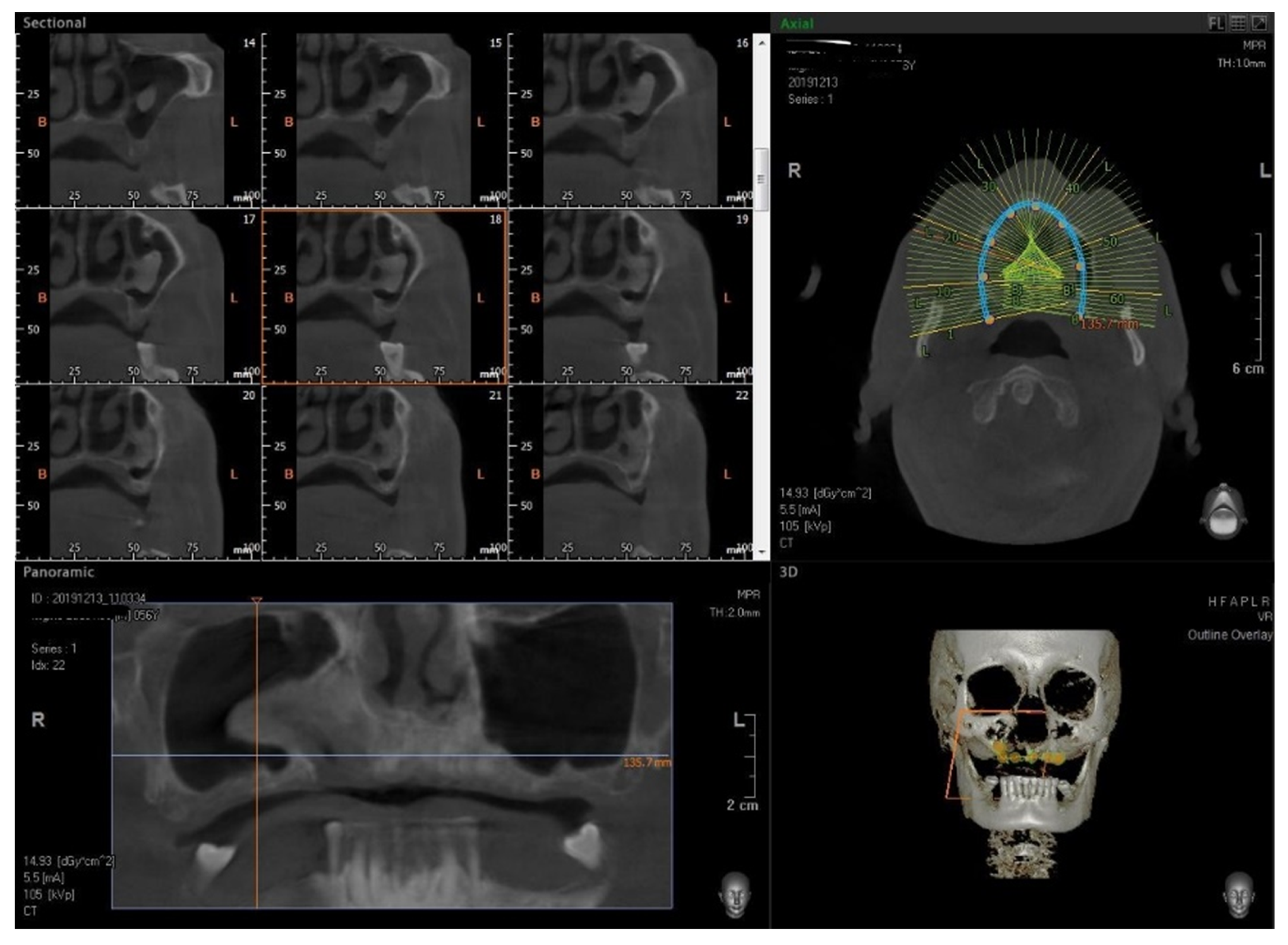
2. Materials and Methods

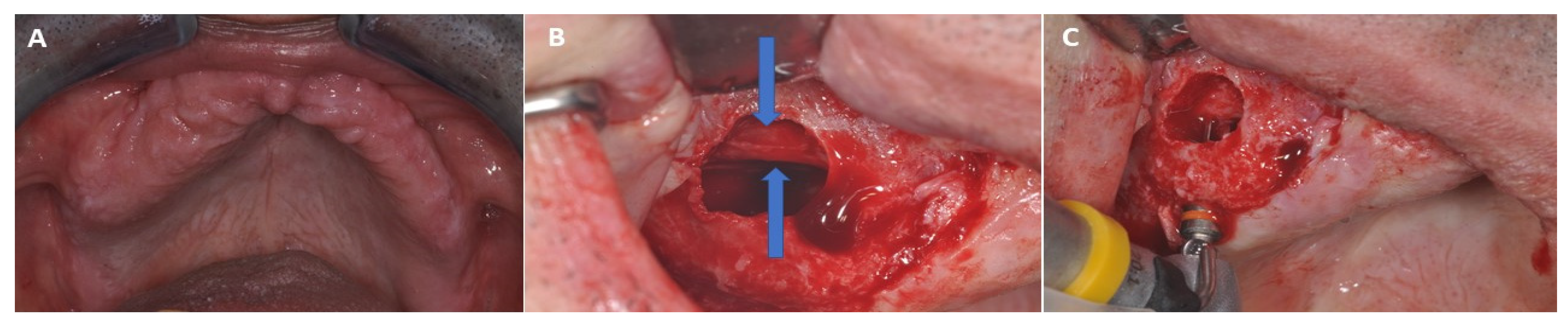
2.1. Surgical Procedure

The case report was conducted in accordance with the ethical laws and the World Medical Association Declaration of Helsinki [31], and the Surgical Case Report (SCARE) guidelines [32]. The present clinical study was based on the ethical laws and the World Medical Association Declaration of Helsinki, and the additional requirements of Italian legislation. Moreover, the University of Chieti-Pescara, Italy, classified the present case reporting to be exempt from ethical review as it carries only negligible risk and involves the use of existing data that contains only non-identifiable data about human beings. A 58-year-old man was referred for rehabilitation of the maxillary. His medical history did not reveal any significant systemic diseases, and he was a non-smoker. The subject reported a history of the removal of a cyst on the left maxillary region approximately 1 year previously. The patient needed rehabilitation with 6 implants and a full prosthesis in the maxilla. The clinical examination of the patient revealed an edentulous state in the maxilla. Radiograph assessment with a Cone Beam Computed Tomography (CBCT) (Vatech Ipax 3D PCH-6500, Fort Lee, NJ, USA) excluded any clinically relevant and radiographically evident pathologies such as odontogenic sinusitis, mucosal thickening, allergy, mucus-retaining cysts, oro-antral fistula, partial to complete sinus obliteration, antroliths, mucoceles, or mucopyoceles, but detected a radiopaque lesion in the right sinus (Figure 1). For this reason, the patient was referred to the Department of Innovative Technology in Medicine and Dentistry of the University “G. d’Annunzio” of Chieti-Pescara in Italy by his dentist for case evaluation. The lesion was probably an osteoma and involved the nasal wall of the right maxillary sinus. It had a lateral axis of 8 mm long and a mesiodistal width of 5 mm. Its location did not interfere with the open lateral wall but interfered with the implant position. The computerized tomography (CT) also showed an insufficient height of crestal bone, and the patient, therefore, needed sinus lifting. The mucosa appeared healthy. No nasal obstruction, epistaxis, drainage purulent nasal, or fever were reported by the patient. Based on recorded anamnestic data alone, the patient reported good general health, was a non-smoker, was taking no medications affecting bone metabolism or wound healing, and there was an absence of any disease. After discussing the options with the patient, he agreed to maintain the lesion and a sinus augmentation with a bone graft. According to the observation of the nasal location of the osteoma and the clinical absence of an oro-nasal pathology, the surgery procedure was planned under local anesthesia and in a dentist’s surgery (Figure 1). Chlorhexidine 0.2% digluconate solution (Curasept® S.p.A., Saronno, Italy) was used for rinsing the mouth for 2 min before surgery. Surgical sites were infiltrated with local Articaine® (Ubistesin® 4%—Espe Dental® AG, Seefeld, Germany) with epinephrine 1:200,000. A modified triangular full-thickness flap was lifted to expose the maxilla lateral sinus wall and crest bone. After removing the crestal bone window, the sinus membrane was gently lifted around the osteoma using primarily the sinus curette (Figure 2). The dental implants were 4.1 mm in diameter and 12 mm in length (2P®, Bone System®, Milan, Italy). They were positioned according to the surgical system and drilling sequence provided by the manufacturer protocol: rosehead drill, twist drill 2.1 diameter, 3.5 diameter drill, and 4.1 drill with a speed of 800 rpm and a 40 Ncm surgical motor unit (NSK®, Surgipro®, Shinagawa, Japan). The final primary stability was assessed by the system’s manual racket torquemeter.
Part of the osteoma was removed for histological analysis while avoiding perforation or tearing of the schneiderian membrane. As a grafting material, bovine bone xenograft (Re-Bone®, Ubgen, Padova, Italy) was used. The vestibular flap was repositioned using the surgical Assumid® (Assut Europe, Aquila, Italy) as previously described [33]. No peri-operative and early postoperative events were reported after the surgical procedures. The histological analysis confirmed the suspected diagnosis of osteoma. The follow-up period of 1 year from the implant surgery revealed no local alteration or lesion recurrency at the level of the treated site.

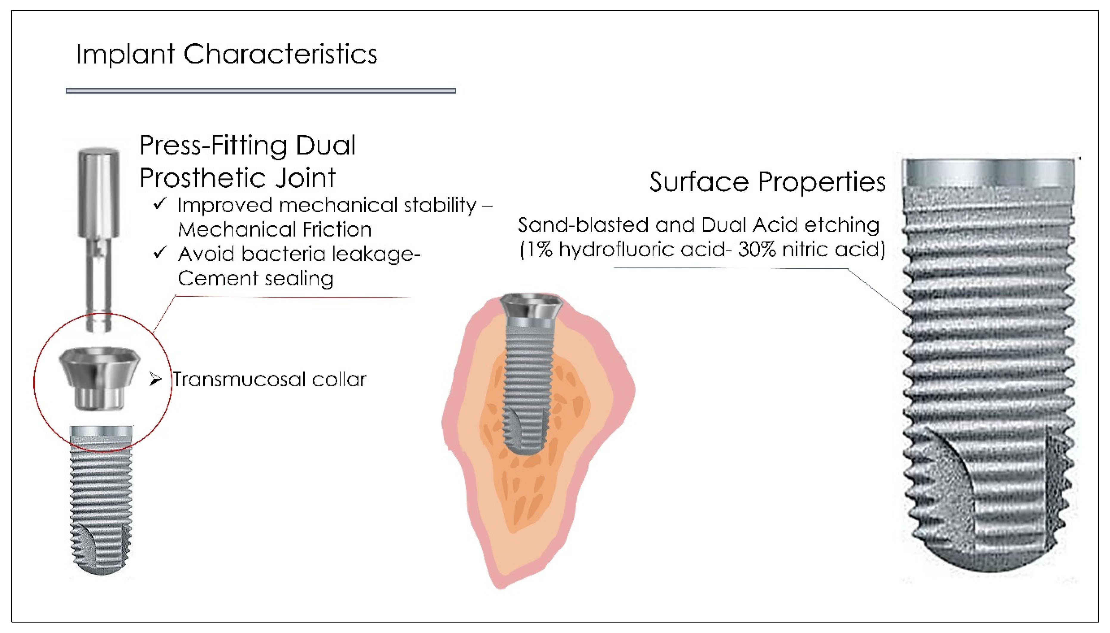
2.2. Dental Implant Characteristics

The implants positioned in the present investigation were composed of 3 components: implant fixture, transmucosal permanent collar tissue level, and prosthetic abutment [34]. The implant-abutment joint was connected by a dual sealing technique: a mechanical frictional press-fitting and the chemical cement sealing [35] (Figure 3). This procedure can produce high mechanical stability and loading strength of the implant system, avoiding bacterial leakage due to the cement sealing [35,36] (Figure 2).
The surface treatment was provided by sandblasting followed by a dual acid etching process [34,35] (mean Ra~2.15 microns) (Figure 3).

2.3. Systematic Literature Review

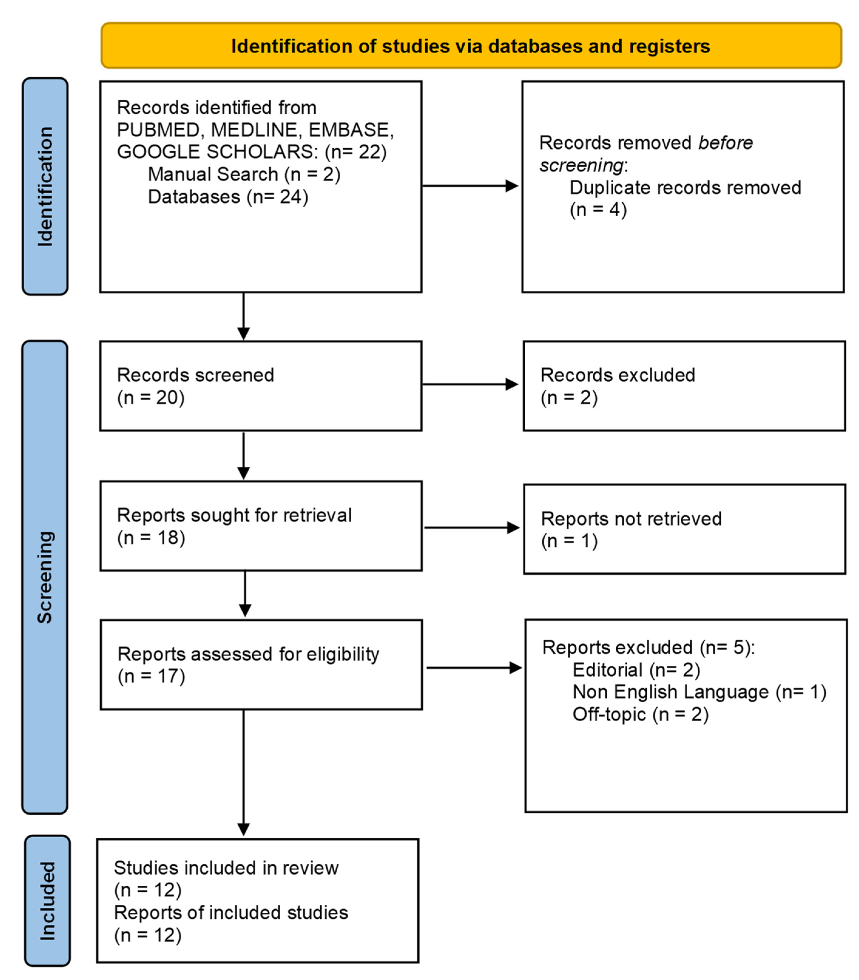
2.3.1. Screening Procedure

The papers selected were assessed following the Standards for Reporting Qualitative Research principles (SRQR) and the PRISMA guidelines. The PICO question has been indicated in Table 1. The search methodology was assessed following a Boolean keyword process described in Table 2. The paper screening was performed through PubMed/MEDLINE, EMBASE, and Cochrane electronic databases (11 January 2022) (PROSPERO Reg. n. 322324).
The titles and abstracts of the articles were collected to perform the first-level screening. The scientific contributions were limited to randomized and non-randomized human clinical trials, prospective and retrospective studies, clinical case reports, and case series with an osteoma of the maxillary bone and nasal/paranasal cavity involvement. The full text was collected and used for further eligibility assessment and the descriptive synthesis.

2.3.2. Inclusion and Exclusion Criteria

The inclusion criteria for the qualitative analysis were limited to human clinical trials, prospective and retrospective studies with a minimum of 1-month follow-up with no restriction of surgical/non-surgical approach, alternative procedures, postoperative sequelae, and the number of stages protocol. The exclusion criteria were limited to literature reviews, editorial letters, in-vitro studies, and laboratory reports. Articles written in a non-English language were not included.

2.3.3. Paper Selection Assessment

The eligibility procedure was performed independently by two expert reviewers (A.S., F.L.). Moreover, a manual search was performed to increase the paper selection. The papers that satisfied the inclusion criteria were included after the removal of any duplicates. The studies excluded were analyzed, and the exclusion reasons were reported.

2.3.4. Article Assessment

The papers were assessed independently using a special electronic form according to the following classification: first author and journal, study model design, subject age, etiology, lesion position, size, treatment, postoperative events, recurrency, number of subjects, histological type, study findings, and principal diagnostic methodology.

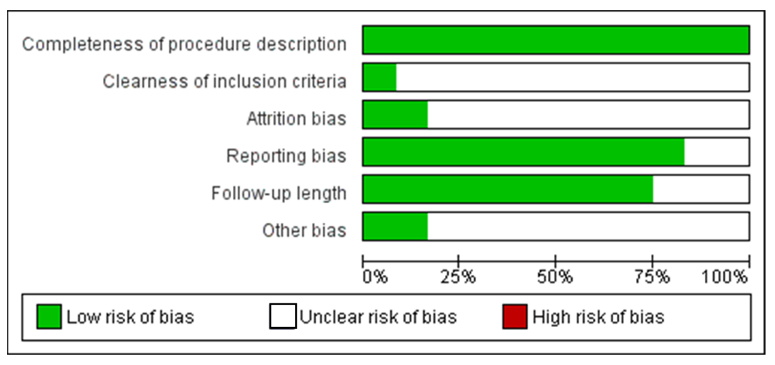
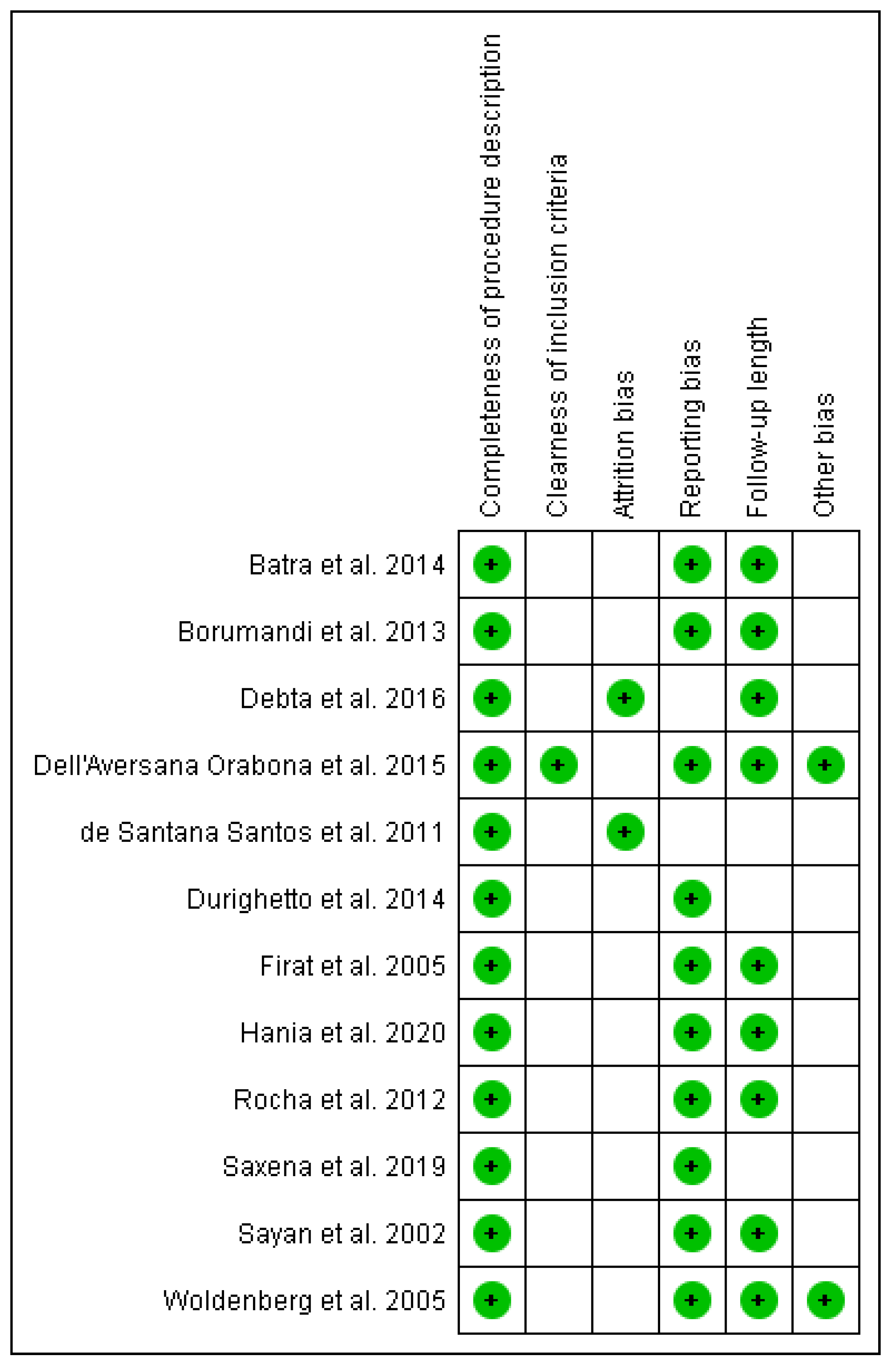
2.3.5. Risk of Bias Measurement

The measurement of the risk of study bias was conducted by the dedicated software RevMan 5.5 (The Nordic Cochrane Centre, The Cochrane Collaboration, Copenhagen, Denmark, 2014). The risk of bias was classified according to the following criteria: completeness of procedure description, clearness of inclusion criteria, attrition bias, reporting bias, follow-up length, and other biases. The criteria were classified as adequate, unclear, or inadequate. The papers included for risk of bias assessment were classified as low risk of bias with a minimum ratio of 4/6 positive parameters and an absence of a negative outcome. Otherwise, the research was categorized as high risk.

3. Results

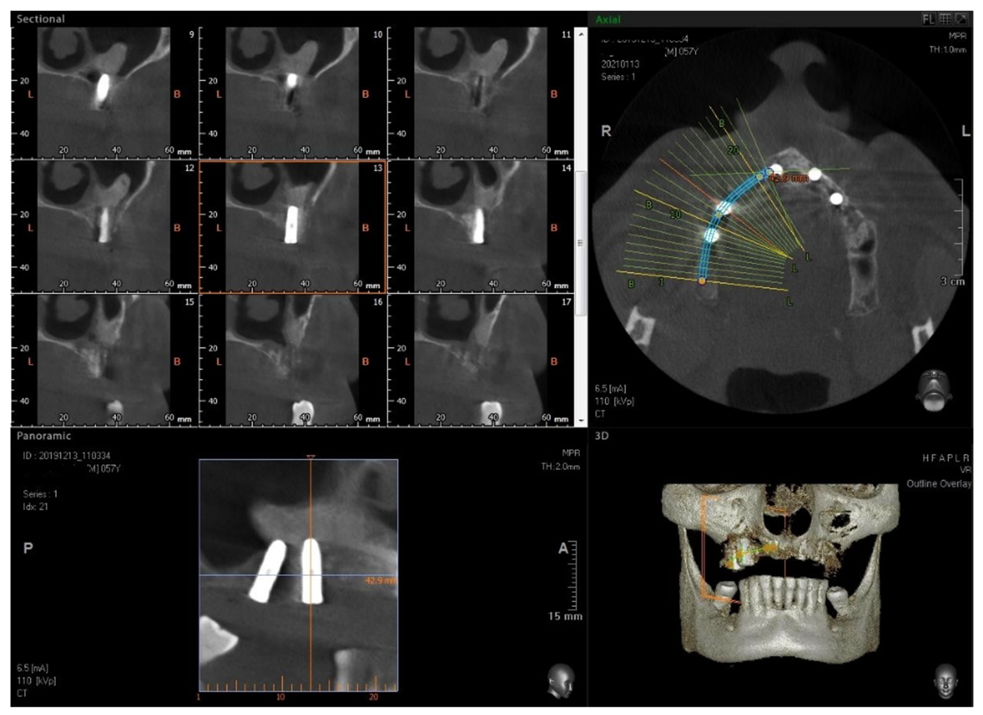
3.1. Surgical Procedure

No signs of inflammation or sinusitis were reported by the patient in the postoperative period. After six months, 6 implants (Bone System Implant, Milano, Italy) were placed in the maxillary, two of which were inserted in the osteoma.
After another four months, healing screws were positioned, and after a further 2 weeks, the provisional crown was positioned, and then, after another 2 months, the final crown. Written informed consent was acquired from the patient for publication in the case report. The patient returned for CBCT 9 months after the final crown placement (Figure 4). The two implants placed in the osteoma were perfectly osseointegrated.

3.2. Article Selection Process

The scientific contribution screening and eligibility process was presented in Figure 3 according to the PRISMA principles. The databases and manual search output identified 24 articles, while duplicates were removed from the selection. After the screening process, 20 articles were assessed, and 2 papers were excluded. Only 1 full text was unavailable. The eligibility process examined 17 articles, excluding 5 papers for the following reasons: 2 editorial letters, 2 off-topic products, and 1 non-English article. A total of 12 scientific contributions were included in the final qualitative analysis (Figure 5).

3.3. Included Articles Characteristics

The article selection identified a total of 9 case reports [37,38,39,40,41,42,43,44,45], 2 case series [46,47], and 1 retrospective study [48], for a total of 58 subjects—35 male and 25 female (Table 3). The patients’ ages were heterogeneous and ranged between 12 and 79 years old. A total of 47 cases showed a maxillary antrum position [37,39,40,41,42,43,44,45], of which 1 subject also had a bone ridge involvement [39], in 2 cases in the hard palate [38,48], 7 cases of maxillary bone ridge involvement [39,41,46,47,48], 1 case of infraorbital rim involvement [42], 1 case in the right piriform [42] (Table 3). The main lesion diameters ranged from 1.0 cm to 6.5 cm at the moment of first diagnosis. In all cases treated, the surgical approach was conservative lesion removal/osteoplasty, while all cases documented received a histological diagnosis confirmation. In all cases, the postoperative events were almost uneventful except for 1 case accompanied by bone deformity, mucosal ulcer, limitation of ATM movement, sensitivity, and headache, which was partially resolved during the healing period [47], nine subjects with severe facial swelling, and 1 patient affected by transitory dysesthesia [46]. The main diagnostic methodology used was histology, which identified only one case of central osteoma, the most common form being peripheral osteoma (Table 3).

3.4. Risk of Bias Assessment

The risk of bias evaluation was discussed in Figure 2 for a total of 12 papers. A total of 2 papers were considered to have a low risk of bias [46,47] (Figure 4 and Figure 5) according to a wide heterogeneity of study model design, intervention treatment, and follow-up period. A total of 10 scientific studies were considered at high risk of bias concerning the assessment criteria [37,38,39,40,41,42,43,44,45,48] (Figure 6 and Figure 7).

4. Discussion

Osteomas are generally asymptomatic and in the majority of cases do not require treatment. The present case report represents a very rare clinical situation that required a major surgical intervention for lateral sinus floor elevation. This procedure is widely used to increase the bone height in the posterior maxilla. In the presented case, we describe for the first time the clinical management of a sinus lifting in a patient with an osteoma that was used for implant support. We decided to use the osteoma for implant support because the CT of the sinus performed 5 years previously showed a similar dimension, so no augmentation was observed of the bone volume of the lesion. Usually, a radiopaque lesion attributable to a similar osteoma is an occasional finding during an orthopantomogram (OPG) performed to program the routine dental treatment. Exceptionally it can cause compression symptoms such as obstruction of the nasolacrimal duct [49], headache, and facial pain depending on the location and size of the tumor. Osteomas are non-odontogenic benign, slow-growing tumors that can develop in the nasal cavity and the paranasal sinuses. Histologically they are characterized by osteoblast deposition and proliferation of atypical compact or cancellous bone with a small marrow space [50,51]. The possible localizations are peripheral or central, arising from the periosteum and endosteum. Extra-skeletal ones arise from the dermis or muscles and are commonly referred to in the literature as osteoma cutis. They can be associated with familial adenomatous polyposis and colorectal adenocarcinoma with autosomal dominant transmission, called Gardner’s syndrome [52]. The rationale of the systematic research query was associated with the lack in the literature of a sinus regeneration approach and dental implant procedure associated with a sinus osteoma, which represents the main novelty in a very rare clinical condition. In fact, the present case is a very rare clinical presentation; a complete state-of-the-art comprehension and literature contextualization through a systematic and rigorous approach is fundamental to determine the differential option treatment.
As reported by the present literature review, the most frequent osteoma sub-type of the bone head is represented by the peripheral form (91.7%) [37,38,39,41,42,43,44,45,46,47,48]. Instead, the central sub-type form (8.3%) is often associated with a previous local trauma/impacted tooth.
No cases of extra-skeletal osteoma of the maxilla have been reported in the present review. No recurrent events or episodes of malignant alterations were reported by the studies included in the present investigation. In this way, the lesion position could limit the implant rehabilitation procedure when in proximity to the sinus base wall, while a very complex and extended surgical resection approach could produce potentially disabling sequelae and the need for a wide regenerative approach for further dental implant positioning [53]. The absence of recurrencies represents the main finding of the literature search, which indicates the modest nature of the lesion upon the clinical severity that, in most cases, is approached by a conservative follow-up. The surgical treatment is often planned in case of large progressive lesions with facial dysmorphism and asymmetries that could also produce functional and aesthetic alterations [53]. In this way, the conservative approach with osteoplasty represents the most common protocol with an early postoperative period, generally characterized by mild symptoms and no relevant events at medium-term follow-up.
In many cases in the literature, the maxillary graft reconstruction with single- or multiple surgical stages is often performed in case of large malignant/benign tumor resection, cysts defects, and wide bone loss [54,55]. As reported by the present literature review, the maxillary osteoma represents a rare occurrence (<0.5% of incidence) that, in case of large residual bone defect, could produce significant functional and aesthetical sequelae [20]. The present case report represents the first case of sinus osteoma used for implant support. Despite the lack of documented cases in the literature, only two cases of osteoma associated with sinus lifting have been described [56]. Carini et al. described a case of simultaneous dental implant positioning in a maxillary ridge osteoma associated with a fresh alveolar socket [29]. In this case, the authors reported a successful dental implant procedure with no regenerative technique procedure associated with a fresh alveolar socket model that represents a very favorable bone defect [29]. Also, Mootaz et al. reported a case of Lateral maxillary sinus floor elevation in the presence of a sinus osteoma. The authors reported a partial excision of the segment impeding on the lateral window [56]. The authors, during sinus lifting, removed only the part of osteoma that impeded lateral window execution to reduce the risk of tearing or perforation of the sinus membrane. The remaining part of the osteoma was kept in place. In the present case, we decided to leave the osteoma because it was asymptomatic. Furthermore, excision is not always indicated and should be considered only when the osteoma is the cause of occurring symptoms. The limits of this approach could be correlated to the variables that could affect the implant site over a long-term period. These include the type of implant-supported prosthesis and prosthetic components stress distribution [57], the loading and masticatory forces discharge [58,59], and the peri-implant and grafted tissues response to the stimuli and eventual infections [60,61,62] that could be determinant in case of an osteoma lesion characterized by a very low recurrence rate [47]. For this purpose, a long-term and accurate clinical and radiological follow-up is necessary.

5. Conclusions

Though this condition requires no treatment, it has been used as dental implant support. This approach could take advantage of a very low recurrence rate of the maxillary antrum osteoma, as documented in the literature. The effectiveness of the present investigation can provide useful guidance for surgeons and dentists for the management of similar clinical situations in operative practice and to avoid a more demolitive surgical and, consequently, a wide regenerative approach for dental implant rehabilitation. However, radiographic monitoring and histological diagnosis are important to exclude other tumor lesions.

Author Contributions

Conceptualization, A.S.; methodology, A.S.; software, A.S. and F.L.; validation, A.S., F.I., G.T. and F.L.; formal analysis, A.S., B.R., F.I., G.T. and F.L.; investigation, A.S. and F.L.; resources, A.S.; data curation, A.S. and F.L.; writing—original draft preparation, A.S., F.I. and F.L.; writing—review and editing, A.S., F.I. and F.L. All authors have read and agreed to the published version of the manuscript.

Funding

This research received no external funding.

Institutional Review Board Statement

The present clinical study was based in accordance with the ethical laws and the World Medical Association Declaration of Helsinki, and the additional requirements of Italian legislation. Moreover, the University of Chieti-Pescara, Italy, classified the present study to be exempt from ethical review as it carries only negligible risk and involves the use of existing data that contains only non-identifiable data about human beings.

Informed Consent Statement

Informed consent was obtained from all subjects involved in the study.

Data Availability Statement

All experimental data to support the findings of this study are available by contacting the corresponding author with a request. The authors have annotated the entire data building process and empirical techniques presented in the paper.

Acknowledgments

The authors acknowledge the support of Carlo Barbone for tomography execution and radiograph elaboration of the present investigation.

Conflicts of Interest

The authors declare no conflict of interest.

References

  1. Buser, D.; Janner, S.F.; Wittneben, J.-G.; Brägger, U.; Ramseier, C.A.; Salvi, G.E. 10-Year Survival and Success Rates of 511 Titanium Implants with a Sandblasted and Acid-Etched Surface: A Retrospective Study in 303 Partially Edentulous Patients. Clin. Implant. Dent. Relat. Res. 2012, 14, 839–851. [Google Scholar] [CrossRef] [PubMed]
  2. Albrektsson, T.; Berglundh, T.; Lindhe, J. Osseointegration: Historic Background and Current Concepts. Clin. Periodontol. Implant. Dent. 2003, 4, 809–820. [Google Scholar]
  3. Mijiritsky, E.; Barone, A.; Cinar, I.C.; Nagy, K.; Shacham, M. 3D Considerations and Outcomes of Immediate Single Implant Insertion and Provisionalization at the Maxillary Esthetic Zone: A Long-Term Retrospective Follow-Up Study of up to 18 Years. J. Clin. Med. 2021, 10, 4138. [Google Scholar] [CrossRef] [PubMed]
  4. Lorusso, F.; Noumbissi, S.; Francesco, I.; Rapone, B.; Khater, A.G.A.; Scarano, A. Scientific Trends in Clinical Research on Zirconia Dental Implants: A Bibliometric Review. Materials 2020, 13, 5534. [Google Scholar] [CrossRef]
  5. Albrektsson, T.; Chrcanovic, B.; Östman, P.-O.; Sennerby, L. Initial and Long-Term Crestal Bone Responses to Modern Dental Implants. Periodontology 2000 2017, 73, 41–50. [Google Scholar] [CrossRef]
  6. Naves, M.M.; Menezes, H.H.M.; Magalhães, D.; Ferreira, J.A.; Ribeiro, S.F.; de Mello, J.D.B.; Costa, H.L. Effect of Macrogeometry on the Surface Topography of Dental Implants. Int. J. Oral Maxillofac. Implants 2015, 30, 789–799. [Google Scholar] [CrossRef] [Green Version]
  7. Ryu, H.-S.; Namgung, C.; Lee, J.-H.; Lim, Y.-J. The Influence of Thread Geometry on Implant Osseointegration under Immediate Loading: A Literature Review. J. Adv. Prosthodont. 2014, 6, 547–554. [Google Scholar] [CrossRef] [Green Version]
  8. Velasco, E.; Monsalve-Guil, L.; Jimenez, A.; Ortiz, I.; Moreno-Muñoz, J.; Nuñez-Marquez, E.; Pegueroles, M.; Pérez, R.A.; Gil, F.J. Importance of the Roughness and Residual Stresses of Dental Implants on Fatigue and Osseointegration Behavior. In Vivo Study in Rabbits. J. Oral Implantol. 2016, 42, 469–476. [Google Scholar] [CrossRef]
  9. Wu-Yuan, C.D.; Eganhouse, K.J.; Keller, J.C.; Walters, K.S. Oral Bacterial Attachment to Titanium Surfaces: A Scanning Electron Microscopy Study. J. Oral Implantol. 1995, 21, 207–213. [Google Scholar]
  10. Scarano, A.; Piattelli, A.; Polimeni, A.; Di Iorio, D.; Carinci, F. Bacterial Adhesion on Commercially Pure Titanium and Anatase-Coated Titanium Healing Screws: An in Vivo Human Study. J. Periodontol. 2010, 81, 1466–1471. [Google Scholar] [CrossRef]
  11. Montegrotto Group for the Study of Peri-implant Disease; Canullo, L.; Schlee, M.; Wagner, W.; Covani, U. International Brainstorming Meeting on Etiologic and Risk Factors of Peri-Implantitis, Montegrotto (Padua, Italy), August 2014. Int. J. Oral Maxillofac. Implants 2015, 30, 1093–1104. [Google Scholar] [CrossRef] [PubMed] [Green Version]
  12. Zandim-Barcelos, D.L.; de Carvalho, G.G.; Sapata, V.M.; Villar, C.C.; Hämmerle, C.; Romito, G.A. Implant-Based Factor as Possible Risk for Peri-Implantitis. Braz. Oral Res. 2019, 33, e067. [Google Scholar] [CrossRef] [PubMed]
  13. Klotz, A.L.; Fobbe, H.; Rammelsberg, P.; Lorenzo Bermejo, J.; Kappel, S. Survival and Success of Tooth-Implant-Supported and Solely Implant-Supported Double-Crown-Retained Overdentures: A Prospective Study over a Period of up to 11 Years. Clin. Oral Implants Res. 2021, 32, 1425–1432. [Google Scholar] [CrossRef] [PubMed]
  14. Mavriqi, L.; Lorusso, F.; Conte, R.; Rapone, B.; Scarano, A. Zygomatic Implant Penetration to the Central Portion of Orbit: A Case Report. BMC Ophthalmol 2021, 21, 121. [Google Scholar] [CrossRef] [PubMed]
  15. Scarano, A.; Barros, R.R.M.; Iezzi, G.; Piattelli, A.; Novaes, A.B. Acellular Dermal Matrix Graft for Gingival Augmentation: A Preliminary Clinical, Histologic, and Ultrastructural Evaluation. J. Periodontol. 2009, 80, 253–259. [Google Scholar] [CrossRef] [PubMed]
  16. Borges, F.L.; Dias, R.O.; Piattelli, A.; Onuma, T.; Gouveia Cardoso, L.A.; Salomão, M.; Scarano, A.; Ayub, E.; Shibli, J.A. Simultaneous Sinus Membrane Elevation and Dental Implant Placement without Bone Graft: A 6-Month Follow-up Study. J. Periodontol. 2011, 82, 403–412. [Google Scholar] [CrossRef] [PubMed]
  17. Bhalla, N.; Dym, H. Update on Maxillary Sinus Augmentation. Dent. Clin. N. Am. 2021, 65, 197–210. [Google Scholar] [CrossRef] [PubMed]
  18. Scarano, A.; Sinjari, B.; Murmura, G.; Lorusso, F. Neurosensory Disturbance of the Inferior Alveolar Nerve After 3025 Implant Placements. Implant Dent. 2017, 26, 735–743. [Google Scholar] [CrossRef] [PubMed]
  19. Schlund, M.; Meeus, J.; Politis, C.; Ferri, J. Management of Sinus Graft Infection-a Systematic Review. Int. J. Oral Maxillofac. Surg. 2022, 51, 690–698. [Google Scholar] [CrossRef] [PubMed]
  20. Minetti, E.; Celko, M.; Contessi, M.; Carini, F.; Gambardella, U.; Giacometti, E.; Santillana, J.; Beca Campoy, T.; Schmitz, J.H.; Libertucci, M.; et al. Implants Survival Rate in Regenerated Sites with Innovative Graft Biomaterials: 1 Year Follow-Up. Materials 2021, 14, 5292. [Google Scholar] [CrossRef] [PubMed]
  21. Inchingolo, F.; Dipalma, G.; Paduanelli, G.; De Oliveira, L.A.; Inchingolo, A.M.; Georgakopoulos, P.I.; Inchingolo, A.D.; Malcangi, G.; Athanasiou, E.; Fotopoulou, E.; et al. Computer-Based Quantification of an Atraumatic Sinus Augmentation Technique Using CBCT. J. Biol. Regul. Homeost. Agents 2019, 33, 31–39. [Google Scholar] [PubMed]
  22. Pjetursson, B.E.; Tan, W.C.; Zwahlen, M.; Lang, N.P. A Systematic Review of the Success of Sinus Floor Elevation and Survival of Implants Inserted in Combination with Sinus Floor Elevation. J. Clin. Periodontol. 2008, 35, 216–240. [Google Scholar] [CrossRef] [PubMed]
  23. Spinato, S.; Bernardello, F.; Galindo-Moreno, P.; Zaffe, D. Maxillary Sinus Augmentation by Crestal Access: A Retrospective Study on Cavity Size and Outcome Correlation. Clin. Oral Implants Res. 2015, 26, 1375–1382. [Google Scholar] [CrossRef] [PubMed]
  24. Stacchi, C.; Lombardi, T.; Ottonelli, R.; Berton, F.; Perinetti, G.; Traini, T. New Bone Formation after Transcrestal Sinus Floor Elevation Was Influenced by Sinus Cavity Dimensions: A Prospective Histologic and Histomorphometric Study. Clin. Oral Implants Res. 2018, 29, 465–479. [Google Scholar] [CrossRef] [PubMed]
  25. Scarano, A.; Degidi, M.; Iezzi, G.; Pecora, G.; Piattelli, M.; Orsini, G.; Caputi, S.; Perrotti, V.; Mangano, C.; Piattelli, A. Maxillary Sinus Augmentation With Different Biomaterials: A Comparative Histologic and Histomorphometric Study in Man. Implant Dent. 2006, 15, 197–207. [Google Scholar] [CrossRef] [PubMed]
  26. Vinci, R.; Teté, G.; Lucchetti, F.R.; Capparé, P.; Gherlone, E.F. Implant Survival Rate in Calvarial Bone Grafts: A Retrospective Clinical Study with 10 Year Follow-Up. Clin. Implant. Dent. Relat. Res. 2019, 21, 662–668. [Google Scholar] [CrossRef] [PubMed]
  27. Trimarchi, M.; Giordano Resti, A.; Vinciguerra, A.; Danè, G.; Bussi, M. Dacryocystorhinostomy: Evolution of Endoscopic Techniques after 498 Cases. Eur. J. Ophthalmol. 2020, 30, 998–1003. [Google Scholar] [CrossRef] [PubMed]
  28. Yacoub, M.-R.; Trimarchi, M.; Cremona, G.; Dal Farra, S.; Ramirez, G.A.; Canti, V.; Della Torre, E.; Baldini, M.; Pignatti, P.; Bussi, M.; et al. Are Atopy and Eosinophilic Bronchial Inflammation Associated with Relapsing Forms of Chronic Rhinosinusitis with Nasal Polyps? Clin. Mol. Allergy 2015, 13, 23. [Google Scholar] [CrossRef] [PubMed] [Green Version]
  29. Carini, F.; Longoni, S.; Simone, M.; Monai, D.; Saggese, V.; Porcaro, G. Central Osteoma of the Maxilla: Implant Rehabilitation with Immediate Loading in Fresh Extraction Socket. Ann. Stomatol. 2014, 5, 10. [Google Scholar]
  30. Nah, K.-S. Osteomas of the Craniofacial Region. Imaging Sci. Dent. 2011, 41, 107–113. [Google Scholar] [CrossRef] [PubMed] [Green Version]
  31. Association, W.M. World Medical Association Declaration of Helsinki: Ethical Principles for Medical Research Involving Human Subjects. JAMA 2013, 310, 2191–2194. [Google Scholar]
  32. Agha, R.A.; Franchi, T.; Sohrabi, C.; Mathew, G.; Kerwan, A.; SCARE Group. The SCARE 2020 Guideline: Updating Consensus Surgical CAse REport (SCARE) Guidelines. Int. J. Surg. 2020, 84, 226–230. [Google Scholar] [CrossRef] [PubMed]
  33. Scarano, A.; Lorusso, F.; Arcangelo, M.; D’Arcangelo, C.; Celletti, R.; de Oliveira, P.S. Lateral Sinus Floor Elevation Performed with Trapezoidal and Modified Triangular Flap Designs: A Randomized Pilot Study of Post-Operative Pain Using Thermal Infrared Imaging. Int. J. Environ. Res. Public Health 2018, 15, 1277. [Google Scholar] [CrossRef] [PubMed] [Green Version]
  34. Assenza, B.; Scarano, A.; Petrone, G.; Iezzi, G.; Thams, U.; San Roman, F.; Piattelli, A. Crestal Bone Remodeling in Loaded and Unloaded Implants and the Microgap: A Histologic Study. Implant Dent. 2003, 12, 235–241. [Google Scholar] [CrossRef] [PubMed]
  35. Assenza, B.; Tripodi, D.; Scarano, A.; Perrotti, V.; Piattelli, A.; Iezzi, G.; D’Ercole, S. Bacterial Leakage in Implants with Different Implant-Abutment Connections: An in Vitro Study. J. Periodontol. 2012, 83, 491–497. [Google Scholar] [CrossRef] [PubMed]
  36. Scarano, A.; Assenza, B.; Inchingolo, F.; Mastrangelo, F.; Lorusso, F. New Implant Design with Midcrestal and Apical Wing Thread for Increased Implant Stability in Single Postextraction Maxillary Implant. Case Rep. Dent. 2019, 2019, 9529248. [Google Scholar] [CrossRef] [PubMed] [Green Version]
  37. Hania, M.; Sharif, M.O. Maxillary Sinus Osteoma: A Case Report and Literature Review. J. Orthod. 2020, 47, 240–244. [Google Scholar] [CrossRef] [PubMed]
  38. Saxena, S.; Ashwin, V.G.; Rajinikanth, D. A Sizeable Solitary Pedunculated Peripheral Osteoma of the Hard Palate: A Case Report. Indian J. Otolaryngol. Head Neck Surg. 2019, 71, 813–815. [Google Scholar] [CrossRef] [PubMed]
  39. Debta, P.; Debta, F.M.; Bussari, S.; Acharya, S.S.; Jeergal, V.A. Cancellous Osteoma of Maxilla: A Rare Case Report. J. Int. Soc. Prev. Community Dent. 2016, 6, 261–264. [Google Scholar] [CrossRef] [PubMed] [Green Version]
  40. de Santana Santos, T.; Frota, R.; Martins-Filho, P.R.S.; Melo, A.R.; de Souza Andrade, E.S.; de Oliveira e Silva, E.D.; Avelar, R.L. Central Osteoma of the Maxilla with Involvement of Paranasal Sinus. J. Craniofac. Surg. 2011, 22, 589–591. [Google Scholar] [CrossRef] [PubMed]
  41. Durighetto, A.F.; de Moraes Ramos, F.M.; da Rocha, M.A.; da Cruz Perez, D.E. Peripheral Osteoma of the Maxilla: Report of a Case. Dentomaxillofac. Radiol. 2007, 36, 308–310. [Google Scholar] [CrossRef] [PubMed]
  42. Batra, N.; Batra, R.; Singh, G.; Gaur, A. Peripheral Osteoma of Maxilla: A Case Report. Natl. J. Maxillofac. Surg. 2014, 5, 240–242. [Google Scholar] [CrossRef] [PubMed] [Green Version]
  43. Rocha, J.F.; Gonçales, A.G.B.; da Silva Sampieri, M.B.; da Silva, A.A.; Matsumoto, M.A.; Gonçales, E.S. Peripheral Osteoma of the Maxillary Sinus: A Case Report. Oral Maxillofac. Surg. 2012, 16, 315–319. [Google Scholar] [CrossRef] [PubMed]
  44. Firat, D.; Sirin, Y.; Bilgic, B.; Ozyuvaci, H. Large Central Osteoma of the Maxillary Antrum. Dentomaxillofac. Radiol. 2005, 34, 322–325. [Google Scholar] [CrossRef]
  45. Borumandi, F.; Lukas, H.; Yousefi, B.; Gaggl, A. Maxillary Sinus Osteoma: From Incidental Finding to Surgical Management. J. Oral Maxillofac. Pathol. 2013, 17, 318. [Google Scholar] [CrossRef] [Green Version]
  46. Dell’Aversana Orabona, G.; Salzano, G.; Iaconetta, G.; Piombino, P.; Ponzo, L.; Santella, A.; Astarita, F.; Solari, D.; Salzano, F.A.; Califano, L. Facial Osteomas: Fourteen Cases and a Review of Literature. Eur. Rev. Med. Pharmacol. Sci. 2015, 19, 1796–1802. [Google Scholar]
  47. Woldenberg, Y.; Nash, M.; Bodner, L. Peripheral Osteoma of the Maxillofacial Region. Diagnosis and Management: A Study of 14 Cases. Med. Oral Patol. Oral Cir. Bucal. 2005, 10 (Suppl. 2), E139–E142. [Google Scholar]
  48. Sayan, N.B.; Uçok, C.; Karasu, H.A.; Günhan, O. Peripheral Osteoma of the Oral and Maxillofacial Region: A Study of 35 New Cases. J. Oral Maxillofac. Surg. 2002, 60, 1299–1301. [Google Scholar] [CrossRef]
  49. Saratziotis, A.; Emanuelli, E. Osteoma of the Medial Wall of the Maxillary Sinus: A Primary Cause of Nasolacrimal Duct Obstruction and Review of the Literature. Case Rep. Otolaryngol. 2014, 2014, 348459. [Google Scholar] [CrossRef]
  50. Bhatt, G.; Gupta, S.; Ghosh, S.; Mohanty, S.; Kumar, P. Central Osteoma of Maxilla Associated with an Impacted Tooth: Report of a Rare Case with Literature Review. Head Neck Pathol. 2019, 13, 554–561. [Google Scholar] [CrossRef]
  51. Dalambiras, S.; Boutsioukis, C.; Tilaveridis, I. Peripheral Osteoma of the Maxilla: Report of an Unusual Case. Oral Surg. Oral Med. Oral Pathol. Oral Radiol. Endodontol. 2005, 100, e19–e24. [Google Scholar] [CrossRef] [PubMed]
  52. Antohi, C.; Haba, D.; Caba, L.; Ciofu, M.L.; Drug, V.-L.; Bărboi, O.-B.; Dobrovăț, B.I.; Pânzaru, M.-C.; Gorduza, N.C.; Lupu, V.V.; et al. Novel Mutation in APC Gene Associated with Multiple Osteomas in a Family and Review of Genotype-Phenotype Correlations of Extracolonic Manifestations in Gardner Syndrome. Diagnostics 2021, 11, 1560. [Google Scholar] [CrossRef] [PubMed]
  53. Chan, H.-L.; Wang, H.-L. Sinus Pathology and Anatomy in Relation to Complications in Lateral Window Sinus Augmentation. Implant Dent. 2011, 20, 406–412. [Google Scholar] [CrossRef] [PubMed] [Green Version]
  54. Kim, H.J.; Lee, K.H.; Park, S.Y.; Kim, H.K. One-Stage Reconstruction for Midfacial Defect after Radical Tumor Resection. Clin. Exp. Otorhinolaryngol. 2012, 5, 53–56. [Google Scholar] [CrossRef]
  55. Adwani, D.; Bhattacharya, A.; Arora, R.S.; Soni, R.; Adwani, N. Resection and Reconstruction of Maxillary Class IIIc Defect in a Case of Adenoid Cystic Carcinoma: Cost-Sensitive Technique without Microvascular Grafts. Case Rep. Dent. 2013, 2013, 865010. [Google Scholar] [CrossRef]
  56. Mlouka, M.; Tlili, M.; Hamrouni, A.; Selmi, R.; Khanfir, F.; Khalfi, M.S.; Ben Amor, F. Lateral Maxillary Sinus Floor Elevation in the Presence of a Sinus Osteoma: A Case Report. Clin. Case Rep. 2021, 9, e04124. [Google Scholar] [CrossRef]
  57. Torkzaban, P.; Ziaei, N.; Tootiaee, B.; Khoshhal, M.; Vafaee, F.; Panahandeh, N. Effect of Implant-Abutment Connection Type on Stress Distribution in Peri-Implant Bone and Abutment Micromovement: A Three-Dimensional Finite Element Analysis. J. Long-Term Eff. Med. Implants 2019, 29, 113–124. [Google Scholar] [CrossRef]
  58. Goellner, M.; Schmitt, J.; Karl, M.; Wichmann, M.; Holst, S. The Effect of Axial and Oblique Loading on the Micromovement of Dental Implants. Int. J. Oral Maxillofac. Implants 2011, 26, 257–264. [Google Scholar]
  59. Olsson, M.; Urde, G.; Andersen, J.B.; Sennerby, L. Early Loading of Maxillary Fixed Cross-Arch Dental Prostheses Supported by Six or Eight Oxidized Titanium Implants: Results after 1 Year of Loading, Case Series. Clin. Implant Dent. Relat. Res. 2003, 5, 81–87. [Google Scholar] [CrossRef]
  60. Piattelli, A.; Scarano, A.; Piattelli, M.; Podda, G. Implant Periapical Lesions: Clinical, Histologic, and Histochemical Aspects. A Case Report. Int. J. Periodontics Restor. Dent. 1998, 18, 181–187. [Google Scholar]
  61. Scarano, A.; Di Domizio, P.; Petrone, G.; Iezzi, G.; Piattelli, A. Implant Periapical Lesion: A Clinical and Histologic Case Report. J. Oral Implantol. 2000, 26, 109–113. [Google Scholar] [CrossRef]
  62. Albrektsson, T.; Canullo, L.; Cochran, D.; De Bruyn, H. “Peri-Implantitis”: A Complication of a Foreign Body or a Man-Made “Disease”. Facts and Fiction: Peri-Implantitis: Facts and Fiction. Clin. Implant. Dent. Relat. Res. 2016, 18, 840–849. [Google Scholar] [CrossRef] [PubMed]
Figure 1. CT detected a radiopaque lesion involving the right nasal wall of the maxillary sinus.
Applsci 12 05435 g001
Figure 2. (A) Edentulous state of patient. (B) clinical aspect of osteoma. (C) Implant bed preparation involving the osteoma.
Applsci 12 05435 g002
Figure 3. Summary of the dental implant characteristics and implant-abutment joint properties.
Applsci 12 05435 g003
Figure 4. A new CBCT was performed after sinus lifting and implant placement. Coronal section shows two-implant placement in the osteoma.
Applsci 12 05435 g004
Figure 5. Prisma flowchart of the database search and study retrieval process.
Applsci 12 05435 g005
Figure 6. Risk of bias graph: judgments for risk of bias criteria as percentages across all included studies.
Applsci 12 05435 g006
Figure 7. Risk of bias summary: judgments of risk of bias criteria for each included study.
Applsci 12 05435 g007
Table 1. The PICO (population, intervention, comparison, outcome) question.
Population\PatientsInterventionComparisonOutcomes
Patient group of interest?What is the main intervention you wish to consider?Is there an alternative intervention to compare?What is the clinical outcome?
Subjects affected by maxillary sinus osteoma.The intervention was total/partial lesion removal and rehabilitation procedure.The comparison was performed with conservative and non-surgical approaches.The complete/partial lesion removal did not produce a recurrence of the lesion.
Table 2. Database search strategy for the article selection.
Search Strategies
Keywordskeyword search: ((maxilla *) AND (antrum OR Sinus) AND (central OR peripheral OR extraskeletal) AND Osteoma)
DatabasesPubMed/Medline, EMBASE, Cochrane electronic databases
Table 3. Studies regarding the maxillary antrum osteoma cases present in the literature.
AuthorJournalStudy DesignAgeEtiologyPositionLesion (s) SizeTreatment ProtocolPost Operative EventsRecurrencySubject (s)TypeStudy Findings
Hania et al. [37]J Orthod.Case report15 yearsPremature extraction of maxillary deciduous caninesMaxillary antrum-Lesion Removal/osteoplastyUneventful-One (1) maleperipheral osteomaTrauma/dental extraction and osteoma correlation
Saxena et al. [38]Indian J Otolaryngol Head Neck SurgCase report38 years-Hard palate5 × 4 × 2 cmLesion Removal/osteoplastyUneventful-One (1) maleperipheral osteomaLesions usually managed conservatively; excepts symptomatic cases.
Debta et al. [39]J Int Soc Prev Community DentCase report37 years Posterior Maxilla/Antrum3 × 1.5 cmLesion Removal/osteoplastyUneventful One (1) femaleperipheral osteomaThe lesion removal is the elective treatment; the recurrence rate is very low.
de Santana Santos et al. [40]J Craniofac SurgCase report44 years-Maxillary antrum-Lesion Removal/osteoplastyUneventful One (1) maleCentral OsteomaThe traumatic factor could induce an endosteal osteoblasts activity and the development of central osteoma.
Durighetto et al. [41]Dentomaxillofac RadiolCase report42 years-Maxillary alveolar process, maxillary sinus,3 cm diameterLesion Removal/osteoplastySmall area of ulceration One (1) maleperipheral osteomano recurrence of the lesion after 6 years
Woldenberg et al. [47]Med Oral Patol Oral CirBucalCase seriesrange 13 to 79 yearsNot determined9 cases Body mandible 3 Temporal Bone 1 Maxilla 1 Maxillary antrumrange 1.0 cm to 4.0 cmLesion Removal/osteoplastydeformity, mucosal ulcer, limitation of ATM movement, sensitivity, and headache-Eight (8) Female; Six (6) maleperipheral osteomaMandibular osteomas may be a genetic marker for the development of colorectal carcinoma
Sayan et al. [48]J Oral Maxillofac SurgRetrospective studyrange 14 to 58 yearsNot determined(a) frontal bone (28.57%), (b) mandible(22.85%) (c) maxilla (14.28%)-Lesion Removal/osteoplasty- Twenty-three (23) males, Twelve (12) femalesperipheral osteomaThe complete surgical removal at the base where it unites with the cortical bone is necessary
Batra et al. [42]Natl J Maxillofac SurgCase report32 years-Maxillary alveolar process, maxillary sinus, infraorbital rim, right piriform6.3 × 6.3 × 6.5 cmLesion Removal/osteoplastyno sensory deficit and the involved teeth were not devitalized One (1) femaleperipheral osteomaSome lesions likely to present as true neoplasm of bone; other lesions may be the alteration of bone as a response to trauma or infection
Rocha et al. [43]Oral Maxillofac SurgCase report18 yearsNot determinedMaxillary sinus3 cm diameterLesion Removal/osteoplasty -One (1) femaleperipheral osteomaOsteomas most frequently occur in the frontal and ethmoid sinuses, and are rare in the maxillary sinus
Firat et al. [44]Dentomaxillofac RadiolCase report15 yearsNot determinedMaxillary sinus-Lesion Removal/osteoplastyImpacted teeth-One (1) maleperipheral osteomaNew research efforts must be made to enlighten particularly the unknown aetiology of osteoma formation
Borumandi et al. [45]J Oral Maxillofac PatholCase report39 yearsNot determinedMaxillary sinus2 cm diameterLesion Removal/osteoplasty One (1) maleperipheral osteomaThe midface osteomas appear frequently in the frontoethmoidal sinuses
Dell’Aversana Orabona et al. [46]Eur Rev Med PharmacolCase seriesrange 24–61 yearsNot determined(a) 3 cases mandibular angle (b) 7 subjects anterior body (c) 4 patients alveolar processes (d) 2 Maxillary lesionsrange 1.0 to 3.8 cmLesion Removal/osteoplasty(a) Nine patients out of the eleven (81.8%) facial swelling (b) 1 subject dysesthesia of the trigeminal nerve (7.14%) (c) 4 cases uneventful-Six (6) male; eight (8) femaleperipheral osteomaCraniofacial osteomas are more frequent in the mandible, with no predilection for any specific age range
Publisher’s Note: MDPI stays neutral with regard to jurisdictional claims in published maps and institutional affiliations.

