Article

Promising Photocytotoxicity of Water-Soluble Phtalocyanine against Planktonic and Biofilm Pseudomonas aeruginosa Isolates from Lower Respiratory Tract and Chronic Wounds

by
Wojciech Szczolko
1,*,
Magdalena Ratajczak
2,
Tomasz Koczorowski
1,
Dorota Kaminska
2,
Tomasz Goslinski
1 and
Jolanta Dlugaszewska
2,*
1
Chair and Department of Chemical Technology of Drugs, Poznan University of Medical Sciences, Grunwaldzka 6, 60-780 Poznan, Poland
2
Chair and Department of Genetics and Pharmaceutical Microbiology, Poznan University of Medical Sciences, Swiecickiego 4, 60-781 Poznan, Poland
*
Authors to whom correspondence should be addressed.
Appl. Sci. 2022, 12(8), 3707; https://doi.org/10.3390/app12083707
Submission received: 15 February 2022 / Revised: 27 March 2022 / Accepted: 6 April 2022 / Published: 7 April 2022
(This article belongs to the Special Issue Heterocyclic Compounds with Potential Biological Activity - Volume II)

Abstract

:
Alternative methods of killing microbes have been extensively researched in connection with the widespread appearance of antibiotic resistance among pathogenic bacteria. In this study, we report on in vitro antimicrobial phototoxicity research of cationic phthalocyanine with 2-(4-N-methylmorpholin-4-ium-4-yl)ethoxy substituents against selected clinical strains of Pseudomonas aeruginosa isolated from the lower respiratory tract and chronic wounds. The microorganisms tested in the research were analyzed in terms of drug resistance and biofilm formation. The photocytotoxic effect of phthalocyanine was determined by the reduction factor of bacteria. The studied cationic phthalocyanine at a concentration of 1.0 × 10⁻⁴ M, when activated by light, revealed a significant reduction factor, ranging from nearly 4 to 6 log, of P. aeruginosa cells when compared to the untreated control group. After single irradiation, a decrease in the number of bacteria in biofilm ranging from 1.3 to 4.2 log was observed, whereas the second treatment significantly improved the bacterial reduction factor from 3.4 to 5.5 log. It is worth mentioning that a boosted cell-death response was observed after the third irradiation, with a bacterial reduction factor ranging from 4.6 to 6.4 log. According to the obtained results, the tested photosensitizer can be considered as a potential antimicrobial photodynamic therapy against multidrug-resistant P. aeruginosa.

1. Introduction

The current emergence and worldwide spread of multidrug-resistant bacteria are of great concern. Among bacteria that have developed multiple mechanisms of resistance against clinically relevant antibiotics, Pseudomonas aeruginosa is particularly noteworthy. P. aeruginosa is an opportunistic, nosocomial pathogen that causes infections of particular danger for immunocompromised and hematological patients, as well as patients with cystic fibrosis or burns [1,2,3,4,5]. This bacterium was found to be one of the most frequent pathogens associated with nosocomial pneumonia (including ventilator-associated pneumonia), septicemia, urinary tract infections, as well as surgical site and wound infections [6,7,8,9,10]. P. aeruginosa is not only naturally resistant but can also acquire further resistance to multiple antibiotic classes, making it extremely difficult to treat. Moreover, P. aeruginosa can survive and multiply in a nutrient-depleted environment, as well as produce many virulence factors. It is also known to produce robust biofilm, which complicates the treatment of severe infections appearing within the lower respiratory tract and chronic wound infections [11,12,13]. Therefore, biofilm eradication appears to be one of the most important steps in the treatment of these infections [14,15,16].
Serious limitations of traditional antibiotic and chemotherapeutic approaches for the treatment of bacterial infections, mainly related to the emergence of multi-drug resistant bacteria, triggered a search for new therapeutic methods. One of these seems to be photodynamic therapy (PDT), also known as photodynamic inactivation (PDI) or photodynamic antimicrobial therapy (PACT), which has shown potential as a non-antibiotic modality treatment for various infections. PACT is the method by which a combination of a sensitizing phototherapeutic agent (photosensitizer), molecular oxygen and visible light destroys microbial cells through the generation of reactive oxygen species (ROS), including singlet oxygen [17,18]. Singlet oxygen and other oxygen radicals cause lethal effects within a variety of bacterial cells [19,20,21,22]. The mechanism of the photodynamic reaction starts with irradiation of a photosensitizer (Ps) with light of an appropriate wavelength. The energy transfer from the excited Ps to oxygen in its triplet state causes the formation of reactive oxygen species (ROS), including singlet oxygen (1O2). ROS react with cell organelles and disrupt their functions as a result of previously mentioned physicochemical processes in PACT. Two main oxidative mechanisms of photoinactivation (PI) are considered for the inactivation of the target cells. The type I pathway involves electron/hydrogen atoms—transfer reactions from a Ps in its triplet state with the participation of a substrate, which leads to the production of radical ions. The type II pathway relies on singlet oxygen (1O2) production in the reaction of a Ps with triplet state oxygen. Both processes lead to ROS, such as 1O2 and free radicals, which can irreversibly alter vital components of cells, finally leading to lethal oxidative damage [23,24].
Very intensive worldwide studies aiming to develop an ideal photosensitizer for PACT are ongoing, with many multidisciplinary research groups having so far evaluated the potential of various agents against different microorganisms [25,26]. The properties of phthalocyanines (Pcs) have met most of the requirements for an ideal photosensitizer in PACT, being fully synthetic macrocyclic compounds belonging to the porphyrinoid family. Pcs, unlike porphyrins, consist of four isoindole heterocycles bound together by meso nitrogen atoms in place of methine groups. Interestingly, they can be easily modified by both the introduction of different metal cations to the macrocyclic core as well as by modification with various functional groups to their periphery [27,28]. Their physicochemical properties, especially optical ones, enable their application as photosensitizers in photodynamic therapy. One of the most critical factors limiting their broader use as photosensitizers is the tendency to form aggregates in solutions, including water-based media.
Several studies for Pcs modified with morpholine moieties in axial, peripheral and non-peripheral positions have been performed in the last fifteen years, including recent works published by the authors [29,30,31,32]. In the latest study, novel octaiodide salt of octa-substituted magnesium(II) phthalocyanine with N-methyl morpholiniumethoxy (Pc+) substituents (Figure 1) was examined in vitro for photocytotoxicity against a broad spectrum of microorganisms. Pc+ revealed promising photosensitizing activity against a plethora of standard bacteria strains, including P. aeruginosa [29].
P. aeruginosa can be particularly dangerous for patients with lung diseases and is notoriously persistent in severe wounds. Treatment of these infections is also hindered by the ability of P. aeruginosa to form antibiotic-resistant biofilms. According to a CDC report, in 2017, multidrug-resistant P. aeruginosa caused an estimated 32,600 infections among hospitalized patients and 2700 estimated deaths in the United States [33]. In the same year it was recognized as one of the most life-threatening bacteria and listed as a priority pathogen for research and development of new antibiotics by the World Health Organization [34].
The potential use of PDT in the treatment of microbial biofilms associated with chronic wounds and the lower respiratory tract is very promising [35,36,37]. Therefore, the objective of the present microbiological study was to evaluate a PACT photosensitizing potential of Pc+ against clinical strains of P. aeruginosa. Clinical strains of P. aeruginosa with different phenotypic features isolated from lower respiratory tract and chronic wound infections were studied in the current research.

2. Materials and Methods

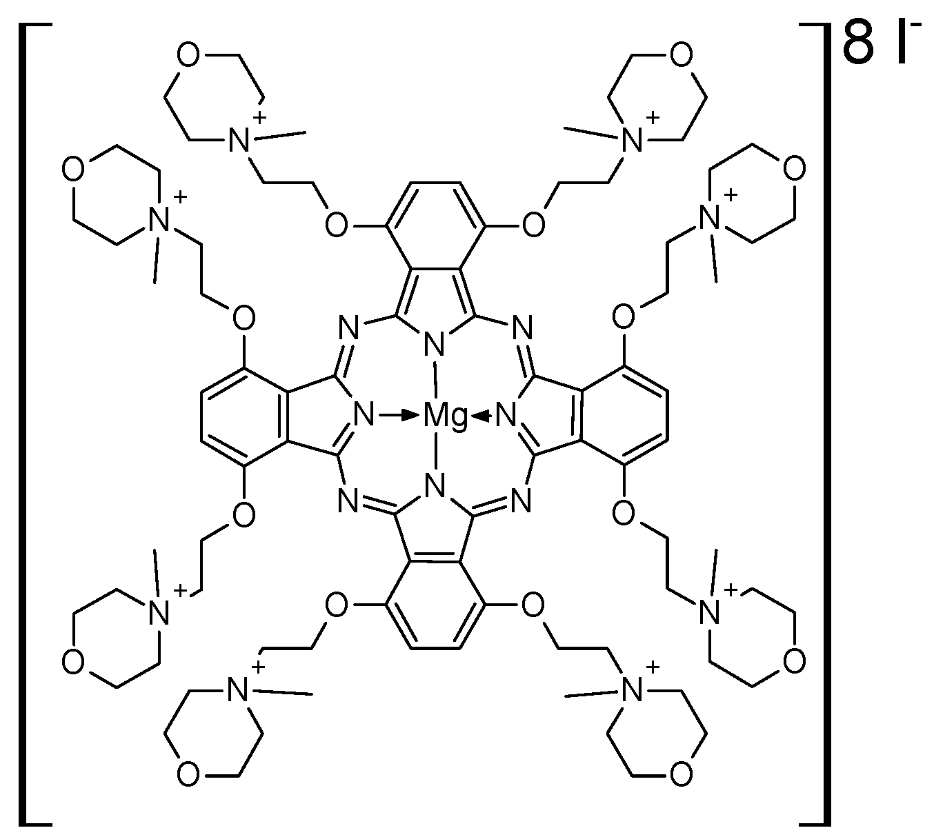
2.1. Photosensitizer and Light Source

Pc+,1,4,8,11,15,18,22,25-octakis[2-(4-N-methylmorpholin-4-ium-4-yl)ethoxy]phthalocyaninato magnesium(II) octaiodide was synthesized and characterized following a recently published procedure [29]. Q-band maximum absorption in the UV–vis of Pc+ in aqueous solution appears at 736 nm. A high-power LED multichip emitter (60 high-efficiency AlGaAs diode chips, Roithner LaserTechnik GmbH, Vienna, Austria) was used for illumination at 735 nm (7.90 V, 0.158 A). Bacteria cells were exposed to a light dose of 1.8 J/cm2 (planktonic form) or 3.6 J/cm2 (biofilm).

2.2. Bacterial Strains

Clinical strains (n = 29) of P. aeruginosa were acquired from the Microbiological Laboratory of Transfiguration of the Lord Clinical Hospital (Poznan University of Medical Sciences, Poland). Strains were isolated from the lower respiratory tract (n = 13) and chronic wounds (n = 16). Selected characteristics of P. aeruginosa strains used in the study are summarized in Table 1. P. aeruginosa PAO1 was used to verify the biofilm formation procedure. P. aeruginosa PAO1, P. aeruginosa ATCC 27853 and P. aeruginosa NCTC 6749 were used to determine conditions of the photodynamic inactivation of biofilm. All strains were stored in cryogenic vials at −70 ± 10 °C. Prior to each experiment, subcultures were prepared on tryptic soy agar (TSA).

2.3. Antimicrobial Susceptibility Testing

  • Sensitivity to antimicrobials:
Assessment of drug susceptibility of P. aeruginosa strains to antibiotics was carried out using the disc diffusion method in accordance with EUCAST recommendations. The following antibiotics were used to test drug susceptibility: piperacillin 30 µg, ceftazidime 10 µg, imipenem 10 µg, meropenem 10 µg, ciprofloxacin 5 µg and tobramycin 10 µg. The test results were interpreted according to EUCAST guidelines [38].
  • Detecting carbapenemases in P. aeruginosa strains:
For P. aeruginosa strains intermediate or resistant to carbapenems, Carba NP test and imipenem–EDTA disk method for metallo-beta-lactamases (MBL), as well as a test with boronic acid for Klebsiella pneumoniae carbapenemases (KPC), were carried out. The test results were interpreted in accordance with EUCAST guidelines [39].

2.4. Quantitative Assessment of Biofilm Formation

To evaluate biofilm formation, a crystal violet assay was used. Each well of a sterile 96-well flat-bottom plastic plate was filled with 200 µL of standardized bacterial suspension in brain–heart infusion broth (BHI) (density c.a. 106 CFU/mL). Negative control wells contained BHI broth only. P. aeruginosa PAO1 was used as a positive control. The plates were closed and incubated aerobically for 24 h at 37 °C. Then, the contents of each well were aspirated, and each well was washed three times with 250 µL of sterile physiological saline. The plates were shaken in order to remove all non-adherent bacteria. After washing, biofilms were stained for 15 min with 2% solution of crystal violet (200 µL). Then, each well was washed three times with 250 µL of sterile physiological saline once again. After the plates were air-dried, 200 µL of 99% methanol per well was added. The absorbance of each well was measured at 590 nm using an Infinite M200 plate reader (Tecan, Austria GmbH, Grödig, Austria). The interpretation of biofilm formation by tested strains was made according to the criteria of Stepanovic et al. [40].
ODc = mean OD of negative control + 3 × standard deviation (SD) of negative control; non-biofilm producer (N): OD ≤ ODc; weak-biofilm producer (W): ODc < OD ≤ 2 × ODc; moderate-biofilm producer (M): 2 × ODc < OD ≤ 4 × ODc; strong-biofilm producer (S): OD > 4 × ODc.

2.5. Effect of PACT on Bacterial Planktonic Form

The tested strains were cultured aerobically in BHI broth at 36 ± 1 °C for 20 h. Then, bacterial cells were harvested by centrifugation (3000 RPM for 15 min at 4 °C), resuspended and diluted with 10 mM PBS (pH = 7.0) to a final concentration of ca. 107 colony forming units (CFUs) per mL. Aliquots of a studied compound were placed in the wells of microtiter plates (except for negative controls (containing PBS)), then a standardized bacterial suspension was added to each well. The samples were incubated for 15 min (incubation time). Bacteria were irradiated at room temperature with high-power LED multichip emitter (60 high-efficiency AlGaAs diode chips, Roithner LaserTechnik GmbH, Vienna, Austria) for 10 min. A control study in darkness was performed simultaneously. Bacterial suspension from each well, after dilution, was grown on TSA plates. After the incubation period (20 h at 36 ± 1 °C), viable bacteria were measured by counting the number of CFUs. Experiments were performed in triplicate. The reduction of living bacteria in each sample was determined by the log reduction factor calculations as follows:
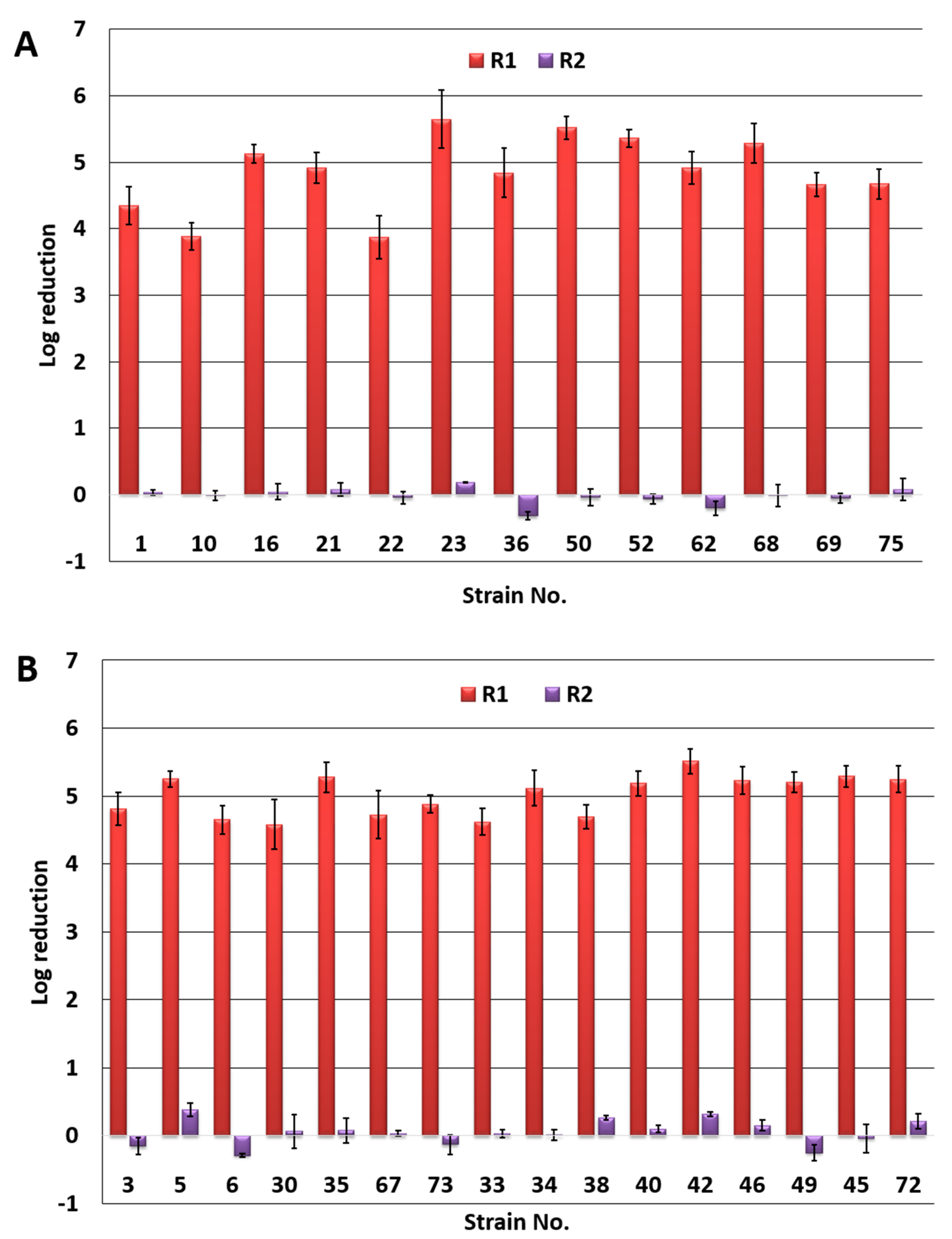
  • photodynamic inactivation—R1 = Log L(-)Ps(-)—Log L(+)Ps(+);
  • dark toxicity—R2 = Log L(-)Ps(-)—Log L(-)Ps(+);
  • L(-)Ps(-)—no irradiation, no Pc+ (negative control)
  • L(-)Ps(+)—no irradiation, Pc+ (dark control)
  • L(+)Ps(+)—irradiation, Pc+

2.6. Effect of PACT on Biofilm

To evaluate the effect of PACT on P. aeruginosa biofilm, viable counts were determined after single, double and triple photoinactivation. For this purpose, biofilms were grown in 96-well microtiter plates for 24 h, as described previously [29]. The medium was removed, and the wells were washed gently with PBS in order to remove planktonic cells. Subsequently, 200 µL aliquots of Ps solution (group Ps+) were added. The samples were incubated for 15 min and irradiated by LED panel (group L+) for 20 min. Next, the solution containing Ps was removed, and the first set of samples was tested for determination of the viable cells of bacteria in biofilm, while another 200 µL aliquot of Ps solution was added to the remaining two sets of samples. The procedure was repeated twice, and the viable cells of bacteria in biofilm were determined after the second and the third treatment. To determine the number of viable cells, the biofilm was scraped carefully and homogenized. Samples were diluted with PBS in series, plated on the TSA plates and incubated 20 h at 36 ± 1 °C. Simultaneously, the sets of samples in the dark condition (group L-) were tested. Irradiated and non-irradiated viable bacteria in samples and the reductions of bacterial cell populations were calculated as described above (Section 2.5).

2.7. Statistical Analysis

The data for CFU/mL were converted to the logarithmic form. The data of antimicrobial activity were expressed as mean ± SD. Pairwise comparisons were made using the unpaired Student’s t-test to compare the decrease of P. aeruginosa strains isolated (i) from the lower respiratory tract and from chronic wounds, and (ii) the decrease of P. aeruginosa strains resistant to all β-lactams and sensitive to all β-lactams. Analysis of variance tests were used to verify differences between the groups of strong-biofilm producers, moderate-biofilm producers and weak-biofilm producers.
A probability value (p) of less than 0.05 was considered significantly different. Statistical analysis was performed with the STATISTICA software, v.13.1.

3. Results

3.1. Resistance Patterns and Ability to Form Biofilm of P. aeruginosa Strains

Selected characteristics of P. aeruginosa strains applied in the study are summarized in Table 1. P. aeruginosa strains exhibited various patterns of resistance to antibacterial agents—from sensitive to resistant to all antibiotics used in the research (two strains isolated from chronic wounds). Four strains (three lower respiratory tract isolates and one chronic wound isolate) exhibited resistance to five of the six drugs used and were MBL(+). It should be highlighted that the highest rates of resistance were noted against carbapenems: meropenem (18 resistant strains, two intermediate strains) and imipenem (14 resistant strains, one intermediate strain). In addition, the most common pattern of resistance was resistance to the above mentioned antibiotics (four lower respiratory tract isolates and three chronic wound isolates) and to meropenem alone (two lower respiratory tract isolates and two chronic wound isolates). In order to identify the mechanism of resistance to carbapenems, disk-based assays and colorimetric tests were also employed.
According to the obtained results, only six strains were resistant to carbapenems due to the presence of metallo-β-lactamases (MBL). No strains positive for Klebsiella pneumoniae carbapenemases (KPC) or OXA-β-lactamases (OXA) were detected.
In general, apart from carbapenems, the tested strains showed resistance to ciprofloxacin and to drugs other than carbapenems beta-lactams (piperacillin and ceftazidime). The lowest antibiotic resistance was observed for tobramycin. In the herein presented study, strains of P. aeruginosa were found to produce biofilm, with most of them (n = 21) producing a massive amount of biofilm (Table 1).

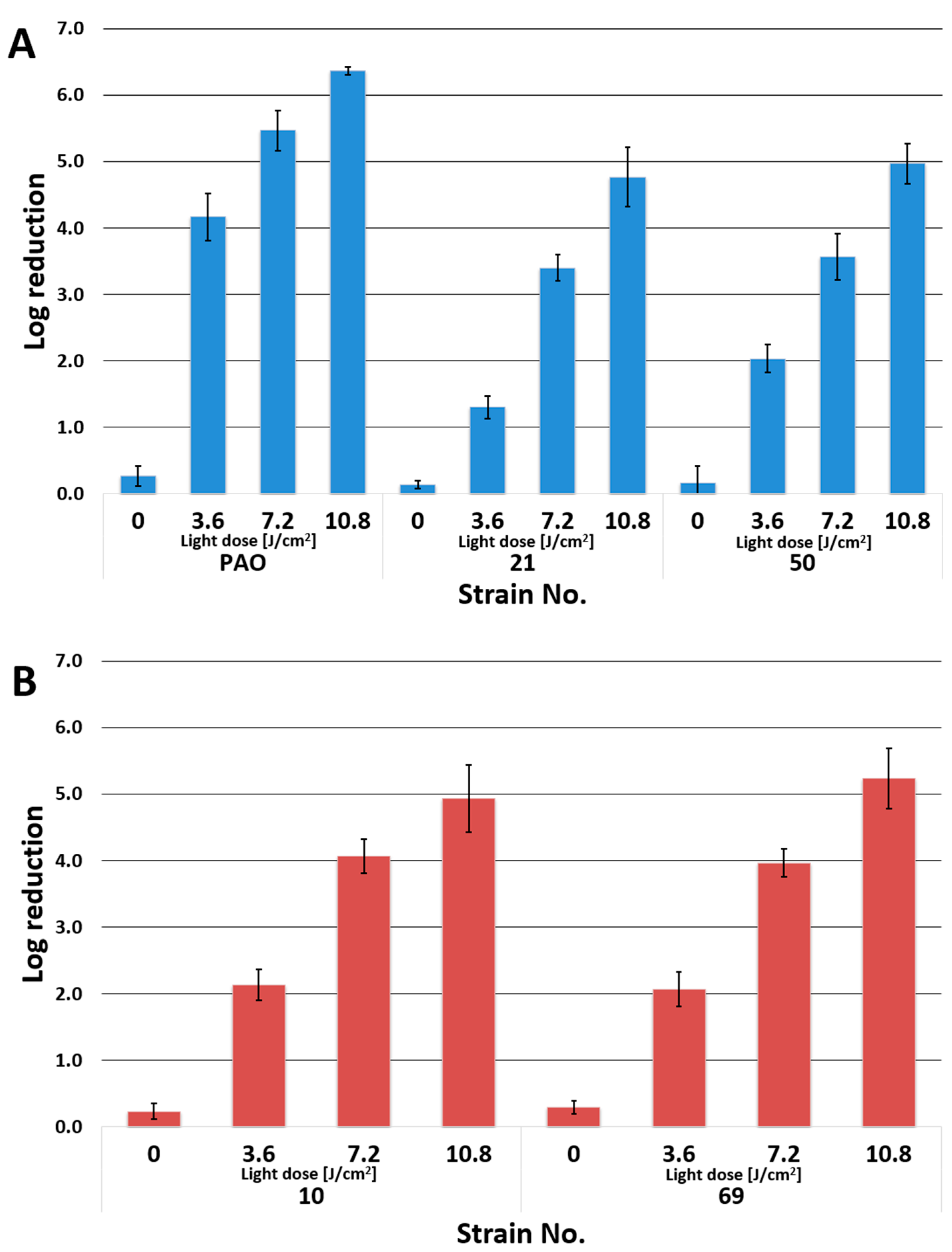
3.2. Photoinactivation of P. aeruginosa by PACT

Synthesis of the studied phthalocyanine Pc+ was based on procedures recently reported by our group [29,31]. The octaiodide salt of the magnesium(II) phthalocyanine derivative Pc+ was synthesized and characterized by mass spectrometry (MALDI TOF, the Advanced Chemical Equipment Instrumentation Facility at the Faculty of Chemistry, Adam Mickiewicz University in Poznan), UV–vis (Hitachi UV–vis U-1900 spectrometer) and NMR spectroscopy (Bruker 400 spectrometer) [29]. Herein presented studies indicate antimicrobial photocytotoxicity of Pc+ against all tested strains of P. aeruginosa. Pc+ at a concentration of 1.0 × 10⁻⁴ M, activated by light [L(+)Ps(+)], revealed a significant decrease in the number of bacterial cells compared to the untreated control group (L(-)Ps(-)). Representative images of P. aeruginosa quantification after treatment with Pc+ at a concentration of 1.0 × 10⁻⁴M in the dark (dark toxicity) and after irradiation with light (photocytotoxicity) are presented in Figure 2. For P. aeruginosa isolated from the lower respiratory tract of patients with pneumonia, effective bactericidal action (Figure 3) with reductions varying from 3.9 log (strains no. 10 and 22) to 5.7 log (strain no. 23) was demonstrated (Figure 3A). The results of the studies performed on P. aeruginosa strains isolated from chronic wound infections (Figure 3B) revealed a decrease in the number of viable cells varying from 4.6 log (strain no. 30) to 5.5 log (strain no. 42). It is worth noting that no significant statistical differences were observed in the degree of microbial reduction either between strains isolated from the lower respiratory tract and chronic wounds, between strains resistant and sensitive to all beta-lactam antibiotics used in the study, or between strains producing different degrees of biofilm (p > 0.05). In the dark, no apparent toxicity was noted in the same conditions (Figure 3A,B). The influence of light was also assessed by the irradiation of a set of samples without a photosensitizer. The control indicated no change in the number of viable cells as compared to the non-irradiated samples (data not shown).
In the studies, the differences in morphology of P. aeruginosa colonies were observed. Colonies isolated from Pc+-treated but non-irradiated samples were smaller and smoother compared to those isolated from non-Pc-treated controls.

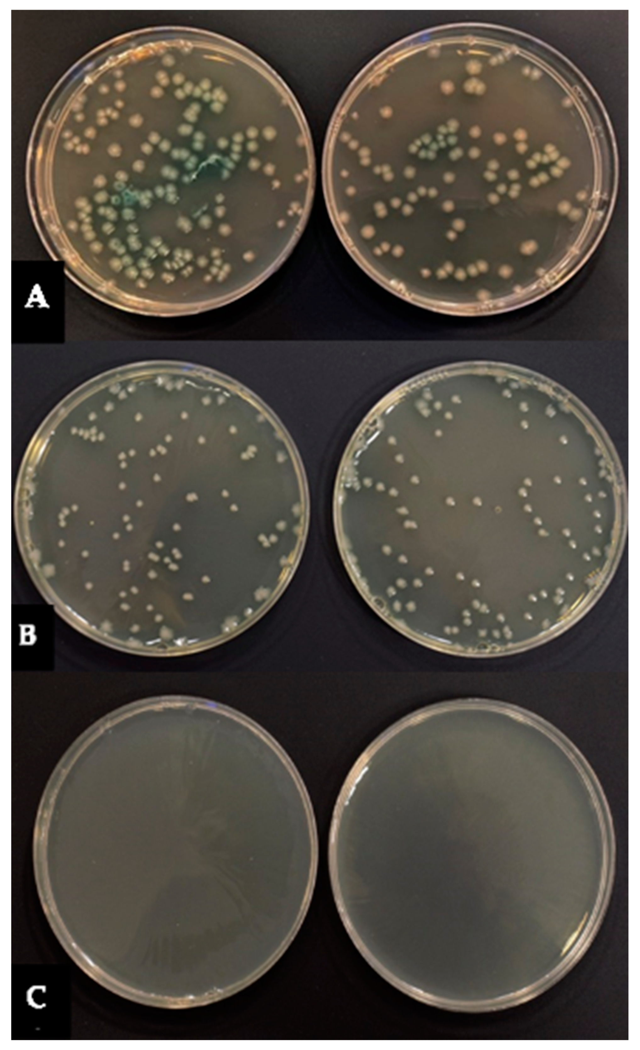
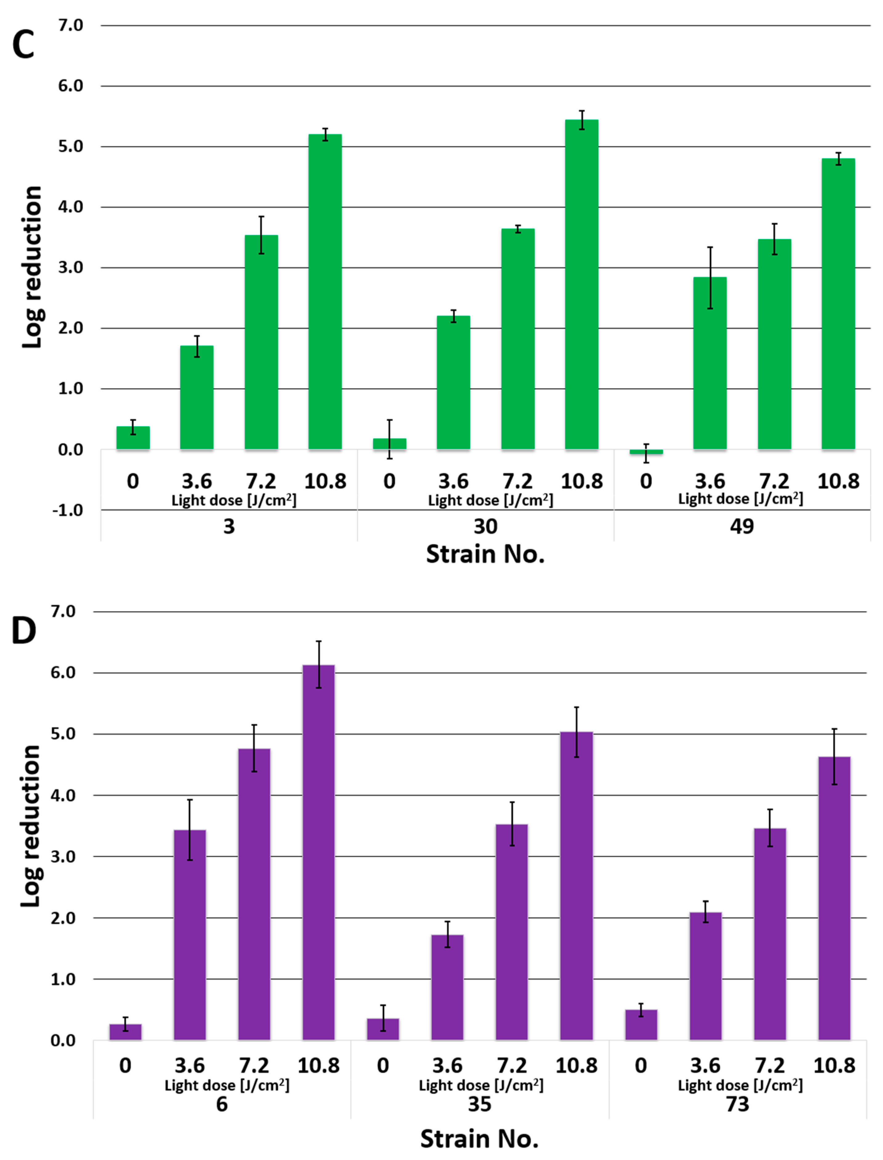
3.3. Effect of PACT on P. aeruginosa Biofilm

To evaluate the effect of PACT on biofilm, P. aeruginosa PAO and ten clinical strains of P. aeruginosa were selected on the basis of origin and biofilm production (strains no. 21, 50—lower respiratory tract and strong biofilm producer; strains no. 10, 69—lower respiratory tract and moderate biofilm producer; strains no. 3, 30, 49—chronic wound and strong biofilm producer; strains no. 6, 35, 73—chronic wound and moderate biofilm producer). First, extensive screening using standard strains of P. aeruginosa (PAO, P. aeruginosa ATCC 27853, P. aeruginosa NCTC 6749) was carried out to determine the lowest Pc+ concentration, incubation and irradiation time (data not shown). For this study we chose Pc+ concentration of 1 × 10⁻⁴ M, 15 min of incubation and 20 min of irradiation time.
As revealed in Figure 4, after single irradiation, a decrease of the number of bacteria in biofilm varied in the range of 1.3–4.2 log between tested strains. It is worth pointing out that the second treatment decreased the number of bacterial significantly, as a log reduction ranging from 3.4 to 5.5 log was noted. Boosted cell-death response was observed after the third irradiation, when the decrease in the number of viable bacterial cells was in the range 4.6–6.4 log. Scanning electron microscopic (SEM) images of a biofilm of P. aeruginosa treated with Pc+ in the dark and after photodynamic therapy using Pc+ are shown in Figure 5. Specific changes in cellular morphology, including alterations in cellular size and structures, were noted.

4. Discussion

In our earlier work, the photocytotoxic effect of Pc+ was assessed against planktonic and biofilm forms of microorganisms, including reference and clinical strains [29]. An extensive drop in cell survival of the planktonic form of P. aeruginosa NCTC 6749 was observed in the photocytotoxicity study with Pc+ against strains isolated from severe and difficult-to-treat infections. In this study, clinical planktonic and sessile strains of P. aeruginosa isolated from chronic wounds and lower respiratory tract infections were also subjected to Pc+ treatment. It is worth noting that the antimicrobial susceptibility patterns and biofilm forms of the evaluated strains isolated from different infection sites were different. Among the strains with strong biofilm biomass production there were both multi-drug resistant as well as antibiotic sensitive strains. However, when analyzing antimicrobial resistance on the ability to form biofilms, it can be concluded that the strong-biofilm producer strains are those which most frequently include microorganisms resistant to more than four antibiotics. In our previous study, Pc+ revealed excellent photocytotoxicity against planktonic cells of both Gram-positive and Gram-negative bacteria. The photoactivity of Pc+ toward P. aeruginosa NCTC 6749 was only slightly lower than that of E. coli and Gram-positive bacteria [28]. For this reason, further examination with the use of clinical strains of P. aeruginosa was included in the current study. In the in vitro antimicrobial photocytotoxicity study against P. aeruginosa, cationic phthalocyanine successfully reduced planktonic bacterial cells, with reduction factor (R1) values much higher than 4 log for nearly all tested strains (except strains no. 10 and 22). It is worth noting that for all tested strains the 3-log criterion for bactericidal activity for antibiotics was achieved. Bactericidal activity of antibacterial agent in vitro is usually defined as a reduction of the number of viable cells by at least 3 log. It is worth noting that for all tested strains the parameter was exceeded.
Moreover, the differences in the sensitivity to antibiotics and the degree of biofilm production by the analyzed strains did not affect their sensitivity to the photocytotoxic effect of the tested phthalocyanine.
The potential of various Pcs for antimicrobial photodynamic therapy was confirmed in various studies [41,42,43]. Spesia et al. presented high photoinactivation of Gram-positive S. mitis and Gram-negative E. coli bacteria by cationic zinc(II) phthalocyanines [41]. In another study, a cationic zinc(II) phthalocyanine derivative was active against both Gram-positive and Gram-negative bacteria. E. coli and S. aureus strains were inactivated with 675 nm light in the presence of 1 µM photosensitizer under 5 min irradiation with a reduction of about 5 log [42]. Other authors studied zinc(II) and ruthenium phthalocyanine derivatives in multi-cationic dendrimers against different microorganisms. Zinc(II) phthalocyanine derivatives at a concentration of 1 μM and light dose of 60 J × cm−2 inactivated S. aureus strains with a reduction factor of 6 log, whereas ruthenium phthalocyanines appeared less harmful against S. aureus, leading to a bactericidal effect only at maximum concentration and light dosage (1 μM and 60 J × cm−2). Photoinactivation of E. coli with a reduction of more than 7 log using zinc(II) phthalocyanine derivatives and in the range of 2–5 log for ruthenium phthalocyanine derivatives with a light dose of 60 J × cm−2 has been noted [43].
A number of various Pcs, either alone or in combinations with different substances, have been tested for a potential photodynamic effect against P. aeruginosa, with differentiated antibacterial photocytotoxicities against P. aeruginosa [44,45,46,47,48,49,50,51]. Ömeroğlu et al. presented the effectiveness of the silicon phthalocyanine derivative against Gram-positive bacteria, S. mutans and S. aureus, with over 3 log reductions. However, no effect was observed on the P. aeruginosa strain photoinactivated and irradiated under the same conditions (approx. 1 log reduction) [50]. In another study, two water-soluble cationic lutetium(III) acetate phthalocyanines (LuPc-5 and LuPc-6) were tested against P. aeruginosa and Candida albicans. Full photoinactivation of tested strains was achieved with LuPc-5 against P. aeruginosa and C. albicans, whereas photoinactivation with the peripherally substituted LuPc-6 was less significant (2 log) [49]. Ke and coworkers presented the photoinactivation efficiency of two zinc(II) phthalocyanine derivatives against Gram-positive and Gram-negative bacterial strains. S. aureus and E. coli were inactivated with a reduction of 4 log in the concentration range of 5 nM to 0.4 µM, whereas the same reduction in the viability of P. aeruginosa appeared in the range of 3.5 to 15.5 µM [47].
Many studies revealed that, although P. aeruginosa could be successfully inactivated, higher doses of photosensitizers and/or light are necessary. The sensitivity to photosensitization of Gram-positive and Gram-negative bacteria differs depending on their extracellular structures. The cell wall of Gram-negative bacteria is more complex than that of Gram-positive bacteria due to the presence of an outer membrane that covers the surface of the peptidoglycan [49,50,51,52,53]. The outer membrane is equipped with hydrophilic surfaces and lipophilic cores, which makes it a very effective barrier against lipophilic and hydrophilic molecules. Moreover, only amphipathic molecules, i.e., molecules with lipophilic backbones and polar/charged flanks, are capable of passing through this barrier [22,54]. Despite this phenomenon, cationic photosensitizers replace an essential LPS-binding (LPS—lipopolysaccharide) of divalent Ca2+ and Mg2+ ions and thereby disrupt outer membrane stability, leading to the formation of channels in the outer membrane that can facilitate photosensitizer uptake [55]. The electrostatic interactions constitute an essential factor initiating cell–drug binding. Therefore, the outer membrane and cell membrane are the sites of action for cationic phototherapeutic agents, which explains the high photocytotoxic potential of the herein studied zinc(II) phthalocyanine against P. aeruginosa strains. The fact that the tested Pc+ strongly absorbs red light means that the photodynamic process in tissues is effective, as red light is characterized by relatively deep tissue penetration.
The huge threat of P. aeruginosa to human health results from its innate resistance to many antimicrobials and its unique ability to form a biofilm. Biofilms often develop on the surface of wounds and is responsible for their chronicity and also complicates treatment. In a similar fashion, infections of the lower respiratory tract develop a biofilm, which makes it important to look for substances that act not only on the planktonic forms of bacteria, but, most importantly, on biofilms. Antibiotics and antiseptics used in the treatment of chronic wound infections are ineffective as antibiofilm agents because these agents have not been designed for killing of microorganisms within a biofilm but have only been designed to specifically focus on killing microorganisms in the planktonic state [37,56,57]. What is interesting is that biofilm production is a persistence strategy of P. aeruginosa for survival. The significance of this strategy is especially highlighted in chronic infections, pneumonia in patients with cystic fibrosis, hospital-acquired pneumonia, as well as infections associated with biomedical devices such as implants or catheters [3,43,58]. Growth in the biofilm contributes to its virulence and protects bacteria not only from antibiotics, but also from disinfectants and other antimicrobials, as well as host defense systems [13,14,59]. In this study, the effect of Pc+ on P. aeruginosa biofilm was assessed. As revealed in Figure 4, after single irradiation, a decrease in the number of bacteria in biofilm varied between tested strains and was observed in the range of 1.3–4.2 log. It is worth noting that a significant bolstering of the photoinactivation efficiency against P. aeruginosa biofilms was achieved by the introduction of the second and the third irradiation step.

5. Conclusions

Chronic wounds and lower respiratory tract infections constitute a complex and interdisciplinary health and social problem. Treatment of these infections is particularly problematic due to the presence of bacteria in the form of a biofilm. Cationic zinc(II) phthalocyanine with 2-(4-N-methylmorpholin-4-ium-4-yl)ethoxy groups subjected to the photocytotoxicity assessment against planktonic and biofilm forms of clinical strains of P. aeruginosa demonstrated promising photocytotoxicity. The studied cationic phthalocyanine at a concentration of 1.0 × 10⁻⁴ M, when activated by light, revealed a significant reduction factor of P. aeruginosa bacteria, ranging from nearly 4 to 6 log when compared to the untreated control group. In the biofilm inactivation study, after single dose irradiation a decrease in the number of bacteria ranging from 1.3 to 4.2 log was observed. P. aeruginosa strains used in this study exhibited various antibiotic resistance patterns and varying degrees of biofilm formation. Nevertheless, the phototoxic effect of Pc+ was not influenced by the degree of biofilm production or antibiotic resistance mechanisms present in the cells. The result is significant regarding the increasing resistance of bacteria to many drugs, especially beta-lactam antibiotics used in routine antibiotic therapy. An interesting increase of photoinactivation efficiency against P. aeruginosa biofilms was achieved by the introduction of the second and the third irradiation steps. The second irradiation of the biofilm significantly improved the bacterial reduction factor from 3.4 to 5.5 log, whereas the boosted cell death response was observed after the third irradiation, when a bacterial reduction factor from 4.6 to 6.4 log was noted. Noteworthy is the fact that repeated exposure of short durations of time can be advantageous in the case of this type of biofilm, and the obtained result could be considered for the basis of a prospective therapy. To sum up, the herein researched phthalocyanine derivative could be considered as a potential photosensitizer against multidrug-resistant P. aeruginosa of potential applicability in the treatment of chronic wound infection.

Author Contributions

Conceptualization, J.D.; methodology, J.D., W.S. and T.K.; investigation, W.S., T.K., M.R. and D.K.; resources, W.S., M.R. and J.D.; writing—original draft preparation, W.S. and M.R.; writing—review and editing, T.G. and J.D.; supervision, J.D. and T.G.; project administration, J.D. and W.S.; funding acquisition, M.R. All authors have read and agreed to the published version of the manuscript.

Funding

The authors acknowledge the financial support for Miniatura project No. 2021/05/X/NZ5/01600 from the National Science Centre, Poland.

Institutional Review Board Statement

Not applicable.

Informed Consent Statement

Not applicable.

Data Availability Statement

Not applicable.

Acknowledgments

The authors would like to acknowledge Poznan University of Medical Sciences Core Facility for providing access to the JEOL JSM-6380LA scanning electron microscope.

Conflicts of Interest

The authors declare that they have no conflict of interest.

References

  1. Brugha, R.E.; Davies, J.C. Pseudomonas aeruginosa in cystic fibrosis: Pathogenesis and new treatments. Br. J. Hosp. Med. 2013, 72, 614–619. [Google Scholar] [CrossRef] [PubMed]
  2. Cattaneo, C.; Antoniazzi, F.; Casari, S.; Ravizzola, G.; Gelmi, M.; Pagani, C.; D’Adda, M.; Morello, E.; Re, A.; Borlenghi, E.; et al. P. aeruginosa bloodstream infections among hematological patients: An old or new question? Ann. Hematol. 2012, 91, 1299–1304. [Google Scholar] [CrossRef] [PubMed]
  3. Church, D.; Elsayed, S.; Reid, O.; Winston, B.; Lindsay, R. Burn Wound Infections. Clin. Microbiol. Rev. 2006, 19, 403–434. [Google Scholar] [CrossRef] [PubMed] [Green Version]
  4. Gaspar, M.C.; Couet, W.; Olivier, J.C.; Pais, A.A.; Sousa, J.J. Pseudomonas aeruginosa infection in cystic fibrosis lung disease and new perspectives of treatment: A review. Eur. J. Clin. Microbiol. Infect. Dis. 2013, 32, 1231–1252. [Google Scholar] [CrossRef]
  5. Ruhnke, M.; Arnold, R.; Gastmeier, P. Infection control issues in patients with haematological malignancies in the era of multidrug-resistant bacteria. Lancet Oncol. 2014, 5, e606–e619. [Google Scholar] [CrossRef]
  6. Azzopardi, E.A.; Azzopardi, E.; Camilleri, L.; Villapalos, J.; Dean, E.; Boyce, D.E.; Dziewulski, P.; Dickson, W.A.; Whitaker, I.S. Gram Negative Wound Infection in Hospitalised Adult Burn Patients-Systematic Review and Metanalysis. PLoS ONE 2014, 9, e95042. [Google Scholar] [CrossRef] [Green Version]
  7. Hilker, R.; Munder, A.; Klockgether, J.; Losada, P.M.; Chouvarine, P.; Cramer, N.; Davenport, C.F.; Dethlefsen, S.; Fischer, S.; Peng, H.; et al. Interclonal gradient of virulence in the Pseudomonas aeruginosa pangenome from disease and environment. Environ. Microbiol. 2015, 17, 29–46. [Google Scholar] [CrossRef]
  8. Lusignani, L.S.; Blacky, A.; Starzengruber, P.; Diab-Elschahawi, M.; Wrba, T.; Presterl, E. A national point prevalence study on healthcare-associated infections and antimicrobial use in Austria. Wien. Klin. Wochenschr. 2016, 128, 89–94. [Google Scholar] [CrossRef]
  9. Sader, H.S.; Castanheira, M.; Duncan, L.R.; Flamm, R.K. Antimicrobial Susceptibility of Enterobacteriaceae and Pseudomonas aeruginosa Isolates from United States Medical Centers Stratified by Infection Type: Results from the International Network for Optimal Resistance Monitoring (INFORM) Surveillance Program, 2015–2016. Diagn. Micr. Infec. Dis. 2018, 92, 69–74. [Google Scholar]
  10. Recio, R.; Villa, J.; Viedma, E.; Orellana, M.A.; Lora-Tamayo, J.; Chaves, F. Bacteraemia due to extensively drug-resistant Pseudomonas aeruginosa sequence type 235 high-risk clone: Facing the perfect storm. Int. J. Antimicrob. Agents 2018, 52, 172–179. [Google Scholar] [CrossRef]
  11. Chatterjee, M.; Anju, C.P.; Biswas, L.; Anil Kumar, V.; Gopi Mohan, C.; Biswas, R. Antibiotic resistance in Pseudomonas aeruginosa and alternative therapeutic options. Int. J. Med. Microbiol. 2016, 306, 48–58. [Google Scholar] [CrossRef] [PubMed]
  12. Potron, A.; Poirel, L.; Nordmann, P. Emerging broad-spectrum resistance in Pseudomonas aeruginosa and Acinetobacter baumannii: Mechanisms and epidemiology. Int. J. Antimicrob. Agents 2015, 45, 568–585. [Google Scholar] [CrossRef] [PubMed] [Green Version]
  13. Husain, F.M.; Qais, F.A.; Ahmad, I.; Hakeem, M.J.; Baig, M.H.; Khan, J.M.; Al-Shabib, N.A. Biosynthesized zinc oxide nanoparticles disrupt established biofilms of pathogenic bacteria. Appl. Sci. 2022, 12, 710. [Google Scholar] [CrossRef]
  14. Mulcahy, L.R.; Isabella, V.M.; Lewis, K. Pseudomonas aeruginosa biofilms in disease. Microb. Ecol. 2014, 68, 1–12. [Google Scholar] [CrossRef] [Green Version]
  15. Pang, Z.; Raudonis, R.; Glick, B.R.; Lin, T.J.; Cheng, Z. Antibiotic resistance in Pseudomonas aeruginosa: Mechanisms and alternative therapeutic strategies. Biotechnol. Adv. 2019, 37, 177–192. [Google Scholar] [CrossRef]
  16. Poonsuk, K.; Tribuddharat, C.; Chuanchuen, R. Simultaneous overexpression of multidrug efflux pumps in Pseudomonas aeruginosa non-cystic fibrosis clinical isolates. Can. J. Microbiol. 2014, 60, 437–443. [Google Scholar] [CrossRef]
  17. Hamblin, M.R. Antimicrobial photodynamic inactivation: A bright new technique to kill resistant microbes. Curr. Opin. Microbiol. 2016, 33, 67–73. [Google Scholar] [CrossRef] [Green Version]
  18. Mahmoudi, H.; Bahador, A.; Pourhajibagher, M.; Alikhani, M.H. Antimicrobial Photodynamic Therapy: An Effective Alternative Approach to Control Bacterial Infections. J. Lasers. Med. Sci. 2018, 9, 154. [Google Scholar] [CrossRef] [Green Version]
  19. Awad, M.M.; Tovmasyan, A.; Craik, J.D.; Batinic-Haberle, I.; Benov, L.T. Important cellular targets for antimicrobial photodynamic therapy. Appl. Microbiol. Biotechnol. 2016, 100, 7679–7688. [Google Scholar] [CrossRef]
  20. Ragàs, X.; He, X.; Agut, M.; Roxo-Rosa, M.; Gonsalves, A.R.; Serra, A.C.; Nonell, S. Singlet Oxygen in Antimicrobial Photodynamic Therapy: Photosensitizer-Dependent Production and Decay in E. coli. Molecules 2013, 18, 2712–2725. [Google Scholar] [CrossRef] [Green Version]
  21. Cieplik, F.; Dongmei Deng, D.; Crielaard, W.; Buchalla, W.; Hellwig, E.; Al-Ahmad, A.; Maisch, T. Antimicrobial photodynamic therapy—What we know and what we don’t. Crit. Rev. Microbiol. 2018, 44, 571–589. [Google Scholar] [CrossRef] [PubMed] [Green Version]
  22. Liu, Y.; Qin, R.; Zaat, S.A.J.; Breukink, E.; Heger, M. Antibacterial photodynamic therapy: Overview of a promising approach to fight antibiotic-resistant bacterial infections. J. Clinic. Transl. Res. 2015, 1, 140–167. [Google Scholar]
  23. Skupin-Mrugalska, P.; Koczorowski, T.; Szczolko, W.; Dlugaszewska, J.; Teubert, A.; Piotrowska-Kempisty, H.; Goslinski, T.; Sobotta, L. Cationic porphyrazines with morpholinoethyl substituents—Syntheses, optical properties, and photocytotoxicities. Dye. Pigment. 2022, 197, 109937. [Google Scholar] [CrossRef]
  24. Benov, L. Photodynamic Therapy: Current Status and Future Directions. Med. Princ. Pract. 2015, 24 (Suppl. 1), 14–28. [Google Scholar] [CrossRef] [PubMed]
  25. Abrahamse, H.; Hamblin, M.R. New photosensitizers for photodynamic therapy. Biochem. J. 2016, 473, 347–364. [Google Scholar] [CrossRef] [Green Version]
  26. Ormond, A.B.; Freeman, H.S. Dye Sensitizers for Photodynamic Therapy. Materials 2013, 6, 817–840. [Google Scholar] [CrossRef] [Green Version]
  27. Donzello, M.P.; Ercolani, D.; Gaberkorn, A.A.; Kudrik, E.V.; Meneghetti, M.; Marcolongo, G.; Rizzoli, C.; Stuzhin, P.A. Synthesis, X-ray crystal structure, UV/Visible linear and nonlinear (optical limiting) spectral properties of symmetrical and unsymmetrical porphyrazines with annulated 1,2,5-thiadiazole and 1,4-diamyloxybenzene moieties. Chem. Eur. J. 2003, 9, 4009–4024. [Google Scholar] [CrossRef]
  28. Yano, S.; Hirohara, S.; Obata, M.; Hagiya, Y.; Ichiro Ogura, S.; Ikeda, A.; Kataoka, H.; Tanaka, M.; Joh, T. Current states and future views in photodynamic therapy. J. Photochem. Photobiol. 2011, 12, 46–67. [Google Scholar] [CrossRef]
  29. Dlugaszewska, J.; Szczolko, W.; Koczorowski, T.; Skupin-Mrugalska, P.; Teubert, A.; Konopka, K.; Kucinska, M.; Murias, M.; Düzgüneş, N.; Mielcarek, J.; et al. Antimicrobial and anticancer photodynamic activity of a phthalocyanine photosensitizer with N-methyl morpholiniumethoxy substituents in nonperipheral positions. J. Inorg. Biochem. 2017, 172, 67–79. [Google Scholar] [CrossRef]
  30. Dur, E.; Bulut, M. Derivatizable novel β-tetra 7-oxycoumarin-3-carboxylate substituted metallophthalocyanines: Synthesis and characterization. Polyhedron 2010, 29, 2689–2695. [Google Scholar] [CrossRef]
  31. Kucinska, M.; Skupin-Mrugalska, P.; Szczolko, W.; Sobotta, L.; Sciepura, M.; Tykarska, E.; Wierzchowski, M.; Teubert, A.; Fedoruk-Wyszomirska, A.; Wyszko, E.; et al. Phthalocyanine derivatives possessing 2-(morpholin-4-yl)ethoxy groups as potential agents for photodynamic therapy. J. Med. Chem. 2015, 58, 2240–2255. [Google Scholar] [CrossRef] [PubMed]
  32. Zhu, Y.J.; Huang, J.D.; Jiang, X.J.; Sun, J.C. Novel silicon phthalocyanines axially modified by morpholine: Synthesis, complexation with serum protein and in vitro photodynamic activity. Inorg. Chem. Commun. 2006, 9, 473–477. [Google Scholar] [CrossRef]
  33. Centers for Disease Control and Prevention. Antibiotic Resistance Threats in the United States; Department of Health and Human Services: Atlanta, GA, USA, 2019.
  34. World Health Organization. Prioritization of Pathogens to Guide Discovery, Research and Development of New Antibiotics for Drug-Resistant Bacterial Infections, Including Tuberculosis; World Health Organization: Geneva, Switzerland, 2017. [Google Scholar]
  35. Grinholc, M.; Szramka, B.; Kurlenda, J.; Graczyk, A.; Bielawski, K.P. Bactericidal effect of photodynamic inactivation against methicillin-resistant and methicillin-susceptible Staphylococcus aureus is strain-dependent. J. Photochem. Photobiol. 2008, 90, 57–63. [Google Scholar] [CrossRef] [PubMed]
  36. Lyon, J.P.; Moreira, L.M.; de Moraes, P.C.G.; dos Santos, F.V.; de Resende, M.A. Photodynamic therapy for pathogenic fungi. Mycoses 2011, 54, 265–271. [Google Scholar] [CrossRef]
  37. Percival, S.L.; Suleman, L.; Francolini, I.; Donelli, G. The effectiveness of photodynamic therapy on planktonic cells and biofilms and its role in wound healing. Future Microbiol. 2014, 9, 1083–1094. [Google Scholar] [CrossRef]
  38. EUCAST. Clinical Breakpoins—Bacteria, Version 10, European Committee on Antimicrobial Susceptibility Testing. Available online: http://www.eucast.org/clinical_breakpoints/2021 (accessed on 26 June 2021).
  39. UCAST. Guidelines for Detection of Resistance Mechanisms and Specific Resistance of Clinical and/or Epidemiological Importance. Version 2.0, July 2017. European Committee on Antimicrobial Susceptibility Testing. Available online: http://www.eucast.org/resistance_mechanisms/2017 (accessed on 12 February 2021).
  40. Stepanovic, S.; Vukovic, D.; Hola, V.; Di Bonaventura, G.; Djukić, S.; Cirković, I.; Ruzicka, F. Quantification of biofilm in microtiter plates: Overview of testing conditions and practical recommendations for assessment of biofilm production by staphylococci. APMIS 2007, 115, 891–899. [Google Scholar] [CrossRef]
  41. Spesia, M.B.; Rovera, M.; Durantini, E.N. Photodynamic inactivation of Escherichia coli and Streptococcus mitis by cationic zinc(II) phthalocyanines in media with blood derivatives. Eur. J. Med. Chem. 2010, 45, 2198–2205. [Google Scholar] [CrossRef]
  42. Segalla, A.; Borsarelli, C.D.; Braslavsky, S.E.; Spikes, J.D.; Roncucci, G.; Dei, D.; Chiti, G.; Jori, G.; Reddi, E. Photophysical, photochemical and antibacterial photosensitizing properties of a novel octacationic Zn(II)-phthalocyanine. Photochem. Photobiol. Sci. 2002, 1, 641–648. [Google Scholar] [CrossRef]
  43. Ruiz-González, R.; Setaro, F.; Gulías, O.; Agut, M.; Uwe Hahn, U.; Torres, T.; Nonell, S. Cationic phthalocyanine dendrimers as potential antimicrobial photosensitisers. Org. Biomol. Chem. 2018, 16, 1037. [Google Scholar] [CrossRef] [Green Version]
  44. Maho, T.; Binois, R.; Brulé-Morabito, F.; Demasure, M.; Douat, C.; Dozias, D.; Bocanegra, P.E.; Goard, I.; Hocqueloux, L.; Le Helloco, C.; et al. Anti-Bacterial Action of Plasma Multi-Jets in the Context of Chronic Wound Healing. Appl. Sci. 2021, 11, 9598. [Google Scholar] [CrossRef]
  45. Ribeiro, C.P.S.; Lourenço, L.M.O. Overview of cationic phthalocyanines for effective photoinactivation of pathogenic microorganisms. J. Photochem. Photobiol. C 2021, 48, 100422. [Google Scholar] [CrossRef]
  46. Mantareva, V.; Kussovski, V.; Angelov, I.; Dieter Wohrle, D.; Roumen Dimitrov, R.; Elka Popovae, E.; Dimitrov, S. Non-aggregated Ga(III)-phthalocyanines in the photodynamic inactivation of planktonic and biofilm cultures of pathogenic microorganisms. Photochem. Photobiol. Sci. 2011, 10, 91–102. [Google Scholar] [CrossRef] [PubMed]
  47. Ke, M.-R.; Eastel, J.M.; Ngai, K.L.K.; Cheung, Y.-Y.; Chan, P.K.S.; Hui, M.; Ng, D.K.P.; Lo, P.-C. Oligolysine-Conjugated Zinc(II) Phthalocyanines as Efficient Photosensitizers for Antimicrobial Photodynamic Therapy. Chem. Asian J. 2014, 9, 1868–1875. [Google Scholar] [CrossRef] [PubMed]
  48. Mantareva, V.; Gol, C.; Kussovski, V.; Durmuş, M.; Ivan Angelov, I. Impact of water-soluble zwitterionic Zn(II) phthalocyanines against pathogenic bacteria. Z. Naturforsch. 2019, 74, 183–191. [Google Scholar] [CrossRef] [PubMed]
  49. Mantareva, V.; Kussovski, V.; Durmuş, M.; Borisova, E.; Angelov, I. Photodynamic inactivation of pathogenic species Pseudomonas aeruginosa and Candida albicans with lutetium (III) acetate phthalocyanines and specific light irradiation. Lasers Med. Sci. 2016, 31, 1591–1598. [Google Scholar] [CrossRef]
  50. Ömeroğlu, I.; Kaya, E.N.; Göksel, M.; Kussovski, V.; Mantareva, V.; Durmuş, M. Axially substituted silicon(IV) phthalocyanine and its quaternized derivative as photosensitizers towards tumor cells and bacterial pathogens. Bioorg. Med. Chem. 2017, 25, 5415–5422. [Google Scholar] [CrossRef]
  51. Nyamu, S.N.; Ombaka, L.; Masika, E.; Ng’ang’a, M. Antimicrobial Photodynamic Activity of Phthalocyanine Derivatives. Adv. Chem. 2018, 2018, 2598062. [Google Scholar] [CrossRef]
  52. Meerovich, G.A.; Akhlyustina, E.V.; Tiganova, I.G.; Lukyanets, E.A.; Makarova, E.A.; Tolordava, E.R.; Yuzhakova, O.A.; Romanishkin, I.D.; Philipova, N.I.; Zhizhimova, Y.S.; et al. Photodynamic inactivation of Pseudomonas aeruginosa bacterial biofilms using new polycationic photosensitizers. Laser Phys. Lett. 2019, 16, 115603. [Google Scholar] [CrossRef]
  53. Pucelik, B.; Paczyński, R.; Dubin, G.; Pereira, M.M.; Arnaut, L.G.; Dąbrowski, J.M. Properties of halogenated and sulfonated porphyrins relevant for the selection of photosensitizers in anticancer and antimicrobial therapies. PLoS ONE 2017, 12, e0185984. [Google Scholar] [CrossRef]
  54. Hamblin, M.R.; Hasan, T. Photodynamic therapy: A new antimicrobial approach to infectious disease? Photochem. Photobiol. Sci. 2004, 3, 436–450. [Google Scholar] [CrossRef] [Green Version]
  55. Denyer, S.P.; Maillard, J.-Y. Cellular impermeability and uptake of biocides and antibiotics in Gram-negative bacteria. J. Appl. Microbiol. 2002, 92, 35–45. [Google Scholar] [CrossRef]
  56. Nikaido, H. Molecular Basis of Bacterial Outer Membrane Permeability Revisited. Microbiol. Mol. Biol. Rev. 2003, 67, 593–656. [Google Scholar] [CrossRef] [Green Version]
  57. Długaszewska, J.; Antczak, M.; Kaczmarek, I.; Jankowiak, R.; Buszkiewicz, M.; Herkowiak, M.; Michalak, K.; Kukuła, H.; Ratajczak, M. In vitro biofilm formation and antibiotic susceptibility of Pseudomonas aeruginosa isolated from airways of patients with cystic fibrosis. J. Med. Sci. 2016, 85, 245–253. [Google Scholar] [CrossRef]
  58. George, S.; Hamblin, M.R.; Kishen, A. Uptake pathways of anionic and cationic photosensitizers into bacteria. Photochem. Photobiol. Sci. 2009, 8, 788–795. [Google Scholar] [CrossRef] [PubMed] [Green Version]
  59. Ratajczak, M.; Kamińska, D.; Nowak-Malczewska, D.M.; Anna Schneider, A.; Dlugaszewska, J. Relationship between antibiotic resistance, biofilm formation, genes coding virulence factors and source of origin of Pseudomonas aeruginosa clinical strains. Ann. Agric. Environ. Med. 2021, 28, 306–313. [Google Scholar] [CrossRef] [PubMed]
Figure 1. The chemical structure of magnesium(II) phthalocyanine with peripheral 2-(4-N-methylmorpholin-4-ium-4-yl)ethoxy substituents.
Applsci 12 03707 g001
Figure 2. Photocytotoxic effect of Pc+ against P. aeruginosa through incubation and plate counting method. Representative plates with bacterial colonies of P. aeruginosa. (A)—number of P. aeruginosa in the control sample not containing the photosensitizer and not irradiated (L- Ps-); (B)—number of P. aeruginosa in the non-irradiated test containing the photosensitizer (dark toxicity (L- Ps+)); (C)—number of P. aeruginosa in the irradiated test containing the photosensitizer (photocytotoxicity (light toxicity) (L+ Ps+)).
Applsci 12 03707 g002
Figure 3. Values of reduction factors (R1 and R2) based on clinical strains of P. aeruginosa treated with Pc+ at a concentration of 1.0 × 10⁴ M in the dark (dark toxicity) and after irradiation with light (photocytotoxicity): (A) photocytotoxicity and toxicity—strains isolated from lower respiratory tract; (B) photocytotoxicity and toxicity—strains isolated from chronic wounds. Abbreviations: R1—photodynamic inactivation; R2—dark toxicity; R1 = Log L(-)Ps(-)—Log L(+)Ps(+); R2 = Log L(-)Ps(-)—Log L(-)Ps(+); L(-)Ps(-)—no irradiation, no Pc+ (negative control); L(-)Ps(+)—no irradiation, Pc+ (dark control); L(+)Ps(+)—irradiation, Pc+.
Applsci 12 03707 g003
Figure 4. Values of reduction factors (R) based on selected clinical strains of P. aeruginosa biofilms treated with Pc+ at a concentration of 1.0 × 10⁴ M in the dark (dark toxicity) and after irradiation (photocytotoxicity): (A) photocytotoxic biofilm inactivation and toxicity—PAO and strains isolated from lower respiratory tract, strong biofilm producers; (B) photocytotoxic biofilm inactivation and toxicity—strains isolated from lower respiratory tract, moderate biofilm producers; (C) photocytotoxic biofilm inactivation and toxicity—strains isolated from chronic wound, strong biofilm producers; (D) photocytotoxic biofilm inactivation and dark toxicity—strains isolated from chronic wound, moderate biofilm producers.
Applsci 12 03707 g004aApplsci 12 03707 g004b
Figure 5. SEM image of a biofilm of Pseudomonas aeruginosa: (A) treated with Pc+ in the non-irradiated test containing a photosensitizer concentration of 1.0 × 10⁻⁴ M—dark toxicity (L- Ps+), and (B) treated with Pc+ at a concentration of 1.0 × 10⁻⁴ M and irradiated by light dose of 3.6 J/cm2—photocytotoxicity (L+ Ps+). Arrows indicate changes in the cellular morphology, including alterations in cellular size and structures, thus suggesting cellular rupture. (SEM × 10,000; scale bar—1 μm).
Applsci 12 03707 g005
Table 1. Characteristics of P. aeruginosa strains using selected phenotypic and genotypic features.
Strain No.Source of IsolationResistance to AntibioticsMBLBiofilm Formation
PRLCAZIPMMEMCIPTOB
1L. resp. tractSSRRSS-S
10L. resp. tractSSRRSS-M
16L. resp. tractSSSSSS-S
21L. resp. tractSSRRRR-S
22L. resp. tractSSRRSS-S
23L. resp. tractSSSSSS-W
36L. resp. tractRRRRRS+S
50L. resp. tractRRRRRS+S
52L. resp. tractSSSRSS-S
62L. resp. tractRRRRRS+S
68L. resp. tractSSSRSS-S
69L. resp. tractSSRRSS-M
75L. resp. tractSSSSRS-S
3chronic woundSSSSSS-S
5chronic woundSSSSSS-W
6chronic woundSSSISS-M
30chronic woundSSSSSS-S
35chronic woundSSSSSS-M
67chronic woundSSRRIS-W
73chronic woundSSSRSS-M
33chronic woundRRRRRR+S
34chronic woundRSSSSS-S
38chronic woundRRRRRR+S
40chronic woundSSRRSS-S
42chronic woundSSISSS-S
46chronic woundSSSRSS-S
49chronic woundRRRRRS+S
45chronic woundSSRRSS-S
72chronic woundSSSISS-S
L. resp. tract—lower respiratory tract; PRL—piperacillin; CAZ—ceftazidime; IPM—imipenem; MEM—meropenem; CIP—ciprofloxacin; TOB—tobramycin; S—sensitive; I—susceptible, increased exposure; R—resistant; MBL—metalobetalactamases; (-)—negative test for MBL; (+)—positive test for MBL; S—strong-biofilm producer; M—moderate-biofilm producer; W—weak-biofilm producer.
Publisher’s Note: MDPI stays neutral with regard to jurisdictional claims in published maps and institutional affiliations.

