Article

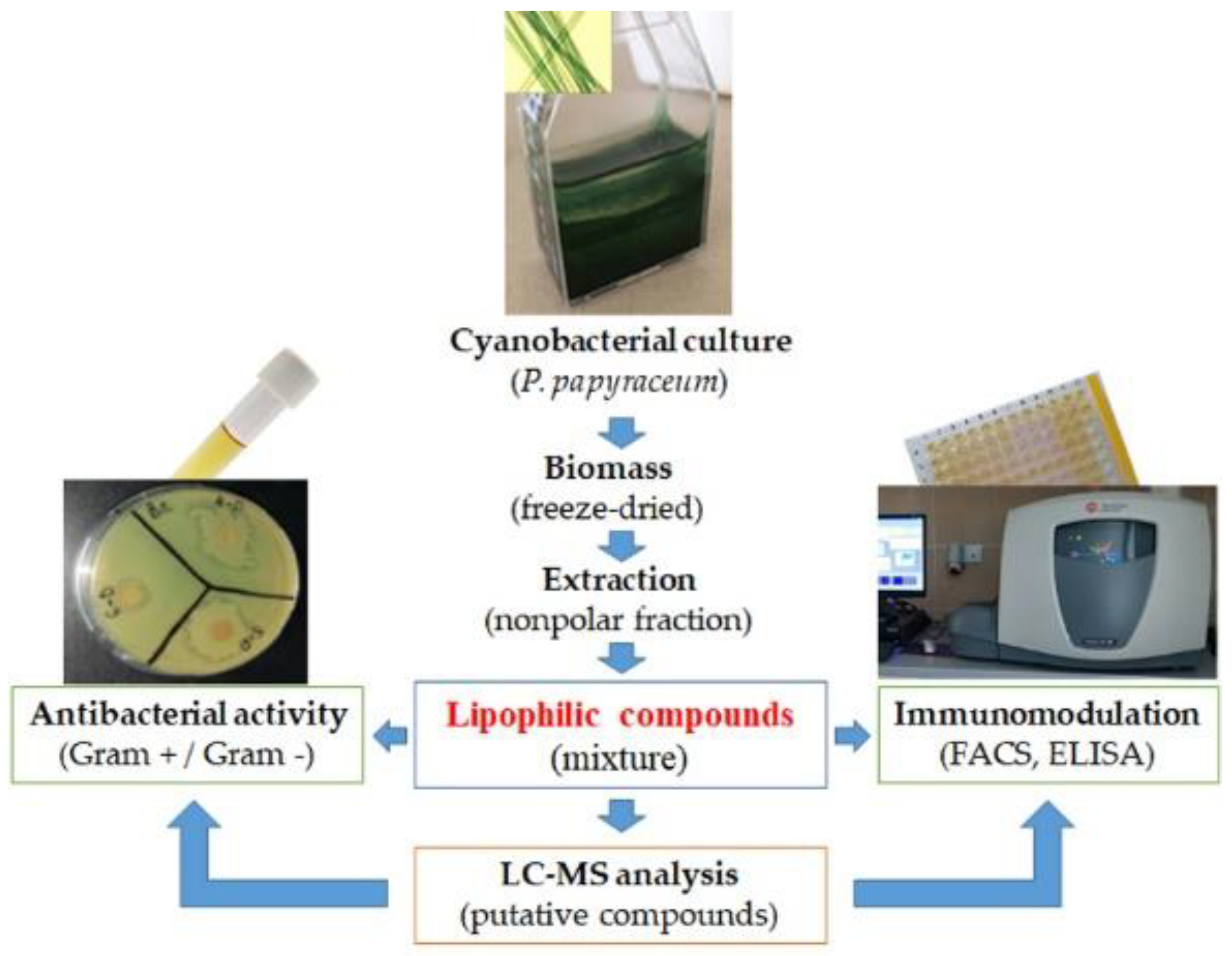
A Novel Approach for Fast Screening of a Complex Cyanobacterial Extract for Immunomodulatory Properties and Antibacterial Activity

1
Faculty of Biology, Plovdiv University “Paisii Hilendarski”, 4000 Plovdiv, Bulgaria
2
InoBioTech Ltd., 1113 Sofia, Bulgaria
*
Author to whom correspondence should be addressed.
Appl. Sci. 2022, 12(6), 2847; https://doi.org/10.3390/app12062847
Submission received: 6 January 2022 / Revised: 4 March 2022 / Accepted: 8 March 2022 / Published: 10 March 2022
(This article belongs to the Special Issue Novel Approaches for Natural Product-Derived Immunomodulators)

Abstract

:
The filamentous cyanobacteria from genus Phormidium are rich natural sources of bioactive compounds that could be exploited as pharmaceuticals or nutraceuticals. In this study, we suggest a novel approach for assessing the immunomodulatory properties of the products derived from cyanobacteria. The influence of Phormidium papyraceum extract on the human leukocyte immunophenotype was evaluated by attempting to link this activity to certain putative compounds identified in the extract. By using three staining panels and flow cytometry, we found that the cyanobacterial extract affected mainly CD4+ T cells upregulating activated CD4+CD152+ T cells (15.75 ± 1.93% treated vs. 4.65 ± 1.41% control) and regulatory CD4+CD25+ T cells (5.36 ± 0.64% treated vs. 1.03 ± 0.08% control). Furthermore, P. papyraceum extract can modulate T cell subpopulations with a CD4+ effector/memory phenotype. Extract-treated cells showed increased production of IL-2 (55 ± 12 pg/mL) and IL-6 (493 ± 64 pg/mL) compared to the untreated, 21 ± 7 pg/mL and 250 ± 39 pg/mL, respectively. No significant changes were observed in the secretion of TNF-α. In addition, P. papyraceum extract displayed antibacterial activity against both Gram-negative (inhibition zone from 18.25 ± 0.50 mm to 20.28 ± 1.50 mm) and Gram-positive (inhibition zone from 10.86 ± 0.85 mm to 17.00 ± 0.82 mm) bacteria. The chemical profile of the cyanobacterial extract was determined using LC–ESI–MS/MS analysis, where at least 112 putative compounds were detected. Many of these compounds have proven different biological activities. We speculated that compounds such as betulin and the macrolide azithromycin (or their analogues) could be responsible for the immunomodulatory potential of the investigated extract. More studies are needed to determine and validate the biological activities of the determined putative compounds.

1. Introduction

Cyanobacteria are an interesting, ancient group of photosynthetic prokaryotes with great potential as an object for scientific research. They have cosmopolitan distribution and are adapted to almost all ecological niches. These organisms can grow in fresh and salt waters, where they are often the predominant part of the phytoplankton. Some of them live in the soil and others are extremophiles and live in hypersaline waters, thermal springs, or on glaciers at the poles [1].
Cyanobacteria are considered to be one of the most important sources of natural products for drug discovery due to their potential to produce a wide spectrum of secondary metabolites and non-ribosomal peptides with different biological and pharmacological activities. In recent years, more than 1630 unique cyanobacterial compounds have been described [2]. Representatives of the genera Microcystis, Nostoc, Anabaena, Aphanisomenon (Aphanisomenon flos-aqua), Lyngbya (Lyngbya majuscula), Spirulina, and Scytonema were reported as producers of compounds with various chemical structures and biological activity, such as lipopeptides, amino acids, fatty acids, macrolides, esters, indoles, alkaloids, amides, lactones, and polysaccharides [3,4]. This identifies the cyanobacteria as a potential source of substances that can be used in the pharmacology, medicine, food, and cosmetics industries, as well as in the biotechnology industry.
Metabolites isolated from cyanobacteria show a variety of biological properties—tumour-suppressing, tumour-promoting, immunomodulatory, anti-inflammatory, antifungal, antibacterial, antiviral (anti-HIV), antiprotozoal, and anthelmintic activities, but they could also be toxic [5,6,7,8,9,10,11]. Some of the isolated cyanobacterial compounds or their chemical analogues with strong anticancer activity have already entered clinical trials (as comprehensively reviewed by Qamar et al. [12]). Typical examples are the cryptophycins, apratoxins, and dolastatins, as well as their synthetic analogues (LY 355073, oxazoline, TZT-1027, cematodin, and synthadotin) [4,12].
Despite intensive research on the bioactive substances produced by cyanobacteria, the potential of the 150 extant cyanobacterial genera, with more than 2600 species, has not yet been studied. Here, we can mention species-rich filamentous cyanobacterial genera, such as Phormidium, Microcoleus, Oscillatoria, Tolypothrix and Leptolyngbya. Most of the bioactive secondary metabolites (49%) have been isolated from the filamentous cyanobacteria belonging to order Oscillatoriales [12].
Species of the genus Phormidium are producers of biologically active substances with antiplasmodial activity. New, natural products, hierridin B and hierridin A, have been isolated from the lipophilic extract of the marine cyanobacterium Phormidium ectocarpi. The isolated hierridin mixture showed antiplasmodial activity towards Plasmodium falciparum [13]. The specific antitumour activity of two Phormidium molle strains was reported by Dzhambazov et al., studying their in vitro cytotoxic and anticancer properties on five cancer (HeLa, Jurkat, U-937, A2058, and RD) and two normal (3T3 and FL) cell lines [6]. Treatment with Phormidium extracts or Phormidium growth media altered the cytoskeletons and microtubule network of adherent cells, causing dose-dependent destruction of the monolayer and morphological changes. An extract concentration of 10 µg/mL showed significant cytotoxicity on HeLa and A2058 cells 24 h after treatment (41% and 60% respectively) as determined with MTT assay, while the viability of other cells was not significantly affected [6]. Portoamides A and B are cyclic peptides isolated from the cyanobacterium Phormidium sp. LEGE 05292 and were shown to be cytotoxic against RPE-1 (normal epithelial) and HCT116 (colon carcinoma) cell lines [14].
The problem is that many of the isolated bioactive cyanobacterial compounds have complicated chemical structures that will be difficult to synthesize. On the other hand, to purify a certain compound, a huge amount of cyanobacterial mass is required. Thus, before starting with purification or chemical synthesis of the desired compound, it is necessary to know the exact mechanism of its action. To speed the process of discovery of new bioactive compounds from cyanobacteria, Ferreira et al. developed an automated library of cyanobacterial fractions in a format of 96-well mother plates that can be easily screened with different bioassays and even coupled with metabolomics analysis [2].
Currently, only 10–20% of the known cyanobacterial secondary metabolites are characterized in terms of their chemical structure and biological potential [15,16,17]. Most common are the lipopeptides (40.2%), amides (9.4%), amino acids (5.6%), macrolides (4.2%), and fatty acids (4.2%) [4]. On the other hand, the variability of chemical structures produced by cyanobacteria allows interaction with different cellular targets, leading to hypotheses about new mechanisms of cyanobacterial action [18,19]. In this study, we suggest a novel approach for assessing the immunomodulatory properties of natural products derived from cyanobacteria. By using three staining panels (for T cells, antigen-presenting cells, and NK cells) and human peripheral blood mononuclear cells (PBMCs) from healthy individuals, the status of the main immunocompetent cells treated with natural products can be easily determined by flow cytometry. In addition, the observed effects can be related to certain compound(s) determined by mass spectrometry within the tested natural product. Thus, the aim of our study was to evaluate the immunomodulatory properties of cyanobacterial extract from Phormidium papyraceum Gomont ex Gomont 1892 (strain PACC 8600) and to determine its chemical composition. The proposed bioactive compounds could be additionally isolated and assessed (when this is possible). Here, we also evaluated the antibacterial activity of the studied cyanobacterial extract.

2. Materials and Methods

2.1. Chemicals and Reagents

Ultra-pure 19MOhm water was prepared by a PURELAB® Ultra Water Purification System (ELGA LabWater, High Wycombe, UK). Acetonitrile (ACN) and methanol (MeOH), Optima™ LC/MS-grade, and HPLC-grade chloroform were purchased from Thermo Fisher Scientific (Fair Lawn, NJ, USA). Formic acid (FA) LC/MS-grade ammonium formate, and HPLC-quality testosterone (as an internal standard) were purchased from Merck KGaA (Darmstadt, Germany).

2.2. Cyanobacterial Biomass and Extraction

Freeze-dried biomass from Phormidium papyraceum Gomont ex Gomont 1892, strain PACC 8600 (Oscillatoriales, Cyanobacteria) was provided by the Plovdiv Algal Culture Collection (PACC), Paisii Hilendarski University of Plovdiv, Bulgaria. Briefly, the cyanobacterial strain was cultured under sterile conditions (75 cm2 culture flasks, TPP, Trasadingen, Switzerland) in alkaline Z-nutrient medium [20], with a photoperiod of 12 h/12 h light and dark, respectively, and at a light intensity of 10 μmol photons s−1 m−2, provided by 40 W cool-white fluorescent tubes. The biomass of P. papyraceum culture was collected by centrifugation, frozen, and freeze-dried.
The cyanobacterial biomass (500 mg) was mixed with 3 mL MeOH and vortexed for 1 min, followed by an ultrasonic bath extraction (Branson 5510R-DTH, Wilmington, NC, USA) for 20 min under periodical vortexing. Then, 6 mL of chloroform were added to the suspension and shaken for 20 min at 15 rpm. Three millilitres of Milli-Q water were added and vortexed for 1 min. The extract was centrifuged at 4000 rpm for 20 min, and the methanol/chloroform fraction was collected and filtered by Millex-FG, 0.20 µm, hydrophobic PTFE filter (Merck KGaA, Darmstadt, Germany). In this study, we were interested more in the nonpolar compounds (methanol/chloroform fraction). Experimental design to assess the immunomodulatory properties, antibacterial activity, and chemical composition of the cyanobacterial extract is shown in Figure 1.
For LC-MS analysis, 2 mL of the filtered, nonpolar fraction was transferred to a standard autosampler vial, capped, and placed in the 4 °C Peltier cooled autosampler tray.
The resting amount of the filtered nonpolar fraction was used for testing of immunomodulatory properties and antibacterial activity. To remove the organic solvents, the filtered fraction was evaporated under a vacuum at 37 °C using a Savant SpeedVac Concentrator (SAVANT Instruments Inc., Farmingdale, NY, USA). Then, the dry residue was dissolved in 50% DMSO aqueous solution (w/v) to a final concentration of 5 mg/mL (5 mg of the dry residue was dissolved in 1 mL of DMSO/water, 1:1) and stored at 4 °C until further use. Working solutions were prepared by additional dilution of the stock (5 mg/mL) in Dulbecco’s Phosphate-Buffered Saline (DPBS, Gibco®, Life Technologies™, Paisley, Scotland, UK).

2.3. Immunomodulatory Properties

A total of 10 healthy subjects (6 males and 4 females, 26–40 years old, mean age 31, non-smokers) were recruited for this study. None of the subjects had received any therapy in the 2 months prior to taking a blood sample. Peripheral venous blood samples were collected from the cubital vein in BD Vacutainer® K2EDTA tubes (Becton, Dickinson and Company, Oakville, ON, Canada) in a clinical laboratory. The basic haematological parameters of all volunteers were within the reference ranges.
Blood samples were centrifuged at 1500× g for 15 min at room temperature, and the serum was discarded. Red blood cells were lysed with 0.84% NH4Cl buffer, and the samples were washed twice with a sterile Dulbecco’s Phosphate-Buffered Saline (D-PBS, Gibco®, Life Technologies™, Paisley, Scotland, UK). The isolated leukocytes were centrifuged at 1500× g for 10 min, and the cells were resuspended in Dulbecco’s Modified Eagle’s Medium (DMEM), supplemented with 10% heat-inactivated fetal bovine serum, and stabilized with antibiotic, antimycotic solution (all from Sigma-Aldrich Chemie GmbH, Steinheim, Germany). This medium was denoted as complete DMEM. Cells were plated (1 × 10⁶ cells/mL) in 12-well plates (TPP, Trasadingen, Switzerland) and treated for 48 h with 4 μL (5 mg/mL) of the cyanobacterial extract to reach a final concentration of 20 μg/mL in complete DMEM (1 mL/well). Our pilot studies for cytotoxicity with the cell lines A549 (epithelial lung carcinoma cells), HT-29 (epithelial colon adenocarcinoma cells), HeLa (epithelial cervix adenocarcinoma cells), U-937 (human histiocytic lymphoma cells), and NIH/3T3 (embryonic mouse fibroblasts) showed that concentrations of the extract up to 100 μg/mL do not have any cytotoxic effects. Untreated cells cultured for 48 h in complete DMEM served as a negative control. Another control included cells treated with an equivalent amount of DMSO (4 μL of DMSO/water, 1:1) as that contained in the extracts. Cells incubated for 48 h with 1 µg/mL leucoagglutinin PHA-L (Sigma-Aldrich, Saint Louis, MO, USA) were used as a positive control. The leukocytes were cultured in complete DMEM at 37 °C, 5% CO2, 95% atmospheric air, in a humidified incubator.
After 48 h, the treated and control cells were collected by centrifugation at 1500× g for 10 min, and the cell pellets were resuspended in FACS buffer (D-PBS, supplemented with 5% fetal bovine serum and 0.05% NaN3). The cells were stained for 20 min at 4 °C with the following fluorochrome-conjugated, anti-human antibodies grouped in 3 panels: (I) CD3 PE-Dazzle™ 594, CD4 FITC, CD8 PE, CD25 PE-Cy5, and CD152 PE-Cy7; (II) HLA-DR-DP FITC, CD19 PE, CD80 PE-Dazzle™ 594, CD11b PE-Cy5, and CD138 PE-Cy7; and (III) CD3 PE-Dazzle™ 594, CD56 FITC, and CD16 PE (all from BioLegend®, San Diego, CA, USA). Finally, the cells were washed twice and resuspended in 300 μL FACS buffer. Immunophenotyping was performed by flow cytometry (FACS) using a Cytomics FC500 instrument (Beckman Coulter Inc., Life Sciences, Indianapolis, IN, USA). The results are presented as mean ± SD (n = 10).
Production of IL-2, IL-6, and TNF-α in the supernatants was assayed by LEGEND MAX™ Human IL-2, IL-6, and TNF-α ELISA kits (BioLegend Inc., San Diego, CA, USA), according to the manufacturer’s instructions. The sensitivity of the assay for IL-2, IL-6, and TNF-α was 4 pg/mL, 1.6 pg/mL, and 3.5 pg/mL, respectively.
The study was conducted in adherence to the Declaration of Helsinki ethical guidelines and was approved by the Local Ethics Committee at the Paisii Hilendarski University of Plovdiv, Bulgaria. Written informed consent was obtained from all participants prior to the initiation of the study.

2.4. Antibacterial Activity
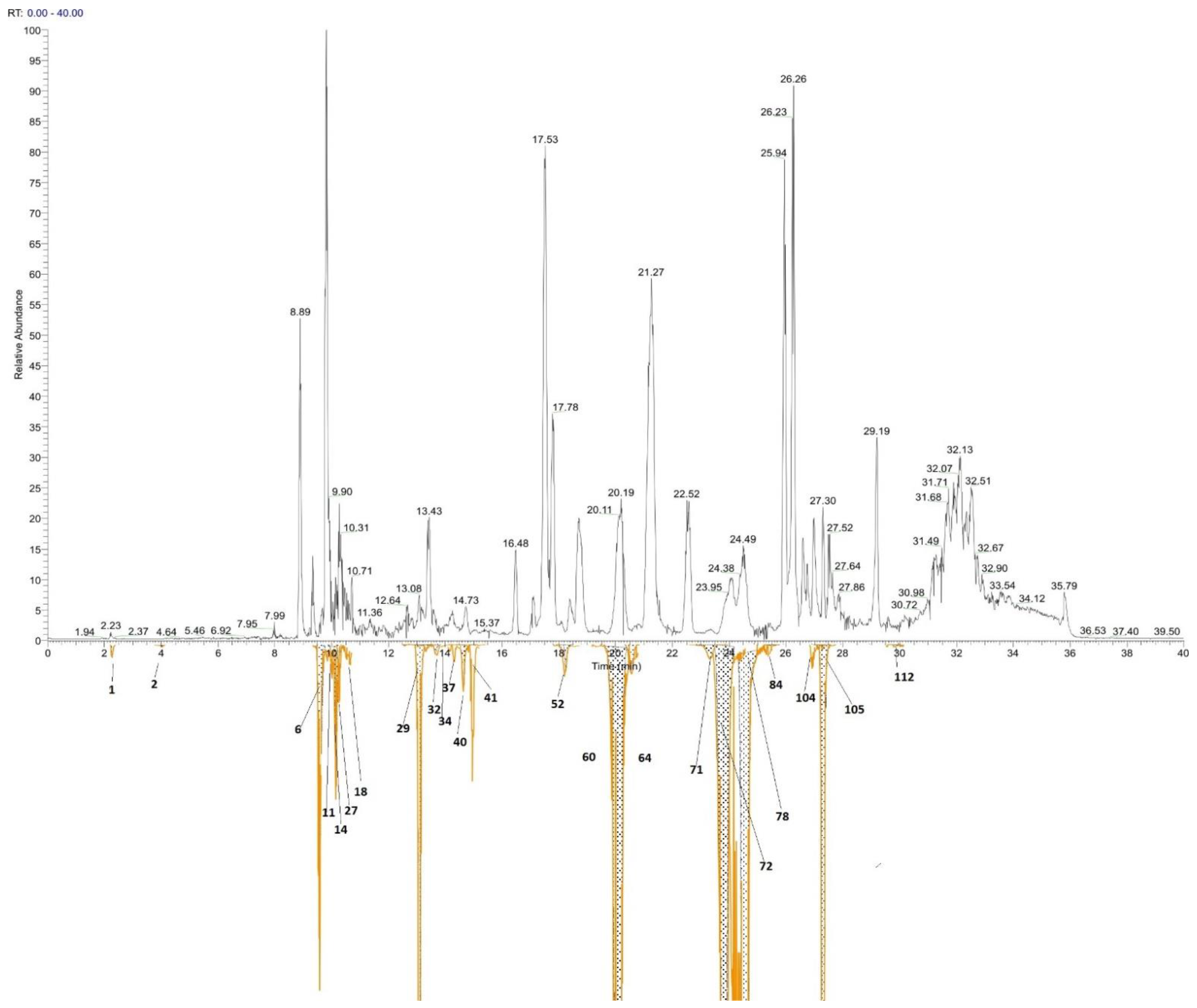
Mueller Hinton medium (agar or broth) and antibiotics were purchased from Sigma-Aldrich Inc., Merck KGaA, Darmstadt, Germany. The antibacterial activity of the P. papyraceum extract was evaluated by the agar diffusion method [21]. Three Gram-negative (Escherichia coli ATCC® 25922™, Pseudomonas aeruginosa ATCC® 27853™, and Proteus mirabilis ATCC® 14153™) and three Gram-positive (Bacillus cereus ATCC® 11778™, Bacillus subtilis ATCC® 11774™, and Staphylococcus aureus ATCC® 12600™) bacterial strains were used in the experiment.
Mueller Hinton agar plates were inoculated with 50 μL overnight-grown at 37 °C bacterial suspensions (concentration 1 × 10⁶ CFU/mL). Then, 20 μL containing 20 μg of the cyanobacterial extract were applied on standard holes in the agar medium with a diameter of 8 mm. A quantity of 20 μL of antibiotics (20 IU penicillin and 20 μg streptomycin) and 20 μL DMSO/water (1:1) served as positive and negative controls. The plates were incubated at 37 °C for 24 h, and then the diameters of the inhibition zones were measured in millimetres. Determinations were performed in four repetitions.

2.5. LC-MS Analysis

2.5.1. Instrumentation

An Accela quaternary HPLC pump with an Accela autosampler, and an HRMS Q-Exactive detector (Thermo Fisher Scientific, Waltham, MA, USA) with H-ESI electrospray were used. Chromatographic separation was performed on a Kinetex EVO C18 150 mm × 3 mm, 2.6 µm core-shell column (Phenomenex Inc., Torrance, CA, USA).

2.5.2. Reverse Phase (RP) Separation

Mobile phases A (90% water, 5% acetonitrile, 5% methanol, 9 mM ammonium formate, and 0.1% formic acid), B (90% acetonitrile, 5% water, 5% methanol, 9 mM ammonium formate, and 0.1% formic acid), and C (95% isopropanol, 5% methanol, and 0.1% formic acid) were used according to the following gradient setup (Table 1).
1 µL of the filtered sample was injected into the chromatographic system. The autosampler tray was maintained at 4 °C, and the injection needle was washed with portions of 1 mL water, 1 mL methanol, 1 mL THF, 1 mL DMSO, 1 mL methanol, and 1 mL water. Thew column oven was kept at 40 °C.

2.5.3. Mass Spectral Conditions

The H-ESI vaporizer temperature was maintained at 250 °C, spray voltage of 4 kV (in positive mode), ion transfer tube temperature of 350 °C (positive mode), sheath gas pressure 55 psi, auxiliary gas flow—10 arbitrary units. Top 5 Full MS/Data Dependent-MS2 were performed in positive ion mode with the following settings: 70,000 FWHM resolution in full MS from 134 to 2000 m/z for non-polar compounds. The automatic gain control (AGC) target was set to 1e6, and the Max IT value was 120 mse for Full MS scans, as well as for fragment spectra scans MS/MS. Settings of 17,500 FWHM resolution with 1.6 m/z quadrupole isolation window of precursor ion, 60 ms maximal trap filling time and 35 NCE (13 eV) HCD stepped collision energy with 50% step (17–53 NCE) were used. A 0.2% underfill ratio and 10 s dynamic exclusion were used.
Accurate mass calibration and tune were performed with the original calibration solution supplied by Thermo Scientific at every 24 h of operation, and room temperature was maintained within 22–26 °C during operation.

2.5.4. Data Treatment

LC-HRMS acquired data from the lipophilic extract of P. papyraceum were processed with Compound Discoverer 3.0 (Thermo Fisher Scientific, Waltham, MA, USA) using an untargeted approach: (I) extracting ion traces for exact mass ions, (II) finding molecular ions and adducts, and (III) grouping ion species and the corresponding fragment spectra (if available) in possible structure candidates packets with exact mass and formula. Based on these, a web database search was performed using ChemSpider, mzCloud, Metabolica, or other mass list resources to generate possible putative structural candidates (putative identifications). Only natural products databases were exploited.
MS data were exported in Excel; defining names consisted of molecular mass and retention time for each feature. Features were further processed with Perseus software. Based on the significance of the features, the corresponding molecular ion extracted chromatograms were reviewed, and when MS/MS data were available, they were further processed to propose hypothetical structures using Compound Discoverer™ Software [22], Sirius 4.8.1 [23,24], and MS-Finder [25].
Generated MS features were reviewed manually where possible and subjected to automatic processing and statistics. MS/MS spectra for annotated compounds with significant fold change and acceptable p-value (<0.05) were subjected to FISh coverage processing, SIRIUS MS/MS processing, and an MS-Finder Search. A limited number of compounds were manually validated by a comparison with experimentally obtained or simulated MS/MS spectra from the METLIN script and mzCloud databases, if such were found available.
Any data processing of metabolites’ manual work outside Compound Discoverer was made using Xcalibur™ (Thermo Fisher Scientific, Hemmel, UK) and Microsoft Excel. For quantitative comparison, metabolites’ m/z ratios were extracted from chromatograms, and chromatographic peaks were integrated with internal standard correction. Testosterone was chosen as an internal standard for positive ionization mode.

2.6. Statistics

Statistical analyses were performed using StatView software (SAS Institute Inc., Cary, NC, USA). Results are presented as mean ± SD from the individual determinations. The non-parametric Mann-Whitney U test was applied to determine differences between the treated and the control groups. p-Values less than 0.05 were considered statistically significant.

3. Results

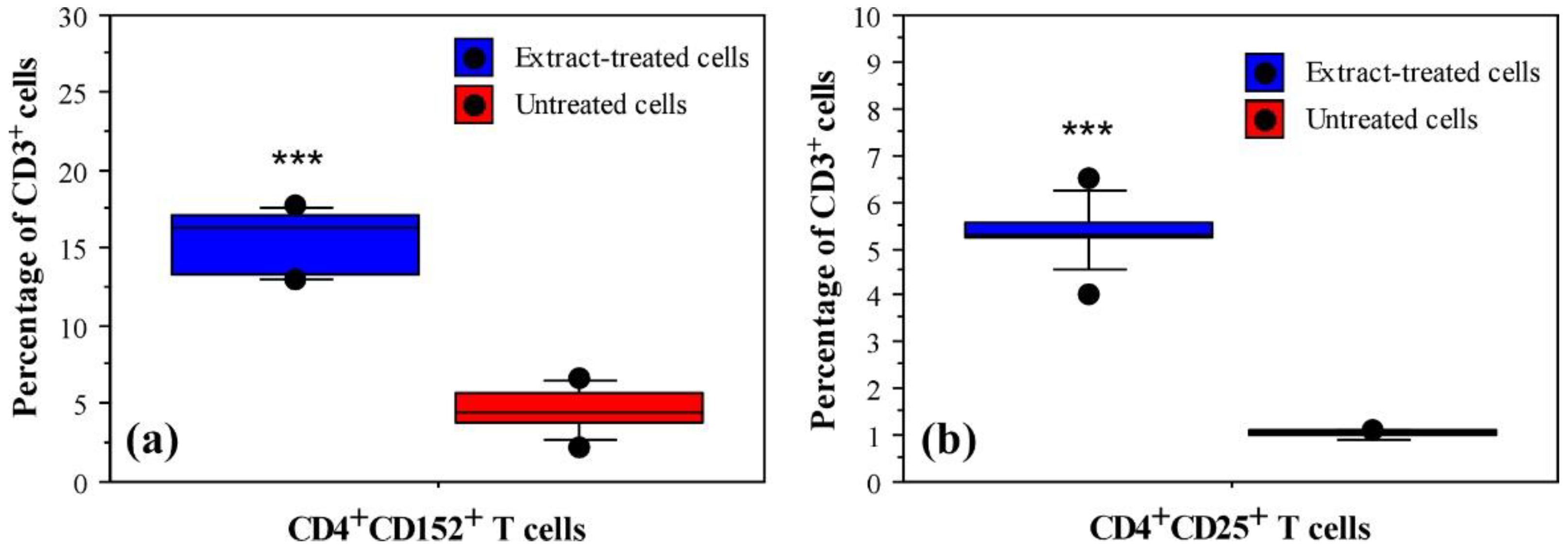
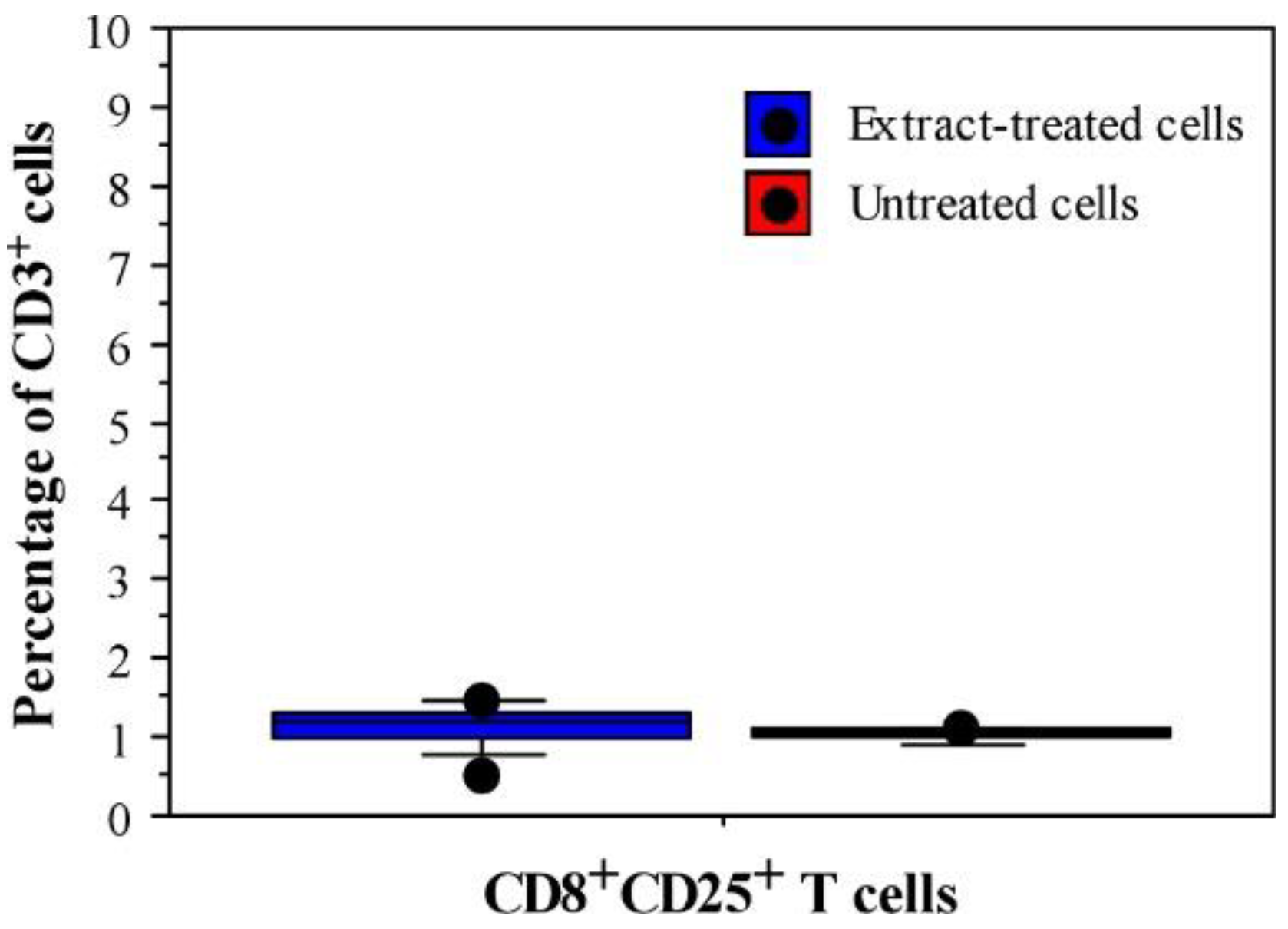
3.1. Immunomodulatory Properties of Phormidium Papyraceum Extract

To determine the influence of the P. papyraceum extract on the immunophenotype and composition of the adaptive immune cells, we designed staining panels that allow us to identify the major subsets of T cells, B cells, antigen-presenting cells (APCs), and NK cells. The expression levels of the main surface markers characterizing the major cell subpopulations were compared between extract-treated and untreated cells (Table 2). No differences between treated and untreated leukocytes were observed for B cells (CD19), plasma cells (CD138), monocytes/macrophages (CD11b), or NK cells (CD16/CD56) (Table 2).
To evaluate the T cell subsets in response to the cyanobacterial extract, we analysed the expression of CD3, CD4, CD8, CD25, and CD152. The data demonstrated a strong activation of the CD4+ T lymphocytes after treatment with P. papyraceum extract with no significant changes of the CD8+ T cell subpopulation (Table 2). As shown in Figure 2, there was a significant increase in the percentages of activated T cells (CD4+CD152+) and regulatory T cells (Tregs, CD4+CD25+) within extract-treated cells compared to untreated cells. No such difference was observed for the CD8+CD25+ population (Figure 3).
The treatment with P. papyraceum extract was able to modulate the levels of some proinflammatory cytokines that are responsible for the cell-mediated immunity and the initial activation of the immune system. Observed activation of the CD4+ T cells by the extract was accompanied with significantly enhanced secretion of IL-2 and IL-6 (2.8 and 2.5-fold), as compared with untreated cells, respectively (Figure 4). In contrast, the secretion of TNF-α was not affected (Figure 4).
Regarding antigen-presenting cells, HLA-DR-DP expression was significantly upregulated after treatment with P. papyraceum extract (Table 2). In contrast, the percentages of CD80+ (B7-1) cells were comparably low without significant differences in either extract-treated or untreated cells (2.26 ± 1.91 and 2.12 ± 0.97, respectively).
Detected expression levels of the selected immune markers on PHA-L-stimulated leukocytes (positive control) are also presented in Table 2.

3.2. Antibacterial Potential of Phormidium Papyraceum Extract

Six bacterial strains (three Gram-negative and three Gram-positive) were used to evaluate the antibacterial potential of the P. papyraceum extract. Antibacterial activity of the tested non-polar fraction was observed against all studied bacterial strains, with the growth-inhibition zone ranging between 10.86 and 20.28 mm (Table 3). Antibiotics (positive controls) showed significant growth-inhibition zones (between 25.78–27.67 mm for the Gram-negative strains and between 24.50–25.00 mm for the Gram-positive strains). No inhibition was observed for the negative control (Table 3).
As shown in Table 3, the highest inhibition zone of 20.28 mm was measured against Escherichia coli, followed by a 19.33 mm zone against Proteus mirabilis. The least growth-inhibition among the studied bacterial strains was against Staphylococcus aureus, with the lowest mean inhibition-zone value of 10.86 mm. Therefore, Escherichia coli was the most susceptible strain to the P. papyraceum extract, and Staphylococcus aureus was the most resistant strain.

3.3. Chemical Composition

The LS-ESI-MS/MS analysis of the non-polar fraction from the P. papyraceum extract revealed the existence of more than 1600 compounds. The chromatographic profile is presented in Figure 5.
The upper chromatogram (black) represents the total ion current chromatogram of P. papyraceum extract covering the mass range from 140 to 2100 m/z. The chromatogram below (orange) represents the superimposed, extracted ion features corresponding only to the putative compounds discussed in Table 4. Each peak in the chromatogram is marked with the same number as in the table. Both chromatograms are autoscaled according to the highest peak in the view.
We were able to determine 112 putative compounds. Their names, retention times (RT), formulas, molecular weights, and peak areas are provided in the Supplementary Materials (Table S1). The major compounds detected were mainly fatty acids and their derivatives, carbamate and carboxylate esters, terpenoids, amides, secondary and tertiary alcohols, and macrolides. Many of these compounds are characterized by antibacterial, antioxidant, antiviral, antifungal, cytotoxic, anticancer, and antimalarial activities, as well as being inhibitors of different enzymes. Compounds with proven biological activity that could be responsible for the effects observed in our study are shown in Table 4.

4. Discussion

Representatives of the genus Phormidium are a promising source of biologically active substances. This was the reason we chose P. papyraceum as a subject in this study. Although we have isolated two fractions (polar and non-polar components), we decided to study in detail the lipophilic fraction because the data on these compounds are limited, and the transport of such constituents across biological membranes is facilitated, in comparison with the large, polar molecules. We applied a novel approach for the fast screening of a complex cyanobacterial extract for immunomodulatory activities, attempting to link the putative compounds found in this mixture to the observed effects.
The immunophenotypic analysis performed after in vitro treatment of human leukocytes with P. papyraceum extract for 48 h showed upregulation of markers typical for the CD4+ T cell subpopulation. Our study demonstrated that the investigated extract induces non-specific immunostimulation similarly to the PHA-L by activation of the CD4+ T cells. We observed upregulation of the CD4+CD152+ (activated) and CD4+CD25+ (regulatory) T cell subpopulations, which was in line with the used positive control (PHA-L) (Table 2) and the results from other authors about the phenotype of T cells stimulated with phytohemagglutinin [27]. However, unlike PHA-L, the P. papyraceum extract does not stimulate CD8+ T cells.
Evaluating how cannabidiol modulates cytokine/chemokine production in monocytes, Sermet et al. also showed upregulation of IL-2 and IL-6 [28]. They demonstrated that cannabidiol could modulate the production of monocyte-derived proinflammatory cytokines, including IL-6, which plays an important role in bridging innate and adaptive immunity. Nowadays, IL-6 is considered an important modulator of the effector functions of CD4+ T cells. IL-6 stimulates T cell proliferation independently of IL-2 and increases the effector/memory T cell population. It prolongs CD4+ T cell survival and increases the migration of activated T cells in vitro [29]. Therefore, the immunomodulatory properties of the P. papyraceum extract are directed to the CD4+ effector/memory T cell subset.
In regard to the increased expression of HLA-DR-DP, two scenarios are possible. Taking into account that the expression of CD19, CD11b, and CD80 was equally low in both groups (extract-treated and untreated leucocytes), one possibility is that this marker was increased and detected on dendritic cells (DCs). Unfortunately, in our staining panels, we did not include antibodies specific for DCs, because we did not expect to detect them in peripheral blood after 48 h in vitro culturing. Another possibility is that P. papyraceum extract upregulated the expression of HLA-DR-DP on the activated CD4+ T cells. Such expression on activated T effector cells was reported in patients with tuberculosis [30].
From the known biological activities of the identified compounds in the non-polar fraction of the extract, we can speculate that the observed immunomodulatory properties are due to the azithromycin (84) and betulin (111), compounds that have been formerly stated to possess such properties [31,32,33]. Other compounds with biological activities that could affect the immunomodulation are autumnolide (6), phomoarcherin B (17), nahuoic acid A (29), 3-hydroxyechinenone (40), nootkatone (59), oligomycin C (78), plakevulin A (101), and anhydroretinol (112). Here, we discuss only the compounds with reported biological activities. The possibility that many of the observed effects may be caused by other compounds found in the investigated extract (Table S1) with unknown yet biological activity should not be overlooked.
Our results revealed that P. papyraceum extract exhibits antibacterial activity against both Gram-negative and Gram-positive bacteria, with a more pronounced effect on the Gram-negative strains. Probably, this activity is due to the compounds diversonol (14), torularhodin (63), tanikolide (70), oligomycin C (78), and azithromycin (84), reported previously as antibacterial agents [31,32,34,35,36,37,38]. Taking into account that many pathogenic bacteria develop resistance to the classical antibiotics, the cyanobacteria can be an alternative source of compounds with antibacterial activity.
For many compounds, fragment spectra were not available in data-dependent MS/MS experiments. For others, the quality was poor due to inappropriate collision energy setting or spectra overlay due to the fragmentation of more than one ion. The quadrupole isolation window was set to 2 Da. In such complex samples, the isolation of just one molecular ion is rarely possible within a reasonable chromatographic separation time and resolution. For these, most of the spectra are overlayed.
Within the non-polar fraction, more than 1,200 compounds were detected, but we were able to determine only 112 putative compounds. Half of these components are known to have some biological activity. For example, compounds 104, 105, and 111 inhibit reverse transcriptase, showing anti-HIV activity [39,40,41,42]. Antitumour activity has been reported for the compounds 6, 16, 18, 41, 63, 71, 101, and 111 [37,42,43,44,45,46,47,48,49,50,51]. Nahuoic acid A (29), oleamide (37), erucamide (52), oligomycin C (78), and plakevulin A (101) are known as inhibitors of different enzymes [52,53,54,55]. Some of identified compounds (17, 18, 34, 37, 41, 59, and 111) are characterized by anti-inflammatory or antimalarial properties [42,43,49,56,57,58].
In addition, other compounds in P. papyraceum extract have been used for treatment of neuropathic pain (gabapentin, 1) [59] or for preparation of liposomes of mixed lipid composition such as 1-(9Z-octadecenoyl)-2-hexadecanoyl-3-beta-d-galactosyl-sn-glycerol (104) [60].
Among the putatively determined compounds given in Table S1, some were chosen to support the observed bioactivities based on biological properties previously reported by other researchers. As this has a supporting role, we provide putative identifications that we do not claim to be confirmed, but which do exhibit some degree of probability. FISh scoring was used to rank the probability. mzCloud similarity and molecular formula elucidation based on isotopic pattern (predicted composition) were actively used as well. Biological possibility and chromatographic behaviour were also taken into account for the selected candidates.
In many cases, there is a huge number of possible structures with absolutely the same exact mass, molecular formula, and almost the same fragmentation pattern, which makes it difficult to precisely identify a compound without a reference substance. In most untargeted workflows, there are no standard substances available, and thus all identifications are putative and should be perceived as candidate structures with certain probability, but further elucidation is required. These putative structures are a good starting point for new work and/or have referential value.
Exact identification and separation of the bioactive compounds is the major limitation for most researchers investigating natural products. Due to the complex chemical structures of natural compounds, it is not easy to use them effectively. On the one hand, their synthesis is difficult or impossible, and on the other hand, isolation and purification are very expensive and time-consuming processes. It is a big challenge for the biological and pharmaceutical industries to develop methods for the fast screening of natural products and the identification/purification of the beneficial bioactive compounds.

5. Conclusions

Applying a novel approach for the fast screening of immunomodulatory properties, we found that the cyanobacterium P. papyraceum is a rich natural source of biologically active components that can modulate the immunophenotype of the main immune cell subsets. Besides this, the cyanobacterial extract showed different biological activities, including antibacterial activity against both Gram-negative and Gram-positive strains. The chemical profile of the non-polar fraction from P. papyraceum extract analysed by LC-ESI-MS/MS showed the presence of at least 112 components, some of which are known to have antitumour, antioxidant, antibacterial, antifungal, antivirus, anti-inflammatory, antineoplastic, antilipidemic, and other activities. Thus, future functional studies of the determined compounds are needed to confirm their biological activities.

Supplementary Materials

The following supporting information can be downloaded at: https://www.mdpi.com/article/10.3390/app12062847/s1: Table S1: Chemical constituents in the non-polar fraction of Phormidium papyraceum extract identified by LC-ESI-MS/MS analysis (positive ion mode).

Author Contributions

Conceptualization, I.T., K.B. and B.D.; methodology, I.T., T.B., K.B., D.M. and B.D.; software, K.B., T.B. and D.M.; validation, I.T., T.B., K.B., D.M. and B.D.; formal analysis, I.T., T.B., K.B., D.M. and B.D.; resources, I.T.; writing—original draft preparation, I.T. and B.D.; writing—review and editing, I.T., T.B., K.B., D.M. and B.D.; visualization, K.B. and B.D.; supervision, B.D.; project administration, I.T.; funding acquisition, I.T. All authors have read and agreed to the published version of the manuscript.

Funding

This research was funded by the Bulgarian National Science Fund, KP-06-N51/5.

Institutional Review Board Statement

The study was conducted according to the guidelines of the Declaration of Helsinki and approved by the Local Ethical Committee at the Plovdiv University “Paisii Hilendarski, Bulgaria (protocol code: 5, date of approval: 10 June 2020).

Informed Consent Statement

The blood sampling procedure was in accordance with The Code of Ethics of the World Medical Association (Declaration of Helsinki) for experiments involving humans. All participants signed a written informed consent prior to the initiation of the study.

Data Availability Statement

Data are contained within the article or available from the corresponding author upon request.

Conflicts of Interest

The authors declare no conflict of interest.

References

  1. Berg, K.A.; Lyra, C.; Sivonen, K.; Paulin, L.; Suomalainen, S.; Tuomi, P.; Rapala, J. High diversity of cultivable heterotrophic bacteria in association with cyanobacterial water blooms. ISME J. 2009, 3, 314–325. [Google Scholar] [CrossRef] [PubMed]
  2. Ferreira, L.; Morais, J.; Preto, M.; Silva, R.; Urbatzka, R.; Vasconcelos, V.; Reis, M. Uncovering the Bioactive Potential of a Cyanobacterial Natural Products Library Aided by Untargeted Metabolomics. Mar. Drugs 2021, 19, 633. [Google Scholar] [CrossRef] [PubMed]
  3. Swain, S.S.; Paidesetty, S.K.; Padhy, R.N. Antibacterial, antifungal and antimycobacterial compounds from cyanobacteria. Biomed. Pharmacother. 2017, 90, 760–776. [Google Scholar] [CrossRef] [PubMed]
  4. Singh, R.K.; Tiwari, S.P.; Rai, A.K.; Mohapatra, T.M. Cyanobacteria: An emerging source for drug discovery. J. Antibiot. 2011, 64, 401–412. [Google Scholar] [CrossRef] [PubMed] [Green Version]
  5. Burja, A.M.; Banaigs, B.; Abou-Mansour, E.; Grant Burgess, J.; Wright, P.C. Marine cyanobacteria—A prolific source of natural products. Tetrahedron 2001, 57, 9347–9377. [Google Scholar] [CrossRef]
  6. Dzhambazov, B.; Teneva, I.; Mladenov, R.; Popov, N. In vitro cytotoxicity and anticancer properties of two Phormidium molle strains (Cyanoprokaryota). Trav. Sci. Univ. Plovdiv Biol.-Plant. 2006, 39, 3–16. [Google Scholar]
  7. Dixit, R.B.; Suseela, M.R. Cyanobacteria: Potential candidates for drug discovery. Antonie Van Leeuwenhoek 2013, 103, 947–961. [Google Scholar] [CrossRef] [PubMed]
  8. Niedermeyer, T. Anti-infective Natural Products from Cyanobacteria. Planta Med. 2015, 81, 1309–1325. [Google Scholar] [CrossRef] [PubMed] [Green Version]
  9. Shishido, T.; Humisto, A.; Jokela, J.; Liu, L.; Wahlsten, M.; Tamrakar, A.; Fewer, D.; Permi, P.; Andreote, A.; Fiore, M.; et al. Antifungal Compounds from Cyanobacteria. Mar. Drugs 2015, 13, 2124–2140. [Google Scholar] [CrossRef] [Green Version]
  10. Wang, M.; Zhang, J.; He, S.; Yan, X. A Review Study on Macrolides Isolated from Cyanobacteria. Mar. Drugs 2017, 15, 126. [Google Scholar] [CrossRef] [Green Version]
  11. Chorus, I.; Welker, M. Toxic Cyanobacteria in Water, 2nd ed.; CRC Press/Taylor & Francis Group: Boca Raton, FL, USA, 2021. [Google Scholar]
  12. Qamar, H.; Hussain, K.; Soni, A.; Khan, A.; Hussain, T.; Chénais, B. Cyanobacteria as Natural Therapeutics and Pharmaceutical Potential: Role in Antitumor Activity and as Nanovectors. Molecules 2021, 26, 247. [Google Scholar] [CrossRef] [PubMed]
  13. Papendorf, O.; König, G.M.; Wright, A.D. Hierridin B and 2,4-dimethoxy-6-heptadecyl-phenol, secondary metabolites from the cyanobacterium Phormidium ectocarpi with antiplasmodial activity. Phytochemistry 1998, 49, 2383–2386. [Google Scholar] [CrossRef]
  14. Sousa, M.L.; Ribeiro, T.; Vasconcelos, V.; Linder, S.; Urbatzka, R. Portoamides A and B are mitochondrial toxins and induce cytotoxicity on the proliferative cell layer of in vitro microtumours. Toxicon 2020, 175, 49–56. [Google Scholar] [CrossRef] [PubMed]
  15. Welker, M.; Marsalek, B.; Sejnohova, L.; von Dohren, H. Detection and identification of oligopeptides in Microcystis (cyanobacteria) colonies: Toward an understanding of metabolic diversity. Peptides 2006, 27, 2090–2103. [Google Scholar] [CrossRef] [PubMed]
  16. Welker, M.; Von Döhren, H. Cyanobacterial peptides—Nature’s own combinatorial biosynthesis. FEMS Microbiol. Rev. 2006, 30, 530–563. [Google Scholar] [CrossRef] [PubMed] [Green Version]
  17. Hrouzek, P.; Tomek, P.; Lukešová, A.; Urban, J.; Voloshko, L.; Pushparaj, B.; Ventura, S.; Lukavský, J.; Štys, D.; Kopecký, J. Cytotoxicity and secondary metabolites production in terrestrial Nostoc strains, originating from different climatic/geographic regions and habitats: Is their cytotoxicity environmentally dependent? Environ. Toxicol. 2011, 26, 345–358. [Google Scholar] [CrossRef] [PubMed]
  18. Hrouzek, P.; Kuzma, M.; Cerny, J.; Novak, P.; Fiser, R.; Simek, P.; Lukesova, A.; Kopecky, J. The cyanobacterial cyclic lipopeptides puwainaphycins F/G are inducing necrosis via cell membrane permeabilization and subsequent unusual actin relocalization. Chem. Res. Toxicol. 2012, 25, 1203–1211. [Google Scholar] [CrossRef] [PubMed]
  19. Jokela, J.; Oftedal, L.; Herfindal, L.; Permi, P.; Wahlsten, M.; Doskeland, S.O.; Sivonen, K. Anabaenolysins, novel cytolytic lipopeptides from benthic Anabaena cyanobacteria. PLoS ONE 2012, 7, e41222. [Google Scholar] [CrossRef] [PubMed] [Green Version]
  20. Teneva, I.; Dzhambazov, B.; Koleva, L.; Mladenov, R.; Schirmer, K. Toxic potential of five freshwater Phormidium species (Cyanoprokaryota). Toxicon 2005, 45, 711–725. [Google Scholar] [CrossRef] [PubMed]
  21. Jorgensen, J.H.; Turnidge, J.D. Manual of Clinical Microbiology, 9th ed.; American Society for Microbiology: Washington, DC, USA, 2007; Volume 1. [Google Scholar]
  22. Compound Discoverer™ Software. Available online: https://www.thermofisher.com/order/catalog/product/OPTON-31061 (accessed on 5 January 2022).
  23. Dührkop, K.; Nothias, L.F.; Fleischauer, M.; Reher, R.; Ludwig, M.; Hoffmann, M.A.; Petras, D.; Gerwick, W.H.; Rousu, J.; Dorrestein, P.C.; et al. Systematic classification of unknown metabolites using high-resolution fragmentation mass spectra. Nat. Biotechnol. 2021, 39, 462–471. [Google Scholar] [CrossRef] [PubMed]
  24. Lehrstuhl Bioinformatik Jena Home Page. Available online: https://bio.informatik.uni-jena.de/software/sirius/ (accessed on 5 January 2022).
  25. Science webpage of Dr. Hiroshi Tsugawa for Computational Mass Spectrometry (CompMS). Available online: http://prime.psc.riken.jp/compms/msfinder/main.html (accessed on 5 January 2022).
  26. PubChem Home Page. Available online: https://pubchem.ncbi.nlm.nih.gov/ (accessed on 5 January 2022).
  27. Caruso, A.; Licenziati, S.; Corulli, M.; Canaris, A.D.; De Francesco, M.A.; Fiorentini, S.; Peroni, L.; Fallacara, F.; Dima, F.; Balsari, A.; et al. Flow cytometric analysis of activation markers on stimulated T cells and their correlation with cell proliferation. Cytometry 1997, 27, 71–76. [Google Scholar] [CrossRef]
  28. Sermet, S.; Li, J.; Bach, A.; Crawford, R.B.; Kaminski, N.E. Cannabidiol selectively modulates interleukin (IL)-1β and IL-6 production in toll-like receptor activated human peripheral blood monocytes. Toxicology 2021, 464, 153016. [Google Scholar] [CrossRef] [PubMed]
  29. Dienz, O.; Rincon, M. The effects of IL-6 on CD4 T cell responses. Clin. Immunol. 2009, 130, 27–33. [Google Scholar] [CrossRef] [PubMed] [Green Version]
  30. Lewinsohn, D.M.; Ahmed, A.; Adiga, V.; Nayak, S.; Uday Kumar, J.A.J.; Dhar, C.; Sahoo, P.N.; Sundararaj, B.K.; Souza, G.D.; Vyakarnam, A. Circulating HLA-DR+CD4+ effector memory T cells resistant to CCR5 and PD-L1 mediated suppression compromise regulatory T cell function in tuberculosis. PLoS Pathog. 2018, 14, e1007289. [Google Scholar] [CrossRef] [Green Version]
  31. Ali, A.S.; Asattar, M.A.; Karim, S.; Kutbi, D.; Aljohani, H.; Bakhshwin, D.; Alsieni, M.; Alkreathy, H.M. Pharmacological basis for the potential role of Azithromycin and Doxycycline in management of COVID-19. Arab. J. Chem. 2021, 14, 102983. [Google Scholar] [CrossRef] [PubMed]
  32. Firth, A.; Prathapan, P. Broad-spectrum therapeutics: A new antimicrobial class. Curr. Res. Pharmacol. Drug Discov. 2021, 2, 100011. [Google Scholar] [CrossRef] [PubMed]
  33. Renda, G.; Gökkaya, İ.; Şöhretoğlu, D. Immunomodulatory properties of triterpenes. Phytochem. Rev. 2021. [Google Scholar] [CrossRef]
  34. Villarino, N.; Martín-Jiménez, T. Pharmacokinetics of macrolides in foals. J. Vet. Pharmacol. Ther. 2013, 36, 1–13. [Google Scholar] [CrossRef]
  35. Siddiqui, I.N.; Zahoor, A.; Hussain, H.; Ahmed, I.; Ahmad, V.U.; Padula, D.; Draeger, S.; Schulz, B.; Meier, K.; Steinert, M.; et al. Diversonol and Blennolide Derivatives from the Endophytic Fungus Microdiplodia sp.: Absolute Configuration of Diversonol. J. Nat. Prod. 2011, 74, 365–373. [Google Scholar] [CrossRef]
  36. Han, S.; Pham, T.-V.; Kim, J.-H.; Lim, Y.-R.; Park, H.-G.; Cha, G.-S.; Yun, C.-H.; Chun, Y.-J.; Kang, L.-W.; Kim, D. Functional characterization of CYP107W1 from Streptomyces avermitilis and biosynthesis of macrolide oligomycin A. Arch. Biochem. Biophys. 2015, 575, 1–7. [Google Scholar] [CrossRef]
  37. Kot, A.M.; Błażejak, S.; Gientka, I.; Kieliszek, M.; Bryś, J. Torulene and torularhodin: “New” fungal carotenoids for industry? Microb. Cell Factories 2018, 17, 49. [Google Scholar] [CrossRef] [PubMed] [Green Version]
  38. Tang, L.; Shang, J.; Song, C.; Yang, R.; Shang, X.; Mao, W.; Bao, D.; Tan, Q. Untargeted Metabolite Profiling of Antimicrobial Compounds in the Brown Film of Lentinula edodes Mycelium via LC–MS/MS Analysis. ACS Omega 2020, 5, 7567–7575. [Google Scholar] [CrossRef] [PubMed] [Green Version]
  39. Loya, S.; Reshef, V.; Mizrachi, E.; Silberstein, C.; Rachamim, Y.; Carmeli, S.; Hizi, A. The inhibition of the reverse transcriptase of HIV-1 by the natural sulfoglycolipids from cyanobacteria: Contribution of different moieties to their high potency. J. Nat. Prod. 1998, 61, 891–895. [Google Scholar] [CrossRef] [PubMed]
  40. Reshef, V.; Mizrachi, E.; Maretzki, T.; Silberstein, C.; Loya, S.; Hizi, A.; Carmeli, S. New acylated sulfoglycolipids and digalactolipids and related known glycolipids from cyanobacteria with a potential to inhibit the reverse transcriptase of HIV-1. J. Nat. Prod. 1997, 60, 1251–1260. [Google Scholar] [CrossRef] [PubMed]
  41. Ma, X.; Li, L.; Zhu, T.; Ba, M.; Li, G.; Gu, Q.; Guo, Y.; Li, D. Phenylspirodrimanes with Anti-HIV Activity from the Sponge-Derived Fungus Stachybotrys chartarum MXH-X73. J. Nat. Prod. 2013, 76, 2298–2306. [Google Scholar] [CrossRef] [PubMed]
  42. Lou, H.; Li, H.; Zhang, S.; Lu, H.; Chen, Q. A Review on Preparation of Betulinic Acid and Its Biological Activities. Molecules 2021, 26, 5583. [Google Scholar] [CrossRef] [PubMed]
  43. Hemtasin, C.; Kanokmedhakul, S.; Kanokmedhakul, K.; Hahnvajanawong, C.; Soytong, K.; Prabpai, S.; Kongsaeree, P. Cytotoxic Pentacyclic and Tetracyclic Aromatic Sesquiterpenes from Phomopsis archeri. J. Nat. Prod. 2011, 74, 609–613. [Google Scholar] [CrossRef] [PubMed]
  44. Rosowsky, A.; Kim, S.H.; Ross, J.; Wick, M.M. Lipophilic 5′-alkyl phosphate esters of 1-.beta.-d-arabinofuranosylcytosine and its N4-acyl and 2,2′-anhydro-3′-O-acyl derivatives as potential prodrugs. J. Med. Chem. 2002, 25, 171–178. [Google Scholar] [CrossRef]
  45. Hamamura, E.K.; Prystasz, M.; Verheyden, J.P.H.; Moffatt, J.G.; Yamaguchi, K.; Uchida, N.; Sato, K.; Nomura, A.; Shiratori, O. Reactions of 2-acyloxyisobutyryl halides with nucleosides. 8. Synthesis and biological evaluation of some 3′-acyl and 3′,5′-diacyl derivatives of 1-.beta.-d-arabinofuranosylcytosine. J. Med. Chem. 2002, 19, 667–674. [Google Scholar] [CrossRef]
  46. Reutrakul, V.; Anantachoke, N.; Pohmakotr, M.; Jaipetch, T.; Sophasan, S.; Yoosook, C.; Kasisit, J.; Napaswat, C.; Santisuk, T.; Tuchinda, P. Cytotoxic and Anti-HIV-1 Caged Xanthones from the Resin and Fruits of Garcinia hanburyi. Planta Med. 2006, 73, 33–40. [Google Scholar] [CrossRef]
  47. Hahnvajanawong, C.; Sahakulboonyarak, T.; Boonmars, T.; Reutrakul, V.; Kerdsin, A.; Boueroy, P. Inhibitory effect of isomorellin on cholangiocarcinoma cells via suppression of NF-κB translocation, the phosphorylated p38 MAPK pathway and MMP-2 and uPA expression. Exp. Ther. Med. 2020, 21, 151. [Google Scholar] [CrossRef] [PubMed]
  48. Liu, Y.; Chen, Y.; Lin, L.; Li, H. Gambogic Acid as a Candidate for Cancer Therapy: A Review. Int. J. Nanomed. 2020, 15, 10385–10399. [Google Scholar] [CrossRef] [PubMed]
  49. Ávila-Román, J.; García-Gil, S.; Rodríguez-Luna, A.; Motilva, V.; Talero, E. Anti-Inflammatory and Anticancer Effects of Microalgal Carotenoids. Mar. Drugs 2021, 19, 531. [Google Scholar] [CrossRef] [PubMed]
  50. Kuramochi, K. Synthetic and Structure-Activity Relationship Studies on Bioactive Natural Products. Biosci. Biotechnol. Biochem. 2014, 77, 446–454. [Google Scholar] [CrossRef] [PubMed]
  51. Maoka, T.; Yasui, H.; Ohmori, A.; Tokuda, H.; Suzuki, N.; Osawa, A.; Shindo, K.; Ishibashi, T. Anti-oxidative, anti-tumor-promoting, and anti-carcinogenic activities of adonirubin and adonixanthin. J. Oleo Sci. 2013, 62, 181–186. [Google Scholar] [CrossRef] [Green Version]
  52. Williams, D.E.; Dalisay, D.S.; Li, F.; Amphlett, J.; Maneerat, W.; Chavez, M.A.G.; Wang, Y.A.; Matainaho, T.; Yu, W.; Brown, P.J.; et al. Nahuoic Acid A Produced by a Streptomyces sp. Isolated From a Marine Sediment Is a Selective SAM-Competitive Inhibitor of the Histone Methyltransferase SETD8. Org. Lett. 2012, 15, 414–417. [Google Scholar] [CrossRef] [PubMed]
  53. Long, J.Z.; Li, W.; Booker, L.; Burston, J.J.; Kinsey, S.G.; Schlosburg, J.E.; Pavón, F.J.; Serrano, A.M.; Selley, D.E.; Parsons, L.H.; et al. Selective blockade of 2-arachidonoylglycerol hydrolysis produces cannabinoid behavioral effects. Nat. Chem. Biol. 2008, 5, 37–44. [Google Scholar] [CrossRef] [PubMed] [Green Version]
  54. Solanki, H.; Pierdet, M.; Thomas, O.P.; Zubia, M. Insights into the Metabolome of the Cyanobacterium Leibleinia gracilis from the Lagoon of Tahiti and First Inspection of Its Variability. Metabolites 2020, 10, 215. [Google Scholar] [CrossRef] [PubMed]
  55. Symersky, J.; Osowski, D.; Walters, D.E.; Mueller, D.M. Oligomycin frames a common drug-binding site in the ATP synthase. Proc. Natl. Acad. Sci. USA 2012, 109, 13961–13965. [Google Scholar] [CrossRef] [Green Version]
  56. Capó, X.; Martorell, M.; Tur, J.A.; Sureda, A.; Pons, A. 5-Dodecanolide, a Compound Isolated from Pig Lard, Presents Powerful Anti-Inflammatory Properties. Molecules 2021, 26, 7363. [Google Scholar] [CrossRef] [PubMed]
  57. Jain, S.; Bisht, A.; Verma, K.; Negi, S.; Paliwal, S.; Sharma, S. The role of fatty acid amide hydrolase enzyme inhibitors in Alzheimer’s disease. Cell Biochem. Funct. 2021. [Google Scholar] [CrossRef] [PubMed]
  58. Chen, C.-M.; Lin, C.-Y.; Chung, Y.-P.; Liu, C.-H.; Huang, K.-T.; Guan, S.-S.; Wu, C.-T.; Liu, S.-H. Protective Effects of Nootkatone on Renal Inflammation, Apoptosis, and Fibrosis in a Unilateral Ureteral Obstructive Mouse Model. Nutrients 2021, 13, 3921. [Google Scholar] [CrossRef] [PubMed]
  59. Mastrangelo, S.; Rivetti, S.; Triarico, S.; Romano, A.; Attinà, G.; Maurizi, P.; Ruggiero, A. Mechanisms, Characteristics, and Treatment of Neuropathic Pain and Peripheral Neuropathy Associated with Dinutuximab in Neuroblastoma Patients. Int. J. Mol. Sci. 2021, 22, 12648. [Google Scholar] [CrossRef] [PubMed]
  60. Karlsson, H.L.; Cronholm, P.; Hedberg, Y.; Tornberg, M.; De Battice, L.; Svedhem, S.; Wallinder, I.O. Cell membrane damage and protein interaction induced by copper containing nanoparticles—Importance of the metal release process. Toxicology 2013, 313, 59–69. [Google Scholar] [CrossRef] [PubMed]
Figure 1. Schematic overview of the experimental design for assessment of the P. papyraceum extract.
Applsci 12 02847 g001
Figure 2. Frequency of some T cell subpopulations after treatment with P. papyraceum extract and without treatment: (a) CD3+CD4+CD152+ cells; (b) CD3+CD4+CD25+ cells. Asterisks denote statistically significant differences from the untreated cells (*** p < 0.001) as determined by Mann-Whitney U test. Data are presented as means ± SD (n = 10).
Applsci 12 02847 g002
Figure 3. Frequency of CD3+CD8+CD25+ T cell subpopulations after treatment with P. papyraceum extract and without treatment. Data are presented as means ± SD (n = 10).
Applsci 12 02847 g003
Figure 4. P. papyraceum-extract-induced secretion of cytokines. IL-2, IL-6, and TNF-α production in the supernatants were assayed by ELISA. Asterisks denote statistically significant differences from the untreated cells (*** p <0.001) as determined by Mann-Whitney U test. Data are presented as means ± SD (n = 10).
Applsci 12 02847 g004
Figure 5. LC-MS mass spectra (FTMS + pESI, positive ion mode) of cyanobacterial extract.
Applsci 12 02847 g005
Table 1. A gradient program for extract RP separation.
No.TimeSolvent ASolvent BSolvent CFlow Rate
(min)(%)(%)(%)(μL/min)
00.085150600
10.270300250
21.065350250
38.05950250
419.001000250
521.001000250
622.506040250
730.002080250
832.500100250
934.000100450
1035.085150600
1142.085150600
Table 2. Immunophenotyping of human leukocytes after in vitro treatment with P. papyraceum extract. The cells were stained using 5-color panels of indicated surface markers and analysed by FACS. Percentages ± SD (n = 10) of specific populations from the total number of cells are shown.
CD MarkersExtract-Treated CellsUntreated Cells
(Negative Control)
PHA-L Treated Cells
(Positive Control)
CD3 (%)22.10 ± 3.92 a11.91 ± 4.4728.30 ± 3.13
CD4 (%)18.87 ± 2.00 a10.57 ± 3.524.37 ± 5.42
CD8 (%)2.10 ± 0.022.29 ± 1.1510.64 ± 1.07
CD25 (%)5.30 ± 1.24 a1.02 ± 0.088.04 ± 1.12
CD152 (%)15.70 ± 2.18 a4.64 ± 2.1832.60 ± 4.35
CD19 (%)3.29 ± 0.194.55 ± 0.513.45 ± 0.47
HLA-DR-DP (%)11.12 ± 1.79 a4.27 ± 0.9912.13 ± 1.80
CD11b (%)1.12 ± 0.381.03 ± 0.021.38 ± 0.22
CD138 (%)2.62 ± 0.391.42 ± 0.091.88 ± 0.19
CD80 (%)2.26 ± 1.912.12 ± 0.971.46 ± 0.13
CD16 (%)1.16 ± 0.021.12 ± 0.041.16 ± 0.05
CD56 (%)1.45 ± 0.902.79 ± 0.294.27 ± 0.56
For comparison (extract-treated vs. untreated cells), the non-parametric Mann-Whitney U test was used. Values that are statistically significant vs. control are given in bold. a p-Value < 0.001.
Table 3. Antibacterial activity of Phormidium papyraceum extract against six bacterial strains. Zones of inhibition (mm) are represented as mean ± SD (n = 4). Penicillin/streptomycin and DMSO/water (1:1) were used as a positive, respectively negative control.
Bacterial StrainsP. Papyraceum
Extract
AntibioticsDMSO/Water
(1:1)
Gram-Negative:
Escherichia coli20.28 ± 1.5025.78 ± 1.820.00 ± 0.00
Pseudomonas aeruginosa18.25 ± 0.5027.67 ± 1.530.00 ± 0.00
Proteus mirabilis19.33 ± 2.0826.25 ± 2.060.00 ± 0.00
Gram-Positive:
Bacillus cereus17.00 ± 0.8224.50 ± 1.000.00 ± 0.00
Bacillus subtilis16.50 ± 1.2925.00 ± 0.580.00 ± 0.00
Staphylococcus aureus10.86 ± 0.8524.58 ± 1.260.00 ± 0.00
Table 4. Putative compounds detected in the non-polar fraction of Phormidium papyraceum extract with proven biological activity. LC-ESI-MS/MS analysis (positive ion mode). The numbers in the first column were kept as they are in Table S1.
No.RT [min]NameFormulaMolecular WeightFISH Coverage ScoreStructure *
12.69GabapentinC9H17NO2171.1342.31 Applsci 12 02847 i001
24.39Istamycin C1C19H37N5O6431.2742.86 Applsci 12 02847 i002
69.82AutumnolideC15H20O5280.1338.78 Applsci 12 02847 i003
1110.04MueggeloneC18H28O3292.2076.92 Applsci 12 02847 i004
1410.40DiversonolC15H18O6294.1141.38 Applsci 12 02847 i005
1710.55Phomoarcherin BC23H28O5384.1926.67 Applsci 12 02847 i006
1810.66Kampanol AC25H32O6428.2228.57 Applsci 12 02847 i007
2912.42Nahuoic acid AC30H50O7522.3526.82 Applsci 12 02847 i008
3213.33Adonirubin (Phoenicoxanthin)C18H18580.3941.98 Applsci 12 02847 i009
3413.59Palmitic amideC16H33NO255.2570.00 Applsci 12 02847 i010
3713.92OleamideC18H35NO281.2791.67 Applsci 12 02847 i011
4014.913-hydroxyechinenoneC40H54O2566.4138.33 Applsci 12 02847 i012
4114.94CanthaxanthinC40H52O2564.3945.00 Applsci 12 02847 i013
5218.40ErucamideC22H43NO337.3375.76 Applsci 12 02847 i014
6020.33NootkatoneC15H22O218.1729.44 Applsci 12 02847 i015
6420.98TorularhodinC40H52O2564.3937.97 Applsci 12 02847 i016
7123.45TanikolideC17H32O3284.2367.65 Applsci 12 02847 i017
7223.96(2S)-3-(beta-d-galactopyrano syloxy)-2-(palmitoyloxy)propyl (9Z,12Z,15Z)-9,12,15-octadecatrienoateC43H76O10752.54100 Applsci 12 02847 i018
7824.65Oligomycin CC45H74O10774.5233.82 Applsci 12 02847 i019
8425.52AzithromycinC38H72N2O12748.5028.22 Applsci 12 02847 i020
10127.21Plakevulin AC23H42O4382.3025.14 Applsci 12 02847 i021
10427.371,2-dipalmitoyl-3-beta-d-galactosyl-sn-glycerolC41H78O10730.5635.38 Applsci 12 02847 i022
10527.391-(9Z-octadecenoyl)-2-hexadecanoyl-3-beta-d-galactosyl-sn-glycerolC43H80O10756.5834.62 Applsci 12 02847 i023
11229.07BetulinC30H50O2442.3866.67 Applsci 12 02847 i024
* Chemical structures are from PubChem [26]. Blue square = N; Red square = O; Gray square = H.
Publisher’s Note: MDPI stays neutral with regard to jurisdictional claims in published maps and institutional affiliations.

