Article

MIC-1 Antlerogenic Stem Cells Homogenate from Cervus elaphus Accelerate Corneal Burn Reepithelization in Rabbits

1
Department of Pharmacology, Wroclaw Medical University, ul. Mikulicza-Radeckiego 2, 50-345 Wroclaw, Poland
2
Department and Clinic of Otolaryngology, Head and Neck Surgery, Wroclaw Medical University, ul. Borowska 213, 50-556 Wrocław, Poland
3
Ophthalmology Clinic, Uniwersytecki Szpital Kliniczny, Borowska 213, 50-556 Wrocław, Poland
4
Department of Pathology, Wroclaw University of Environmemntal and Life Sciences, C.K. Norwida 31, 50-375 Wroclaw, Poland
5
Department of Food Chemistry and Biocatalysis, Wroclaw University of Environmental and Life Sciences, C.K. Norwida 25, 50-375 Wroclaw, Poland
*
Author to whom correspondence should be addressed.
Appl. Sci. 2022, 12(5), 2468; https://doi.org/10.3390/app12052468
Submission received: 22 November 2021 / Revised: 9 February 2022 / Accepted: 23 February 2022 / Published: 26 February 2022
(This article belongs to the Special Issue Diagnosis and Treatment of Dry Eye Disease)

Abstract

:
Deer antler is the only mammalian organ that can fully grow back once lost from its pedicle. Antler regeneration is a stem cell-based process. Therefore, antlers probably offer the most pertinent model for studying organ regeneration in mammals. Evaluation of the effect of deer antler stem cells on the healing of superficial and deep rabbit corneal wounds was performed. Thirty-six New Zealeand White rabbits were used in this study in superficial and deep denaturation models, and corneal erosion was performed with n-heptanol placed on the cornea for 30 and NaOH for 90 s. Antler stem cells in drop formulation with hyaluronate was used. As a control, sodium hyaluronate in the superficial model and protein-free calf blood dialysate (Solcoseryl) in the deep model were administered. In superficial corneal damage, a reduction in the area of the damaged cornea was observed from day 3 of the experiment to an adequate level: 45% in the test group and 52% in the control group relative to the baseline damage (100%). Between days 3 and 7, on average, a smaller lesion area was observed in the group receiving antler stem cells. The use of antler stem cells has resulted in a marked improvement in cornea clarity. According to the 5-point scale of corneal opacity evaluation, where 1 is completely clear and 5 is completely opaque, the first statistically significant changes were observed after 4 weeks of treatment: 3.0 in the study group, 4.1 in the control with Solcoseryl, and 4.4 in the control group. After 9 weeks, these values were, 2.5, 3.8, and 4.1, respectively. The present preliminary study shows the promising results of antlerogenic stem cells of Cervus elaphus topically applied for the treatment of corneal injury. A deeper understanding of the developmental mechanisms involved in antler renewal can be useful for controlling regeneration cornea processes.

1. Introduction

Deer antler is the only mammalian organ that can fully grow back once lost from its pedicle. Therefore, antlers probably offer the most pertinent model for studying organ regeneration in mammals. Antler regeneration is a stem-cell-based process, and antler stem cells reside in the periosteum of the pedicle, the permanent bony protuberance, from which antler regeneration occurs [1].
Pedicle periosteum (PP) cells are direct derivatives of cells resident in the antlerogenic periosteum (AP), a tissue that exists in prepubertal deer calves and can induce ectopic antler formation when transplanted elsewhere in the deer body. Both AP and PP cells express key embryonic stem cell markers (STRO-1, CD133,CD271 POU5F1, SOX2, and NANOG) [2,3] and can be induced to differentiate into multiple cell lineages in vitro, and, therefore, they are termed antler stem cells, and antler regeneration is a stem cell-based epimorphic regeneration [4].
The antler stem cells express both classic mesenchymal stem cells (MSCs) and embryonic stem cells (ESCs) and are able to differentiate into multiple cell types in vitro. Thus, ASC were defined as MSCs but with partial ESC attributes [5].
The cornea is a highly specialized transparent tissue, which is an important refractive center. It is essential for normal vision, and any disturbances of the cornea usually leads to a decrease in visual acuity or even loss of vision. Being the most anterior part of the eyeball, it is also more vulnerable than other structures to trauma from impact, infection, or surgery.
The cornea is made up of several layers: the anterior epithelium, the anterior limiting lamina, the Bowman’s membrane, the stroma, the posterior limiting lamina, Descemet’s membrane, and the posterior epithelium, the endothelium [6]. Depending on the extent of corneal damage and which layers of the cornea were damaged in the repair process, there is an inflammatory reaction with or without fibrosis, which usually leads to opacity and scar formation, followed by permanent visual impairment [7].
However, by far the most common result of various corneal injuries is superficial corneal erosion, which does not result in scar formation after healing. It involves only the corneal epithelium, very well innervated, whose damage causes the patient severe pain, as well as tearing, conjunctival redness, swelling, and great discomfort. When the erosion is large, multiple corneal erosions are present, or the patient is taking medication to inhibit re-epithelialization, the recovery process is prolonged, resulting in longer pain for the patient [8].
The way to rebuild a damaged cornea is to heal or replace it. Initially, a variety of therapeutic substances are used, but if these are ineffective, surgical methods are used [9].
The introduction of substances that accelerate corneal reepithelialization could significantly help patients by relieving the usually very severe pain. One of the directions of current research on substances accelerating wound healing is stem cells, whose potential therapeutic application is also being tested in ophthalmology. Successfully mesenchymal stem cells and limbal epithelial stem cells for ocular surface reconstruction and reepitalizsation was used [10].
One of the most serious eye surface injuries is a chemical corneal burn. It can cause permanent loss of transparency, ingrowth of new abnormal vessels, secondary glaucoma, and lead to blindness [11].
In an alkali burn corneal wound healing model, it was investigated whether systemically transplanted mesenchymal stem cells (MSCs) could nest and implant into tissue to promote corneal wound healing. At the 1-month follow-up, it was found that systemically transplanted MSCs could implant into the injured cornea to accelerate wound healing through differentiation, proliferation, and synergy with hematopoietic stem cells.
Although there are known and applied in corneal regeneration stem cells, e.g., limbal epithelial, embryogenic, stromal, etc. in all cases here is a practical limitation in their availability as well as ethical issues [12]. Due to this fact, it seems to be rational to look for stem cells, which could be always available isolated from non-conventional sources. Although they have been introduced on the market as active compounds in anti-aging cosmetics (Revitacell and Biocervin), medical application of ACS is still under development.
This study aimed to evaluate the effect of stem cells from red deer antlers (ASC, antler stem cells) on corneal wound healing after superficial and deep damage following exposure to n-heptanol.

2. Materials and Methods

This study was approved by the Animal Research Ethics Committee in Wroclaw, Poland. (Nr 7/2011 from 16 March 2011.)

2.1. Preparation of Stem Cells

The formulation used for the experiment is made of antlerogenic stem cells of Cervus elaphus cultured under standard conditions [13]. After homogenization of 500,000 cells, we received 1 mL of drops.
The MIC1 cell line was isolated from antlers at the Wroclaw Medical University. The preparations used in the experiment and the cell line placed in drop and gel media were also prepared at the Wroclaw Medical University.
From the terminal lateral sections of growing antlers of red deer (Cervus elaphus), in the period of most intensive growth, sections with an average thickness of 0.5 cm and with an area of 1 to 2 cm2, in the amount of 2–3 g, were taken as sterile, under anaesthesia conducted with a remote injection system (during grooming procedures in the Wrocław Zoological Garden). The collected cuttings were mechanically minced to obtain microscopic fragments of about 100–900 micrometres in size. Half of the fragmented tissue was left for microscopic and electron-microscopic examination. Proliferating cells were isolated from the fragmented antlers to be cultured, using the migration phenomenon. The isolated cells were placed in culture bottles. Medium was used as growth fluid: SmGM-2 SingleQuots from CAMBREX, supplemented with L-glutamine at 1 mM/mL, penicillin at 100 UI/mL, streptomycin at 0.1 mg/mL from Merck. The cells were placed in a incubator where they grew under standard conditions at 5% CO2 and +37 °C. The line was run for four months with a yield of approximately 5 million cells per week. The cultured cell line was frozen and placed in liquid nitrogen dishes.
For the experiment, it was thawed and then further cultured in the same way for the time needed to produce the necessary number of stem cells. After obtaining mono-layers in culture bottles, the cells were trypsinised, centrifuged, and washed.
After obtaining a complete monolayer, cells were detached from the bottom of the culture bottle using 0.05% trypsin with 0.02% ethylenediaminetetraacetic acid (EDTA) and transferred to centrifuge tubes. Cells were then washed twice in PBS and centrifuged. The cell line was resuspended in 0.9% sodium chloride at a rate of 20 million per one mL growth fluid. The homogeneous suspension was cooled in a water-cooled homogenization chamber to approximately 4 °C. In a continuous water-cooled steel chamber, cell disruption was performed using a 20 kHz ultrasound over 30 s. The solution was titrated in biological units—the extract obtained from 1 million cells was taken as 1 unit. Eye drops were made on saline medium at a concentration of 1 unit per 1 mL and gel for the eye on a prepared substrate: 2 mg carbomer and excipients: cetrimide, sodium hydroxide, sorbitol, water for injection contained in 1 g of gel, 1 biological unit of homogenate.

2.2. Animals

Thirty-six New Zealand white rabbits, both sexes, weight 2–3 kg, were used in this study. All animals were housed in individual cages with unrestricted access to water and food, on a 12 h light:12 h dark cycle. Prior to the experiment, the eyes of all rabbits were examined to exclude existing abnormalities of the cornea, conjunctiva, and other anterior ocular structures (anterior chamber and iris). For this purpose, a fluorescein test (evaluation of a cobalt filter of the anterior segment after prior administration of a 2% fluorescein solution into the conjunctival sac) and slit-lamp examination were performed.

2.3. Superficial Denaturation Animal Model

Erosion of the corneal epithelium was performed in both eyes: after local anesthesia, Sol. Alcaine (proxymethacaine hydrochloride 5 mg/mL, Alcon), a 3 mm diameter disc (Whatmana nr 1 tissue paper) soaked in pure n-heptanol (UQF, Wrocław, Poland) was placed on the cornea and allowed to stand for 30 s, according to approach 5 and 6. After removal, the eye was washed with an isotonic aqueous solution of 0.9% NaCl based on the protocol proposed by Chung [14]. The right eye was treated with ASC whereas for the left was Hyal-Drop Multi.

2.4. Deep Denaturation Animal Model

Erosion of the corneal epithelium was performed in both eyes after local anesthesia, Sol. Alcaine (proxymetacaine hydrochloride, 5 mg/mL, Alcon), a 3 mm diameter disc (Whatman nr 1 tissue paper) soaked in n-heptanol (Sigma-Aldrich, Burlington, MA, USA) was placed on the cornea and allowed to stand for 90 s. After removal, the eye was washed with an isotonic aqueous solution of 0.9% NaCl, and the experiment approach was based on the protocol proposed by Chung [14]. The right eye was treated with ASC or Solcoseryl whereas for the left was Vidisic.

2.5. Epithelial Wound Healing

To assess the rate of corneal wound healing, rabbit eyes were stained with 2% fluorescein solution and examined using a slit lamp, fluorescein staining, and photography of the cornea with an interval of 10 h between the generation of corneal erosion and the beginning of measurements. This time corresponds to the lag phase of corneal wound healing observed in vivo.
During the experiment, rabbits received the homogenate in the drops in the superficial denaturation model, in gel in the deep denaturation model, in each case one drop to the right eye, 3 times a day to the last day. The vehicle: sodium hyaluronate (commercially available as Hyal-Drop Multi, containing 2.4 mg/mL of polymer Bausch & Lomb, Laval, QC, Canada) in superficial and carbomer (Vidisic gel, 2 mg/g, Bausch & Lomb) in deep denaturation model was administered to the left eye, which is the control with the same schedule and amount. As a positive control for the deep denaturation animal model, protein-free calf blood dialysis was administered (Solcoseryl, 4.15 mg/g, MEDA, Solna, Sweden). Solcoseryl is widely used for promotion of corneal healing. Eyes were examined after 10, 24, 48, and 72 h. In the case of incomplete healing of corneal wounds within 72 h, examinations were continued every 24 h until complete epithelial recovery. The corneal injury and its rate of healing in the study group compared to controls, as well as the condition of the rest of the anterior segment of the eyeball, were evaluated. During each examination, a photograph of the corneal surface was taken, and the damaged area was measured. The decrease in wound area over time was evaluated. The experiment was terminated when 100% closure of each corneal wound was achieved. In the deep denaturation model group, the study was continued until week 9, examining the effect of the administered substances on the presence of corneal opacity. The experiment design is presented in Table 1.

2.6. Histological Studies

Each cornea was isolated post mortem for histological examination. Tissue sections were fixed in 7% buffered formalin solution, embedded in paraffin, and cut into 4 µm thick slices and stained with hematoxylin and eosin method. Specimens were blindly examined in the Olympus BX-53 light microscope coupled with a UC90 photographic camera by an experienced pathomorphologist. Anterior corneal epithelial thickness measurements were performed using StemCell Standard v 1.0 software.

2.7. Methodology for Assessing the Extent of Eye Damage

The eye was photographed with a Nikon D80 camera with a Vivitar 100 mm f 3.5 macro lens at a scale of 1:1 from a fixed distance from the eye. The image was edited in GIimp 2 software. The diameter of the lesion was measured using a tape measure and the opacity of the damaged cornea was assessed. Assessments were made weekly from day 2 of the experiment for 9 weeks. The surface of the damaged cornea was measured in pixels, analyzing the extent of damage using the computer program Adobe Photoshop CS4 Extended.
A corneal damage rating scale after the epithelialization period was assessed according to Gupta et al. [15] with slight modification (Table 2).

2.8. Statistical Analysis

All data are presented as mean. Student’s t-test (unpaired) was used as indicated in the text to compare continuous variables. The statistical significance level was set at p < 0.05. Statistical analysis was performed using Dell Statistica version 13.

3. Results

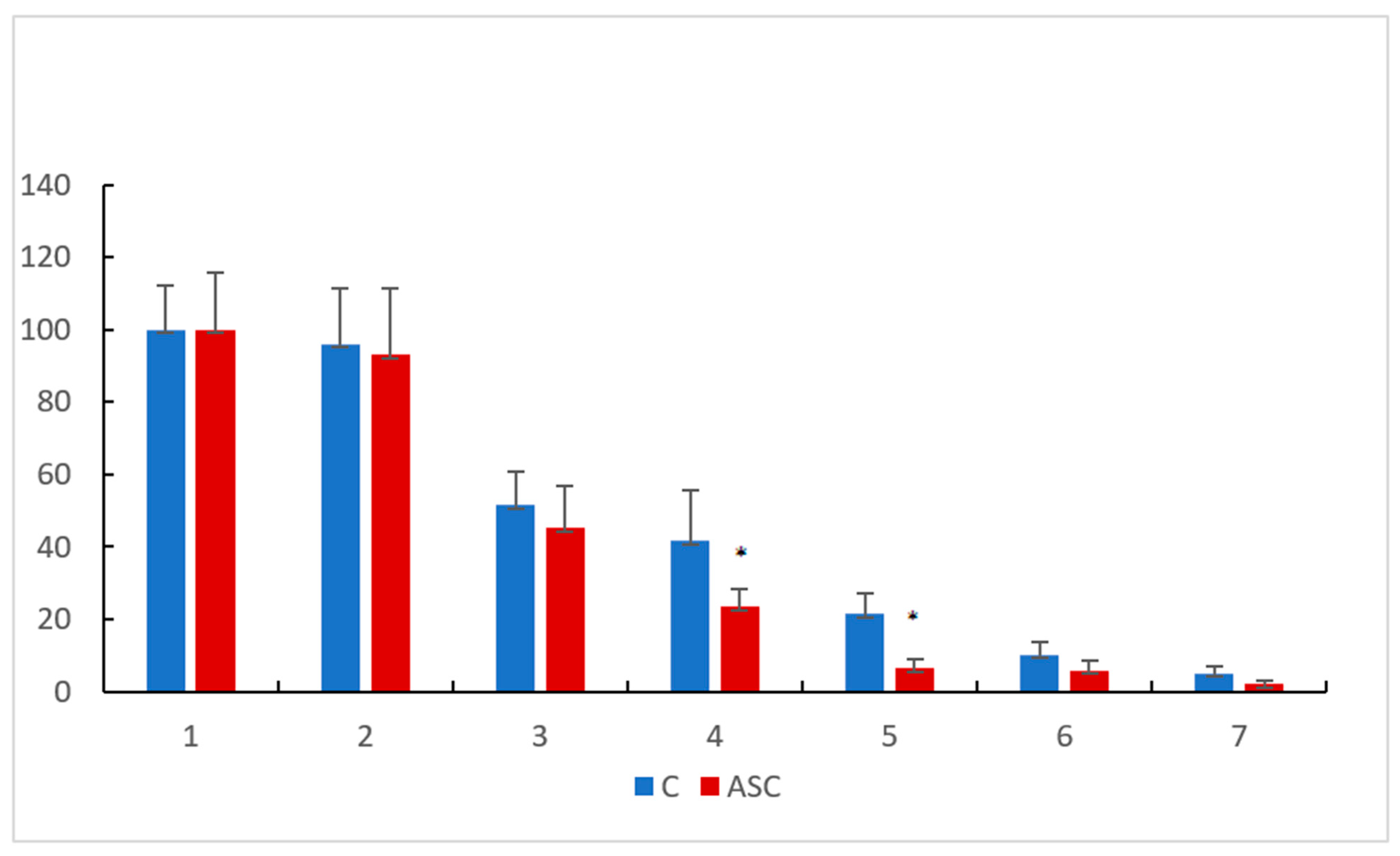
3.1. Superficial Damage
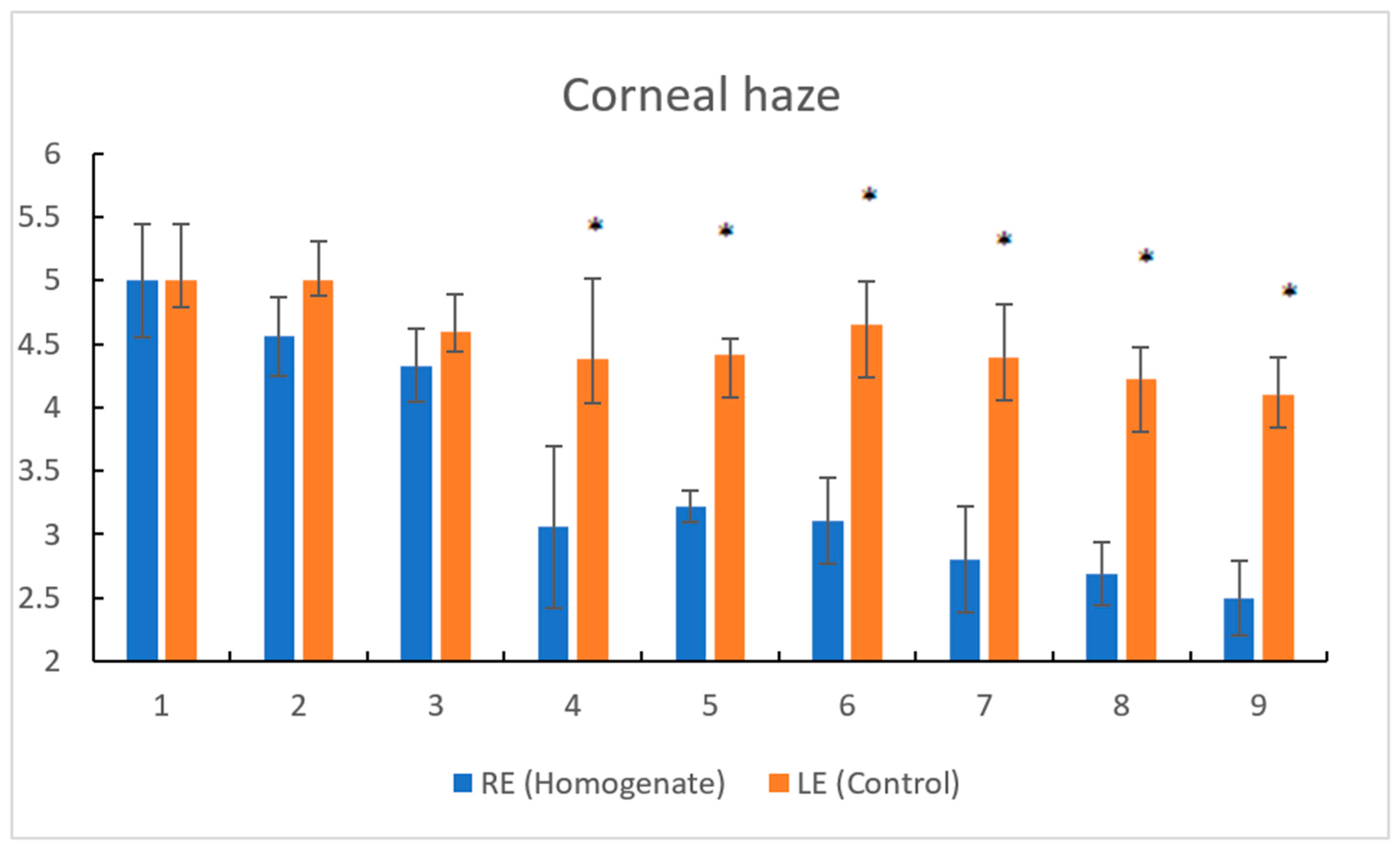
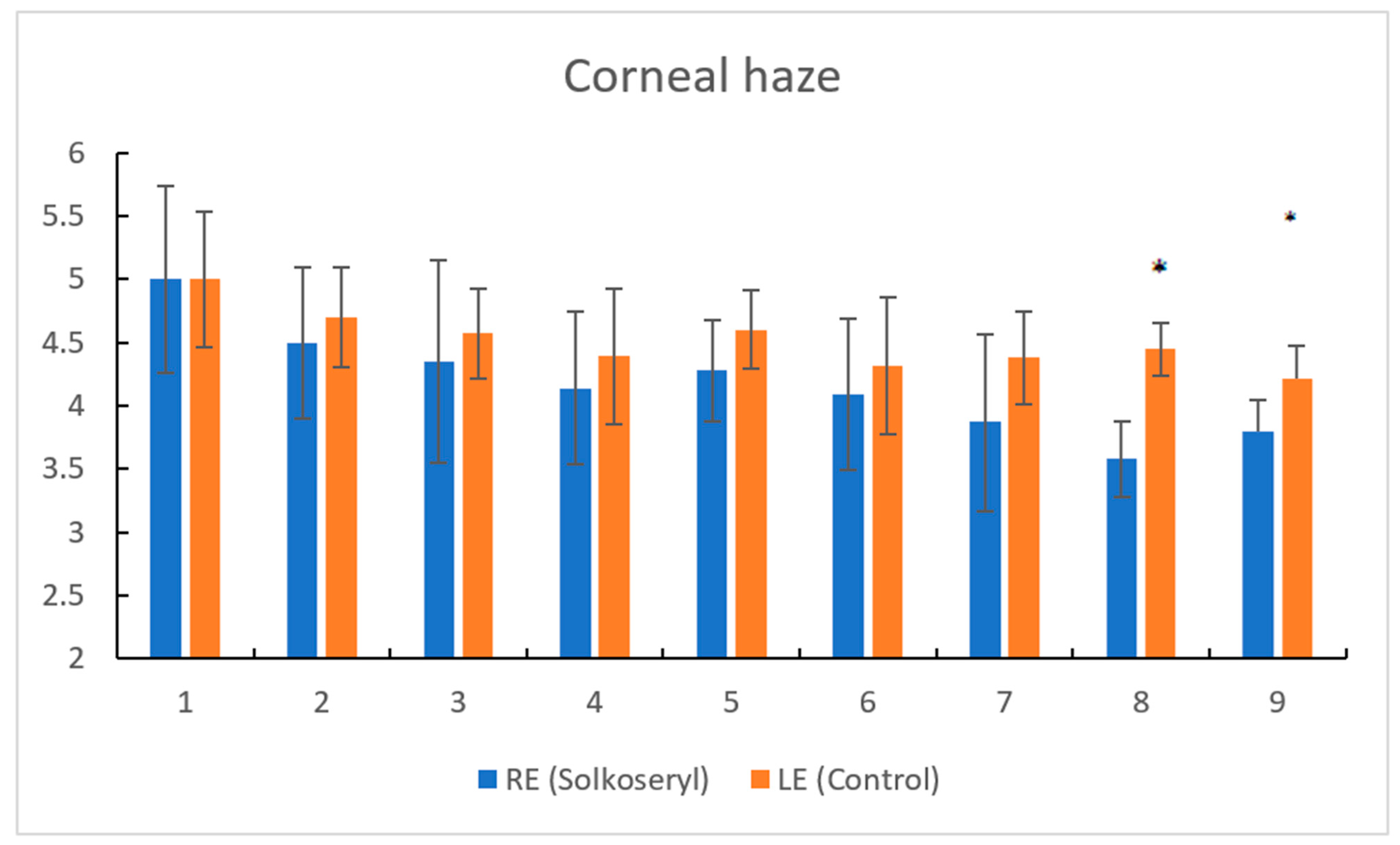
The results in a superficial damage group were averaged, assessing the reduction in the surface of the wound over time. Pictures of eyes in the superficial denaturation model are available in Supplementary Materials File S1. Corneal healing treated by the investigated preparations followed on the second day and on the third day in the control group. No opacity formation was observed in any cornea after a period of epithelial regeneration. In the study, the surface area of the epithelium of the damaged cornea was measured and stained with fluorescein after illumination with a UV lamp operating at 485 nm. After the first day of the preparation application, no differences between the groups were observed, and the measured damage was close to the initial value. However, already 48 h after the eye drops, a decrease in the surface area of the damaged cornea was observed to the appropriate level: 45% in the ASC group and 52% in the control group compared to the initial damage. The drop in the surface area lasted until the end of the experiment, i.e., until day 7, when complete healing of the epithelium was observed. Between the third and seventh days, an average smaller surface area of the ASC-recipient lesion was observed in the group receiving ASC (Figure 1).

3.2. Deep Damage

In the case of heptanol-induced corneal opacity, the use of ASC resulted in a marked improvement in clarity. Pictures of eyes in the deep denaturation model are available in Supplementary Materials (File S2—Solcoseryl and File S3—ASC). According to the assumed 5-stage corneal opacity score scale, where 1—completely clear and 5—completely opaque, the first statistically significant changes were observed already after 4 weeks of administration (Figure 2). The clarity in comparison to the initial value increased by about two units with an average value of 3.06. It should be noted that the improvement in clarity was observed after the second week of administration of ACS, but these changes were not statistically significant. The use of eye drops with Solcoseryl after the fourth week of administration brought only moderate improvement (to about 2.5) (Figure 3). The observed regeneration after ASC application was much more effective compared to the Solcoseryl administration. In his case, a reduction in corneal damage was observed only after eight weeks of application, and the final value of corneal haze was as high as 3.8.
The observed ASC regenerative activity is similar to that of amniotic membrane mesenchymal bone marrow stem cells. For alkali injury (0.5 N NaOH) in rats, Jiang et al. observed an improvement in corneal clarity only in the 10th week of use of the examined maternal cells. This improvement was approximately 1.5 points on a comparative scale for corneal opacity [16]. The effect of accelerating the regeneration of corneal damage (mechanically scraped), by bone marrow-derived mesenchymal stem cells was observed by Lan et al. The acceleration of the restoration of corneal transparency using MSCs was demonstrated by elevated levels of hepatocyte growth factor after mechanical removal of corneal epithelium and anterior stroma [17].

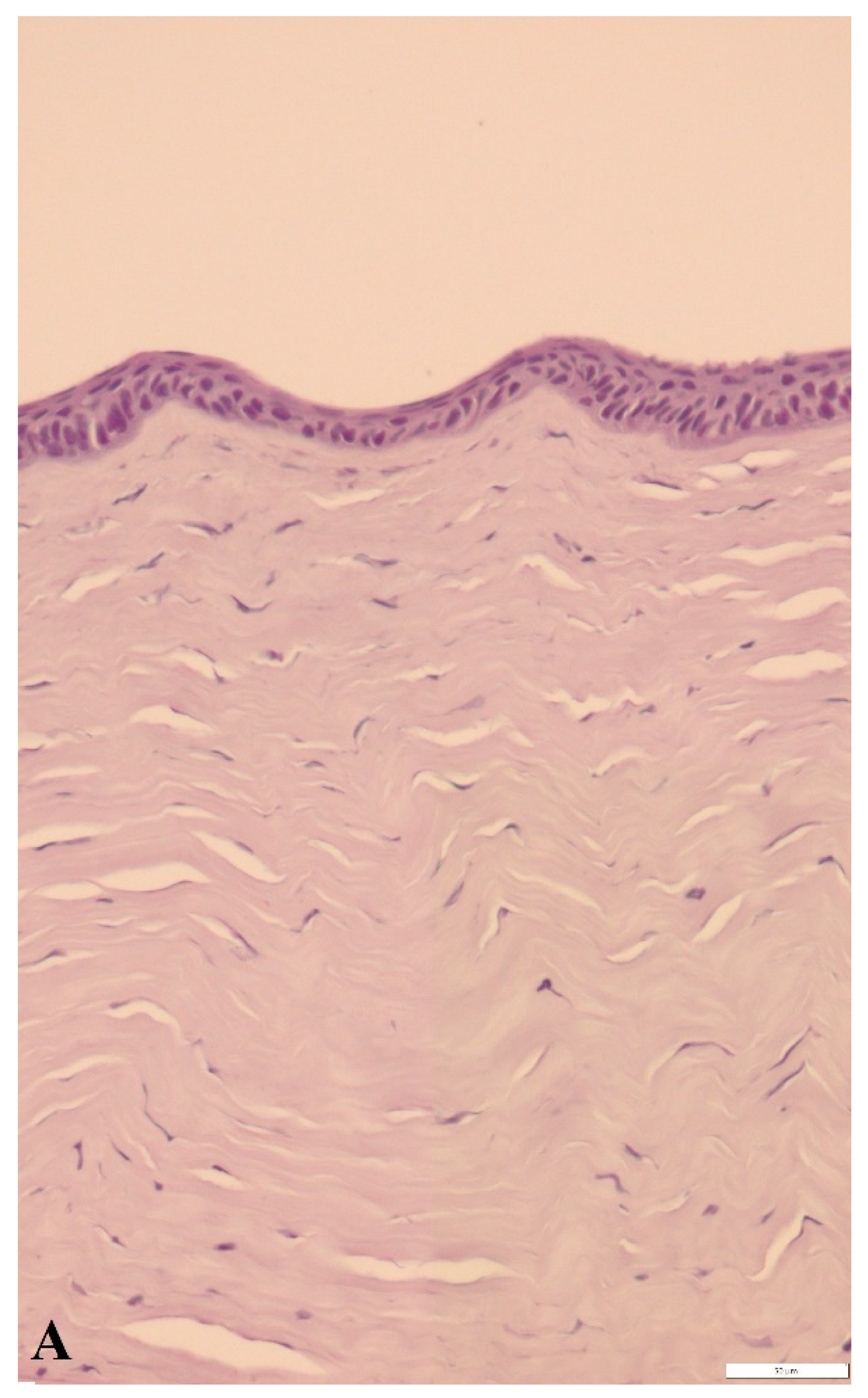
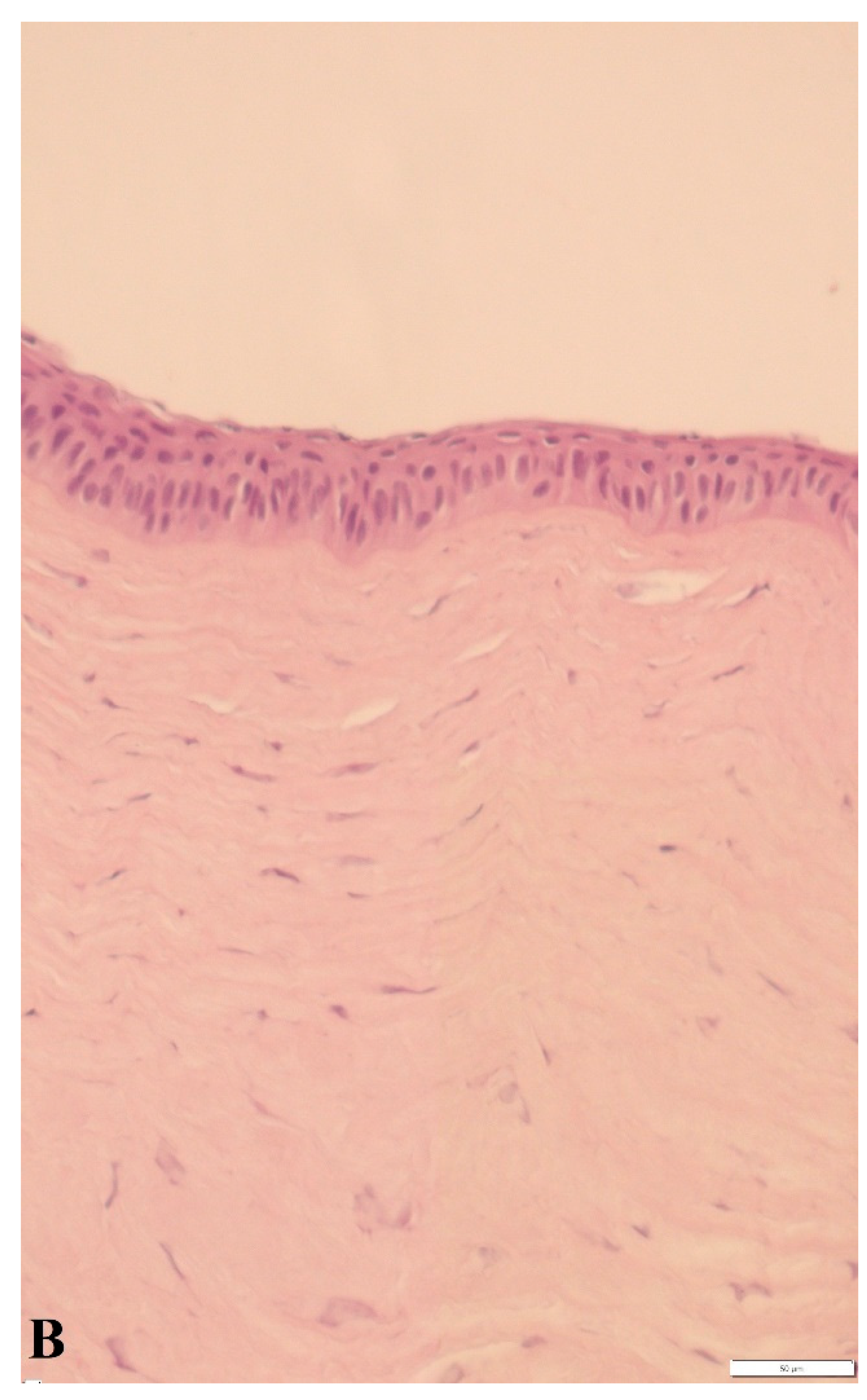
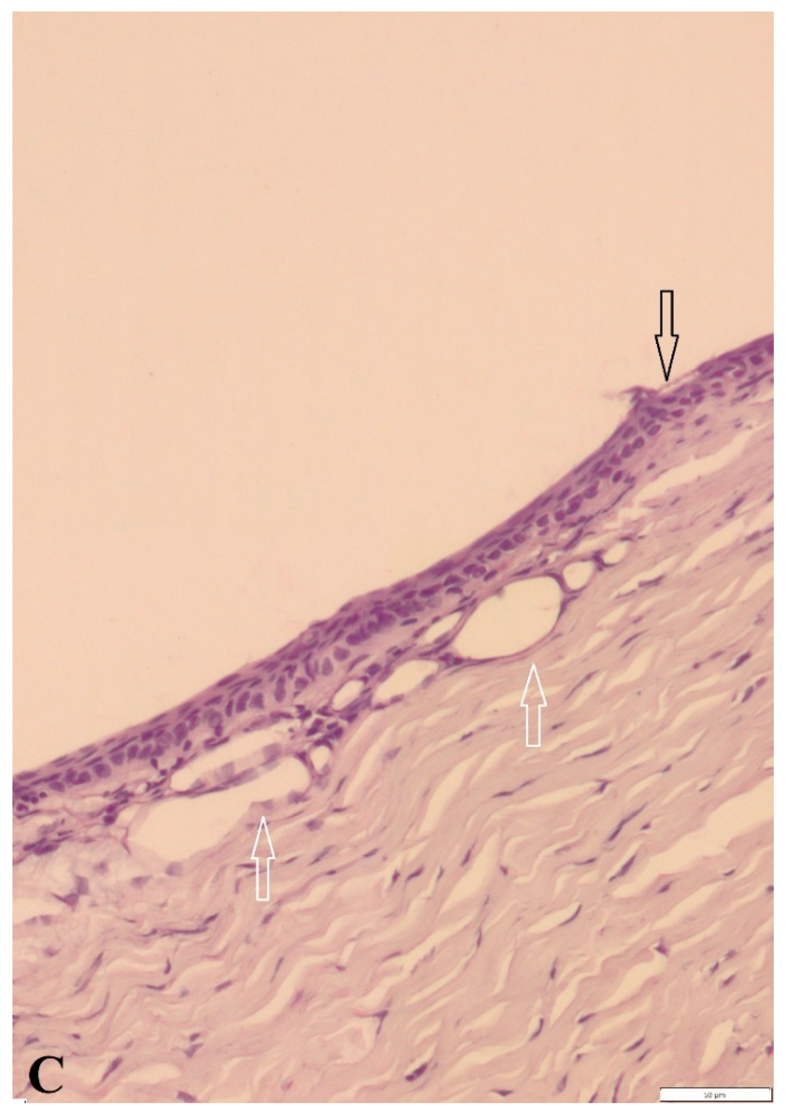
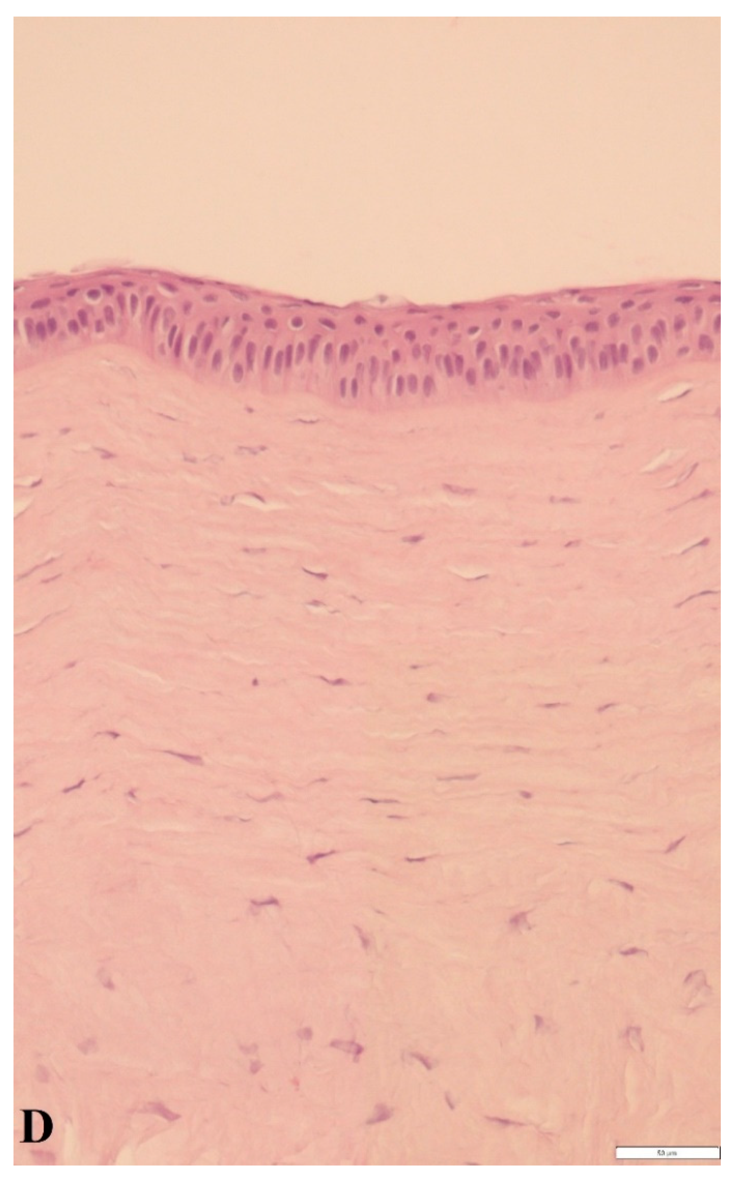
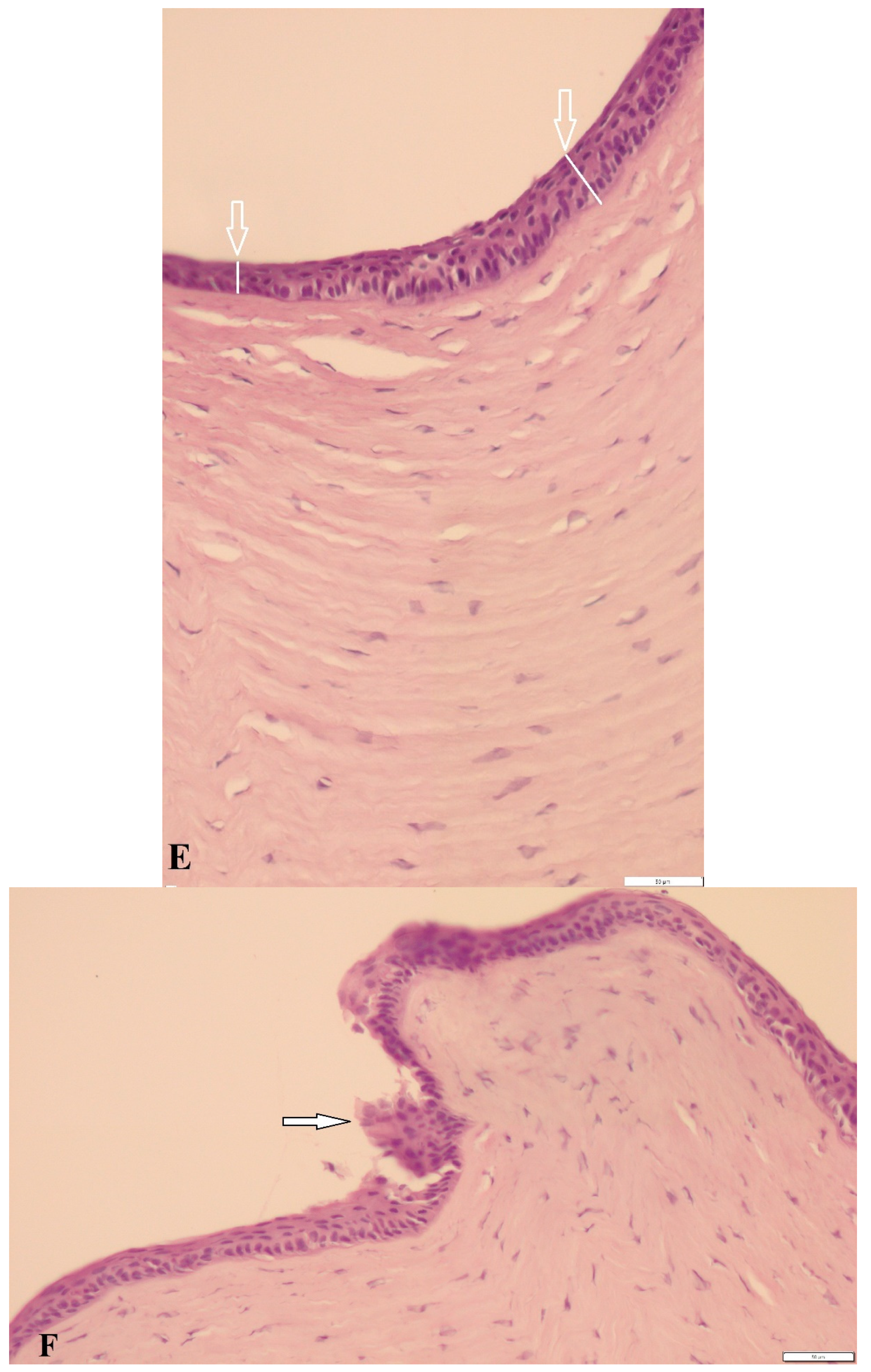
3.3. Histological Findings and Epidermal Thickness

For superficial corneal epithelial damage, histopathological examination on day 7 of the experiment showed no difference between the two study groups. The application of both ASC and the vehicle had an identical effect on the corneal epithelial pattern. Only trace damage to the surface of the anterior corneal epithelium was noted in the microscopic image. This study confirms the healing rate result shown in Figure 4 This effect is consistent with the results presented by Li et al. [4] where the healing of the corneal epithelial cells after alcohol burn was studied.
In the deep denaturation model group, the changes were more diverse: significant epithelial damage, corneal stromal dilation, or anterior epithelial detachment was observed. After 9 weeks of the experiment (deep denaturation model), both the homogenate and Solcoseryl groups showed more anterior corneal epithelial thickening compared with the control group, and the difference was statistically significant (Table 3). The average epithelial thickness was 20% higher with ASC and over 42% higher with Solcoseryl compared to the control group. The phenomenon of corneal changing after chemical (NaOH) burn was described by Reim et al. [18].

4. Discussion

One of the most serious corneal injuries is burns. Chemical injury of the eye may produce extensive damage to the ocular surface epithelium, cornea, and anterior segment, resulting in permanent visual impairment. Immediately following chemical injury, it is important to estimate and clinically grade the severity of limbal stem cell injury and intraocular penetration of the noxious agent [11]. Immediate therapy is directed toward prompt irrigation and removal of any remaining reservoir of chemical contact with the eye. Initial medical therapy is directed at promoting re-epithelialization and transdifferentiation of the ocular surface, augmenting corneal repair by supporting keratocyte collagen production and minimizing ulceration related to collagenase activity, and controlling inflammation [19]. At the 1-month follow-up, it was found that systemically transplanted MSCs could implant into the injured cornea to accelerate wound healing through differentiation, proliferation, and synergy with hematopoietic stem cells [20].
Human limbal niche cells (LNC) or bone-marrow-derived mesenchymal stem cells (BMMSC) were used to prevent limbal stem cell deficiency (LSCD) in an alkali burn rabbit model, and their results were compared. In contrast to the control group, the severity of LSCD in both the LNC and BMMSC transplantation groups was dramatically reduced as shown by smaller epithelial cell defects, decreased fluorescein sodium staining, decreased neovascularization, and decreased goblet cell density. Interestingly, the LNC group was shown to more effectively prevent LSCD than the BMMSC group [21].
The effect of cultured human adipose tissue-derived stem cells on the regeneration of the rabbit cornea after alkaline chemical burn was studied, using human adipose tissue-derived stem cells as the source material. Immediately after the chemical burn, the experimental eye received a single subconjunctival injection of a stem cell suspension. Transplantation of cultured human adipose tissue-derived stem cells as a treatment for chemical corneal burn was found to promote cell renewal and help repair damage to the rabbit cornea [22].
Acute phase treatment includes a broad spectrum topical antibiotic, cycloplegic, and antiglaucoma therapy [23]. Oral treatment with doxycycline was found to be very effective in preventing corneal angiogenesis and inflammation of alkali-burned corneas [24].
Positive results in oxidative disorder management have been achieved with tear supplements, particularly based on hyaluronic acid, topically administered corticosteroids, and prophylactic antibiotic use. Chemically modified and cross-linked hyaluronan gels improved corneal wound healing after alkali injury in rabbits. The wound closure rate and thickness of the corneal epithelium in eyes treated with hyaluronan gel were significantly greater than in untreated injured eyes.
Acceleration of corneal healing can occur after administration of low-molecular organic compounds [6]. High solution of retinol palmitate to treat corneal damage with n-heptanol. However, effective treatment required eye drops six times a day at a dose of over 1000 mU/mL. At the same time, complete healing of the cornea was observed on the 12th day after the damage.
Burgalassi et al. [7]. showed that a polysaccharide fraction isolated from tamarind (Tama-rindus indica L.) seeds slightly but significantly increased the wound healing rate caused by surface alcohol damage.
Sekundo et al. examined the effect of antioxidants (allopurinol and acetylcysteine) and corticosteroids (prednisolone acetate) in eye drops in the healing of corneal alkali burns [25]. These drugs were effective particularly in the early treatment of experimental corneal burns. Saud et al. described a favorable effect of subconjunctival injection of triamcinolone after corneal alkali burns in rabbits [26]. Triamcinolone was very effective for the treatment of acute ocular burn because it reduced the corneal inflammatory process, opacity, and vascularization. It was also checked in transdifferentiated bone marrow or hair follicle stem cells [27]. One therapeutic option is to use drops from the patient’s own serum. Human serum contains a physiological plethora of bioactive elements naturally released by activated platelets, which might have a significant effect on the regeneration of corneal layers by stimulating cell growth [28]. Calf blood dialysate is also used to accelerate wound healing of the skin, mucosa, and cornea (Solcoseryl) [29].
The effect confirming the regenerative properties of the substance is the acceleration of the growth rate of the corneal epithelium compared to the control group. The turned-over of epithelium cells could approximate from 7 to 10 days. Although multiplication and acquisition of human-derived maternal cells is possible and has good therapeutic results, it is expensive and requires constant cooperation with the cell culture laboratory. Standardization of such obtained cells is also problematic. An additional obstacle of the above-mentioned cellular therapies is the necessity of many days of hospitalization of the treated patient. These problems are solved by administering standardized stem cells in eye drops grown from deer antlers. Multiplied laboratory stem cells are used to treat corneal damage. They may come from a healthy eye, the same patient, the same donor, as well as from the oral mucosa [30]. Proven usability of bulge-derived, immature dental pulp, umbilical cord, induced pluripotent, mesenchymal, or embryonic stem cells are successful in the treatment of corneal lesions. In 2020, Qiu et al. adipose-tissue-derived stem cells were used to treat model rabbit n-heptanol corneal injury [31]. They proved that ocular surface reconstruction can be improved by using applied stem cells. Corneal wounds can be angled by using maternal cells from the intact eye. Gimeo et al. applied autologous limbal epithelial cell transplantation improved after corneal injury with alkaline [32]. Long-term observation of the effects of limbal stem cell treatment on corneal damage with heptanol showed that the corneal healing effect persisted even after one year [33]. In the conducted experiment, we noticed a statistically significant increase in the mean thickness of the anterior corneal epithelium for the group treated with the homogenate of deer antler stem cells. For the homogenate group, it was the mean value of 19.2, compared to the value of 15.2 in the control group. It should also be emphasized here that there were no statistically significant differences in epithelial thickness between the homogenate and Solcoseryl groups. One idea for a new therapy is trying to use stem cells from the antlers of the deer Cervus elaphus [1].
The renewal of the corneal and regeneration is mediated by the limbus stem cells that are located in the thick zone between the bulbar conjunctiva and the cornea [34]. Recent research has proven that the regenerative action of deer stem cells is connected with pleiotrophin as a multifunctional growth factor [35]. Wang et al. proved that deer antler cells belong to a novel type of cells that sustain full regeneration of mammalian organs [36].
There are the apparent lack of any specific “antlerogenic molecular mechanisms” suggesting that the secret of deers’ ability to regenerate antlers lies in the particular cues to which multipotential progenitor/stem cells in an antler’s “regeneration territory” are exposed. This in turn suggests that with appropriate manipulation of the environment, pluripotential cells in other adult mammalian tissues could be stimulated to increase the healing capacity of organs, even if not to regenerate them completely. The benefits of increasing individuals’ own capacity for regeneration and repair are self-evident [37].
Comparisons between the healing process of the stumps of an amputated mouse limb and early regeneration of antlers suggest that the stump of a mouse limb cannot regenerate because of the limited potential of periosteal cells in long bones to proliferate [4].
Healing of the casting wound in deers typically involves no or only minor scarring, making antlers interesting subjects for researchers attempting to reduce scar formation during wound healing in humans, especially in the cornea. Additionally, despite the enormous growth rate, the antlers appear to be resistant to malignant transformation, which furthermore offers research opportunities for cancer biology [38].
Antler stem cells (ASC) effectively accelerated the rate of wound closure and improved the quality of skin wound healing in rats, which may be due to the transformation of wound skin fibroblasts into their fetal counterparts. Therefore, they could potentially be developed as a novel therapy for scarless wound healing [39].
In a rat model of CCl4-induced liver fibrosis, ASCs were shown to effectively alleviate liver fibrosis and inhibit activation of hepatic stellate cells HSCs. In the future, ASCs may serve as a new source of stem cells for the treatment of liver fibrosis in the clinic [40].
The homogenate of antlerogenic stem cells has been checked by Kiełbowicz et al., 2020 [41]. In this study, the authors proved the acceleration of corneal healing in mechanical lesion (done with trephine) in rabbits. Additionally, the ACS weakened or arrested the development of side effects.
Dong et al. identify key pathways, molecular/cellular functions, and top-down regulators involved in mammalian organ regeneration. Antler regeneration is known to be initiated and sustained by neuron-derived stem cells in different states of activation. Therefore, antler stem cells can be used as a model to study the proteins and pathways involved in maintaining the stem cell niche and their activation and differentiation during organ formation. In this study, the MSC markers CD73, CD90, and CD105 were examined within the antler tip. The central role of stem cell activation in the development of this mammalian organ was confirmed by the localization of MSC markers within the antler growth center. The highest number of unique proteins (87) was found in the growth center. Antler stem cell activation has been implicated in multiple biological processes and signaling pathways, such as Hippo and canonical Wnt signaling [42].
Li et al. identified proteins that were differentially expressed between antler stem cells and somatic cells (facial periosteum). They found activation of several molecular pathways (PI3K/Akt, ERK/MAPK, p38 MAPK, etc.) during proliferation. The transcription factors POU5F1, SOX2, NANOG, and MYC, which are key markers of embryonic stem cells, were also expressed. Expression of these proteins was confirmed in both cultured cells and fresh tissues. Thus, the molecular pathways and transcription factors identified in the study are common to embryonic and adult stem cells. However, the expression of embryonic stem cell transcription factors would suggest that antler stem cells are potentially an intermediate stem cell type between embryonic cells and more specialized tissue-specific stem cells, such as those residing in muscle, fat, or derived from the haematopoietic system [2].
Recent studies have shown that rapid elongation of the main bundle and antler branches is a controlled form of tumor growth, regulated by the TP73 and ADAMTS18 genes. Osteoclastogenesis as well as osteogenic and chondrogenic differentiation are also involved in this process. As a unique stem-cell-based organ regeneration process in mammals, deer antlers provide a first-rate model system to study regeneration mechanisms in mammalian tissues. Novel ASCs could provide cell-based therapies for regenerative medicine and bone remodeling for clinical applications [43,44].
Rabbit limbal stem cells (LSCs) were studied in an animal model of limbal stem cell deficiency (LSCD). During in vitro culture, LSCs underwent epithelial-mesenchymal transformation. Mesenchymal LSCs were shown to be a highly proliferative population and capable of restoring the corneal epithelium in rabbits. On the 90th day after transplantation of mesenchymal-like LSCs to rabbits with previously formed LSCD, normal corneal epithelium was restored; vascularization was absent; and goblet cells were absent. In the control group, opacity and neovascularization of the stroma was observed [45].
Li et al. showed that also human limbal niche cells or bone-marrow-derived mesenchymal stem cells can be used to prevent limbal stem cell deficiency (LSCD) in an alkali burn rabbit model. Human limbal niche cells are a more potent source than bone marrow-derived mesenchymal stem cells in preventing LSCD in the rabbit alkali burn model [21].
A greater understanding of the mechanisms that regulate the regeneration of antlers may provide a valuable insight to aid the field of regenerative medicine.

5. Conclusions

In conclusion, the present preliminary study shows the promising results of antlerogenic stem cells of Cervus elaphus topically applied for the treatment of corneal injury in a New Zealand rabbit model. In case of superficial corneal damage by n-heptanol, a reduction in the area of damaged cornea was observed from day 3, in comparison with the hyaluronic acid composition. Corneal haze was reduced significantly effectively when ASC was applied, in comparison with Solcoseryl. A deeper understanding of the developmental mechanisms involved in antler renewal can be useful for controlling regeneration cornea processes.

Supplementary Materials

The following supporting information can be downloaded at: https://www.mdpi.com/article/10.3390/app12052468/s1, File S1: Pictures of Eyes Superficial Denaturation; File S2: Deep Denaturation—Solcoseryl; File S3: Deep Denaturation—ASC.

Author Contributions

W.D. and M.B.: conceptualization, investigation. D.S.: conceptualization, writing—review and editing, investigation, methodology, data curation; S.D.: histological analysis. A.S. (Adam Szeląg): coordination. A.S. (Antoni Szumny): writing—review and editing, data. All authors have read and agreed to the published version of the manuscript.

Funding

This research received no external funding.

Institutional Review Board Statement

All experimental procedures used in this study were approved by the II Local Ethics Committee in Wroclaw, Poland (permission No 7/2011).

Informed Consent Statement

Not applicable.

Data Availability Statement

The data are available in Department of Pharmacology of Wrocław Medical University and Department of Pathology in Wroclaw University of Environmental and Life Sciences.

Conflicts of Interest

The authors declare no conflict of interest.

References

  1. Ba, H.; Wang, D.; Li, C. MicroRNA profiling of antler stem cells in potentiated and dormant states and their potential roles in antler regeneration. Mol. Genet. Genom. 2016, 291, 943–955. [Google Scholar] [CrossRef] [PubMed]
  2. Li, C.; Harper, A.; Puddick, J.; Wang, W.; McMahon, C. Proteomes and Signalling Pathways of Antler Stem Cells. PLoS ONE 2012, 7, e30026. [Google Scholar] [CrossRef] [PubMed] [Green Version]
  3. Rolf, H.J.; Kierdorf, U.; Kierdorf, H.; Schulz, J.; Seymour, N.; Schliephake, H.; Napp, J.; Niebert, S.; Wölfel, H.; Wiese, K.G. Localization and characterization of STRO-1 cells in the deer pedicle and regenerating antler. PLoS ONE 2008, 3, e2064. [Google Scholar] [CrossRef] [Green Version]
  4. Lee, H.S.; Lee, J.H.; Kim, C.E.; Yang, J.W. Anti-neovascular effect of chondrocyte-derived extracellular matrix on corneal alkaline burns in rabbits. Graefe’s Arch. Clin. Exp. Ophthalmol. 2014, 252, 951–961. [Google Scholar] [CrossRef] [PubMed]
  5. Zhang, W.; Ke, C.-H.; Guo, H.-H.; Xiao, L. Antler stem cells and their potential in wound healing and bone regeneration. World J. Stem Cells 2021, 13, 1049–1057. [Google Scholar] [CrossRef]
  6. Lwigale, P.Y. Corneal development: Different cells from a common progenitor. Prog. Mol. Biol. Transl. Sci. 2015, 134, 43–59. [Google Scholar] [PubMed]
  7. Shi, L.; Chang, Y.; Yang, Y.; Zhang, Y.; Yu, F.-S.X.; Wu, X. Activation of JNK signaling mediates connective tissue growth factor expression and scar formation in corneal wound healing. PLoS ONE 2012, 7, e32128. [Google Scholar] [CrossRef] [Green Version]
  8. Willmann, D.; Fu, L.; Melanson, S.W. Corneal Injury; StatPearls: Treasure Island, FL, USA, 2021. [Google Scholar]
  9. Ljubimov, A.V.; Saghizadeh, M. Progress in corneal wound healing. Prog. Retin. Eye Res. 2015, 49, 17–45. [Google Scholar] [CrossRef] [Green Version]
  10. Holan, V.; Trosan, P.; Cejka, C.; Javorkova, E.; Zajicova, A.; Hermankova, B.; Chudickova, M.; Cejkova, J. A comparative study of the therapeutic potential of mesenchymal stem cells and limbal epithelial stem cells for ocular surface reconstruction. Stem Cells Transl. Med. 2015, 4, 1052–1063. [Google Scholar] [CrossRef] [PubMed]
  11. Hamill, C.E.; Bozorg, S.; Chang, H.-Y.P.; Lee, H.; Sayegh, R.R.; Shukla, A.N.; Chodosh, J. Corneal alkali burns: A review of the literature and proposed protocol for evaluation and treatment. Int. Ophthalmol. Clin. 2013, 53, 185–194. [Google Scholar] [CrossRef]
  12. Saghizadeh, M.; Kramerov, A.A.; Svendsen, C.N.; Ljubimov, A.V. Concise review: Stem cells for corneal wound healing. Stem Cells 2017, 35, 2105–2114. [Google Scholar] [CrossRef] [Green Version]
  13. Cegielski, M.; Dziewiszek, W.; Zabel, M.; Dzięgiel, P.; Iżycki, D.; Zatoński, M.; Bochnia, M. Experimental application of xenogenous antlerogenic cells in replacement of auricular cartilage in rabbits. Xenotransplantation 2008, 15, 374–383. [Google Scholar] [CrossRef]
  14. Chung, J.-H.; Kim, W.-K.; Lee, J.-S.; Pae, Y.-S.; Kim, H.-J. Effect of topical Na-hyaluronan on hemidesmosome formation in n-heptanol-induced corneal injury. Ophthalmic Res. 1998, 30, 96–100. [Google Scholar] [CrossRef]
  15. Gupta, N.; Kalaivani, M.; Tandon, R. Comparison of prognostic value of Roper Hall and Dua classification systems in acute ocular burns. Br. J. Ophthalmol. 2011, 95, 194–198. [Google Scholar] [CrossRef] [PubMed]
  16. Jiang, T.-S.; Cai, L.; Ji, W.-Y.; Hui, Y.-N.; Wang, Y.-S.; Hu, D.; Zhu, J. Reconstruction of the corneal epithelium with induced marrow mesenchymal stem cells in rats. Mol. Vis. 2010, 16, 1304–1316. [Google Scholar] [PubMed]
  17. Mittal, S.K.; Omoto, M.; Amouzegar, A.; Sahu, A.; Rezazadeh, A.; Katikireddy, K.R.; Shah, D.I.; Sahu, S.K.; Chauhan, S.K. Restoration of Corneal Transparency by Mesenchymal Stem Cells. Stem Cell Rep. 2016, 7, 583–590. [Google Scholar] [CrossRef] [Green Version]
  18. Reim, M.; Bahrke, C.; Kuckelkorn, R.; Kuwert, T. Investigation of enzyme activities in severe burns of the anterior eye segment. Graefe’s Arch. Clin. Exp. Ophthalmol. 1993, 231, 308–312. [Google Scholar] [CrossRef] [PubMed]
  19. Wagoner, M.D. Chemical injuries of the eye: Current concepts in pathophysiology and therapy. Surv. Ophthalmol. 1997, 41, 275–313. [Google Scholar] [CrossRef]
  20. Ye, J.; Yao, K.; Kim, J.C. Mesenchymal stem cell transplantation in a rabbit corneal alkali burn model: Engraftment and involvement in wound healing. Eye 2006, 20, 482–490. [Google Scholar] [CrossRef] [Green Version]
  21. Li, G.; Zhang, Y.; Cai, S.; Sun, M.; Wang, J.; Li, S.; Li, X.; Tighe, S.; Chen, S.; Xie, H.; et al. Human limbal niche cells are a powerful regenerative source for the prevention of limbal stem cell deficiency in a rabbit model. Sci. Rep. 2018, 8, 6566. [Google Scholar] [CrossRef]
  22. Lin, H.F.; Lai, Y.C.; Tai, C.F.; Tsai, J.L.; Hsu, H.C.; Hsu, R.F.; Lu, S.N.; Feng, N.H.; Chai, C.Y.; Lee, C.H. Effects of cultured human adipose-derived stem cells transplantation on rabbit cornea regeneration after alkaline chemical burn. Kaohsiung J. Med. Sci. 2013, 29, 14–18. [Google Scholar] [CrossRef] [Green Version]
  23. Singh, P.; Tyagi, M.; Kumar, Y.; Gupta, K.; Sharma, P. Ocular chemical injuries and their management. Oman J. Ophthalmol. 2013, 6, 83–86. [Google Scholar] [CrossRef]
  24. Cejkova, J.; Trosan, P.; Cejka, C.; Lencova, A.; Zajicova, A.; Javorkova, E.; Kubinova, S.; Sykova, E.; Holan, V. Suppression of alkali-induced oxidative injury in the cornea by mesenchymal stem cells growing on nanofiber scaffolds and transferred onto the damaged corneal surface. Exp. Eye Res. 2013, 116, 312–323. [Google Scholar] [CrossRef]
  25. Sekundo, W.; Augustin, A.; Strempel, I. Topical allopurinol or corticosteroids and acetylcysteine in the early treatment of experimental corneal alkali burns: A pilot study. Eur. J. Ophthalmol. 2002, 12, 366–372. [Google Scholar] [CrossRef]
  26. Saud, E.E.; Moraes Jr, H.V.; Marculino, L.G.; Gomes, J.A.P.; Allodi, S.; Miguel, N.C. Clinical and histopathological outcomes of subconjunctival triamcinolone injection for the treatment of acute ocular alkali burn in rabbits. Cornea 2012, 31, 181–187. [Google Scholar] [CrossRef]
  27. Eberwein, P.; Reinhard, T. Current and future therapeutic options in limbal stem cell insufficiency. Klinische Monatsblatter fur Augenheilkunde 2012, 229, 1178–1184. [Google Scholar]
  28. De Pascale, M.R.; Lanza, M.; Sommese, L.; Napoli, C. Human serum eye drops in eye alterations: An insight and a critical analysis. J. Ophthalmol. 2015, 2015, 396410. [Google Scholar] [CrossRef] [Green Version]
  29. Konturek, S.J.; Drozdowicz, D.; Pytko-Polonczyk, J.; Brzozowski, T.; Bielanski, W. Solcoseryl in prevention of stress-induced gastric lesions and healing of chronic ulcers. J. Physiol. Pharmacol. 1991, 42, 73–84. [Google Scholar] [PubMed]
  30. Bains, K.K.; Fukuoka, H.; Hammond, G.M.; Sotozono, C.; Quantock, A.J. Recovering vision in corneal epithelial stem cell deficient eyes. Contact Lens Anterior Eye 2019, 42, 350–358. [Google Scholar] [CrossRef] [Green Version]
  31. Qiu, T.; Cui, L.; Xu, J.-J.; Hong, J.-X.; Xiang, J. Reconstruction of the ocular surface by autologous transplantation of rabbit adipose tissue-derived stem cells on amniotic membrane. Ann. Transl. Med. 2020, 8, 1062. [Google Scholar] [CrossRef] [PubMed]
  32. Gimeno, F.L.; Lavigne, V.; Gatto, S.; Croxatto, J.O.; Correa, L.; Gallo, J.E. Advances in corneal stem-cell transplantation in rabbits with severe ocular alkali burns. J. Cataract Refract. Surg. 2007, 33, 1958–1965. [Google Scholar] [CrossRef] [PubMed]
  33. Ti, S.-E.; Anderson, D.; Touhami, A.; Kim, C.; Tseng, S.C. Factors affecting outcome following transplantation of ex vivo expanded limbal epithelium on amniotic membrane for total limbal deficiency in rabbits. Investig. Ophthalmol. Vis. Sci. 2002, 43, 2584–2592. [Google Scholar]
  34. Pellegrini, G.; De Luca, M. Eyes on the prize: Limbal stem cells and corneal restoration. Cell Stem Cell 2014, 15, 121–122. [Google Scholar] [CrossRef] [PubMed] [Green Version]
  35. Dong, Z.; Li, C.; Coates, D. PTN−PTPRZ signalling is involved in deer antler stem cell regulation during tissue regeneration. J. Cell. Physiol. 2021, 236, 3752–3769. [Google Scholar] [CrossRef] [PubMed]
  36. Wang, D.; Berg, D.; Ba, H.; Sun, H.; Wang, Z.; Li, C. Deer antler stem cells are a novel type of cells that sustain full regeneration of a mammalian organ—Deer antler. Cell Death Dis. 2019, 10, 443. [Google Scholar] [CrossRef] [Green Version]
  37. Price, J.; Faucheux, C.; Allen, S. Deer antlers as a model of Mammalian regeneration. Curr. Top. Dev. Biol. 2005, 67, 1–48. [Google Scholar] [PubMed]
  38. Kierdorf, U.; Kierdorf, H. Deer antlers–a model of mammalian appendage regeneration: An extensive review. Gerontology 2011, 57, 53–65. [Google Scholar] [CrossRef]
  39. Rong, X.; Chu, W.; Zhang, H.; Wang, Y.; Qi, X.; Zhang, G.; Wang, Y.; Li, C. Antler stem cell-conditioned medium stimulates regenerative wound healing in rats. Stem Cell Res. Ther. 2019, 10, 326. [Google Scholar] [CrossRef] [PubMed] [Green Version]
  40. Rong, X.; Yang, Y.; Zhang, G.; Zhang, H.; Li, C.; Wang, Y. Antler stem cells as a novel stem cell source for reducing liver fibrosis. Cell Tissue Res. 2020, 379, 195–206. [Google Scholar] [CrossRef] [PubMed] [Green Version]
  41. Kiełbowicz, M.; Kuropka, P.; Cegielski, M.; Kiełbowicz, Z.; Trębacz, P.; Hebel, M.; Aleksiewicz, R. Influence of antlerogenic stem cells on the healing of lesions in the corneal epithelium and corneal stroma in rabbits. Pol. J. Vet. Sci. 2020, 23, 281–290. [Google Scholar] [CrossRef]
  42. Dong, Z.; Haines, S.; Coates, D. Proteomic Profiling of Stem Cell Tissues during Regeneration of Deer Antler: A Model of Mammalian Organ Regeneration. J. Proteome Res. 2020, 19, 1760–1775. [Google Scholar] [CrossRef]
  43. Feleke, M.; Bennett, S.; Chen, J.; Hu, X.; Williams, D.; Xu, J. New physiological insights into the phenomena of deer antler: A unique model for skeletal tissue regeneration. J. Orthop. Transl. 2021, 27, 57–66. [Google Scholar] [CrossRef]
  44. Dąbrowska, N.; Kiełbowicz, Z.; Nowacki, W.; Bajzert, J.; Reichert, P.; Bieżyński, J.; Zebrowski, J.; Haczkiewicz, K.; Cegielski, M. Antlerogenic stem cells: Molecular features and potential in rabbit bone regeneration. Connect. Tissue Res. 2016, 57, 539–554. [Google Scholar] [CrossRef]
  45. Selver, O.B.; Durak, I.; Gürdal, M.; Baysal, K.; Ates, H.; Ozbek, Z.; Wang, Z.; Wu, A.; Wolosin, J.M. Corneal recovery in a rabbit limbal stem cell deficiency model by autologous grafts of tertiary outgrowths from cultivated limbal biopsy explants. Mol. Vis. 2016, 22, 138–149. [Google Scholar]
Figure 1. The surface area (in pixels) of the damaged cornea (Y-axis) vs to time (days, X-axis) staining with fluorescein. The field of primary n-heptanol damage was assumed to be 100%. Red: ASC; blue: control. Number of eyes: 24. *—statistically significant.
Applsci 12 02468 g001
Figure 2. Corneal damage scores (Y-axis) from the start of the experiment to week 9, on a 5-point scale (1—cornea completely clear and 5—cornea completely opaque) in the homogenate-treated group compared to the control. Observations were made at 9 weeks (X-axis). RE—right eye and LE—left eye. *—statistically significant. Number of eyes: 24.
Applsci 12 02468 g002
Figure 3. Corneal damage scores (Y-axis) from the start of the experiment to week 9, on a five-point scale (1—cornea completely clear and 5—cornea completely opaque) in the Solcoseryl-treated group compared to controls. Observations were made at 9 weeks (X-axis). RE—right eye, LE—left eye. *—statistically significant. Number of eyes: 24.
Applsci 12 02468 g003
Figure 4. Microscopic examination of cornea in deep denaturation model. Examples of micrographs: (A,B)—superficial damage to the corneal epithelium, (CF)—severe damage to the corneal epithelium. HE staining, magnification 200×, (A,D)—control, standard, homogeneous anterior corneal epithelium, the correct stroma of the cornea, normal, homogeneous. (B)—ASC group, normal epithelium, slightly thicker than in control. (C)—Solcoseryl, regenerated epithelium, in places with a non-lumpy, slightly damaged surface (black arrow), focal detachment from the corneal essence (white arrows). (E)—ASC group, normal epithelium, non-uniform thickness (white lines and white arrows), stable, normal corneal stroma. (F)—Solcoseryl, epithelium with a visible defect (arrow), areas with a non-rough surface, the stroma of the cornea without changes.
Applsci 12 02468 g004aApplsci 12 02468 g004bApplsci 12 02468 g004cApplsci 12 02468 g004dApplsci 12 02468 g004e
Table 1. Diagram of the experiment. RE—right eye, LE—left eye, ASC—antler stem cells, Lacrimal (sodium hyaluronate in commercial preparation), Vidisic (carbomer in commercial preparation), Solcoseryl (commercial preparation).
GroupSuperficial Denaturation Model
No. 1
12 rabbits
RE—ASC + sodium hyaluronate in drops solution (Hyal-Drop Multi)LE—sodium hyaluronate in drops solution (Hyal-Drop Multi)
Deep denaturation model
No. 2
12 rabbits
RE—ASC + carbomer in gel (Vidisic)LE—carbomer in gel (Vidisic)
No. 3
12 rabbits
RE—Solcoseryl in gelLE—carbomer in gel (Vidisic)
Table 2. Grading of corneal clarity on the basis of corneal haze.
GradeOcular Details
1Cornea completely clear
2Iris details visible
3Pupillary margin visible, iris details not visible
4Pupillary margin not visible
5Cornea completely opaque
Table 3. Mean corneal anterior epithelial thickness (µm) measured by histo-pathology 9 weeks after corneal injury.
Study GroupControl Group
Homogenate19.2 ± 3.615.2 ± 2.8
Solcoseryl20.4 ± 4.313.9 ± 3.5
Publisher’s Note: MDPI stays neutral with regard to jurisdictional claims in published maps and institutional affiliations.

