Article

Does the Behavior of Pediatric Patients towards Dental Treatment Influence the Pattern of Adjacent (Sound, Decayed, and Filled) Proximal Surfaces in Primary Molars?

1
Department of Preventive Dental Sciences, College of Dentistry, Imam Abdulrahman Bin Faisal University, Dammam 31441, Saudi Arabia
2
Dental Internship Program, College of Dentistry, Imam Abdulrahman Bin Faisal University, Dammam 31441, Saudi Arabia
*
Author to whom correspondence should be addressed.
Appl. Sci. 2022, 12(4), 1910; https://doi.org/10.3390/app12041910
Submission received: 30 December 2021 / Revised: 7 February 2022 / Accepted: 7 February 2022 / Published: 11 February 2022
(This article belongs to the Special Issue Applied Sciences in Oral Health and Clinical Dentistry)

Abstract

:
This retrospective study aimed to investigate the influence of patient behavior toward dental treatment on the presence of adjacent (sound, decayed, or filled) proximal surfaces in the primary molars. The study included a records review of 2226 pediatric patients. Records were eligible if the patients were in primary or mixed dentition, and if they included a complete set of bitewings and periapical intraoral radiographs of all their primary molars. Four calibrated reviewers used intraoral radiographs to record the sound, decayed, and filled proximal distal surfaces of the first primary molars and the mesial surfaces of the second primary molars. Demographical variables were reported from patients’ medical records including gender, age, medical history, nationality, and patient behavior. Patients’ behaviors were evaluated using Frankl’s Behavior Rating Scale. The Pearson correlation was used to test the association between the sound, decayed, and filled distal surfaces of the first primary molars and the mesial surface of the second primary molar. High correlations of ≥0.6 were further tested in simple and multiple linear regression models to test the influence of patients’ behaviors on these correlations after adjusting for demographical factors. All tests were performed at 5% significance level A. A total of 1194 records met the inclusion criteria and were analyzed. There was a high positive correlation between the sound, decayed, and filled distal surfaces of the first primary molar and similar surfaces on the mesial of the second primary molars (0.66, 0.61, and 0.60, respectively). Compared to cooperative patients, the adjusted estimate of the mean decayed mesial surface of the second primary molar increased significantly for non-cooperative patients by 0.1 (95% CI = 0.16–0.53). On the contrary, the adjusted estimate of the mean sound mesial surfaces of the second primary molars decreased significantly by 0.09 for non-cooperative patients, compared to those who were cooperative (95% CI = −0.52–−0.15). Patient cooperation did not significantly influence the mean of the filled mesial surfaces of the second primary molars (adjusted B = 0.01, 95% CI = −0.05–0.09). Educating parents and clinicians about the influence of children’s behaviors on oral health is highly encouraged to improve treatment outcomes and reduce the progression of dental caries. The implementation of specific behavior management techniques is also important to reduce dental fear and anxiety.

1. Introduction

Dental caries is considered one of the most common diseases worldwide despite the noticeable increase in oral and dental health awareness [1]. It can lead to pain, suffering, poor oral hygiene, and reduced quality of life for adolescents and adults [2]. Dental caries affected 2.4 billion people in 2017 alone [3]. Caries prevalence is estimated to affect approximately 80% of individuals in developing countries, and approximately 90% of those caries are left untreated [2,3,4,5]. In Saudi Arabia, despite the free dental services provided by the government, the prevalence of dental caries remains high [2]. During the last decade, the prevalence of caries in individuals with primary dentition in Saudi Arabia was reported to reach 95% [6]. In addition, previous studies in Riyadh, Jeddah, and Al-Hassa reported an average (decayed, missing, filled primary teeth) dmft score of approximately six [7,8,9,10,11]. This high prevalence and incidence of dental caries in children could be related to increased sugar consumption, dietary habits, and reduced awareness of oral health and diseases [4]. In fact, most Saudi children are at high risk for dental caries [5] despite the efforts of the Ministry of Health to provide dental care programs, such as the National Initiative, “Prevent Tooth Caries”, that embraces the existing partnership between the Saudi Ministry of Health and the Ministry of Education [12].
Proximal caries is a category of dental caries that develops between two adjacent teeth. It is crucial to detect proximal caries early in the development of primary dentition due to the rapid progression of caries between the adjacent surfaces of the teeth [13]. In comparison to permanent teeth, primary teeth have thinner enamel and dentine, a lesser degree of remineralization, and wider dentinal tubules [13]. The early detection of proximal caries in these teeth is a fundamental component of minimally invasive dentistry because the prevention of caries’ progression to adjacent teeth is less destructive to tooth structures [14]. One method to define proximal caries in clinical studies is the use of the term caries adjacent to restoration (CAR) [15]. In one study, caries was significantly more prevalent in intact proximal surfaces next to recently placed composite posterior proximal restorations than on the contralateral control surface that did not have a restoration in contact (OR = 2.6, CI = 1.2–5.3) [16]. Caries adjacent to caries (CAC) is another method to define proximal caries in clinical studies [17]. A strong association has been reported between the caries status of a tooth and the caries status of adjacent teeth in 3–5-year-old children [17].
Children’s behaviors during dental treatment have a considerable impact on their oral health and the severity of dental caries [18]. They affect the distribution of caries, treatment outcomes, and their oral and systemic health status [19]. There are several factors that influence children’s behaviors, such as their cognitive skills, family, and society, which consequently affect their oral health [20]. Treatment administration during pediatric dental visits can be terminated or interrupted due to children’s negative behaviors, which results in poor oral health outcomes [21]. The influence of children’s behaviors towards dental treatment on the relationship between the adjacent proximal surfaces (sound, decayed, or filled) in primary molars has not been well studied. This study will assist clinical practitioners in the improved management and prevention of the progression of dental caries to achieve more favorable dental treatment outcomes. Therefore, the present study aimed to investigate the influence of pediatric patient behavior on the presence of the adjacent proximal surfaces (sound, decayed, or filled) in the primary molars.

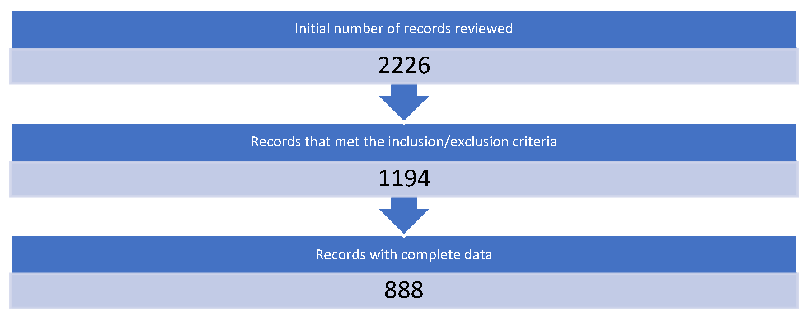
2. Materials and Methods
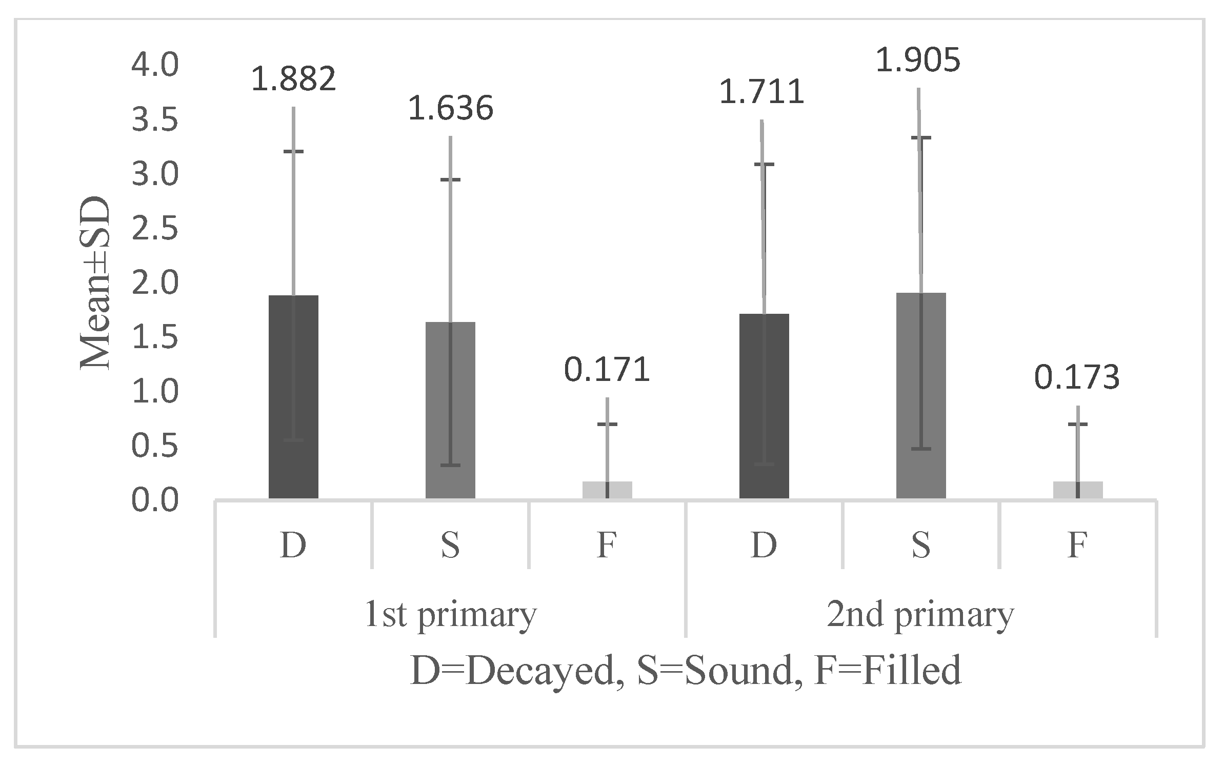
This retrospective study included a records review of pediatric patients who visited the dental hospital at the College of Dentistry at Imam Abdulrahman Bin Faisal University (IAU) in Dammam, Saudi Arabia. The study was conducted in accordance with the Declaration of Helsinki and was approved by the Institutional Review Board at Imam Abdulrahman Bin Faisal University (#2017-02-48).
The records of all patients who visited the dental hospital over a six-year period (2015–2020) were reviewed. For records to be included in the study, they had to meet the following inclusion criteria: (1) records must contain a complete set of bitewings and periapical intraoral radiographs of all the primary molars; (2) patients must be in primary or mixed dentition. Patients with exfoliated primary teeth, trauma that caused tooth loss, missing teeth due to extraction, and patients with syndromes and medical conditions that affect caries patterns or the number of teeth were excluded. The identified radiographs of these patients were examined using a standardized method by means of the MiPACS Dental Enterprise Viewer, software version 3.1.1404 (Medicor imaging, Charlotte, NC, USA). The magnification option in the software was used as needed. Patients’ identifiable information (name and ID) was not recorded, and confidentiality was maintained. The legal guardians of the pediatric patients signed an informed consent form prior to the radiographic examination.
The sample size was calculated prior to the completion of the study. The calculation was based on the following assumptions: confidence intervals = 95%; margin of error = 2%; proportion of the population with caries on the proximal surfaces of the primary molars = 20%; and the total population size = 2000 (https://www.calculator.net/sample-size-calculator, accessed on 29 August 2020. The estimated sample size was 870 records.
A total of four medical and radiographic records reviewers completed three consecutive training and calibration sessions prior to the completion of the study. The training consisted of radiographic cases of randomly selected radiographic records of bitewings and periapical views of the first and second primary molars. Reviewers were asked to record the sound, decayed, or filled proximal distal surfaces of the first primary molars and the mesial surfaces of the second primary molars. Caries was recorded if the proximal radiolucency reached half the thickness of the enamel or more. Caries that reached the pulp and recurrent caries beneath a failed restoration was recorded as caries. Responses were compared to those of a gold standard pediatric dentist using Kappa statistics to determine inter- and intra-examiner reliability.
Demographical data (gender, age, nationality, medical history, and patient behavior) were recorded for each patient based on the information available in their medical records. Significant medical history was considered if the patient had any medical conditions recorded on file, including medications or medication allergies. Patient behavior was recorded based on Frankl’s Behavior Rating Scale (FBRS): definitely negative (−−); negative (−); positive (+); and definitely positive (++) [22]. This instrument has shown satisfactory reliability and validity [23], and it is the routinely used method to report patient behavior in the medical records of all pediatric patients who attend the dental hospital. Children’s behaviors and radiographs were extracted from their initial visit records.
The Statistical Package for the Social Sciences (SPSS), version 25.0.0.2 (IBM Corp., Armonk, NY, USA), was used to analyze the data. The frequencies and percentages were calculated for categorical demographical variables. The means and standard deviations were calculated for patients’ ages and to report the means of the sound, decayed, and filled distal surfaces of the first primary molars and the mesial surfaces of the second primary molars. The association between the sound, decayed, and filled adjacent proximal surfaces of the distal first primary molars and the mesial surfaces of the second primary molars was tested using the Pearson correlation. High correlations of ≥0.6 were further tested in simple and multiple linear regression models to test the influence of patients’ behaviors on these correlations after adjusting for other demographical factors. The outcome variables were the sound, decayed, and filled mesial surfaces of the second primary molars. The main predictors were the sound, decayed, and filled distal surfaces of the first primary molars, in addition to patient behavior. Behaviors that were reported as definitely negative or negative on the FBRS were considered uncooperative, while positive or definitely positive ratings on the FBRS were considered cooperative. The confounding factors were the demographical variables (gender, age, nationality, and medical history). All tests were performed at a 5% significance level.

3. Results

The results of the weighted Kappa revealed substantial agreement between the inter- and intra-examiner reliability (Kappa ≥ 0.8). A total of 2226 pediatric patient records were reviewed. Of these, 1194 records met the inclusion and exclusion criteria and were included in this study. However, the final regression analysis was performed on full, complete records without missing data (n = 888) (Figure 1).
Table 1 presents the demographical distribution of the study participants. The data contained an almost equal distribution of males and females (53% and 47%, respectively), with a mean age of 7.5- ± 2-years-old. They were mostly Saudis (79%) who were healthy, with no significant medical history (91%). Based on the patient records, only 20% of the patients were identified as uncooperative (rated as negative or definitely negative on the FBRS).
The means of the sound, decayed, and filled distal surfaces of the first primary molars were 1.64, 1.89, and 0.17, respectively. On the other hand, the means of the sound, decayed, and filled mesial surfaces of the second primary molars were 1.91, 1.71, and 0.17, respectively (Figure 2). Table 2 presents the Pearson correlations between the distal surfaces of the first primary molars and the mesial surfaces of the second primary molars. There was a high positive correlation of 0.61 between the decayed distal surfaces of the first primary molars and the mesial surfaces of the second primary molars. Additionally, the sound surfaces and filled surfaces of the distal first primary molars were highly positively correlated with similar surface conditions in the mesial of the second primary molars (0.66 and 0.6, respectively).
Table 3 shows the linear regression models used to determine the influence of patient behavior on the association between similar adjacent surfaces of the primary molars. Based on the results from those models, B estimates of the sound, decayed, and filled distal surfaces of the first primary molars were the strongest predictors of similar sound, decayed, or filled surfaces in the mesial surfaces of the second primary molars (B = 0.62, 0.61, and 0.61, respectively). These estimates were statistically significant in crude and adjusted models. Compared to cooperative patients, the adjusted estimate of the mean decayed mesial surfaces of the second primary molars was estimated to increase significantly for non-cooperative patients by 0.1 (95% CI = 0.16–0.53), and the adjusted R-squared = 0.4. On the contrary, the adjusted estimate of the mean sound mesial surfaces of the second primary molars significantly decreased by 0.09 for non-cooperative patients, as compared to those who were cooperative, with a 95% CI of −0.52–−0.15, and the adjusted R-squared = 0.4. However, patient cooperation did not significantly influence the mean estimate of the filled mesial surfaces of the second primary molars (adjusted B-estimate 0.01 with a 95% CI of −0.05–0.09), and the adjusted R-squared = 0.4. Additionally, an increase in patient age was associated with a 0.06 increase in carious mesial surfaces of the second primary molars in crude and adjusted models. Patient age only had an inverse relationship with the mean of the sound mesial surfaces of the second primary molars in the univariate model.

4. Discussion

The present study aimed to investigate the influence of patients’ behaviors on the presence of the proximal sound, decayed, and filled first and second primary molars adjacent to each other, as shown in intraoral radiographs. The overall results showed that the sound, decayed, and filled surfaces of the distal first primary molars were associated with similar adjacent surfaces of the mesial second primary molars. Negative patient behavior significantly increased the mean number of decayed surfaces but decreased the mean number of sound surfaces of the distal second primary molars adjacent to similar mesial surfaces of the first primary molars.
Our study found that the distal surfaces of the first primary molars contained a higher proportion of caries, with a mean of 1.89 compared to the mesial surfaces of the second primary molars. This finding is in accordance with Elfrink et al., who reported significantly higher caries on the proximal surfaces of the first primary molars (mean dmfs of 0.16–0.19) compared to the second primary molars (mean dmfs of 0.08–0.12) [24]. This finding could be explained by the earlier eruption of the first primary molars and the spreading pattern of caries to the adjacent surfaces of the mesial second primary molars [16]. Masoud et al. reported that the second primary molars are more vulnerable to dental caries in general compared to the first primary molars in Saudi children. In his study, caries in the second molars accounted for 51.21% of all decayed teeth, whereas the first primary molars accounted for only 31.31% [19]. This, in fact, is due to the establishment of adjacent contact with the first primary molars once the second primary molars erupt into the oral cavity, which results in increased susceptibility to caries. On the other hand, our findings showed that the mean of the sound mesial surfaces of the second primary molars was higher than the distal surfaces of the first primary molars. This result is logically explained by the fact that the spread of caries to the mesial surfaces of the second primary molars is a result of caries in the distal surfaces of the first primary molars. Gomez et al. highlighted how dental caries could spread to the proximal surfaces of teeth adjacent to a carious proximal surface of another tooth [24]. In addition, the second primary molars erupt later than the first primary molars, and their susceptibility to caries and exposure to cariogenic bacteria in the oral cavity starts after the earlier erupted first primary molars [16]. Therefore, the second primary molars have a higher mean of sound mesial surfaces than the first primary molars. Consequently, it is critical to provide early treatment for the first primary molars before caries spreads to the adjacent surfaces of the second primary molars. The mean of filled surfaces of the first primary and second primary molars in our study was found to be similar (0.171 and 0.173, respectively). This finding could be explained by the fact that adjacent primary molars usually undergo operative procedures simultaneously, and therefore, the mean of the filled adjacent surfaces of these molars was similar in our study.
The linear correlation between different surface conditions of the first and second primary molars showed that similar adjacent surfaces were highly correlated. The sound, decayed, and filled distal surfaces of the first primary molars were correlated with similar adjacent mesial surfaces of the second primary molars. The correlation observed in our study was similar to a study by Afroughi et al., in which the researchers measured the effects of adjacent teeth on the prevalence of caries; they found a strong association between the caries status of a tooth and the caries status of its adjacent teeth [17].
The results of our study showed a significant relationship between children’s cooperation and the presence of caries on the proximal surfaces of the primary molars. This finding is in accordance with another study that supports the fact that children’s cooperation has an immense impact on their oral health [18]. Patient non-cooperation has been associated with dental fear and anxiety in the literature. The American Academy of Pediatric Dentistry uses the term “behavior guidance” in reference to modifying patient behavior, which includes anxiety and fear, in the dental setting [25]. The aim of behavior guidance is to maintain communication, build children’s confidence to overcome their fear of the dental chair, improve parental awareness, and provide the best possible oral healthcare in a comfortable and effective way [25]. A cross-sectional study completed in Saudi Arabia found that fearful children were more likely to have a minimum of one untreated dental decay compared to cooperative children [26]. Another study among Saudi children found similar results with regard to dental fear and caries. In that study, children with dental fear had more carious teeth and fewer sound permanent teeth [27].
Children’s negative behaviors can lead to treatment termination or incomplete dental therapy, resulting in fewer restorative treatments, and hence a lower association with filled surfaces [19]. Alternative behavioral management options, such as computerized local anesthesia, oral sedation, and general anesthesia, although effective in managing patient behavior, tend to be costly and are associated with a possible relapse of oral health issues [28,29,30,31,32,33]. Children who undergo dental treatment under general anesthesia were more likely to experience poor oral health and recurrent early childhood caries after 19–24 months [31]. Thus, it is highly important to educate parents, dental students, and clinical practitioners about the influence of children’s behaviors on oral health outcomes to improve oral health status and reduce the progression of dental caries. The implementation of community-based programs for schoolchildren to desensitize undesired behaviors, and encourage good cooperation, is also recommended. This could be accomplished by developing oral health education platforms for parents to raise their awareness regarding oral health and oral hygiene practices. On the other hand, it is also recommended that children with severe caries receive greater attention and are provided specific behavior management techniques based on their age to reduce the incidence of dental fear and anxiety [18].
Our study had several limitations. Since this was a retrospective study that extracted patient information from medical records, it was not possible to obtain additional information. These limitations could have been improved by the inclusion of additional patient factors that are known to influence caries patterns, such as the frequency of consumption of cariogenic foods, brushing practices, fluoride exposure, and parental socioeconomic background. According to the AAPD, socioeconomic status is considered a risk factor for developing caries [34]. Tickotsky et al. reported an accelerated caries progression rate (47.9%) between the first and second visits of 5–12-year-old children from families of low socioeconomic class [35]. Additionally, a literature review completed by Reisine et al. found a strong relationship between toothbrushing and the prevalence of caries. This review also showed that the duration of bottle use did not increase the risk of caries, but the bottle contents, such as milk with sugar or juice, did increase this risk [36]. However, another possible limitation of the present study was the recording of patients’ behaviors from patient records, which might have resulted in unintentional operator bias. Nevertheless, pediatric patient behavior is routinely reported in the records at the dental hospital using the FBRS, which is a validated tool with high inter-examiner reliability. Additionally, the relationship observed between the influence of patient behavior on the proximal adjacent surfaces of the primary molars was based on cross-sectional data; therefore, temporal relationships cannot be established. Only associations can be drawn from the studied relationships without a definitive direction. However, this study could serve as a baseline for future longitudinal prospective studies, with more confounding factors targeting different regions in Saudi Arabia.

5. Conclusions

The present study showed that the proximal surfaces of the first primary molars are highly correlated with the similar adjacent surfaces of the second primary molars. However, patient behavior could influence this relationship. Patients’ negative behaviors towards dental treatment were found to increase the mean of decayed mesial surfaces of the second primary molars adjacent to the decayed first primary molars while decreasing the mean of the sound mesial surfaces of the second primary molars adjacent to the sound distal surfaces of the first primary molars. An understanding of the overall caries distribution would help public health agencies provide preventable and cost-effective methods to reduce their incidence. This will eventually lead to improved oral and systemic health, patient experience, and social well-being. The implementation of specific behavioral management techniques is also important to reduce dental fear and anxiety.

Author Contributions

Methodology, E.B.; data curation, E.B.; writing—original draft preparation, E.B., J.A., H.A., R.A., and S.A.; writing—review and editing, E.B. and K.S.A.-K.; project administration, E.B. All authors have read and agreed to the published version of the manuscript.

Funding

This research received no external funding.

Institutional Review Board Statement

The study was reviewed and approved by the Institutional Review Board at Imam Abdulrahman Bin Faisal University.

Informed Consent Statement

Informed consent was obtained from all subjects involved in the study for intraoral radiographs.

Data Availability Statement

Data are contained within the article.

Conflicts of Interest

The authors declare no conflict of interest.

References

  1. Al-Meedani, L.A.; Al-Dlaigan, Y.H. Prevalence of dental caries and associated social risk factors among preschool children in Riyadh, Saudi Arabia. Pak. J. Med. Sci. Q. 2016, 32, 452. [Google Scholar] [CrossRef] [PubMed]
  2. Kathmandu, R.Y. The burden of restorative dental treatment for children in Third World countries. Int. Dent. J. 2002, 52, 1–9. [Google Scholar]
  3. Pitts, N.B.; Zero, D.T.; Marsh, P.D.; Ekstrand, K.; Weintraub, J.A.; Ramos-Gomez, F.; Tagami, J.; Twetman, S.; Tsakos, G.; Ismail, A. Dental Caries. Nat. Rev. Dis. Primers 2017, 3, 17030. [Google Scholar] [CrossRef] [PubMed] [Green Version]
  4. Farsi, N.; Merdad, L.; Mirdad, S. Caries Risk Assessment in Preschool Children in Saudi Arabia. Oral Heal. Prev Dent 2013, 11, 271–280. [Google Scholar]
  5. Farooqi, F.A.; Khabeer, A.; Moheet, I.; Khan, S.Q.; Farooq, I. Prevalence of dental caries in primary and permanent teeth and its relation with tooth brushing habits among schoolchildren in Eastern Saudi Arabia. Saudi Med, J. 2015, 36, 737–742. [Google Scholar] [CrossRef]
  6. Al-Ansari, A.A. Prevalence, severity, and secular trends of dental caries among various saudi populations: A literature review. Saudi, J. Med. Med. Sci. 2014, 2, 142. [Google Scholar] [CrossRef]
  7. Khan, N.B.; Al-Ghannam, N.A.; Al-Shammery, A.R.; Wyne, A.H. Caries in primary school children: Prevalence, severity and pattern in Al-Ahsa, Saudi Arabia. Saudi Dent. J. 2001, 13, 71–74. [Google Scholar]
  8. Al-Wazzan, K.A. Dental caries prevalence in 6–7 year-old schoolchildren in Riyadh region: A comparative study with the 1987–Oral Health Survey of Saudi Arabia Phase, I. Saudi Dent. J. 2004, 16, 54–61. [Google Scholar]
  9. Salwa, A.; Al, S. Dental caries prevalence among 12–14 year-old schoolchildren in Riyadh: A 14 year follow-up study of the oral health survey of Saudi Arabia phase, I. Saudi Dent. J. 2006, 18, 2–7. [Google Scholar]
  10. Amin, T.T.; Al-Abad, B.M. Oral hygiene practices, dental knowledge, dietary habits and their relation to caries among male primary school children in Al Hassa, Saudi Arabia. Int. J. Dent. Hyg. 2008, 6, 361–370. [Google Scholar] [CrossRef]
  11. Al Agili, D.E.; Niazy, H.A.; Pass, M.A. Prevalence and socioeconomic determinants of dental sealant use among schoolchildren in Saudi Arabia. East. Mediterr. Health J. 2012, 18, 1209–1216. [Google Scholar] [CrossRef] [PubMed]
  12. Ministry of Health, Saudi Arabia; Virajsilp, V.; Thearmontree, A.; Aryatawong, S.; Paiboonwarachat, D. Comparison of proximal caries detection in primary teeth between laser fluorescence and bitewing radiography. Pediatr. Dent. 2005, 27, 493–499. Available online: https://www.moh.gov.sa/en/Ministry/Projects/tooth-decay/Pages/default.aspx (accessed on 30 September 2021).
  13. Shimada, Y.; Burrow, M.F.; Araki, K.; Zhou, Y.; Hosaka, K.; Sadr, A.; Yoshiyama, M.; Miyazaki, T.; Sumi, Y.; Tagami, J. 3D imaging of proximal caries in posterior teeth using optical coherence tomography. Sci. Rep. 2020, 10, 15754. [Google Scholar] [CrossRef]
  14. Kamburoğlu, K.; Kolsuz, E.; Murat, S.; Yüksel, S.; Özen, T. Proximal caries detection accuracy using intraoral bitewing radiography, extraoral bitewing radiography and panoramic radiography. DMFR 2012, 41, 450–459. [Google Scholar] [CrossRef] [PubMed] [Green Version]
  15. Skudutyte-Rysstad, R.; Tveit, A.B.; Espelid, I.; Kopperud, S.E. Posterior composites and new caries on adjacent surfaces-any association? Longitudinal study with a split-mouth design. BMC Oral Health 2016, 16, 1–6. [Google Scholar] [CrossRef] [Green Version]
  16. Afroughi, S.; Faghihzadeh, S.; Khaledi, M.; Ghandehari Motlagh, M. Effects of adjacent teeth on caries status of a deciduous tooth in 3–5 years-old children. Armaghane Danesh 2010, 15, 253–261. [Google Scholar]
  17. Yang, C.; Zou, H.; Zou, J. Analysis on dental uncooperative behaviors of the first-visit children in clinic. West China J. Stomatol. 2011, 29, 501–504. [Google Scholar]
  18. Ibrahim Masoud, B.D. Prevalence and distribution of caries in the primary dentition in a cosmopolitan Saudi population. Saudi Dent. J. 1995, 7, 23–28. [Google Scholar]
  19. Lee, C.Y.; Ting, C.C.; Wu, J.H.; Lee, K.T.; Chen, H.S.; Chang, Y.Y. Dental visiting behaviours among primary schoolchildren: Application of the health belief model. Int. J. Dent. Hyg. 2018, 16, e88–e95. [Google Scholar] [CrossRef]
  20. Buldur, B. Behavior management in pediatric dentistry: An overview and interpretation. Pesqui. Bras. Odontopediatr. Clin. Integr. 2019, 19, 4649. [Google Scholar] [CrossRef]
  21. Narayan, V.; Samuel, S. Appropriateness of various behavior rating scales used in pediatric dentistry: A Review. J. Glob. Oral Health 2020, 2, 112–117. [Google Scholar] [CrossRef] [Green Version]
  22. Lakhani, B.; Indushekar, K.R.; Garg, S.; Singh, N.; Tomer, E. Behavior assessment using frankl rating scale and identification of personality in pediatric dental operatory. J. Child Adolesc. Behav. 2017, 5, 1–5. [Google Scholar] [CrossRef]
  23. Elfrink, M.E.C.; Veerkamp, J.S.J.; Kalsbeek, H. Caries pattern in primary molars in Dutch 5-year-old children. Eur. Arch. Paediatr. Dent. 2006, 7, 236–240. [Google Scholar] [CrossRef]
  24. Gomez, S.S.; Emilson, C.-G.; Corvalan, G.C.; Quiroz, M. Efficacy of sealing the mesial surfaces of first permanent molars with respect to the status of the distal surfaces of the second primary molars in children at high caries-risk. Eur. Arch. Paediatr. Dent. 2013, 15, 65–73. [Google Scholar] [CrossRef]
  25. Academy of Pediatric Dentistry. Behavior guidance for the pediatric dental patient. In The Reference Manual of Pediatric Dentistry; American Academy of Pediatric Dentistry: Chicago, IL, USA, 2021; pp. 306–324. [Google Scholar]
  26. Panda, S.; Quadri, M.; Hadi, I.H.; Jably, R.M.; Hamzi, A.M.; Jafer, M.A. Does Dental Fear in Children Predict Untreated Dental Caries? An Analytical Cross-Sectional Study. Children 2021, 8, 382. [Google Scholar] [CrossRef] [PubMed]
  27. Alsadat, F.A.; El-Housseiny, A.A.; Alamoudi, N.M.; Elderwi, D.A.; Ainosa, A.M.; Dardeer, F.M. Dental fear in primary school children and its relation to dental caries. Niger. J. Clin. Pract. 2018, 21, 1454–1460. [Google Scholar] [PubMed]
  28. Dicker, R.C.; Coronado, F.; Koo, D.; Parrish, R.G. Principles of Epidemiology in Public Health Practice, An Introduction to Applied Epidemiology and Biostatistics, 3rd ed.; Department of Health and Human Services, Centers for Disease Control and Prevention (CDC), Office of Workforce and Career Development: Atlanta, GA, USA, 2006. [Google Scholar]
  29. Schroth, R.J.; Quiñonez, C.; Shwart, L.; Wagar, B. Brandon WagarTreating Early Childhood Caries Under General Anesthesia: A National Review of Canadian Data. J. Can. Dent. Assoc. 2016, 82, g20. [Google Scholar]
  30. Lee, J.Y.; Vann, W.F.; Roberts, M.W. A cost analysis of treating pediatric dental patients using general anesthesia versus conscious sedation. Pediatr. Dent. 2000, 22, 27–32. [Google Scholar]
  31. Bedard, A.D.; Gamble, J. Early childhood caries: Recurrence after comprehensive dental treatment under general anaesthesia. Eur. Arch. Paediatr. Dent. 2010, 11, 269–273. [Google Scholar]
  32. Patini, R.; Staderini, E.; Cantiani, M.; Camodeca, A.; Guglielmi, F.; Gallenzi, P. Dental anaesthesia for children–effects of a computer-controlled delivery system on pain and heart rate: A andomized clinical trial. Br. J. Oral Maxillofac. Surg. 2018, 56, 744–749. [Google Scholar] [CrossRef]
  33. Ashley, P.F.; Chaudhary, M.; Lourenço-Matharu, L. Sedation of children undergoing dental treatment. Cochrane Database Syst. Rev. 2018, 2018, CD003877. [Google Scholar] [CrossRef] [Green Version]
  34. American Academy of Pediatric Dentistry. Guideline on caries-risk assessment and management for infants, children, and adolescents. Pediatr. Dent. 2013, 35, E157–E164. [Google Scholar]
  35. Tickotsky, N.; Petel, R.; Araki, R.; Moskovitz, M. Caries Progression Rate in Primary Teeth: A Retrospective Study. J. Clin. Pediatr. Dent. 2017, 41, 358–361. [Google Scholar] [CrossRef] [PubMed]
  36. Reisine, S.T.; Psoter, W. Socioeconomic Status and Selected Behavioral Determinants as Risk Factors for Dental Caries. J. Dent. Educ. 2001, 65, 1009–1016. [Google Scholar] [CrossRef] [PubMed]
Figure 1. Flow chart of the study participants.
Applsci 12 01910 g001
Figure 2. Mean ± SD of the decayed, sound, and filled distal surfaces of the first primary molars, and the mesial surfaces of the second primary molars (n = 1194).
Applsci 12 01910 g002
Table 1. Description of the study participants.
n (%)
Gender n = 1194
Male
Female
634 (53.1)
560 (46.9)
Nationality n = 1194
Saudi
Non-Saudi
942 (78.9)
252 (21.1)
Medical History n = 1194
Healthy
Non-healthy
1087 (91.0)
107 (9.0)
Patient Behavior n = 888
Cooperative
Non-cooperative
710 (80.0)
178 (20.0)
Mean ± SD
Age (years) n = 11947.51 ± 2.0
Table 2. Pearson correlations between adjacent decayed, sound, and filled 1st and 2nd primary molars in the study sample (n = 1194).
2nd Primary M Decay2nd Primary M Sound2nd Primary M Filled
1st Primary D Decay0.61 (p < 0.001) *−0.44 (p < 0.001)−0.19 (p < 0.001)
1st Primary D Sound−0.5 (p < 0.001)0.66 (p < 0.001) *−0.14 (p < 0.001)
1st Primary D Filled−0.14 (p < 0.001)−0.14 (p < 0.001)0.60 (p < 0.001) *
* Correlations ≥ 0.6.
Table 3. Linear regression models showing the influence of patients’ behaviors on the associations between the sound, carious, and filled 1st and 2nd primary molars, adjusted for demographical variables (n = 888).
Variables
Reference group
Univariate
Regression Coefficient (95% CI)
Multivariate
Regression Coefficient (95% CI)
2nd Primary Mesial Decay
Models for decayed surface adjacent to decayed1st Primary Distal Decay0.61 (0.59–0.68) *0.61 (0.58–0.69) *
Gender (female)0.00 (−0.15–0.16)0.02 (−0.08–0.21)
Age−0.04 (−0.07–0.01)0.06 (0.01–0.08) *
Nationality (Saudi)0.05 (−0.35–0.03)−0.02 (−0.25–0.11)
Medical History (healthy)−0.04 (−0.48–0.07)−0.05 (−0.53–0.00)
Patient Cooperation (non-cooperative)0.09 (0.07–0.52) *0.1 (0.16–0.53) *
2nd Primary Mesial Sound
Models for sound surface adjacent to sound1st Primary Distal Sound0.62 (0.62–0.72) *0.60 (0.61–0.72)*
Gender (female)0.00 (−0.16–0.17)−0.02 (−0.20–0.09)
Age−0.07 (−0.9–−0.01) *−0.05 (−0.08–0.00)
Nationality (Saudi)0.03 (−0.9–0.31)0.02 (−0.1–0.27)
Medical History (healthy)0.07 (0.05–0.62) *−0.04 (−0.47–0.07)
Patient Cooperation (non-cooperative)−0.07 (−0.48–−0.02) *−0.09 (−0.52–−0.15) *
2nd Primary Mesial Filled
Models for filled surface adjacent to filled1st Primary Distal Filled0.61 (−0.05–0.10)0.61 (0.55–0.66) *
Gender (female)0.001 (−0.06–0.06)0.00 (−0.06–0.06)
Age0.03 (−0.01–0.02)0.01 (−0.01–0.02)
Nationality (Saudi)0.01 (−0.06–0.09)0.00 (−0.06–0.07)
Medical History (healthy)0.00 (−0.10–0.11)0.02 (−0.07–0.14)
Patient Cooperation (non-cooperative)0.01 (−0.08–0.11)0.01 (−0.05–0.09)
* Statistically significant at p = 0.05.
Publisher’s Note: MDPI stays neutral with regard to jurisdictional claims in published maps and institutional affiliations.

